Falco 28 - International Wildlife Consultants Ltd.
Falco 28 - International Wildlife Consultants Ltd.
Falco 28 - International Wildlife Consultants Ltd.

Create successful ePaper yourself
Turn your PDF publications into a flip-book with our unique Google optimized e-Paper software.
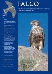
desired locations and examine the organs of concern. For<br />
educational purposes, a necropsy was performed in a Cooper’s<br />
Hawk (Accipiter cooperii) to visualize the locations of the<br />
endoscope in the bird during laparoscopy.<br />
The wild bird had been found and submitted to the Tufts<br />
<strong>Wildlife</strong> Clinic, with a comminuted open fracture of the<br />
left humerus which could not be repaired to restore flight.<br />
Therefore, the bird was anaesthetized with 3% isoflurane<br />
and humanely euthanized by intravenous injection of<br />
pentobarbital sodium solution. The photographs were taken<br />
two hours after death during necropsy.<br />
The cadaver has been placed in dorsal position with the<br />
sternum and the abdominal body wall removed. Cranial is<br />
on the left side and caudal on the right side of the pictures.<br />
The endoscope is represented by an orthopaedic wire in the<br />
photographs and a red line in the drawings.<br />
3c<br />
Figures 3. Illustration (3a, modified image, Evans 1982)<br />
and photographs (3b, 3c) of the endoscopy of the left cranial<br />
thoracic airsac. The endoscope is pushed cranially from the<br />
caudal into the cranial thoracic airsac. In 3c, the membrane<br />
separating cranial and caudal airsac has been removed and a<br />
more cranial view is visualizing the ostia of the airsacs. Cr<br />
ta= cranial thoracic airsac, c ta= caudal thoracic airsac, aa=<br />
abdominal airsac, H= heart, L= liver, O= ostium.<br />
3a<br />
4a<br />
3b<br />
4b<br />
26