chronOS. Bone Graft Substitute. Osteoconductive ... - Osteosyntese
chronOS. Bone Graft Substitute. Osteoconductive ... - Osteosyntese chronOS. Bone Graft Substitute. Osteoconductive ... - Osteosyntese
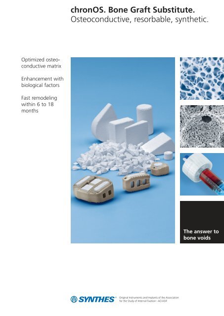
chronOS. Bone Graft Substitute. Osteoconductive, resorbable, synthetic. Optimized osteoconductive matrix Enhancement with biological factors Fast remodeling within 6 to 18 months The answer to bone voids
- Page 3 and 4: Table of Contents Introduction Over
- Page 5 and 6: Indications chronOS implants should
- Page 7 and 8: Spine surgery - Case description Th
- Page 9 and 10: Enhancement with Biological Factors
- Page 11 and 12: Fast Remodeling Within 6 to 18 Mont
- Page 13 and 14: chronOS Granules* Art. No. Diameter
- Page 15 and 16: SynCage PROmotive (prefilled), 2838
- Page 17: Potential complications of autologo
<strong>chronOS</strong>. <strong>Bone</strong> <strong>Graft</strong> <strong>Substitute</strong>.<br />
<strong>Osteoconductive</strong>, resorbable, synthetic.<br />
Optimized osteoconductive<br />
matrix<br />
Enhancement with<br />
biological factors<br />
Fast remodeling<br />
within 6 to 18<br />
months<br />
The answer to<br />
bone voids
Table of Contents<br />
Introduction<br />
Overview 2<br />
Indications 3<br />
Clinical Examples<br />
Trauma and Orthopedics 4<br />
Spine Surgery 5<br />
Features and Benefits<br />
Optimized <strong>Osteoconductive</strong> Matrix 6<br />
Enhancement with Biological Factors 7<br />
Fast Remodeling Within 6 to 18 Months 9<br />
Ordering Information<br />
10<br />
Bibliography<br />
14<br />
Synthes 1
Overview<br />
<strong>chronOS</strong> is a fully synthetic and resorbable bone graft substitute<br />
consisting of pure -tricalcium phosphate with a compressive<br />
strength similar to that of cancellous bone.<br />
The interconnected porous structure of <strong>chronOS</strong> acts as an<br />
osteoconductive matrix for the ingrowth of bone cells and<br />
blood vessels. Normally, <strong>chronOS</strong> implants are resorbed and<br />
completely remodeled into host bone within 6 to 18<br />
months.<br />
Easy to use<br />
<strong>chronOS</strong> is an off-the-shelf product and is available as granules<br />
of different sizes and as pre-shaped cylinders, blocks<br />
and wedges to precisely fit into critical size bony defects in<br />
trauma, spine and cranio-maxillofacial surgery. Furthermore,<br />
cages pre-filled with <strong>chronOS</strong> are available as vertebral interbody<br />
fusion implants.<br />
Long clinical experience<br />
<strong>chronOS</strong> products are widely and successfully used for more<br />
than 25 years. As early as 1988, Eggli et al. had suggested<br />
that <strong>chronOS</strong> underwent osteoclastic resorption and in 1990<br />
Pochon wrote about <strong>chronOS</strong> as an advantageous bone<br />
graft for bone defects in children (under the name Ceros-<br />
82). Since then, several published studies have shown the<br />
excellent clinical performance of <strong>chronOS</strong> (see bibliography).<br />
Avoids bone harvesting<br />
Autologous bone grafting is associated with several shortcomings<br />
and potential complications. Donor site pain and<br />
other morbidities are a major concern. Depending on the<br />
harvesting site and the quantity, more than 30% of all<br />
patients still suffer from donor site pain 2 years after discharge<br />
(McKay, 2002). Furthermore, bone harvesting can be<br />
limited by insufficient volume or quality (e. g. in osteoporosis)<br />
available.<br />
<strong>chronOS</strong> is an ideal alternative to autologous bone. It avoids<br />
donor site morbidity and shortens the duration of the overall<br />
surgery. Having a synthetic origin, <strong>chronOS</strong> offers the advantage<br />
of uniform quality and unlimited availability.<br />
% Patients with Donor Site Pain<br />
Incidence of donor site pain<br />
100%<br />
100%<br />
82.6%<br />
90%<br />
80%<br />
70%<br />
56%<br />
60%<br />
43.2%<br />
50%<br />
40%<br />
30%<br />
20%<br />
10%<br />
0% Discharge 6 Weeks 3 Months 6 Months<br />
34.6%<br />
12 Months<br />
31.6%<br />
24 Months<br />
Incidence of donor site pain for autograft control patients (McKay, 2002)<br />
2 Synthes <strong>chronOS</strong>. <strong>Bone</strong> <strong>Graft</strong> <strong>Substitute</strong> Brochure
Indications<br />
<strong>chronOS</strong> implants should be used as bone void fillers or<br />
augmentation material in zones requiring cancellous rather<br />
than cortical bone. This includes the filling of bone defects<br />
after trauma, reconstruction or correction in non-load bearing<br />
indications only.<br />
Depending on the size, voids of undefined geometric shape<br />
can be filled with granules or combinations of granules and<br />
blocks. Voids with defined geometric shape can be filled with<br />
blocks, wedges or cylinders.<br />
Trauma and orthopedics<br />
For example filling of voids caused by cysts or osteotomies,<br />
filling of defects arising from impacted fractures, refilling of<br />
cancellous bone harvesting sites, arthrodesis, non-unions and<br />
pseudoarthrosis.<br />
Spine surgery<br />
For example postero-lateral fusion, interbody fusion (as cage<br />
filling material), vertebrectomies (as filling material of the vertebral<br />
implants), refilling of bone graft harvesting sites.<br />
Cranio-maxillofacial surgery<br />
For example reconstruction of mandibular cyst defects and<br />
voids after tooth socket extractions and the maxillary sinus.<br />
Synthes 3
Clinical Examples<br />
Trauma and Orthopedics<br />
– Case description<br />
Correction of a varus leg axis with an open wedge high<br />
tibial osteotomy (OWHTO).<br />
– Treatment<br />
Osteotomy stabilized medially with a TomoFix plate and<br />
the gap filled with a <strong>chronOS</strong> wedge that was previously<br />
perfused with the patient's own blood.<br />
– Outcome<br />
Osteotomy healed and <strong>chronOS</strong> completely remodeled<br />
after 12 months.<br />
– Reference<br />
Van Hemert et al, 2004.<br />
Post-op, 1½ months<br />
Post-op, 12 months<br />
– Case description<br />
A 30-year-old female patient reported a broken small finger<br />
on the right hand. Upon examination a tumor and<br />
bone dystrophy in the proximal phalange was discovered.<br />
– Treatment<br />
Stable fixation with osteosynthesis plate and screws and<br />
filling of the bone void with <strong>chronOS</strong> Granules (vol.: 5 ml,<br />
: 1.4-2.8 mm). For further stabilization, the small finger<br />
was fixed to the ring finger for the first 2 weeks.<br />
– Outcome<br />
One year after the surgery the patient was very satisfied<br />
and did not notice the slight deficit in flexion and extension<br />
at the proximal interphalangeal joint. The <strong>chronOS</strong><br />
Granules had almost completely remodeled into host<br />
bone.<br />
Pre-op Post-op, 3 months Post-op, 12 months<br />
– Reference<br />
Philippe Chelius, MD, Troyes, France.<br />
4 Synthes <strong>chronOS</strong>. <strong>Bone</strong> <strong>Graft</strong> <strong>Substitute</strong> Brochure
Spine surgery<br />
– Case description<br />
The ability of <strong>chronOS</strong> Granules to achieve dorsal spondylodesis<br />
in adolescent idiopathic scoliosis was evaluated and<br />
followed by clinical examination, x-ray and CT scan.<br />
– Treatment<br />
USS titanium posterior fixation was complemented with<br />
posterolateral grafting. The grafting was performed either<br />
using autologous bone mixed with <strong>chronOS</strong> Granules or<br />
autologous bone mixed with allograft.<br />
– Outcome<br />
In both groups posterolateral segments had fused after<br />
6 months. No pseudarthrosis was observed. <strong>chronOS</strong> appears<br />
to be a valuable alternative to allograft for applications<br />
in the spine, even if large amounts of graft material is<br />
needed.<br />
Intra-operative Post-op Post-op, 6 months<br />
– Reference<br />
Muschik et al, 2001.<br />
– Case description<br />
Patient with severe back pain, resistant to therapy, was diagnosed<br />
with degenerative disc disease at L5-S1.<br />
– Treatment<br />
SynCage PROmotive perfused with bone marrow aspirate<br />
was used in anterior lumbar interbody fusion (ALIF) supported<br />
by posterior fixation.<br />
– Outcome<br />
A CT scan 3 months postoperatively showed the contact<br />
between the endplates and <strong>chronOS</strong> Inserts by the disrupted<br />
white line (arrow). 11 months postoperatively the<br />
<strong>chronOS</strong> remodeling and bony fusion were almost accomplished.<br />
– Reference<br />
Dr. med. Ch. Bach, University Hospital Innsbruck, Austria.<br />
Post-op, 3 months<br />
Post-op, 11 months<br />
Synthes 5
Optimized <strong>Osteoconductive</strong> Matrix<br />
Optimized scaffold<br />
To induce the bone remodeling process, osteoconductivity is<br />
a prerequisite. It is mainly influenced by three factors: the<br />
overall porosity, the interconnected macropores and the micropores.<br />
<strong>chronOS</strong> has been designed to optimize these features<br />
in order to mimic cancellous bone and provide an ideal<br />
scaffold for bone tissue infiltration.<br />
Overall porosity<br />
<strong>chronOS</strong> has a total porosity of 60% for the granules and<br />
70% for the preformed shapes. <strong>chronOS</strong> benefits from the<br />
highest possible degree of porosity, without compromising<br />
the mechanical integrity.<br />
Cancellous bone<br />
Interconnected macropores<br />
The macropores of <strong>chronOS</strong> are distributed mainly within a<br />
range of 100 to 500 μm. This provides the optimal condition<br />
for vascularization and migration of osteoclasts and osteoblasts<br />
(Gazdag, 1995). In addition, the macropores are interconnected<br />
to allow bone formation throughout the entire<br />
implant.<br />
Distribution<br />
40%<br />
<strong>chronOS</strong><br />
30%<br />
20%<br />
10%<br />
0% 500<br />
Pore size, μm<br />
Distribution of macropores: more than 95% of all macropores<br />
have a diameter between 100 and 500 μm.<br />
Micropores<br />
<strong>chronOS</strong> contains micropores, which are defined as the<br />
space within the material smaller than 10 μm. The microporosity<br />
accelerates the remodeling process by increasing<br />
the surface area and allowing for circulation of body fluids.<br />
6 Synthes <strong>chronOS</strong>. <strong>Bone</strong> <strong>Graft</strong> <strong>Substitute</strong> Brochure
Enhancement with Biological Factors<br />
Description<br />
<strong>chronOS</strong> implants contain a significant amount of air in its<br />
pores. Impregnation of the porous material with bone marrow<br />
or blood not only removes the air but also introduces<br />
blood cells, growth factors and, in the case of bone marrow,<br />
osteoprogoniter cells into the bone graft substitute. The<br />
combination of <strong>chronOS</strong> with bone marrow accelerates and<br />
enhances osteointegration and is a valuable alternative to<br />
autologous or allogenic bone graft material (Stoll et al, 2004,<br />
and Becker et al, 2006).<br />
Design<br />
In order to make the osteoinductive and osteogenic potential<br />
of autologous bone marrow available, Synthes has developed<br />
the <strong>chronOS</strong> Perfusion Concept allowing the efficient,<br />
intra-operative impregnation of <strong>chronOS</strong> products with the<br />
patient's own bone marrow.<br />
<strong>chronOS</strong> Perfusion Concept<br />
Step 1<br />
Aspiration of bone marrow<br />
Step 2<br />
Perfusion under vacuum<br />
Step 3<br />
Implantation of <strong>chronOS</strong><br />
Synthes 7
Enhanced osteointegration<br />
An animal study evaluating the osteoinductive and osteogenic<br />
potential of autologous bone marrow showed that<br />
osteointegration was significantly more pronounced for<br />
<strong>chronOS</strong> implants impregnated with bone marrow rather<br />
than with blood.<br />
12 weeks after surgery, the <strong>chronOS</strong> Cylinders implanted<br />
into critical-size defects in the metaphysis of sheep tibiae<br />
were mostly remodeled into host bone when bone marrow<br />
was used to perfuse the implant. On the other hand, the<br />
bone substitute material was still clearly visible when impregnation<br />
was performed with venous blood.<br />
For details see: Stoll et al, 2004, and Becker et al, 2006.<br />
Blood<br />
Post-op, 6 weeks<br />
<strong>Bone</strong> Marrow<br />
Post-op, 6 weeks<br />
Additional information on the <strong>chronOS</strong> Perfusion Concept is<br />
available in the corresponding Flyer (036.000.890) and Technique<br />
Guide (036.000.745).<br />
Post-op, 12 weeks<br />
Post-op, 12 weeks<br />
8 Synthes <strong>chronOS</strong>. <strong>Bone</strong> <strong>Graft</strong> <strong>Substitute</strong> Brochure
Fast Remodeling Within<br />
6 to 18 Months<br />
Rapid resorption of -tricalcium phosphate<br />
Differences in chemical composition of biomaterials have<br />
profound effects on their in vivo behavior. <strong>chronOS</strong> consists<br />
of pure -tricalcium phosphate and is structurally and chemically<br />
similar to bone. Osteoclasts resorb <strong>chronOS</strong> like natural<br />
bone and degrade it rapidly. Hydroxyapatite, in contrast, resorbs<br />
very slowly (Buser et al, 1998). Due to the chemical<br />
composition, <strong>chronOS</strong> implants are initially radio-opaque.<br />
Formation of new host bone<br />
While resorption is taking place, new bone is being formed:<br />
osteoblasts fill the lacunae created by osteoclast by producing<br />
extracellular matrix, which is subsequently calcified. As<br />
a result of both the choice of the specific chemical composition<br />
and the optimized scaffold as described previously,<br />
<strong>chronOS</strong> leads to a faster and more effective formation of<br />
new bone than other bone graft substitutes.<br />
Fraction of total defect volume<br />
Resorption of bone graft <strong>chronOS</strong> Hydroxyapatite<br />
40%<br />
30%<br />
20%<br />
10%<br />
18.5 %<br />
0% 4<br />
34.1%<br />
7.5 %<br />
28.1%<br />
5.8 %<br />
12 24<br />
Weeks after surgery<br />
30.6%<br />
Resorption of -tricalcium phosphate (<strong>chronOS</strong>) is significantly faster than for<br />
hydroxyapatite (animal model, see Buser et al,1998, for details).<br />
Formation of new host bone<br />
<strong>chronOS</strong><br />
Hydroxyapatite<br />
Replaced in 6 to 18 months<br />
The key to success of <strong>chronOS</strong> is the remodeling process. Resorption<br />
and new bone formation happen simultaneously.<br />
Timing is the critical factor for a bone graft to remodel into<br />
natural bone. If the resorption is too rapid, the osteoblasts<br />
lose the scaffold needed for the formation of new bone. If<br />
the resorption is too slow or incomplete, the graft will not be<br />
replaced by bone in an adequate time span. <strong>chronOS</strong> has<br />
been designed to remodel in an ideal time span. It is being<br />
replaced in the human body by host bone in 6 to 18 months;<br />
depending on the indication and the patient's conditions.<br />
No adverse reactions<br />
All investigations, according to ISO 10993 series, demonstrate<br />
the excellent biocompatibility of <strong>chronOS</strong>. No adverse<br />
reactions have been observed in the more than 25 years of<br />
clinical applications (see bibliography).<br />
Fraction of total defect volume<br />
80%<br />
60%<br />
40%<br />
20%<br />
23.3 % 20.7%<br />
64.2 %<br />
42.8%<br />
69.7 %<br />
0% 4 12 24<br />
Weeks after surgery<br />
49.0%<br />
-tricalcium phosphate (<strong>chronOS</strong>) is remodeled faster and more efficiently into<br />
new host bone than hydroxyapatite (animal model, see Buser et al, 1998, for<br />
details).<br />
Remodeling and substitution of <strong>chronOS</strong> (24 weeks in an animal model). Some<br />
<strong>chronOS</strong> Granules are still lined by woven bone, other parts are directly covered by<br />
lamellar bone, or are exposed to the marrow space (arrow) where they undergo<br />
degradation by osteoclasts (Buser et al, 1998).<br />
Synthes 9
Ordering Information<br />
<strong>chronOS</strong> Preforms<br />
<strong>chronOS</strong> Cylinders<br />
Art. No. Diameter Length Perfusion device<br />
07.710.030S 8.5 mm 25 mm Syringe M<br />
07.710.031S 9.5 mm 25 mm Syringe M<br />
07.710.032S 10.5 mm 25 mm Syringe M<br />
07.710.033S 12.5 mm 25 mm Syringe M<br />
07.710.035S 14.0 mm 25 mm Syringe M<br />
07.710.038S 15.15 mm 20 mm Syringe L<br />
07.710.039S 17.55 mm 20 mm Syringe L<br />
<strong>chronOS</strong> Blocks<br />
Art. No. Size (mm) Perfusion device<br />
07.710.042S 5510 Syringe S<br />
07.710.045S 12.512.510 Syringe L<br />
07.710.047S 202010 Syringe L<br />
<strong>chronOS</strong> Wedges<br />
Art. No. Angle Size (mm) Perfusion device<br />
07.710.050S 10° 25206 Container<br />
07.710.051S 14° 25208 Container<br />
07.710.052S 18° 252010 Container<br />
07.710.053S 22° 252012 Container<br />
07.710.054S 26° 252014 Container<br />
<strong>chronOS</strong> Wedges, semi-circular<br />
Art. No. Angle Size (mm) Perfusion device<br />
07.710.057S 7° 25357 Container<br />
07.710.060S 10° 253510 Container<br />
07.710.063S 13° 253513 Container<br />
10 Synthes <strong>chronOS</strong>. <strong>Bone</strong> <strong>Graft</strong> <strong>Substitute</strong> Brochure
<strong>chronOS</strong> Granules*<br />
Art. No. Diameter Content<br />
710.000S 0.5–0.7 mm 0.5 ml<br />
710.001S 0.7–1.4 mm 0.5 ml<br />
710.002S 0.7–1.4 mm 1.0 ml<br />
710.003S 0.7–1.4 mm 2.5 ml<br />
710.011S 1.4–2.8 mm 2.5 ml<br />
710.014S 1.4–2.8 mm 5.0 ml<br />
710.019S 1.4–2.8 mm 10.0 ml<br />
710.021S 1.4–2.8 mm 20.0 ml<br />
710.024S 2.8–5.6 mm 2.5 ml<br />
710.025S 2.8–5.6 mm 5.0 ml<br />
710.026S 2.8–5.6 mm 10.0 ml<br />
710.027S 2.8–5.6 mm 20.0 ml<br />
* <strong>chronOS</strong> Granules are not offered in a perfusion device.<br />
They can easily be perfused with autologous bone marrow<br />
or blood by mixing its sterile cup package or a sterile bowl,<br />
respectively.<br />
<strong>chronOS</strong> Inserts for Cervios <strong>chronOS</strong>, wedge-shaped<br />
Art. No. Height Fits to Perfusion device<br />
Cervios cage<br />
710.921S 5 mm 889.921S Syringe S<br />
710.922S 6 mm 889.922S Syringe S<br />
710.923S 7 mm 889.923S Syringe S<br />
710.924S 8 mm 889.924S Syringe S<br />
710.925S 9 mm 889.925S Syringe S<br />
710.926S 10 mm 889.926S Syringe S<br />
<strong>chronOS</strong> Inserts for Cervios <strong>chronOS</strong>, curved<br />
Art. No. Height Fits to Perfusion device<br />
Cervios cage<br />
710.931S 5 mm 889.931S Syringe S<br />
710.932S 6 mm 889.932S Syringe S<br />
710.933S 7 mm 889.933S Syringe S<br />
710.934S 8 mm 889.934S Syringe S<br />
710.935S 9 mm 889.935S Syringe S<br />
710.936S 10 mm 889.936S Syringe S<br />
Synthes 11
Ordering Information<br />
Cervios <strong>chronOS</strong> (prefilled), wedge-shaped<br />
Art. No. Height Perfusion device<br />
870.921S 5 mm Syringe L<br />
870.922S 6 mm Syringe L<br />
870.923S 7 mm Syringe L<br />
870.924S 8 mm Syringe L<br />
870.925S 9 mm Syringe L<br />
870.926S 10 mm Syringe L<br />
Cervios <strong>chronOS</strong> (prefilled), curved<br />
Art. No. Height Perfusion device<br />
870.931S 5 mm Syringe L<br />
870.932S 6 mm Syringe L<br />
870.933S 7 mm Syringe L<br />
870.934S 8 mm Syringe L<br />
870.935S 9 mm Syringe L<br />
870.936S 10 mm Syringe L<br />
Plivios <strong>chronOS</strong> (prefilled)<br />
Art. No. Height Perfusion device<br />
870.984S 7 mm Syringe L<br />
870.985S 9 mm Syringe L<br />
870.986S 11 mm Syringe L<br />
870.987S 13 mm Syringe L<br />
870.988S 15 mm Syringe L<br />
870.989S 17 mm Syringe L<br />
SynCage PROmotive (prefilled), 2430 mm, 12°<br />
Art. No. Height Perfusion device<br />
08.802.851S 12 mm Container<br />
08.802.852S 13.5 mm Container<br />
08.802.854S 15 mm Container<br />
08.802.856S 17 mm Container<br />
08.802.858S 19 mm Container<br />
12 Synthes <strong>chronOS</strong>. <strong>Bone</strong> <strong>Graft</strong> <strong>Substitute</strong> Brochure
SynCage PROmotive (prefilled), 2838 mm, 10°<br />
Art. No. Height Perfusion device<br />
08.802.871S 12 mm Container<br />
08.802.872S 13.5 mm Container<br />
08.802.874S 15 mm Container<br />
08.802.876S 17 mm Container<br />
08.802.878S 19 mm Container<br />
SynCage PROmotive (prefilled), 2838 mm, 12°<br />
Art. No. Height Perfusion device<br />
08.802.899S 12 mm Container<br />
08.802.900S 13.5 mm Container<br />
08.802.901S 15 mm Container<br />
08.802.902S 17 mm Container<br />
08.802.903S 19 mm Container<br />
<strong>Bone</strong> Marrow Aspiration System (BMAS)<br />
Art. No. Diameter Length Syringe<br />
710.110S 11 ga 10 cm 20 ml<br />
710.115S 11 ga 15 cm 20 ml<br />
Synthes 13
Bibliography<br />
Optimized scaffold<br />
To induce the bone remodeling process, osteoconductivity<br />
must occur. It is mainly influenced by three factors:<br />
i) the overall porosity:<br />
– Toth JM et al. (1995) Evaluation of porous biphasic calcium<br />
phosphate ceramics for anterior cervical interbody fusion in<br />
a caprine model. Spine 20(20):2203–2210.<br />
ii) the interconnected macropores:<br />
– Lu JX, Flautre B et al. (1999) Role of interconnections in<br />
porous bioceramics on bone recolonization in vitro and<br />
vivo. J Mater Sci Mater Med 10:111–120.<br />
iii) micropores:<br />
– Chang BS et al. (2000) Osteoconduction at porous hydroxy-apatite<br />
with various pore configurations. Biomaterials<br />
21(12):1291–1298.<br />
– Gazdag AR, Lane JM, Glaser D, et al. (1995) Alternatives to<br />
autogenous bone graft: efficacy and indications. J Am<br />
Acad Orthop Surg 3(1):1–8.<br />
– Daculsi G. (1990) Effect of macroporosity for osseous<br />
substitution of calcium phosphate ceramics. Biomaterials<br />
11:86–87.<br />
– Eggli PS et al. (1988) Porous hydroxyapatite and tricalcium<br />
phosphate cylinders with two different pore size ranges<br />
implanted in the cancellous bone of rabbits: A comparative<br />
histomorphometric and histologic study of bony ingrowth<br />
and implant substitution. Clin Orthop (232):127–138.<br />
<strong>chronOS</strong> induces remodeling process<br />
The remodeling process (simultaneous resorption and new<br />
bone formation) is possible due to the specific chemical composition<br />
and the optimized scaffold of <strong>chronOS</strong>. <strong>chronOS</strong><br />
consists of pure -tricalcium phosphate which remodels<br />
completely.<br />
i) <strong>chronOS</strong> remodels in vivo:<br />
– Wheeler D. (2005) <strong>Graft</strong>ing of massive tibial subchondral<br />
bone defects in a Caprine Model using<br />
-Tricalcium phosphate versus autograft. J Orthop Trauma<br />
19(2):85–91.<br />
– Buser D et al. (1998) Evaluation of filling materials<br />
in membrane protected bone defects: A comperative<br />
histomorphometric study in the mandible<br />
of miniature pigs. Clin Oral Implants Res 9 (3):<br />
137–150.<br />
– Leutenegger H. (1993/94) Integration und Resorption<br />
von Kalziumphoshatkeramiken zur Defektauffüllung<br />
bei Tibiakopffrakturen. Helv Chir 60:<br />
1061–1066.<br />
– Waisbrod H et al. (1986) A pilot study of the value<br />
of ceramics for bone replacement. Arch Orthop<br />
Trauma Surg 105:298–301.<br />
ii) Chemical composition is vital for resorption:<br />
– Koerten HK. (1999) Degradation of calcium phosphate<br />
ceramics. J Biomed Mater Res 44(1):78–86.<br />
– LeGeros RZ et al. (1988) Signifiance of the porosity<br />
and physical chemistry of calcium phosphate ceramics:<br />
Biodegradation-bioresorption. Ann N Y<br />
Acad Sci 523:268–271.<br />
iii) Microporosity accelerates the remodeling process:<br />
– Yokozeki H et al. (1998) Influence of surface microstructure<br />
on the reaction of the active ceramics<br />
in vitro. J Mater Sci: Mat in Med 9:381ff.<br />
– Klein CP et al. (1985) Interaction of biodegradable<br />
-whitlockite ceramics with bone tissue: An in vitro<br />
study. Biomaterials 6:189–198.<br />
Enhancing <strong>chronOS</strong> with biological properties<br />
The biological characteristics of <strong>chronOS</strong> can be improved<br />
by combining <strong>chronOS</strong> with patient bone marrow<br />
or blood, making <strong>chronOS</strong> potentially osteoinductive<br />
or osteogenic, respectively.<br />
– Becker S et al. (2006) Osteopromotion by a -TCP/<br />
bone marrow hybrid implant for use in spine surgery.<br />
Spine 31(1):11–17.<br />
– Stoll T et al. (2004) New Aspects in Osteoinduction.<br />
Mat.-wiss. u. Werkstofftech 35(4):198–202.<br />
14 Synthes <strong>chronOS</strong>. <strong>Bone</strong> <strong>Graft</strong> <strong>Substitute</strong> Brochure
Potential complications of autologous bone grafting<br />
Autologous bone grafting is associated with several shortcomings<br />
and potential complications. <strong>chronOS</strong> is an advantageous<br />
alternative to bone harvesting.<br />
– Muschler GF et al. (2003) Spine fusion using cell matrix<br />
composites enriched in bone marrow-derived cells. Clin<br />
Orth Rel Res 407:102–118.<br />
– McKay B, Sandhu HS. (2002) Use of recombinant human<br />
bone morphogenetic protein-2 in spinal fusion applications.<br />
SPINE 27(16S):66-85.<br />
– Connolly J. (1995) Injectable bone marrow preparations to<br />
stimulate osteogenic repair. Clin Orth Rel Res 313:8–18.<br />
– Tiedeman J et al. (1991) Healing of a large nonossifying fibroma<br />
after grafting with bone matrix and marrow. Clin<br />
Orth Rel Res 265:302–305.<br />
– Connolly J et al. (1989) Autologous marrow injection for<br />
delayed unions of the tibia: a preliminary report. J Orth<br />
Trauma 3(4):276–282.<br />
Clinical studies<br />
– Van Hemert W et al. (2004) Tricalcium phosphate granules<br />
or rigid wedge preforms in open wedge high tibial osteotomy:<br />
a radiological study with a new evaluation system.<br />
Knee 11(6):451–456.<br />
– Pavlov PW. (2003) Anterior decompression for cervical<br />
spondylotic myelopathy. Eur Spine J 12(Suppl 2):188–194.<br />
– Muschik M et al. (2001) Beta-tricalcium phosphate as a<br />
bone substitute for dorsal spinal fusion in adolescent idiopathic<br />
scoliosis: Preliminary results of a prospective clinical<br />
study. Eur Spine J 10:178–184.<br />
– Pochon JP. (2000) (Case Report) Juvenile Knochenzysten;<br />
Die operative Versorgung juveniler Knochenzysten mit<br />
Beta-Tricalciumphosphat-Keramik (<strong>chronOS</strong>-Granulat).<br />
– Muschik M et al. (2000) Beta-tricalcium phosphate as a<br />
bone substitute and autograft for spinal fusion: A comparative<br />
prospective study in adolescent idiopathic scoliosis.<br />
– Meiss L. (1999) Stimulation of bone regeneration by fragmented<br />
cortical bone on porous calcium phosphate ceramics<br />
(tricalcium phosphate and hydroxyapatite): An experimental<br />
study and preliminary clinical results. Neuere<br />
Ergebnisse in der Osteologie Springer Willert+ Heug Hrsg.<br />
– Pochon J-P. (1990) Knochenersatzplastiken mit Trikalziumphosphatkeramik<br />
im Kindesalter. Aktuelle Probl Chir Orthop<br />
(Switzerland) 36:1–51.<br />
Synthes 15
Presented by:<br />
0123<br />
036.000.305 SM_707856 AC 71060012 © Synthes 2007 Subject to modifications


