Research Profile - Department of Materials Science and Metallurgy ...
Research Profile - Department of Materials Science and Metallurgy ...
Research Profile - Department of Materials Science and Metallurgy ...

You also want an ePaper? Increase the reach of your titles
YUMPU automatically turns print PDFs into web optimized ePapers that Google loves.
Paul Midgley<br />
Pr<strong>of</strong>essor <strong>of</strong> <strong>Materials</strong> <strong>Science</strong><br />
BSc University <strong>of</strong> Bristol<br />
MSc University <strong>of</strong> Bristol<br />
PhD University <strong>of</strong> Bristol<br />
+44 (0) 1223 334561<br />
pam33@cam.ac.uk<br />
www-hrem.msm.cam.ac.uk<br />
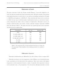
Electron Microscopy<br />
My research is based on the development <strong>and</strong> application <strong>of</strong><br />
new electron microscopy techniques to study the structural <strong>and</strong><br />
functional properties <strong>of</strong> a variety <strong>of</strong> materials with high spatial<br />
resolution in 2 <strong>and</strong> 3 dimensions.<br />
Electron tomography<br />
By recording a tilt-series <strong>of</strong> transmission electron micrographs,<br />
using a variety <strong>of</strong> novel imaging modes, electron tomography can<br />
be used to reconstruct the 3-dimensional morphology, defect<br />
structure <strong>and</strong> composition <strong>of</strong> materials systems with nanometre<br />
resolution in all 3 dimensions. <strong>Materials</strong> studied range from cell<br />
sections <strong>and</strong> bacteria to heterogeneous catalysts, semiconductor<br />
quantum dots <strong>and</strong> aerospace alloys. Recently we have started a<br />
research programme developing mesoscale tomography using<br />
the dual beam SEM-FIB to link the length scales probed by TEM<br />
with those investigated by X-ray methods.<br />
Electron holography <strong>and</strong> phase-sensitive imaging<br />
Quantitative <strong>of</strong>f-axis <strong>and</strong> in-line (Fresnel) electron holography is<br />
being used to study the electrostatic potentials at unbiased <strong>and</strong><br />
biased device junctions in two <strong>and</strong> three dimensions. Magnetic<br />
fields in ferromagnetic thin films <strong>and</strong> nanoparticle arrays are<br />
also being mapped with electron holography. Low-temperature<br />
microscopy is being carried out to investigate the structure <strong>and</strong><br />
dynamics <strong>of</strong> flux vortices in high T c<br />
superconductors. Vortex<br />
contrast is amplified by energy-filtered Lorentz imaging or by the<br />
use <strong>of</strong> a phase plate.<br />
Electron crystallography<br />
Precession electron diffraction is being developed to determine<br />
the atomic structure <strong>of</strong> sub-micrometre particles. This technique,<br />
which minimizes the effects <strong>of</strong> dynamical interaction, can<br />
now be used to determine atomic positions to an accuracy<br />
approaching that <strong>of</strong> X-ray diffraction. Why the technique works,<br />
<strong>and</strong> over what range <strong>of</strong> sample thickness <strong>and</strong> precession angle,<br />
is being investigated. The technique is being used to determine<br />
the structure <strong>of</strong> inorganic <strong>and</strong> organic crystals, including metal<br />
oxides, metal-organic frameworks <strong>and</strong> pharmaceutical crystals.<br />
Nanoscale structures<br />
Novel structures, including mesoporous catalysts, organic <strong>and</strong><br />
inorganic nanotubes, <strong>and</strong> semiconducting nanowires, are being<br />
characterized by high-resolution electron microscopy, electron<br />
diffraction <strong>and</strong> electron tomography. The cellular toxicity <strong>of</strong><br />
fullerenes <strong>and</strong> carbon nanotubes is investigated by chemical<br />
assay <strong>and</strong> electron tomography.<br />
PA Midgley & M Weyl<strong>and</strong>, “3D electron microscopy in the physical<br />
sciences: the development <strong>of</strong> Z-contrast <strong>and</strong> EFTEM tomography”<br />
Ultramicroscopy 96, 413–431 (2003).<br />
I Arslan, TJV Yates, ND Browning & PA Midgley, “Embedded<br />
nanostructures revealed in three dimensions” <strong>Science</strong> 309, 2195–2198<br />
(2005).<br />
MH Gass, K Koziol, AH Windle & PA Midgley, “Four-dimensional spectral<br />
tomography <strong>of</strong> carbonaceous nanocomposites” Nano Lett. 6, 376–379<br />
(2006).<br />
JS Barnard, J Sharp, JR Tong & PA Midgley, “High-resolution threedimensional<br />
imaging <strong>of</strong> dislocations” <strong>Science</strong> 313, 319 (2006).<br />
3D electron-tomographic reconstructions showing (left) a<br />
carbon nanotube-nylon composite <strong>and</strong> (right) a heterogeneous<br />
catalyst composed <strong>of</strong> bimetallic nanoparticles supported by<br />
mesoporous silica<br />
30 <strong>Research</strong> <strong>Pr<strong>of</strong>ile</strong>