Interesting Case Series Breast Augmentation - ePlasty
Interesting Case Series Breast Augmentation - ePlasty
Interesting Case Series Breast Augmentation - ePlasty

Create successful ePaper yourself
Turn your PDF publications into a flip-book with our unique Google optimized e-Paper software.
<strong>Interesting</strong> <strong>Case</strong> <strong>Series</strong><br />
<strong>Breast</strong> <strong>Augmentation</strong><br />
Sachin M. Shridharani, MD, Justin L. Bellamy, BS, Mark M. Mofid, MD,<br />
and Navin K. Singh, MD<br />
Department of Plastic Surgery, The Johns Hopkins University School of Medicine, Baltimore, Md<br />
Correspondence: sshridh1@jhmi.edu<br />
DESCRIPTION<br />
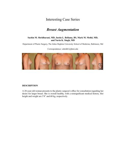
A 28-year-old woman presents to the plastic surgeon’s office for consultation regarding her<br />
desire for larger breast. She is overall healthy, with a nonsignificant medical history. Her<br />
height and weight are 5 ′ 8 ′′ and 60 kg, respectively.
QUESTIONS<br />
1. What are the available options for breast implants? What are their advantages<br />
and disadvantages?<br />
2. What are the common surgical approaches for breast augmentation? What<br />
are their advantages and disadvantages?<br />
3. What is capsular contracture?<br />
4. How does one diagnose an implant rupture?<br />
5. Does breast augmentation surgery impact breast cancer screening with<br />
mammography?
DISCUSSION<br />
Since 2006, augmentation mammoplasty has remained the most commonly performed<br />
cosmetic surgical procedure in the United States, with nearly 300 000 patients annually<br />
undergoing the procedure. 1 The modern era of silicone elastomer-shelled prostheses (breast<br />
implants filled with either silicone or saline) began in early 1960s and has seen fluctuation<br />
in practice trends regarding which implant to use. A thorough understanding of the most<br />
recent recommendations remains important in providing the best short- and long-term care<br />
for patients desiring breast augmentation.<br />
The 2 primary types of breast implants used today are saline- or silicone-filled. In<br />
1992, the US Food and Drug Administration issued a moratorium on silicone implants<br />
because of an alleged link to autoimmune disease. Surgeons defaulted to the use of salinefilled<br />
implants as the predominant implant; however, these implants have otherwise been<br />
considered a second choice to silicone implants in the United States and abroad because<br />
of increased rupture/deflation rates and problems with underfilling and overfilling. These<br />
issues may result in a need for revision surgery and dissatisfactory aesthetic result. After extensive<br />
population-based studies failed to show a correlation between autoimmune disease<br />
and silicone implants, the moratorium was lifted in 2006, and silicone breast implants came<br />
back into popularity in the United States. 2 The most recent generation of silicone implants<br />
features a barrier layer to reduce the silicone bleeding phenomenon seen in older implants.<br />
Textured implants were initially developed to mimic polyurethane implant shells that were<br />
shown to have very low rates of capsular contracture but are not available in the United<br />
States. Unfortunately, there is little high-level evidence that surface texturizing alone reduces<br />
capsular contracture. 3,4 The primary utility of texturized implants is to reduce implant<br />
rotation in anatomic-shaped prostheses. Innovation regarding silicone chemical processing,<br />
including extensive cross-linking of the filler, has led to the development of cohesive gel<br />
implants. Cohesive gel implants are supple, yet able to maintain their shape without being<br />
deformed by the surrounding soft-tissue envelope or gravitational effects over time. In<br />
addition to aesthetic benefit, these “form stable” or “gummy-bear” implants may reduce<br />
long-term implant-related complications. 5−7<br />
Implants are available in 2 general shapes: round or anatomic. The indications for<br />
either type vary, and surgeons should be comfortable using one or the other as dictated by<br />
patient goals and anatomy. Round implants may be suitable for patients when rotation of an<br />
anatomic-shaped implant would be of concern. These circumstances include patients exhibiting<br />
increased athletic activity and revision surgery after confirmed rotation deformity.<br />
In addition, patients deemed candidates suited for round implants include those patients in<br />
whom the round shape would be less noticeable (fuller patients, patients with good skin<br />
quality, or patients requiring smaller volumes). Furthermore, patients who explicitly desire<br />
an overfilled or large breast appearance may be excellent candidates for round implants.<br />
Anatomic-shaped implants are well suited for patients desiring a natural appearance or those<br />
with mild ptosis or pseudoptosis. One should note, however, that a “natural” appearance<br />
can be achieved with either shape, and significant ptosis with increased skin envelope laxity<br />
may lead to increased risk of rotation with anatomic implants. Patients with constricted<br />
lower-pole breasts and thoracic hypoplasia may also benefit from form-stable anatomicshaped<br />
implants. Many surgeons have advocated for tissue-based planning—preoperative<br />
breast dimensional measurements dictating implant selection. These efforts have improved
outcomes and reduced reoperation. The goal of tissue-based planning is to select an implant<br />
that will fill the breast while simultaneously respecting natural anatomy, matching<br />
the breast footplate, and minimizing breast distortion. 8−10 Rather than cup-size, the width<br />
and skin-stretch measurements of the soft tissue envelope are used to determine optimal<br />
fill volume. 9 Alternatively, a saline-filled breast sizer can be inserted, filled, and removed<br />
intraoperatively to objectively determine the optimal volume for the permanent implant.<br />
Several access incisions/approaches are employed: inframammary, periareolar,<br />
transaxillary, and transumbilical (saline only). There are advantages and disadvantages<br />
to each approach. Inframammary incisions provide the most control, allowing the surgeon<br />
to set the location of the inframammary fold while minimizing implant trauma or contamination;<br />
however, without careful planning the scar may not be ideal. The periareolar<br />
incision allows good access and disguise of the scar; however, the incision may increase<br />
implant contamination (due to transection of parenchymal ducts often colonized by Staphylococcus<br />
epidermidis) 11 and resultant capsular contracture. Further, the scar is placed in<br />
the central area of interest in the breast, and for those prone to hypertrophic scarring, this<br />
would be a riskier incisional choice. The transaxillary approach results in no scar on the<br />
breast; however, the remote access site reduces control for pectoralis major release unless<br />
endoscopically assisted. Furthermore, placing larger silicone gel implants can be challenging<br />
via the transaxillary approach because of limited incision size. Should revision surgery<br />
be required after transaxillary or transumbilical approaches, inframammary incisions are<br />
often ultimately required.<br />
The main surgical planes of implant placement are subglandular or subpectoral/submuscular<br />
with release of the inferior pectoral origin. Total submuscular planes<br />
of dissection have been described but have limited indications. Subglandular placement<br />
of implants may be appropriate in patients with sufficient overlying soft tissue in the upper<br />
pole of the breast (determined by a pinch test of >1-2 cm). The subglandular space<br />
is viewed by some to represent a more natural plane yielding a more natural-appearing<br />
augmentation. 12 In addition, placing implants in this plane allows for correction of mild<br />
breast ptosis without having to perform a separate procedure on the breast mound to elevate<br />
the nipple-areola complex. Alternatively, subpectoral implant placement provides its own<br />
benefits. There is some evidence to suggest that it may provide improved capsular contracture<br />
rates, 13 although high-level evidence is limited. Similarly, it has been suggested that<br />
subpectoral placement may improve breast visibility on mammography. 14 In recent years,<br />
the subpectoral approach has been refined, using the dual plane technique to combine good<br />
upper and medial cover with improved draping of the lower pole over the implant and less<br />
pectoralis animation. 15 Three types of dual plane approaches have been described, with<br />
each level describing increasing degree of release of anterior pectoral fascial attachments<br />
from overlying glandular tissue. Dual plane I features division of the inferior pectoral origin<br />
without further fascial release, dual plane II adds release of anterior pectoral fascial<br />
attachments to the level of the inferior areolar border and rotation of the inferior origin of<br />
the pectoralis, while dual plane III involves fascial release and rotation at the level of the<br />
superior areolar border.<br />
Regardless of the surgical approach, the surgeon should be familiar with complications<br />
specific to breast augmentation. Capsular contracture remains one of the most troubling and<br />
frequently reported complications of augmentation mammoplasty, and thus reduction in its<br />
occurrence has been the topic of numerous studies. 16 <strong>Breast</strong> prostheses, being a foreign
ody, invariably develop a capsule as a protective immune reaction. Capsular contracture<br />
describes the pathologic activation of this capsule that results in a constrictive fibrosis that<br />
deforms and impairs the aesthetic result. Several causal theories, involving endogenous (eg,<br />
due to host-implant interaction) and exogenous (eg, due to subclinical infection, biofilms)<br />
contributions, have been proposed, although the exact cause is multifactorial and remains<br />
elusive. Baker’s original clinical classification of capsular contracture remains a simple tool<br />
to describe severity, where grades I (normal capsule) and II (palpable but nonvisible) are<br />
considered acceptable and grades III (palpable and visible) and IV (painful, hard, and very<br />
constrictive) represent pathologic contracture. 17 While capsular contracture mostly happens<br />
within the first 2 years, there is a long-term cumulative increase in capsular contracture risk<br />
over time 18−20 that may require surgical capsulectomy with implant replacement.<br />
An important consideration after augmentation mammoplasty includes breast imaging.<br />
First, the surgeon should be aware of the possibility of implant rupture. Intracapsular implant<br />
rupture (those not extending beyond the fibrous capsule around the implant) represents 80%<br />
to 90% of implant ruptures. 21,22 Diagnosis is generally more straightforward with salinefilled<br />
implants. Rupture is followed quickly by total/near-total deflation. If patients have<br />
silicone-filled breast implants, these ruptures can be diagnosed with magnetic resonance<br />
imaging by the presence of multiple curvilinear low-signal-intensity lines within the silicone<br />
gel (known as “linguine sign”) on T2-weighted imaging. The rarer extracapsular rupture can<br />
be identified by identification of free, high-signal-intensity silicone in surrounding breast<br />
tissue. Second, imaging considerations for breast cancer screening remains an important<br />
issue after augmentation. While many studies suggest a trend toward lower cancer rates<br />
in patients with implants (perhaps reflecting the patient population, smaller breast size,<br />
etc), breast prostheses do create certain mammography challenges. Both silicone and saline<br />
implants create dense radio-opaque shadows and limit maximal compression of breast<br />
tissue during standard mammography that may interfere with detection of smaller subtle<br />
lesions. 23 Capsular contracture can further reduce sensitivity by as much as 30% with Baker<br />
I/II, or by more than 50% with Baker III/IV. 14,24 To counteract these effects, the Eklund<br />
Pushback technique, 25 which involves pushing the implant posteriorly toward the chest wall<br />
and pulling the breast tissue forward, can be employed by the radiologist to significantly<br />
increase the amount of breast tissue visualized. Despite this theoretical hindrance to optimal<br />
mammography screening, studies have failed to demonstrate any significant delay in cancer<br />
detection. 26 In addition, overall breast cancer survival rates are ultimately not impacted by<br />
breast augmentation. 27 Last, the increasing use of magnetic resonance imaging in breast<br />
imaging may obviate the limitations associated with mammography.
DESCRIPTION<br />
The patient underwent placement of 300 mL round, smooth, silicone gel–filled implants<br />
through an inframammary fold incision in a dual plane approach. She did well in the<br />
postoperative picture and was pleased with the overall aesthetic outcome. Given earlier is<br />
her picture at 1-year follow-up.<br />
REFERENCES<br />
1. American Society of Plastic Surgeons. 2010 plastic surgery procedural statistics. http://www.<br />
plasticsurgery.org/Documents/news-resources/statistics/2012-Plastic-Surgery-Statistics/Cosmetic-<br />
Procedure-Trends-2012.pdf. Published 2010. Accessed April 18, 2013<br />
2. US Food and Drug Administration. FDA update on the safety of silicone gel–filled breast implants. www.<br />
fda.gov/downloads/MedicalDevices/ProductsandMedicalProcedures/ImplantsandProesthetics/<strong>Breast</strong><br />
Implants/UCM260090.pdf. Published 2011. Accessed April 18, 2013.<br />
3. Barnsley GP, Sigurdson LJ, Barnsley SE. Textured surface breast implants in the prevention of capsular<br />
contracture among breast augmentation patients: a meta-analysis of randomized controlled trials. Plast<br />
Reconstr Surg. 2006;117:2182-90.<br />
4. Poeppl N, Schreml S, Lichtenegger F, Lenich A, Eisenmann-Klein M, Prantl L. Does the surface<br />
structure of implants have an impact on the formation of a capsular contracture? Aesthetic Plast Surg.<br />
2007;31:133-9.<br />
5. Stevens WG, Hirsch EM, Tenenbaum MJ, Acevedo M. A prospective study of 708 form-stable silicone<br />
gel breast implants. Aesthet Surg J. 2010;30:693-701.<br />
6. Jewell ML, Jewell JL. A comparison of outcomes involving highly cohesive, form-stable breast implants<br />
from two manufacturers in patients undergoing primary breast augmentation. Aesthet Surg J. 2010;30:51-<br />
65.<br />
7. Henriksen TF, Fryzek JP, Holmich LR, et al. Surgical intervention and capsular contracture after breast<br />
augmentation: a prospective study of risk factors. Ann Plast Surg. 2005;54:343-51.<br />
8. Adams WP Jr. The process of breast augmentation: four sequential steps for optimizing outcomes for<br />
patients. Plast Reconstr Surg. 2008;122:1892-900.<br />
9. Tebbetts JB. A system for breast implant selection based on patient tissue characteristics and implant-soft<br />
tissue dynamics. Plast Reconstr Surg. 2002;109:1396-409; discussion 1410-5.<br />
10. Tebbetts JB, Adams WP. Five critical decisions in breast augmentation using five measurements in 5<br />
minutes: the high five decision support process. Plast Reconstr Surg. 2005;116:2005-16.<br />
11. Maxwell GP, Hartley RW Jr. <strong>Breast</strong> augmentation. In: Mathes SJ, ed. Plastic Surgery. Vol. VI. 2nd ed.<br />
London, United Kingdom: Elsevier Health Sciences; 2006:1-34.<br />
12. Lesavoy MA. <strong>Breast</strong> augmentation. In: Mathes SJ, ed. Plastic Surgery. Vol. VI. 2nd ed. London, United<br />
Kingdom: Elsevier Health Sciences; 2006:35-46.<br />
13. Spear SL, Carter ME, Ganz JC. The correction of capsular contracture by conversion to “dual-plane”<br />
positioning: technique and outcomes. Plast Reconstr Surg. 2003;112:456-66.<br />
14. Handel N, Silverstein MJ, Gamagami P, Jensen JA, Collins A. Factors affecting mammographic visualization<br />
of the breast after augmentation mammaplasty. JAMA. 1992;268:1913-7.<br />
15. Tebbetts JB. Dual plane breast augmentation: optimizing implant-soft-tissue relationships in a wide<br />
range of breast types. Plast Reconstr Surg. 2001;107:1255-72.<br />
16. Mofid M. Acellular dermal matrix in cosmetic breast procedures and capsular contracture. Aesthet Surg<br />
J. 2011;31:77s-84s.<br />
17. Spear SL, Baker JL Jr. Classification of capsular contracture after prosthetic breast reconstruction.Plast<br />
Reconstr Surg. 1995;96:1119-23; discussion 1124.<br />
18. Malata CM, Feldberg L, Coleman DJ, Foo IT, Sharpe DT. Textured or smooth implants for breast<br />
augmentation? Three year follow-up of a prospective randomised controlled trial. Br J Plast Surg.<br />
1997;50:99-105.
19. Hakelius L, Ohlsen L. Tendency to capsular contracture around smooth and textured gel-filled silicone<br />
mammary implants: a five-year follow-up. Plast Reconstr Surg. 1997;100:1566-9.<br />
20. Collis N, Coleman D, Foo IT, Sharpe DT. Ten-year review of a prospective randomized controlled trial of<br />
textured versus smooth subglandular silicone gel breast implants. Plast Reconstr Surg. 2000;106:786-91.<br />
21. Gorczyca DP, Sinha S, Ahn CY, et al. Silicone breast implants in vivo: MR imaging. Radiology.<br />
1992;185:407-10.<br />
22. Gorczyca DP. MR imaging of breast implants. Magn Reson Imaging Clin N Am. 1994;2:659-72.<br />
23. Carlson GW, Curley SA, Martin JE, Fornage BD, Ames FC. The detection of breast cancer after<br />
augmentation mammaplasty. Plast Reconstr Surg. 1993;91:837-40.<br />
24. Silverstein MJ, Handel N, Gamagami P. The effect of silicone-gel-filled implants on mammography.<br />
Cancer. 1991;68:1159-63.<br />
25. Eklund GW, Busby RC, Miller SH, Job JS. Improved imaging of the augmented breast. AJRAmJ<br />
Roentgenol. 1988;151:469-73.<br />
26. Deapen D. <strong>Breast</strong> implants and breast cancer: a review of incidence, detection, mortality, and survival.<br />
Plast Reconstr Surg. 2007;120:70S-80S.<br />
27. Birdsell DC, Jenkins H, Berkel H. <strong>Breast</strong> cancer diagnosis and survival in women with and without<br />
breast implants. Plast Reconstr Surg. 1993;92:795-800.<br />
Shridharani et al. <strong>Breast</strong> <strong>Augmentation</strong>. www.<strong>ePlasty</strong>.com, <strong>Interesting</strong> <strong>Case</strong>, June 13, 2013