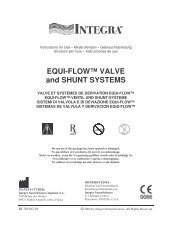
Pudenz Peritoneal Catheter
Pudenz Peritoneal Catheter
Pudenz Peritoneal Catheter

You also want an ePaper? Increase the reach of your titles
YUMPU automatically turns print PDFs into web optimized ePapers that Google loves.
102309-732-02.QXD 8/26/2004 8:16 AM Page 1<br />
<strong>Pudenz</strong> <strong>Peritoneal</strong> <strong>Catheter</strong><br />
Sterile For Single Use Only<br />
2<br />
STERILE EO 0123<br />
NL850-1380 NL850-1381 NL850-1382<br />
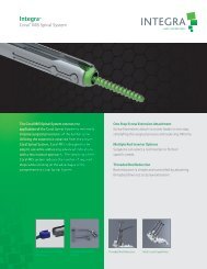
Description<br />
The silicone elastomer <strong>Pudenz</strong><br />
<strong>Peritoneal</strong> <strong>Catheter</strong> is designed to<br />
deliver cerebrospinal fluid (CSF) to the<br />
peritoneal cavity through a slit valve<br />
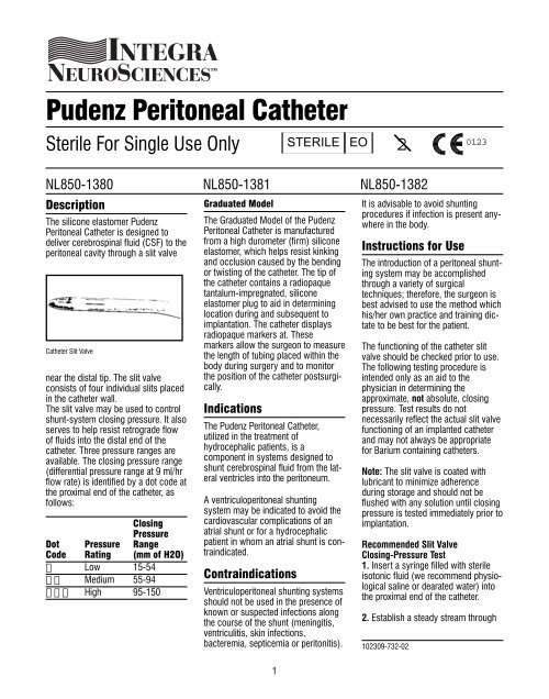
<strong>Catheter</strong> Slit Valve<br />
near the distal tip. The slit valve<br />
consists of four individual slits placed<br />
in the catheter wall.<br />
The slit valve may be used to control<br />
shunt-system closing pressure. It also<br />
serves to help resist retrograde flow<br />
of fluids into the distal end of the<br />
catheter. Three pressure ranges are<br />
available. The closing pressure range<br />
(differential pressure range at 9 ml/hr<br />
flow rate) is identified by a dot code at<br />
the proximal end of the catheter, as<br />
follows:<br />
Closing<br />
Pressure<br />
Dot Pressure Range<br />
Code Rating (mm of H2O)<br />
Low 15-54<br />
Medium 55-94<br />
High 95-150<br />
Graduated Model<br />
The Graduated Model of the <strong>Pudenz</strong><br />
<strong>Peritoneal</strong> <strong>Catheter</strong> is manufactured<br />
from a high durometer (firm) silicone<br />
elastomer, which helps resist kinking<br />
and occlusion caused by the bending<br />
or twisting of the catheter. The tip of<br />
the catheter contains a radiopaque<br />
tantalum-impregnated, silicone<br />
elastomer plug to aid in determining<br />
location during and subsequent to<br />
implantation. The catheter displays<br />
radiopaque markers at. These<br />
markers allow the surgeon to measure<br />
the length of tubing placed within the<br />
body during surgery and to monitor<br />
the position of the catheter postsurgically.<br />
Indications<br />
The <strong>Pudenz</strong> <strong>Peritoneal</strong> <strong>Catheter</strong>,<br />
utilized in the treatment of<br />
hydrocephalic patients, is a<br />
component in systems designed to<br />
shunt cerebrospinal fluid from the lateral<br />
ventricles into the peritoneum.<br />
A ventriculoperitoneal shunting<br />
system may be indicated to avoid the<br />
cardiovascular complications of an<br />
atrial shunt or for a hydrocephalic<br />
patient in whom an atrial shunt is contraindicated.<br />
Contraindications<br />
Ventriculoperitoneal shunting systems<br />
should not be used in the presence of<br />
known or suspected infections along<br />
the course of the shunt (meningitis,<br />
ventriculitis, skin infections,<br />
bacteremia, septicemia or peritonitis).<br />
1<br />
It is advisable to avoid shunting<br />
procedures if infection is present anywhere<br />
in the body.<br />
Instructions for Use<br />
The introduction of a peritoneal shunting<br />
system may be accomplished<br />
through a variety of surgical<br />
techniques; therefore, the surgeon is<br />
best advised to use the method which<br />
his/her own practice and training dictate<br />
to be best for the patient.<br />
The functioning of the catheter slit<br />
valve should be checked prior to use.<br />
The following testing procedure is<br />
intended only as an aid to the<br />
physician in determining the<br />
approximate, not absolute, closing<br />
pressure. Test results do not<br />
necessarily reflect the actual slit valve<br />
functioning of an implanted catheter<br />
and may not always be appropriate<br />
for Barium containing catheters.<br />
Note: The slit valve is coated with<br />
lubricant to minimize adherence<br />
during storage and should not be<br />
flushed with any solution until closing<br />
pressure is tested immediately prior to<br />
implantation.<br />
Recommended Slit Valve<br />
Closing-Pressure Test<br />
1. Insert a syringe filled with sterile<br />
isotonic fluid (we recommend physiological<br />
saline or dearated water) into<br />
the proximal end of the catheter.<br />
2. Establish a steady stream through<br />
102309-732-02
102309-732-02.QXD 8/26/2004 8:16 AM Page 2<br />
each of the four slits by gently rolling<br />
the slit valve region of the catheter<br />
between thumb and forefinger while<br />
flushing the catheter with fluid.<br />
Note: Any mechanical or fluid manipulation<br />
of the slit valve may temporarily<br />
lower the normal operating<br />
pressure.<br />
3. Hold both ends of the catheter in<br />
one hand.<br />
4. After filling the catheter with testing<br />
solution, remove the syringe and<br />
slowly lower the distal end until the<br />
catheter is hanging vertically. Fluid<br />
should run freely from the slits.<br />
Starting from the moment fluid begins<br />
to flow from the slits, allow 1 1 ¼2 minutes<br />
for the fluid level to stabilize.<br />
Note: While waiting, inspect for air<br />
bubbles. If air bubbles are present,<br />
start the test procedure over.<br />
Caution: Extreme care must be<br />
taken during this test to eliminate<br />
any air bubbles in the column of<br />
testing fluid. One bubble is enough<br />
to impede or even completely stop<br />
the descent of fluid and give a false<br />
reading.<br />
Should a bubble occur, flush out the<br />
catheter completely and carefully<br />
refill per the instructions.<br />
5. Measure the column of fluid in centimeters<br />
from the top of the slit valve<br />
to the top of the fluid column in the<br />
catheter to obtain the closingpressure<br />
reading.<br />
How Supplied<br />
The <strong>Pudenz</strong> <strong>Peritoneal</strong> <strong>Catheter</strong> is<br />
supplied individually in a sterile, double-wrap,<br />
pyrogen-free packaging<br />
system. The double-wrap system<br />
facilitates the preferred method of<br />
sterile product transfer from the circulating<br />
area to the sterile field.<br />
Do Not Resterilize<br />
This product is for Single Use Only.<br />
Warnings<br />
Hydrocephalic patients with<br />
cerebrospinal fluid shunting systems<br />
must be kept under close observation<br />
for signs and symptoms of increasing<br />
intracranial pressure due to shunting<br />
failure. These signs and symptoms<br />
may vary from patient to patient.<br />
Increasing intracranial pressure is<br />
characterized by headache, vomiting,<br />
irritability, listlessness, drowsiness<br />
and other signs of deterioration of<br />
consciousness and nuchal rigidity. In<br />
the infant, increased scalp tension at<br />
the anterior fontanelle and congestion<br />
of scalp veins will be noted.<br />
Perforation of abdominal viscera may<br />
occur from any foreign object retained<br />
in the abdomen.<br />
An obscure fever in patients with a<br />
ventriculoperitoneal shunt should suggest<br />
the possibility of a shunt-associated<br />
lowgrade peritonitis.<br />
A patient with a ventriculoperitoneal<br />
shunt in whom gram-negative<br />
ventriculitis occurs, recurs, or persists<br />
should be checked for a possible perforation<br />
of the intestinal wall by the<br />
peritoneal catheter.<br />
If the peritoneal catheter becomes disconnected,<br />
peristaltic action may<br />
draw the catheter completely into the<br />
abdominal cavity.<br />
This product has not been tested for<br />
drug compatibility and therefore is not<br />
intended for drug administration.<br />
Integra NeuroSciences makes no<br />
claim for or representation as to the<br />
performance characteristics of a<br />
shunting system if this product is<br />
used in conjunction with components<br />
of other manufacturers.<br />
Silicone tubing may be easily cut or<br />
torn when instruments are used to<br />
secure it to a connector. The use of<br />
instruments to attach silicone<br />
catheters to connectors should be<br />
avoided. When instruments are used,<br />
carefully inspect the tubing for nicks<br />
or other damage prior to closure.<br />
Precautions<br />
Prior to surgery, prospective patients<br />
or their representatives should be<br />
informed of the possible complications<br />
associated with the use of this<br />
product.<br />
Fluid flow through the slit valve should<br />
be verified prior to implantation. (See<br />
Instructions For Use.)<br />
The slits of the valve should not be<br />
removed, lengthened or altered in any<br />
other manner.<br />
<strong>Catheter</strong> tubing should be carefully<br />
secured to a connector with ligatures<br />
in such a manner as to avoid cutting<br />
or strangulation of the tubing.<br />
Kinking of the catheter tubing may<br />
result in shunt blockage.<br />
Although a ventriculoperitoneal shunting<br />
system may function for years<br />
without revision in adults, it cannot be<br />
considered to remain permanently<br />
operational in infants or young<br />
children. A child’s growth results in<br />
the progressive withdrawal of the peritoneal<br />
catheter, and the catheter may<br />
require periodic revisions.<br />
Complications<br />
Complications which may result from<br />
the use of this product include the<br />
risks associated with the medication<br />
and methods utilized in the surgical<br />
procedure, as well as the patient’s<br />
response, reaction or degree of<br />
intolerance to any foreign object<br />
implanted in the body.<br />
The principal complications<br />
associated with cerebrosinal fluid<br />
shunting into the peritoneum are<br />
shunt obstruction, functional failure of<br />
the shunt system, infection or<br />
intracranial hypotension.<br />
Shunt obstruction may occur in either<br />
the proximal ventricular catheter or in<br />
the distal, peritoneal catheter.<br />
Ventricular catheters may be<br />
obstructed by particulate matter such<br />
as blood clots, fibrin or brain<br />
2
102309-732-02.QXD 8/26/2004 8:16 AM Page 3<br />
fragments. If not properly located in<br />
the lateral ventricle, the catheter may<br />
become embedded in the ventricular<br />
wall or choroid plexus. Less<br />
commonly, the catheter may be<br />
obstructed by excessive reduction of<br />
ventricular size to slit-like proportions.<br />
<strong>Peritoneal</strong> catheters may also be<br />
obstructed by particulate matter.<br />
<strong>Peritoneal</strong> catheters may be<br />
obstructed by the omentum or coiled<br />
loops of bowel.<br />
Functional failure of the shunt system<br />
due to separation of its component<br />
parts can result in serious<br />
complications. <strong>Peritoneal</strong> catheters<br />
may migrate completely into the peritoneal<br />
cavity. Volvulus and perforation<br />
of intraabdominal viscera may occur<br />
or the catheter may be extruded.<br />
Infection is a common and serious<br />
complication of a shunting system<br />
and is most frequently caused by skin<br />
contaminants. Septicemia, which<br />
occurs most frequently in debilitated<br />
infants, can result from infections<br />
anywhere in the body and may<br />
develop with few or no symptoms. It<br />
may occur as a result of a wound<br />
infection. The presence of a foreign<br />
body (i.e. the shunting system) may<br />
trigger ventriculitis or a dormant<br />
meningitis. Intracranial infection may<br />
then be disseminated throughout the<br />
body via the distal catheter. Lesions<br />
developing from the breakdown of<br />
skin or tissue over the shunting<br />
system may also serve as foci of serious<br />
infections. In the event of an<br />
infection, removal of the shunt system<br />
is indicated in addition to the appropriate<br />
therapy.<br />
Excessive lowering of intracranial<br />
pressure may result in complications,<br />
particularly in the infant. These<br />
include subdural hematomas,<br />
markedly sunken fontanelles, overriding<br />
of cranial bones, and the<br />
conversion of a communicating to a<br />
non-communicating hydrocephalus<br />
due to obstruction of the aqueduct of<br />
Sylvius.<br />
Failure of the shunting system may be<br />
evidenced by any or all of the<br />
following: continuing symptoms of<br />
increased intracranial pressure, the<br />
subcutaneous exudation of CSF along<br />
the pathway of the shunt, and leakage<br />
of fluid through the surgical wound.<br />
These failures require immediate<br />
replacement of the shunting system<br />
or of the affected component.<br />
Product Information<br />
Disclosure<br />
Integra NeuroSciences has exercised<br />
reasonable care in the choice of materials<br />
and manufacture of this product.<br />
Integra NeuroSciences excludes all<br />
warranties, whether express or<br />
implied by operation of law or<br />
otherwise, including, but not limited<br />
to, any implied warranties of<br />
merchantability or fitness. Integra<br />
NeuroSciences shall not be liable for<br />
any incidental or consequential loss,<br />
damage, or use of the product. Integra<br />
NeuroSciences neither assumes nor<br />
authorizes any other person to<br />
assume for it, any other or additional<br />
liability or responsibility in connection<br />
with this device.<br />
Product Order Information<br />
All products can be ordered through<br />
your Integra NeuroSciences Neuro<br />
Specialist or customer service<br />
representative or by contacting :<br />
Integra NeuroSciences<br />
311 Enterprise Drive<br />
Plainsboro, NJ 08536 USA<br />
Telephone: 1-800-654-2873<br />
Outside the US: 1-609-275-0500<br />
Fax: 609-275-5363<br />
or<br />
Integra NeuroSciences<br />
Newbury Road, Andover<br />
Hampshire SP10 4DR England<br />
Tel: +44(0) 1264-345-700<br />
Fax: +44 (0) 1264-332-113<br />
Caution: Federal (USA) law<br />
restricts this device to sale by or<br />
on the order of a physician. Do not<br />
use if the package has been<br />
opened or damaged.<br />
Symbols Used On Labeling<br />
2<br />
LOT<br />
STERILE EO<br />
See instructions for use<br />
Expiration date<br />
Do not reuse after opening<br />
Lot number<br />
Sterile unless package is<br />
opened or damaged.<br />
Method of sterilizationethylene<br />
oxide.<br />
0123 Product complies with<br />
requirements of directive<br />
93/42/EEC for medical<br />
devices.<br />
Bibliography<br />
Foltz, E.; Shurtleff, D.B. “Conversion<br />
of Communicating Hydrocephalus to<br />
Stenosis or Occlusion of the Aqueduct<br />
during Ventricular Shunt.” Journal of<br />
Neurosurgery. 1966: 24; 520-529.<br />
Forward, K.R.; Fewer, D.; Stiver, H.G.<br />
“Cerebrospinal Fluid Shunt Infections;<br />
A Review of 35 Infections In 32<br />
Patients.” Journal of Neurosurgery.<br />
1983 September: 59; 389-394.<br />
Ignelzi, R.J.; Kirsch, W.M. “Follow-up<br />
Analysis of Ventriculoperitoneal and<br />
Ventriculoatrial Shunts for Hydrocephalus.”<br />
Journal of Neurosurgery.<br />
1975 June: 42; 679-682.<br />
Illingworth, R.D.; Logue, V.; Symon L.;<br />
et al. “The Ventriculocaval Shunt in<br />
the Treatment of Adult Hydrocephalus:<br />
Results and Complications in 101<br />
Patients.” Journal of Neurosurgery.<br />
1979 December 35; 681-685.<br />
Kuwamura, K.; Kokunai, T.<br />
“Intraventricular Hematoma<br />
Secondary to a Ventriculoperitoneal<br />
Shunt.” Neurosurgery. 1982 March:<br />
10(3); 384-386.<br />
3
102309-732-02.QXD 8/26/2004 8:16 AM Page 4<br />
Little, J.R.; Rhoton Jr., A.L.; Mellinger,<br />
J.F. “Comparison of Ventriculoperitoneal<br />
and Ventriculoatrial Shunts<br />
for Hydrocephalus in Children.” Mayo<br />
Clinic Proceedings. 1972 June: 47;<br />
396-401.<br />
McCullough, D.C.; Fox, J.L.; et al.<br />
“Effects of CSF Shunts on Intracranial<br />
Pressure and CSF Dynamics.”<br />
Cisternography and Hydrocephalus,<br />
edited by John C. Harbert. Springfield,<br />
Illinois 1972.<br />
Milhorat, Thomas H. Hydrocephalus<br />
and the Cerebrospinal Fluid. The<br />
Williams and Wilkins Co., Baltimore,<br />
1972.<br />
Portnoy, H.D.; Croissant, P.D.<br />
“Combined Drainage of Ventricular<br />
and Subdural Fluid.” Surgical<br />
Neurology. 1974 January: 2, 41-42.<br />
Sells, C.J.; Loeser, J.D. “Peritonitis<br />
Following Perforation of the Bowel:<br />
A Rare Complication of Ventriculoperitoneal<br />
Shunt.” Journal of<br />
Pediatrics. 1973 November: 83; 823-<br />
824.<br />
Sugar, O.; Bailey O.T. “Subcutaneous<br />
Reaction to Silicone in Ventriculoperitoneal<br />
Shunts.” Journal of<br />
Neurosurgery. 1974 September: 41;<br />
367-371.<br />
Weiss, S.R.; Raskind, R. “Twenty-Two<br />
Cases of Hydrocephalus Treated with<br />
Silastic Ventriculoperitoneal Shunt.”<br />
International Surgery. 1969 January:<br />
51; 13-19.<br />
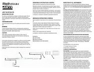
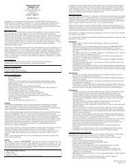
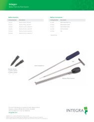
Dimensioned Illustrations (All Dimensions Nominal)<br />
Model Catalog Number Description<br />
Graduated NL850-1380 <strong>Pudenz</strong> <strong>Peritoneal</strong> <strong>Catheter</strong>, Low Pressure<br />
NL850-1381<br />
<strong>Pudenz</strong> <strong>Peritoneal</strong> <strong>Catheter</strong>, Medium Pressure<br />
NL850-1382<br />
<strong>Pudenz</strong> <strong>Peritoneal</strong> <strong>Catheter</strong>, High Pressure<br />
Graduated Model<br />
1.3mm ID, 2.5mm OD<br />
1cm<br />
Typical 1cm<br />
90cm<br />
Integra NeuroSciences<br />
311 Enterprise Drive, Plainsboro, NJ 08536 USA © Copyright 2002 Integra NeuroSciences. All rights reserved.<br />
4