View PDF - Mycosphere-online journal
View PDF - Mycosphere-online journal
View PDF - Mycosphere-online journal

You also want an ePaper? Increase the reach of your titles
YUMPU automatically turns print PDFs into web optimized ePapers that Google loves.
<strong>Mycosphere</strong> 4 (4): 734–743 (2013) ISSN 2077 7019<br />
www.mycosphere.org Article <strong>Mycosphere</strong><br />
Copyright © 2013<br />
Online Edition<br />
Doi 10.5943/mycosphere/4/4/10<br />
In-vitro evaluation of some Indian lichens against human pathogenic<br />
bacteria<br />
Srivastava P 1 , Logesh AR 2 , Upreti DK 2 , Dhole TN 3 and Srivastava A 4<br />
1 Department of Botany, Dr. R.M.L. Avadh University, Faizabad, India<br />
2 Lichenology laboratory, Plant Biodiversity Conservation Biology and Herbarium Division, CSIR-National Botanical<br />
Research Institute, Lucknow, India<br />
3 Department of Microbiology, Sanjay Gandhi Post Graduate Institute of Medical Sciences, Lucknow, India<br />
4 Department of Pathology & Microbiology, Saraswati Dental & Medical College, Lucknow, India<br />
Email: upretidk@rediffmail.com<br />
Srivastava P, Logesh AR, Upreti DK, Dhole TN, Srivastava A 2013 – In-vitro evaluation of some<br />
Indian lichens against human pathogenic bacteria. <strong>Mycosphere</strong> 4(4), 734–743, Doi<br />
10.5943/mycosphere/4/4/10<br />
Abstract<br />
Antimicrobial activity of the acetone, methanol and ethanol extracts of some common<br />
lichen species such as Usnea longissima Ach., Everniastrum cirrhatum (Fr.) Hale, Peltigera<br />
polydactylon (Neck.) Hoffm. and Sulcaria sulcata (Lév.) Bystr. ex Brodo & D. Hawksw., were<br />
screened in vitro against six clinically important pathogenic bacteria, Staphylococcus aureus,<br />
Streptococcus faecalis, Bacillus cereus, Pseudomonas aeruginosa, Salmonella typhimurium and<br />
Escherichia coli by Kirby-Bauer technique of disc diffusion method. Minimum inhibitory<br />
concentration was taken out by Broth micro dilution method according to the NCCLS guidelines.<br />
Acetone, methanol and ethanol extracts of the investigated lichens showed relatively strong<br />
antimicrobial activity against all the gram positive bacteria and two gram negative bacteria. It was<br />
found that the inhibition zone of tested bacteria against lichen extracts varied between 7.6 – 30.7<br />
mm diameters. The lowest MIC value was observed to be as low as 6.25 µg/ml against B. cereus of<br />
U. longissima. Generally the lichen extracts tested demonstrated antimicrobial effect which<br />
suggests a possibility of their use in treatment of various diseases caused by these and similar<br />
microorganisms.<br />
Key words – Kirby-Bauer method – Lichenized fungi – MIC – pathogenic bacteria.<br />
Introduction<br />
Lichens are composite organisms consisting of symbiotic association of a fungus (the<br />
mycobiont) with a photosynthetic partner (the photobiont or phycobiont), usually either a green<br />
alga or cyanobacterium. Lichens are unique in nature and physiology because they look and behave<br />
quite differently from their component organisms. Lichens produce a wide range of organic<br />
compounds that can be grouped as primary metabolites and secondary metabolites (Elix 1996).<br />
Primary metabolites such as proteins, lipids, carbohydrates, and some other organic compounds are<br />
produced by both the partners and are needed for the lichen’s metabolism and structure. Secondary<br />
metabolites are produced by the fungus alone and secreted onto the surface of lichen’s hyphae<br />
either in amorphous forms or as crystals. The secondary metabolites include aliphatic,<br />
Submitted 25 June 2013, Accepted 21 July 2013, Published <strong>online</strong> 5 August 2013<br />
Corresponding Author: Upreti DK – e-mail – upretidk@rediffmail.com 734
cycloaliphatic, aromatic and terpenic components are extra cellular products of relatively low<br />
molecular weight usually insoluble in water and can be extracted into organic solvents (Ötzürk et<br />
al. 1999). They make even more than 30% of the dry mass of thalus (Galun 1988). Up till now,<br />
about 350 biologically active secondary metabolites of lichens have been discovered and<br />
approximately 200 have been characterized (Chand et al. 2009, Tay et al. 2004). Lichen secondary<br />
metabolites have been investigated mostly for chemotaxonomic purposes and in connection with<br />
their potential as phytomedicines and natural biopesticides (Dayan and Romagni, 2001). Many<br />
lichens have been used for human or animal nutrition and used as dyes, perfumes, in preparation of<br />
alcohol and in the pharmaceutical industries (Kirmizigül et al. 2003, Richardson 1988, Romagni<br />
2002). Lichen metabolites exert a wide variety of biological actions including antibiotic,<br />
antimycobacterial, antiviral, antiinflammatory, analgesic, antipyretic, antiproliferative and<br />
cytotoxic effects (Muller K. 2001). A number of lichen species are listed as having use as in folk<br />
medicine for treatment of stomach diseases, diabetes, whooping cough, pulmonary tuberculosis,<br />
cancer treatment and skin diseases (Baytop 1999, Huneck 1999, Richardson 1991). Medicinal<br />
activities of some lichens and their components are known, such as: antiviral, anti-tumor, antiinflammatory,<br />
analgesic, antipyretic, antiproliferative and antiprotozoal (Lawrey 1986, Halama and<br />
Van Haluvin 2004 and Huneck 1999). However, only very limited numbers of lichen substances<br />
have been screened only very limited numbers of lichen substances have been screened for their<br />
biological activities and their therapeutic potential in medicine. This is certainly due to the<br />
difficulties encountered in identification of the species, collection of bulk quantities, and the<br />
isolation of pure substances for structure determination and testing activity. Recently, possibility<br />
for by passing some of these former difficulties have arisen by the introduction of new techniques.<br />
This includes axenic cultivation for production of the genuine compounds or new ones, extraction<br />
of focused compounds, or synthesis of natural products or their derivatives for testing (Boustie and<br />
Grube 2005).<br />
According to one estimate, 50% of all lichens have antibiotic properties (Sharnoff 1997).<br />
The development and spread of microbial resistance to the available antibiotics has prompted<br />
investigators to study antimicrobial substances from other sources. India being a mega diversity<br />
country exhibit rich diversity and luxuriance of lichens together with other group of plants. A<br />
number of vascular plants from the country are known having their potential medicinal use;<br />
however, so far the studies and enumeration of medicinal non vascular plants are not available from<br />
the country. In India, few studies regarding use of lichens against plant pathogen are available<br />
(Tiwari et al. 2011a, Tiwari et al. 2012b). Since, there has not been much work done on the<br />
antimicrobial activity of Indian lichens, especially against human pathogens. Thus the aim of the<br />
present study was to conduct in vitro evaluation of antibacterial activity with acetone, ethanol and<br />
aqueous extracts from the lichens against human pathogenic bacteria.<br />
Materials & Methods<br />
Collection and identification of lichens<br />
The lichen species were collected from different parts of the Himalayas in Uttarakhand and<br />
Himachal Pradesh. Lichen specimens were air dried at room temperature and identified by studying<br />
their morphology, anatomy and chemistry and authenticated using the standard literatures (Awasthi<br />
2007, Orange et al. 2001). The voucher specimens are deposited in herbarium of National Botanical<br />
Research Institute, Lucknow (LWG).<br />
Extraction<br />
For extraction, air-dried lichen samples were ground, then 10 g portions were taken and<br />
added to 100 ml of solvents of acetone, ethanol and methanol. The mixtures were sonicated for 30<br />
minutes, then left at room temperature for seven days and flasks were sealed with parafilm so that<br />
the solvent cannot evaporate. The extracts were filtered over Whatman No 1 filter paper, and the<br />
filtrates were sterilized by membrane filtration using 0.45 μm pore size filters. The extracts were<br />
735
then evaporated to dryness under reduced pressure and re-dissolved in respective solvents to attain<br />
the required concentration 0.2 mg/ml for antibacterial screening. These extracts were kept at 4°C<br />
till used.<br />
Specimens used in the present study<br />
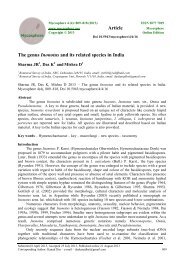
Fig.1<br />
1. Everniastrum cirrhatum (Fr.) Hale.<br />
India – Uttarakhand, Pithoragarh district, Narayan Swami Ashram. 02.11.2009, D.K. Upreti & al,<br />
09-013411-LWG.<br />
2. Peltigera polydactylon (Neck.) Hoffm.<br />
India – Uttarakhand, Pithoragarh district, Munsiyari, Khuliya top. 31.10.2009. D.K. Upreti & al.,<br />
09-023666-LWG.<br />
3. Sulcaria sulcata (Lév.) Bystr. ex Brodo<br />
India – Uttarakhand, Pithoragarh district, Munsiyari, Khuliya Top, 17.11.2006. Y. Joshi & R.<br />
Bajpai. 06-006935-LWG.<br />
4. Usnea longissima Ach.<br />
India – Uttarakhand, Pithoragarh district, Narayan Swami Ashram. 02.11.2009. D.K. Upreti & al.,<br />
09-012179-LWG.<br />
Fig. 1 – 1 Everniastrum cirrhatum (Fr.) Hale; 2 Peltigera polydactylon (Neck.) Hoffm.; 3 Sulcaria<br />
sulcata (Lév.) Bystr. ex Brodo; 4 Usnea longissima Ach.
Identification of lichen acids<br />
To identify the lichen substances present, the extracted lichen crude extracts were dissolved<br />
in acetone to a final concentration of 1 mg/ml. The crude extracts were then spotted on silica gel<br />
thin layer chromatography (TLC) plates (silica gel 60 F254 aluminum plates, Merck) and run in<br />
solvent systems A (36:9:1 toluene/dioxane/glacial acetic acid). The plates were kept for air drying<br />
at room temperature. Before spraying, the dried plates were examined in day light for pigments and<br />
for fluorescence or quenching under short and long wave length ultraviolet (U.V) light.<br />
Subsequently, each TLC plate was then sprayed with 10% sulfuric acid and heated at 110°C for 10<br />
minutes to visualize the lichen substances (Santos and Mondragon 1969). The Rf values for each<br />
spot were determined and compared with selected lichen compound standards: (1) norstictic acid,<br />
(2) usnic acid, (3) salazinic acid, (4) caperatic : usnic acid, 25:1, (5) protocetraric acid, (6) stictic :<br />
constictic acid, 2:1, (7) diffractaic acid, (8) barbatic acid, and (9) galbinic acid. The lichen acids<br />
standards were generously provided by Prof. Dr. Jack A. Elix, Australian National University,<br />
Canberra, Australia.<br />
Antibacterial activity of lichen extracts<br />
The antimicrobial activity of lichen extracts against tested bacteria was determined<br />
employing Kirby-Bauer technique of disk diffusion method (Bauer et al. 1966, NCCLS 1993) The<br />
lichen crude extracts were then tested for their inhibitory activities against representative test<br />
bacterial gram-positive bacteria such as Staphylococcus aureus (ATCC 25923), Streptococcus<br />
faecalis (ATCC 33186) and Bacillus cereus (ATCC 14579); gram-negative bacteria such as<br />
Escherichia coli (ATCC 25922), Pseudomonas aeruginosa (ATCC 29853), Salmonella<br />
typhimurium (ATCC 13311). Bacterial cell suspension was prepared from a 24-hour old culture,<br />
adjusted to 0.5 McFarland standard, and swabbed on petri plates pre-filled with 25 ml Mueller-<br />
Hinton Agar (MHA, Hi-Media). Antibiotic disks (Whatmann) measuring 6 mm in diameter were<br />
then placed onto the inoculated MHA plates (two disks per plate). To determine the sensitivity of<br />
lichen crude extracts, to each paper disk 10 μl of 0.2 mg/ml concentration of lichen crude extract<br />
was added .The positive control were Gentamycin (for gram positive bacteria) and Ceftriaxone (for<br />
gram negative bacteria) and the negative control were the solvents acetone, methanol, ethanol<br />
respectively. Plates were then incubated at 37°C for 18-24 hrs. Following incubation, zones of<br />
inhibition including paper disks were then measured with a ruler and recorded.<br />
The minimal inhibitory concentration (MIC) of the crude extract was determined by micro<br />
dilution techniques in Mueller-Hinton Broth (MHB), according to National Committee for Clinical<br />
Laboratory Standard, USA guidelines (NCCLS, 2002). A series of two fold dilutions with<br />
concentrations ranging from 200 µg/ml to 0.195 µg/ml of extract was used in the experiment<br />
against S. aureus, S. faecalis and B. cereus, P. aeruginosa, and E. coli. The starting solutions of<br />
extracts and component were obtained by measuring a certain quantity of extract and dissolving it<br />
in dimethylsulphoxide (DMSO). Two-fold dilutions of extracts and components were prepared in<br />
Mueller-Hinton broth (MHB) for bacterial cultures. The inoculates were prepared in the same<br />
medium at a density adjusted to a 0.5 McFarland turbidity standard colony forming units, and<br />
diluted 1:10 for the broth micro dilution procedure .Then 100 µl of diluted extracts and 100 µl of<br />
bacterial suspensions were dispensed in 96 well sterile microtitre plate. The microtitre plates were<br />
incubated at 37 0 C and MIC was determined after 24 h of incubation. The MIC was determined by<br />
establishing visible growth of the microorganisms. The boundary dilution without any visible<br />
growth was defined as the MIC for the tested microorganism at the given concentration. Untreated<br />
bacteria were taken as Positive control and MHB was taken as negative control. All experiments<br />
were performed in triplicate.<br />
The results of the antibacterial screening for disc diffusion assay are expressed as mean ±<br />
SD of three replicates in each test and for MIC, the boundary dilution without any visible growth<br />
was defined as the MIC for the tested microorganism at the given concentration.<br />
737
Results<br />
Identification of lichen compounds<br />
The results of thin layer chromatography showed that lichen substances were present in all<br />
the four lichens used for the study. Usnea longissima showed presence of four compounds; stictic<br />
acid complex at Rf class 3, barbatic acid at Rf class 4, diffractic acid at Rf class 4 and usnic acid at<br />
Rf class 6. Everniastrum cirrhatum shown salazinic acid at Rf class 2, protolichesterinic acid<br />
between Rf classes 3-4, atranorin at Rf class 7. In Peltigera polydactyla, two compounds were<br />
detected such as gyrophoric acid at Rf class 3 and tenuiorin at Rf class 7. In lichen Sulcaria sulcata<br />
has pulvinic acid at Rf class 1, virensic acid at Rf class 3-4, psoromic acid at Rf class 4 and vulpinic<br />
acid at Rf class 6-7 were present.<br />
Antibacterial activity of extracts<br />
The present study confirmed the presence of antibacterial substance in all the extracts of<br />
tested lichens and the results were presented (Table 1). The majority of acetone, methanol and<br />
ethanol extracts of U. longissima exhibited activity against the gram positive Staphylococcus<br />
aureus, Streptococcus faecalis and Bacillus cereus. Importantly the ethanol extract showed activity<br />
against two gram negative bacteria, Pseudomonas aeruginosa and E. coli. No activity was recorded<br />
against S. typhimurium. Against S. aureus, the methanol extract was most active with a mean zone<br />
of 18.4 ± 1.4 mm diameters. Against S. faecalis and B. cereus, again the zone of methanol extract<br />
was better than acetone and ethanol extract and the mean zone of inhibition was 22.6 ± 1.4 mm and<br />
30.7 ± 0.9 mm diameters respectively. The activity of ethanol extract against P. aeruginosa and E.<br />
coli was 16.3 ± 0.5 mm and 18.8 ± 0.8 mm diameters respectively. The inhibitory effect of solvent<br />
alone on microorganisms was nil.<br />
E. cirrhatum extracts were active against all the gram positive and a single gram negative<br />
bacteria. Against S. aureus, the activity of ethanol extract was greater than acetone and methanol<br />
extract and the calculated zone of inhibition against ethanol extract was 20.2 ± 1.1 mm diameters.<br />
The zone of inhibition noted for ethanol extract against S. faecalis and B. cereus was 16.3 ± 0.5 mm<br />
and 24.1 ± 0.6 mm diameters respectively. Against P. aeruginosa, only ethanol extract was<br />
effective and the calculated zone of inhibition was 19.3 ± 1.1 mm.<br />
Ethanol extract of P. polydactylon showed activity against S. aureus with a mean zone of<br />
10.8 ± 0.8 mm diameters. Both the acetone and ethanol extract showed activity against S. faecalis<br />
with a mean zone of 9.6 ± 0.5 mm and 11 ± 1.1 mm diameters respectively. Against B. cereus, only<br />
ethanol extract was effective with a mean zone of 15.1 ± 0.7 mm. Only acetone extract shown<br />
activity against P. aeruginosa with a zone of 7.6 ± 0.5 mm.<br />
All the three extracts of S. sulcata showed activity against S. aureus and B. cereus. Against<br />
S. aureus, the acetone extract was most effective and the calculated zone was 17.1 ± 0.8 mm<br />
diameters while against B. cereus, the zone of acetone extract (8.7 ± 1.2 mm) was more than<br />
methanol and ethanol extract. Marked activity was shown by ethanol against P. aeruginosa and the<br />
mean zone of inhibition was 7.6 ± 0.5 mm diameters. No activity was noted against S. faecalis, E.<br />
coli and S. typhimurium<br />
Minimum Inhibitory Concentration<br />
The results of minimum inhibitory concentration of lichen U. longissima ranged within the<br />
range of 3.125-200 µg/ml. The least MIC value was calculated against B. cereus (6.25 µg/ml). The<br />
MIC results of E. cirrhatum were varying between 3.125-200 µg/ml. Lowest MIC was observed<br />
against S. faecalis (12.5 µg/ml). The MIC value noted for P. polydactylon against all the tested<br />
bacteria was 200 µg/ml. The MIC value calculated for the extract of S. sulcata against all the tested<br />
bacteria was 200 µg/ml (Table 2).
Table1 Results of zone of inhibition (mm) of extracts of Usnea longissima, Everniastrum cirrhatum, Peltigera<br />
polydactylon and Sulcaria sulcata against tested microorganisms.<br />
S. Bacteria Usnea longissima Everniastrum cirrhatum Peltigera polydactylon Sulcaria sulcata<br />
No.<br />
Ac.<br />
extract<br />
Me.<br />
extract<br />
Et.<br />
extract<br />
Ac.<br />
extract<br />
Me.<br />
extract<br />
Et.<br />
extract<br />
Ac.<br />
extract<br />
Me.<br />
extract<br />
Et.<br />
extract<br />
Ac.<br />
extract<br />
Me.<br />
extract<br />
Et.<br />
extract<br />
1. Staphlyococcus 13.2 ± 18.4 ± 17.6 ± 18.5 ± 12.8 ± 20.2 ± 0.0 ± 0.0 ± 10.8 ± 17.1 ± 13.4 ± 14.9 ±<br />
aureus<br />
0.7 1.4 1.1 0.8 1.6 1.1 0.0 0.0 0.8 0.8 0.5 0.9<br />
2. Streptococcus 21.5 ± 22.6 ± 20.3 ± 14.4 ± 15.4 ± 16.3 ± 9.6 ± 0.0 ± 11 ± 0.0 ± 0.0 ± 0.0 ±<br />
faecalis 0.9 1.4 0.5 0.7 0.8 0.5 0.5 0.0 1.1 0.0 0.0 0.0<br />
3. Bacillus cereus 19.4 ± 30.7 ± 20.4 ± 28.3 ± 21.4 ± 24.1 ± 0.0 ± 0.0 ± 15.1 ± 8.7 ± 8.6 ± 8.3 ±<br />
0.7 0.9 0.7 1.5 1 0.6 0.0 0.0 0.7 1.2 1.2 0.5<br />
4. Escherichia coli 0.0 ± 0.0 ± 16.3 ± 0.0 ± 0.0 ± 0.0 ± 0.0 ± 0.0 ± 0.0 ± 0.0 ± 0.0 ± 0.0 ±<br />
0.0 0.0 0.5 0.0 0.0 0.0 0.0 0.0 0.0 0.0 0.0 0.0<br />
5. Pseudomonas 8.4 ± 8.7 ± 18.8 ± 0.0 ± 10.6 ± 19.3 ± 7.6 ± 0.0 ± 0.0 ± 0.0 ± 0.0 ± 7.6 ±<br />
aeruginosa 0.6 0.3 0.5 0.0 0.5 1.1 0.5 0.0 0.0 0.0 0.0 0.5<br />
6. Salmonella 0.0 ± 0.0 ± 0.0 ± 0.0 ± 0.0 ± 0.0 ± 0.0 ± 0.0 ± 0.0 ± 0.0 ± 0.0 ± 0.0 ±<br />
typhimurium 0.0 0.0 0.0 0.0 0.0 0.0 0.0 0.0 0.0 0.0 0.0 0.0<br />
(*values are in mean ± Standard deviation, n=3)<br />
(Ac. = Acetone, Me. = Methanol, Et. = Ethanol)<br />
Table 2 Results of Minimum Inhibitory Concentration (MIC) of extracts of Usnea longissima, Everniastrum cirrhatum,<br />
Peltigera polydactylon and Sulcaria sulcata against tested microorganisms.<br />
S.No Bacterial pathogen Lichen Species<br />
(MIC in µg/ml)<br />
Usnea longissima Everniastrum cirrhatum Peltigera polydactylon Sulcaria sulcata<br />
1. Staphlyococcus aureus 3.125 3.125 200 200<br />
2. Streptococcus faecalis 25 12.5 200 NA<br />
3. Bacillus cereus 6.25 3.125 200 200<br />
4. Pseudomonas aeruginosa 200 200 200 200<br />
5. Escherichia coli 200 NA NA NA<br />
NA= No Activity<br />
739
Discussion<br />
Lichens produce antibiotic secondary metabolites that provide defense against most of the<br />
pathogens in nature (Molnar and Farkas 2010). The tested lichen extracts showed a relatively<br />
strong antimicrobial activity. The screening results showed that nearly all the extracts of the tested<br />
lichens showed antibacterial activity against gram positive as well as gram negative bacteria.<br />
Antibacterial activity observed in this present investigation depended on the sort of the extract, its<br />
concentration and the tested microorganisms. Similar results were also noticed by other<br />
investigators (Rankovic et al. 2007), there is no antibacterial activity of the extracts against S.<br />
typhimurium was detected. Strong antibacterial activity was given by all the tested lichens against<br />
S. aureus, but extracts of E. cirrhatum showed larger zone of inhibition against this bacterium.<br />
Dülger et al. (1997, 1998) have found that although 4 different extracts of a macrofungus have<br />
inhibition effect against B. subtilis ATCC 6633 and other some gram positive and negative bacteria,<br />
and they do not have effect against E. coli ATCC 11230, S. epidermidis NRRL B-4377, S. aureus<br />
ATCC 6538P (Dülger et al. 1998). Activity against S. faecalis was given by all the lichens except<br />
S. sulcata. Among all the tested lichens, U. longissima was most effective against this bacterium.<br />
Rowe et al. (1999) reported that the lichens from Turkey, Evernia prunastri, Pseudovernia<br />
furfuracea and Alectoria capillaris were active against gram-positive bacteria and Candida<br />
albicans.<br />
U. longissima showed the maximum activity against B. cereus. Similar to U. longissima,<br />
extracts of E. cirrhatum and P. polydactylon were also showing better activity against B. cereus. It<br />
has been determined that lichens have shown the inhibition effect against a lot of bacteria such as<br />
Bacillus, Pseudomonas, E. coli, Streptococcus, Staphylococcus, Enterococcus, Mycobacterium<br />
(Esimone and Adikwn 1999, Perry et al. 1999). However, S. sulcata showed poor activity against<br />
B. cereus. The previous studies carried out by Burkholder et al. (1944), Rowe et al. (1989) and<br />
Silva et al. (1986) indicated that the lichens inhibit mostly gram positive bacteria. Even though<br />
most of the lichens have been reported to be active against gram positive bacteria, but it is of great<br />
interest to note that the extracts of U. longissima, E. cirrhatum, P. polydactylon and S. sulcata<br />
inhibited the growth of gram negative bacteria together with gram positive bacteria. All the extracts<br />
of four lichens showed activity against P. aeruginosa except U. longissima which showed activity<br />
against both gram negative bacteria such as P. aeruginosa and E. coli.<br />
All the three extracts of U. longissima, methanol extract was found to have larger zone of<br />
inhibitions while according to Gulluce et al. (2006) and the methanol extract of the lichen Parmelia<br />
saxatilis had a strong antimicrobial influence. Results of U. longissima are comparable to<br />
Thippeswamy et al.(2011) where Ethanol extract of Usnea Longissima exhibited significant<br />
antibacterial activity and antifungal activity with 1mg/ml Agar well diffusion method against the<br />
Gram positive and Gram negative bacteria. Ethanol extracts of E. cirrhatum showed maximum<br />
inhibition as compared to acetone and methanol extracts while in the studies done by Swathi et al.<br />
(2010), Methanol extract of E. cirrhatum caused more inhibition of Streptococcus epidermidis than<br />
other bacteria whereas P. aeruginosa was least inhibited. The MIC was found to be least for S.<br />
epidermidis. Similar to Parmelia kamstchandalis tested using disc diffusion method by Mazid et<br />
al. (1999). Ethanol extract of both P. polydactyla and S. sulcata show inhibitory activity against<br />
large number of bacteria. According to Burkholder et al. (1944), out of 100 species of American<br />
lichens, only 52% of lichens showed activity against gram positive bacteria. Silva et al. (1986) also<br />
observed that most of the Brazilian lichens were active against gram positive bacteria.<br />
In the present study, the extracts showed activity with different MIC values against the<br />
same microorganisms. Although the obtained MIC values for all extracts were varying between<br />
6.25-200 μg/ml and for each microorganism. Madamombe and Afolayan (2003) reported<br />
significant activity of Usnea barbata against gram positive bacteria with MIC as low as 100 µg/ml<br />
on B. subtilis, S. faecalis, M. viridans and S. aureus. However in our study, the MIC value<br />
calculated against Usnea longissima was as low as 6.25 µg/ml against B. cereus and S. aureus<br />
respectively.
Both U. longissima and E. cirrhatum have a higher activity against gram positive as well as<br />
gram negative bacteria. However, P. polydactylon and S. sulcata were also effective against tested<br />
bacteria but the zones of inhibitions were quite smaller. Owing to pronounced antimicrobial activity<br />
of some of their secondary metabolites, lichens (along with algae, micro fungi and higher plants)<br />
are attracting much attention among researchers as significant new sources of bioactive substance.<br />
(Hostettmann et. al. 1997, Ingolfsdottir et al. 1997). Lichen compounds do have antimicrobial<br />
property that must be investigated to determine that what compounds are useful and how best to<br />
extract them in order to provide some legitimacy to a potential medical goldmine. Hence, there is<br />
an interest in the potential uses of antibiotics derived from lichens for the pharmaceutical industry<br />
in the future. In addition, the data may also suggest that the extracts of lichen species tested possess<br />
compounds with antibacterial properties, which require further studies to determine antimicrobial<br />
agents for therapy of infectious diseases in humans.<br />
Acknowledgements<br />
The authors grateful to The Director, CSIR-National Botanical Research Institute, Lucknow,<br />
and the Head, Department of Microbiology, SGPGI, Lucknow and India for providing necessary<br />
laboratory facilities to carry out this work. The authors are also grateful to Department of<br />
Biotechnology, Government of India for financial support.<br />
References<br />
Awasthi DD. 2007 – A compendium of the Macrolichens from India, Nepal and Sri Lanka. Bishen<br />
Singh Mahendra Pal Singh, Dehra Dun, India.<br />
Bauer AW, Kirby WM, Sherris JC and Turck M. 1966 – Antibiotic Susceptibility testing by a<br />
standardized single disk method. American Journal of Clinical Pathology 45, 493–496.<br />
Baytop T. 1999 – Therapy with Medicinal plants in Turkey - Past and Present. Nobel publishers 2 nd<br />
Edition. Istanbul.<br />
Boustie J and Grube M. 2005 – Lichens - a promising source of bioactive secondary metabolites.<br />
Plant Genetic Resources: Characterization and Utilization. 3, 273–287.<br />
Burkholder PR, Evans AW, McVeigh I and Thornton HK. 1944 – Antibiotic activity of lichens.<br />
Proceedings on National Academic Science, USA, 30(9), 250–255.<br />
Chand P, Singh M and Rai M. 2009 – Antibacterial activity of some Indian lichens. Journal of<br />
ecophysiology and occupational health 9, 23–29<br />
Dayan FE and Romagni RJ. 2001 – Lichens as a potential source of pesticides. Pestic Outlook. 12:<br />
229–232.<br />
Dülger B, Gücin F and Aslan A. 1998 – Cetraria islandica (L.) Ach. Likenin Antimikrobiyal<br />
Aktivitesi. Tropical Journal of Biology 22, 111–118.<br />
Dülger B, Gücin F, Kara A and Aslan A. 1997 – Usnea florida (L) Wigg. Likeninin Antimikrobiyal<br />
Aktivitesi. Tropical Journal of Biology 21, 103–108.<br />
Elix JA. 1996 – Biochemistry and secondary metabolites. In: Lichen Biology (Nash III T. H., ed.).<br />
Cambridge University Press, Cambridge, 154–181.<br />
Esimone, CO and Adikwn MU. 1999 – Antimicrobial activty of the cytotoxicity of Ramalina<br />
farinaceae. Fitoterapia 7, 428–431.<br />
Galun M. 1988 – CRC Handbook of Lichenology. CRC Press, Boca Raton, Florida.<br />
Gulluce M, Aslan A, Sokmen M, Sahin F, Adiguzel A, Agar G and Sokmen A. 2006 – Screening<br />
the antioxidant and antimicrobial properties of the lichens Parmelia saxatilis, Platismatia<br />
glauca, Ramalina pollinaria, Ramalina polymorpha and Umbilicaria nylanderiana.<br />
Phytomedicine 13, 515–521.<br />
Halama P and van Haluwin C. 2004 – Antifungal activity of lichen extracts and lichenic acids.<br />
biocontrol 49, 95–107.<br />
741
Hostettmann K, Wolfender JL and Rodriguez S. 1997 – Rapid detections and subsequent isolation<br />
of bioactive constituents of crude plant extracts, Planta Medica 63(1), 2–10.<br />
Huneck S. 1999 – The significance of lichens and their metabolites. Naturwissenschaften 86, 559–<br />
576.<br />
Ingolfsdottir K, Halmarsdottir MA, Guojonsdottir GA, Brynjolfsdottir A, Sigurdsson A and Stein-<br />
Grimsson O. 1997 – In vitro susceptibility of Helicobacter pylori to protolichesterinic acid<br />
from Cetraria islandica. Antimicrobial Agents Chemotherapy 41, 215–217.<br />
Kirmizigül S, Koz Ö, Anil H and Içli S. 2003. – Isolation and structure elucidation of novel natural<br />
products from Turkish lichens. Turkish Journal of Chemistry 27, 493–500.<br />
Lawrey JD. 1986 – Biological role of lichen substances. Bryologist 89, 111–122.<br />
Madamombe IT and Afolayan AJ. 2003 – Evaluation of antimicrobial activity of extracts from<br />
South African Usnea barbata. Pharmaceutical biology 41(3), 199–202.<br />
Mazid MA, Hasan CM and Rashi MA. 1999 – Antibacterial activity of Parmelia kamstchandalis.<br />
Fitoterapia. 70, 615–617.<br />
Molnar K and Farkas E. 2010 – Current Results on Biological Activities of Lichen Secondary<br />
Metabolites: a Review. Z Naturforsch. 65c: 157–173.<br />
Muller K. 2001 – Pharmaceutically relevant metabolites from lichens. Appl Microbiol Biotechnol.<br />
56: 9–16.<br />
NCCLS. 1993 – Performance standards for antimicrobial disk susceptibility tests. 1993. Approved<br />
standard. NCCLS document M2-A5. Wayne, Pa.<br />
NCCLS. 2002 – Performance standards for antimicrobial susceptibility testing. Twelfth<br />
informational supplement. NCCLS document M100-S12. NCCLS Wayne, Pa.<br />
Orange A, James PW, White FJ. 2001 – Microchemical Methods for the Identification of Lichens.<br />
London, British Lichen Society.<br />
Ötzürk S, Güvenç S, Arikan N and Yýlmaz Ö. 1999 – Effect of usnic acid on mitotic index in root<br />
tips of Allium cepa L. Lagascalia. 21, 47–52.<br />
Perry NB, Benn MH, Brennan NJ, Burgess EJ, Ellis G, Galloway DJ, Lorimer SD and Tangney S.<br />
1999 – Antimicrobial, antiviral and cytotoxic activity of New Zeland lichens. Lichenologist<br />
31, 627–636.<br />
Rankovic B, Mišic M, Sukdolak S and Milosavljevic D. 2007 – Antimicrobial activity of the lichen<br />
Aspicilia cinerea, Collema cristatum, Ochrolechia androgyna, Physcia aipolia and Physcia<br />
caesia. Italian <strong>journal</strong> of food science 4, 461–469.<br />
Richardson DHS. 1988 – Medicinal and other economic aspects of lichens. In: Galun M ed. CRC<br />
Handbook of lichenology. Boca Raton, Florida, CRC Press pp. 93-108<br />
Richardson DHS. 1991 – Lichen and man. In: Hawksworth DL., ed. Frontiers in Mycology.<br />
Wallingford, CAB International pp. 187–210.<br />
Romagni JG and Dayan FE. 2002 – Structural diversity of lichen metabolites and their potential<br />
use, in Advances in Microbial Toxin Research and its Biotechnological Exploitation,<br />
Upadhyay RK Ed., New York. Kluwer Academic, Plenum Publishers pp. 151–169.<br />
Rowe JG, Garcia MD, Saenz Rodriguez MT. 1999 – Some lichen products have antimicrobial<br />
activity. Zeitschrift für Naturforschung 54(7-8), 605–609.<br />
Rowe JG, Saenz MT, Garcia MD. 1989 – Contribution a a’l e’ tudedel’ activiteantibacterienne de<br />
queques lichens du sudde l’Espagne. Pharmaceutical francaise 47, 89–94.<br />
Santos P and Mondragon A. 1969 – Studies on the Philippine lichens, II thin-layer chromatographic<br />
study of the constituents of some lichen species. Philippines Journal of Sciences 98, 297–303.<br />
Sharnoff SD. 1997 – Lichens and people. Online:http://www.lichen.com/people.html<br />
Silva DA, Oliveira J, Maileite JE, Paulo MQ and Filho LX. 1986 – Antimicrobial activity of<br />
Brazilian lichens. Biological Society Broteriana. 59(C), 87–96.<br />
Swathi D, Suchitha Y, Prashith Kekuda TR, Venugopal TM, Vinayaka KS, Mallikarjun N,<br />
Raghavendra HL. 2010 – Antimicrobial, Anthelmintic And Insecticidal Activity Of A<br />
Macrolichen Everniastrum cirrhatum (Fr.) Hale. Int. J. Drug Dev. & Res. 2(4):780–789
Tay T, Özdemir TA, Yilmaz M, Türk H and Kivanc M. 2004 – Evolution of the antimicrobial<br />
activity of the acetone extract of the lichen Ramalina farinacea and its (+) - usnic acid,<br />
norstictic acid, and protocetraric acid constituents. Zeitschrift für Naturforschung 59, 384–<br />
388.<br />
Thippeswamy B, Naveenkumar KJ, Bodharthi JG and Shivaprasad SR. 2011 – Antimicrobial<br />
activity of ethanolic extract of Usnea longissima. Journal of Experimental Sciences 2(12):<br />
01–03<br />
Tiwari P, Rai H, Upreti DK, Trivedi S, Shukla P. 2011a – Assessment of antifungal activity of<br />
some himalayan foliose lichens against plant pathogenic fungi. American Journal of Plant<br />
Sciences 2: 841–846.<br />
Tiwari P, Rai H, Upreti DK, Trivedi S, Shukla P. 2011b – Antifungal Activity of a Common<br />
Himalayan Foliose Lichen Parmotrema tinctorum ( Despr. ex Nyl.) Hale. Nature and Science<br />
9(9): 167–171<br />
743