synchrotron radiation in natural science - Polskie Towarzystwo ...
synchrotron radiation in natural science - Polskie Towarzystwo ...
synchrotron radiation in natural science - Polskie Towarzystwo ...

You also want an ePaper? Increase the reach of your titles
YUMPU automatically turns print PDFs into web optimized ePapers that Google loves.
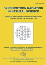
ISSN 1644-7190<br />
SYNCHROTRON RADIATION<br />
IN NATURAL SCIENCE<br />
Bullet<strong>in</strong> of the Polish Synchrotron Radiation Society<br />
Volume 11, Number 1-2, May 2012<br />
Includes: News, regular papers, <strong>in</strong>formation on PTPS,<br />
Programme and Abstracts of the 11th International<br />
Symposium and School on Synchrotron Radiation<br />
<strong>in</strong> Natural Science (ISSRNS-11),<br />
(Cracow, Poland, 20-25 May, 2012)<br />
Organised by:<br />
Polish Synchrotron Radiation Society and Institute of Nuclear Physics PAN, Cracow,<br />
under the honorary patronage of Mayor of the Royal City of Cracow Jacek Majchrowski,<br />
General Director of the Institute of Nuclear Physics, PAN Marek Jeżabek,<br />
and Rector of the Jagiellonian University <strong>in</strong> Kraków Karol Musioł<br />
Sponsored by: M<strong>in</strong>istry of Science and Higher Education, Hamamatsu<br />
Bruker, Panalitycal, Netzsch, Spectro-Lab
SYNCHROTRON RADIATION IN NATURAL SCIENCE<br />
Bullet<strong>in</strong> of the Polish Synchrotron Radiation Society<br />
Address: al. Lotników 32/46, 02-668 Warsaw, Poland,<br />
phone/fax 228436034, e-mail: paszk@ifpan.edu.pl<br />
Editorial Board<br />
Wojciech Paszkowicz, Editor<br />
Institute of Physics<br />
Polish Academy of Sciences<br />
al. Lotników 32/46<br />
02-668 Warsaw<br />
e-mail: paszk@ifpan.edu.pl<br />
Paweł Piszora<br />
Faculty of Chemistry<br />
A. Mickiewicz University<br />
ul. Grunwaldzka 6<br />
60-780 Poznań<br />
e-mail: pawel@amu.edu.pl<br />
Zbigniew Kaszkur, Secretary<br />
Institute of Physical Chemistry<br />
Polish Academy of Sciences<br />
Kasprzaka 44/52<br />
01-224 Warsaw, Poland<br />
e-mail: zkaszkur@ichf.edu.pl<br />
Radosław Przeniosło<br />
Institute of Experimental Phyics<br />
Warsaw University<br />
ul. Hoża 69<br />
00-681 Warszawa<br />
e-mail:<br />
radek.przenioslo@fuw.edu.pl)<br />
Bogdan J. Kowalski<br />
Institute of Physics<br />
Polish Academy of Sciences<br />
al. Lotników 32/46<br />
02-668 W arsaw, Poland<br />
e-mail: kowab@ifpan.edu.pl<br />
Wojciech Szuszkiewicz<br />
Institute of Physics<br />
Polish Academy of Sciences<br />
al. Lotników 32/46<br />
02-668 W arsaw, Poland<br />
e-mail: szusz@ifpan.edu.pl<br />
Krystyna Jabłońska<br />
Institute of Physics<br />
Polish Academy of Sciences<br />
al. Lotników 32/46<br />
02-668 Warsaw<br />
e-mail: jablo@ifpan.edu.pl<br />
Wojciech Kwiatek<br />
Institute of Nuclear Physics<br />
Polish Academy of Sciences<br />
ul. Radzikowskiego 152<br />
31-342 Kraków<br />
e-mail:<br />
wojciech.kwiatek@ifj.edu.pl<br />
Advisory Board<br />
Czesław Kapusta<br />
Department of Solid State Physics<br />
AGH University of Science and<br />
Technology<br />
al. Mickiewicza 30<br />
30-059 Kraków,<br />
e-mail: kapusta@agh.edu.pl<br />
Marek Stankiewicz<br />
Institute of Physics<br />
Jagellonian University<br />
ul. Reymonta 4<br />
30-059 Kraków<br />
e-mail: m.j.stankiewicz@uj.edu.pl<br />
Maciej Kozak<br />
Faculty of Physics<br />
A. Mickiewicz University<br />
ul. Umultowska 85<br />
61-614 Poznań<br />
e-mail: mkozak@amu.edu.pl<br />
Jacek Szade<br />
Institute of Physics<br />
Silesian University<br />
ul. Uniwersytecka 4<br />
40-007 Katowice<br />
e-mail: szade@us.edu.pl<br />
Note for contributors: Contributions <strong>in</strong> English (preferred) or <strong>in</strong> Polish should be sent to the Editor.<br />
The topics <strong>in</strong>clude: <strong>synchrotron</strong> and alternative <strong>radiation</strong> sources such as free electron lasers,<br />
beaml<strong>in</strong>e <strong>in</strong>strumentation, experimental and theoretical results connected with application of various<br />
methods and approaches (x-ray scatter<strong>in</strong>g, x-ray diffraction, x-ray absorption, fluorescence and<br />
photoelectron spectroscopies, magnetic dichroism, etc.) <strong>in</strong> connection with application of <strong>synchrotron</strong><br />
<strong>radiation</strong> <strong>in</strong> physics, chemistry, crystallography, materials <strong>science</strong> and life <strong>science</strong>s.<br />
Layout by Arkadiusz Zarzycki. Cover design by W. Paszkowicz. Figure on the cover page:<br />
Illustration from the abstract "Wavelet analysis of X-ray absorption anisotropy: Accuracy<br />
and limitations of atomic structure imag<strong>in</strong>g" by D.T. Dul et al. (this issue).<br />
SYNCHROTRON RADIATION IN NATURAL SCIENCE is published and distributed by the Polish<br />
Synchrotron Radiation Society (PSRS). Detailed <strong>in</strong>formation on PSRS is given on cover page 3.<br />
ISSN 1644-7190<br />
Volume 11, No. 1-2, 2012, © Polish Synchrotron Radiation Society
Synchrotron Radiation <strong>in</strong> Natural Science Vol. 11, No.1 – 2 (2012)<br />
SOLARIS — NATIONAL SYNCHROTRON RADIATION CENTRE,<br />
THE POLISH RESEARCH INFRASTRUCTURE ROADMAP FACILITY<br />
STATUS IN SPRING 2012<br />
Wojciech Paszkowicz 1 and Marek Stankiewicz 2<br />
1 Institute of Physics, Polish Academy of Sciences, ul. Lotników 32, 02-668 Warszawa, Poland<br />
2 National Synchrotron Radiation Centre SOLARIS, Jagiellonian University,<br />
ul. Gronostajowa 7/P.1.6, 30-387 Kraków, Poland<br />
Interaction of the electromagnetic <strong>radiation</strong> with<br />
matter is fundamental to the universe. From the big<br />
bang up to the current times such <strong>in</strong>teractions have<br />
been activat<strong>in</strong>g the processes and phenomena at the<br />
atomic level up to the scale of the whole Universe.<br />
The <strong>radiation</strong> <strong>in</strong>fluences short and long term processes<br />
on our globe at the micro and macro scales<br />
affect<strong>in</strong>g such areas as the plant growth, earth atmosphere,<br />
climate changes, weather, etc.<br />
In the Universe the Stars are <strong>natural</strong> sources of<br />
EM <strong>radiation</strong>. In our planetary system the Sun<br />
supplies the Earth with a very broad spectrum of<br />
electromagnetic <strong>radiation</strong> spread<strong>in</strong>g from the <strong>in</strong>frared,<br />
through the visible region to X-rays. Significant<br />
part of the <strong>radiation</strong> <strong>in</strong>teracts with and is absorbed<br />
by the atmosphere, the transmitted part <strong>in</strong>teracts<br />
with the Earth ecosystem provid<strong>in</strong>g the necessary<br />
energy, catalyz<strong>in</strong>g reactions and stimulat<strong>in</strong>g biological<br />
processes.<br />
The knowledge and control of the <strong>radiation</strong> <strong>in</strong>teraction<br />
with matter is therefore extremely important<br />
and allow<strong>in</strong>g for understand<strong>in</strong>g the ongo<strong>in</strong>g<br />
reactions and processes and open<strong>in</strong>g the possibilities<br />
of their control.<br />
Until 1970 there was no man made source of such<br />
properties and <strong>in</strong>tensities enabl<strong>in</strong>g for research of<br />
EM <strong>radiation</strong> stimulated processes <strong>in</strong> such a broad<br />
spectrum of energies. This situation has changed<br />
<strong>in</strong> 1970 when the first <strong>synchrotron</strong> <strong>radiation</strong> source<br />
was built.<br />
The advent of <strong>synchrotron</strong> <strong>radiation</strong> sources and<br />
their constant development and improvement has<br />
revolutionized the research <strong>in</strong> many areas of fundamental<br />
as well as applied <strong>science</strong>. S<strong>in</strong>ce then the potential<br />
of such facilities has been widely recognized<br />
and led to the development and construction of very<br />
high <strong>in</strong>tensity light sources (<strong>synchrotron</strong>s and free<br />
electron lasers) emitt<strong>in</strong>g the <strong>radiation</strong> of unprecedented<br />
properties:<br />
• broad spectral range and tunability (opportunity<br />
of selection of a s<strong>in</strong>gle energy),<br />
• high collimation, with opportunity for focus<strong>in</strong>g<br />
down to the size of the order of 10 nm,<br />
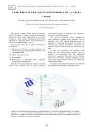
Figure 1 : SOLARIS facility layout.<br />
I
Synchrotron Radiation <strong>in</strong> Natural Science Vol. 11, No.1 – 2 (2012)<br />
• high coherence,<br />
• extremely high <strong>in</strong>tensity,<br />
• specific time structure (pulses down to femtosecond<br />
scale and up to gigawatt power).<br />
Synchrotrons, despite be<strong>in</strong>g big devices (their<br />
circumference ranges from several tens to several<br />
thousand of meters), emit a very compact photon<br />
beam, which can be described as a light scalpel<br />
that can operate on the surface or <strong>in</strong>side the <strong>in</strong>vestigated<br />
objects. It is not only the photon beam size<br />
but also the precision of the applied photon energy<br />
that means the light scalpel has unique applications.<br />
This k<strong>in</strong>d of site specific surgery opens up a vast<br />
range of research that is available only with <strong>synchrotron</strong><br />
<strong>radiation</strong>. The systems <strong>in</strong>vestigated range<br />
from s<strong>in</strong>gle atoms and molecules, through biological<br />
complex molecules (prote<strong>in</strong>s, DNA) to bulk materials<br />
and crystals.<br />
Over the last three decades, <strong>synchrotron</strong> light<br />
has supported cutt<strong>in</strong>g-edge research <strong>in</strong> physics,<br />
chemistry, biology and material <strong>science</strong>s, and has<br />
opened up many new areas of research <strong>in</strong> fields<br />
such as medic<strong>in</strong>e, geological and environmental<br />
studies, structural genomics and archaeology. The<br />
<strong>synchrotron</strong> <strong>radiation</strong> centers are truly multidiscipl<strong>in</strong>ary<br />
and multi-user facilities.<br />
Many tens of such sources have been built <strong>in</strong> all<br />
developed countries hav<strong>in</strong>g population of 40 million<br />
or more, but also <strong>in</strong> less populated Canada, Australia,<br />
Switzerland, Taiwan, Sweden, Denmark and<br />
S<strong>in</strong>gapore or just develop<strong>in</strong>g Brazil and Thailand.<br />
Each of the exist<strong>in</strong>g sources is surrounded by tens<br />
of beaml<strong>in</strong>es where specific tasks are performed by<br />
the users. It is difficult to imag<strong>in</strong>e a technologically<br />
advanced country without at least one <strong>in</strong>tense light<br />
source; the number of sources <strong>in</strong> each of most developed<br />
countries, USA and Japan, is of the order<br />
of twenty.<br />
The unique properties of the <strong>radiation</strong> and the<br />
huge research and development potential offered by<br />
<strong>synchrotron</strong> light sources have been explored by the<br />
community of Polish scientists from the very beg<strong>in</strong>n<strong>in</strong>g<br />
and their scientific output is perceptible, especially<br />
<strong>in</strong> the time follow<strong>in</strong>g the access to European<br />
Community. The <strong>synchrotron</strong> <strong>radiation</strong> users<br />
<strong>in</strong> Poland have formalized the existence of their<br />
community by creation of Polish Synchrotron Radiation<br />
Society (PSRS) <strong>in</strong> 1990. In 1998 the efforts<br />
to construct a <strong>synchrotron</strong> <strong>radiation</strong> source <strong>in</strong><br />
Poland emerged. The goal was achieved <strong>in</strong> 2010<br />
when the project “National Centre of Electromagnetic<br />
Radiation for Research Applications” (stage<br />
I) was granted. The project is run by Jagiellonian<br />
University and the facility will be located with<strong>in</strong><br />
the new University campus area, the new location<br />
for the Science Faculties and the site of the Jagiellonian<br />
Centre of Innovation - the Life Science Park.<br />
Construction of the build<strong>in</strong>g accommodat<strong>in</strong>g<br />
the Polish Synchrotron “SOLARIS” started <strong>in</strong><br />
December 2011, the project completion is scheduled<br />
for September 2014. The SOLARIS build<strong>in</strong>g<br />
(for an artistic view see Fig. 1) will be ready <strong>in</strong><br />
autumn 2013 and then the <strong>in</strong>stallation of the mach<strong>in</strong>e<br />
will start. The project is run <strong>in</strong> a very close<br />
collaboration with MAX-lab <strong>in</strong> Lund. SOLARIS<br />
<strong>synchrotron</strong> r<strong>in</strong>g (96 m circumference) is a tw<strong>in</strong> to<br />
the new 1.5 GeV facility of MAX IV project. Us<strong>in</strong>g<br />
the same design allowed for a fundamental reduction<br />
of the development and construction costs. Moreover,<br />
the modern, technologically advanced, novel<br />
Swedish design leads to improved parameters of the<br />
r<strong>in</strong>g. The storage r<strong>in</strong>g is composed of 12 magnet<br />
blocks form<strong>in</strong>g a 12 double bend achromatic structures.<br />
Differences between the two projects will <strong>in</strong>clude<br />
the <strong>in</strong>jector and the beaml<strong>in</strong>es. SOLARIS,<br />
and its tw<strong>in</strong> Swedish counterpart, are the first facilities<br />
<strong>in</strong> the new generation of compact, high current,<br />
high brilliance 1.5 GeV <strong>synchrotron</strong>s.<br />
The <strong>synchrotron</strong> will be capable of deliver<strong>in</strong>g <strong>radiation</strong><br />
<strong>in</strong> a broad spectral range. Its characteristics:<br />
• particle energy 1.5 GeV (<strong>in</strong>jection 0.6 –<br />
0.7 GeV),<br />
• current 500 mA,<br />
• <strong>radiation</strong> energy at bend<strong>in</strong>g magnets — optimal<br />
at 0.1 – 5 keV (nom<strong>in</strong>al critical energy<br />
2 keV),<br />
• <strong>radiation</strong> energy at wigglers — optimal at 2 –<br />
20 keV, <strong>radiation</strong> available at still higher energies<br />
(∼ 30 or more) (nom<strong>in</strong>al critical energy<br />
6 keV),<br />
• <strong>radiation</strong> energy at undulators — <strong>in</strong>dividually<br />
tuned <strong>in</strong> a broad range beaml<strong>in</strong>es<br />
are considerably better than those for older 1.5 GeV<br />
mach<strong>in</strong>es.<br />
Both, the bend<strong>in</strong>g magnets and <strong>in</strong>sertion devices<br />
<strong>in</strong>stalled <strong>in</strong> the straight sections will be used for generation<br />
of <strong>radiation</strong>. Installation of up to 20 beaml<strong>in</strong>es<br />
and still more experimental end stations will be<br />
possible. There is an opportunity to build 10 beaml<strong>in</strong>es<br />
at bend<strong>in</strong>g magnets and 10 beaml<strong>in</strong>es at wigglers<br />
(undulators). Each beaml<strong>in</strong>e may have more<br />
than one endstation, so there is a perspective that<br />
the number of endstations will be of the order of 40.<br />
The formal and f<strong>in</strong>ancial status of the beaml<strong>in</strong>es<br />
is under consideration. The budget of the project<br />
<strong>in</strong>cludes one experimental l<strong>in</strong>e. However, the <strong>in</strong>itiatives<br />
for the next beaml<strong>in</strong>es have emerged. The applications<br />
for fund<strong>in</strong>g of EXAFS beaml<strong>in</strong>e (led by<br />
University of Silesia), U-ARPES beaml<strong>in</strong>e (led by<br />
Jagiellonian University), have been submitted and<br />
further two applications for: diffraction beaml<strong>in</strong>e<br />
with 3 endstations (led by A. Mickiewicz University),<br />
and for <strong>in</strong> the <strong>in</strong>frared range studies beaml<strong>in</strong>e<br />
(led by University of Rzeszów,) are be<strong>in</strong>g completed.<br />
The community is asked to provide new beaml<strong>in</strong>e<br />
projects.<br />
II
Synchrotron Radiation <strong>in</strong> Natural Science Vol. 11, No.1 – 2 (2012)<br />
There are various scientific, economic, educational<br />
and social benefits which SOLARIS will br<strong>in</strong>g<br />
to the community:<br />
• as all <strong>in</strong>tense light sources, it will become<br />
a center of modern materials <strong>science</strong>, solid<br />
state physics and chemistry and will be helpful<br />
<strong>in</strong> other doma<strong>in</strong>s (medic<strong>in</strong>e, m<strong>in</strong>eralogy,<br />
archaeometry, biology, forensic studies, ...),<br />
• SOLARIS will attract foreign groups to conduct<br />
or participate <strong>in</strong> experiments here, promot<strong>in</strong>g<br />
thus the experiment-base scientific exchange<br />
and collaboration on the basis of its<br />
unique beaml<strong>in</strong>es,<br />
• SOLARIS is go<strong>in</strong>g to be the first research <strong>in</strong>frastructure<br />
of such substantial size and potential<br />
constructed of the region. It will thus<br />
reduce the asymmetry still observed between<br />
the older and newer parts of EU, and hopefully<br />
will <strong>in</strong>itiate further actions of this k<strong>in</strong>d <strong>in</strong> the<br />
region (an important example is the project<br />
for build<strong>in</strong>g a free electron laser, POLFEL, <strong>in</strong><br />
Świerk near Warsaw),<br />
• it will play an important role <strong>in</strong> education at<br />
the graduate and post graduate level,<br />
• as for the first time advanced material and device<br />
studies will become possible, Polish enterprises<br />
hav<strong>in</strong>g access to SOLARIS will have an<br />
opportunity to become <strong>in</strong>ternationally competitive,<br />
• reduction of the outflow of highly qualified<br />
manpower.<br />
It is noteworthy, that on 20th March 2010, the<br />
SOLARIS Project has been awarded by the European<br />
Medal for the Functional and Application<br />
Program and Technological Concept of SOLARIS<br />
(Fig. 2). The award was given <strong>in</strong> concert by three<br />
<strong>in</strong>stitutions: Integration office of European Union,<br />
Bus<strong>in</strong>ess Centre Club and the Socio-Economic European<br />
Committee.<br />
Figure 2 : The European Medal for the Functional and<br />
Application Program and technological Concept of SO-<br />
LARIS awarded <strong>in</strong> 2010 after acceptation of the project<br />
by the M<strong>in</strong>istry.<br />
(For more details see pages 1 – 4 of this issue.)<br />
III
Synchrotron Radiation <strong>in</strong> Natural Science Vol. 11, No.1 – 2 (2012)<br />
Content<br />
W. Paszkowicz and M.J. Stankiewicz<br />
C.J. Bocchetta, P. Goryl, K. Królas,<br />
M. M̷lynarczyk, M.J. Stankiewicz,<br />
P. Tracz, ̷L. Walczak, A. Wawrzyniak,<br />
M. Eriksson, J. Ahlback,<br />
A. Andersson, P. Fernandes Tavares,<br />
M. Johansson, D. Kumbaro,<br />
S.C. Leeman, L. Malmgren, J. Modeer,<br />
S. Thor<strong>in</strong>, D. E<strong>in</strong>feld, and<br />
E. Al-dmour<br />
M. Kozak, W. Rypniewski, and<br />
M. Jaskólski<br />
SOLARIS — National Synchrotron Radiation<br />
Centre, the Polish research <strong>in</strong>frastructure roadmap<br />
facility. Status <strong>in</strong> spr<strong>in</strong>g 2012<br />
Content<br />
Project status of the Polish Synchrotron Radiation<br />
Facility SOLARIS<br />
Koncepcja budowy l<strong>in</strong>ii pomiarowej<br />
MX/SAXS/XRD w NCPS SOLARIS<br />
C. Habfast and G. Admans ESRF Upgrade Programme Reaches Halfway Mark 10<br />
A. Kisiel<br />
Dwadzieścia lat PTPS z perspektywy Prezesów.<br />
Wyst¸apienie na sesji KSUPS-9 w dniu 26 września<br />
2011 r.<br />
The 9 th National Meet<strong>in</strong>g of Synchrotron Radiation<br />
Users (Warsaw 2011)<br />
I<br />
IV<br />
1<br />
5<br />
14<br />
17<br />
ISSRNS–11 — 11 th International School and Symposium on Synchrotron<br />
Radiation <strong>in</strong> Natural Science<br />
Welcome to the 11 th ISSRNS 18<br />
ISSRNS-11 — Information 19<br />
Conference Schedule 21<br />
ISSRNS–11 — Invited lecturers and oral presentations<br />
C.M. Schneider, M. Patt, V. Feyer,<br />
C. Wiemann, I.P. Krug, F. Nickel,<br />
D. Gottlob, and S. Cramm<br />
Photoelectron spectronanoscopy — opportunities<br />
and challenges<br />
L 01<br />
Ext.<br />
24<br />
W. Rypniewski, P.H. Ma̷lecki, and<br />
C.V. Vorgias<br />
P. Dumas<br />
Work<strong>in</strong>g hard and <strong>in</strong> the cold: Chit<strong>in</strong>ase from<br />
M. mar<strong>in</strong>a<br />
What role does <strong>synchrotron</strong> <strong>in</strong>frared<br />
micro-spectroscopy play <strong>in</strong> biomedical applications?<br />
L 02 27<br />
L 03 28<br />
M. Korbas, T.C. MacDonald,<br />
N.J. Sylva<strong>in</strong>, I.J. Picker<strong>in</strong>g,<br />
G.N. George, and P.H. Krone<br />
Shedd<strong>in</strong>g <strong>synchrotron</strong> light on mercury toxicity<br />
L 04<br />
Ext.<br />
29<br />
J. Chwiej, J. Kutoras<strong>in</strong>ska,<br />
K. Janeczko, K. Gzielo-Jurek,<br />
L. Uram, K. Appel, R. Simon, and<br />
Z. Setkowicz<br />
J. Czapla W.M. Kwiatek, J. Lekki,<br />
J. Dulińska, R. Ste<strong>in</strong><strong>in</strong>ger, and<br />
J. Göttlicher<br />
The progress of elemental anomalies of hippocampal<br />
formation <strong>in</strong> pilocarp<strong>in</strong>e model of temporal lobe<br />
epilepsy — X-ray fluorescence microscopy study<br />
The chemical species of sulphur <strong>in</strong> prostate cancer<br />
cells studied by XANES<br />
O 01 31<br />
O 02 32<br />
IV
Synchrotron Radiation <strong>in</strong> Natural Science Vol. 11, No.1 – 2 (2012)<br />
B. Ziaja-Motyka<br />
M. Gilski<br />
European X-ray free electron laser: Status and<br />
applications<br />
Atomic resolution macromolecular crystallography<br />
with <strong>synchrotron</strong> <strong>radiation</strong><br />
L 05 33<br />
L 06 34<br />
Z. Pietralik and M. Kozak<br />
Complexation of nucleic acids by cationic gem<strong>in</strong>i<br />
surfactant<br />
O 03 35<br />
J. Kutoras<strong>in</strong>ska, Z. Setkowicz,<br />
K. Janeczko, C. Sandt, P. Dumas, and<br />
J. Chwiej<br />
Investigation of differences <strong>in</strong> frequency of creat<strong>in</strong>e<br />
<strong>in</strong>clusions with<strong>in</strong> hippocampal formation between<br />
the acute and latent periods of pilocarp<strong>in</strong>e model of<br />
TLE-SRFTIR microspectroscopy study<br />
O 04 36<br />
M. Stankiewicz SOLARIS — new light for Polish research L 07 37<br />
P. Olko and M. Jeżabek<br />
National Centre for Hadron Radiotherapy<br />
— Bronowice Cyclotron Centre<br />
L 08<br />
Ext.<br />
38<br />
G. Wrochna, J.B. Pe̷lka, R. Nietubyć,<br />
R. Sobierajski, and J. Sekutowicz<br />
H. Tomizawa<br />
POLFEL – Polish Free Electron Laser from THz to<br />
XUV<br />
The features and design overview of state-of-the-art<br />
XFEL<br />
L 09 40<br />
L 10 41<br />
A. Galler, W. Gawelda, K. Haldrup,<br />
K. Kjaer, T. van Driel, A.M. March,<br />
G. Doumy, E. Kanter, D. Ray,<br />
R. Dunford, J. Uhlig, S. Canton,<br />
G. Smolentsev, D. Fritz,<br />
M. Cammarata, H. Lemke,<br />
U. Bergmann, R. Alonso Mori, N. Sás,<br />
A. Bordage, G. Vankó, E. Gallo,<br />
P. Glatzel, K. Gaffney, V. Sundström,<br />
M.M. Nielsen, L. Young,<br />
S. Southworth, and C. Bressler<br />
Observ<strong>in</strong>g molecular reactions via simultaneous<br />
ultrafast X-ray spectroscopy and scatter<strong>in</strong>g<br />
L 11<br />
Ext.<br />
42<br />
G. Dietler<br />
M. Gateshki, H. te Nijenhuis,<br />
D. Beckers, A. Kharchenko, and<br />
M. Fransen<br />
T. Togashi, E.J. Takahashi,<br />
K. Midorikawa, M. Aoyama,<br />
K. Yamakawa, T. Sato, A. Iwasaki,<br />
S. Owada, K. Yamanouchi, T. Hara,<br />
S. Matsubara, T. Ohshima, Y. Otake,<br />
H. Tanaka, T. Tanaka, H. Tomizawa,<br />
T. Watanabe, M. Yabashi, and<br />
T. Ishikawa<br />
V. Petříček and M. Dušek<br />
C.J. Bocchetta<br />
P. Rudolf<br />
Interplay between topology and statistical<br />
properties of DNA: A polymer physics approach<br />
High-energy X-ray scatter<strong>in</strong>g studies of<br />
nanomaterials us<strong>in</strong>g a laboratory system<br />
Seed<strong>in</strong>g of extreme ultraviolet free electron laser<br />
with high-order harmonic<br />
Solv<strong>in</strong>g and ref<strong>in</strong><strong>in</strong>g difficult structures by the<br />
program package JANA2006<br />
Techniques and technologies for ultra bright<br />
<strong>synchrotron</strong> light sources<br />
Graphene growth on Cu(111): Microscopic<br />
angle-resolved photoemission and scann<strong>in</strong>g<br />
tunnel<strong>in</strong>g microscopy <strong>in</strong>vestigations<br />
L 12 44<br />
O 05 45<br />
L 13 46<br />
L 14 47<br />
O 06 48<br />
L 15 49<br />
V
Synchrotron Radiation <strong>in</strong> Natural Science Vol. 11, No.1 – 2 (2012)<br />
V.M. Kaganer<br />
X-ray diffraction peak profiles from relaxed<br />
epitaxial films<br />
L 16 50<br />
A. Bartnik, P. Wachulak,<br />
H. Fiedorowicz, R. Jarocki,<br />
J. Kostecki, and M. Szczurek.<br />
Lum<strong>in</strong>escence of gases <strong>in</strong>duced with EUV pulses<br />
from a laser plasma source<br />
O 07<br />
Ext.<br />
51<br />
P.W. Wachulak,<br />
A. Baranowska-Korczyc, D. Pánek,<br />
P. Brůža, A. Bartnik, J. Kostecki,<br />
L. Wȩgrzyński, R. Jarocki,<br />
M. Szczurek, D. Elbaum, and<br />
H. Fiedorowicz<br />
A. Marcelli, K. Zhang, Z. Wu, V. Della<br />
Corte, A. Rotundi, G. Della Ventura,<br />
M. Ferrari, F.J.M. Rietmeijer, and<br />
E. Pace<br />
H. Oyanagi<br />
F. Masiello, S.H. Connell, and<br />
J. Härtwig<br />
C. Pettenkofer and A. Hofmann<br />
Imag<strong>in</strong>g <strong>in</strong> nanoscale us<strong>in</strong>g laser-plasma sources of<br />
extreme ultraviolet (EUV)<br />
X-ray CT scan of stratospheric micron-sized dust<br />
particles: An attempt to a non-destructive<br />
morphological reconstruction<br />
Nanocrystals and small clusters <strong>in</strong>vestigated by<br />
<strong>synchrotron</strong> <strong>radiation</strong> and microfluidics<br />
Measurement of residual stra<strong>in</strong>s with quantitative<br />
X-ray topography<br />
Energy convert<strong>in</strong>g <strong>in</strong>terfaces studied by <strong>synchrotron</strong><br />
<strong>radiation</strong><br />
O 08 53<br />
L 17 54<br />
L 18 55<br />
O 09 56<br />
L 19 57<br />
M. Taniguchi<br />
Electronic and sp<strong>in</strong> structures of solids by means of<br />
<strong>synchrotron</strong> <strong>radiation</strong> photoemission<br />
L 20<br />
Ext.<br />
58<br />
K. Lawniczak-Jablonska, A. Wolska,<br />
M.T. Klepka, and V. Sessi<br />
XMCD studies of the magnetic properties of<br />
nanoclusters <strong>in</strong> GaAs matrix<br />
O 10 60<br />
C.S. Fadley<br />
Prob<strong>in</strong>g the electronic and magnetic properties<br />
of bulk materials and buried layers and <strong>in</strong>terfaces<br />
with stand<strong>in</strong>g-wave and hard-x-ray photoemission<br />
L 21<br />
Ext.<br />
61<br />
M. Sikora<br />
A. Rogalev and F. Wilhelm<br />
Electronic structure of A 2 FeReO 6 double<br />
perovskites probed with Re 2p RXES<br />
X-ray Magnetic Circular Dichroism under high<br />
magnetic field<br />
O 11 63<br />
O 12 64<br />
P. Zajdel, A. Kisiel, A. Szytu̷la,<br />
P. Starowicz, J. Goraus, J. Konior,<br />
A. Banaś, A. Balerna, G. C<strong>in</strong>que,<br />
A. Grilli<br />
Valence of consituents of selected rare earth<br />
silicides — XANES and LAPW numerical study<br />
O 13<br />
Ext.<br />
65<br />
A. Fernández-Pacheco, A. Szkudlarek,<br />
L.E. Serrano-Ramón, T. Tyliszczak,<br />
Cz. Kapusta, M.R. Ibarra, and<br />
J.M. De Teresa<br />
Cz. Kapusta, M. Sikora,<br />
J. Przewoźnik, J. Żukrowski,<br />
J. Fedotova, and J. Kasiuk<br />
Studies of cobalt nanoconstrictions by scann<strong>in</strong>g<br />
transmission X-ray microscopy and micromagnetic<br />
simulations<br />
Study of magnetoresistive nanogranular films<br />
with X-ray spectroscopies<br />
O 14 67<br />
L 22 68<br />
E. Guziewicz, B.A. Orlowski,<br />
A. Reszka, L. Wachnicki,<br />
S. Gieraltowska, M. Godlewski,<br />
I.A. Kowalik, B.J. Kowalski, and<br />
R.L. Johnson<br />
Resonant photoemission of 4f electrons on clean<br />
semiconductor surfaces<br />
L 23<br />
Ext.<br />
69<br />
VI
Synchrotron Radiation <strong>in</strong> Natural Science Vol. 11, No.1 – 2 (2012)<br />
J. Kubacki, D. Kajewski, J. Szade,<br />
A. Köhl, Ch. Lenser, R. Dittmann,<br />
K. Szot, and K. Schulte<br />
P. Goryl, C.J. Bocchetta,<br />
M.J. Stankiewicz, A.I. Wawrzyniak,<br />
K. Wawrzyniak, M. Zaj¸ac, ̷L. Żytniak,<br />
and D. Spruce<br />
M. Kozak, M. Taube, M. Murawska,<br />
V. L<strong>in</strong>dström, and A. Grubb<br />
H. Drozdowski, A. Romaniuk, and,<br />
Z. B̷laszczak<br />
K. Won-<strong>in</strong>, T. Sako,<br />
W. Pattanasiriwisana,<br />
S. Tancharakorn, and P. Dararutana<br />
Resonant photoemission studies of Fe doped<br />
SrTiO 3 th<strong>in</strong> films<br />
The Solaris concepts for the beaml<strong>in</strong>es control<br />
systems<br />
Structural studies of covalently stabilised oligomers<br />
of human cystat<strong>in</strong> C<br />
Structure and <strong>in</strong>termolecular <strong>in</strong>teractions <strong>in</strong><br />
selected b<strong>in</strong>ary solutions studied by X-ray methods<br />
Characterization of ancient burnt rice excavated <strong>in</strong><br />
Thailand archaeological sites<br />
O 15 71<br />
O 16 72<br />
O 17 73<br />
O 18 74<br />
O 19 75<br />
J. Kowalska, W.M. Kwiatek,<br />
M. Gajda, K. Appel, and P. Dumas<br />
Effect of AVE 0991 – angiotens<strong>in</strong>-(1-7) receptor<br />
agonist treatment on elemental and biomolecules<br />
distribution <strong>in</strong> atherosclerotic plaques of<br />
apoE-knockout mice<br />
O 20<br />
Ext.<br />
76<br />
A. Kubala-Kukuś, D. Banaś,<br />
M. Pajek, J. Szlachetko, J.-Cl. Dousse,<br />
J. Hoszowska, Y. Kayser, S. Nowak,<br />
P. Jagodziński, J. Sus<strong>in</strong>i, and<br />
M. Salomé<br />
Synchrotron <strong>radiation</strong> based micro X-ray<br />
fluorescence analysis of the calibration samples used<br />
<strong>in</strong> surface sensitive TXRF and GEXRF techniques<br />
O 21<br />
Ext.<br />
78<br />
ISSRNS–11 — Poster presentations<br />
K. Bal<strong>in</strong>, J. Szade, and Z. Cel<strong>in</strong>ski<br />
D. Banaś, J. Braziewicz,<br />
A. Kubala-Kukuś, U. Majewska,<br />
M. Pajek, J. Wudarczyk-Moćko,<br />
K. Czech, M. Garnuszek,<br />
P. S̷lomkiewicz, and B. Szczepanik<br />
K. Banas, A.M. Banas, M. Gajda,<br />
W.M. Kwiatek, B. Pawlicki, and<br />
M.B.H. Breese<br />
A. Baranowska-Korczyc, K. Fronc,<br />
J.B. Pe̷lka, K. Sobczak, P. D̷lużewski,<br />
and D. Elbaum<br />
Reversible valency transitions of europium <strong>in</strong> MBE<br />
grown Eu-Mn th<strong>in</strong> films<br />
Study of absorption properties of chemically<br />
modified halloysite samples with X-ray fluorescence<br />
and X-ray powder diffraction methods<br />
Analysis of <strong>synchrotron</strong> <strong>radiation</strong> <strong>in</strong>duced X-ray<br />
emission spectra with R environment<br />
Structural studies of magnetic Fe doped ZnO<br />
nanofibers<br />
P 01 80<br />
P 02 81<br />
P 03 82<br />
P 04 83<br />
M.R. Bartosik, C.J. Bocchetta,<br />
P. Goryl, M.J. Stankiewicz, P. Tracz,<br />
SOLARIS — National Synchrotron Radiation<br />
̷L. Walczak, A.I. Wawrzyniak,<br />
Centre, project progress, May 2012<br />
K. Wawrzyniak, J. Wiechecki,<br />
M. Zaj¸ac, and ̷L. Żytniak<br />
P 05<br />
Ext.<br />
84<br />
J. Bielecki, E. Lipiec, and<br />
W.M. Kwiatek<br />
K. Biernacka, M. Sikora, Cz. Kapusta,<br />
A. Brudnik, K. Zakrzewska, and<br />
M. Radecka<br />
First-pr<strong>in</strong>ciple approach to <strong>in</strong>terpretation of<br />
changes <strong>in</strong> IR spectra of cellular DNA<br />
X-ray absorption and emission spectroscopy of<br />
titanium dioxide with modified anionic sublattice<br />
P 06 86<br />
P 07 87<br />
VII
Synchrotron Radiation <strong>in</strong> Natural Science Vol. 11, No.1 – 2 (2012)<br />
M. Brancewicz, A. Andrejczuk,<br />
E. Żukowski, L. Dobrzyński,<br />
Y. Sakurai, and M. Itou<br />
K.M. D¸abrowski, D.T. Dul,<br />
M. Tolkiehn, D.V. Novikov, and<br />
P. Korecki<br />
Compton profile of Mg s<strong>in</strong>gle crystal: High<br />
resolution experiment and theory<br />
Detection of X-ray abasorption anisotropy us<strong>in</strong>g<br />
fluorescence <strong>radiation</strong> for atomic resolved imag<strong>in</strong>g<br />
P 08 88<br />
P 09 89<br />
I.N. Demchenko, R. M<strong>in</strong>ikayev,<br />
T. Tyliszczak, M. Chernyshova,<br />
K.M. Yu, J.D. Denl<strong>in</strong>ger, D. Speaks,<br />
and W. Walukiewicz<br />
Electronic structure of irradiated CdO th<strong>in</strong> films P 10 90<br />
A. Dowiercia̷l, A. Jarmu̷la,<br />
W.R. Rypniewski, T. Fr¸aczyk,<br />
P. Wilk, and W. Rode<br />
H. Drozdowski, T. Ha̷las, and<br />
Z. B̷laszczak<br />
H. Drozdowski, A. Romaniuk, and<br />
Z. B̷laszczak<br />
J. Dudala,<br />
M. Szczerbowska-Boruchowska,<br />
M. Lankosz, and M. Bialas<br />
D.T. Dul and P. Korecki<br />
E. Dynowska, J.Z. Domaga̷la,<br />
P. Romanowski, E. Janik, P. Wojnar,<br />
and W. Caliebe<br />
E. Dynowska, W. Paszkowicz,<br />
P. Aleshkevych, L. G̷ladczuk,<br />
W. Szuszkiewicz, S. Müller,<br />
C. Ronn<strong>in</strong>g, and W. Caliebe<br />
K. Dziedzic-Kocurek and J. Stanek<br />
Search<strong>in</strong>g for the differences between trich<strong>in</strong>ella<br />
spiralis and mouse thymidylate synthases: A quest<br />
for species-specific drugs<br />
A molecular structure study of<br />
1,3,5-trichlorobenzene<br />
The determ<strong>in</strong>ation of molecular structure of<br />
chloroanisoles by X-ray diffraction<br />
Distribution of biomolecules <strong>in</strong> the adrenal gland<br />
tumors — FTIR results compared with histological<br />
view of samples<br />
Wavelet analysis of X-ray absorption anisotropy:<br />
Accuracy and limitations of atomic structure<br />
imag<strong>in</strong>g<br />
Structural characterization of the core-shell<br />
ZnTe/ZnMgTe nanowires<br />
Observation of extremely slow order<strong>in</strong>g effects <strong>in</strong><br />
Co-implanted ZnO<br />
XANES evaluation of iron local structures <strong>in</strong><br />
monomer and dimersed forms of porphyr<strong>in</strong>s<br />
P 11 91<br />
P 12 92<br />
P 13 93<br />
P 14 94<br />
P 15 95<br />
P 16 96<br />
P 17 97<br />
P 18 98<br />
O.N. Ermakova, R. M<strong>in</strong>ikayev,<br />
H. Dabkowska, C. Lathe, J. de Groot,<br />
and W. Paszkowicz<br />
Elastic properties of praseodymium orthovanadate P 19 99<br />
E. Guziewicz, M.I. ̷Lukasiewicz,<br />
K. Kopalko, L. Wachnicki,<br />
M. Godlewski<br />
Co 3d states <strong>in</strong> ferromagnetic and paramagnetic<br />
(Zn, Co)O films – resonant photoemission studies<br />
P 20 100<br />
P. Jagodziński, M. Pajek, D. Banaś,<br />
A. Kubala-Kukuś, J. Szlachetko,<br />
J.-Cl. Dousse, J. Hoszowska,<br />
Y. Kayser, and S. Nowak<br />
Simulations of X-ray transmission <strong>in</strong> polycapillaries<br />
for <strong>synchrotron</strong> <strong>radiation</strong> applications<br />
P 21 102<br />
A. Jarmu̷la, A. Dowiercia̷l, P. Wilk,<br />
W.R. Rypniewski, B. Kierdaszuk, and<br />
W. Rode<br />
Crystal structures of mouse thymidylate synthase<br />
<strong>in</strong> b<strong>in</strong>ary complex with a strong <strong>in</strong>hibitor,<br />
N(4)-OH-dCMP, and ternary complex with<br />
N(4)-OH-dCMP and the cofactor product,<br />
dihydrofolate<br />
P 22 103<br />
VIII
Synchrotron Radiation <strong>in</strong> Natural Science Vol. 11, No.1 – 2 (2012)<br />
M. Johansson, R. Nietubyć, and<br />
A. Wawrzyniak<br />
L<strong>in</strong>ac and storage r<strong>in</strong>g magnets for SOLARIS<br />
<strong>synchrotron</strong><br />
P 23 104<br />
Z. Kaszkur<br />
XRD study of uniformity and <strong>in</strong>terdiffusion <strong>in</strong><br />
PdCo and PdAg nanoalloys<br />
P 24 105<br />
W. Kida and M. Kozak<br />
Influence of gem<strong>in</strong>i surfactants with different cha<strong>in</strong><br />
length on the structure of DPPC bilayers<br />
P 25 106<br />
D. Kl<strong>in</strong>ger, R. Sobierajski, J. Pelka,<br />
E. Lusakowska, D. Żymierska,<br />
W. Wierzchowski, K. Wieteska,<br />
T. Balcer, J. Chalupský, V. Hájková,<br />
T. Burian, A.J. Gleeson, L. Juha,<br />
K. Tiedtke, S. Toleikis, L. Vyšín,<br />
H. Wabnitz, and J. Gaud<strong>in</strong><br />
Damage of two-component materials such as GaAs,<br />
ZnO, SiO 2 created after ir<strong>radiation</strong> by ultra-short<br />
VUV laser pulses<br />
P 26 107<br />
W. Knoff, M.A. Pietrzyk, A. Reszka,<br />
B.A. Or̷lowski, T. Story, and<br />
R.L. Johnson<br />
Photoemission study of amorphous and crystall<strong>in</strong>e<br />
GeTe and (Ge,Mn)Te semiconductors<br />
P 27 108<br />
B. Korczyc, A. Bartnik, J. Kostecki,<br />
and H. Fiedorowicz<br />
Changes <strong>in</strong> chemical and physical structure of<br />
polymers under EUV <strong>radiation</strong><br />
P 28 109<br />
I.A. Kowalik, M.I. Lukasiewicz,<br />
E. Guziewicz, M. Godlewski,<br />
F.J. Luque, M.A. N<strong>in</strong>o, A. Zakharov,<br />
and D. Arvanitis<br />
Electronic structure and magnetism of (Zn,Co)O<br />
films: A soft x-ray spectroscopy study<br />
P 29 110<br />
B.J. Kowalski, R. Nietubyć, and<br />
J. Sadowski<br />
Resonant and angle-resolved photoemission<br />
spectroscopy of Ga 1−x Mn x Sb<br />
P 30 111<br />
A. Kubala-Kukuś,<br />
M. Ludwikowska-Kȩdzia, D. Banaś,<br />
J. Braziewicz, U. Majewska, M Pajek,<br />
and J. Wudarczyk-Moćko<br />
Application of the X-ray fluorescence analysis and<br />
X-ray diffraction <strong>in</strong> geochemical studies of till<br />
samples<br />
P 31 112<br />
K. Lawniczak-Jablonska, M.T. Klepka,<br />
A. Wolska, and M.A. Borysiewicz<br />
The X-ray absorption studies of the Ti-Si-C films<br />
stoichiometry <strong>in</strong> function of the technological<br />
parameters<br />
P 32 113<br />
E. Lipiec, G. Birarda, J. Lekki,<br />
L. Vaccari, A. Wiecheć, and<br />
W.M. Kwiatek<br />
First approach of the FTIR microspectroscopy for<br />
study<strong>in</strong>g the effect of ionis<strong>in</strong>g <strong>radiation</strong> <strong>in</strong> s<strong>in</strong>gle<br />
cells<br />
P 33 114<br />
A.F. Mabied, S. Nozawa, M. Hosh<strong>in</strong>o,<br />
A. Tomita, T. Sato, and S. Adachi<br />
S<strong>in</strong>gular value decomposition analysis of<br />
time-resolved powder diffraction data<br />
P 34 115<br />
M. Ma̷lachowski and M. Kozak<br />
Crystallisation of polymer phases and its <strong>in</strong>fluence<br />
on structure and mechanical properties of<br />
multilayered polymer systems<br />
P 35 116<br />
IX
Synchrotron Radiation <strong>in</strong> Natural Science Vol. 11, No.1 – 2 (2012)<br />
P. Mazalski, Z. Kurant, A. Maziewski,<br />
M.O. Liedke, J. Fassbender,<br />
L.T. Baczewski, A. Wawro,<br />
A. Rogalev, and F. Wilhelm<br />
XAS/XMCD studies of Pt/Co/Pt nanostructures<br />
with out-of-plane magnetization <strong>in</strong>duced by Ga +<br />
ions low fluence ir<strong>radiation</strong><br />
P 36 117<br />
J. Mesjasz-Przybylowicz, A. Barnabas,<br />
I. Yousef, P. Dumas, F. Jamme,<br />
Ch. Sandt, F. Guillon, P. Sechogela,<br />
and W. Przybylowicz<br />
Differences and similarities <strong>in</strong> roots of the nickel<br />
hyperaccumulat<strong>in</strong>g and non-accumulat<strong>in</strong>g<br />
genotypes of Senecio coronatus from South Africa<br />
P 37 118<br />
J. Mesjasz-Przybylowicz,<br />
E. Montargès-Pelletier, A. Barnabas,<br />
G. Echevarria, V. Briois, P. Sechogela,<br />
S. Groeber, and W. Przybylowicz<br />
Distribution and speciation of nickel <strong>in</strong><br />
hyperaccumulat<strong>in</strong>g plants from South Africa<br />
P 38 119<br />
R. M<strong>in</strong>ikayev, E. Dynowska, T. Story,<br />
A. Szczerbakow, A.M.T. Bell,<br />
D. Trots, and W. Szuszkiewicz<br />
X-ray studies of thermal properties of Pb 1−x Cd x Te<br />
solid solution <strong>in</strong> a broad temperature range<br />
P 39 120<br />
R. M<strong>in</strong>ikayev, W. Paszkowicz,<br />
P. Piszora, M. Knapp, C. Bähtz, and<br />
S. Podsiad̷lo<br />
Thermal expansion of gallium nitride P 40 121<br />
M. Murawska, A. Grubb, and<br />
M. Kozak<br />
Small angle X-ray scatter<strong>in</strong>g (SAXS) studies of<br />
monomeric human cystat<strong>in</strong> C <strong>in</strong> solution<br />
P 41 122<br />
M. Murawska, K. Smolarek,<br />
A. Skrzypczak, and M. Kozak<br />
Rod-like morphology of silver nanoparticles<br />
produced <strong>in</strong> cationic gem<strong>in</strong>i surfactants systems<br />
P 42 123<br />
M. Murawska, M. Wiatr,<br />
P. Nowakowski, K. Szutkowski,<br />
A. Skrzypczak, and M. Kozak<br />
The structure and morphology of gold nanoparticles<br />
produced <strong>in</strong> cationic gem<strong>in</strong>i surfactants systems<br />
P 43 124<br />
A. Nasr, U. Werthenbach,<br />
H.W. Schenk, and A.H. Walenta<br />
Multi-channel ionization chamber development for<br />
<strong>synchrotron</strong> beam flactuation monitor<strong>in</strong>g and time<br />
resolve measurement<br />
P 44 126<br />
B.A. Orlowski, A. Szczerbakow,<br />
P. Dziawa, K. Gas, A. Reszka,<br />
S. Thiess, and W. Drube<br />
Photoemission b<strong>in</strong>d<strong>in</strong>g energy local change caused<br />
by crystall<strong>in</strong>e local structure<br />
P 45<br />
Ext.<br />
127<br />
C. Paluszkiewicz, W.M. Kwiatek, and<br />
E. Stodolak<br />
Characterization of polymer nanocomposites by<br />
mikro SR-FTIR spectroscopy<br />
P 46<br />
Ext.<br />
129<br />
W. Paszkowicz, O.N. Ermakova,<br />
W. Wierzchowski, K. Wieteska,<br />
M. Berkowski, M. G̷lowacki,<br />
H. D¸abkowska, J. Domaga̷la,<br />
J. B¸ak-Misiuk, and C. Paulmann<br />
Topographic and high-resolution diffraction study<br />
of defect structure of RVO 4 s<strong>in</strong>gle crystals<br />
P 47<br />
Ext.<br />
131<br />
X
Synchrotron Radiation <strong>in</strong> Natural Science Vol. 11, No.1 – 2 (2012)<br />
W. Paszkowicz, R. M<strong>in</strong>ikayev,<br />
P. Piszora, M. Knapp, D. Trots, and<br />
R. Bacewicz<br />
Thermal expansion of CuInSe 2<br />
P 48<br />
Ext.<br />
133<br />
W. Paszkowicz, P. Piszora,<br />
R. M<strong>in</strong>ikayev, M. Brunelli, and<br />
A. Fitch<br />
Thermal expansion of polycrystall<strong>in</strong>e cBN <strong>in</strong> the<br />
low-temperature range<br />
P 49 135<br />
J.B. Pe̷lka, O. Cho̷luj-Dziewiecka,<br />
J. Lorkiewicz, R. Nietubyć,<br />
J. Sekutowicz, R. Sobierajski,<br />
J. Szewiński, T. Wasiewicz, and<br />
G. Wrochna<br />
Terahertz FEL source at the Polish National Center<br />
POLFEL. A conceptual design<br />
P 50 136<br />
Z. Pietralik, M. Krȩcisz, and M. Kozak<br />
Spectroscopic and structural studies of <strong>in</strong>teractions<br />
between gem<strong>in</strong>i surfactants and<br />
phosphatidylochol<strong>in</strong>e derivatives<br />
P 51 137<br />
Z. Pietralik, R. Krzysztoń, and<br />
M. Kozak<br />
Structural analysis of selected gem<strong>in</strong>i surfactant<br />
(1-imidazolilo-3-decylooxymethyl) pentane chloride<br />
lipoplexes<br />
P 52 138<br />
Z. Pietralik, I. Mucha-Kruczyńska, and<br />
M. Kozak<br />
FTIR analysis of prote<strong>in</strong> secondary structure <strong>in</strong><br />
solid and solution states<br />
P 53 139<br />
P. Piszora and J. Darul<br />
Hydrogen reduction of LiMn 2 O 4 : Identification of<br />
products with <strong>synchrotron</strong> X-ray powder diffraction<br />
P 54 140<br />
A. Romaniuk, H. Drozdowski, and<br />
Z. B̷laszczak<br />
X-ray diffraction study of some liquid b<strong>in</strong>ary<br />
solutions<br />
P 55 141<br />
P. Romanowski, J. B¸ak-Misiuk,<br />
K. Sobczak, P. Dziawa, E. Dynowska,<br />
A. Szczepańska, and A. Misiuk<br />
Mn 4 Si 7 nano<strong>in</strong>clusions <strong>in</strong> Mn-implanted Si P 56 142<br />
K. Schneider, M. Sikora, J. Stȩpień,<br />
K. Biernacka, Cz. Kapusta, D. Zajac,<br />
K. Michalow-Mauke, Th. Graule,<br />
A. Vital, K. Zakrzewska, and M. Rekas<br />
XAS study of Mo doped TiO 2 nanoparticle<br />
materials<br />
P 57 143<br />
W. S̷lawiński, R. Przenios̷lo,<br />
I. Sosnowska, and V. Petricek<br />
Helical screw-type magnetic structure of the<br />
multiferroics CaMn 7 O 12<br />
P 58 144<br />
R. Sobierajski, J.B. Pe̷lka,<br />
R. Nietubyć, G. Wrochna, and<br />
J. Sekutowicz<br />
XFEL — European X-ray Free Electron Laser P 59 145<br />
XI
Synchrotron Radiation <strong>in</strong> Natural Science Vol. 11, No.1 – 2 (2012)<br />
R. Sobierajski, D. Kl<strong>in</strong>ger,<br />
P. D̷lużewski, M. Klepka, J. Gaud<strong>in</strong>,<br />
C. Özkan, J. Chalupský, S. Bajt,<br />
T. Burian, L. Vyšín, N. Coppola,<br />
S.D. Farahani, H.N. Chapman,<br />
G. Galasso, V. Hájková, M. Harmand,<br />
L. Juha, M. Jurek, R. Loch, S. Mölle,<br />
M. Nagasono, H. S<strong>in</strong>n, K. Saskl,<br />
J. Schulz, P. Sovak, S. Toleikis,<br />
K. Tiedtke, T. Tschentscher, and<br />
J. Krzyw<strong>in</strong>ski<br />
Damage of multilayer optics under ir<strong>radiation</strong> with<br />
multiple femtosecond XUV pulses<br />
P 60 146<br />
W. Szczerba, J. Kaiser, H. Riesemeier,<br />
U. Re<strong>in</strong>holz, M. Radtke, L. Yu,<br />
M. Ballauff, and A.F. Thünemann<br />
On the local structure of catalytic Au/Pd<br />
nanoparticles stabilized on spherical polyelectrolyte<br />
brushes<br />
P 61 147<br />
W. Szczerba, H. Riesemeier,<br />
U. Re<strong>in</strong>holz, M. Radtke, and<br />
A.F. Thünemann<br />
Comb<strong>in</strong>ed small angle X-ray scatter<strong>in</strong>g and X-ray<br />
absorption spectroscopy studies of electrochrome<br />
metallopolyelectrolytes<br />
P 62 148<br />
W. Szczerba, H. Riesemeier,<br />
U. Re<strong>in</strong>holz, M. Radtke,<br />
A.F. Thünemann, A. Kaupner, and<br />
C. Giordano<br />
Local structure of iron carbide nanoparticles P 63 149<br />
M. Szczerbowska-Boruchowska,<br />
M. Lankosz, M. Czyzycki,<br />
A. Wandzilak, P. Wrobel,<br />
E. Radwanska, and D. Adamek<br />
Synchrotron <strong>radiation</strong> based studies of the<br />
elemental composition and chemical forms of Fe<br />
and Zn <strong>in</strong> bra<strong>in</strong> gliomas<br />
P 64 150<br />
M. Szczerbowska-Boruchowska,<br />
P. Wrobel, A. Sorowka, E. Radwanska,<br />
and D. Adamek<br />
Evaluation of the variability <strong>in</strong> elemental<br />
composition of dopam<strong>in</strong>egric neurons <strong>in</strong> senile<br />
bra<strong>in</strong>s us<strong>in</strong>g <strong>synchrotron</strong> <strong>radiation</strong> based X-ray<br />
fluorescence<br />
P 65 151<br />
J. Szlachetko, M. Nachtegaal,<br />
J.-Cl. Dousse, J. Hoszowska,<br />
E. Kleymenov, M. Janousch, J. Sa,<br />
O.V. Safonova, and J.A. van Bokhoven<br />
Direct XES to XAS relation for off-resonant<br />
excitations at L3 absorption edge: Towards<br />
high-resolution XAS at s<strong>in</strong>gle-shot<br />
P 66 152<br />
Cz. Ślusarczyk<br />
Structure development dur<strong>in</strong>g isothermal<br />
crystallization of high-density polyethylene:<br />
Synchrotron small-angle X-ray study<br />
P 67 153<br />
W. Tokarz, M. Kowalik, R. Zalecki,<br />
and A. Ko̷lodziejczyk<br />
Electronic band structure of<br />
La 0.67 Pb 0.33 Mn 0.92 Fe 0.08 O 3<br />
P 68 154<br />
L. Walczak, J. Alhback, M. Berglund,<br />
E. Al-dmour, J. Pasquaud,<br />
P. Fernandes Tavares, M. Eriksson,<br />
D. E<strong>in</strong>feld, C.J. Bocchetta, and<br />
M.J. Stankiewicz<br />
Vacuum system of the Polish light source<br />
— SOLARIS<br />
P 69 155<br />
XII
Synchrotron Radiation <strong>in</strong> Natural Science Vol. 11, No.1 – 2 (2012)<br />
D. Wardecki, R. Przenios̷lo, A. Fitch,<br />
M. Bukowski, and R. Hempelmann<br />
Size dependence of microstra<strong>in</strong> fluctuations <strong>in</strong><br />
nanocrystall<strong>in</strong>e chromium<br />
P 70 156<br />
A. Wiecheć, E. Lipiec, J. Lekki,<br />
M. Wide̷l, and W.M. Kwiatek<br />
SR-FTIR spectroscopy <strong>in</strong> study of the double stand<br />
breaks <strong>in</strong> s<strong>in</strong>gle cells irradiated by proton<br />
microbeam<br />
P 71 157<br />
K. Wieteska, W. Wierzchowski,<br />
A. Mal<strong>in</strong>owska, M. Lefeld-Sosnowska,<br />
M. Swirkowicz, T. Lukasiewicz, and<br />
C. Paulmann<br />
Synchrotron diffraction topography of SBN<br />
(Sr x Ba 1−x Nb 2 O 6 ) and CBN (Ca x Ba 1−x Nb 2 O 6 )<br />
crystals<br />
P 72<br />
Ext.<br />
158<br />
W. Wierzchowski, K. Wieteska,<br />
D. Kl<strong>in</strong>ger, R. Sobierajski, J. Pelka,<br />
D. Zymierska, T. Balcer, and<br />
C. Paulmann<br />
The <strong>in</strong>vestigations of the damages <strong>in</strong>duced by flash<br />
pulses <strong>in</strong> silicon crystals by means of white beam<br />
<strong>synchrotron</strong> section topography<br />
P 73<br />
Ext.<br />
160<br />
A. Wolska, K. Lawniczak-Jablonska,<br />
M.T. Klepka, and V. Sessi<br />
XMCD studies of the GaSb:MnSb layers on the<br />
GaSb and GaAs substrates<br />
P 74 162<br />
D.A. Zajac, A. Bikowski,<br />
M. V<strong>in</strong>nichenko, and K. Ellmer<br />
Near-order structure of transparent conduct<strong>in</strong>g<br />
oxides: X-ray absorption study of Al-doped ZnO<br />
and ZnMgO films<br />
P 75<br />
Ext.<br />
163<br />
D.A. Zaj¸ac, W.M. Woch, J. Stȩpień,<br />
Cz. Kapusta, A. Ko̷lodziejczyk,<br />
H. Sudra, and G. Gritzner<br />
XANES and EXAFS study of<br />
(Tl 0.5 Pb 0.5 )Sr 2 (Ca 1−x Gd x )Cu 2 O z superconductors<br />
P 76 164<br />
P. Zajdel, I. Jendrzejewska, J. Goraus,<br />
T. Goryczka, and T. Mydlarz<br />
Local electronic structure and physical properties of<br />
Zn 1−x Ni x Cr 2 Se 4<br />
P 77 165<br />
A. Katrusiak<br />
A. Kuczumow<br />
Synchrotron Light News 166<br />
ISSRNS climbs to the top: Three proceed<strong>in</strong>gs<br />
papers on the Radiation Physics and Chemistry<br />
Top25 list<br />
Frolic GOATS workshops on high pressure<br />
diffraction<br />
Jubileuszowe X Krajowe Sympozjum<br />
Użytkowników Promieniowania Synchrotronowego<br />
180<br />
181<br />
182<br />
Future conferences and workshops 183<br />
Conference proceed<strong>in</strong>gs of meet<strong>in</strong>gs organised by<br />
Polish Synchrotron Radiation Society (1992 – 2012)<br />
185<br />
Authors Index 186<br />
Subject Index 189<br />
XIII
Synchrotron Radiation <strong>in</strong> Natural Science Vol. 11, No.1 – 2 (2012)<br />
PROJECT STATUS OF THE POLISH SYNCHROTRON<br />
RADIATION FACILITY SOLARIS 1)<br />
C.J. Bocchetta 1∗ , P. Goryl 1 , K. Królas 1 , M. M̷lynarczyk 1 , M.J. Stankiewicz 1 , P. Tracz 1 ,<br />
̷L. Walczak 1 , A. Wawrzyniak 1 , M. Eriksson 2 , J. Ahlback 2 , A. Andersson 2 ,<br />
P. Fernandes Tavares 2 , M. Johansson 2 , D. Kumbaro 2 , S.C. Leeman 2 , L. Malmgren 2 ,<br />
J. Modeer 2 , S. Thor<strong>in</strong> 2 , D. E<strong>in</strong>feld 3 , and E. Al-dmour 3<br />
1 National Synchrotron Radiation Centre Solaris at the Jagiellonian University, Kraków, Poland<br />
2 MAX-lab, Lund, Sweden<br />
3 CELLS-ALBA Synchrotron, Cerdanyola del Valles, Spa<strong>in</strong><br />
Abstract The Polish <strong>synchrotron</strong> <strong>radiation</strong> facility Solaris is be<strong>in</strong>g built at the Jagiellonian<br />
University <strong>in</strong> Krakow. The project is based on an identical copy of the 1.5 GeV storage r<strong>in</strong>g<br />
be<strong>in</strong>g concurrently built for the MAX IV project <strong>in</strong> Lund, Sweden. A general description<br />
of the facility is given together with a status of activities. Unique features associated with<br />
Solaris are outl<strong>in</strong>ed, such as <strong>in</strong>frastructure, the <strong>in</strong>jector and operational characteristics.<br />
∗ e-mail: carlo.bocchetta@uj.edu.pl<br />
1. INTRODUCTION<br />
The first ideas for a national <strong>synchrotron</strong> <strong>radiation</strong><br />
facility <strong>in</strong> Poland were put forward <strong>in</strong> 1998 and<br />
several proposals made <strong>in</strong> subsequent years. In 2008<br />
the government pledged funds of 143 MPLN with<br />
formal allocation after approval of a feasibility study<br />
from the Jagiellonian University for the construction<br />
of a light source with such a budget. In 2009<br />
this study, based on the <strong>in</strong>novative ideas and technology<br />
of MAX-lab (ref. MAX III, [2]), was submitted<br />
and the National Synchrotron Radiation Centre<br />
Solaris was approved for construction <strong>in</strong> February<br />
2010 us<strong>in</strong>g EU structural funds. The facility will be<br />
built on land allocated by the Jagiellonian university<br />
on the new campus <strong>in</strong> Krakow. In December<br />
2010 an agreement was signed between the Jagiellonian<br />
University and Lund University Sweden, for<br />
the mutual cooperation and shar<strong>in</strong>g of ideas and designs<br />
related to the construction of the two facilities.<br />
Solaris will be an identical copy of the 1.5 GeV r<strong>in</strong>g<br />
of the MAX IV project and will use identical parts<br />
of the l<strong>in</strong>ac <strong>in</strong>jector and transfer l<strong>in</strong>e [2, 3]. Major<br />
differences between the two mach<strong>in</strong>es are the <strong>in</strong>frastructures,<br />
the lower energy l<strong>in</strong>ac and the beaml<strong>in</strong>es.<br />
2. FACILITY<br />
Build<strong>in</strong>g<br />
The <strong>synchrotron</strong> <strong>radiation</strong> facility will be built at<br />
the campus III site of the Jagiellonian University<br />
<strong>in</strong> the city of Krakow. The land with an area of<br />
∼ 22000 m 2 will site the mach<strong>in</strong>e, experimental<br />
hall, auxiliary service build<strong>in</strong>gs, laboratories, offices<br />
and auditorium. The contract for the design<br />
and construction was awarded <strong>in</strong> the March 2011<br />
to the consortium of companies: ALPINE Construction<br />
Polska Spó̷lka z o.o. and Przedsiebiorstwo<br />
Budowlano-Produkcyjne ̷LȨGPRZEM Spó̷lka z o.o.<br />
The build<strong>in</strong>g permit was granted <strong>in</strong> December 2011<br />
and the construction is <strong>in</strong> progress. The build<strong>in</strong>g is<br />
composed of a l<strong>in</strong>ac tunnel and an adjacent modulator<br />
and service gallery placed below the storage r<strong>in</strong>g<br />
level. The length of the tunnel ∼ 100 m, with<strong>in</strong> the<br />
constra<strong>in</strong>ts of land availability, foresees an upgrade<br />
to the l<strong>in</strong>ac to <strong>in</strong>crease its energy for top-up <strong>in</strong>jection.<br />
All services, power, HVAC and cool<strong>in</strong>g will be<br />
built with this upgrade <strong>in</strong> m<strong>in</strong>d. The experimental<br />
hall for beaml<strong>in</strong>es houses the storage r<strong>in</strong>g tunnel.<br />
The experimental hall has a surface area of 3000 m 2<br />
and provision is made for its future extension on<br />
one side by 600 m 2 . Access to the storage r<strong>in</strong>g tunnel<br />
will be through chicanes on the <strong>in</strong>ner side and<br />
the roof shield<strong>in</strong>g will be removable for mach<strong>in</strong>e <strong>in</strong>stallation<br />
and ma<strong>in</strong>tenance. All equipment for the<br />
storage r<strong>in</strong>g will be housed on the <strong>in</strong>ner side of the<br />
r<strong>in</strong>g tunnel. A crane, rated at 8 tonnes, spann<strong>in</strong>g<br />
the experimental hall, will be used for mach<strong>in</strong>e and<br />
beaml<strong>in</strong>e <strong>in</strong>stallation and ma<strong>in</strong>tenance.<br />
Injector<br />
The l<strong>in</strong>ac <strong>in</strong>jector for Solaris will <strong>in</strong>itially be operated<br />
at 550 MeV with options for a full energy<br />
upgrade. The l<strong>in</strong>ac is composed of an RF gun and<br />
six normal conduct<strong>in</strong>g 3 GHz accelerat<strong>in</strong>g sections,<br />
of length 5.2 m, grouped <strong>in</strong>to three units conta<strong>in</strong><strong>in</strong>g<br />
two accelerat<strong>in</strong>g sections [2]. Each unit will be<br />
powered through SLED cavities fed by a solid-state<br />
modulator driv<strong>in</strong>g a klystron. The l<strong>in</strong>ac sections<br />
are be<strong>in</strong>g manufactured by Research Instruments<br />
GmbH (D) and will have a guaranteed performance<br />
of 20 MV/m. The RF power from the SLED cavities<br />
feed<strong>in</strong>g each unit will be split equally to the<br />
two l<strong>in</strong>ac sections. The first unit, however, will be<br />
configured to deliver RF power to the gun too.<br />
1
Synchrotron Radiation <strong>in</strong> Natural Science Vol. 11, No.1 – 2 (2012)<br />
Figure 1 : Concept layout of the facility. The experimental hall can be extended by 10 m on the right hand side at<br />
a later date. Above the l<strong>in</strong>ac tunnel ample covered space is available for pre-assembly and general laboratories.<br />
In this case the first l<strong>in</strong>ac section will be given more<br />
RF power. The solid-state modulators, ordered and<br />
be<strong>in</strong>g manufactured by ScandiNova Systems AB<br />
(S), will power 35 MW klystrons at 10 Hz. The<br />
electron gun will be an upgraded version of that<br />
presently used at MAXlab. It will have a BaO<br />
cathode and a 180 ◦ bend<strong>in</strong>g magnet will be used<br />
for energy filter<strong>in</strong>g.<br />
Transfer l<strong>in</strong>e<br />
The beam is transferred to the storage r<strong>in</strong>g via a<br />
27 ◦ vertical ramp. The l<strong>in</strong>ac will <strong>in</strong>itially be placed<br />
close to the storage to reduce the length of transfer<br />
l<strong>in</strong>e and the gun relocated when the full energy<br />
upgrade will be performed. The vertical ramp is<br />
optically mirror symmetric and composed of two<br />
pulsed 10 ◦ magnets, two dipole magnets deflect<strong>in</strong>g<br />
17 ◦ and six quadrupoles. The pulsed dipole magnet<br />
<strong>in</strong> the l<strong>in</strong>ac tunnel <strong>in</strong> comb<strong>in</strong>ation with a kicker<br />
magnet will <strong>in</strong> the future be used to share the l<strong>in</strong>ac<br />
beam between topp<strong>in</strong>g up and possible FEL experiments.<br />
The pulsed septum magnet at the end<br />
of the transfer l<strong>in</strong>e is a vertical Lamberston type,<br />
deflect<strong>in</strong>g the beam <strong>in</strong>to the horizontal plane. All<br />
magnets and power supplies are identical to MAX<br />
IV systems.<br />
Injection process<br />
Injection <strong>in</strong>to the storage r<strong>in</strong>g will be performed<br />
with a pulsed sextupole magnet [4, 5]. The scheme<br />
has many advantages over a conventional four-kicker<br />
<strong>in</strong>jection bump especially for top-up operation. In<br />
the case of Solaris with straight section lengths<br />
of 3.5 m, a fourkicker scheme would require it to<br />
span two achromat sections that conta<strong>in</strong> strong<br />
sextupoles and large dispersion that would affect the<br />
stored beam. Furthermore the conventional scheme<br />
would reduce the available space for <strong>in</strong>sertion devices.<br />
The use of a pulsed sextupole magnet will<br />
simplify the scheme and circumvent the aforementioned<br />
disadvantages. The <strong>in</strong>jection dynamics at a<br />
lower energy compared to the MAX IV case is considered<br />
<strong>in</strong> reference [4]. Care must be taken <strong>in</strong> the<br />
design of the pulsed sextupole magnet and associated<br />
power supply given the 320 ns revolution time<br />
of the storage r<strong>in</strong>g, s<strong>in</strong>ce a two turn <strong>in</strong>jection scheme<br />
is less efficient compared to a s<strong>in</strong>gle turn scheme.<br />
The possibility of us<strong>in</strong>g a s<strong>in</strong>gle dipole kicker is also<br />
be<strong>in</strong>g evaluated to help facilitate the commission<strong>in</strong>g<br />
of the pulsed sextupole scheme [6]. Once accumulated<br />
the beam will be ramped to its f<strong>in</strong>al energy of<br />
1.54 GeV. The behaviour and response of the magnets<br />
dur<strong>in</strong>g ramp<strong>in</strong>g is expected to be similar to<br />
that of MAX III.<br />
Figure 2 : Magnet half block with coils <strong>in</strong> red [7].<br />
2
Synchrotron Radiation <strong>in</strong> Natural Science Vol. 11, No.1 – 2 (2012)<br />
Figure 3 : Prelim<strong>in</strong>ary vacuum chamber <strong>in</strong> sta<strong>in</strong>less steel with antechambers [9].<br />
Storage R<strong>in</strong>g Technology<br />
The storage r<strong>in</strong>g will be technologically identical to<br />
the MAX IV 1.5 GeV r<strong>in</strong>g and is composed of 12<br />
magnet blocks form<strong>in</strong>g a 12 double bend achromatic<br />
structure. Bend<strong>in</strong>g magnet <strong>synchrotron</strong> <strong>radiation</strong><br />
will be extracted for users from the first dipole <strong>in</strong><br />
the achromat. The iron blocks mach<strong>in</strong>ed to high<br />
precision will conta<strong>in</strong> all magnetic elements allow<strong>in</strong>g<br />
for a very compact design. The iron for the<br />
magnets has been purchased and is be<strong>in</strong>g thermally<br />
treated prior to mach<strong>in</strong><strong>in</strong>g. Magnet design [7] is<br />
<strong>in</strong> the f<strong>in</strong>al stages of completion and is be<strong>in</strong>g performed<br />
<strong>in</strong> parallel with the design activities of the<br />
vacuum chamber.<br />
An evaluation has been performed of the technology<br />
to be used for the vacuum system. Both a wholly<br />
NEG coated chamber, similar to that adopted for<br />
the MAX IV 3.0 GeV r<strong>in</strong>g [8], and a conventional<br />
system with antechamber and absorbers were exam<strong>in</strong>ed.<br />
A conventional sta<strong>in</strong>less-steel system was<br />
chosen on the basis of manufacturer availability,<br />
costs, technology requirements and project timeschedule.<br />
The vacuum system is be<strong>in</strong>g designed and<br />
the construction draw<strong>in</strong>gs prepared by the group<br />
from CELLS-ALBA Synchrotron <strong>in</strong> Cedanyola del<br />
Valles (E) <strong>in</strong> collaboration with MAXlab and Solaris.<br />
The system and magnet configuration foresees<br />
extraction of bend<strong>in</strong>g magnet <strong>radiation</strong> at either 3<br />
or 7.5 degrees from the first bend<strong>in</strong>g magnet of the<br />
achromat.<br />
The storage r<strong>in</strong>g RF system is composed of two<br />
100 MHz cavities similar to those used <strong>in</strong> MAX II<br />
and III. The cavities are normal conduct<strong>in</strong>g and of<br />
the capacity loaded type that have relatively high<br />
frequency higher order modes compared to pill-box<br />
type cavities. The cavities will be equipped with<br />
higher order mode coupl<strong>in</strong>g loops that will extract<br />
the residual high frequency modes. Cavities and<br />
couplers have been ordered and are be<strong>in</strong>g manufactured<br />
by Research Instruments GmbH. The r<strong>in</strong>g<br />
also foresees operation with two passive Landau<br />
cavities at 300 MHz as designed by MAX-lab similar<br />
to the ma<strong>in</strong> cavities [10]. Either solid state<br />
or tetrode amplifiers will power the ma<strong>in</strong> cavities.<br />
Two such amplifiers will be comb<strong>in</strong>ed to provide<br />
60 kW of power per cavity via a circulator. The RF<br />
units will be controlled with a digital LLRF system.<br />
Optics and Dynamics<br />
The <strong>in</strong>tegrated magnets permit an ultra-compact<br />
double bend achromatic structure with low emittance<br />
and zero dispersion <strong>in</strong> the straight sections.<br />
The compact magnet design has three quadrupoles<br />
that focus <strong>in</strong> the horizontal plane while the vertical<br />
focus<strong>in</strong>g is done by the gradient <strong>in</strong> the dipoles. Pole<br />
strips on the bend<strong>in</strong>g magnets will allow tun<strong>in</strong>g of<br />
the vertical focus<strong>in</strong>g. The focus<strong>in</strong>g sextupoles have<br />
also been <strong>in</strong>tegrated <strong>in</strong>to the focus<strong>in</strong>g quadrupoles.<br />
Recently the lattice has been optimised for the<br />
ramped operation <strong>in</strong> Solaris where Touschek lifetime<br />
is important s<strong>in</strong>ce the facility will not operate<br />
<strong>in</strong> top-up mode but <strong>in</strong> decay mode. The optimisation<br />
has focused on <strong>in</strong>creas<strong>in</strong>g the momentum acceptance<br />
by ensur<strong>in</strong>g the lattice momentum acceptance<br />
match the RF acceptance of 4%. Together<br />
with the use of Landau cavities the Touschek lifetime<br />
at 500 mA is expected to reach 13 hours [6].<br />
In each magnet block there will be three BPMs and<br />
three horizontal/vertical corrector coils mounted on<br />
the sextupole magnets. Two of the BPMs will be<br />
positioned at the ends of the achromatic block and<br />
one <strong>in</strong> the centre.<br />
Table 1 : 1.5 GeV Storage R<strong>in</strong>g Parameters<br />
Current<br />
500 mA<br />
Circumference<br />
96 m<br />
Horizontal emittance<br />
(bare lattice) 6 nm rad<br />
Coupl<strong>in</strong>g 1%<br />
Tunes Q x, Q y 11.22, 3.14<br />
Natural chromaticities ξ x, ξ y −22.9, −17.1<br />
Momentum compaction 3.04 × 10 −3<br />
Momentum acceptance 4%<br />
3
Synchrotron Radiation <strong>in</strong> Natural Science Vol. 11, No.1 – 2 (2012)<br />
Beaml<strong>in</strong>es<br />
For the first phase of the project one beaml<strong>in</strong>e is<br />
planned to be f<strong>in</strong>anced from the project budget.<br />
The beaml<strong>in</strong>e will use bend<strong>in</strong>g magnet <strong>radiation</strong><br />
and will have a X-PEEM/XAS/XMCD end-station.<br />
This activity is <strong>in</strong> cooperation with PSI. Fund<strong>in</strong>g<br />
proposals have been submitted for additional beaml<strong>in</strong>es<br />
from undulators.<br />
3. SCHEDULE AND MILESTONES<br />
The project deadl<strong>in</strong>e for first light is the third quarter<br />
of 2014. The build<strong>in</strong>g construction has started<br />
and the beneficial occupancy of the build<strong>in</strong>g is programmed<br />
for the end of August 2013. Component<br />
schedules and purchas<strong>in</strong>g milestones are l<strong>in</strong>ked to<br />
the MAX IV project schedule and are compatible<br />
with Solaris <strong>in</strong>stallation.<br />
4. CONCLUSIONS<br />
The Solaris project is a prime example of the benefits<br />
of shar<strong>in</strong>g of state-of-the-art knowledge and resources<br />
for the rapid establishment of a national<br />
research <strong>in</strong>frastructure. Scientific collaboration is<br />
certa<strong>in</strong>ly not new <strong>in</strong> the field of accelerators but the<br />
direct utilisation of a design and its complete replication<br />
is unique. The collaboration maximises the<br />
utilisation of human and f<strong>in</strong>ancial capital lead<strong>in</strong>g to<br />
more effective and efficient use of public funds. The<br />
collaboration permits quick tra<strong>in</strong><strong>in</strong>g of new people<br />
with an <strong>in</strong>itial focus on mobility and network<strong>in</strong>g and<br />
an optimal use of mentorship and expert knowledge.<br />
Procurement efforts are rendered more effective by<br />
not duplicat<strong>in</strong>g tasks and allow <strong>in</strong>dustry to program<br />
its response to large-scale research <strong>in</strong>frastructure requirements.<br />
The advantages also extend to build<strong>in</strong>g<br />
design and construction s<strong>in</strong>ce critical knowledge is<br />
shared. Furthermore there is benefit of the Solaris-<br />
MAX IV collaboration on other European laboratories<br />
from collaborations that are unique to either<br />
Solaris or MAX IV which extend the network and<br />
knowledge base.<br />
Acknowledgments: The work was supported by<br />
the European Regional Development Fund with<strong>in</strong><br />
the frame of the Innovative Economy Operational<br />
Program:POIG.02.01.00-12-213/09.<br />
References<br />
[1] MAX III reference.<br />
[2] MAX IV Detailed Design Report,<br />
http://www.maxlab.lu.se/maxlab/max4/<strong>in</strong>dex.html.<br />
[3] M. Eriksson et al., “The MAX IV Synchrotron Light<br />
Source,” THPC058, this conference.<br />
[4] A.I. Wawrzyniak et al., “Injector layout and beam<br />
<strong>in</strong>jection <strong>in</strong>to Solaris”, THPC123, this conference.<br />
[5] S.C. Leemann, Particle Accelerator Conference, New<br />
York, USA, THP214 (2011).<br />
[6] S.C. Leeman, “Recent improvements to the lattices<br />
for the MAX IV storage r<strong>in</strong>gs,” THPC056, this conference.<br />
[7] M. Johansson, “Design of the MAX IV/Solaris<br />
1.5 GeV storage r<strong>in</strong>g magnets”, WEPO016, this conference.<br />
[8] J. Ahlback, “Vacuum system design for the MAX IV<br />
3 GeV r<strong>in</strong>g”, TUPS016, this conference.<br />
[9] E. D’Amour, ALBA, private communication.<br />
[10] Å. Andersson, “The 100 MHz RF system for the<br />
MAX IV storage r<strong>in</strong>gs”, MOPC051, this conference.<br />
1) Updated version of the paper presented at Proceed<strong>in</strong>gs of IPAC2011, San Sebastián, Spa<strong>in</strong>.<br />
4
Synchrotron Radiation <strong>in</strong> Natural Science Vol. 11, No.1 – 2 (2012)<br />
KONCEPCJA BUDOWY LINII POMIAROWEJ MX/SAXS/XRD<br />
W NCPS SOLARIS<br />
M. Kozak 1∗ , W. Rypniewski 2 , and M. Jaskólski 2,3<br />
1 Wydzia̷l Fizyki, Uniwersytet im. Adama Mickiewicza, ul. Umultowska 85, 61-614 Poznań, Polska<br />
2 Instytut Chemii Bioorganicznej <strong>Polskie</strong>j Akademii Nauk, ul. Noskowskiego 12/14, 61-704 Poznań, Polska<br />
3 Wydzia̷l Chemii, Uniwersytet im. Adama Mickiewicza, ul. Grunwaldzka 6, 60-780 Poznań, Polska<br />
Keywords: <strong>synchrotron</strong> <strong>radiation</strong>, <strong>synchrotron</strong>, SOLARIS, beam l<strong>in</strong>e, prote<strong>in</strong> crystallography, small-angle X-ray<br />
scatter<strong>in</strong>g, SAXS, structural biology, materials <strong>science</strong>, powder diffraction, tuneable <strong>synchrotron</strong> <strong>radiation</strong><br />
∗ e-mail: mkozak@amu.edu.pl<br />
W grudniu 2011 za<strong>in</strong>icjowana zosta̷la budowa pierwszego polskiego <strong>synchrotron</strong>u — Narodowego<br />
Centrum Promieniowania Synchrotronowego SOLARIS w Krakowie. Parametry<br />
energetyczne <strong>synchrotron</strong>u SOLARIS (1.5 GeV) pozwalaj¸a na zaprojektowanie l<strong>in</strong>ii pomiarowej<br />
wykorzystuj¸acej promieniowanie <strong>synchrotron</strong>owe z zakresu twardego promieniowania<br />
rentgenowskiego, która bȩdzie mog̷la być wykorzystywana na potrzeby biokrystalografii<br />
oraz badań materia̷lowych. Planowana l<strong>in</strong>ia pomiarowa posiadać bȩdzie trzy końcowe<br />
stacje pomiarowe — biokrystalograficzn¸a (MX), ma̷lok¸atowego rozpraszania promieniowania<br />
rentgenowskiego (SAXS) i dyfrakcji promieniowania <strong>synchrotron</strong>owego na materia̷lach<br />
polikrystalicznych (XRD).<br />
In December 2011 the construction of the first Polish <strong>synchrotron</strong> — The National Synchrotron<br />
Radiation Centre SOLARIS — was <strong>in</strong>itiated <strong>in</strong> Kraków. The energy parameters<br />
of SOLARIS (1.5 GeV) allow plann<strong>in</strong>g of a beam l<strong>in</strong>e utilis<strong>in</strong>g the <strong>synchrotron</strong> <strong>radiation</strong><br />
(hard X-rays range), that can be used <strong>in</strong> biocrystallography and material <strong>science</strong>. The<br />
planned beam l<strong>in</strong>e has three end-stations — for biocrystallography (MX), small-angle X-<br />
ray scatter<strong>in</strong>g (SAXS) and diffraction of <strong>synchrotron</strong> <strong>radiation</strong> on polycrystall<strong>in</strong>e materials<br />
(XRD).<br />
Rozwój niektórych dziedz<strong>in</strong> nauki stymulowany<br />
jest dostȩpem do źróde̷l promieniowania elektromagnetycznego.<br />
Należ¸a do nich przede wszystkim<br />
te dziedz<strong>in</strong>y badań, które wykorzystuj¸a<br />
zjawisko dyfrakcji czy rozpraszania promieniowania<br />
rentgenowskiego, miȩdzy <strong>in</strong>nymi jest to krystalografia.<br />
Jeżeli prześledzimy jej rozwój od pocz¸atku<br />
ubieg̷lego wieku z ̷latwości¸a zauważymy jak ścis̷le<br />
s¸a te powi¸azania. Praktycznie na każdym etapie<br />
odkrycia naukowe stymulowane by̷ly rozwojem<br />
nowych źróde̷l promieniowania rentgenowskiego, a<br />
szczególnie spektakularne odkrycia ostatnich lat<br />
ściśle wi¸aż¸a siȩ z dostȩpem do promieniowania <strong>synchrotron</strong>owego<br />
[1, 2].<br />
Od pocz¸atku XX wieku Nagrodȩ Nobla za<br />
badania zwi¸azane z krystalografi¸a przyznano<br />
33 razy http://www.iucr.org/people/nobel-prize).<br />
Szczególnie dynamiczny rozkwit biokrystalografii<br />
zapocz¸atkowa̷ly w latach 60-tych wiekopomne prace<br />
Watsona i Cricka [3, 4] oraz Perutza i Kendrew<br />
[5, 6]. Od tego czasu Nagrodȩ Nobla za badania<br />
krystalograficzne struktury biomoleku̷l przyznano<br />
szesnaście razy, ostatnio w 2009 roku za<br />
zbadanie struktury rybosomu — ogromnego kompleksu<br />
̷lańcuchów RNA oraz bia̷lek, na którym w<br />
każdej żywej komórce na Ziemi zachodzi biosynteza<br />
bia̷lek [7]-[9]. Dziȩki biokrystalografii uzyskiwane<br />
s¸a modele strukturalne bia̷lek o rozdzielczości<br />
atomowej, które s̷luż¸a miȩdzy <strong>in</strong>nymi do projektowania<br />
leków. Najbardziej spektakularnym tego<br />
przyk̷ladem s¸a leki stosowane w leczeniu AIDS,<br />
które powsta̷ly dziȩki poznaniu struktury bia̷lek<br />
wirusa HIV [10]-[12]. Zaprojektowana w oparciu o<br />
wyniki badań krystalograficznych i stosowana obecnie<br />
zmodyfikowana <strong>in</strong>sul<strong>in</strong>a jest ̷latwiej przyswajalna<br />
i ma przed̷lużony czas dzia̷lania [13]. Co<br />
wiȩcej badania krystalograficzne pozwalaj¸a także na<br />
poznanie struktury nie tylko pojedynczych bia̷lek<br />
wirusa, ale nawet ca̷lych wirusów [14]. Trudno wiȩc<br />
przecenić znaczenie tego typu badań w opracowywaniu<br />
nowych leków, szczepionek czy nowych terapii.<br />
Od po̷lowy lat 80-tych ubieg̷lego wieku biokrystalografia<br />
stymulowana jest dostȩpem do źróde̷l<br />
promieniowania <strong>synchrotron</strong>owego. Dobrym<br />
odzwierciedleniem tego trendu jest nie tylko wzrastaj¸aca<br />
liczba struktur bia̷lkowych deponowanych<br />
w bazie Prote<strong>in</strong> Data Bank http://www.pdb.org)<br />
ale przede wszystkim relacja liczby struktur<br />
5
Synchrotron Radiation <strong>in</strong> Natural Science Vol. 11, No.1 – 2 (2012)<br />
rozwi¸azywanych z wykorzystaniem promieniowania<br />
<strong>synchrotron</strong>owego do ca̷lkowitej liczby zdeponowanych<br />
struktur. Porównuj¸ac tylko ostatnie<br />
15-lecie warto zauważyć, że w 1996 roku<br />
zdeponowano w PDB 1148 struktur bia̷lkowych<br />
rozwi¸azanych w oparciu o dane dyfrakcyjne z<br />
czego 275 stanowi̷ly struktury zbadane z pomoc¸a<br />
promieniowania <strong>synchrotron</strong>owego (co stanowi 24%<br />
ogólnej liczby struktur rentgenowskich). Z kolei<br />
w roku 2011 na 8359 zdeponowanych struktur<br />
makromoleku̷l uzyskanych z użyciem promieniowania<br />
rentgenowskiego przypada aż 7076 struktur<br />
rozwi¸azanych w oparciu o dane <strong>synchrotron</strong>owe<br />
(czyli oko̷lo 85%).<br />
Z upowszechnieniem siȩ dostȩpu do źróde̷l <strong>synchrotron</strong>owych<br />
zwi¸azany jest też <strong>in</strong>tensywny rozwój<br />
metod badania struktury biomoleku̷l w roztworach<br />
w oparciu o ma̷lok¸atowe rozpraszanie promieniowania<br />
<strong>synchrotron</strong>owego. Technika ta stanowi komplementarne<br />
wobec krystalografii narzȩdzie badawcze<br />
pozwalaj¸ace na badanie struktur makromoleku̷l w<br />
ich <strong>natural</strong>nym środowisku i jest niezwykle atrakcyjn¸a<br />
do badań tych uk̷ladów, dla których nie<br />
można uzyskać kryszta̷lów (bia̷lka o strukturze dynamicznej,<br />
z̷lożone kompleksy multimetryczne, czy<br />
niektóre peptydy) [15].<br />
Biokrystalografia w Polsce<br />
Polscy naukowcy od pocz¸atku brali aktywny<br />
udzia̷l w badaniach biokrystalograficznych. Należy<br />
tu wymienić przede wszystkim prof. Tadeusza<br />
Baranowskiego, który w 1939 roku jako pierwszy<br />
Polak przeprowadzi̷l krystalizacjȩ bia̷lek [16]-[18].<br />
W latach powojennych polska szko̷la krystalografii<br />
rozwija̷la siȩ nadal, ale w wyniku pog̷lȩbiaj¸acych<br />
siȩ trudności z prowadzeniem nowoczesnych badań<br />
w Polsce, jak również z <strong>in</strong>nych wzglȩdów,<br />
wiȩkszość polskich biokrystalografów wyemigrowa̷la<br />
na Zachód.<br />
Dopiero w okresie ostatnich kilkunastu lat<br />
ta sytuacja zaczȩ̷la siȩ zmieniać. Powsta̷lo w<br />
kraju kilka nowoczesnych ośrodków i krystalografia<br />
makromoleku̷l w Polsce znów rozwija siȩ dynamicznie.<br />
Ten rozwój nie by̷lby możliwy bez dostȩpu<br />
do promieniowania <strong>synchrotron</strong>owego. Dotychczas<br />
dość swobodnie można by̷lo korzystać z l<strong>in</strong>ii <strong>synchrotron</strong>owych<br />
za granic¸a. Jednak w najbliższym<br />
czasie w dużym stopniu ograniczone maj¸a zostać<br />
programy miȩdzynarodowe f<strong>in</strong>ansuj¸ace dostȩp do<br />
tych ośrodków dla badaczy europejskich. Aby<br />
podtrzymać poziom i zapewnić dalszy rozwój badań<br />
biostrukturalnych w Polsce zaistnia̷la pilna potrzeba<br />
zapewnienia polskim biokrystalografom warunków<br />
do pracy w kraju w oparciu o narodowe centrum<br />
<strong>synchrotron</strong>owe wyposażone we w̷lasne l<strong>in</strong>ie <strong>synchrotron</strong>owe.<br />
Już na etapie prac nad projektem<br />
koncepcyjnym polskiego <strong>synchrotron</strong>u pojawi̷l siȩ<br />
pomys̷l budowy w nim l<strong>in</strong>ii pomiarowej przeznaczonej<br />
do badań biokrystalograficznych. W grudniu<br />
2011 rozpoczȩ̷la siȩ budowa d̷lugo wyczekiwanego<br />
pierwszego polskiego <strong>synchrotron</strong>u — Narodowego<br />
Centrum Promieniowania Synchrotronowego SO-<br />
6<br />
LARIS w Krakowie. Znaj¸ac uwarunkowania konstrukcyjne,<br />
możliwości badawcze tego urz¸adzenia<br />
oraz uwarunkowania f<strong>in</strong>ansowe na obecnym etapie<br />
możliwe jest zaproponowanie l<strong>in</strong>ii pomiarowej<br />
opartej o promieniowanie <strong>synchrotron</strong>owe z zakresu<br />
twardego promieniowania rentgenowskiego, która<br />
bȩdzie mog̷la być wykorzystywana na potrzeby biologii<br />
strukturalnej i badań materia̷lowych. W<br />
środowisku poznańskich naukowców z Uniwersytetu<br />
im. Adama Mickiewicza oraz Instytutu Chemii<br />
Bioorganicznej PAN powsta̷l projekt takiej l<strong>in</strong>ii.<br />
Koncepcja l<strong>in</strong>ii<br />
Bior¸ac pod uwagȩ parametry <strong>synchrotron</strong>u<br />
(obwód 96 m, energia 1.5 GeV), wielkość hali<br />
eksperymentalnej (50 m ×60 m) oraz możliwość<br />
lokalizacji na jego obwodzie 9 – 10 stacji pomiarowych<br />
należy w pierwszym rzȩdzie rozważyć<br />
budowȩ l<strong>in</strong>ii, która zapewnia̷laby dostȩp do <strong>in</strong>frastruktury<br />
pomiarowej dla możliwie najszerszej<br />
grupy badaczy. Nie bez znaczenia obok badań<br />
krystalograficznych s¸a także badania strukturalne<br />
uk̷ladów niekrystalicznych takich jak polimery i<br />
nanokompozyty polimerowe, szk̷la, b̷lony biologiczne,<br />
nośniki leków czy szczepionek. Tego<br />
typu obiekty badane s¸a również z wykorzystaniem<br />
promieniowania <strong>synchrotron</strong>owego i techniki<br />
rozpraszania ma̷lok¸atowego. Osobnym i<br />
niezwykle istotnym aspektem dla postȩpu technologicznego<br />
w kraju s¸a także badania strukturalne<br />
nowych materia̷lów (nanokompozytów, polimerów,<br />
pó̷lprzewodników itp.), które również bȩd¸a mog̷ly<br />
rozwijać siȩ przy udziale nowej l<strong>in</strong>ii pomiarowej.<br />
Warto nadmienić, że tego typu l<strong>in</strong>ie (z̷lożone<br />
z kilku końcowych stacji pomiarowych) w <strong>synchrotron</strong>ach<br />
za granic¸a, ze wzglȩdu na sw¸a specyfikȩ,<br />
s¸a najbardziej produktywne w przeliczeniu na<br />
liczbȩ i wagȩ publikacji naukowych. Wyniki tych<br />
badań s¸a publikowane w najlepszych czasopismach<br />
naukowych i maj¸a zawsze najsilniejszy oddźwiȩk w<br />
środowisku naukowym.<br />
Dlatego przedstawiona poniżej koncepcja<br />
obejmuje stworzenie obok końcowej stacji pomiarowej<br />
przeznaczonej do badań biokrystalograficznych<br />
budowȩ dwóch stacji końcowych do pomiarów<br />
ma̷lok¸atowego rozpraszania promieniowania<br />
rentgenowskiego (SAXS) i dyfrakcji na uk̷ladach<br />
polikrystalicznych. Potencjalna lokalizacja stacji,<br />
wstȩpnie uzgodniona z dyrekcj¸a <strong>synchrotron</strong>u SO-<br />
LARIS zaprezentowana zosta̷la na rysunku 1.<br />
Z uwagi na wymagania optyki rentgenowskiej<br />
oraz powierzchniȩ niezbȩdn¸a do konstrukcji<br />
poszczególnych stacji pomiarowych konieczna jest<br />
lokalizacja stacji w możliwie najd̷luższym (36 m) i<br />
przestronnym miejscu hali pomiarowej (segment C).<br />
Projekt przewiduje umieszczenie stacji w narożniku<br />
hali pomiarowej bezpośrednio s¸asiaduj¸acym z segmentem<br />
B.<br />
Przeprowadzone wcześniej konsultacje z zespo̷lami<br />
zajmuj¸acymi siȩ konstrukcj¸a aparatury pomiarowej<br />
w ośrodkach <strong>synchrotron</strong>owych (miȩdzy
Synchrotron Radiation <strong>in</strong> Natural Science Vol. 11, No.1 – 2 (2012)<br />
Rysunek 1 : Lokalizacja stacji MX/SAXS/XRD (jako szary wielobok) na planie <strong>synchrotron</strong>u SOLARIS. Plan sytuacyjny<br />
<strong>synchrotron</strong>u SOLARIS zosta̷l udostȩpniony dziȩki uprzejmości dyrekcji NCPS.<br />
<strong>in</strong>nymi w MAX-lab Lund czy DESY Hamburg)<br />
oraz z firmami zajmuj¸acymi siȩ budow¸a komponentów<br />
lub ca̷lych <strong>synchrotron</strong>owych l<strong>in</strong>ii pomiarowych<br />
pozwoli̷ly stworzyć spójn¸a koncepcjȩ oraz<br />
realistycznie oszacować wymagane środki f<strong>in</strong>ansowe.<br />
Projekt tej l<strong>in</strong>ii przewiduje wyposażenie jej w<br />
urz¸adzenia pomiarowe najnowszej generacji, co zapewni<br />
jej konkurencyjność wobec <strong>in</strong>nych ośrodków<br />
na świecie.<br />
Lund (Szwecja) [19]. L<strong>in</strong>ia I911 zawiera aż piȩć<br />
stacji końcowych (jedn¸a pracuj¸ac¸a w szerszym zakresie<br />
energii i dostosowan¸a do techniki dostrojonej<br />
dyfrakcji anomalnej (Multiwavelength Anomalous<br />
Diffraction - MAD) [20], oraz cztery pracuj¸ace przy<br />
sta̷lej d̷lugości fali). L<strong>in</strong>ia ta za<strong>in</strong>stalowana zosta̷la<br />
na pierścieniu akumulacyjnym MAX-II o obwodzie<br />
(90 m) i parametrach zbliżonych do planowanych<br />
dla pierścienia akumulacyjnego w <strong>synchrotron</strong>ie SO-<br />
LARIS<br />
Wiggler nadprzewodz¸acy i optyka<br />
rentgenowska<br />
Energia krytyczna na magnesach ug<strong>in</strong>aj¸acych<br />
dla <strong>synchrotron</strong>u SOLARIS wynosi 1.96 keV.<br />
Promieniowanie rentgenowskie w poż¸adanym zakresie<br />
energetycznym (6 – 18 keV) uzyskiwane bȩdzie<br />
wiȩc za pomoc¸a wigglera nadprzewodz¸acego w polu<br />
B 0 = 3.5 – 4 T (energia krytyczna 5.5 – 6 keV),<br />
który zapewni nie tylko odpowiedni zakres energetyczny<br />
ale też wysok¸a <strong>in</strong>tensywność promieniowania.<br />
Przyk̷ladowy teoretyczny zakres widmowy dla tego<br />
typu wigglera zaprezentowany zosta̷l na rysunku 2.<br />
Elementy optyki stacji bȩd¸a dostosowane do<br />
wymagań krystalografii bia̷lek. W szczególności<br />
konieczne s¸a lustra kolimuj¸ace pozwalaj¸ace na<br />
osi¸agniȩcie wymaganej dla pomiarów z wykorzystaniem<br />
rozpraszania anomalnego wysokiej rozdzielczości<br />
energetycznej.<br />
Koncepcja l<strong>in</strong>ii pomiarowej z̷lożonej z kilku<br />
końcowych stacji pomiarowych jest najbardziej<br />
efektywnym wykorzystaniem miejsca oraz wi¸azki<br />
promieniowania rentgenowskiego emitowanej z jednego<br />
wigglera. W ostatnich latach podobna konstrukcja<br />
zosta̷la zbudowana i z powodzeniem uruchomiona<br />
w ośrodku <strong>synchrotron</strong>owym MAX-lab w<br />
Rysunek 2 : Teoretyczny profil energetyczny<br />
promieniowania <strong>synchrotron</strong>owego generowanego z<br />
użyciem wigglera nadprzewodz¸acego 3.75 T. Na wykresie<br />
zaznaczony zosta̷l zakres energetyczny możliwy do<br />
wykorzystania przez planowan¸a l<strong>in</strong>iȩ.<br />
Stacje końcowe<br />
Pierwsza stacja końcowa przeznaczona do<br />
badań dyfrakcyjnych monokryszta̷lów biomakromoleku̷l<br />
- bia̷lek i kwasów nukle<strong>in</strong>owych (MX)<br />
7
Synchrotron Radiation <strong>in</strong> Natural Science Vol. 11, No.1 – 2 (2012)<br />
pracować pow<strong>in</strong>na w pe̷lnym zakresie energetycznym<br />
(6 – 18 keV). Oprócz standardowego goniostatu<br />
do badań monokryszta̷lów wyposażona<br />
pow<strong>in</strong>na być także w system umożliwiaj¸acy pe̷ln¸a<br />
automatyzacjȩ pomiarów dyfrakcyjnych oraz w<br />
przysz̷lości docelowo umożliwiać prowadzenie pomiarów<br />
zdalnych. Tego typu rozwi¸azania s¸a<br />
już rutynowo stosowane w najlepszych l<strong>in</strong>iach<br />
biokrystalograficznych [21]-[24]. Z uwagi na podatność<br />
próbek biologicznych na destrukcjȩ <strong>in</strong>dukowan¸a<br />
promieniowaniem rentgenowskim pomiary<br />
bȩd¸a prowadzone w warunkach kriogenicznych.<br />
Z kolei dziȩki wykorzystaniu pe̷lnego zakresu energetycznego<br />
stacji możliwe bȩdzie rejestrowanie<br />
danych dyfrakcyjnych w eksperymentach opartych<br />
na technice MAD [20]. Aby w pe̷lni wykorzystać<br />
możliwości stacji na potrzeby techniki MAD jej energetyczna<br />
zdolność rozdzielcza pow<strong>in</strong>na wynosić<br />
∆E/E = 10 −4 .<br />
Druga stacja końcowa (pomiarowa) pow<strong>in</strong>na<br />
także pracować w tym zakresie energetycznym<br />
co stacja MX (6 – 18 keV, ∆E/E = 10 −4 )<br />
i być wyposażona w wysokiej klasy optykȩ<br />
umożliwiaj¸ac¸a pomiary uk̷ladów biologicznych w<br />
roztworach (bioSAXS). Rejestracja danych oparta<br />
bȩdzie o najlepszy dostȩpny na światowym rynku<br />
detektor pó̷lprzewodnikowy (np. typu PILATUS<br />
1M) oraz system pomiarowy umożliwiaj¸acy pomiary<br />
w szerokim zakresie temperatur, przynajmniej<br />
od 80 K do 573 K. Z uwagi na potencjalne<br />
szerokie wykorzystanie tej stacji pomiarowej<br />
także przez fizyków materii miȩkkiej oraz<br />
badaczy polimerów system wyposażony pow<strong>in</strong>ien<br />
być w uk̷lad umożliwiaj¸acy wyt̷laczanie próbek<br />
polimerowych w trakcie pomiarów oraz z<strong>in</strong>tegrowany<br />
kalorymetr skan<strong>in</strong>gowy do rejestracji<br />
efektów energetycznych towarzysz¸acym przejściom<br />
fazowym generowanym w funkcji temperatury.<br />
Z kolei trzecia stacja pomiarowa operować bȩdzie<br />
przy sta̷lej energii (15 keV) i wyposażona zostanie<br />
w dyfraktometr proszkowy wraz z wysokiej klasy<br />
detektorem l<strong>in</strong>iowym lub powierzchniowym. Ca̷ly<br />
system pozwala̷l bȩdzie na pomiary dyfrakcyjne w<br />
szerokim zakresie temperatur (60 – 1500 K) oraz<br />
pomiary ciśnieniowe.<br />
Podsumowanie<br />
L<strong>in</strong>ie o podobnym charakterze do planowanej<br />
funkcjonuj¸a w prawie wszystkich <strong>synchrotron</strong>ach<br />
na świecie. Warto ponownie podkreślić, że w<br />
wiȩkszości przypadków tego typu l<strong>in</strong>ie pomiarowe<br />
należ¸a do najbardziej produktywnych naukowo l<strong>in</strong>ii<br />
badawczych. Wielkość środowiska potencjalnych<br />
użytkowników stacji MX w Polsce oszacować można<br />
na 12 grup biokrystalograficznych oraz przynajmniej<br />
5 kolejnych grup badawczych sporadycznie korzystaj¸acych<br />
z technik biokrystalograficznych, co<br />
daje szacunkow¸a liczbȩ 50 – 60 potencjalnych<br />
użytkowników stacji MX. Szacunki zapotrzebowania<br />
na czas pomiarowy bazuj¸ace tylko na wykorzystaniu<br />
l<strong>in</strong>ii przez te grupy, nie licz¸ac potencjalnych<br />
użytkowników z zagranicy (w szczególności<br />
z Europy środkowej), to ponad 100 dni pomiarowych.<br />
Co wiȩcej w zwi¸azku z rozwojem biologii<br />
strukturalnej w Polsce i powstawaniem w<br />
kraju nowych grup biokrystalograficznych należy<br />
szacować, że liczebność tej grupy użytkowników<br />
ma tendencje wzrostowe. Jeśli chodzi o pozosta̷le<br />
końcowe stacje pomiarowe (SAXS, XRD) to z uwagi<br />
na szerszy (multidyscypl<strong>in</strong>arny) profil badań liczba<br />
użytkowników jest znacznie wiȩksza.<br />
W powyższych szacunkach uwzglȩdnić należy<br />
również efekt generowania zapotrzebowania poprzez<br />
dostȩpność technologii (technology-driven research)<br />
oraz prowadzone przez personel l<strong>in</strong>ii badania<br />
w̷lasne (<strong>in</strong>-house) w efekcie można powiedzieć, że<br />
możliwości planowanej l<strong>in</strong>ii pomiarowej bȩd¸a w<br />
pe̷lni wykorzystywane.<br />
Acknowledgments: The work was supported by<br />
the European Regional Development Fund with<strong>in</strong> the<br />
frame of the Innovative Economy Operational Program:<br />
POIG.02.01.00-12-213/09.<br />
References<br />
[1] Z. Dauter, M. Jaskolski, A. Wlodawer, “Impact of<br />
<strong>synchrotron</strong> <strong>radiation</strong> on macromolecular crystallography:<br />
A personal view,” J. Synchrotron Rad. 17<br />
(2010) 433 – 444.<br />
[2] Z. Dauter, A. Wlodawer, M. Jaskolski,<br />
“Promieniowanie rentgenowskie ze źróde̷l <strong>synchrotron</strong>owych<br />
jako katalizator rozwoju krystalografii<br />
bia̷lek”, Synchrotron Radiation <strong>in</strong> Natural<br />
Science 9 (2010) 4 – 16.<br />
[3] J.D. Watson, F.H.C. Crick, “Molecular structure of<br />
nucleic acids — a structure for deoxyribose nucleic<br />
acid,” Nature 171 (1953) 737 – 738.<br />
[4] J.D. Watson, F.H.C. Crick, “Genetical implications<br />
of the structure of deoxyribonucleic acid,” Nature<br />
171 (1953) 964 – 967.<br />
[5] J.C. Kendrew, R.E. Dickerson, B.E. Strandberg,<br />
R.G. Hart, D.R. Davies, D.C. Phillips, V.C. Shore,<br />
“Structure of myoglob<strong>in</strong> — 3-dimensional Fourier<br />
synthesis at 2 Å resolution,” Nature 185 (1960)<br />
422 – 427.<br />
[6] M.F. Perutz, M.G. Rossmann, A.F. Cullis, H.<br />
Muirhead, G. Will, A.C.T. North, “Structure of<br />
haemoglob<strong>in</strong> — 3-dimensional Fourier synthesis at<br />
5.5 Å resolution, obta<strong>in</strong>ed by X-ray analysis,” Nature<br />
185 (1960) 416 – 422.<br />
[7] N. Ban, P. Nissen, J. Hansen, P.B. Moore, T.A.<br />
Steitz, “The complete atomic structure of the large<br />
ribosomal subunit at 2.4 angstrom resolution,” Science<br />
289 (2000) 905 – 920.<br />
[8] J. Harms, F. Schluenzen, R. Zarivach, A. Bashan,<br />
S. Gat, I. Agmon, H. Bartels, F. Franceschi, A.<br />
Yonath, “High resolution structure of the large ribosomal<br />
subunit from a mesophilic Eubacterium,”<br />
Cell 107 (2001) 679 – 688.<br />
[9] B.T. Wimberly, D.E. Brodersen, W.M. Clemons,<br />
R.J. Morgan-Warren, A.P. Carter, C. Vonrhe<strong>in</strong>, T.<br />
8
Synchrotron Radiation <strong>in</strong> Natural Science Vol. 11, No.1 – 2 (2012)<br />
Hartsch, V. Ramakrishnan, “”Structure of the 30S<br />
ribosomal subunit,” Nature 407 (2000) 327 – 339.<br />
[10] A. Wlodawer, M. Miller, M. Jaskolski, B.K. Sathyanarayana,<br />
E. Baldw<strong>in</strong>, I.T. Weber, L.M. Selk, L.<br />
Clawson, J. Schneider, S.B.H. Kent, “Conserved<br />
fold<strong>in</strong>g <strong>in</strong> retroviral proteases — crystal-structure<br />
of a synthetic HIV-1 protease,” Science 245 (1989)<br />
616 – 621.<br />
[11] M. Jaskolski, “From atomic resolution to molecular<br />
giants: An overview of crystallographic studies<br />
of biological macromolecules with <strong>synchrotron</strong> <strong>radiation</strong>,”<br />
Acta Phys. Polon. A 117 (2009) 257 – 263;<br />
Synchrotron Radiation <strong>in</strong> Natural Science 9 (2009)<br />
17 – 23.<br />
[12] Z. Dauter, M. Jaskolski, “Zastosowanie<br />
promieniowania <strong>synchrotron</strong>owego w krystalografii<br />
bia̷lek,” <strong>in</strong>: Promieniowanie <strong>synchrotron</strong>owe<br />
w spektroskopii i badaniach strukturalnych. Wybrane<br />
zagadnienia, B.J. Kowalski, W. Paszkowicz, E.A.<br />
Gorlich, eds., pp. 305 – 328 (PTPS Kraków, 2011).<br />
[13] J.L. Whitt<strong>in</strong>gham, I. Jonassen, S. Havelund, S.M.<br />
Roberts, E.J. Dodson, C.S. Verma, A.J. Wilk<strong>in</strong>son,<br />
G.G. Dodson, “Crystallographic and solution studies<br />
of N-lithocholyl <strong>in</strong>sul<strong>in</strong>: A new generation of<br />
prolonged-act<strong>in</strong>g human <strong>in</strong>sul<strong>in</strong>s,” Biochemistry 43<br />
(2004) 5987 – 5995.<br />
[14] S.A. Wynne, R.A. Crowther, A.G.W. Leslie, “The<br />
crystal structure of the human hepatitis B virus capsid,”<br />
Mol. Cell 3 (1999) 771 – 780.<br />
[15] C.D. Putnam, M. Hammel, G.L. Hura, J.A. Ta<strong>in</strong>er,<br />
“X-ray solution scatter<strong>in</strong>g (SAXS) comb<strong>in</strong>ed with<br />
crystallography and computation: Def<strong>in</strong><strong>in</strong>g accurate<br />
macromolecular structures, conformations and assemblies<br />
<strong>in</strong> solution,” Quarterly Rev. Biophys. 40<br />
(2007) 191 – 285.<br />
[16] T. Baranowski, “Die Isolierung von kristallisierten<br />
Prote<strong>in</strong>en aus Kan<strong>in</strong>chenmuskel,” Z. Physiol. Chem.<br />
260 (1939) 43 – 55.<br />
[17] L. Chrobak, T. Baranowski, “Über verschiedene<br />
Kristalle des A-Myogens aus Kan<strong>in</strong>chenmuskelature.<br />
Comptes Rendus,” (Doklady) l’Academie des Science<br />
de l’USSR XXVIII (1940) 724 – 725.<br />
[18] T. Baranowski, ”Crystall<strong>in</strong>e glycerophosphate dehydrogenase<br />
from rabbit muscle,” J. Biol. Chem.<br />
180 (1949) 535 – 541.<br />
[19] C.B. Mammen, T. Ursby, M. Thunnissen, and J.<br />
Als-Nielsen, “Bent diamond crystals and multilayer<br />
based optics at the new 5-Station Prote<strong>in</strong> Crystallography<br />
Beaml<strong>in</strong>e ‘Cassiopeia’ at MAX-lab, AIP<br />
Conference Proceed<strong>in</strong>gs 705 (2004) 808 – 811.<br />
[20] W.A. Hendrickson, “Determ<strong>in</strong>ation of macromolecular<br />
structures from anomalous diffraction of <strong>synchrotron</strong><br />
<strong>radiation</strong>,” Science 254 (1991) 51 – 58.<br />
[21] G. Snell, C. Cork, R. Nordmeyer, E. Cornell, G.<br />
Meigs, D. Yegian, J. Jaklevic, J. J<strong>in</strong>, R.C. Stevens,<br />
T. Earnest, “Automated sample mount<strong>in</strong>g and alignment<br />
system for biological crystallography at a <strong>synchrotron</strong><br />
source,” Structure 12 (2004) 537 – 545.<br />
[22] S.M. Soltis, A.E. Cohen, A. Deacon, T. Eriksson,<br />
A. Gonzalez, S. McPhillips, H. Chui, P. Dunten, M.<br />
Hollenbeck, I. Mathews, M. Miller, P. Moorhead,<br />
R.P. Phizackerley, C. Smith, J. Song, H. van dem Bedem,<br />
P. Ellis, P. Kuhn, T. McPhillips, N. Sauter, K.<br />
Sharp, I. Tsyba, G. Wolf, “New paradigm for macromolecular<br />
crystallography experiments at SSRL: Automated<br />
crystal screen<strong>in</strong>g and remote data collection,”<br />
Acta Cryst. D 64 (2008) 1210 – 1221.<br />
[23] X.D. Wang, M. Gleaves, D. Meredith, R. Allan, C.<br />
Nave, “E-<strong>science</strong> technologies <strong>in</strong> <strong>synchrotron</strong> <strong>radiation</strong><br />
beaml<strong>in</strong>e — Remote access and automation (A<br />
case study for high throughput prote<strong>in</strong> crystallography),”<br />
Macromol. Res. 14 (2006) 140 – 145.<br />
[24] M. Gilski, “Automation and remote <strong>synchrotron</strong><br />
data collection,” Acta Phys. Polon. A 114 (2008)<br />
331 – 338.<br />
9
Synchrotron Radiation <strong>in</strong> Natural Science Vol. 11, No.1 – 2 (2012)<br />
ESRF UPGRADE PROGRAMME REACHES HALFWAY MARK<br />
Claus Habfast and Gary Admans<br />
ESRF Communication Group, Grenoble, France<br />
In 2012, the ESRF reached the peak activity of the<br />
first phase of its Upgrade Programme. This <strong>in</strong>cluded<br />
a 5-month shutdown until May 2012, the first<br />
time ever <strong>in</strong> 18 years that the user operation was<br />
shut down for such an extended period of time.<br />
The first new beaml<strong>in</strong>es are now available for user<br />
operation, and many more will become operational<br />
until the end of Phase I of the upgrade <strong>in</strong> 2015. Remarkably,<br />
the performance of the ESRF for its users<br />
did not drop significantly despite a period of heavy<br />
works. The X-ray source performs better than ever,<br />
with record values for the mean time between failure<br />
and availability. Although the number of hours<br />
of user operations is slightly lower than <strong>in</strong> the past<br />
years, due to the construction-related shutdown periods,<br />
the <strong>in</strong>terest <strong>in</strong> the ESRF rema<strong>in</strong>s high, and<br />
the number of proposals did not decrease, on the<br />
contrary!<br />
Upgrade Beaml<strong>in</strong>es<br />
Eight Upgrade Beaml<strong>in</strong>e Projects are a core deliverable<br />
of the Upgrade Programme. These are now<br />
all under development, and most of them are already<br />
under construction. Together, the eight Upgrade<br />
Beaml<strong>in</strong>e Projects actually comprise 11 different<br />
new beaml<strong>in</strong>es with 15 <strong>in</strong>dependently operable<br />
end stations (Figure 1).<br />
In the follow<strong>in</strong>g, short descriptions are given of these<br />
eight projects, <strong>in</strong>clud<strong>in</strong>g the open<strong>in</strong>g dates of the<br />
end stations for users and whom to contact for any<br />
enquiry:<br />
ID01 Diffraction imag<strong>in</strong>g for nanoanalysis<br />
Long beaml<strong>in</strong>e for nano-X-ray diffraction across a<br />
wide energy range (2.2 – 50 keV), offer<strong>in</strong>g coherent<br />
imag<strong>in</strong>g of <strong>in</strong>dividual nanostructures as well<br />
as basic surface diffraction and small-angle scatter<strong>in</strong>g.<br />
Comb<strong>in</strong>es X-ray diffraction with atomic-force<br />
microscopy to allow <strong>in</strong>vestigations of the structurefunction<br />
relationship at the nanoscale. Targets the<br />
study of properties of device-like structures <strong>in</strong> unprecedented<br />
detail.<br />
Open late 2014, scientist <strong>in</strong> charge: Tobias Schulli,<br />
schulli@esrf.fr.<br />
Figure 1 : ESRF experimental hall.<br />
10
Synchrotron Radiation <strong>in</strong> Natural Science Vol. 11, No.1 – 2 (2012)<br />
ID31 High-energy beaml<strong>in</strong>e for buried <strong>in</strong>terface<br />
structures and materials process<strong>in</strong>g<br />
(previously ID15)<br />
Long beaml<strong>in</strong>e cover<strong>in</strong>g energies 30 – 150 keV for<br />
the study of work<strong>in</strong>g devices <strong>in</strong> situ, with new optics<br />
allow<strong>in</strong>g the beam size to be reduced to 200 nm<br />
at the push of a button. The smaller spot size will<br />
allow users to study less perfect, more realistic, <strong>in</strong>terfaces<br />
to understand the <strong>in</strong>terplay between microscopic<br />
material properties and macroscopic device<br />
performance — <strong>in</strong> particular concern<strong>in</strong>g advanced<br />
materials for fuel cells, organic solar cells, rechargeable<br />
batteries and catalytic materials.<br />
Open early 2015, scientist <strong>in</strong> charge: Veijo<br />
Honkimäki, honkimak@esrf.fr.<br />
ID16 Nano-imag<strong>in</strong>g and nano-analysis<br />
(previously ID22)<br />
Long, high-brilliance beaml<strong>in</strong>e provid<strong>in</strong>g nanofocused<br />
beams for two end stations. Operated <strong>in</strong> a<br />
cryogenic environment, the nano-imag<strong>in</strong>g end station<br />
will focus hard X-rays at specific energies to<br />
a spot size as small as 15 nm and comb<strong>in</strong>e fluorescence<br />
analysis and nanotomography. The nanoanalysis<br />
end station will provide a monochromatic<br />
beam tunable <strong>in</strong> a large energy range, offer<strong>in</strong>g a<br />
multianalysis nanoprobe for spectroscopic studies.<br />
ID16 will focus on biomedical research, for <strong>in</strong>stance<br />
allow<strong>in</strong>g subcellular processes to be studied, as well<br />
as environmental <strong>science</strong>s, energy and nanotechnology.<br />
Open February 2014, scientist <strong>in</strong> charge:<br />
Cloetens, cloetens@esrf.fr.<br />
Peter<br />
ID20 Inelastic hard X-ray scatter<strong>in</strong>g for<br />
electronic spectroscopy (previously ID16)<br />
Two end stations offer<strong>in</strong>g a spectroscopic tool with<br />
all the advantages of a hard X-ray probe — bulk<br />
<strong>in</strong>formation, high-penetrat<strong>in</strong>g power and elemental<br />
and sp<strong>in</strong> sensitivity — designed to enhance the<br />
ESRF’s <strong>in</strong>elastic scatter<strong>in</strong>g programme. The upgrade<br />
will decrease the size of the beam from around<br />
100 microns to 10 microns, allow<strong>in</strong>g experiments <strong>in</strong><br />
extreme conditions, for example at very high pressure.<br />
The energy range will also <strong>in</strong>crease from 6 –<br />
10 KeV to 5 – 20 KeV, enabl<strong>in</strong>g resonant experiments<br />
at a large number of absorption edges with<br />
<strong>in</strong>creased compatibility with more complex sample<br />
environments. F<strong>in</strong>ally, more lum<strong>in</strong>ous spectrometers<br />
will broaden the scientific impact of <strong>in</strong>elastic<br />
X-ray scatter<strong>in</strong>g.<br />
Open March 2013, scientist <strong>in</strong> charge: Giulio<br />
Monaco, gmonaco@esrf.fr.<br />
ID32 Soft X-rays for magnetic and electronic<br />
spectroscopy (previously ID08)<br />
State-of-the art facility for soft X-ray absorption<br />
spectroscopy and very high-energy resolution<br />
resonant <strong>in</strong>elastic X-ray scatter<strong>in</strong>g, with<br />
sophisticated sample environments and tunable X-<br />
ray beam sizes rang<strong>in</strong>g from microns to hundreds<br />
of microns. The beaml<strong>in</strong>e will provide new facilities<br />
for users to study the electronic and magnetic<br />
properties of materials, offer<strong>in</strong>g magnetic dichroism<br />
techniques and soft resonant <strong>in</strong>elastic X-ray scatter<strong>in</strong>g<br />
to meet the demands of an expand<strong>in</strong>g user<br />
community.<br />
Open August 2014, scientist <strong>in</strong> charge: Nick<br />
Brookes, brookes@esrf.fr.<br />
ID02 / ID09 Time-resolved experiments<br />
(previously ID02 / ID09B)<br />
Two <strong>in</strong>dependent beaml<strong>in</strong>es. The long beaml<strong>in</strong>e<br />
ID02 will extend SAXS to ultra-small (microradian)<br />
angles with sub-millisecond time resolution, push<strong>in</strong>g<br />
the technique’s applicability to systems rang<strong>in</strong>g<br />
from colloidal plasmas to highly self-assembled<br />
biomimetic systems. ID09 is dedicated to timeresolved<br />
diffraction and scatter<strong>in</strong>g, with picosecond<br />
laser pulses <strong>in</strong>itiat<strong>in</strong>g structural changes <strong>in</strong> the sample<br />
that can then be probed with ultrashort X-ray<br />
pulses.<br />
Open: ID02 April 2014, scientist <strong>in</strong> charge:<br />
Theyencheri Narayanan, narayan@esrf.fr<br />
ID09 operational, scientist <strong>in</strong> charge: Michael Wulff,<br />
wulff@esrf.fr.<br />
ID30/BM29 Massively automated sample<br />
selection <strong>in</strong>tegrated facility for macromolecular<br />
crystallography<br />
A unique resource based on second-generation automation<br />
for macromolecular crystallography experiments,<br />
designed to help structural biologists<br />
tackle ever more ambitious projects, such as complex<br />
membranes. The hub of the project is a<br />
sample-evaluation and sort<strong>in</strong>g facility (MASSIF),<br />
from which the most suitable crystals for data collection<br />
will be distributed to the best suited of<br />
seven end stations (MASSIF-1/-2/-3, ID23-1/-2,<br />
ID29 or ID30B). Such screen<strong>in</strong>g is vital to cope with<br />
the problem of <strong>in</strong>ter- and <strong>in</strong>tra-sample variations<br />
<strong>in</strong> modern macromolecular crystallography experiments.<br />
Open: BM29A June 2012; ID30A May 2013; ID30B<br />
May 2014, scientist <strong>in</strong> charge: Christoph Mueller-<br />
Dieckmann, christoph.mueller dieckmann@esrf.fr.<br />
ID24/BM23 Time resolved and extreme<br />
conditions X-ray absorption spectroscopy<br />
(previously ID24/BM29)<br />
High-brilliance energy dispersive X-ray absorption<br />
spectroscopy (EDXAS) allows users to study the<br />
local and electronic structure of matter <strong>in</strong> real time<br />
and <strong>in</strong> situ; the behaviour of matter under extreme<br />
pressures and temperatures, such as those <strong>in</strong> the<br />
Earth’s core, or the structure-function relationship<br />
<strong>in</strong> <strong>in</strong>dustrially-relevant catalysts. Two <strong>in</strong>dependent<br />
end stations (EDXAS S “small spot” and EDXAS L<br />
“large spot”) on ID24 comb<strong>in</strong>ed with the general<br />
11
Synchrotron Radiation <strong>in</strong> Natural Science Vol. 11, No.1 – 2 (2012)<br />
purpose EXAFS station on BM23 will permit X-<br />
ray absorption spectroscopy <strong>in</strong> sample volumes 20<br />
times smaller and with time resolution a 1000 times<br />
better than before.<br />
Open: BM23 s<strong>in</strong>ce November 2011; EDXAS S<br />
May 2012; EDXAS L Sep 2012, scientist <strong>in</strong> charge:<br />
Sakura Pascarelli, sakura@esrf.fr.<br />
Beaml<strong>in</strong>e Refurbishments<br />
Dur<strong>in</strong>g the Upgrade, every ESRF beaml<strong>in</strong>e will undergo<br />
at least some form of refurbishment, and those<br />
with only light improvements will be candidates for<br />
Phase II of the upgrade beg<strong>in</strong>n<strong>in</strong>g <strong>in</strong> 2015. The<br />
national ”CRG” beaml<strong>in</strong>es, which <strong>in</strong>clude the Italian<br />
GILDA beaml<strong>in</strong>e, do not receive ESRF fund<strong>in</strong>g,<br />
but they stand to benefit from improvements to the<br />
X-ray source, sample environments and larger experimental<br />
halls.<br />
Major refurbishments to the beaml<strong>in</strong>es ID19 and<br />
ID10 are now near<strong>in</strong>g completion, and they will<br />
boost ESRF’s imag<strong>in</strong>g, soft-matter and <strong>in</strong>terface<br />
<strong>science</strong> as of the summer of 2012:<br />
Today, palaeontology represents more than 35% of<br />
the ID19 microtomography proposals. After more<br />
than ten years of operation, ID19 is currently undergo<strong>in</strong>g<br />
an <strong>in</strong>-depth refurbishment with palaeontology<br />
as the <strong>science</strong> driver, but benefit<strong>in</strong>g also other<br />
research areas such as materials <strong>science</strong>, eng<strong>in</strong>eer<strong>in</strong>g,<br />
environmental <strong>science</strong>s and biology. ID19 will<br />
provide a high-flux p<strong>in</strong>k beam allow<strong>in</strong>g multi-scale<br />
imag<strong>in</strong>g at sub-micron resolution of objects measur<strong>in</strong>g<br />
up to 40 cm across.<br />
The refurbishment will be complete by spr<strong>in</strong>g 2013.<br />
The neighbour<strong>in</strong>g ID17 beaml<strong>in</strong>e will be equipped<br />
with a new sample stage for large fossil scann<strong>in</strong>g<br />
plus a refurbished monochromator for higher-energy<br />
operation and eventually a new detector. Contacts:<br />
Paul Tafforeau, paul.tafforeau@esrf.fr (ID19) and<br />
Alberto Brav<strong>in</strong>, brav<strong>in</strong>@esrf.fr (ID17).<br />
In parallel, the ID10 beaml<strong>in</strong>e complex — previously<br />
known as the Troika I, II and III beaml<strong>in</strong>es<br />
— will restart <strong>in</strong> June 2012 after an <strong>in</strong>-depth refurbishment<br />
under the new name “soft <strong>in</strong>terfaces and<br />
coherent scatter<strong>in</strong>g” (SICS). ID10A/C and ID10B<br />
have been transformed <strong>in</strong>to one beaml<strong>in</strong>e with two<br />
end stations work<strong>in</strong>g <strong>in</strong> time-shar<strong>in</strong>g mode. One<br />
station (SICS-CS) will be devoted to X-ray photon<br />
correlation spectroscopy and coherent X-ray diffraction<br />
imag<strong>in</strong>g, while the other (SICS-LSIS) will offer<br />
liquid surfaces and <strong>in</strong>terfaces scatter<strong>in</strong>g based<br />
on X-ray reflectivity and graz<strong>in</strong>g <strong>in</strong>cidence scatter<strong>in</strong>g.<br />
Each station will benefit from <strong>in</strong>dependent optics<br />
and <strong>in</strong>strumentation optimised for each of the<br />
techniques, and will be served by two different silicon<br />
monochromators. Contacts: Oleg Konovalov,<br />
konovalo@esrf.fr (SICS-LSIS) and Yuriy Chushk<strong>in</strong>,<br />
yuriy.chushk<strong>in</strong>@esrf.fr (SICS-CS).<br />
Instrumentation and Data Handl<strong>in</strong>g<br />
Develop<strong>in</strong>g state-of-the-art <strong>in</strong>strumentation is another<br />
pillar of the Upgrade Programme. Driven by<br />
the projects for new beaml<strong>in</strong>es, the need for new<br />
technologies was identified <strong>in</strong> the fields of X-ray<br />
mirror eng<strong>in</strong>eer<strong>in</strong>g, diamond technologies, nanofocuss<strong>in</strong>g<br />
optics, pixel detectors, on-l<strong>in</strong>e data analysis<br />
and high-rate data collection.<br />
Handl<strong>in</strong>g the massive flux of data com<strong>in</strong>g <strong>in</strong> particular<br />
from the latest generation of 2D detectors<br />
is a challenge which requires a coord<strong>in</strong>ated<br />
approach between different groups at the ESRF.<br />
Today, the petabyte has become the standard<br />
unit for data-<strong>in</strong>tense facilities like the LHC at<br />
CERN (15 petabytes/year) or the ESRF (several<br />
petabytes/year).<br />
Work has started to optimise the <strong>in</strong>tegration of the<br />
data flow from the detectors <strong>in</strong>to the ESRF IT <strong>in</strong>frastructure<br />
<strong>in</strong> order to m<strong>in</strong>imise bottlenecks between<br />
data collection and the actual data analysis.<br />
Already <strong>in</strong> 2011, a new data centre was <strong>in</strong>augurated,<br />
equipped with state-of-the-art file servers capable<br />
of stor<strong>in</strong>g almost 1 petabyte of data, a tape-based<br />
archiv<strong>in</strong>g facility of several petabytes, comput<strong>in</strong>g<br />
clusters with a peak performance of 15 teraflops<br />
and an extensive 10 Gbit/s Ethernet <strong>in</strong>frastructure.<br />
This can easily be extended thanks to pre-<strong>in</strong>stalled<br />
power, cool<strong>in</strong>g and network<strong>in</strong>g resources, allow<strong>in</strong>g a<br />
flexible response to chang<strong>in</strong>g demands of the users<br />
for storage, data analysis capacity and data backup,<br />
for many years to come. Plan of the ESRF experimental<br />
hall (Figure 1) show<strong>in</strong>g the location of<br />
the beaml<strong>in</strong>es at the end of phase I of the ESRF<br />
Upgrade Programme. Refurbished and Upgraded<br />
beaml<strong>in</strong>es are marked <strong>in</strong> green; new build<strong>in</strong>gs are<br />
outl<strong>in</strong>ed <strong>in</strong> red (Image credit: ESRF/M. Collignon)<br />
View of construction works for the Belledonne<br />
experimental hall extension area, March 2012 (Figure<br />
2). About 8500 m 2 of new experimental halls,<br />
laboratory and office space is be<strong>in</strong>g created. The<br />
build<strong>in</strong>gs are scheduled for completion by June<br />
2013. (Image credit: ESRF/C. Argoud).<br />
Inauguration of Upgrade Beaml<strong>in</strong>e ID24.<br />
Ribbons were cut dur<strong>in</strong>g the <strong>in</strong>auguration ceremony<br />
on 11 November 2011 to mark the open<strong>in</strong>g<br />
of two new beaml<strong>in</strong>e branches (Figure 3). Left:<br />
High-pressure / extreme conditions branch. From<br />
left to right: Francesco Sette, ESRF Director General,<br />
Sakura Pascarelli, Scientist <strong>in</strong> charge of ID24,<br />
Harald Reichert, ESRF Research Director. Right:<br />
Chemistry branch. From left to right: Michel van<br />
der Rest, vice-chairman ESRF Council, Geneviève<br />
Fioraso, Députée de l‘Isère et Adjo<strong>in</strong>te au Maire<br />
chargée de l‘Economie, l‘Emploi, l‘Université, la<br />
Recherche, Rafael Abela, Chairman ESRF SAC.<br />
(Image credit: ESRF/C. Argoud).<br />
12
Synchrotron Radiation <strong>in</strong> Natural Science Vol. 11, No.1 – 2 (2012)<br />
Figure 2 : construction works for the Belledonne experimental hall.<br />
Figure 3 : Inauguration of Upgrade Beaml<strong>in</strong>e ID24.<br />
13
Synchrotron Radiation <strong>in</strong> Natural Science Vol. 11, No.1 – 2 (2012)<br />
Dwadzieścia lat PTPS z perspektywy Prezesów.<br />
Wyst¸apienie na sesji KSUPS-9 w dniu 26 września 2011 r.<br />
Twenty years of PTPS from the perspective of the Presidents. Speech at the<br />
KSUPS-9 session on 26 September 2011. The author rem<strong>in</strong>ds the two decades of<br />
the activity of Polish Synchrotron Radiation Society seen from the perspective of the first<br />
presidents. International collaboration, organization of conferences and <strong>in</strong>itiative of build<strong>in</strong>g<br />
<strong>radiation</strong> sources <strong>in</strong> Poland are briefly presented.<br />
Szanowni Państwo,<br />
Obchodzona przez nas dzisiaj dwudziesta<br />
rocznica utworzenia <strong>Polskie</strong>go Towarzystwa<br />
Promieniowania Synchrotronowego jest wdziȩczn¸a<br />
okazj¸a do przypomnienia i podsumowania<br />
niektórych wydarzeń sprzed 20 lat. Entuzjazm,<br />
z którym podejmowaliśmy decyzjȩ o utworzeniu<br />
Towarzystwa i d¸ażeniu do jego rozwoju, trwa do<br />
dnia dzisiejszego. Napawa to uzasadnionym optymizmem<br />
na przysz̷le lata.<br />
W ostatnim zeszycie Biuletynu PTPS w impresjach<br />
rocznicowych, skupi̷lem siȩ na kilku szczegó̷lach<br />
rozwoju naszego Towarzystwa. Dzisiaj chcia̷lbym<br />
wskazać komu szczególnie zawdziȩczamy obecn¸a<br />
dobr¸a kondycjȩ Towarzystwa.<br />
Jest godne uwagi to, że powstanie PTPS<br />
za<strong>in</strong>icjowane w trzy miesi¸ace po zarejestrowaniu<br />
Europejskiego Towarzystwa Promieniowania Synchrotronowego,<br />
ma już swoj¸a dwudziestoletni¸a historiȩ,<br />
gdy dzia̷lalność organizacyjna Europejskiego<br />
Towarzystwa Promieniowania Synchrotronowego<br />
zanik̷la po oko̷lo sześciu latach, a W̷loskie <strong>Towarzystwo</strong><br />
utworzone w 1992 roku, jako swoisty<br />
zwi¸azek zawodowy użytkowników promieniowania<br />
<strong>synchrotron</strong>owego również zag<strong>in</strong>ȩ̷lo w niepamiȩci.<br />
Z perspektywy m<strong>in</strong>ionych lat widać, że<br />
obecne istnienie PTPS zawdziȩczamy ogromnemu i<br />
trwa̷lemu entuzjazmowi wielu cz̷lonków za̷lożycieli<br />
Towarzystwa i ich nastȩpców. Tym wszystkim<br />
animatorom Towarzystwa należy siȩ nasza<br />
wdziȩczność.<br />
W pierwszym rzȩdzie chcia̷lbym wspomnieć o<br />
trzech nieżyj¸acych już za̷lożycielach Towarzystwa<br />
prof. Julianie Auleytnerze, Jacku Grochowskim i<br />
Jerzym Gronkowskim. Prof. J. Auleytner, najstarszy<br />
z nas wszystkich, cz̷lowiek bardzo serdeczny,<br />
przyjacielski i bezpośredni, by̷ly dyrektor Instytutu<br />
Fizyki PAN, z w̷lasnego wyboru vice prezes Towarzystwa<br />
kolejnych kadencji Zarz¸adu, dzieli̷l siȩ<br />
z nami ogromn¸a kompetencj¸a naukow¸a i wielkim<br />
doświadczeniem w zarz¸adzaniu nauk¸a. By̷l moderatorem<br />
i uczestnikiem wiȩkszości dzia̷lań Towarzystwa.<br />
Koledzy Jacek Grochowski z Uniwersytetu<br />
Jagiellońskiego i Jerzy Gronkowski z Uniwersytetu<br />
Warszawskiego zarażali nas swoim entuzjazmem<br />
oraz nowymi pomys̷lami rozwoju i popularyzacji<br />
dzia̷lalności Towarzystwa. Wśród nas<br />
żyj¸acych szczególn¸a aktywności¸a odznaczali siȩ w<br />
dzia̷lalności Towarzystwa niezmiennie od dwudziestu<br />
lat nasz skarbnik Wojciech Kwiatek oraz kolejni<br />
prezesi Towarzystwa Bronis̷law Or̷lowski i Krystyna<br />
̷Lawniczak-Jab̷lońska. Również koledzy za̷lożyciele<br />
Towarzystwa profesorowie Maria Lefeld-Sosnowska,<br />
Izabela Sosnowska, Ewa Sobczak, Andrzej Burian i<br />
Jerzy Pielaszek nie ża̷lowali czasu na wspó̷lpracȩ,<br />
dzia̷lali we w̷ladzach PTPS oraz s̷lużyli nam zawsze<br />
pomoc¸a i celn¸a rad¸a. Jednakże bez codziennej,<br />
czasem ma̷lo efektownej, dzia̷lalności koleżanek<br />
i kolegów m̷lodszej generacji z Krakowa Marty<br />
Zimnal-Starnawskiej, Barbary Pukowskiej, Andrzeja<br />
Rodzika, Ewy Such i Krystyny Stankiewicz<br />
oraz z Warszawy Elżbiety Dynowskiej, Hal<strong>in</strong>y<br />
Granat, Bogdana Kowalskiego, Wojciecha Paszkowicza<br />
i Danuty Żymierskiej, dzisiejszy sukces Towarzystwa<br />
nie by̷lby możliwy. Przypom<strong>in</strong>am<br />
nazwiska zas̷lużonej ,,starej gwardii” bo niektórzy<br />
z wymienionych rozluźnili z nami kontakty i s¸a<br />
nieobecni na tej sali.<br />
Istnienie PTPS w dobrym zdrowiu jest wynikiem<br />
realizacji istotnych za̷lożeń statutowych. W ramach<br />
realizacji u̷latwiania dostȩpu polskim użytkownikom<br />
do promieniowania <strong>synchrotron</strong>owego, w wyniku<br />
wieloletnich starań PTPS a nastȩpnie Instytutu<br />
Fizyki PAN w Warszawie, zosta̷la zawarta umowa<br />
o wspó̷lpracy z ESRF w Grenoble na zasadach<br />
Wspó̷lpracuj¸acej Grupy Badawczej. Niestety<br />
umowa ta wygas̷la w tym roku. Ubocznym,<br />
ale bardzo ważnym rezultatem żmudnych negocjacji<br />
PTPS z KBN i Państwow¸a Agencj¸a Atomistyki<br />
o dostȩp polskich użytkowników do źróde̷l<br />
promieniowania <strong>synchrotron</strong>owego, by̷lo przebudowywanie<br />
świadomości w polskim środowisku nauk<br />
przyrodniczych w kwestii podzia̷lu środków f<strong>in</strong>ansowych<br />
na badania w różnych dziedz<strong>in</strong>ach fizyki,<br />
w tym w szczególności w kwestii uczestniczenia w<br />
Polsce badaczy w kosztownych badaniach z użyciem<br />
promieniowania <strong>synchrotron</strong>owego. Trzeba bowiem<br />
pamiȩtać, że w latach siedemdziesi¸atych ubieg̷lego<br />
14
Synchrotron Radiation <strong>in</strong> Natural Science Vol. 11, No.1 – 2 (2012)<br />
stulecia bardzo istotnym i ówcześnie aktualnym<br />
problemem by̷la potrzeba zmodyfikowania przekonania,<br />
dość powszechnego w światowym, a także<br />
polskim środowisku naukowym, że spośród uprawianych<br />
dziedz<strong>in</strong> fizyki tylko niektóre wymagaj¸a<br />
wysokich nak̷ladów f<strong>in</strong>ansowych. Do tych<br />
wyróżnionych dziedz<strong>in</strong> należa̷ly wtedy kriogenika<br />
oraz badania z zakresu fizyki j¸adrowej i fizyki<br />
wysokich energii. Stosowanie w fizyce j¸adrowej<br />
i w fizyce wysokich energii bardzo kosztownych<br />
akceleratorów cz¸astek oraz skomplikowanej i drogiej<br />
aparatury pomiarowej, wynika̷lo z potrzeby<br />
pog̷lȩbienia rezultatów badań z pierwszej po̷lowy<br />
XX wieku, fundamentalnych dla poznania w̷lasności<br />
j¸adra atomowego. Ważnym i powszechnie akceptowanym<br />
wyj¸atkiem by̷lo stosowanie reaktorów neutronowych<br />
w badaniach z fizyki i fizyko-chemii cia̷la<br />
sta̷lego. Przekonanie decydentów, że <strong>in</strong>ne badania<br />
w dziedz<strong>in</strong>ie fizyki, chemii, biologii i medycyny<br />
mog¸a potrzebować również bardzo dużych nak̷ladów<br />
f<strong>in</strong>ansowych, wymaga̷lo czasu i bardzo przekonywuj¸acej<br />
argumentacji. W polskim środowisku<br />
naukowym ta wyraźna potrzeba skrystalizowa̷la<br />
siȩ dopiero w ostatnim dziesiȩcioleciu. W moim<br />
przekonaniu decyzja Rz¸adu dotycz¸aca przyznania<br />
znacznych środków f<strong>in</strong>ansowych Uniwersytetowi<br />
Jagiellońskiemu na budowȩ polskiego <strong>synchrotron</strong>u,<br />
jest logicznym wynikiem naszych wcześniejszych<br />
trudnych negocjacji na wysokim szczeblu w KBN,<br />
które formowa̷ly pozytywne nastawienie w polskim<br />
środowisku nauk przyrodniczych w zakresie wsparcia<br />
w Polsce bardzo kosztownych badań z użyciem<br />
promieniowania <strong>synchrotron</strong>owego.<br />
Równie ważnym celem statutowym by̷lo podnoszenie<br />
kwalifikacji naukowych cz̷lonków Towarzystwa.<br />
Cel ten zosta̷l w pe̷lni osi¸agniȩty przez<br />
bezpośredni kontakt cz̷lonków i sympatyków PTPS<br />
w czasie organizowanych przez <strong>Towarzystwo</strong> szkó̷l<br />
i sympozjów z czynnym udzia̷lem wielu przedstawicieli<br />
różnych ośrodków <strong>synchrotron</strong>owych Europy,<br />
USA i Japonii z którymi wspó̷lpracowali poprzednio<br />
cz̷lonkowie Towarzystwa. Warto przypomnieć,<br />
że pocz¸atki dzia̷lalności Towarzystwa by̷ly<br />
nie̷latwe. Pierwsza Szko̷la i Sympozjum zosta̷la zorganizowana<br />
dla jeszcze nielicznej grupy cz̷lonków<br />
Towarzystwa, z udzia̷lem ponad 20 wyk̷ladowców<br />
z zagranicy, g̷lównie kierowników grup badawczych<br />
z którymi wspó̷lpracowali badacze z Polski. Entuzjazm<br />
organizatorów kolejnych Szkó̷l oraz poziom<br />
i różnorodność tematyki sprawia̷l, że uczestnikami<br />
bywali również m̷lodzi pracownicy naukowi i studenci<br />
przysy̷lani z europejskich laboratoriów <strong>synchrotron</strong>owych.<br />
Tutaj należy przypomnieć bardzo<br />
aktywne i bez<strong>in</strong>teresowne w̷l¸aczenie siȩ szeregu<br />
wybitnych przedstawicieli światowych ośrodków<br />
<strong>synchrotron</strong>owych w szkolenie polskiego środowiska<br />
użytkowników promieniowania <strong>synchrotron</strong>owego i<br />
organizacjȩ naszych Szkó̷l i Sympozjów. Wśród<br />
grona kilkudziesiȩciu aktywnych wyk̷ladowców pierwszych<br />
kilku Szkó̷l i Sympozjum warto przypomnieć<br />
nazwiska kilkunastu, którzy pomagali nam najbardziej<br />
aktywnie. Wśród nich byli wybitni profesorowie<br />
Joseph. Norgren, i Per Olaf. Nilsson ze<br />
Szwecji, Jurgen Haertwig, Ruprecht Haensel i Ake<br />
Kvick, z ESRF Grenoble, Zbigniew Dauter, Klaus<br />
E<strong>in</strong>horn i Ricardo Manzke z Niemiec; Emilio Buratt<strong>in</strong>i,<br />
Stefano Logomars<strong>in</strong>o, Giorgio. Margaritondo,<br />
Settimio Mobilio, Mario Piacent<strong>in</strong>i i Paulo<br />
Perfetti z W̷loch; Pierre Lagarde, Cecile Malgrange<br />
i Albert Renoprez, z Francji, Peter Fratzl i Peter<br />
Langner z Austrii, Fred Schlachter, i Franz Himpsel<br />
z USA oraz Akito Kakizaki i Masaki Taniguchi z<br />
Japonii. Oprócz swojej obecności rekomendowali<br />
oni i nak̷laniali również do uczestniczenia w naszych<br />
Sympozjach wielu swoich wspó̷lpracowników i<br />
uczniów. Udzielona pomoc przez wszystkich<br />
zagranicznych uczestników owocowa̷la wysokim<br />
poziomem kolejnych Sympozjów. Wszystkim<br />
wymienionym i kilkudziesiȩciu niewymiennym z<br />
nazwiska wyk̷ladowcom i uczestnikom jesteśmy<br />
w<strong>in</strong>ni bardzo serdeczn¸a wdziȩczność. Myśl¸ac o<br />
tym po latach, nie potrafiȩ dociec sk¸ad bra̷la<br />
siȩ z ich strony bez<strong>in</strong>teresowna determ<strong>in</strong>acja pomocy<br />
polskim użytkownikom promieniowania <strong>synchrotron</strong>owego.<br />
Podsumowuj¸ac ten wyrywkowy obraz<br />
dzia̷lalności PTPS, można spogl¸adać z uzasadnionym<br />
optymizmem w przyszlość naszego<br />
Towarzystwa. Z mojej perspektywy, widzȩ<br />
dalej PTPS jako bardzo ważny stymulator<br />
wartościowej dzia̷lalności naukowej polskich<br />
użytkowników promieniowania <strong>synchrotron</strong>owego,<br />
a w szczególności również jako źród̷lo zasilaj¸ace<br />
programy badawcze budowanego polskiego <strong>synchrotron</strong>u.<br />
Przed budowniczymi <strong>synchrotron</strong>u stoi<br />
zadanie oddania obiektu do użytku. Jest to jednak<br />
tylko czȩść zadania. Przed cz̷lonkami PTPS<br />
i przysz̷lymi użytkownikami pozyskiwanymi pilnie<br />
z zagranicy stoi, nie zapisane żadn¸a umow¸a,<br />
wype̷lnienie w przysz̷lości treści¸a badawcz¸a budowanej<br />
<strong>in</strong>westycji. Wieloletnie moje doświadczenie<br />
badawcze dyktuje mi scenariusz, w którym zrzeszeni<br />
w PTPS aktywni użytkownicy promieniowania <strong>synchrotron</strong>owego<br />
odegraj¸a kluczow¸a rolȩ, niezależnie<br />
od aktualnej świadomości budowniczych i obecnego<br />
zaangażowania cz̷lonków Towarzystwa w budowȩ<br />
przysz̷lych l<strong>in</strong>ii pomiarowych.<br />
Na zakończenie życzȩ naszemu Towarzystwu<br />
dalszego rozwoju i wielu lat bardzo aktywnej<br />
dzia̷lalności a cz̷lonkom i sympatykom Towarzystwa<br />
życzȩ wartościowych rezultatów badawczych.<br />
Dziȩkujȩ serdecznie za uwagȩ.<br />
Andrzej Kisiel<br />
15
Synchrotron Radiation <strong>in</strong> Natural Science Vol. 11, No.1 – 2 (2012)<br />
Photographic impressions of the first International School and Symposium on Synchrotron<br />
Radiation <strong>in</strong> Natural Science, Jaszowiec, Poland, 13 – 21 May 1992.<br />
Pierwszy rz¸ad: (1) Marek Wo̷lcyrz, (2) Kai Uwe Gawlik, (3) Grzegorz Kowalski, (4) Jerzy Gronkowski , (5)<br />
Pawe̷l Serda, (6) Izabela Sosnowska, (7) Krystyna ̷Lawniczak-Jab̷lońska, (8) Ewa Sobczak, (9) Leena Sisko<br />
Johansson, (11) Thomas Wroblewski, (12) Jacek Grochowski , (13) R. Markowski, (14) Cecile Malgrange;<br />
Drugi rz¸ad: (1) Krzysztof Maur<strong>in</strong>, (2) Krzysztof Wieteska, (5) J. Szade, (6) Bogdan J. Kowalski, (7) Sonke<br />
Harm, (8) Bogus̷law Mierzwa, (10) Maria Lefeld Sosnowska, (11) Wojciech Wierzchowski, (12) Wojciech<br />
Paszkowicz, (13) Bronis̷law A. Or̷lowski, (14) Andrzej Burian, (16) Andrzej Kisiel, (17) Ruprecht Haensel,<br />
(19) Julian Auleytner , (20) Joseph Nordgren, (22) Giorgio Margaritondo, (25) Mariella Antonetti, (26)<br />
Ryszard Iwanowski, (28) Ake Kvick, (30) Jerzy Pielaszek, (31) Elżbieta Dynowska;<br />
Trzeci rz¸ad: (1) Albert-Jean Renouprez, (3) Maciej Szymański, (4) Juerg Osterwalder, (8) Wojciech Kwiatek,<br />
(9) Andrzej Rodzik, (10) Marta Zimnal Starnawska, (11) Tomasz Harasimowicz.<br />
Pierwszy rz¸ad: Julian Auleytner , Andrzej Kisiel,<br />
Ewa Sobczak;<br />
Drugi rz¸ad: Cecile Malgrange, Bronis̷law A. Or̷lowski<br />
Jacek Grochowski , Andrzej Kisiel.<br />
∗ Pomoc w uzupe̷lnieniu identyfikacji uczestników mile widziana.<br />
16
Synchrotron Radiation <strong>in</strong> Natural Science Vol. 11, No.1 – 2 (2012)<br />
The 9 th National Meet<strong>in</strong>g of Synchrotron Radiation Users (Warsaw 2011)<br />
The 9th National Meet<strong>in</strong>g of Synchrotron Radiation<br />
Users was held <strong>in</strong> the Institute of Physical<br />
Chemistry of the Polish Academy of Science, Warsaw,<br />
on 26 th – 27 th of September 2011. The national<br />
meet<strong>in</strong>gs are organized biennially <strong>in</strong>terlaced<br />
with International Schools and Symposia on Synchrotron<br />
Radiation <strong>in</strong> Natural Science. It gathered<br />
80 participants (see photo below) who presented 13<br />
<strong>in</strong>vited lectures, 9 oral presentations and 37 posters.<br />
Most participants came from Polish scientific <strong>in</strong>stitutions<br />
although some represented foreign centers<br />
(Canada, Sweden and France). Dur<strong>in</strong>g the meet<strong>in</strong>g<br />
two workshops were organized: one devoted to <strong>in</strong>terpretation<br />
of XANES data, “X-ray Absorption Near<br />
Edge Structure — Muff<strong>in</strong> T<strong>in</strong> Model and beyond”<br />
(Dr. Keisuke Hatada, Laboratori Nazionali di Frascati<br />
dell’INFN, Italy), the other was a tutorial to a<br />
popular crystallographic software suite JANA2006<br />
(Prof. Václav Petříček, Institute of Physics, Prague<br />
ASCR, Czech Republic).<br />
Among the <strong>in</strong>vited lectures, the participants<br />
had an opportunity to listen about recent progress<br />
<strong>in</strong> construction of the first Polish <strong>synchrotron</strong> <strong>in</strong><br />
Kraków (SOLARIS - the National Synchrotron Radiation<br />
Center at Jagiellonian University) as well<br />
as about a new Polish <strong>in</strong>itiative POLFEL - free<br />
electron UV laser. On a purely scientific part, the<br />
participants could acqua<strong>in</strong>t themselves with, e.g. a<br />
new approaches <strong>in</strong> prote<strong>in</strong> crystallography structure<br />
solution (“The first crystal structure of monomeric<br />
retroviral protease solved by onl<strong>in</strong>e game players”)<br />
as well as new developments <strong>in</strong> the coherent X-ray<br />
diffraction imag<strong>in</strong>g technique. A number of review<br />
lectures cover<strong>in</strong>g, e.g. x-ray absorption, magnetic<br />
spectroscopy/microscopy or presentations of <strong>in</strong>terest<strong>in</strong>g<br />
applications of diffraction experiments could<br />
serve as a valuable guide for students and less experienced<br />
researchers. Selected contributions to the<br />
meet<strong>in</strong>g were published <strong>in</strong> Acta Physica Polonica<br />
A (4 th issue of vol. 121, 2012), edited by W.<br />
Paszkowicz and Z. Kaszkur. The conference materials<br />
were pr<strong>in</strong>ted with<strong>in</strong> a special issue of the Polish<br />
Synchrotron Radiation Society (PSRS) bullet<strong>in</strong><br />
“Synchrotron Radiation <strong>in</strong> Natural Science” (ISSN<br />
1644 – 7190) — this part was edited under Pielaszek<br />
Research software provid<strong>in</strong>g also the conference web<br />
management.<br />
The Meet<strong>in</strong>g offered an opportunity to celebrate<br />
20 years of the PSRS. It was marked by a special<br />
occasional session and a banquet <strong>in</strong> the even<strong>in</strong>g. To<br />
celebrate the PSRS anniversary a team of its members,<br />
advanced users of <strong>synchrotron</strong> <strong>radiation</strong>, prepared<br />
for young colleagues a tutorial book (<strong>in</strong> Polish).<br />
It describes the techniques most frequently<br />
used by Polish <strong>synchrotron</strong> users to share the experience<br />
with newcomers. The book was presented to<br />
the public dur<strong>in</strong>g the meet<strong>in</strong>g. The Society plans to<br />
convert this tutorial <strong>in</strong>to regular textbook after receiv<strong>in</strong>g<br />
sponsorship for professional editorial work.<br />
The Meet<strong>in</strong>g has received f<strong>in</strong>ancial support from<br />
the Polish M<strong>in</strong>istry of Science and Higher Education<br />
as well as material support from Prevac and Bruker.<br />
The organizers take this opportunity to thank<br />
all colleagues contribut<strong>in</strong>g to the organization and<br />
management of the event.<br />
Zbigniew Kaszkur<br />
(the Meet<strong>in</strong>g Chairman)<br />
Participants of KSUPS 2011.<br />
17
ISSRNS 2012 / Synchrotron Radiation <strong>in</strong> Natural Science Vol. 11, No 1 – 2 (2012)<br />
Welcome to the 11 th ISSRNS<br />
On behalf of the Programme Committee of the 11 th International School and Symposium on Synchrotron<br />
Radiation <strong>in</strong> Natural Science (ISSRNS) we are pleased to welcome you to this School and Symposium.<br />
The ISSRNS is a series of meet<strong>in</strong>gs, organized every two years s<strong>in</strong>ce 1992, devoted to recent advances<br />
and new techniques employ<strong>in</strong>g <strong>synchrotron</strong> <strong>radiation</strong> <strong>in</strong> physics, chemistry, materials <strong>science</strong>, biology and<br />
medic<strong>in</strong>e. The aim of this conference is to propagate the newest possibilities and achievements of the <strong>synchrotron</strong><br />
related <strong>science</strong> as well as to enable the participants to learn and share the knowledge. Among<br />
the <strong>in</strong>vited speakers are usually the specialists from <strong>synchrotron</strong> centers and the experts <strong>in</strong> different applications<br />
of the <strong>synchrotron</strong> <strong>radiation</strong>. ISSRNS meet<strong>in</strong>gs owe their success ma<strong>in</strong>ly to the lecturers from<br />
various <strong>synchrotron</strong> and scientific centers <strong>in</strong> the world, <strong>in</strong> particular: J. Dove, V. Holý, G. Margaritondo,<br />
N. Mårtensson, G. Materlik, T.H. Metzger, D.L. Nagy, P.-O. Nilsson, J. Nordgren, B. Ravel, J.J. Rehr, W.<br />
Stirl<strong>in</strong>g, M. Taniguchi, E. Weckert, and many others.<br />
This year the 11 th ISSRNS is organized by Polish Synchrotron Radiation Society <strong>in</strong> cooperation with<br />
Henryk Niewodniczański Institute of Nuclear Physics Polish Academy of Sciences, Kraków, Poland and<br />
under Honorary Patronage by Mayor of the Royal City of Kraków Jacek Majchrowski, General Director of<br />
the Institute of Nuclear Physics PAN Marek Jeżabek, and Rector of the Jagiellonian University <strong>in</strong> Kraków<br />
Karol Musio̷l.<br />
S<strong>in</strong>ce foundation stone ceremony for National Synchrotron Radiation Centre SOLARIS, will take place<br />
<strong>in</strong> May <strong>in</strong> Kraków the Board of Polish Synchrotron Radiation Society decided to organize the meet<strong>in</strong>g <strong>in</strong><br />
Kraków-Tyniec. The conference venue is very close to the place where SOLARIS will be built.<br />
. . . “The Royal City of Kraków, is the major center of education <strong>in</strong> Poland. More than ten university<br />
or academy-level <strong>in</strong>stitutions educate 170, 000 students. Kraków, is now a city of culture and <strong>science</strong>, with<br />
22 <strong>in</strong>stitutions of academic education, numerous theatres, c<strong>in</strong>emas, museums, galleries, music centers and<br />
cabarets. In 1978 Kraków was placed on the UNESCO list of world heritage. The town is located 219 m above<br />
sea level, on the Vistula River, 100 km from the Tatra Mounta<strong>in</strong>s and 300 km from Warsaw the recent capital<br />
of Poland. The historic centre of Kraków, the former capital of Poland (till the 17 th century), is situated at<br />
the foot of the Royal Wawel Castle. The 13 th -century merchants’ town has Europe’s largest market square<br />
and numerous historical houses, palaces and churches with their magnificent <strong>in</strong>teriors. Further evidence of<br />
the town’s fasc<strong>in</strong>at<strong>in</strong>g history is provided by the remnants of the 14 th -century fortifications and the medieval<br />
site of Kazimierz with its ancient synagogues <strong>in</strong> the southern part of town, Jagiellonian University and the<br />
Gothic cathedral on the Wawel hill where the k<strong>in</strong>gs of Poland were buried” . . .<br />
The conference venue is <strong>in</strong> a comfortable hotel which is situated <strong>in</strong> the <strong>natural</strong> surround<strong>in</strong>gs of the Royal<br />
City of Kraków. It is situated near the Benedict<strong>in</strong>e Abbey <strong>in</strong> Tyniec, by the Vistula River, next to a leisure<br />
center with a sports hall, an <strong>in</strong>door swimm<strong>in</strong>g pool, fitness facilities and a state-of-the-art kayak and raft<br />
course.<br />
We hope you will f<strong>in</strong>d 11 th ISSRNS as a very successful and remarkable event that could comb<strong>in</strong>e scientific<br />
purpose with possible rest.<br />
Wojciech M. Kwiatek<br />
Chairman<br />
18
ISSRNS 2012 / Synchrotron Radiation <strong>in</strong> Natural Science Vol. 11, No 1 – 2 (2012)<br />
11 th International School and Symposium on Synchrotron Radiation<br />
<strong>in</strong> Natural Science (ISSRNS’2012)<br />
May 20 th – 25 th , 2012<br />
Kraków Poland<br />
Organized by Polish Synchrotron Radiation Society<br />
<strong>in</strong> cooperation with Institute of Nuclear Physics Polish Academy of Science<br />
and under the honorary patronage of:<br />
Mayor of the Royal City<br />
of Kraków<br />
Jacek Majchrowski<br />
General Director of the Institute<br />
of Nuclear Physics, Polish<br />
Academy of Science<br />
Marek Jeżabek<br />
Rector of the Jagiellonian<br />
University <strong>in</strong> Kraków<br />
Karol Musio̷l<br />
INTERNATIONAL ADVISORY BOARD<br />
• Dieter E<strong>in</strong>feld<br />
– ALBA Synchrotro Light Facility, Barcelona, Spa<strong>in</strong><br />
• Pawel Grochulski<br />
– Canadian Light Source, University of Saskatchewan, Saskatoon, Canada<br />
• Jürgen Härtwig<br />
– European Synchrotron Radiation Facility, Grenoble, France<br />
• Udo He<strong>in</strong>emann<br />
– Max-Delbrück-Centrum für Molekulare Mediz<strong>in</strong>, Berl<strong>in</strong>, Germany,<br />
• Krystyna Jab̷lońska<br />
– Institute of Physics, Polish Academy of Sciences, Warszawa, Poland<br />
• Marek Jeżabek<br />
– Institute of Nuclear Physics, Polish Academy of Science, Kraków,<br />
Poland<br />
• Augusto Marcelli<br />
– Laboratori Nazionali di Frascati, Frascati, Italy<br />
• Václav Petříček<br />
– Institute of Physics, Academy of Sciences of the Czech Republic, Praha,<br />
Czech Republic<br />
• Kenya Shimada<br />
– Hiroshima University Hiroshima Synchrotron Radiation Center,<br />
Hiroshima, Japan<br />
• Hiromitsu Tomizawa<br />
– RIKEN, SPr<strong>in</strong>g-8 Center, Kouto, Japan<br />
19
ISSRNS 2012 / Synchrotron Radiation <strong>in</strong> Natural Science Vol. 11, No 1 – 2 (2012)<br />
ORGANIZING COMMITTEE<br />
• Wojciech M. Kwiatek (Chairman)<br />
– Institute of Nuclear Physics, Polish Academy of Science, Kraków,<br />
Poland<br />
• Czes̷lawa Paluszkiewicz (Secretary)<br />
– AGH University of Science and Technology, Kraków, Poland<br />
• Wojciech Paszkowicz (Proceed<strong>in</strong>gs Editor)<br />
– Institute of Physics, Polish Academy of Science, Warszawa, Poland<br />
• Jakub Bielecki<br />
– Institute of Nuclear Physics, Polish Academy of Science, Kraków,<br />
Poland, and Świerk Comput<strong>in</strong>g Centre Project, National Centre for<br />
Nuclear Research, Otwock-Świerk, Poland<br />
• Joanna Czapla<br />
– Institute of Nuclear Physics, Polish Academy of Science, Kraków,<br />
Poland<br />
• Maciej Kozak<br />
– Adam Mickiewicz University, Poznań, Poland<br />
• Janusz Lekki<br />
– Institute of Nuclear Physics, Polish Academy of Science, Kraków,<br />
Poland<br />
• Ewel<strong>in</strong>a Lipiec<br />
– Institute of Nuclear Physics, Polish Academy of Science, Kraków,<br />
Poland<br />
• Anna Zachara<br />
– Institute of Nuclear Physics, Polish Academy of Science, Kraków,<br />
Poland<br />
• Arkadiusz Zarzycki<br />
– Institute of Nuclear Physics, Polish Academy of Science, Kraków,<br />
Poland<br />
SPONSORS<br />
M<strong>in</strong>istry of Science<br />
and High Education<br />
Hamamatsu Photonics<br />
BRUKER<br />
NETZSCH<br />
Renishaw Sp. z o.o.<br />
PANalytical B. V. Sp. z o.o.<br />
Spectro-Lab<br />
20
ISSRNS 2012 / Synchrotron Radiation <strong>in</strong> Natural Science Vol. 11, No 1 – 2 (2012)<br />
Conference Schedule<br />
SUNDAY 20 th May 2012<br />
17.00−17.20<br />
17.20−18.00<br />
18.00−18.40<br />
19.00−21.00<br />
Open<strong>in</strong>g<br />
Claus M. Schneider, Juelich, Germany<br />
Photoelectron spectronanoscopy — opportunities and challenges<br />
Wojciech Rypniewski, Poznań, Poland<br />
Work<strong>in</strong>g hard and <strong>in</strong> the cold: Chit<strong>in</strong>ase from M. mar<strong>in</strong>a<br />
Welcome Grill<br />
L 1 24<br />
L 2 27<br />
MONDAY 21 st May 2012<br />
7.30 − 8.45 Breakfast<br />
9.00 − 9.40<br />
9.40 − 10.20<br />
10.20−10.40<br />
10.40−11.00<br />
11.00−11.30<br />
11.30−12.10<br />
12.10−12.50<br />
12.50−13.10<br />
13.10−13.30<br />
13.30−15.00<br />
17.00−19.00<br />
19.00−21.00<br />
21.00−22.30<br />
Paul Dumas, Cédex, France<br />
What role does <strong>synchrotron</strong> <strong>in</strong>frared micro-spectroscopy play <strong>in</strong> biomedical applications?<br />
Ma̷lgorzata Korbas, Saskatoon, Canada<br />
Shedd<strong>in</strong>g <strong>synchrotron</strong> light on mercury toxicity<br />
Joanna Chwiej, Juelich, Germany<br />
The progress of elemental anomalies of hippocampal formation <strong>in</strong> pilocarp<strong>in</strong>e<br />
model of temporal lobe epilepsy — X-ray fluorescence microscopy study<br />
Joanna Czapla, Kraków, Poland<br />
The chemical species of sulphur <strong>in</strong> prostate cancer cells studied by XANES<br />
Coffee Break<br />
Beata Ziaja-Motyka, Hamburg, Germany<br />
European X-ray free electron laser: Status and applications<br />
Miros̷law Gilski, Poznań, Poland<br />
Atomic resolution macromolecular crystallography with <strong>synchrotron</strong> <strong>radiation</strong><br />
Zuzanna Pietralik, Poznań, Poland<br />
Complexation of nucleic acids by cationic gem<strong>in</strong>i surfactant<br />
Justyna Kutoras<strong>in</strong>ska, Kraków, Poland<br />
Investigation of differences <strong>in</strong> frequency of creat<strong>in</strong>e <strong>in</strong>clusions with<strong>in</strong> hippocampal<br />
formation between the acute and latent periods of pilocarp<strong>in</strong>e model of TLE-<br />
SRFTIR microspectroscopy study<br />
Lunch<br />
Special Session <strong>in</strong> City Hall<br />
Marek Stankiewicz, Kraków, Poland<br />
SOLARIS — new light for Polish research<br />
Pawe̷l Olko, Kraków, Poland<br />
National Centre for Hadron Radiotherapy — Bronowice Cyclotron Centre<br />
Grzegorz Wrochna, Warszawa, Poland<br />
POLFEL — towards a THz FEL source <strong>in</strong> Poland<br />
Reception at City Hall<br />
Kraków by night<br />
L 3 28<br />
L 4 29<br />
O 1 31<br />
O 2 32<br />
L 5 33<br />
L 6 34<br />
0 3 35<br />
0 4 36<br />
L 7 37<br />
L 8 38<br />
L 9 40<br />
21
ISSRNS 2012 / Synchrotron Radiation <strong>in</strong> Natural Science Vol. 11, No 1 – 2 (2012)<br />
TUESDAY 22 nd May 2012<br />
7.30 − 8.45 Breakfast<br />
9.00 − 15.00 Conference Excursion<br />
15.00−16.20<br />
16.20−17.00<br />
17.00−17.40<br />
17.40−18.00<br />
18.00−18.40<br />
18.40−19.00<br />
19.00−20.00<br />
20.00−22.00<br />
Lunch<br />
Hiromitsu Tomizawa, Hyogo, Japan<br />
The features and design overview of state-of-the-art XFEL<br />
Christian Bressler, Hamburg, Germany<br />
Observ<strong>in</strong>g molecular reactions via simultaneous ultrafast X-ray spectroscopy and<br />
scatter<strong>in</strong>g<br />
Coffee Break<br />
Giovanni Dietler, Lausanne, Switzerland<br />
Interplay between topology and statistical properties of DNA: A polymer physics<br />
approach<br />
L 10 41<br />
L 11 42<br />
L 12 44<br />
Milen Gateshki, Almelo, The Netherlands<br />
High-energy X-ray scatter<strong>in</strong>g studies of nanomaterials us<strong>in</strong>g a laboratory system 0 5 45<br />
D<strong>in</strong>ner<br />
Poster Session / JANA2006 Workshop<br />
WEDNESDAY 23 th May 2012<br />
7.30 − 8.45 Breakfast<br />
9.00 − 9.40<br />
9.40 − 10.20<br />
10.20−11.00<br />
11.00−11.30<br />
11.30−12.10<br />
12.10−12.50<br />
12.50−13.10<br />
13.10−13.30<br />
13.30−15.00<br />
15.00−15.40<br />
15.40−16.20<br />
16.20−16.40<br />
16.40−19.00<br />
19.00−22.30<br />
Tadashi Togashi, Hyogo, Japan<br />
Seed<strong>in</strong>g of extreme ultraviolet free electron laser with high-order harmonic<br />
Václav Petříček, Praha, Czech Republic<br />
Solv<strong>in</strong>g and ref<strong>in</strong><strong>in</strong>g difficult structures by the program package JANA2006<br />
Carlo J. Bocchetta, Kraków, Poland<br />
Techniques and technologies for ultra bright <strong>synchrotron</strong> light sources<br />
Coffee Break<br />
Petra Rudolf, Gron<strong>in</strong>gen, The Netherlands<br />
Graphene growth on Cu(111): Microscopic angle-resolved photoemission and<br />
scann<strong>in</strong>g tunnel<strong>in</strong>g microscopy <strong>in</strong>vestigations<br />
Vladimir M. Kaganer, Berl<strong>in</strong>, Germany<br />
X-ray diffraction peak profiles from relaxed epitaxial films<br />
Andrzej Bartnik, Warsaw, Poland<br />
Lum<strong>in</strong>escence of gases <strong>in</strong>duced with EUV pulses from a laser plasma source<br />
Przemys̷law W. Wachulak, Juelich, Germany<br />
Imag<strong>in</strong>g <strong>in</strong> nanoscale us<strong>in</strong>g laser-plasma sources of extreme ultraviolet (EUV)<br />
Lunch<br />
Augusto Marcelli, Frascati, Italy<br />
X-ray CT scan of stratospheric micron-sized dust particles: An attempt to a nondestructive<br />
morphological reconstruction<br />
Hiroyuki Oyanagi, Tsukuba, Japan<br />
Nanocrystals and small clusters <strong>in</strong>vestigated by <strong>synchrotron</strong> <strong>radiation</strong> and microfluidics<br />
Jürgen Härtwig, Grenoble, France<br />
Measurement of residual stra<strong>in</strong>s with quantitative X-ray topography<br />
Visit to Niepo̷lomice Royal Castle<br />
Conference D<strong>in</strong>ner<br />
L 13 46<br />
L 14 47<br />
O 6 48<br />
L 15 49<br />
L 16 50<br />
0 7 51<br />
0 8 53<br />
L 17 54<br />
L 18 55<br />
0 9 56<br />
22
ISSRNS 2012 / Synchrotron Radiation <strong>in</strong> Natural Science Vol. 11, No 1 – 2 (2012)<br />
THURSDAY 24 th May 2012<br />
7.30 − 8.45 Breakfast<br />
9.00 − 9.40<br />
9.40 − 10.20<br />
10.20−10.40<br />
10.40−13.10<br />
13.30−15.00<br />
15.00−15.40<br />
15.40−16.00<br />
16.00−16.20<br />
16.20−16.40<br />
16.40−17.00<br />
17.00−17.20<br />
17.20−19.00<br />
Christian Pettenkofer, Berl<strong>in</strong>, Germany<br />
Energy convert<strong>in</strong>g <strong>in</strong>terfaces studied by <strong>synchrotron</strong> <strong>radiation</strong><br />
Masaki Taniguchi, Hiroshima, Japan<br />
Electronic and sp<strong>in</strong> structures of solids by means of <strong>synchrotron</strong> <strong>radiation</strong> photoemission<br />
Krystyna Lawniczak-Jablonska, Warsaw, Poland<br />
XMCD studies of the magnetic properties of nanoclusters <strong>in</strong> GaAs matrix<br />
Visit to Tyniec Monastry<br />
Lunch<br />
Charles S. Fadley, Berkeley, USA<br />
Prob<strong>in</strong>g the electronic and magnetic properties of bulk materials and buried<br />
layers and <strong>in</strong>terfaces with stand<strong>in</strong>g-wave and hard-x-ray photoemission<br />
Marc<strong>in</strong> Sikora, Kraków, Poland<br />
Electronic structure of A 2 FeReO 6 double perovskites probed with Re 2p RXES<br />
Andrei Rogalev, Grenoble, France<br />
X-ray Magnetic Circular Dichroism under high magnetic field<br />
Pawe̷l Zajdel, Katowice, Poland<br />
Valence of consituents of selected rare earth silicides — XANES and LAPW<br />
numerical study<br />
Aleksandra Szkudlarek, Krakow, Poland<br />
Studies of cobalt nanoconstrictions by scann<strong>in</strong>g transmission X-ray microscopy<br />
and micromagnetic simulations<br />
Coffee Break<br />
Poster Session<br />
L 19 57<br />
L 20 58<br />
O 10 60<br />
L 21 61<br />
0 11 63<br />
0 12 64<br />
0 13 65<br />
0 14 67<br />
19.00−20.00<br />
20.00−22.00<br />
D<strong>in</strong>ner<br />
PTPS Meet<strong>in</strong>g<br />
FRIDAY 25 th May 2012<br />
7.30 − 8.45 Breakfast<br />
9.00 − 9.40<br />
9.40 − 10.20<br />
10.20−10.40<br />
10.40−11.00<br />
11.00−11.30<br />
11.30−11.50<br />
11.50−12.10<br />
12.10−12.30<br />
12.30−12.50<br />
12.50−13.10<br />
13.10−13.30<br />
13.30−15.00<br />
Czes̷law Kapusta, Krakow, Poland<br />
Study of magnetoresistive nanogranular films with X-ray spectroscopies<br />
Elżbieta Guziewicz, Warsaw, Poland<br />
Resonant photoemission of 4f electrons on clean semiconductor surfaces<br />
Jerzy Kubacki, Katowice, Poland<br />
Resonant photoemission studies of Fe doped SrTiO 3 th<strong>in</strong> films<br />
Piotr Goryl, Kraków, Poland<br />
The Solaris concepts for the beaml<strong>in</strong>es control systems<br />
Coffee Break<br />
Maciej Kozak, Poznań, Poland<br />
Structural studies of covalently stabilised oligomers of human cystat<strong>in</strong> C<br />
Henryk Drozdowski, Poznań, Poland<br />
Structure and <strong>in</strong>termolecular <strong>in</strong>teractions <strong>in</strong> selected b<strong>in</strong>ary solutions studied by<br />
X-ray methods<br />
Pisutti Dararutana, Bangkok, Thailand<br />
Characterization of ancient burnt rice excavated <strong>in</strong> Thailand archaeological sites<br />
Joanna Kowalska, Kraków, Poland<br />
Effect of AVE 0991 – angiotens<strong>in</strong>-(1-7) receptor agonist treatment on elemental<br />
and biomolecules distribution <strong>in</strong> atherosclerotic plaques of apoE-knockout mice<br />
Aldona Kubala-Kukuś, Kielce, Poland<br />
Synchrotron <strong>radiation</strong> based micro X-ray fluorescence analysis of the calibration<br />
samples used <strong>in</strong> surface sensitive TXRF and GEXRF techniques<br />
Clos<strong>in</strong>g Remarks<br />
Lunch<br />
L 22 68<br />
L 23 69<br />
O 15 71<br />
O 16 72<br />
O 17 73<br />
O 18 74<br />
O 19 75<br />
O 20 76<br />
O 21 78<br />
23
L 01 ISSRNS 2012: Abstracts / Synchrotron Radiation <strong>in</strong> Natural Science Vol. 11, No 1 – 2 (2012)<br />
PHOTOELECTRON SPECTRONANOSCOPY — OPPORTUNITIES<br />
AND CHALLENGES<br />
C.M. Schneider 1,2∗ , M. Patt 1 , V. Feyer 1,3 , C. Wiemann 1 , I.P. Krug 1 , F. Nickel 1 ,<br />
D. Gottlob , and S. Cramm 1<br />
1 Peter Grünberg Institute (PGI-6) and JARA-FIT, Research Center Jülich, D – 52425 Jülich, Germany<br />
2 Faculty of Physics and Center for Nano<strong>in</strong>tegration Duisburg-Essen (CENIDE), Universität<br />
Duisburg-Essen, D – 47048 Duisburg, Germany<br />
3 S<strong>in</strong>crotrone Trieste S.C.p.A., S.S. 14, km 163.5 <strong>in</strong> Area Science Park, 34012 Basovizza, Trieste, Italy<br />
Keywords: <strong>synchrotron</strong> <strong>radiation</strong>, spectromicroscopy, photoemission microscopy<br />
∗ e-mail: c.m.schneider@fz-juelich.de<br />
The progress <strong>in</strong> key technologies is <strong>in</strong>timately<br />
connected to the synthesis and understand<strong>in</strong>g of<br />
new materials as well as the ref<strong>in</strong>ement and optimization<br />
of known material systems. Together<br />
with improv<strong>in</strong>g the functionality of a material<br />
system, however, often its structural, chemical<br />
and electronic complexity <strong>in</strong>creases. A major<br />
challenge <strong>in</strong> understand<strong>in</strong>g the properties of these<br />
complicated layered structures is the determ<strong>in</strong>ation<br />
of the chemical state, electronic or magnetic<br />
state / structure <strong>in</strong> a layer or at an <strong>in</strong>terface.<br />
With respect to the <strong>in</strong>vestigation of nanosized objects,<br />
a breakthrough has been achieved by establish<strong>in</strong>g<br />
total yield x-ray photoemission electron microscopy<br />
(XPEEM) as a very versatile nanoscopy<br />
technique for static and dynamic problems [1, 2].<br />
Even more <strong>in</strong>formation on the details of the electronic<br />
states is provided by direct photoelectrons.<br />
In order to discrim<strong>in</strong>ate these <strong>in</strong> the yield spectrum<br />
an energy-filter<strong>in</strong>g <strong>in</strong> XPEEM must be used<br />
[3]-[5]. We will focus on a two specific approaches,<br />
which we have established dur<strong>in</strong>g the last two years.<br />
The first one comb<strong>in</strong>es an electrostatic PEEM column<br />
with a double-hemispherical energy analyser to<br />
permit laterally resolved XPS studies. This <strong>in</strong>strument<br />
has been co<strong>in</strong>ed “NanoESCA” and has been<br />
recently <strong>in</strong>stalled at the soft x-ray nanospectroscopy<br />
beaml<strong>in</strong>e at the storage r<strong>in</strong>g facility Elettra (Italy)<br />
[6]. The second approach employs an aberrationcorrected<br />
LEEM/PEEM <strong>in</strong>strument [5], which has<br />
been <strong>in</strong>stalled at the Jülich soft x-ray beaml<strong>in</strong>e at<br />
BESSY-II (Berl<strong>in</strong>).<br />
The NanoESCA <strong>in</strong>strument — The system<br />
at ELETTRA features a novel electrostatic lens<br />
system with 30 kV extraction voltage, a doublehemispherical<br />
energy filter [7], a s<strong>in</strong>gle-event count<strong>in</strong>g<br />
detector unit, and a 5-axis sample manipulator<br />
with liquid helium cool<strong>in</strong>g. The system provides<br />
two operation modes: (i) spatially resolved<br />
photoelectron imag<strong>in</strong>g with a lateral resolution of<br />
< 100 nm, and (ii) mapp<strong>in</strong>g of the photoelectron<br />
angular distribution (k-space microscopy). It is also<br />
equipped with a surface <strong>science</strong> preparation chamber<br />
and a load-lock system to <strong>in</strong>troduce samples<br />
without break<strong>in</strong>g the UHV.<br />
Soft XPS Imag<strong>in</strong>g — A first test has been carried<br />
out on a grat<strong>in</strong>g-type sample with variable l<strong>in</strong>e<br />
Figure 1 : Soft x-ray photoemission nanoscopy from<br />
a grat<strong>in</strong>g-type Au/SiO 2/Si sample (photon energy<br />
200 eV).<br />
spac<strong>in</strong>g. For this purpose, 320 nm wide grooves<br />
have been milled by a focused ion beam <strong>in</strong>to a Au<br />
film on a SiO 2 /Si template, leav<strong>in</strong>g beh<strong>in</strong>d Au l<strong>in</strong>es<br />
with widths rang<strong>in</strong>g from 900 nm down to 30 nm.<br />
The data (Fig. 1) show a scann<strong>in</strong>g electron microscope<br />
image compared to XPS images on the Au<br />
4f (E B = 87.8 eV) and Si 2p (E B = 106.8 eV)<br />
photoemission l<strong>in</strong>es. In the Si 2p l<strong>in</strong>e we can discern<br />
an image contrast even down to the smallest<br />
Au l<strong>in</strong>e. A quantitative analysis of the lateral resolution<br />
is obta<strong>in</strong>ed on the basis of a l<strong>in</strong>e scan across<br />
the Au pattern reproduced <strong>in</strong> the bottom part of<br />
the figure. This l<strong>in</strong>e scan is the average of horizontal<br />
pixel l<strong>in</strong>es <strong>in</strong> the Au 4f image. Fitt<strong>in</strong>g the experimental<br />
data by a set of Lorentzians and step<br />
functions yields a value of 141 nm for the FWHM<br />
of the Lorentzians. This is a measure of the lateral<br />
resolution obta<strong>in</strong>ed <strong>in</strong> this experiment.<br />
Hard XPS Imag<strong>in</strong>g — Fig. 2 displays the first result<br />
obta<strong>in</strong>ed by hard x-ray nanoscopy from a Au/Si<br />
checkerboard calibration sample. The photon energy<br />
has been 6.5 keV. Because of the low photo-<br />
24
ISSRNS 2012: Abstracts / Synchrotron Radiation <strong>in</strong> Natural Science Vol. 11, No 1 – 2 (2012) L 01<br />
Figure 2 : Hard x-ray photoemission<br />
nanoscopy from a checkerboard<br />
Au/SiO 2/Si sample (photon energy<br />
6500 eV).<br />
emission cross section <strong>in</strong> hard x-ray photoemission<br />
and the result<strong>in</strong>g low signal levels, we operated<br />
the microscope with the largest available contrast<br />
aperture (500 µm), which limits the lateral resolution.<br />
The data demonstrate clearly that elementselective<br />
imag<strong>in</strong>g on the Au 4f and Si 2p is feasible.<br />
In both cases the k<strong>in</strong>etic energy of the photoelectrons<br />
is more than 6.4 keV. The <strong>in</strong>elastic mean free<br />
path of the photoelectrons at this k<strong>in</strong>etic energy is<br />
about 10 nm, which is still well below the thickness<br />
of the Au layer. Therefore, we see a clear<br />
contrast <strong>in</strong>version <strong>in</strong> the images when switch<strong>in</strong>g between<br />
the Au 4f and Si 2p photoemission signal.<br />
A further analysis of the images reveals a lateral resolution<br />
of about 450 nm, which agrees well with the<br />
expectations from electron-optical simulations for<br />
the chosen contrast aperture. We could also demonstrate<br />
that it is possible to image the <strong>in</strong>terface <strong>in</strong><br />
a Au/SrTiO 3 layer structure through the 10 nm<br />
thick Au top layer.<br />
Reciprocal Space Mapp<strong>in</strong>g — By us<strong>in</strong>g a diffraction<br />
lens beh<strong>in</strong>d the objective the angular distribu-<br />
tion rather than the spatial distribution of the photoelectrons<br />
is mapped onto the image detector. By<br />
choos<strong>in</strong>g appropriate k<strong>in</strong>etic energies of the photoelectrons<br />
this allows one to image a cut through<br />
the energy distribution curves. In particular, if<br />
the k<strong>in</strong>etic energy corresponds to the Fermi energy<br />
E F we are able to directly map the Fermi surface.<br />
This is demonstrated <strong>in</strong> Fig. 3 for a Ag(111) surface.<br />
The photon energy of hν = 21.2 eV gives<br />
access to the first Brillou<strong>in</strong> zone (BZ) which is seen<br />
as a circle around the center of the image. In addition,<br />
small parts of the neighbor<strong>in</strong>g BZs are seen.<br />
The angular field of view is not limited by the microscope<br />
sett<strong>in</strong>gs but the maximum k-vector transfer<br />
at this photon energy. This becomes obvious by<br />
<strong>in</strong>creas<strong>in</strong>g the photon energy to hν = 40.8 eV. Now<br />
the wave vector transfer allows one to probe a larger<br />
portion of the nearest neighbor BZs and to access<br />
also the next nearest neighbor BZs. This operation<br />
mode has also been employed to study topological<br />
<strong>in</strong>sulators.<br />
We will also report on first experiences with the<br />
aberration corrected PEEM/LEEM system at the<br />
<strong>synchrotron</strong> <strong>radiation</strong> source BESSY <strong>in</strong> Berl<strong>in</strong>. This<br />
<strong>in</strong>strument is ma<strong>in</strong>ly dedicated to time-resolved<br />
studies of magnetization dynamics with an emphasis<br />
on high lateral resolution rather than spectroscopic<br />
capabilities.<br />
Figure 3 : Fermi surface mapp<strong>in</strong>g from a Ag(111) surface<br />
at two different photon energies.<br />
Acknowledgments: This work has been f<strong>in</strong>ancially<br />
supported by the Deutsche Forschungsgeme<strong>in</strong>schaft<br />
through SFB 491, SFB 917 and the German M<strong>in</strong>istry<br />
of Education and Research BMBF (contract No.<br />
05KS7UK1).<br />
25
L 01 ISSRNS 2012: Abstracts / Synchrotron Radiation <strong>in</strong> Natural Science Vol. 11, No 1 – 2 (2012)<br />
References<br />
[1] J. Feng and A. Scholl, Photoemission Electron<br />
Microscopy <strong>in</strong>: Science of Microscopy, vol. I,<br />
P.W. Hawkes and J.C.H. Spence, Eds. (Spr<strong>in</strong>ger,<br />
Berl<strong>in</strong>, 2007).<br />
[2] G. Schönhense, H.J. Elmers, S.A. Nepijko, and<br />
C.M. Schneider, “Time-resolved photoemission electron<br />
microscopy,” Adv. Im. El. Phys. 142 (2006)<br />
159.<br />
[3] A. Locatelli and E. Bauer, “Recent advances <strong>in</strong><br />
chemical and magnetic imag<strong>in</strong>g of surfaces and <strong>in</strong>terfaces<br />
by XPEEM,” J. Phys.: Cond. Matter 20<br />
(2008) 093002.<br />
[4] M. Escher, K. W<strong>in</strong>kler, O. Renault, and N. Barrett,<br />
“Applications of high lateral and energy resolution<br />
imag<strong>in</strong>g XPS with a double hemispherical analyser<br />
based spectromicroscope,” J. Electron Spectr. Rel.<br />
Phenom. 178 - 179 (2010) 303.<br />
[5] R.M. Tromp, J.B. Hannon, A.W. Ellis, W. Wan,<br />
A. Berghaus, and O. Schaff, “A new aberrationcorrected,<br />
energy-filtered LEEM / PEEM <strong>in</strong>strument.<br />
I. Pr<strong>in</strong>ciples and design,” Ultramicroscopy<br />
110 (2010) 852.<br />
[6] C. Wiemann, M. Patt, I.P. Krug, N.B. Weber,<br />
M. Escher, M. Merkel, and C.M. Schneider, “A new<br />
nanospectroscopy tool with <strong>synchrotron</strong> <strong>radiation</strong>:<br />
NanoESCA@Elettra,” e-J. Surf. Sci. Nanotech. 9<br />
(2011) 395.<br />
[7] M. Escher, N. Weber, M. Merkel, C. Ziethen,<br />
P. Bernhard, G. Schönhense, S. Schmidt, F. Förster,<br />
F. Re<strong>in</strong>ert, B. Krömker, and D. Funnemann, “NanoESCA:<br />
A novel energy filter for imag<strong>in</strong>g x-ray photoemission<br />
spectroscopy,” J. Phys.: Cond. Matter<br />
17 (2005) S1329.<br />
26
ISSRNS 2012: Abstracts / Synchrotron Radiation <strong>in</strong> Natural Science Vol. 11, No 1 – 2 (2012) L 02<br />
WORKING HARD AND IN THE COLD: CHITINASE FROM M. mar<strong>in</strong>a<br />
W. Rypniewski 1∗ , P.H. Ma̷lecki 1 , and C.V. Vorgias 2<br />
1 Institute of Bioorganic Chemistry PAN, Noskowskiego 12/14, 61–704 Poznań, Poland<br />
2 Department of Biochemistry and Molecular Biology, National and Kapodistrian University of Athens,<br />
Panepistimiopolis-Zographou, 15784 Athens, Greece<br />
Keywords: <strong>synchrotron</strong> <strong>radiation</strong>, macromolecular crystallography, chit<strong>in</strong>ase, psychrophile<br />
∗ e-mail: wojtekr@ibch.poznan.pl<br />
A wide variety of microorganisms are present<br />
<strong>in</strong> cold environment, display<strong>in</strong>g a diverse range of<br />
adaptations. The major part of the mar<strong>in</strong>e biosphere<br />
is characterized by permanent low temperatures<br />
(-2 – 10 ◦ C) and therefore is a good source<br />
of cold-adapted mar<strong>in</strong>e bacteria, the so-called psychrophilic<br />
bacteria. Chit<strong>in</strong> is a very abundant<br />
<strong>in</strong>soluble biopolymer <strong>in</strong> the mar<strong>in</strong>e environment<br />
and is composed of l<strong>in</strong>ear cha<strong>in</strong>s of β-1,4-l<strong>in</strong>ked<br />
N-acetyl-D-glucosam<strong>in</strong>e (NAG) residues that are<br />
highly cross-l<strong>in</strong>ked by hydrogen bonds. Chit<strong>in</strong> is<br />
abundant <strong>in</strong> nature, second after cellulose, as a crucial<br />
structural component of the cell walls of fungi<br />
and certa<strong>in</strong> green algae, and as a major constituent<br />
of shells, cuticles and exoskeletons of worms, molluscs<br />
and arthropods, <strong>in</strong>clud<strong>in</strong>g crustaceans and <strong>in</strong>sects.<br />
Chit<strong>in</strong> and its partially deacetylated derivative,<br />
chitosan, as well as other derivatives exhibit<br />
<strong>in</strong>terest<strong>in</strong>g properties and constitute a valuable raw<br />
material for biomedical, agricultural, cosmetics, and<br />
<strong>in</strong>novative biotechnological applications. In the<br />
aquatic biosphere, approximately 10 11 tons of chit<strong>in</strong><br />
are produced annually.<br />
Chit<strong>in</strong>ases (EC 3.2.1.14) hydrolyse the β-1,4-<br />
l<strong>in</strong>kages <strong>in</strong> chit<strong>in</strong>. Chit<strong>in</strong>ases produced by psychrophilic<br />
bacteria, responsible for degradation of<br />
the krill chit<strong>in</strong>, should have high catalytic activities<br />
under these low-temperature conditions and most<br />
often, if not always, exhibit high thermosensitivity.<br />
These properties can be very useful for various applications.<br />
We report the crystal structure of a chit<strong>in</strong>ase<br />
from the psychrophilic bacterium Moritella mar<strong>in</strong>a.<br />
The enzyme has been exam<strong>in</strong>ed <strong>in</strong> complexes with<br />
the reaction <strong>in</strong>termediate, with the reaction product<br />
and <strong>in</strong> an unliganded form. The enzyme consists<br />
of 528 am<strong>in</strong>o-acid residues arranged <strong>in</strong>to four<br />
doma<strong>in</strong>s: a β/α-barrel that <strong>in</strong>cludes the substrateb<strong>in</strong>d<strong>in</strong>g<br />
and the active site, two elongated doma<strong>in</strong>s<br />
hav<strong>in</strong>g an Ig-like fold, and a small chit<strong>in</strong>-b<strong>in</strong>d<strong>in</strong>g<br />
doma<strong>in</strong> (Figure 1). The enzyme is active <strong>in</strong> the crystal<br />
form. It has reduced a NAG 4 substrate, added<br />
to the cryo-protect<strong>in</strong>g solution, to an oxazol<strong>in</strong>ium<br />
reaction <strong>in</strong>termediate. In another experiment it reduced<br />
NAG 3 to NAG 2 . The results of its activity<br />
are clear <strong>in</strong> the electron density and have been exam<strong>in</strong>ed<br />
<strong>in</strong> order to determ<strong>in</strong>e the basis of substrate<br />
recognition and the enzymatic activity.<br />
Comparisons with related enzymes have been<br />
used to identify features that are conserved, and<br />
therefore are presumed to be essential, and those<br />
that have been evolv<strong>in</strong>g and are therefore likely to<br />
be the enzyme’s response to the specific environment.<br />
Psychrophilic enzymes have been proposed to<br />
possess some “additional flexibility” that allows<br />
them to function efficiently at low temperature. We<br />
have exam<strong>in</strong>ed the crystal structure of the psychrophilic<br />
chit<strong>in</strong>ase look<strong>in</strong>g for such features.<br />
Figure 1 : Ribbon diagram of chit<strong>in</strong>ase from M. mar<strong>in</strong>a.<br />
Acknowledgments: This work was supported by the<br />
Foundation for Polish Science and EU structural funds.<br />
References<br />
[1] E. Stefanidi, C.E. Vorgias, “Molecular analysis of<br />
the gene encod<strong>in</strong>g a new chit<strong>in</strong>ase from the mar<strong>in</strong>e<br />
psychrophilic bacterium Moritella mar<strong>in</strong>a and<br />
biochemical characterization of the recomb<strong>in</strong>ant enzyme,”<br />
Extremophiles. 12 (2008) 541.<br />
27
L 03 ISSRNS 2012: Abstracts / Synchrotron Radiation <strong>in</strong> Natural Science Vol. 11, No 1 – 2 (2012)<br />
WHAT ROLE DOES SYNCHROTRON INFRARED<br />
MICRO-SPECTROSCOPY PLAY IN BIOMEDICAL APPLICATIONS?<br />
P. Dumas ∗<br />
SOLEIL Synchrotron, BP48, 91192 Gif Sur Yvette Cédex, France<br />
∗ e-mail: paul.dumas@<strong>synchrotron</strong>-soleil.fr<br />
Synchrotron <strong>radiation</strong> is best known as a source<br />
of X-rays, but it is also a source for VUV to<br />
soft-X-rays photons. However, it was realised <strong>in</strong><br />
the early 90’s that this source can also be exploited<br />
<strong>in</strong> the <strong>in</strong>frared energy doma<strong>in</strong>. Even if <strong>in</strong><br />
some frequency regions (roughly 30 microns and below)<br />
the total photon flux emitted by the <strong>synchrotron</strong><br />
source rema<strong>in</strong>s <strong>in</strong>ferior to that of a thermal<br />
source, the brightness of a <strong>synchrotron</strong> largely<br />
exceeds that of a laboratory thermal source. Enhancement<br />
as high as three orders of magnitude has<br />
been achieved <strong>in</strong> terms of brightness (or brilliance<br />
or spectral radiance). This is essentially the feature<br />
that is most exploited <strong>in</strong> <strong>in</strong>frared spectroscopy<br />
and microscopy. Besides brightness, other characteristics<br />
like time structure (few picoseconds duration)<br />
and well-def<strong>in</strong>ed polarization states are be<strong>in</strong>g<br />
also exploited.<br />
The ma<strong>in</strong> sources of <strong>in</strong>frared photons from storage<br />
r<strong>in</strong>gs are edge emission and bend<strong>in</strong>g magnet<br />
emission, and many recent <strong>in</strong>frared beaml<strong>in</strong>es exploit<br />
both sources to double their experimental capacity.<br />
Synchrotron <strong>in</strong>frared spectroscopy and microspectroscopy<br />
held a big promise <strong>in</strong> medical diagnosis.<br />
It is a powerful methology to identify metabolic<br />
changes occurr<strong>in</strong>g at s<strong>in</strong>gle cell level.<br />
The brightness advantage, comb<strong>in</strong>ed with a high<br />
beam stability (especially on recent third generation<br />
<strong>synchrotron</strong>), has resulted <strong>in</strong> fast data record<strong>in</strong>g<br />
at high lateral resolution, with very good spectral<br />
quality (S/N). There exist several biological<br />
and biomedical applications which emphasize on<br />
the promis<strong>in</strong>g future of <strong>synchrotron</strong> <strong>in</strong>frared microspectroscopy<br />
<strong>in</strong> these scientific discipl<strong>in</strong>es.<br />
Among relevant examples, I will focus on the follow<strong>in</strong>g:<br />
• Ur<strong>in</strong>ary stone disease, constitutes a major<br />
health problem and is affect<strong>in</strong>g an <strong>in</strong>creas<strong>in</strong>g<br />
number of people. Calcium oxalate, calcium<br />
phosphate, uric acid, ammonium hydrogen<br />
urate and magnesium ammonium phosphate<br />
are the ma<strong>in</strong> components of stones. Very<br />
small crystals <strong>in</strong> the kidney biopsy sample,<br />
of 2.8-dihydroxyaden<strong>in</strong>e (2.8-DHA) were identified,<br />
and this has direct relevant therapeutic<br />
implications. IR <strong>synchrotron</strong> microscopy<br />
is actually rout<strong>in</strong>ely used by doctors from Hospital<br />
for a rapid screen<strong>in</strong>g of kidney sections.<br />
This results <strong>in</strong> a direct implication <strong>in</strong> the patient<br />
therapy and recovery.<br />
• The potential changes occurr<strong>in</strong>g <strong>in</strong><br />
hematopoietic cells express<strong>in</strong>g BCR-ABl<br />
and BCR-ABL carry<strong>in</strong>g T3151 mutation, conferr<strong>in</strong>g<br />
resistance to most tyros<strong>in</strong>e k<strong>in</strong>ase <strong>in</strong>hibitors<br />
currently used has been evaluated us<strong>in</strong>g<br />
IR micro-spectroscopy. The use of the <strong>synchrotron</strong><br />
is fully justified due to the small<br />
dimension of human leukemia cells.<br />
• Stem cells research is a very important research<br />
topic nowadays. Several studies have<br />
shown that IR microscopy can determ<strong>in</strong>e<br />
the differentiation state of the stem cells. But<br />
more importantly, we have been able to show<br />
that <strong>synchrotron</strong> IR microscopy can assess<br />
unambiguously the reprogrammation of stem<br />
cells, which is more difficult otherwise.<br />
• Liver steatosis is a severe disease that can<br />
lead to hepatosteatosis, cirrhosis and cancer.<br />
The precise determ<strong>in</strong>ation of the steatosis<br />
content dur<strong>in</strong>g the liver transplant is crucial<br />
with recommendation to select livers exhibit<strong>in</strong>g<br />
no more than 20% steatosis. This drastic<br />
recommendation contrasts with the <strong>in</strong>capacity<br />
of usual histological methods to rigorously<br />
provide an objective and non-biased<br />
assessment of steatosis. Synchrotron <strong>in</strong>frared<br />
microspectroscopy has helped determ<strong>in</strong><strong>in</strong>g<br />
the presence of micro vesicle with lipids<br />
content that can be directly related to<br />
the lipidomic HPLC tests. The database established<br />
with the <strong>synchrotron</strong> source is actually<br />
used to condition an IR thermal source<br />
based microscope, to be set up <strong>in</strong> hospital for<br />
direct diagnostic dur<strong>in</strong>g liver transplant.<br />
28
ISSRNS 2012: Abstracts / Synchrotron Radiation <strong>in</strong> Natural Science Vol. 11, No 1 – 2 (2012) L 04<br />
SHEDDING SYNCHROTRON LIGHT ON MERCURY TOXICITY<br />
M. Korbas 1∗ , T.C. MacDonald 2,3,4 , N.J. Sylva<strong>in</strong> 4 , I.J. Picker<strong>in</strong>g 2 ,<br />
G.N. George 2,3,4 and, P.H. Krone 3,4<br />
1 Canadian Light Source Inc., Saskatoon, SK, Canada<br />
2 Molecular and Environmental Science Research Group, Department of Geological Sciences, University<br />
of Saskatchewan, Saskatoon, SK, Canada<br />
3 Toxicology Centre, University of Saskatchewan, Saskatoon, SK, Canada<br />
4 Department of Anatomy and Cell Biology, University of Saskatchewan, Saskatoon, SK, Canada<br />
∗ e-mail: Gosia.Korbas@lightsource.ca<br />
Mercury is among the most problematic toxicants<br />
to which human populations are exposed.<br />
The nature and the extent of mercury toxicity depend<br />
largely on its molecular form. Neurotoxic<br />
methylmercury (CH 3 Hg + ) compounds are particularly<br />
<strong>in</strong>sidious due to the latency <strong>in</strong> the onset of<br />
toxic symptoms. Low level but widespread human<br />
exposure to methylmercury occurs through consumption<br />
of fish and shellfish. The WHO has estimated<br />
that over one billion people worldwide depend<br />
on fish for daily nutrition and thus may be<br />
at higher risk. Despite public health concerns, relatively<br />
little is known about the biochemical mechanisms<br />
underly<strong>in</strong>g the neurotoxicity of methylmercury.<br />
To some extent, this gap <strong>in</strong> our knowledge is<br />
caused by a lack of techniques suitable to probe<br />
chemical form of mercury as well as its localization<br />
directly <strong>in</strong> situ. Two <strong>synchrotron</strong>-based methods<br />
offer great possibilities <strong>in</strong> this respect. Synchrotron<br />
X-ray absorption spectroscopy (XAS) can<br />
reveal chemical speciation of the elements of <strong>in</strong>terest<br />
<strong>in</strong> <strong>in</strong>tact biological tissues whereas <strong>synchrotron</strong><br />
X-ray fluorescence imag<strong>in</strong>g (XFI) can directly visualize<br />
distributions of these elements down to the cellular<br />
and even subcellular levels (Figure 1).<br />
Figure 1 : Pr<strong>in</strong>ciples of X-ray fluorescence imag<strong>in</strong>g. The elemental distribution images (lower panel) are built by raster<br />
scann<strong>in</strong>g a specimen, here, a 6 micron-thick section of a zebrafish larval eye (upper panel, left), <strong>in</strong> the <strong>synchrotron</strong><br />
micro-beam and collect<strong>in</strong>g the excited X-ray fluorescence signal (upper panel, right) from each irradiated spot.<br />
Maps are then obta<strong>in</strong>ed by filter<strong>in</strong>g from each spectrum the X-ray fluorescence signal at the energies specific for<br />
the respective element.<br />
29
L 04 ISSRNS 2012: Abstracts / Synchrotron Radiation <strong>in</strong> Natural Science Vol. 11, No 1 – 2 (2012)<br />
To understand the molecular pathways<br />
by which mercury affects the function of the targeted<br />
organ, we have comb<strong>in</strong>ed the zebrafish model<br />
system with <strong>synchrotron</strong> techniques. By tak<strong>in</strong>g<br />
advantage of the remarkably high elemental specificity<br />
and spatial resolution of XFI, we have revealed<br />
specific accumulation of methylmercury <strong>in</strong> the eye<br />
tissue (lens epithelium / fibers and ret<strong>in</strong>a) [1]-[3].<br />
Recently, by tak<strong>in</strong>g advantage of a sub-micron X-<br />
ray beam spot size offered by the 2-ID-D beaml<strong>in</strong>e<br />
at the Advanced Photon Source (Argonne, USA),<br />
we have been able to p<strong>in</strong>po<strong>in</strong>t an exact cellular<br />
localization of methylmercury with<strong>in</strong> the ret<strong>in</strong>a<br />
and other target organs.<br />
In addition, us<strong>in</strong>g the same approach, we have<br />
also <strong>in</strong>vestigated the efficacy of two mercury chelators,<br />
meso-2,3-dimercaptosucc<strong>in</strong>ic acid (DMSA)<br />
and alpha-lipoic acid (ALA), <strong>in</strong> treat<strong>in</strong>g mercury<br />
<strong>in</strong>toxication <strong>in</strong> develop<strong>in</strong>g zebrafish larvae follow<strong>in</strong>g<br />
their acute exposures to organic and <strong>in</strong>organic mercury<br />
toxicants.<br />
This presentation will showcase some of the recent<br />
studies <strong>in</strong>to the toxicology of mercury us<strong>in</strong>g<br />
<strong>synchrotron</strong> techniques. A ma<strong>in</strong> focus will be<br />
on the tissue and cell specific accumulation of different<br />
mercury compounds <strong>in</strong> develop<strong>in</strong>g vertebrates,<br />
with a special emphasis on mercury uptake by sensory<br />
organs. The efficacy of the studied chelators <strong>in</strong><br />
reduc<strong>in</strong>g mercury burdens will also be highlighted.<br />
In addition, I will briefly <strong>in</strong>troduce the BioXAS<br />
Facility, a currently constructed suite of three beaml<strong>in</strong>es<br />
at the Canadian Light Source, which has<br />
been specifically tailored for biological and healthrelated<br />
studies of metals <strong>in</strong> liv<strong>in</strong>g systems us<strong>in</strong>g<br />
XAS and XFI.<br />
References<br />
[1] M. Korbas, S.R. Blech<strong>in</strong>ger, I.J. Picker<strong>in</strong>g,<br />
P.H. Krone, G.N. George “Localiz<strong>in</strong>g organomercury<br />
uptake and accumulation <strong>in</strong> zebrafish larvae at<br />
the tissue and cellular level,” Proc. Natl. Acad. Sci.<br />
U.S.A. 105 (2008) 12108 – 12112.<br />
[2] M. Korbas, P.H. Krone, I.J. Picker<strong>in</strong>g, G.N. George,<br />
“Dynamic accumulation and redistribution of<br />
methylmercury <strong>in</strong> the lens of develop<strong>in</strong>g zebrafish<br />
embryos and larvae,” J. Biol. Inorg. Chem. 15<br />
(2010) 1137 – 1145.<br />
[3] M. Korbas, T.C. MacDonald, I.J. Picker<strong>in</strong>g,<br />
G.N. George, P.H. Krone, “Chemical form matters:<br />
Differential accumulation of mercury follow<strong>in</strong>g <strong>in</strong>organic<br />
and organic mercury exposures <strong>in</strong> zebrafish<br />
larvae,” ACS Chem. Biol. 7 (2012) 411 – 420.<br />
30
ISSRNS 2012: Abstracts / Synchrotron Radiation <strong>in</strong> Natural Science Vol. 11, No 1 – 2 (2012) O 01<br />
THE PROGRESS OF ELEMENTAL ANOMALIES OF HIPPOCAMPAL<br />
FORMATION IN PILOCARPINE MODEL OF TEMPORAL LOBE<br />
EPILEPSY — X-RAY FLUORESCENCE MICROSCOPY STUDY<br />
J. Chwiej 1∗ , J. Kutoras<strong>in</strong>ska 1 , K. Janeczko 2 , K. Gzielo-Jurek 2 , L. Uram 2 ,<br />
K. Appel 3 , R. Simon 4 , and Z. Setkowicz 2<br />
1 AGH University of Science and Technology, Faculty of Physics and Applied<br />
Computer Science, Krakow, Poland<br />
2 Jagiellonian University, Faculty of Biology and Earth Sciences, Department<br />
of Neuroanatomy, Krakow, Poland<br />
3 Deutsches Elektronen-Synchrotron (DESY), Hamburg, Germany<br />
3 Institut fur Synchrotronstrahlung, Research Centre Karlsruhe, Karlsruhe, Germany<br />
Keywords: pilocarp<strong>in</strong>e model of epilepsy, topographic and quantitative elemental analysis,<br />
X-ray fluorescence microscopy, <strong>synchrotron</strong> <strong>radiation</strong><br />
∗ e-mail: Joanna.Chwiej@fis.agh.edu.pl<br />
Although epilepsy has been a serious problem of<br />
cl<strong>in</strong>ical neurology for many years, the mechanisms of<br />
its pathogenesis are still not fully understood. The<br />
analysis of nervous tissue from the period of epileptogenesis<br />
is possible based on the animal models of<br />
the disease. Animal models of epilepsy help better<br />
understand the mechanisms lead<strong>in</strong>g to spontaneous<br />
seizure activity, allow observations of the progress<br />
and character of seizures as well as evaluation of<br />
the action of new antiepileptic drugs [1, 2]. The<br />
most frequently occurr<strong>in</strong>g type of epilepsy <strong>in</strong> adults<br />
is the temporal lobe epilepsy (TLE) and the most<br />
frequently used and highly isomorphic with human<br />
cases of TLE animal model is one with seizures <strong>in</strong>duced<br />
with pilocarp<strong>in</strong>e.<br />
Adm<strong>in</strong>istration of pilocarp<strong>in</strong>e <strong>in</strong> rats evokes sequential<br />
behavioral and electrographic changes that<br />
can be divided <strong>in</strong>to three dist<strong>in</strong>ct periods: an acute<br />
period that builds up progressively <strong>in</strong>to a limbic<br />
status epilepticus (24 h), a silent (latent) period<br />
with progressive normalization of EEG and behavior<br />
(from a few to a few dozen days) and a chronic<br />
period with spontaneous recurrent seizures [3].<br />
The ma<strong>in</strong> purpose of the present <strong>in</strong>vestigations<br />
was the analysis of the dynamics of elemental<br />
changes observed <strong>in</strong> rat hippocampus as a result<br />
of pilocarp<strong>in</strong>e <strong>in</strong>duced seizures. For the topographic<br />
and quantitative elemental analysis of tissues,<br />
taken from animals 3 hours (SE3H group)<br />
and 1 (SE24H), 4 (SE4D) and 7 (SE7D) days from<br />
pilocarp<strong>in</strong>e adm<strong>in</strong>istration, X-ray fluorescence microscopy<br />
was applied. The measurements were carried<br />
out at HASYLAB beaml<strong>in</strong>e L and at ANKA<br />
beaml<strong>in</strong>e FLUO. The 17 keV beams focused us<strong>in</strong>g<br />
the polycapillary optics to 15 and 12 µm were used<br />
for the study. The analysis of the differences <strong>in</strong> the<br />
hippocampal accumulation of S, K, Ca, Fe, Cu and<br />
Zn between the analyzed animal groups showed that<br />
seizure <strong>in</strong>duced excitotoxicity, mossy fiber sprout<strong>in</strong>g<br />
and iron <strong>in</strong>duced oxidative stress are the mechanisms<br />
<strong>in</strong>volved <strong>in</strong> the neurodegenerative processes<br />
which may f<strong>in</strong>ally lead to spontaneous seizures <strong>in</strong><br />
the chronic period of pilocarp<strong>in</strong>e model.<br />
Acknowledgments: This work was supported by the<br />
Polish M<strong>in</strong>istry of Science and Higher Education and<br />
the follow<strong>in</strong>g grants:<br />
− Polish M<strong>in</strong>istry of Science and Higher Education<br />
grant IUVENTUS PLUS no. JP2010005370,<br />
− HASYLAB experimental grant I-20110056 EC,<br />
− ANKA experimental grant BIO-1.<br />
References<br />
[1] W. Loscher, “Animals models of epilepsy<br />
for the development of antiepileptogenic and<br />
disease-modify<strong>in</strong>g drugs. A comparison of the pharmacology<br />
of k<strong>in</strong>dl<strong>in</strong>g and post-status epilepticus<br />
models of temporal lobe epilepsy,” Epilepsy Res. 50<br />
(2002) 105 – 123.<br />
[2] A.K. Sharma, R.Y. Reams, W.H. Jordan,<br />
M.A. Miller, H.L. Thacker, P.W. Snyder, “Mesial<br />
temporal lobe epilepsy: pathogenesis, <strong>in</strong>duced rodent<br />
models and lesions,” Toxicol. Path. 35 (2007)<br />
984 – 999.<br />
[3] F.A. Scorza, R.M. Arida, M. da Graca Naffah-<br />
Mazzacoratti, D.A. Scerni, L. Calderazzo, E.A.<br />
Cavalheiro, “The pilocarp<strong>in</strong>e model of epilepsy:<br />
What have we learned?,” An. Acad. Bras. Cienc.<br />
81 (2009) 345 – 365.<br />
31
O 02 ISSRNS 2012: Abstracts / Synchrotron Radiation <strong>in</strong> Natural Science Vol. 11, No 1 – 2 (2012)<br />
THE CHEMICAL SPECIES OF SULPHUR IN PROSTATE CANCER<br />
CELLS STUDIED BY XANES<br />
J. Czapla 1∗ , W.M. Kwiatek 1 , J. Lekki 1 , J. Dulińska 2 , R. Ste<strong>in</strong><strong>in</strong>ger 3 , and J. Göttlicher 3<br />
1 Institute of Nuclear Physics PAN, ul. Radzikowskiego 152, 31–342 Kraków, Poland<br />
2 Chair of Medical Biochemistry, Jagiellonian University Medical College,<br />
Kopernika 7, 31–034 Kraków, Poland<br />
3 Karlsruhe Institute of Technology, Institute for Synchrotron Radiation,<br />
Hermann-von-Helmholtz-Platz 1, D–76344 Eggenste<strong>in</strong>-Leopoldshafen, Germany<br />
Keywords: XANES, prostate cancer, sulphur<br />
∗ e-mail: joanna.czapla@ifj.edu.pl<br />
The role of sulphur <strong>in</strong> prostate cancer progression<br />
may be significant for understand<strong>in</strong>g the process<br />
of carc<strong>in</strong>ogenesis. This work, based on XANES<br />
spectroscopy, is focus<strong>in</strong>g on determ<strong>in</strong>ation of sulphur<br />
chemical species occurr<strong>in</strong>g <strong>in</strong> prostate cancer<br />
cell l<strong>in</strong>es. Changes <strong>in</strong> the ratio of oxidized and reduced<br />
sulphur forms may <strong>in</strong>dicate changes <strong>in</strong> redox<br />
balance due to the oxidation stress. Oxidation<br />
stress, the biochemical condition characterized by<br />
an imbalance between cellular oxidiz<strong>in</strong>g and reduc<strong>in</strong>g<br />
species, provides unusual oxidiz<strong>in</strong>g conditions <strong>in</strong><br />
vivo, characterized by the presence of reactive oxygen<br />
species that can cause oxidative damage to<br />
biomolecules [1]. Such a damage may lead to carc<strong>in</strong>ogenesis<br />
and that is the reason of this type of<br />
studies.<br />
The experimental material consisted of four<br />
commercially available cell l<strong>in</strong>es: three from metastasized<br />
prostate cancer (PC3, LNCaP, DU145) and<br />
one from the peripheral zone of the prostate, used<br />
as a control (PZ-HPV-7). The experiment was performed<br />
at the SUL-X beaml<strong>in</strong>e of the <strong>synchrotron</strong><br />
<strong>radiation</strong> source ANKA, Karlsruhe (Germany). The<br />
K-edge XANES spectra of sulphur were analysed<br />
<strong>in</strong> order to establish sulphur species that occur <strong>in</strong><br />
prostate cancer cells and whether there are any differences<br />
between their content <strong>in</strong> the various cell<br />
l<strong>in</strong>es. As an example the comparison between<br />
XANES spectrum from DU145 and control cell l<strong>in</strong>es<br />
is presented <strong>in</strong> Fig. 1.<br />
The results obta<strong>in</strong>ed are discussed <strong>in</strong> terms of<br />
the differences <strong>in</strong> cells morphology and sulphur biochemistry.<br />
Acknowledgments: The research lead<strong>in</strong>g to these results<br />
has received fund<strong>in</strong>g from the European Community’s<br />
Seventh Framework Programme (FP7/2007 –<br />
2013) under grant agreement n ◦ 226716 (proposal BIO-<br />
12). We acknowledge ANKA for support and grant<strong>in</strong>g<br />
beamtime <strong>in</strong> the proposal BIO-12.<br />
References<br />
Figure 1 : The k-edge XANES spectra of sulphur obta<strong>in</strong>ed<br />
from DU145 cells and control cells.<br />
[1] C. Jacob, G.I. Giles, N.M. Giles, H. Sies, “Sulfur<br />
and selenium: the role of oxidation state <strong>in</strong> prote<strong>in</strong><br />
structure and function,” Angew. Chem. Int. Ed. 42<br />
(2003) 4742.<br />
32
ISSRNS 2012: Abstracts / Synchrotron Radiation <strong>in</strong> Natural Science Vol. 11, No 1 – 2 (2012) L 05<br />
EUROPEAN X-RAY FREE ELECTRON LASER:<br />
STATUS AND APPLICATIONS<br />
B. Ziaja-Motyka ∗<br />
CFEL, DESY and INP, Kraków<br />
∗ e-mail: beata.ziaja-motyka@desy.de<br />
In my presentation a concise overview of the<br />
present status of the research with free-electronlasers<br />
(FELs) will be given. I will expla<strong>in</strong> the operat<strong>in</strong>g<br />
pr<strong>in</strong>ciple of FELs and present their potential<br />
applications to imag<strong>in</strong>g of s<strong>in</strong>gle particles and timeresolved<br />
studies of ultrafast dynamics <strong>in</strong> atomic,<br />
condensed matter, plasma physics and <strong>in</strong> materials<br />
<strong>science</strong> <strong>in</strong> the context of experiments planned<br />
at the European X-ray FEL facility. F<strong>in</strong>ally, I will<br />
discuss the basics on the <strong>in</strong>teraction of FEL <strong>radiation</strong><br />
with matter that has implications for all FEL<br />
applications.<br />
33
L 06 ISSRNS 2012: Abstracts / Synchrotron Radiation <strong>in</strong> Natural Science Vol. 11, No 1 – 2 (2012)<br />
ATOMIC RESOLUTION MACROMOLECULAR CRYSTALLOGRAPHY<br />
WITH SYNCHROTRON RADIATION<br />
M. Gilski ∗<br />
Departament of Crystallography, Faculty of Chemistry, A. Mickiewicz University, Poznań, Poland<br />
and Center for Biocrystallographic Research, Institute of Bioorganic Chemistry,<br />
Polish Academy of Sciences, Poznań, Poland<br />
Keywords: atomic resolution structures, X-ray crystallography, <strong>synchrotron</strong> <strong>radiation</strong><br />
∗ e-mail: mirek@amu.edu.pl<br />
The recent developments at modern thirdgeneration<br />
<strong>synchrotron</strong> <strong>radiation</strong> facilities with<br />
highly sensitive, fast and reliable detectors have a<br />
huge impact on macromolecular X-ray crystallography.<br />
High-brilliance <strong>synchrotron</strong> sources allow<br />
to determ<strong>in</strong>e macromolecule structures with atomic<br />
and subatomic resolution. In connection with a<br />
number of methodological improvements and new<br />
crystallographic software rang<strong>in</strong>g from data process<strong>in</strong>g<br />
to ref<strong>in</strong>ement [1], all it gave the opportunity<br />
to determ<strong>in</strong>ation of the macromolecular structures<br />
with unprecedented extremely high resolution and<br />
quality, at a level traditionally reserved for small<br />
molecules.<br />
At this resolution, <strong>in</strong>dividual atoms are clearly<br />
resolved and f<strong>in</strong>e details of the structures become<br />
visible direct <strong>in</strong> the electron density maps.<br />
The ma<strong>in</strong> importance of such structures is the<br />
possibility of hav<strong>in</strong>g broader <strong>in</strong>sights <strong>in</strong>to macromolecule<br />
function. At very high resolution, hydrogen<br />
atoms can be seen <strong>in</strong> electron density maps<br />
and the detailed <strong>in</strong>formation about the protonation<br />
states of catalytically important residues can be<br />
studied, what often is critical for full understand<strong>in</strong>g<br />
of molecular mechanisms.<br />
The high data to parameter ratio permits the ref<strong>in</strong>ement<br />
of <strong>in</strong>dividual anisotropic atomic displacement<br />
parameters and the <strong>in</strong>formation on the mobility<br />
of a macromolecule and dynamic processes can<br />
be read from the crystal structure.<br />
Atomic resolution gives the opportunity for clear<br />
def<strong>in</strong>ition of multiple conformations, although the<br />
proportion of disordered residues is higher at higher<br />
resolution, and the disorder is seen as dist<strong>in</strong>ct alternative<br />
conformations.<br />
Water <strong>in</strong> macromolecule crystal occupies 30 –<br />
70 % of the unit cell and plays an important role <strong>in</strong><br />
macromolecule’s function and stabilization. Ultrahigh<br />
resolution data allows to ref<strong>in</strong>e water molecules<br />
with anisotropic displacement parameters and ref<strong>in</strong>e<br />
them with fractional occupancies. In this situation<br />
analys<strong>in</strong>g the subtle hydrogen bond network, <strong>in</strong>volv<strong>in</strong>g<br />
precisely located water molecules, is possible.<br />
Atomic resolution structures can be ref<strong>in</strong>ed without<br />
or with only weak stereochemical restra<strong>in</strong>ts.<br />
Macromolecular models ref<strong>in</strong>ed at ultrahigh resolutions,<br />
for well ordered structures, can be used for<br />
validation and improvement of stereochemical restra<strong>in</strong>t<br />
libraries [1, 2], commonly used dur<strong>in</strong>g ref<strong>in</strong>ement<br />
of lower resolution structures.<br />
References<br />
[1] M. Gilski, “Data process<strong>in</strong>g programs for analysis<br />
of diffraction images of macromolecular crystals<br />
recorded us<strong>in</strong>g <strong>synchrotron</strong> <strong>radiation</strong>,” Acta Phys.<br />
Pol. A 121(4) (2012) 871 – 875.<br />
[2] M. Jaskolski, M. Gilski, Z. Dauter, A. Wlodawer,<br />
“Stereochemical restra<strong>in</strong>ts revisited: How accurate<br />
are ref<strong>in</strong>ement targets and how much should prote<strong>in</strong><br />
structures be allowed to deviate from them,” Acta<br />
Cryst. D63 (2007) 611 – 620.<br />
[3] M. Jaskolski, M. Gilski, Z. Dauter, A. Wlodawer,<br />
“Numerology versus reality: A voice <strong>in</strong> a recent dispute,”<br />
Acta Cryst. D63 (2007) 1282 – 1283.<br />
[4] K. Brzez<strong>in</strong>ski et al., “High regularity of Z-DNA revealed<br />
by ultra high-resolution crystal structure at<br />
0.55 Å,” Nucleic Acids Res. 39 (2011) 6238 – 6248.<br />
Figure 1 : Fragment of electron<br />
density map of extremely high resolution<br />
structure of Z-DNA hexamer<br />
duplex d(CGCGCG) [4]. Cyt3-<br />
Gua10 base pair with the correspond<strong>in</strong>g<br />
F obs map at 1.5σ contour<br />
level, resolution 0.53 Å.<br />
34
ISSRNS 2012: Abstracts / Synchrotron Radiation <strong>in</strong> Natural Science Vol. 11, No 1 – 2 (2012) O 03<br />
COMPLEXATION OF NUCLEIC ACIDS<br />
BY CATIONIC GEMINI SURFACTANT<br />
Z. Pietralik and M. Kozak ∗<br />
Department of Macromolecular Physics, Faculty of Physics, Adam Mickiewicz<br />
University, Umultowska 85, 61–614 Poznań, Poland<br />
Keywords: gene therapy, gem<strong>in</strong>i surfactants<br />
∗ e-mail: mkozak@amu.edu.pl<br />
Complexation of DNA is a great issue for drug<br />
delivery and gene therapy development [1]. Many<br />
amphiphilic compounds have been tested for possible<br />
applications <strong>in</strong> these fields, among them, gem<strong>in</strong>i<br />
surfactants (consist<strong>in</strong>g of 2 cationic head groups<br />
and 2 hydrophobic tails l<strong>in</strong>ked by a spacer group)<br />
appear to be very promis<strong>in</strong>g candidates because of<br />
their high tranfection efficiency and low cytotoxicity<br />
[2].<br />
The aim of this work was to <strong>in</strong>vestigate the<br />
<strong>in</strong>teraction of DNA (from Salmon sperm) with<br />
cationic gem<strong>in</strong>i surfactant 1, 1’-(1, 4 butane)bis3-<br />
dodecyloxymethylimidazolium chloride and to determ<strong>in</strong>e<br />
the ability of this gem<strong>in</strong>i surfactant to complexation<br />
of DNA molecules.<br />
The ability of surfactant to complex DNA was<br />
evaluated by runn<strong>in</strong>g the agarose gel electrophoresis.<br />
Circular dichroism (CD) experiments were carried<br />
out with Jasco J-815 CD spectrometer. A series<br />
of the SAXS data sets were collected at DESY, at<br />
the EMBL beam l<strong>in</strong>e X33 (Hamburg, Germany) [3],<br />
us<strong>in</strong>g <strong>synchrotron</strong> <strong>radiation</strong> with a wavelength 0.15<br />
nm and Pilatus detector. Measurements were performed<br />
at 20 ◦ C for the scatter<strong>in</strong>g vector 0.05 < s <<br />
5.0 nm −1 . All data sets were normalized to the <strong>in</strong>cident<br />
beam <strong>in</strong>tensity, corrected for detector response<br />
and scatter<strong>in</strong>g from the buffer was subtracted us<strong>in</strong>g<br />
PRIMUS [4].<br />
CD spectrum shows that with <strong>in</strong>creas<strong>in</strong>g concentration<br />
of gem<strong>in</strong>i surfactant, DNA undergoes conformational<br />
changes form native B-form to chiral<br />
Ψ-phase. In this phase DNA has a highly condensed<br />
structure with surfactant <strong>in</strong> the outer layer. The<br />
SAXS measurements and agarose gel electrophoresis<br />
also <strong>in</strong>dicated that the gem<strong>in</strong>i surfactant studied<br />
forms stable complexes with DNA molecules.<br />
Acknowledgments: The research was supported<br />
by research grant (UMO-2011/01/B/ST5/00846) from<br />
National Science Centre (Poland).<br />
References<br />
[1] M. Cao, M. Deng, X-L. Wang, Y. Wang, “Decompaction<br />
of cationic Gem<strong>in</strong>i surfactant-<strong>in</strong>duced DNA<br />
condensates by -cyclodextr<strong>in</strong>or snionic surfactant,”<br />
J. Phys. Chem. B 112 (2008) 13648 – 13654.<br />
[2] P. Camilleri, A. Kremer, A. Edwards, K.H. Jenn<strong>in</strong>gs,<br />
O. Jenk<strong>in</strong>s, I. Marshall, C. McGregor, W. Nevelle,<br />
S.Q. Rice, R.J. Smith, M.J. Wilkison, A.J. Kirby, “A<br />
novel class of cationic Gem<strong>in</strong>i surfactants show<strong>in</strong>g efficient<br />
<strong>in</strong> vitro gene transfection properties,” Chem.<br />
Commun. 14 (2000) 1252 – 1254.<br />
[3] M.W. Roessle, R. Klaer<strong>in</strong>g, U. Ristau, B. Robrahn,<br />
D. Jahn, T. Gehrmann, P. Konarev, A. Round,<br />
S. Fiedler, C. Hermes, D.I. Svergun, “Upgrade of<br />
the small-angle X-ray scatter<strong>in</strong>g beaml<strong>in</strong>e X33 at<br />
the European Molecular Biology Laboratory, Hamburg,”<br />
J. Appl. Crystallogr. 40 (2007) 190 – 194.<br />
[4] P.V. Konarev, V.V. Volkov, A.V. Sokolova,<br />
M.H.J. Koch, D.I. Svergun, “PRIMUS: A W<strong>in</strong>dows<br />
PC-based system for small-angle scatter<strong>in</strong>g<br />
data analysis,” J. Appl. Crystallogr. 36 (2003)<br />
1277 – 1282.<br />
35
O 04 ISSRNS 2012: Abstracts / Synchrotron Radiation <strong>in</strong> Natural Science Vol. 11, No 1 – 2 (2012)<br />
INVESTIGATION OF DIFFERENCES IN FREQUENCY OF CREATINE<br />
INCLUSIONS WITHIN HIPPOCAMPAL FORMATION BETWEEN<br />
THE ACUTE AND LATENT PERIODS OF PILOCARPINE MODEL<br />
OF TLE-SRFTIR MICROSPECTROSCOPY STUDY<br />
J. Kutoras<strong>in</strong>ska 1∗ , Z. Setkowicz 2 , K. Janeczko 2 , C. Sandt 3 , P. Dumas 3 , and J. Chwiej 1<br />
1 AGH-University of Science and Technology, Faculty of Physics<br />
and Applied Computer Science, Krakow, Poland<br />
2 Jagiellonian University, Faculty of Biology and Earth Sciences,<br />
Department of Neuroanatomy, Krakow, Poland<br />
3 SOLEIL, St Aub<strong>in</strong>, France<br />
Keywords: epilepsy, creat<strong>in</strong>e, <strong>synchrotron</strong> <strong>radiation</strong>, SR FTIR microspectroscopy<br />
∗ e-mail: Justyna.Kutoras<strong>in</strong>ska@fis.agh.edu.pl<br />
Epilepsy is one of the most common neurological<br />
disorders and despite of the longtime history of<br />
researches under this disease the etiology of almost<br />
70% of its cases is still unknown. The <strong>in</strong>vestigation<br />
under the pathogenesis of epilepsy are rarely carried<br />
out based on the human tissues, which can be obta<strong>in</strong>ed<br />
only post mortem or dur<strong>in</strong>g the resection of<br />
epileptic foci. Therefore, different animal models of<br />
epileptic seizures are used.<br />
Epidemiological studies shows that the most frequently<br />
occurr<strong>in</strong>g type of epilepsy <strong>in</strong> adults is temporal<br />
lobe epilepsy (TLE) and the ma<strong>in</strong> features of<br />
TLE are [1]:<br />
1. localization of epileptic foci <strong>in</strong> the limbic system,<br />
2. a seizure-free time <strong>in</strong>terval follow<strong>in</strong>g the precipitat<strong>in</strong>g<br />
<strong>in</strong>jury,<br />
3. the presence of hippocampal sclerosis lead<strong>in</strong>g<br />
to reorganization of neuronal networks [2].<br />
Highly isomorphic with human cases of TLE is<br />
animal model of epilepsy with pilocarp<strong>in</strong>e <strong>in</strong>duced<br />
seizures. After pilocarp<strong>in</strong>e adm<strong>in</strong>istration <strong>in</strong> animals<br />
three dist<strong>in</strong>ct periods can be dist<strong>in</strong>guished <strong>in</strong><br />
their behaviors. They are as follows:<br />
1. an acute period occurr<strong>in</strong>g dur<strong>in</strong>g the first 24<br />
hours after pilocarp<strong>in</strong>e <strong>in</strong>jection,<br />
2. a silent period with progressive normalization<br />
of EEG and behavior and last<strong>in</strong>g from around<br />
4 to 44 days,<br />
3. a chronic period <strong>in</strong> which spontaneous recurrent<br />
seizures may occur [3].<br />
The present work is the cont<strong>in</strong>uation of our previous<br />
research <strong>in</strong> frame of which the frequency of<br />
creat<strong>in</strong>e <strong>in</strong>clusions <strong>in</strong> hippocampal formation was<br />
exam<strong>in</strong>ed for rats <strong>in</strong> the acute period after pilocarp<strong>in</strong>e<br />
<strong>in</strong>jection. The comparison of epileptic animals<br />
with controls showed an <strong>in</strong>creased accumulation<br />
of creat<strong>in</strong>e deposits which were localized ma<strong>in</strong>ly<br />
<strong>in</strong> the dentate gyrus hippocampal area and <strong>in</strong> the<br />
multiform cell layer. What is more, obta<strong>in</strong>ed data<br />
po<strong>in</strong>ted to statistically important correlation between<br />
the number of <strong>in</strong>clusions and the total time<br />
of seizure activity with<strong>in</strong> the observation period [4].<br />
The purpose of the present study was comparison<br />
of the number of creat<strong>in</strong>e <strong>in</strong>clusions <strong>in</strong> hippocampal<br />
formation for the acute and latent periods<br />
after <strong>in</strong>jection of pilocarp<strong>in</strong>e. As <strong>in</strong> our previous research,<br />
<strong>synchrotron</strong> Fourier-transform <strong>in</strong>frared microspectroscopy<br />
(SRFTIR) was used for the detection<br />
of creat<strong>in</strong>e <strong>in</strong> tissues. The measurements were<br />
done at SMIS beaml<strong>in</strong>e of SOLEIL. The use of <strong>synchrotron</strong><br />
source of <strong>in</strong>frared <strong>radiation</strong> allowed us to<br />
carry out the research with the spatial resolution of<br />
10 µm. Because SRFTIR microspectroscopy is non<strong>in</strong>vasive<br />
and nondestructive method we were able<br />
to verify obta<strong>in</strong>ed results with the use of Raman<br />
microspectroscopy [5].<br />
Results of the present work showed that accumulation<br />
of creat<strong>in</strong>e <strong>in</strong>clusions <strong>in</strong> the epileptic hippocampus<br />
is not only temporary but permanent effect<br />
of pilocarp<strong>in</strong>e <strong>in</strong>duced seizures.<br />
Acknowledgments: This work was supported by Polish<br />
M<strong>in</strong>istry of Science and Higher Education and Polish<br />
National Science Centre grant: 2921/B/T02/2011/40.<br />
We acknowledge SOLEIL for provision of <strong>synchrotron</strong><br />
<strong>radiation</strong> facilities (Proposal ID 20090749 and<br />
20100664).<br />
References<br />
[1] E.J, Pedley, Epilepsy: A Comprehensive Textbook<br />
(Williams & Wilk<strong>in</strong>s, Philadelphia 2008).<br />
[2] G. Curia, D. Longo, G. Biag<strong>in</strong>i, R.S.G. Jones,<br />
M. Avoli, “The pilocarp<strong>in</strong>e model of temporal lobe<br />
epilepsy,” J. Neurosci. Meth. 172 (2008) 143 – 157.<br />
[3] F.A. Scorza, R.M. Arida, M. Da Graca Naffah-<br />
Mazzacoratti, D.A. Scerni, L. Carderazzo, E.A. Cavalheiro,<br />
“The pilocarp<strong>in</strong>e model of epilepsy: What<br />
have we learned?,” An. Acad. Cienc. 81 (2009)<br />
345 – 365.<br />
[4] J. Dul<strong>in</strong>ska, Z. Setkowicz, K. Janeczko, C. Sandt,<br />
P. Dumas, L. Uram, K. Gzielo-Jurek, J. Chwiej<br />
“Synchrotron <strong>radiation</strong> Fourier-transform microspectroscopy<br />
study showed an <strong>in</strong>creased frequency<br />
of creat<strong>in</strong>e <strong>in</strong>clusions <strong>in</strong> the rat hippocampal<br />
formation follow<strong>in</strong>g pilocarp<strong>in</strong>e-<strong>in</strong>duced seizures,”<br />
Anal. Bioanal. Chem. 402 (2012) 2267 – 2274.<br />
[5] B. Stuart Modern Infrared Spectroscopy (ACOL<br />
University of Greenwich, UK 1996).<br />
36
ISSRNS 2012: Abstracts / Synchrotron Radiation <strong>in</strong> Natural Science Vol. 11, No 1 – 2 (2012) L 07<br />
SOLARIS — NEW LIGHT FOR POLISH RESEARCH<br />
Marek Stankiewicz ∗<br />
National Synchrotron Radiation Centre SOLARIS Jagiellonian University,<br />
ul. Gronostajowa 7/P.1.6, 30–387 Kraków, Poland<br />
∗ e-mail: m.j.stankiewicz@uj.edu.pl<br />
The current status of the Solaris project will be<br />
presented.<br />
The first Polish <strong>synchrotron</strong> <strong>radiation</strong> facility<br />
Solaris is be<strong>in</strong>g built at the Jagiellonian University<br />
III-rd Campus <strong>in</strong> Krakow. The project named the<br />
National Centre of Electromagnetic Radiation for<br />
Research Applications was granted at the beg<strong>in</strong>n<strong>in</strong>g<br />
of 2010 and is run by Jagiellonian University. It is<br />
f<strong>in</strong>anced from the European Structural Funds. The<br />
<strong>in</strong>itiative has been supported by the strong community<br />
of few hundred Polish <strong>synchrotron</strong> <strong>radiation</strong><br />
users collaborat<strong>in</strong>g with <strong>synchrotron</strong> facilities<br />
abroad. The project is based on an identical copy<br />
of the 1.5 GeV storage r<strong>in</strong>g be<strong>in</strong>g concurrently built<br />
<strong>in</strong> Lund, Sweden, by MAX-laboratory. In December<br />
2010 an agreement was signed between the Jagiellonian<br />
University and Lund University for the mutual<br />
cooperation and shar<strong>in</strong>g of ideas and designs<br />
related to the construction of the two facilities.<br />
At the first stage, the project will deliver the<br />
<strong>synchrotron</strong> <strong>radiation</strong> facility consist<strong>in</strong>g of:<br />
• Electron <strong>in</strong>jection system <strong>in</strong>clud<strong>in</strong>g electron<br />
source and 400 – 700 MeV l<strong>in</strong>ear accelerator<br />
(this accelerator will have the option to be upgraded<br />
<strong>in</strong> the future to the full <strong>in</strong>jection energy<br />
of 1.5 GeV);<br />
• 96 m circumference, 1.5 GeV, 500 mA storage<br />
r<strong>in</strong>g with 12 bend<strong>in</strong>g magnets separated<br />
by 3.5 m long straight sections;<br />
• 1 bend<strong>in</strong>g magnet <strong>radiation</strong> based experimental<br />
beaml<strong>in</strong>e with a multi-grat<strong>in</strong>g monochromator<br />
and VUV/Soft X-ray electron spectroscopy<br />
end-station;<br />
• accommodat<strong>in</strong>g build<strong>in</strong>gs.<br />
The potential of the facility is vast. The project<br />
assumes and allows for a broad scope of future upgrades<br />
and development. The <strong>synchrotron</strong> will be<br />
capable of deliver<strong>in</strong>g <strong>radiation</strong> from both the bend<strong>in</strong>g<br />
magnets and <strong>in</strong>sertion devices <strong>in</strong>stalled <strong>in</strong> the<br />
straight sections. Installation of up to 20 beaml<strong>in</strong>es<br />
and correspond<strong>in</strong>g experimental end stations is feasible<br />
and will provide research options for many research<br />
groups to work simultaneously. The build<strong>in</strong>g<br />
complex, apart from the <strong>synchrotron</strong> <strong>in</strong>stallation,<br />
will also accommodate all the necessary auxiliary<br />
facilities, e.g. workshops, preparatory laboratories,<br />
staff and adm<strong>in</strong>istration offices.<br />
The Polish <strong>synchrotron</strong> is go<strong>in</strong>g to be the first research<br />
<strong>in</strong>frastructure of such substantial size and potential<br />
constructed <strong>in</strong> this part of Europe. The <strong>synchrotron</strong>,<br />
be<strong>in</strong>g a large-scale, multi-user and multidiscipl<strong>in</strong>ary<br />
facility represents a very efficient <strong>in</strong>vestment<br />
<strong>in</strong> research by provid<strong>in</strong>g state-of-the-art research<br />
opportunities for many research groups. The<br />
expected benefits are not limited only to the scientific<br />
community. The availability of such a technologically<br />
advanced facility also contributes to developments<br />
<strong>in</strong> such areas like enhanc<strong>in</strong>g education<br />
and tra<strong>in</strong><strong>in</strong>g, stimulat<strong>in</strong>g hi-tech companies and services,<br />
provid<strong>in</strong>g new options for the research oriented<br />
<strong>in</strong>dustry, creation of new jobs.<br />
37
L 08 ISSRNS 2012: Abstracts / Synchrotron Radiation <strong>in</strong> Natural Science Vol. 11, No 1 – 2 (2012)<br />
NATIONAL CENTRE FOR HADRON RADIOTHERAPY<br />
— BRONOWICE CYCLOTRON CENTRE<br />
P. Olko ∗ and M. Jeżabek<br />
Institute of Nuclear Physics PAN, Radzikowskiego 152, 31–342 Kraków, Poland<br />
Keywords: proton radiotherapy, cancer<br />
∗ e-mail: pawel.olko@ifj.edu.pl<br />
The basic factor which determ<strong>in</strong>es the success<br />
of radiotherapy is deliver<strong>in</strong>g the highest possible<br />
dose of ioniz<strong>in</strong>g <strong>radiation</strong> to the tumor volume while<br />
spar<strong>in</strong>g the neighbor<strong>in</strong>g critical organs and healthy<br />
tissues. Protons with energies from about 60 MeV<br />
to 250 MeV are useful for cancer treatment because<br />
of the phenomenon of the Bragg peak i.e. <strong>in</strong>creas<strong>in</strong>g<br />
of energy deposition at the end of protons path<br />
<strong>in</strong> tissue. Therefore, the unwanted doses to healthy<br />
organs, particularly the entrance dose, are m<strong>in</strong>imal<br />
as compared to MV X-rays used <strong>in</strong> conventional<br />
radiotherapy. This is of particular importance to<br />
pediatric patients <strong>in</strong> whom the probability of later<br />
<strong>radiation</strong>-<strong>in</strong>duced cancer should be m<strong>in</strong>imized.<br />
In February 18, 2011 a series of proton ir<strong>radiation</strong><br />
of the first two patients has been completed<br />
at the Institute of Nuclear Physics (IFJ PAN) <strong>in</strong><br />
Kraków, Poland. Later on, additional eleven patients<br />
suffer<strong>in</strong>g from eye melanoma, underwent a<br />
series of ir<strong>radiation</strong> with 60 MeV proton beam<br />
us<strong>in</strong>g home-developed AIC-144 cyclotron. Physicists<br />
collaborated with physicians from the Cl<strong>in</strong>ic<br />
of Ophthalmology and Ophthalmic Oncology Collegium<br />
Medicum Jagiellonian University. The AIC-<br />
144 isochronous cyclotron was designed at IFJ PAN<br />
at the end of 80’s and adapted to proton radiotherapy<br />
between 2008 and 2010. The beam delivery<br />
and the treatment room were also developed by IFJ<br />
PAN eng<strong>in</strong>eers, technicians and software developers.<br />
The facility, first <strong>in</strong> Poland and <strong>in</strong> the neighbor<strong>in</strong>g<br />
Central-Eastern European countries, is able to satisfy<br />
all national needs of ocular melanoma therapy.<br />
60 MeV protons travel <strong>in</strong> water at the depth<br />
of about 30 mm which is sufficient to treat eyes<br />
but cannot be used to cure the deeply seated tumors.<br />
On September 13, 2006 representatives of<br />
ten major Polish scientific and medical <strong>in</strong>stitutions<br />
established the consortium of The National Hadron<br />
Radiotherapy Centre (Polish acronym: NCRH) coord<strong>in</strong>ated<br />
by the Institute of Nuclear Physics (IFJ<br />
PAN) <strong>in</strong> Kraków. With<strong>in</strong> its long-term strategy, the<br />
Consortium foresees a two-stage development: <strong>in</strong> its<br />
first stage, a 230 – 250 MeV proton accelerator with<br />
a horizontal experimental beam for research and a<br />
treatment beam with a proton gantry, will be <strong>in</strong>stalled<br />
at IFJ PAN <strong>in</strong> Kraków. In the second stage,<br />
a dedicated cl<strong>in</strong>ical centre, with proton and C-12<br />
beams, will be established <strong>in</strong> Warsaw.<br />
Poland, as a new member of European Union,<br />
received <strong>in</strong> years 2007 – 2013 support for reconstruction<br />
of its <strong>in</strong>frastructure. In 2009 – 2010 IFJ PAN<br />
received about 50 MEuro (85% from EU structural<br />
funds, 15% from Polish government) to f<strong>in</strong>ance the<br />
project of <strong>in</strong>stallation <strong>in</strong> Krakow the new Proteus C-<br />
235 230 MeV proton cyclotron from the Ion Beam<br />
Application (IBA), Belgium. On August 2, 2010<br />
Figure 1 : The architectural concept of the NCRH facility at IFJ PAN <strong>in</strong> Kraków. The centre will be operational <strong>in</strong><br />
December 2012.<br />
38
ISSRNS 2012: Abstracts / Synchrotron Radiation <strong>in</strong> Natural Science Vol. 11, No 1 – 2 (2012) L 08<br />
the contract between IFJ PAN and IBA has been<br />
signed for turn-key delivery of the 230 MeV Proteus<br />
C-235 proton cyclotron with a horizontal experimental<br />
beam for research and for the eye l<strong>in</strong>e. The<br />
energy selector will allow to use the proton beam <strong>in</strong><br />
energy range from 70 MeV to 230 MeV. The construction<br />
of the facility at the premises of IFJ PAN<br />
at Bronowice started <strong>in</strong> March 2011 and the facility<br />
will be fully operational at the end of December<br />
2012 (see Fig. 1).<br />
In December 2011 the additional contract was<br />
signed for turnkey <strong>in</strong>stallation of the dedicated<br />
proton gantry, its hous<strong>in</strong>g, and the medical build<strong>in</strong>g.<br />
The 0 – 360 ◦ proton gantry with the 3 mm<br />
and 9 mm scann<strong>in</strong>g pencil beams, Patient Position<strong>in</strong>g<br />
System with robotic arm, remote position<strong>in</strong>g<br />
will be <strong>in</strong>stalled and put <strong>in</strong> operation <strong>in</strong> the mid<br />
of 2014.<br />
Acknowledgments:<br />
This work was supported from<br />
the project “National Centre for Hadron Radiotherapy<br />
— Bronowice Cyclotron Centre” f<strong>in</strong>anced from the<br />
Operational Programme Innovative Economy.<br />
39
L 09 ISSRNS 2012: Abstracts / Synchrotron Radiation <strong>in</strong> Natural Science Vol. 11, No 1 – 2 (2012)<br />
POLFEL – POLISH FREE ELECTRON LASER FROM THz TO XUV<br />
G. Wrochna 1∗ , J.B. Pe̷lka 2 , R. Nietubyć 1,3 , R. Sobierajski 2 and J. Sekutowicz 4<br />
1 National Centre for Nuclear Research, A. So̷ltana 7, PL 05–400 Otwock, Poland<br />
2 Institute of Physics, Polish Academy of Sciences, al. Lotników 32/46, PL 02–668 Warsaw, Poland<br />
3 National Synchrotron Radiation Centre “Solaris,” Jagiellonian University,<br />
Gronostajowa 7/p.1.6, PL 30–387 Kraków, Poland<br />
4 Deutsches Elektronen Synchrotron, Notkestrasse 85, D–22607 Hamburg, Germany<br />
Keywords: Free Electron Laser, THz sources, 4 th generation sources<br />
∗ e-mail: G.Wrochna@ncbj.gov.pl<br />
Free Electron Lasers (FEL) based on the effect of<br />
self-amplified spontaneous emission (SASE), break<br />
fundamental barriers that limit both usual optical<br />
lasers (<strong>in</strong> wavelength and tunability) and conventional<br />
<strong>synchrotron</strong> <strong>radiation</strong> sources (<strong>in</strong> coherence,<br />
pulse length and <strong>in</strong>tensity). SASE-FELs can<br />
produce a monochromatic <strong>radiation</strong> <strong>in</strong> femtosecond<br />
pulses with a peak power up to several GW, <strong>in</strong> the<br />
wide spectral range from terahertz waves down to<br />
hard x-rays. Both the peak brilliance and a number<br />
of photons <strong>in</strong> the coherence volume exceeds<br />
that atta<strong>in</strong>able with the best 3 rd generation <strong>synchrotron</strong><br />
beaml<strong>in</strong>es by up to n<strong>in</strong>e orders of magnitude.<br />
The unique SASE-FEL <strong>radiation</strong> properties<br />
open up previously unavailable opportunities of<br />
prob<strong>in</strong>g the condensed matter with atomic spatial<br />
and femtosecond temporal resolution.<br />
In recent years, the ideas of many pioneer<strong>in</strong>g research<br />
methods mak<strong>in</strong>g use of these new, 4 th generation<br />
light sources, have been confirmed experimentally.<br />
The SASE-FELs have recently become<br />
<strong>in</strong>dispensable complementary to the 3 rd generation<br />
<strong>synchrotron</strong>s, demonstrat<strong>in</strong>g their key importance<br />
<strong>in</strong> address<strong>in</strong>g the challenges of modern <strong>science</strong> and<br />
technology.<br />
Currently operat<strong>in</strong>g <strong>in</strong> Europe are only three<br />
SASE-FEL facilities (work<strong>in</strong>g <strong>in</strong> the wavelength<br />
range of VUV and shorter): FLASH <strong>in</strong> Hamburg,<br />
SPARC <strong>in</strong> Frascati and FERMI at ELETTRA<br />
<strong>synchrotron</strong> <strong>in</strong> Trieste. Access to these devices is<br />
very limited, as compared to needs. Until 2016, only<br />
3 or 4 new sources of this type will be built <strong>in</strong> Europe.<br />
In this number may be Polish free electron<br />
laser POLFEL, which construction <strong>in</strong> the com<strong>in</strong>g<br />
years has been proposed. Apart from the SASE-<br />
FEL laser facility, POLFEL will also form a scientific<br />
center, focus<strong>in</strong>g its activities on the development<br />
of experimental methods and operat<strong>in</strong>g electromagnetic<br />
<strong>radiation</strong> for basic research and technology<br />
purposes. It will be also <strong>in</strong>volved <strong>in</strong> the development<br />
of new <strong>in</strong>tense photon sources for technology<br />
and medic<strong>in</strong>e. It is proposed to locate the<br />
facility at the National Centre for Nuclear Research<br />
<strong>in</strong> Świerk near Warsaw.<br />
The lecture is aimed at the presentation of concept<br />
and applications of the Terahertz-FEL source<br />
which is proposed as the first stage of construction,<br />
thereafter, <strong>in</strong> the next steps, to be supplemented<br />
with XUV to soft x-rays emission capability.<br />
The research capabilities of the powerful longwavelength<br />
and tunable photon source, have been<br />
addressed to basic research <strong>in</strong> the wide range of<br />
discipl<strong>in</strong>es: from medic<strong>in</strong>e, biology, chemistry to<br />
atomic and high energy density physics and astrophysics.<br />
Proposed facility represents a unique tool<br />
for surface eng<strong>in</strong>eer<strong>in</strong>g and other processes which<br />
utilize the ir<strong>radiation</strong>, as well as for substances identification<br />
for security-related applications.<br />
40
ISSRNS 2012: Abstracts / Synchrotron Radiation <strong>in</strong> Natural Science Vol. 11, No 1 – 2 (2012) L 10<br />
THE FEATURES AND DESIGN OVERVIEW<br />
OF STATE-OF-THE-ART XFEL<br />
Hiromitsu Tomizawa 1,2∗<br />
1 Japan Synchrotron Radiation Research Institute, Kouto 1–1–1, Sayo, Hyogo 679–5198, Japan<br />
2 RIKEN, SPr<strong>in</strong>g-8 Center, Kouto 1–1–1, Sayo, Hyogo 679–5148, Japan<br />
Keywords: X-ray Free Electron Laser<br />
∗ e-mail: hiro@spr<strong>in</strong>g8.or.jp<br />
In the last decade, XFELs (X-ray Free Electron<br />
Laser) [1]-[3] have been constructed and operated <strong>in</strong><br />
the world as the next generation of 3rd-generation<br />
<strong>synchrotron</strong> <strong>radiation</strong> sources. The underly<strong>in</strong>g theory<br />
of a high-ga<strong>in</strong> FEL has existed for three decades<br />
[4]. For VUV and XFELs, contrary to conventional<br />
optical lasers, laser cavity mirrors can no longer be<br />
applied due to their low reflectivities <strong>in</strong> normal <strong>in</strong>cidence<br />
geometry and high absorption of their materials<br />
<strong>in</strong> the VUV and X-ray regions. S<strong>in</strong>ce a s<strong>in</strong>glepass<br />
SASE (Self Amplified Spontaneous Emission)<br />
FEL operates <strong>in</strong> the high-ga<strong>in</strong> regime, it does not<br />
require any optical cavity and it can be las<strong>in</strong>g <strong>in</strong> the<br />
VUV and X-ray regime.<br />
In this lecture, the basic physics of SASE FEL<br />
will be expla<strong>in</strong>ed <strong>in</strong> comparison to conventional optical<br />
laser and <strong>synchrotron</strong> <strong>radiation</strong> sources. The<br />
FEL <strong>radiation</strong> is expla<strong>in</strong>ed <strong>in</strong> classical physics as<br />
<strong>in</strong>teractions between <strong>radiation</strong> and electrons, and<br />
even simpler than the physics of conventional optical<br />
lasers. Conversely, the XFEL mach<strong>in</strong>e has more<br />
varieties (complicated) <strong>in</strong> technology. The XFEL<br />
consists of a high-brightness <strong>in</strong>jector, a l<strong>in</strong>earaccelerator<br />
(LINAC) and a long undulator section.<br />
S<strong>in</strong>ce the electrons <strong>in</strong> the FEL are not bound to<br />
(“free” from) atoms, and not limited to specific<br />
transitions between levels, the wavelength of the<br />
FEL is tunable over a wide range depend<strong>in</strong>g on accelerator<br />
energy and undulator parameters. These<br />
three ma<strong>in</strong> components are required for all of XFEL<br />
mach<strong>in</strong>es, but can be modified depend<strong>in</strong>g upon<br />
technological choice for aim<strong>in</strong>g user experiments.<br />
There are three XFEL mach<strong>in</strong>es: Euro-XFEL (EU),<br />
LCLS (USA), SACLA (Japan), and all three are already<br />
operat<strong>in</strong>g or under construction [1]-[3]. The<br />
different choice of technologies of each facility will<br />
be compared with the performance of the mach<strong>in</strong>e.<br />
For <strong>in</strong>stance, SACLA uses thermionic electron gun<br />
and sub-harmonic bunchers, which have a very high<br />
bunch<strong>in</strong>g factor (∼ 3000) with velocity bunch<strong>in</strong>g <strong>in</strong><br />
the low energy region and magnetic bunch<strong>in</strong>g <strong>in</strong> the<br />
high energy. The other XFEL facilities use laserexcited<br />
photocathode RF guns without any velocity<br />
bunch<strong>in</strong>g system.<br />
Recently, the second generation of XFEL comes<br />
up as a seeded FEL scheme to improve the longitud<strong>in</strong>al<br />
(temporal) coherency. The seed<strong>in</strong>g scheme<br />
has two major technologies. One is seeded directly<br />
with femtosecond-laser-driven HHG (Higher Harmonic<br />
Generation) [5]. It should be reliable for<br />
the soft X-ray region. The other is so-called “selfseed<strong>in</strong>g,”<br />
seeded simply with narrow-band-filtered<br />
SASE generated at the upstream part of undulator<br />
section (monochromatization of ∼ 10 5 ) [6]. It is<br />
feasible for the hard X-ray region at present.<br />
In the near future, we will have three “extreme”<br />
light sources: petawatt-class conventional optical<br />
laser, 3rd-generation <strong>synchrotron</strong> <strong>radiation</strong> source,<br />
and full-coherent seeded XFEL. These light sources<br />
are a complementary tr<strong>in</strong>ity to discover the dynamical<br />
nature of a variety of different materials, <strong>in</strong>clud<strong>in</strong>g<br />
the nature of life <strong>in</strong> vivo. The synergy users experiments<br />
utiliz<strong>in</strong>g those light sources will be openly<br />
discussed.<br />
Figure 1 : Historical developments of X-ray light source<br />
technologies.<br />
References<br />
[1] TESLA Technical Design Report, DESY TESLA Report<br />
2001 – 23, DESY TESLA-FEL 2001 – 05 (2001).<br />
[2] L<strong>in</strong>ac Coherent Light Source Design Study Report,<br />
SLAC-R-593, UC – 414 (2002).<br />
[3] SCSS XFEL Conceptual Design Report, RIKEN<br />
Harima Institute/SPr<strong>in</strong>g-8 (2005).<br />
[4] R. Bonifacio et al., Opt. Commun. 50 (1984) 373;<br />
K.-J. Kim, Phys. Rev. Lett. 57 (1986) 1871.<br />
[5] T. Togashi et al., Opt. Express 19 (2011) 317.<br />
[6] G. Geloni et al., DESY 10 – 053 (2010) 1.<br />
41
L 11 ISSRNS 2012: Abstracts / Synchrotron Radiation <strong>in</strong> Natural Science Vol. 11, No 1 – 2 (2012)<br />
OBSERVING MOLECULAR REACTIONS VIA SIMULTANEOUS<br />
ULTRAFAST X-RAY SPECTROSCOPY AND SCATTERING<br />
A. Galler 1 , W. Gawelda 1 , K. Haldrup 2 , K. Kjaer 2 , T. van Driel 2 , A.M. March 3 ,<br />
G. Doumy 3 , E. Kanter 3 , D. Ray 3 , R. Dunford 3 , J. Uhlig 4 , S. Canton 4 , G. Smolentsev 4 ,<br />
D. Fritz 5 , M. Cammarata 5 , H. Lemke 5 , U. Bergmann 5 , R. Alonso Mori 5 , N. Sás 6 ,<br />
A. Bordage 6 , G. Vankó 6 , E. Gallo 7 , P. Glatzel 7 , K. Gaffney 8 , V. Sundström 4 ,<br />
M.M. Nielsen 2 , L. Young 3 , S. Southworth 3 , and C. Bressler 1∗<br />
1 European X-ray Freee Electron Laser Facility, Hamburg, Germany<br />
2 Danish Technical University, Riso, Denmark<br />
3 APS, Advanced Photon Source, Argonne National Lab, Chicago, USA<br />
4 University of Lund, Lund, Sweden<br />
5 LCLS, L<strong>in</strong>ear Coherent Light Source, Stanford, USA<br />
6 KFKI, Research Institute for Partivcle and Nuclear Physics, Budapest, Hungary<br />
7 ESRF, European Synchrotron Facility, Grenoble, France<br />
8 SLAC/PULSE, Stanford L<strong>in</strong>ear Accelerator, Stanford, USA<br />
Keywords: time-resolved, ultrafast, structural dynamics<br />
∗ e-mail: christian.bressler@xfel.eu<br />
Over the past decade ultrafast structural dynamics<br />
has been a rapidly grow<strong>in</strong>g field aim<strong>in</strong>g to<br />
deliver unprecedented detail of understand<strong>in</strong>g the<br />
<strong>in</strong>itial steps <strong>in</strong> complex chemical reactions e.g. dur<strong>in</strong>g<br />
light-<strong>in</strong>duced sp<strong>in</strong> switch<strong>in</strong>g, or dur<strong>in</strong>g a photocatalytic<br />
event. Such processes play a vital role <strong>in</strong><br />
chemistry and biology next to technological applications<br />
<strong>in</strong>clud<strong>in</strong>g efficient charge transport <strong>in</strong> solar<br />
energy converters and ultrafast switchable molecular<br />
magnets.<br />
All these processes are either triggered on or occur<br />
on a femto- to picosecond timescale, therefore a<br />
pump probe technique exploit<strong>in</strong>g ultrashort pulses<br />
is mandatory. The other prerequisite is an x-ray<br />
tool, which can determ<strong>in</strong>e the electronic, sp<strong>in</strong>, and<br />
geometric degrees of freedom on the atomic length<br />
scale. Transition metal based sp<strong>in</strong> crossover (SCO)<br />
systems are of particular <strong>in</strong>terest due to their possible<br />
application <strong>in</strong> quantum-computers, but also<br />
<strong>in</strong> the quest for an efficient artificial solar energy<br />
cell. Light <strong>in</strong>duced sp<strong>in</strong> switch<strong>in</strong>g systems offer the<br />
advantage of an ultrafast trigger for study<strong>in</strong>g their<br />
dynamic magnetic properties. In [Fe(bpy) 3 ] 2+ , the<br />
transition between the low sp<strong>in</strong> (LS) ground and the<br />
high sp<strong>in</strong> (HS) excited states is governed by a complex<br />
<strong>in</strong>terplay of changes <strong>in</strong> the electronic and sp<strong>in</strong><br />
configuration next to geometric structural modifications<br />
[1]. New charge transfer materials also undergoes<br />
such complex processes, whose optimization<br />
is highly desirable for photocatalytic applications.<br />
We thus <strong>in</strong>vestigated a bi-metallic (Ru-Co)<br />
compound where the metal atoms are connected<br />
through a system of bipyrid<strong>in</strong>e (bpy) ligands. The<br />
detailed understand<strong>in</strong>g of the charge transport from<br />
the Ru donor to the Co acceptor <strong>in</strong>clud<strong>in</strong>g oxidization<br />
(Ru), subsequent reduction (Co), next to geometrical<br />
structure changes, and f<strong>in</strong>ally the expected<br />
sp<strong>in</strong> state changes on the acceptor ion is of high <strong>in</strong>terest<br />
for both understand<strong>in</strong>g the mechanism and<br />
f<strong>in</strong>d<strong>in</strong>g ways to improve its efficiency. We report<br />
on both ps and fs studies of different Fe-based sp<strong>in</strong><br />
transition compounds as well as on a bimetallic Ru-<br />
Co compound. These studies were carried out with<br />
high S/N, and comb<strong>in</strong><strong>in</strong>g – on a shot-to-shot basis<br />
– x-ray emission and x-ray diffuse scatter<strong>in</strong>g techniques.<br />
Previous x-ray spectroscopic studies performed<br />
at kHz repetition rates yielded results with suitable<br />
signal to noise ratios (S/N) after rather long collection<br />
times at <strong>synchrotron</strong>s, which so far prevented<br />
a wide scale application of this method. Exploit<strong>in</strong>g<br />
MHz laser sources is a straightforward improvement<br />
for a much faster data collection rate, and<br />
we performed benchmark experiments at the ESRF<br />
(ID26) and the APS (7ID) at pump-probe repetition<br />
rates up to that of the x-ray source. In order<br />
to unravel the complex behavior of photo-excited<br />
molecules we implemented a suite of different spectroscopic<br />
and scatter<strong>in</strong>g techniques. X-ray absorption<br />
spectroscopy (XAS) with its sensitivity to local<br />
electronic and geometric structural changes [2],<br />
1s x-ray emission spectroscopy (XES) which as a<br />
direct probe of the sp<strong>in</strong> state via the exchange <strong>in</strong>teraction<br />
between 2p and 3d orbitals [3], and x-ray<br />
diffuse scatter<strong>in</strong>g (XDS), which yields complementary<br />
<strong>in</strong>formation about the local as well as the global<br />
structure of the studied molecule.<br />
Among the <strong>in</strong>vestigated systems we focus here<br />
on [Fe(bpy) 3 ] 2+ and the RuCo molecules. The fs<br />
XAS on [Fe(bpy) 3 ] 2+ confirmed an earlier slic<strong>in</strong>g<br />
result (us<strong>in</strong>g only 10 photons/pulse at 2 kHz) [4],<br />
<strong>in</strong>clud<strong>in</strong>g the switch<strong>in</strong>g time, which matches nicely<br />
the XES-derived sp<strong>in</strong> switch<strong>in</strong>g time, thus determ<strong>in</strong><strong>in</strong>g<br />
the HS state formation of [Fe(bpy) 3 ] 2+ directly.<br />
The limited time resolution (<strong>in</strong>strument response)<br />
of about 250 fs leaves the issue about the<br />
(sequential) order of the elementary steps <strong>in</strong> sp<strong>in</strong><br />
switch<strong>in</strong>g still unsettled.<br />
42
ISSRNS 2012: Abstracts / Synchrotron Radiation <strong>in</strong> Natural Science Vol. 11, No 1 – 2 (2012) L 11<br />
These experiments were performed simultaneously<br />
to the x-ray diffuse scatter<strong>in</strong>g patterns<br />
(Fig 1.), which reveal a rich variety of changes on<br />
the fs, but also few ps time scales. In comb<strong>in</strong>ation<br />
with our ps scatter<strong>in</strong>g results on this compound we<br />
can enrich our understand<strong>in</strong>g of the geometric structural<br />
changes, but now <strong>in</strong>clud<strong>in</strong>g a first glimpse at<br />
the solvent cage dynamics.<br />
While our dynamic studies on the RuCo system<br />
at <strong>synchrotron</strong> sources revealed an x-<strong>radiation</strong> <strong>in</strong>duced<br />
conversion of the RuCo compound, we nevertheless<br />
unambiguously confirmed the electron transfer<br />
time to 200 ps. Utiliz<strong>in</strong>g ultrashort x-ray pulses<br />
from LCLS we confirmed this result, but <strong>in</strong>terest<strong>in</strong>gly<br />
also discovered a new ultrafast component for<br />
the electron transport time on the Co acceptor,<br />
which contributes to 1 / 3 of the total signal. S<strong>in</strong>ce<br />
we exploited the characteristic XES signal <strong>in</strong> the Co<br />
acceptor, this arrival time also <strong>in</strong>cludes an ultrafast<br />
SCO process.<br />
Us<strong>in</strong>g XES <strong>in</strong> concert with XDS tools we have<br />
<strong>in</strong>vestigated a collection of sp<strong>in</strong> transition systems,<br />
on both the ps and fs time scales, with hitherto<br />
unprecedented S/N. The analysis of these studies<br />
should flow <strong>in</strong>to the analysis of more complex compounds,<br />
like the bpy-based bimetallic RuCo compound,<br />
which revealed at the LCLS its unusual<br />
temporal behaviour. As an outlook, these studies<br />
should allow a more mature <strong>in</strong>vestigation of the<br />
dynamic properties <strong>in</strong> complex molecular systems,<br />
which now can <strong>in</strong>clude dynamic guest-host <strong>in</strong>teractions<br />
<strong>in</strong> the liquid phase.<br />
Acknowledgments: This work was supported by the<br />
Deutsche Forschungsgeme<strong>in</strong>schaft (SfB925, TPA4), and<br />
by the European XFEL.<br />
References<br />
[1] W. Gawelda et al., “Structural determ<strong>in</strong>ation of a<br />
short-lived excited iron(II) complex by picosecond<br />
X-ray absorption spectroscopy,” Phys. Rev. Lett. 98<br />
(2007) 057401.<br />
[2] C. Bressler and M. Chergui, “Molecular structural<br />
dynamics probed by ultrafast X-ray absorption spectroscopy,”<br />
Ann. Rev. Phys. Chem. 61 (2010) 263.<br />
Figure 1 : Simultaneous XDS and XES (lower <strong>in</strong>set) of<br />
photoexcited [Fe(bpy) 3] 2+ (upper <strong>in</strong>set illustrat<strong>in</strong>g the<br />
geometry of low sp<strong>in</strong> <strong>in</strong> blue and high sp<strong>in</strong> <strong>in</strong> red) show<strong>in</strong>g<br />
different femtosecond modulations for the scatter<strong>in</strong>g<br />
pattern scann<strong>in</strong>g the pump probe delay and the transient<br />
K α1 XES for a fixed pump probe delay of 500 fs.<br />
[3] G. Vankó et al., “Picosecond time-resolved X-ray<br />
emission spectroscopy: Ultrafast sp<strong>in</strong>-state determ<strong>in</strong>ation<br />
<strong>in</strong> an iron complex,” Angew. Chem. Int. Ed.<br />
94 (2010) 5910.<br />
[4] C. Bressler et al., “Femtosecond XANES study of<br />
the light-<strong>in</strong>duced sp<strong>in</strong> crossover dynamics <strong>in</strong> an<br />
iron(II) complex,” Science 323 (2009) 489.<br />
43
L 12 ISSRNS 2012: Abstracts / Synchrotron Radiation <strong>in</strong> Natural Science Vol. 11, No 1 – 2 (2012)<br />
INTERPLAY BETWEEN TOPOLOGY AND STATISTICAL PROPERTIES<br />
OF DNA: A POLYMER PHYSICS APPROACH<br />
Giovanni Dietler ∗<br />
Laboratoire de Physique de la Matière Vivante, Ecole Polytechnique Fédérale de Lausanne (EPFL),<br />
CH–1015 Lausanne, Switzerland<br />
Keywords: DNA, Atomic Force Microscope, polymer physics<br />
∗ e-mail: giovanni.dietler@epfl.ch<br />
The talk will present how to use DNA of various<br />
topological forms (l<strong>in</strong>ear, circular and knotted<br />
DNA) <strong>in</strong> order to study polymer physics. On the<br />
other side, I will show the benefits of this approach<br />
for the study of DNA and its function <strong>in</strong>side the cell<br />
and what it can be ga<strong>in</strong>ed from polymer physics.<br />
Examples will be presented for l<strong>in</strong>ear DNA of various<br />
lengths, circular DNA as isolated molecules<br />
and <strong>in</strong> concentrated forms, and knotted DNA. For<br />
these studies, Atomic Force Microscopy (AFM) images<br />
of DNA were analyzed and <strong>in</strong>terpreted us<strong>in</strong>g<br />
polymer physics concepts. It turns out that AFM<br />
images deliver a wealth of detailed data never available<br />
before and that now it is possible to compare<br />
theoretical predictions for l<strong>in</strong>ear, circular and knotted<br />
polymers with real polymers.<br />
Acknowledgments: This work was supported by the<br />
Swiss National Science Foundation.<br />
References<br />
Figure 1 : Circular DNA of various lengths imaged by<br />
Atomic Force Microscope. (a) 350 nm image size; (b)<br />
2 µm image size; (c) 3 µm image size; (d) 2.5 µm image<br />
size.<br />
Figure 2 : Concentrated “solution” of circular DNA.<br />
[1] T. Sakaue, G. Witz, G. Dietler, and H. Wada, “Universal<br />
bond correlation function for two-dimensional<br />
polymer r<strong>in</strong>gs,” EPL 96 (2010) 68002.<br />
[2] F. Valle, M. Favre, P. de los Rios, A. Rosa, and<br />
G. Dietler, “Scal<strong>in</strong>g exponents and probability distributions<br />
of DNA end-to-end distance,” Phys. Rev.<br />
Lett. 95 (2005) 158105.<br />
[3] E. Ercol<strong>in</strong>i, F. Valle, J. Adamcik, R. Metzler,<br />
P. de los Rios, J. Roca, and G. Dietler, “Fractal dimension<br />
and localization of DNA knots,” Phys. Rev.<br />
Lett. 98 (2007) 058102.<br />
[4] G. Witz, K. Rechendorff, J. Adamcik, and G. Dietler,<br />
“Conformation of circular DNA <strong>in</strong> two dimensions,”<br />
Phys. Rev. Lett. 101 (2008) 148103.<br />
[5] K. Rechendorff, G. Witz, J. Adamcik, and G. Dietler,<br />
“Persistence length and scal<strong>in</strong>g properties<br />
of s<strong>in</strong>gle-stranded DNA adsorbed on modified<br />
graphite,” J. Chem. Phys. 131 (2009) 095103.<br />
[6] F. Drube, K. Alim, G. Witz, G. Dietler, and E. Frey,<br />
“Excluded volume effects on semiflexible r<strong>in</strong>g polymers,”<br />
Nano Lett. 10 (2010) 1445.<br />
[7] J.-H. Jeon, J. Adamcik, G. Dietler, and R. Metzler,<br />
“Supercoil<strong>in</strong>g <strong>in</strong>duces denaturation bubbles <strong>in</strong> circular<br />
DNA,” Phys. Rev. Lett. 105 (2010) 208101.<br />
[8] J. Adamcik, J.-M. Jung, J. Flakowski, P. de los Rios,<br />
G. Dietler, and R. Mezzenga, “Understand<strong>in</strong>g amyloid<br />
aggregation by statistical analysis of atomic<br />
force microscopy images,” Nat. Nanotechnol. 5<br />
(2010) 423.<br />
44
ISSRNS 2012: Abstracts / Synchrotron Radiation <strong>in</strong> Natural Science Vol. 11, No 1 – 2 (2012) O 05<br />
HIGH-ENERGY X-RAY SCATTERING STUDIES OF NANOMATERIALS<br />
USING A LABORATORY SYSTEM<br />
M. Gateshki ∗ , H. te Nijenhuis, D. Beckers, A. Kharchenko, and M. Fransen<br />
PANalytical B.V., Almelo, The Netherlands<br />
Keywords: X-ray diffraction, pair distribution function, nanocrystall<strong>in</strong>e materials<br />
∗ e-mail: milen.gateshki@PANalytical.com<br />
The <strong>in</strong>creased <strong>in</strong>terest <strong>in</strong> recent years regard<strong>in</strong>g<br />
the properties and applications of nanomaterials<br />
has also created the need to characterize the structures<br />
of these materials. However, due to the lack of<br />
long-range atomic order<strong>in</strong>g, the structures of nanostructured<br />
and amorphous materials are not accessible<br />
by conventional diffraction methods used to<br />
study crystall<strong>in</strong>e materials. One of the most promis<strong>in</strong>g<br />
techniques to study nanostructures us<strong>in</strong>g X-ray<br />
diffraction is by us<strong>in</strong>g the total scatter<strong>in</strong>g (Bragg<br />
peaks and diffuse scatter<strong>in</strong>g) from the samples and<br />
the pair distribution function (PDF) analysis. The<br />
pair distribution function provides the probability of<br />
f<strong>in</strong>d<strong>in</strong>g atoms separated by a certa<strong>in</strong> distance. This<br />
function is not direction-dependent; it only looks at<br />
the absolute value of the distance between the nearest<br />
neighbors, the next nearest neighbors and so on.<br />
The method can therefore also be used to analyze<br />
non-crystall<strong>in</strong>e materials. From experimental po<strong>in</strong>t<br />
of view a typical PDF analysis requires the use of<br />
<strong>in</strong>tense high- energy X-ray <strong>radiation</strong> (E ≥ 20 KeV)<br />
and a wide 2θ range.<br />
After the <strong>in</strong>itial feasibility studies regard<strong>in</strong>g the<br />
use of standard laboratory diffraction equipment for<br />
PDF analysis [1] this application has been further<br />
developed to achieve improved data quality and to<br />
extend the range of materials, geometrical configurations<br />
and X-ray optical components that can be<br />
used for PDF experiments. Studies performed on<br />
different nanocrystall<strong>in</strong>e and amorphous materials<br />
of scientific and technological <strong>in</strong>terest, <strong>in</strong>clud<strong>in</strong>g organic<br />
substances, oxides, metallic alloys, etc. have<br />
demonstrated that PDF analysis with a laboratory<br />
diffractometer can be a valuable tool for structural<br />
characterization of nanomaterials.<br />
References<br />
[1] J. te Nijenhuis, M. Gateshki, M.J. Fransen,<br />
Z. Kristallogr. Suppl. 30 (2009) 163.<br />
45
L 13 ISSRNS 2012: Abstracts / Synchrotron Radiation <strong>in</strong> Natural Science Vol. 11, No 1 – 2 (2012)<br />
SEEDING OF EXTREME ULTRAVIOLET FREE ELECTRON LASER<br />
WITH HIGH-ORDER HARMONIC<br />
T. Togashi 1∗ , E.J. Takahashi 2 , K. Midorikawa 2 , M. Aoyama 3 , K. Yamakawa 3 , T. Sato 4,5 ,<br />
A. Iwasaki 5 , S. Owada 5 , K. Yamanouchi 5 , T. Hara 4,1 , S. Matsubara 1 , T. Ohshima 4 ,<br />
Y. Otake 4,1 , H. Tanaka 4,1 , T. Tanaka 4 , H. Tomizawa 4,1 , T. Watanabe 1 ,<br />
M. Yabashi 4,1 , and T. Ishikawa 4<br />
1 Japan Synchrotron Radiation Research Institute, Kouto 1–1–1, Sayo, Hyogo 679–5198, Japan<br />
2 RIKEN Advanced Science Institute, Hirosawa 2–1, Wako, Saitama 351–0198, Japan<br />
3 Japan Atomic Energy Agency, Umemidai 8–1, Kizugawa, Kyoto 619–0215, Japan<br />
4 RIKEN, SPr<strong>in</strong>g-8 Center, Kouto 1–1–1, Sayo, Hyogo 679–5148, Japan<br />
5 The University of Tokyo, Hongo 7–3–1, Bunkyo-ku, Tokyo 113–0033, Japan<br />
Keywords: free electron laser, high-order harmonics, extreme ultraviolet<br />
∗ e-mail: tadashit@spr<strong>in</strong>g8.or.jp<br />
Frontiers <strong>in</strong> optical <strong>science</strong> <strong>in</strong> the shortwavelength<br />
region have greatly been expanded by<br />
the advent of <strong>in</strong>tense, s<strong>in</strong>gle-pass free-electron lasers<br />
(FELs) based on a self-amplified spontaneous emission<br />
(SASE) scheme [1, 2]. However, their temporal<br />
profile and frequency-doma<strong>in</strong> spectra are composed<br />
of random and uncontrollable spikes, exhibit<strong>in</strong>g<br />
shot-to-shot fluctuation orig<strong>in</strong>at<strong>in</strong>g from the<br />
stochastic start-up process <strong>in</strong>tr<strong>in</strong>sic to spontaneous<br />
<strong>radiation</strong>. The most straightforward method for<br />
improv<strong>in</strong>g the temporal coherence of FEL is to <strong>in</strong>ject<br />
the high-order harmonic (HH) generated by a<br />
laser <strong>in</strong>to an FEL amplifier. In this paper, we fist<br />
demonstrate [3] the 13th harmonic (61 nm) [4] of a<br />
Ti:sapphire laser <strong>in</strong> the plateau region was <strong>in</strong>jected<br />
as the SPr<strong>in</strong>g-8 Compact SASE Source test accelerator<br />
[2].<br />
Figure 1 shows the recorded spectra of the FEL<br />
<strong>radiation</strong> <strong>in</strong> fifty successive shots both without (a)<br />
and with (b) the HH <strong>in</strong>jection. They exhibit sharp<br />
<strong>in</strong>creases <strong>in</strong> the spectral <strong>in</strong>tensity for several shots<br />
shown as red l<strong>in</strong>es <strong>in</strong> Fig. 1 (b). The small eventnumber<br />
ratio of the enhancement, which is typically<br />
∼ 10 shots per 1000 shots, is caused by the tim<strong>in</strong>g<br />
jitter between the seed<strong>in</strong>g laser pulse and the electron<br />
bunch. In addition, we <strong>in</strong>vestigated the resonance<br />
effect by chang<strong>in</strong>g the deflection parameter,<br />
K, of the undulator (i.e., the central wavelength<br />
of undulator <strong>radiation</strong>) by vary<strong>in</strong>g the undulator<br />
gap. The high-<strong>in</strong>tensity pulses are observable only<br />
<strong>in</strong> the vic<strong>in</strong>ity of the conditions with the central<br />
wavelength of 61.2 nm, as shown <strong>in</strong> the <strong>in</strong>set of Fig.<br />
1 (a). This resonance behavior can be regarded as<br />
an evidence of the successful operation of the seeded<br />
FEL <strong>in</strong> this EUV region.<br />
When the amplification conditions were fulfilled,<br />
strong enhancement of the <strong>radiation</strong> <strong>in</strong>tensity by a<br />
factor of 650 was observed. The random and uncontrollable<br />
spikes, which appeared <strong>in</strong> the spectra of<br />
the SASE based FEL <strong>radiation</strong> without the seed<strong>in</strong>g<br />
source, were found to be suppressed drastically to<br />
form to a narrow-band, s<strong>in</strong>gle peak profile at 61.2<br />
nm. The properties of the seeded FEL <strong>radiation</strong><br />
were well reproduced by numerical simulations. We<br />
will discuss the future precept of the seeded FEL<br />
scheme to the shorter wavelength region.<br />
Figure 1 : Spectra of FEL <strong>radiation</strong> <strong>in</strong> fifty successive<br />
shots without (a) and with (b) HH <strong>in</strong>jection. The red<br />
l<strong>in</strong>es <strong>in</strong> (b) show profiles that have higher <strong>in</strong>tensities<br />
above the threshold level. The <strong>in</strong>set shows an appearance<br />
probability of the high-<strong>in</strong>tensity condition as a<br />
function of the deviation of K-value, ∆K = K −1.37944<br />
(lower axis), and the central wavelength of the undulator<br />
<strong>radiation</strong> (upper axis).<br />
References<br />
[1] W. Ackermann et al., “Operation of a free-electron<br />
laser from the extreme ultraviolet to the water w<strong>in</strong>dow,”<br />
Nature Phot. 1 (2007) 336.; P. Emma et<br />
al., “First las<strong>in</strong>g and operation of an angstromwavelength<br />
free-electron laser,” Nature Photonics 4<br />
(2010) 641.<br />
[2] T. Sh<strong>in</strong>take et al., “A compact free-electron laser for<br />
generat<strong>in</strong>g coherent <strong>radiation</strong> <strong>in</strong> the extreme ultraviolet<br />
region,” Nature Phot. 2 (2008) 555.<br />
[3] T. Togashi et al., “Extreme ultraviolet free electron<br />
laser seeded with high-order harmonic of Ti:sapphire<br />
laser” Opt. Express 19 (2011) 317.<br />
[4] E. Takahashi et al., “Generation of 10-uJ coherent<br />
extreme-ultraviolet light by use of high-order harmonics,”<br />
Opt. Lett. 27 (2002) 1920.<br />
46
ISSRNS 2012: Abstracts / Synchrotron Radiation <strong>in</strong> Natural Science Vol. 11, No 1 – 2 (2012) L 14<br />
SOLVING AND REFINING DIFFICULT STRUCTURES<br />
BY THE PROGRAM PACKAGE JANA2006<br />
V. Petříček ∗ and M. Dušek<br />
Institute of Physics, Academy of Sciences of the Czech Republic v.v.i.,<br />
Na Slovance 10, 182 21 Praha 8, Czech Republic<br />
Keywords: structure analysis, modulated crystals, program Jana2006<br />
∗ e-mail: petricek@fzu.cz<br />
The solution and ref<strong>in</strong>ement of crystal structures<br />
play a crucial role is characterization of new materials<br />
<strong>in</strong> very different branches of <strong>science</strong> deal<strong>in</strong>g<br />
with crystall<strong>in</strong>e solids. New experimental<br />
techniques, namely us<strong>in</strong>g of area detectors, make<br />
the procedure for describ<strong>in</strong>g of crystal structures<br />
much easier and more effective. Thus a standard<br />
crystal structure can be measured, solved and ref<strong>in</strong>ed<br />
[1] with<strong>in</strong> several hours. However, many<br />
other compounds cannot be characterized as standard<br />
ones as they are affected by tw<strong>in</strong>n<strong>in</strong>g, modulations<br />
and disorder of different k<strong>in</strong>ds. These effects<br />
not only complicate structure solution but <strong>in</strong> many<br />
cases they are strongly correlated with physical<br />
and chemical properties of the <strong>in</strong>vestigated materials.<br />
This fact is the ma<strong>in</strong> driv<strong>in</strong>g force for develop<strong>in</strong>g<br />
of the program package Jana2006 [2].<br />
The system can be used to solve and ref<strong>in</strong>e<br />
regular, modulated and composite crystal structures<br />
aga<strong>in</strong>st powder or s<strong>in</strong>gle crystal data collected<br />
with X-ray laboratory diffractometers, <strong>synchrotron</strong><br />
or neutron sources. The latest version allows<br />
so called jo<strong>in</strong>t-ref<strong>in</strong>ement <strong>in</strong> which several data<br />
sets can be comb<strong>in</strong>ed together.<br />
Probably the most common problem <strong>in</strong> crystal<br />
structure analysis is tw<strong>in</strong>n<strong>in</strong>g <strong>in</strong> crystals. Several<br />
different examples <strong>in</strong>clud<strong>in</strong>g multiphase systems<br />
will be presented to show how to handle data<br />
and how to solve and ref<strong>in</strong>e such structures.<br />
Modulated and composite crystals represent another<br />
category of complicated structures. The frequency<br />
of their occurrence has considerably<br />
grown up with <strong>in</strong>creas<strong>in</strong>g usage of modern<br />
CCD diffractometers, which give full <strong>in</strong>formation<br />
about the large portion of diffraction space. Thus<br />
detection of satellite reflections is simple as well as<br />
their <strong>in</strong>tegration, which is an option of the data process<strong>in</strong>g<br />
software supplied with most diffractometers.<br />
Thus the way from experiment to the solution process<br />
is relatively straightforward.<br />
The superspace theory, as developed by de Wolff,<br />
Janssen and Janner [3], is used to make a symmetry<br />
analysis which leads to the most probable superspace<br />
group. Recently developed technique called<br />
“charge flipp<strong>in</strong>g” [4] allows for ab <strong>in</strong>itio solution<br />
of aperiodic structures and opens the field for nonspecialists.<br />
Modulations <strong>in</strong> the crystal, affect<strong>in</strong>g various<br />
structural parameters (site occupancies, atomic positions<br />
and also atomic displacement parameters),<br />
can be usually described as a comb<strong>in</strong>ation of cont<strong>in</strong>uous<br />
harmonic functions. There are cases, however,<br />
when the modulation has a discont<strong>in</strong>uous character,<br />
which cannot be sufficiently described with the cont<strong>in</strong>uous<br />
harmonic model. Discont<strong>in</strong>uity <strong>in</strong> modulation<br />
can be recognized already <strong>in</strong> the diffraction<br />
pattern by presence of strong higher order satellites<br />
and needs for efficient description special modulation<br />
functions of crenel or saw-tooth shape [5].<br />
These functions have been <strong>in</strong>troduced <strong>in</strong> Jana program<br />
and recently a new algorithm has been developed<br />
that facilitates their application.<br />
The lecture will be made as an <strong>in</strong>troduction<br />
to the Jana2006 workshop.<br />
Acknowledgments: This work was supported by Premium<br />
Academiae of the Czech Academy of Sciences.<br />
References<br />
[1] G.M. Sheldrick, “A short history of SHELX,” Acta<br />
Cryst. A 64 (2008) 112 – 122.<br />
[2] V. Petříček, M. Dušek, L. Palat<strong>in</strong>us, Jana2006 —<br />
the crystallographic comput<strong>in</strong>g system (Praha, Czech<br />
Republic 2006).<br />
[3] P.M. de Wolff, T. Janssen, A. Janner, “The superspace<br />
groups for <strong>in</strong>commensurate crystal structures<br />
with a one-dimensional modulation,” Acta Cryst. A<br />
37 (1981) 625 – 636.<br />
[4] L. Palat<strong>in</strong>us, G. Chapuis, “SUPERFLIP — a computer<br />
program for the solution of crystal structures<br />
by charge flipp<strong>in</strong>g <strong>in</strong> arbitrary dimensions,” J. Appl.<br />
Cryst. 40 (2007) 786 – 790.<br />
[5] V. Petříček, A. van der Lee, M. Eva<strong>in</strong>, “On the use of<br />
crenel functions for occupationally modulated structures,”<br />
Acta Cryst. A 51 (1995) 529 – 535.<br />
[6] V. Petříček, Y. Gao, P. Lee, P. Coppens, “X-ray<br />
analysis of the <strong>in</strong>commensurate modulation <strong>in</strong> the<br />
2:2:1:2 Bi-Sr-Ca-Cu-O superconductor <strong>in</strong>clud<strong>in</strong>g the<br />
oxygen atoms,” Phys. Rev. B 42 (1990) 387 – 392.<br />
47
O 06 ISSRNS 2012: Abstracts / Synchrotron Radiation <strong>in</strong> Natural Science Vol. 11, No 1 – 2 (2012)<br />
TECHNIQUES AND TECHNOLOGIES<br />
FOR ULTRA BRIGHT SYNCHROTRON LIGHT SOURCES<br />
Carlo J. Bocchetta ∗<br />
Synchrotron Radiation Center SOLARIS, Jagiellonian University,<br />
ul. Gronostajowa 7/P-1.6, 30–387 Kraków, Poland<br />
∗ e-mail: carlo.bocchetta@uj.edu.pl<br />
The designs of the latest generation of <strong>synchrotron</strong><br />
light sources, both under construction and<br />
planned, have the objectives of ultra-low emittance<br />
and low energy spread, extreme beam stability and<br />
full exploitation of photon energies <strong>in</strong> the range<br />
of a few electron-volts to hard X-Rays with excellent<br />
coherence and time structure. Recent developments<br />
on lattice design have brought the electron<br />
beam emittance to sub nmrad values. As a consequence<br />
the adopted hardware and software technologies<br />
have to match requirements and guarantee<br />
cont<strong>in</strong>ued performance. When design<strong>in</strong>g a <strong>synchrotron</strong><br />
<strong>radiation</strong> source based on a storage r<strong>in</strong>g<br />
there is the need to take <strong>in</strong>to account the strong <strong>in</strong>terdependence<br />
between many parameters: the photon<br />
energy is related to beam energy and the type of<br />
<strong>in</strong>sertion device; the brightness of the source is related<br />
to the beam energy, the emittance, the circulat<strong>in</strong>g<br />
current, beam stability and <strong>in</strong>sertion devices;<br />
source stability is related to the circulat<strong>in</strong>g current<br />
and consequent beam <strong>in</strong>stabilities, to transmitted<br />
vibrations and the ability to stabilise passively and<br />
actively; the time structure of the source is related<br />
to bunch lengths, bunch currents and to the type of<br />
accelerator or slic<strong>in</strong>g technique used.<br />
Ultra low emittance accelerator designs for r<strong>in</strong>g<br />
based light sources benefit from the use of many<br />
magnets <strong>in</strong> an achromat structure and/or the use<br />
of damp<strong>in</strong>g wigglers. Optimal designs of the of the<br />
magnet lattice require many families of sextupole<br />
magnets to maximise the transverse and momentum<br />
dynamic apertures, often with the use of octupole<br />
magnets. The use of conventional separated<br />
function magnets is often not the most favourable<br />
solution and magnets that have a comb<strong>in</strong>ation of<br />
multipoles are more appropriate to reach the desired<br />
emittances. Such magnets should be compact, with<br />
small apertures and strong fields and have a low<br />
cost, both <strong>in</strong> production and operation. Modern <strong>in</strong>dustrial<br />
techniques are available for the production<br />
of exotic magnets hav<strong>in</strong>g exceptional field properties.<br />
The small apertures necessitate narrow gap<br />
vacuum chambers which <strong>in</strong> turn require state-ofthe-art<br />
solutions for pump<strong>in</strong>g given the low vacuum<br />
conductance of the chambers. The design of the<br />
vacuum chamber is strongly l<strong>in</strong>ked to the magnetic<br />
and mechanical design of the magnet. To reach a<br />
technologically feasible design, the adoption of Non-<br />
Evaporable Getter th<strong>in</strong> film technology is often the<br />
only means of pump<strong>in</strong>g the vacuum chamber. Small<br />
bore vacuum chambers may <strong>in</strong> turn have adverse<br />
effects on the beam caus<strong>in</strong>g <strong>in</strong>stabilities through<br />
generated wake-fields and will necessitate the implementation<br />
of Landau cavities and sophisticated<br />
feedback systems. Landau cavities additionally benefit<br />
mach<strong>in</strong>e operation by decreas<strong>in</strong>g longitud<strong>in</strong>al<br />
bunch densities. This latter effect is particularly<br />
important given the very high electron bunch densities<br />
that can compromise the f<strong>in</strong>al emittance value<br />
because of <strong>in</strong>tra-beam scatter<strong>in</strong>g.<br />
Beam stability is of paramount importance <strong>in</strong><br />
ma<strong>in</strong>ta<strong>in</strong><strong>in</strong>g the ultra-bright characteristics of the<br />
photon beams. The accelerator and its hous<strong>in</strong>g<br />
have to be treated and designed together. Unavoidable<br />
cultural noise has to be isolated and damped.<br />
Care has to be taken <strong>in</strong> the design of all components<br />
of the <strong>in</strong>fra-structure from magnet support<br />
stands to experimental floors, from water <strong>in</strong>stallations<br />
to the landscap<strong>in</strong>g of areas around the facility.<br />
Ground vibration <strong>in</strong> the range of tens of nanometers<br />
can be amplified by supports and the magnetic lattice<br />
to levels that compromise facility performance.<br />
In addition to passive mechanisms towards beam<br />
stability many active systems are also required us<strong>in</strong>g<br />
state-of-the-art technology. Active systems can<br />
range from fast multibunch feedback systems (100’s<br />
MHz) to top-up operations for thermal stability and<br />
improved feedback operational ranges. Top-up operation<br />
is essential to reach full performance and<br />
must be <strong>in</strong>visible to users. Operational ease of the<br />
facility is governed by the use of modern control systems<br />
that <strong>in</strong>tegrate a multitude of different systems<br />
and allow both operators and users transparent access<br />
to beam parameters and sett<strong>in</strong>gs.<br />
The talk will give a broad overview of state-ofthe-art<br />
<strong>synchrotron</strong> light source design, both r<strong>in</strong>g<br />
and l<strong>in</strong>ac based. Examples from on-go<strong>in</strong>g design<br />
and construction will be shown, together with examples<br />
of systems and solutions to technological requirements<br />
that guarantee performance.<br />
48
ISSRNS 2012: Abstracts / Synchrotron Radiation <strong>in</strong> Natural Science Vol. 11, No 1 – 2 (2012) L 15<br />
GRAPHENE GROWTH ON CU(111): MICROSCOPIC ANGLE-RESOLVED<br />
PHOTOEMISSION AND SCANNING TUNNELING<br />
MICROSCOPY INVESTIGATIONS<br />
P. Rudolf ∗<br />
Zernike Institute for Advanced Materials, University of Gron<strong>in</strong>gen,<br />
Nijenborgh 4, NL–9747 AG Gron<strong>in</strong>gen, The Netherlands<br />
∗ e-mail: p.rudolf@rug.nl<br />
The <strong>in</strong>vestigation of graphene grown on a copper<br />
foil by chemical vapor deposition has been widely<br />
explored <strong>in</strong> last few years. However, only <strong>in</strong> a very<br />
few experiments the graphene growth on highlyordered<br />
copper surfaces (i.e. s<strong>in</strong>gle crystals) has<br />
been studied. In these studies, graphene was grown<br />
<strong>in</strong>-situ under ultrahigh vacuum conditions.<br />
Our studies focus on graphene on Cu(111) grown<br />
ex-situ <strong>in</strong> a vacuum furnace by chemical vapor<br />
deposition of methane. All <strong>in</strong>vestigations have<br />
been carried out <strong>in</strong> an ultrahigh vacuum environment<br />
while the samples were annealed at 400 ◦ C<br />
<strong>in</strong> order to remove surface contam<strong>in</strong>ants. Angleresolved<br />
photoemission spectroscopy with a microscopic<br />
photon beam resulted <strong>in</strong> the detection of a<br />
clear band structure exhibit<strong>in</strong>g a Dirac cone shape.<br />
Scann<strong>in</strong>g tunnel<strong>in</strong>g microscopy experiments showed<br />
that the Cu surface is covered ma<strong>in</strong>ly by a s<strong>in</strong>gle<br />
layer of graphene. Different rotational doma<strong>in</strong>s<br />
were detected, some show<strong>in</strong>g a Moire pattern.<br />
In addition, the role of oxygen on the growth<br />
process and its contribution to the modification of<br />
the morphology of the copper/graphene is also presented.<br />
49
L 16 ISSRNS 2012: Abstracts / Synchrotron Radiation <strong>in</strong> Natural Science Vol. 11, No 1 – 2 (2012)<br />
X-RAY DIFFRACTION PEAK PROFILES<br />
FROM RELAXED EPITAXIAL FILMS<br />
Vladimir M. Kaganer ∗<br />
Paul Drude Institute for Solid State Electronics, Hausvogteiplatz 5 – 7, 10117 Berl<strong>in</strong>, Germany<br />
Keywords: epitaxy, films, relaxation, dislocations<br />
∗ e-mail: kaganer@pdi-berl<strong>in</strong>.de<br />
Epitaxial films relax because of the lattice<br />
parameter mismatch between the film and the substrate.<br />
Misfit dislocations are formed at the <strong>in</strong>terface,<br />
and thread<strong>in</strong>g dislocations go from the <strong>in</strong>terface<br />
to the surface. The x-ray diffraction peak<br />
profiles are sensitive to the dislocation stra<strong>in</strong> fields<br />
and conta<strong>in</strong> <strong>in</strong>formation on the dislocation densities,<br />
Burgers vectors, and positional correlations of<br />
the dislocations. The aim of this talk is to discuss<br />
ma<strong>in</strong> features of the diffraction patterns due<br />
to dislocations <strong>in</strong> epitaxial films, and demonstrate<br />
possibilities of obta<strong>in</strong><strong>in</strong>g <strong>in</strong>formation on dislocation<br />
distributions from the x-ray diffraction experiments<br />
[1]-[5].<br />
Dislocations develop positional correlations to<br />
reduce elastic energy of the relaxed epitaxial film.<br />
The m<strong>in</strong>imum of elastic energy of misfit dislocations<br />
is reached for periodic dislocations. The edge<br />
(Lomer) dislocations glide along the <strong>in</strong>terface and<br />
can approach this state, while the 60 ◦ dislocations<br />
that glide <strong>in</strong> the <strong>in</strong>cl<strong>in</strong>ed planes have less possibility<br />
to order. The case of large dislocation densities,<br />
when the mean distance between misfit dislocations<br />
is much smaller than the film thickness, is most important<br />
experimentally and relatively easy for calculations.<br />
Positional correlations are described by<br />
a s<strong>in</strong>gle parameter g ≤ which is the mean-squared<br />
fluctuation of the number of dislocations on a given<br />
<strong>in</strong>terval [1, 5]. The case of low dislocation densities<br />
is more complex and <strong>in</strong>volves the correlation function<br />
for dislocation positions [3].<br />
The positional correlations of thread<strong>in</strong>g dislocations<br />
are directed to screen the long-range stra<strong>in</strong><br />
fields of the dislocations. The x-ray diffraction profiles<br />
from thread<strong>in</strong>g dislocations <strong>in</strong> GaN epitaxial<br />
films [3] are well described by the Krivoglaz-Wilkens<br />
correlation function G(x) ∝ exp[−ρx 2 ln(R/x)],<br />
where ρ is the dislocation density and R is a screen<strong>in</strong>g<br />
distance. From the fits of the experimental profiles,<br />
this distance occurs comparable with the mean<br />
distance between dislocations, so that just neighbor<br />
dislocations screen each other.<br />
If statistical properties of the dislocation distribution<br />
are known (or postulated), the x-ray scattered<br />
<strong>in</strong>tensity can be calculated by the Monte Carlo<br />
method [3]-[5]. The Monte Carlo <strong>in</strong>tegration is<br />
performed with<strong>in</strong> the k<strong>in</strong>ematical diffraction theory<br />
without any further simplifications or approximations.<br />
Dislocation positions are generated on random<br />
with desired positional correlations. The average<br />
over an ensemble of dislocations is performed<br />
by the Monte Carlo method. The spatial <strong>in</strong>tegration<br />
over the film can be performed either by the<br />
Monte Carlo method [3, 5] or by usual quadrature<br />
formulas [4].<br />
The misfit dislocations are generated <strong>in</strong> the<br />
Monte Carlo calculations as Markov cha<strong>in</strong>s. A<br />
choice of the probability distribution for distances<br />
between neighbour dislocations allows a cont<strong>in</strong>uous<br />
variation from completely uncorrelated dislocations<br />
to almost periodic ones. The satellite reflections appear<br />
for highly enough correlations [3].<br />
For thread<strong>in</strong>g dislocations, the screen<strong>in</strong>g of the<br />
long-range stra<strong>in</strong> is provided by dislocation pairs<br />
with opposite Burgers vectors of two dislocations <strong>in</strong><br />
the pair. The mean distance between dislocations<br />
serves as a screen<strong>in</strong>g distance. It can be larger than<br />
the mean distance between dislocations, so that the<br />
pairs overlap, rather than make separated dislocation<br />
dipoles [4].<br />
Thus, the x-ray diffraction profiles can be evaluated<br />
for a wide class of distributions of misfit and<br />
thread<strong>in</strong>g dislocations <strong>in</strong> epitaxial films. A comb<strong>in</strong>ation<br />
of the approximate calculations and the direct<br />
Monte Carlo model<strong>in</strong>g provides a solid ground<br />
for the dislocation density determ<strong>in</strong>ation and the<br />
detailed description of dislocation distributions <strong>in</strong><br />
relaxed epitaxial films.<br />
References<br />
[1] V.M. Kaganer, R. Köhler, M. Schmidbauer,<br />
R. Opitz, and B. Jenichen, Phys. Rev. B 55 (1997)<br />
1793.<br />
[2] V.M. Kaganer, O. Brandt, A. Trampert, and<br />
K.H. Ploog, Phys. Rev. B 72 (2005) 045423.<br />
[3] V.M. Kaganer and K.K. Sabelfeld, Phys. Rev. B 80<br />
(2009) 184105.<br />
[4] V.M. Kaganer and K.K. Sabelfeld, Acta Cryst. A 66<br />
(2010) 703.<br />
[5] V.M. Kaganer and K.K. Sabelfeld, Phys. Status Solidi<br />
A 208 (2011) 2563.<br />
50
ISSRNS 2012: Abstracts / Synchrotron Radiation <strong>in</strong> Natural Science Vol. 11, No 1 – 2 (2012) O 07<br />
LUMINESCENCE OF GASES INDUCED WITH EUV PULSES<br />
FROM A LASER PLASMA SOURCE<br />
A. Bartnik ∗ , P. Wachulak, H. Fiedorowicz, R. Jarocki, J. Kostecki, and M. Szczurek<br />
Institute of Optoelectronics, Military University of Technology, Kaliskiego 2, 00–908 Warsaw, Poland<br />
Keywords: hot plasma, extreme ultraviolet, photoionization<br />
∗ e-mail: abartnik@wat.edu.pl<br />
Ionization of a gaseous medium can be obta<strong>in</strong>ed<br />
by an electrical discharge or <strong>in</strong>tense laser pulse ir<strong>radiation</strong>.<br />
In both cases the electron collisional ionization<br />
is a dom<strong>in</strong>at<strong>in</strong>g mechanism lead<strong>in</strong>g to plasma<br />
creation. In both cases electrons are accelerated by<br />
an electric field and some threshold must be exceeded<br />
to <strong>in</strong>itialize the discharge or a laser spark.<br />
Quite different possibility offers ir<strong>radiation</strong> with X-<br />
rays or extreme ultraviolet (EUV). In this case a<br />
s<strong>in</strong>gle photon carries enough energy to ionize any<br />
atom or molecule. Thus ionization is possible even<br />
with low <strong>in</strong>tensity <strong>radiation</strong> beams. Some photoionization<br />
experiments were performed on high power<br />
laser or Z-p<strong>in</strong>ch facilities for laboratory simulation<br />
of astrophysical plasmas [1, 2].<br />
In this work different gases were irradiated with<br />
a focused EUV beam from a laser-plasma source. In<br />
the experiments, a 10-Hz laser-plasma EUV source,<br />
based on a double-stream gas-puff target, irradiated<br />
with the 3-ns/0.8J Nd:YAG laser pulse, was<br />
used. The <strong>radiation</strong> was focused us<strong>in</strong>g a goldplated<br />
graz<strong>in</strong>g <strong>in</strong>cidence ellipsoidal collector, manufactured<br />
<strong>in</strong> Reflex s.r.o., Czech Republic. The<br />
collector allowed for effective focus<strong>in</strong>g of <strong>radiation</strong><br />
emitted from Kr/Xe plasma <strong>in</strong> the wavelength range<br />
λ = 9 ÷ 70 nm. The most <strong>in</strong>tense emission was<br />
<strong>in</strong> the relatively narrow spectral region centered at<br />
λ = 11 ± 1 nm. The spectral <strong>in</strong>tensity at longer<br />
wavelength range was much smaller, however, the<br />
spectrally <strong>in</strong>tegrated <strong>in</strong>tensities <strong>in</strong> both ranges were<br />
comparable. The EUV fluence <strong>in</strong> the focal plane of<br />
the collector exceeded 60 mJ/cm 2 <strong>in</strong> the center of<br />
the focal spot. Detailed description of the source<br />
and parameters of the focused EUV <strong>radiation</strong> can<br />
be found elsewhere [3].<br />
Ir<strong>radiation</strong> of gases <strong>in</strong>jected <strong>in</strong>to the <strong>in</strong>teraction<br />
region resulted <strong>in</strong> ionization and excitation of atoms<br />
and molecules. Spectra <strong>in</strong> EUV/VUV range were<br />
measured us<strong>in</strong>g a graz<strong>in</strong>g <strong>in</strong>cidence, flat-field spectrometer<br />
(McPherson Model 251), equipped with<br />
a 450 l<strong>in</strong>es/mm toroidal grat<strong>in</strong>g. Examples of<br />
the spectra for neon and helium gases are shown<br />
<strong>in</strong> Fig. 1.<br />
In both cases the most <strong>in</strong>tense emission l<strong>in</strong>es<br />
were assigned to s<strong>in</strong>gly charged ions. The other<br />
emission l<strong>in</strong>es belong to neutral or doubly ionized<br />
atoms. The spectra were excited <strong>in</strong> low density<br />
gases of approximately 2% of atmospheric density,<br />
however <strong>in</strong>crease to 10% of atmospheric density, the<br />
spectra can be also obta<strong>in</strong>ed. Both spectra consist<br />
of multiple l<strong>in</strong>es with one dom<strong>in</strong>at<strong>in</strong>g l<strong>in</strong>e, correspo-<br />
Figure 1 : Spectra of gases ionized with EUV <strong>radiation</strong><br />
pulses from a laser-plasma source: a) helium, b) neon<br />
nd<strong>in</strong>g to s<strong>in</strong>gly ionized atoms. In case of helium<br />
ions the l<strong>in</strong>e corresponds to 1s-2p radiative transitions,<br />
<strong>in</strong> case of neon to 2s 2 2p 5 − 2s2p 6 transitions.<br />
The other helium l<strong>in</strong>es correspond to 1s−np transitions<br />
<strong>in</strong> He II ions or 1s2 − 1snp transitions. These<br />
l<strong>in</strong>es are much less <strong>in</strong>tense compar<strong>in</strong>g to the dom<strong>in</strong>at<strong>in</strong>g<br />
l<strong>in</strong>e, thus quasi - monochromatic <strong>radiation</strong><br />
can be obta<strong>in</strong>ed. This is different situation compar<strong>in</strong>g<br />
to dense He plasma, where a relative <strong>in</strong>tensity<br />
of the 1s − 3p is significantly higher. In case of<br />
neon spectrum there are multiple Ne II l<strong>in</strong>es correspond<strong>in</strong>g<br />
to 2s 2 2p 5 −2s 2 2p 4 nl transitions. Two very<br />
weak l<strong>in</strong>es can be assigned to 2s 2 2p 4 -2s2p 5 transitions<br />
<strong>in</strong> Ne III ions. There are also two l<strong>in</strong>es that<br />
correspond to 2s 2 2p 6 − 2s2p 5 3s of neutral atoms.<br />
This spectrum is quite different compar<strong>in</strong>g to neon<br />
spectra excited by free electron laser FLASH with a<br />
wavelength 32 nm. The dramatic difference corresponds<br />
especially to two Ne II l<strong>in</strong>es: 2s 2 2p 5 − 2s2p 6<br />
at the wavelength 46.1 nm and 2s 2 2p 5 − 2s2p 4 3s at<br />
44.6 nm. The <strong>in</strong>tensity ratio of these l<strong>in</strong>es <strong>in</strong> both<br />
51
O 07 ISSRNS 2012: Abstracts / Synchrotron Radiation <strong>in</strong> Natural Science Vol. 11, No 1 – 2 (2012)<br />
cases is opposite. These l<strong>in</strong>es come from transitions<br />
<strong>in</strong> the same ion species, thus the difference cannot<br />
be referred to different ionization degree. It is thus<br />
connected with different excitation mechanism.<br />
Some differences were also observed <strong>in</strong> case,<br />
when the gases were excited with <strong>radiation</strong> of the<br />
laser-plasma EUV source limited to the wavelength<br />
range close to the emission maximum at 11 nm.<br />
The spectra for helium and neon are presented <strong>in</strong><br />
Fig. 2. Also <strong>in</strong> this case some <strong>in</strong>terest<strong>in</strong>g effects can<br />
be noticed. Ir<strong>radiation</strong> of helium gas with the short<br />
wavelength part of EUV <strong>radiation</strong> selected with a<br />
Zr 140 nm filter results <strong>in</strong> relative <strong>in</strong>crease of <strong>in</strong>tensity<br />
of the 1s 2 −1s2p l<strong>in</strong>e <strong>in</strong> respect to 1s 2 −1s3p and<br />
He II l<strong>in</strong>es. In case of neon there was an opposite<br />
effect: strong decrease of relative <strong>in</strong>tensities of<br />
Ne I emission l<strong>in</strong>es <strong>in</strong> respect to Ne II l<strong>in</strong>es. It is<br />
probably due to lower excitation probability of the<br />
2s 2 2p 5 3s states by electron impact compar<strong>in</strong>g to excitation<br />
of 2s 2 p6 or 2s 2 2p 4 nl states <strong>in</strong> Ne II ions <strong>in</strong><br />
case of high energy photoelectrons (about 100 eV).<br />
Without the Zr filter photoelectrons can have much<br />
smaller energies, because of the long-wavelength tail<br />
of the EUV spectrum and the excitation probability<br />
of Ne I electronic states is higher. Apart from that<br />
ir<strong>radiation</strong> of Ne gas through the Zr filter results<br />
<strong>in</strong> relative decrease of <strong>in</strong>tensity, of an emission l<strong>in</strong>e<br />
correspond<strong>in</strong>g to the 2s 2 2p 5 −2s 2 2p 4 3s transition at<br />
44.6 nm. It should be po<strong>in</strong>ted out that <strong>in</strong> case of the<br />
above mentioned experiment with FLASH, <strong>in</strong>tensity<br />
of this l<strong>in</strong>e was the highest. On the other hand relative<br />
<strong>in</strong>tensities of another two spectral l<strong>in</strong>es, correspond<strong>in</strong>g<br />
to transitions between the same subshells<br />
(with different sp<strong>in</strong> configurations), at the wavelengths<br />
36.0 nm, 40.5 nm respectively, rema<strong>in</strong> almost<br />
unaltered.<br />
The Zr filter used for wavelength selection reduces<br />
of course the total flux of the EUV <strong>radiation</strong><br />
from the plasma source. Thus the above mentioned<br />
effects can be related both to narrow<strong>in</strong>g of<br />
the EUV spectrum and decrease of the ir<strong>radiation</strong><br />
energy. Additional measurements with lower ir<strong>radiation</strong><br />
flux should be performed to clarify this issue.<br />
Acknowledgments: This work was supported by the<br />
grant No. N N202 174939 of the M<strong>in</strong>istry of Science<br />
and Higher Education of Poland, the European Commission’s<br />
Seventh Framework Program (LASERLAB-<br />
EUROPE-grant agreement 228334 and partially funded<br />
by EU from EUROPEAN REGIONAL DEVELOP-<br />
MENT FUND, project number: WND - POiG.02.01.00<br />
- 14 - 095/09.<br />
References<br />
[1] R.C. Manc<strong>in</strong>i, J.E. Bailey, J.F. Hawley, T. Kallman,<br />
M. Witthoeft, S.J. Rose, H. Takabe, Phys. Plasmas<br />
16 (2009) 041001.<br />
Figure 2 : Spectra of gases ionized with <strong>radiation</strong> of<br />
the laser-plasma EUV source limited to the wavelength<br />
range close to the emission maximum at 11 nm, selected<br />
by Zr filter. EUV <strong>radiation</strong> pulses from a laser-plasma<br />
source: a) helium, b) neon.<br />
[2] S. Fujioka, H. Takabe, N. Yamamoto, D. Salzmann,<br />
F. Wang, H. Nishimura, Y. Li, Q. Dong, S. Wang,<br />
Y. Zhang, Y. Rhee, Y. Lee, J. Han, M. Tanabe,<br />
T. Fujiwara, Y. Nakabayashi, G. Zhao, J. Zhang,<br />
K. Mima, Nature Phys. 5 (2009) 821 – 825.<br />
[3] A. Bartnik, H. Fiedorowicz, R. Jarocki, J. Kostecki,<br />
M. Szczurek, P.W. Wachulak, Nucl. Inst. Meth.<br />
Phys. Res. A 647 (2011) 125 – 131.<br />
52
ISSRNS 2012: Abstracts / Synchrotron Radiation <strong>in</strong> Natural Science Vol. 11, No 1 – 2 (2012) O 08<br />
IMAGING IN NANOSCALE USING LASER-PLASMA SOURCES<br />
OF EXTREME ULTRAVIOLET (EUV)<br />
P.W. Wachulak 1∗ , A. Baranowska-Korczyc 2 , D. Pánek 3 , P. Brůža 3 , A. Bartnik 1 ,<br />
J. Kostecki 1 , L. Wȩgrzyński 1 , R. Jarocki 1 , M. Szczurek 1 , D. Elbaum 2 , and H. Fiedorowicz 1<br />
1 Institute of Optoelectronics, Military University of Technology, 2 Kaliskiego Str., 00–908 Warsaw, Poland<br />
2 Institute of Physics, Polish Academy of Sciences, 32/46 Aleje Lotników, 02–668 Warsaw, Poland<br />
3 Faculty of Biomedical Eng<strong>in</strong>eer<strong>in</strong>g, Czech Technical University, Nám. Sítná 3105, Kladno, Czech Republic<br />
Keywords: nanoimag<strong>in</strong>g, extreme ultraviolet EUV, microscopy, Fresnel zone plates<br />
∗ e-mail: wachulak@gmail.com<br />
Recent rapid developments of nano<strong>science</strong> and<br />
nanotechnology require nanometer scale resolution<br />
imag<strong>in</strong>g tools and methods. One of the methods,<br />
extensively studied for the last few decades, is an<br />
extreme ultraviolet (EUV) and soft X-ray (SXR)<br />
microscopy, based on Fresnel zone plates. The <strong>in</strong>troduction<br />
of compact sources of bright EUV and<br />
SXR <strong>radiation</strong> paved the way for the development<br />
of tabletop microscopes that can render images of<br />
nanoscale objects with exposures as short as a few<br />
seconds and spatial resolution approach<strong>in</strong>g that of<br />
<strong>synchrotron</strong>-based microscopes [1, 2].<br />
In this paper, we report on applications of<br />
a desk-top microscopy us<strong>in</strong>g a laser-plasma EUV<br />
source based on a gas-puff target for studies of morphology<br />
of th<strong>in</strong> silicon membranes coated with NaCl<br />
crystals and ZnO nanowires. Previously measured<br />
spatial resolution of this microscope, reach<strong>in</strong>g 50<br />
nm, allows for acquisition of images of various objects<br />
with high spatial resolution and field of view<br />
approach<strong>in</strong>g 50 × 50 µm 2 <strong>in</strong> a very compact setup.<br />
Utilization of the short wavelength EUV <strong>radiation</strong><br />
allows to demonstrate the <strong>in</strong>tr<strong>in</strong>sic advantage of this<br />
<strong>radiation</strong> for extraction of additional <strong>in</strong>formation<br />
about the <strong>in</strong>vestigated object, which cannot be obta<strong>in</strong>ed<br />
directly from optical micrographs and SEM<br />
images. Moreover, this microscope does not require<br />
additional sample modification necessary for SEM<br />
microscopy.<br />
Both, NaCl coated membranes and ZnO coated<br />
polymer nanowires were imaged us<strong>in</strong>g an EUV microscopy<br />
setup, reported previously <strong>in</strong> [1, 2]. NaCl<br />
was crystallized on a 15 nm thick non-porous silicon<br />
membrane (SPI Supplies) with dimensions 0.07×1.5<br />
mm 2 . The NaCl crystals were prepared from phosphate<br />
buffered sal<strong>in</strong>e (PBS) solution. Dur<strong>in</strong>g the<br />
process of re-crystallization stress was <strong>in</strong>troduced<br />
to the membrane caus<strong>in</strong>g random cracks of various<br />
sizes to appear <strong>in</strong> the NaCl crystals and the silicon<br />
membrane. Sample EUV image of cracks <strong>in</strong> the<br />
membrane are presented <strong>in</strong> Fig. 1. ZnO coated polymer<br />
nanowires were prepared by electrosp<strong>in</strong>n<strong>in</strong>g on<br />
top of a gold mesh. Later they were annealed at<br />
500 ◦ C for 4 hours. Sample image of ZnO coated<br />
nanowires are presented <strong>in</strong> Fig. 2.<br />
Figure 2 : EUV image of the sample, ∼ 20 × 20 µm 2<br />
with ZnO coated polymer nanowires.<br />
Acknowledgments: The research was supported by<br />
the Foundation for Polish Science under the HOMING<br />
2009 Programme (grant number HOM2009/14B), Military<br />
University of Technology project number 08-662,<br />
Also, the support of GA AV CR KAN300100702 project<br />
is gratefully acknowledged.<br />
References<br />
Figure 1 : EUV image of the sample, ∼ 50 × 50 µm 2<br />
with re-crystallized NaCl.<br />
53<br />
[1] P.W. Wachulak, A. Bartnik, and H. Fiedorowicz,<br />
Opt. Lett. 35 (2010) 2337 – 2339.<br />
[2] P.W. Wachulak, A. Bartnik, H. Fiedorowicz, and<br />
J. Kostecki, Opt. Express 19 (2011) 9541 – 9550.
L 17 ISSRNS 2012: Abstracts / Synchrotron Radiation <strong>in</strong> Natural Science Vol. 11, No 1 – 2 (2012)<br />
X-RAY CT SCAN OF STRATOSPHERIC MICRON-SIZED DUST<br />
PARTICLES: AN ATTEMPT TO A NON-DESTRUCTIVE<br />
MORPHOLOGICAL RECONSTRUCTION<br />
A. Marcelli 1,2∗ , K. Zhang 3 , Z. Wu 3,4 , V. Della Corte 5 , A. Rotundi 5 , G. Della Ventura 6 ,<br />
M. Ferrari 5 , F.J.M. Rietmeijer 7 , and E. Pace 8<br />
1 INFN - Laboratori Nazionali di Frascati, 00044 Frascati, Italy<br />
2 University of Science and Technology of Ch<strong>in</strong>a, CAS, Hefei 230026, P.R. Ch<strong>in</strong>a<br />
3 Beij<strong>in</strong>g Synchrotron Radiation Facility, IHEP, CAS, Beij<strong>in</strong>g 100049, P.R. Ch<strong>in</strong>a<br />
4 National Synchrotron Radiation Laboratory, University of Science and Technology of Ch<strong>in</strong>a,<br />
Hefei, Anhui 230029, P.R. Ch<strong>in</strong>a<br />
5 Dipartimento Scienze Applicate, Università degli Studi di Napoli “Parthenope,” Napoli 80134, Italy<br />
6 Dipartimento Scienze Geologiche, Università degli Studi Roma Tre, Roma 00146, Italy<br />
7 Department of Earth and Planetary Sciences, University of New Mexico, Albuquerque,<br />
NM 87131–0001, USA<br />
8 Dipartimento Astronomia e Scienza dello Spazio, Università degli Studi di Firenze, Firenze, Italy<br />
∗ e-mail: marcelli@lnf.<strong>in</strong>fn.it<br />
Aerosols and dust are key elements <strong>in</strong> many m<strong>in</strong>eralogical,<br />
geochemical and physical processes. As<br />
an example, w<strong>in</strong>dblown m<strong>in</strong>eral dust plays a key<br />
role <strong>in</strong> the climate system, and permits evaluation of<br />
climate and environmental phenomena. Similarly,<br />
<strong>in</strong>terplanetary dusts are the build<strong>in</strong>g blocks from<br />
which planetesimals accreted <strong>in</strong> the early solar system<br />
lead to the formation of planets, comets and<br />
asteroids [1].<br />
We will present <strong>in</strong> this contribution the status of<br />
the physical and chemical characterization of stratospheric<br />
micron-sized dust particles with sizes rang<strong>in</strong>g<br />
from 0.3 to 20 µm. Collection of uncontam<strong>in</strong>ated<br />
dust has been performed by the DUSTER<br />
<strong>in</strong>strument [2, 3] on board of stratospheric balloons<br />
fly<strong>in</strong>g at altitudes of 35 – 40 km. Prelim<strong>in</strong>ary characterization<br />
of gra<strong>in</strong> morphology and size by Field<br />
Emission Scann<strong>in</strong>g Electron Microscopy (FESEM),<br />
study of the m<strong>in</strong>eralogy and the carbonaceous component<br />
by InfraRed (IR) micro-spectroscopy and<br />
Raman micro-spectroscopy have been performed at<br />
the Cosmic Physics Laboratory of the University<br />
of Naples “Parthenope” / INAF-OAC [4]. In addition,<br />
a <strong>synchrotron</strong> X-ray CT scan <strong>in</strong>vestigation<br />
of two gra<strong>in</strong>s from the DUSTER collection with<br />
micrometer size have been performed obta<strong>in</strong><strong>in</strong>g a<br />
full 3D reconstruction of these particles. Indeed,<br />
analytical capabilities <strong>in</strong> m<strong>in</strong>eralogy, geochemistry<br />
and physics of organic/<strong>in</strong>organic matter has undergone<br />
a tremendous development <strong>in</strong> the last few<br />
years, due to the availability of <strong>synchrotron</strong>-light<br />
based microscope-based techniques. Nano-CT experiments<br />
were performed with a full-field transmission<br />
x-ray microscope (TXM) <strong>in</strong> the range 5 to<br />
12 keV at the Beij<strong>in</strong>g Synchrotron Radiation Facility<br />
(BSRF) operat<strong>in</strong>g at 2.5 GeV with an available<br />
spatial resolution < 30 nm.<br />
Results show that this non-destructive 3D X-<br />
ray imag<strong>in</strong>g technique is unique and ideal to visualize<br />
hidden <strong>in</strong>ternal structures of these micronsized<br />
gra<strong>in</strong>s collected <strong>in</strong> the stratosphere. Actually,<br />
experiments may simultaneously provide a detailed<br />
morphological and crystal-chemical characterization<br />
of dusts, contribut<strong>in</strong>g to the identification of their<br />
orig<strong>in</strong>: terrestrial vs. extraterrestrial.<br />
References<br />
[1] D.E. Brownlee, The Orig<strong>in</strong> and Role of Dust <strong>in</strong> the<br />
Early Solar System (LPI Technical Report 94 – 02<br />
for Lunar and Planetary Institute, Workshop on<br />
analysis of IDPs, December 1994).<br />
[2] A. Ciucci, P. Palumbo, R. Brunetto, V. Della<br />
Corte, S. De Angelis, A. Rotundi, F.J.M. Rietmeijer,<br />
E. Zona, L. Colangeli, F. Esposito, E. Mazzetta<br />
Epifani, V. Mennella, S. Inarta, S. Peterzen, S. Masi,<br />
R. Ibba, “DUSTER (Dust <strong>in</strong> the Upper Stratosphere<br />
Track<strong>in</strong>g Experiment and Retrieval) prelim<strong>in</strong>ary<br />
analysis,” Mem. S.A.It. Suppl., 16 (2011) 119 –<br />
124.<br />
[3] V. Della Corte, P. Palumbo, S. De Angelis,<br />
A. Ciucci, R. Brunetto, A. Rotundi, F.J.M.<br />
Rietmeijer, E. Zona, E. Bussoletti, L. Colangeli,<br />
F. Esposito, E. Mazzotta Epifani, V. Mennella,<br />
S. Peterzen, S. Masi, R. Ibba, “DUSTER (Dust <strong>in</strong><br />
the Upper Stratosphere Track<strong>in</strong>g Experiment and<br />
Return): A balloon-borne dust particle collector,”<br />
Mem. S.A.It., 16 (2011) 14 – 20.<br />
[4] S. De Angelis, V. Della Corte, G.A. Baratta,<br />
F.J.M. Rietmeijer, R. Brunetto, P. Palumbo,<br />
A. Ciucci, A. Rotundi, “Raman Micro-Spectroscopy<br />
performed on extraterrestrial particles,” Spectrosc.<br />
Lett. 44 (2011) 549 – 553.<br />
54
ISSRNS 2012: Abstracts / Synchrotron Radiation <strong>in</strong> Natural Science Vol. 11, No 1 – 2 (2012) L 18<br />
NANOCRYSTALS AND SMALL CLUSTERS<br />
INVESTIGATED BY SYNCHROTRON RADIATION<br />
AND MICROFLUIDICS<br />
Hiroyuki Oyanagi ∗<br />
Electronics and Photonics Research Institute, AIST, 1–1–1 Umezono, Tsukuba 305–8568, Japan<br />
Keywords: <strong>synchrotron</strong> <strong>radiation</strong>, nanocrystal, small cluster, microfluidics, X-ray absorption spectroscopy<br />
∗ e-mail: h.oyanagi@aist.go.jp<br />
Recently, small clusters (SCs) formed by N<br />
atoms where N < 50 attract keen attention, associated<br />
with recent demands on understand<strong>in</strong>g microscopic<br />
mechanisms of <strong>in</strong>itial growth (nucleation)<br />
of nanocrystals (NC’s) and state of “monomers”.<br />
Comb<strong>in</strong><strong>in</strong>g <strong>synchrotron</strong> <strong>radiation</strong> and x-ray absorption<br />
spectroscopy (XAS) with microfluidics allows<br />
us to study the <strong>in</strong>itial process with<strong>in</strong> a limited volume<br />
(v 0 < 1 mm 3 ) <strong>in</strong>-situ. Microfluidic cell [1] is<br />
a microchannel device along which a chemical reaction<br />
occurs <strong>in</strong> a lamilar flow [2]. For <strong>in</strong>vestigat<strong>in</strong>g<br />
time-dependent structures of NCs, “monomers” or<br />
SCs, a high-sensitivity is needed which is realized<br />
by high brilliance x-ray beam available from <strong>in</strong>sertion<br />
devices at the 3 rd generation facilities (ca. 10 12<br />
photons per sec) and modern x-ray detectors. Here,<br />
we describe <strong>in</strong>-situ XAS studies us<strong>in</strong>g microfluidics<br />
to illustrate the capability described above, demonstrated<br />
by a couple of applications, i.e., i) the structural<br />
and k<strong>in</strong>etics studies dur<strong>in</strong>g the <strong>in</strong>itial stage<br />
of CdSe NCs [3] and ii) copper SCs (N = 13 – 19)<br />
photo-<strong>in</strong>duced by <strong>in</strong>tense x-ray beam.<br />
Colloidal semiconductor NCs, sometimes called<br />
quantum dots, became popular due to their sizetunable<br />
optical properties and a variety of <strong>in</strong>dustrial<br />
applications. We demonstrated that timedependent<br />
EXAS (conventionally used as an average<br />
local probe) is <strong>in</strong>formative on higher order structures,<br />
i.e., NC size and density if bond formation<br />
k<strong>in</strong>etics is analyzed [4, 5]. The second application<br />
is copper SCs formed by a reduc<strong>in</strong>g reaction<br />
<strong>in</strong> organic solvent under photo-ir<strong>radiation</strong>. The<br />
local structure of SCs prepared <strong>in</strong> organic solution<br />
by reduc<strong>in</strong>g Cu(II) hexafluoroacetylacetonate<br />
[Cu(hfac) 2 ] was studied as-grown by XANES and<br />
EXAFS. The Cu K-XANES spectra <strong>in</strong>dicated the<br />
formation of copper SCs by ligand-exchange with<br />
oleylam<strong>in</strong>e and a subsequent reduc<strong>in</strong>g by diphenylsilane.<br />
The multiple-scatter<strong>in</strong>g (MS) XANES calculation<br />
for various model SCs suggests that the SCs<br />
consist of 13 – 19 atoms that are characterized by a<br />
similar fcc-like local structure although the SCs are<br />
expected to be <strong>in</strong>sulat<strong>in</strong>g based on the electronic<br />
state calculated by DFT on possible models.<br />
Acknowledgments: The authors express his thanks<br />
to the collaborators; Z. H. Sun, Y. Jiang, M. Uehara,<br />
H. Nakamura, K. Yamashita, Y. Orimoto, L. Zhang,<br />
C. Lee, A. Fukano and H. Maeda.<br />
References<br />
[1] H. Nakamura et al., Chem. Commun. 2 (2002) 2844.<br />
[2] H. Holman et al., Anal. Chem. 81 (2009) 8564.<br />
[3] M. Uehara et al., Appl. Phys. Lett. 94 (2009) 063104.<br />
[4] Z. Sun et al., J. Phys. Chem. C 113 (2009) 18608.<br />
[5] H. Oyanagi et al., unpublished.<br />
Figure 1 : Schematic pr<strong>in</strong>ciple of <strong>in</strong>-situ XAS.<br />
55
O 09 ISSRNS 2012: Abstracts / Synchrotron Radiation <strong>in</strong> Natural Science Vol. 11, No 1 – 2 (2012)<br />
MEASUREMENT OF RESIDUAL STRAINS WITH QUANTITATIVE<br />
X-RAY TOPOGRAPHY<br />
F. Masiello 1,2 , S.H. Connell 3 , and J. Härtwig 1∗<br />
1 European Synchrotron Radiation Facility, Grenoble, France<br />
2 PANalytical B.V., Almelo, The Netherlands<br />
3 University of Johannesburg, South Africa<br />
∗ e-mail: haertwig@esrf.fr<br />
Due to its excellent optical, thermal and<br />
mechanical properties synthetic s<strong>in</strong>gle-crystall<strong>in</strong>e<br />
diamond is a very well suited material for several<br />
X-ray optical elements to be used <strong>in</strong> 3 rd generation<br />
X-ray sources. In the case of 4 th generation<br />
X-ray sources like the XFEL <strong>in</strong> Hamburg it<br />
is probably the only possible material for some applications.<br />
Diamond is used for Bragg diffract<strong>in</strong>g<br />
elements like monochromators, beam splitters or<br />
phase plates / polarisers. However, the beam quality<br />
should not be spoiled by those elements. Thus,<br />
a high perfection <strong>in</strong> the crystal bulk and very good<br />
surface quality are crucial.<br />
In recent years, considerable progress has been<br />
made <strong>in</strong> the field of the HPHT synthesis methods<br />
(high-pressure high-temperature). This has allowed<br />
the growth of diamond crystals with l<strong>in</strong>ear<br />
dimensions of around 10 mm and with low nitrogen<br />
content (below 40 ppb <strong>in</strong>stead of hundreds of<br />
ppm). The result is a material with extended areas<br />
(20 mm 2 and more) that are free of macroscopic<br />
defects like dislocations, stack<strong>in</strong>g faults and <strong>in</strong>clusions.<br />
The residual stra<strong>in</strong>s <strong>in</strong> these areas may be on<br />
an extremely low level (smaller than 10 −7 ), fulfill<strong>in</strong>g<br />
the str<strong>in</strong>gent requirements on crystal quality. The<br />
sources of residual stra<strong>in</strong>s are long range (over millimetres!)<br />
stra<strong>in</strong> fields of still exist<strong>in</strong>g dislocations,<br />
or local variations of the impurity concentrations.<br />
Surface scratches, which may even not be visible under<br />
an optical microscope and other imperfections at<br />
the surface, also play a role. Such low stra<strong>in</strong> levels<br />
are far away from the detection limit of standard<br />
methods of X-ray diffraction. The classical measurement<br />
of the FWHM of rock<strong>in</strong>g curves is far too<br />
<strong>in</strong>sensitive. Even such popular X-ray topography<br />
methods like Lang topography (laboratory) or <strong>synchrotron</strong><br />
white beam topography are not sufficiently<br />
sensitive to the levels of stra<strong>in</strong> which are required<br />
to be measured, even by some orders of magnitude.<br />
Our goal was twofold. On the one hand we<br />
wanted to push the detection limit for residual<br />
stra<strong>in</strong>s as far (low) as possible, and on the other<br />
hand, we wanted to obta<strong>in</strong> quantitative results<br />
with spatial resolution, based on X-ray topographs.<br />
Thus, to vary the stra<strong>in</strong> sensitivity and to measure<br />
extremely low stra<strong>in</strong> values, we had to use sophisticated<br />
non-dispersive double crystal diffraction topography<br />
methods (“plane” wave topography). The<br />
idea was to use a non-dispersive (n,-m)-setup with<br />
a bendable silicon monochromator, comb<strong>in</strong>ed with<br />
high-order reflections and to also use rather high<br />
X-ray energies. In this way narrow rock<strong>in</strong>g curves<br />
with extremely steep flanks could be obta<strong>in</strong>ed. This<br />
results <strong>in</strong> extreme stra<strong>in</strong> sensitivity when us<strong>in</strong>g the<br />
steepest part of the flank. We were able to achieve<br />
detection limits even down to 10 −9 . Quantitative<br />
2D-analysis of local stra<strong>in</strong> was possible with two different<br />
experimental methods. We shall demonstrate<br />
this based on results obta<strong>in</strong>ed from diamond plates<br />
purchased from Element Six.<br />
56
ISSRNS 2012: Abstracts / Synchrotron Radiation <strong>in</strong> Natural Science Vol. 11, No 1 – 2 (2012) L 19<br />
ENERGY CONVERTING INTERFACES STUDIED<br />
BY SYNCHROTRON RADIATION<br />
C. Pettenkofer ∗ and A. Hofmann<br />
Helmholtz-Zentrum-Berl<strong>in</strong>, Albert-E<strong>in</strong>ste<strong>in</strong> Str.15 12489 Berl<strong>in</strong>, Germany<br />
Keywords: <strong>synchrotron</strong> <strong>radiation</strong><br />
∗ e-mail: pettenkofer@helmholtz-berl<strong>in</strong>.de<br />
The electronic properties of <strong>in</strong>terfaces <strong>in</strong> semiconductor<br />
devices are crucially dependent on the<br />
detailed atomic structure of the contact plane.<br />
Most studies on solar cell <strong>in</strong>terfaces are carried<br />
out on technologically prepared <strong>in</strong>terfaces. In this<br />
study we start from idealized s<strong>in</strong>gle crystall<strong>in</strong>e <strong>in</strong>terfaces<br />
prepared by MBE, MOMBE, ALD etc. under<br />
very well def<strong>in</strong>ed UHV conditions and <strong>in</strong>vestigated<br />
<strong>in</strong> situ by UPS, XPS, LEED, STM and XPEEM.<br />
In particular we report on our attempts to<br />
model the junction <strong>in</strong> chalcopyrite th<strong>in</strong> films by<br />
well def<strong>in</strong>ed <strong>in</strong>terfaces to clarify the <strong>in</strong>fluence of<br />
gra<strong>in</strong> boundaries, lateral <strong>in</strong>homogenities and chemical<br />
variations across and aside the contact plane.<br />
Chalcopyrites of the Type CuInX 2 (X = S, Se) were<br />
grown by MBE as s<strong>in</strong>gle crystall<strong>in</strong>e samples <strong>in</strong> various<br />
orientations and were studied by surface analytical<br />
tools like XPS, UPS, LEED, STM and XPEEM<br />
<strong>in</strong> situ. Especially the application of <strong>synchrotron</strong><br />
<strong>radiation</strong> <strong>in</strong> photo emission experiments is an extremely<br />
powerful tool to ga<strong>in</strong> <strong>in</strong>sight <strong>in</strong>to the morphology<br />
and structure of hetero contacts. In a s<strong>in</strong>gle<br />
deposition experiment it is possible to determ<strong>in</strong>e the<br />
band alignment, band bend<strong>in</strong>g, chemical reacted <strong>in</strong>terfaces<br />
and their crystall<strong>in</strong>e structure with high accuracy.<br />
By follow<strong>in</strong>g the development of the contact<br />
phase to ZnO, ZnSe, ZnS step by step <strong>in</strong> an UHV<br />
environment, all properties of the <strong>in</strong>terface are determ<strong>in</strong>ed<br />
on an atomic scale with high resolution.<br />
Beside the formation of an ordered vacancy compound<br />
of the absorber the existence of various <strong>in</strong>terfacial<br />
layers are detected and their <strong>in</strong>fluence on<br />
the parameters of a cell is discussed.<br />
For CuInSe 2 the formation of Cu poor <strong>in</strong>terface<br />
layers is observed by SRXPS by the formation of the<br />
<strong>in</strong>terface to ZnSe buffer layers. The development of<br />
Cu-poor surface phases was discussed by Zunger et<br />
al. and is here detected unambiguously.<br />
To determ<strong>in</strong>e the band alignment valence band<br />
spectra have to be recorded to obta<strong>in</strong> the valence<br />
band onset. Here we will show that the right value<br />
can only be obta<strong>in</strong>ed by us<strong>in</strong>g <strong>synchrotron</strong> <strong>radiation</strong><br />
as the correct position of the valence band <strong>in</strong><br />
k-space has to be determ<strong>in</strong>ed at the Γ-po<strong>in</strong>t.<br />
Further details on <strong>in</strong>terface properties will be<br />
given by present<strong>in</strong>g XPEEM results on energy convert<strong>in</strong>g<br />
<strong>in</strong>terfaces.<br />
Figure 1 : Valence band for CuInSe 2 (112) for different<br />
photon energies.<br />
Figure 2 : Valence band offsets <strong>in</strong> the<br />
CuInSe 2/ZnSe/ZnO heterojunction as determ<strong>in</strong>ed<br />
by photoemission data and the calculation of Wei and<br />
Zunger.<br />
57
L 20 ISSRNS 2012: Abstracts / Synchrotron Radiation <strong>in</strong> Natural Science Vol. 11, No 1 – 2 (2012)<br />
ELECTRONIC AND SPIN STRUCTURES OF SOLIDS<br />
BY MEANS OF SYNCHROTRON RADIATION PHOTOEMISSION<br />
Masaki Taniguchi 1,2∗<br />
1 Hiroshima Synchrotron Radiation Center, Hiroshima University, Kagamiyama 2–313,<br />
Higashi-Hiroshima 739–0046, Japan<br />
2 Graduate School of Science, Hiroshima University, Kagamiyama 1-3-1,<br />
Higashi-Hiroshima, 739–8526, Japan<br />
Keywords: high-resolution ARPES, unconventional superconductor, topological <strong>in</strong>sulator,<br />
Rashba system, sp<strong>in</strong> structure<br />
∗ e-mail: taniguch@hiroshima-u.ac.jp<br />
Synchrotron <strong>radiation</strong> photoemission is a powerful<br />
tool to directly clarify the electronic and sp<strong>in</strong><br />
structures of solids that are directly related to the<br />
physical properties. Angle-resolved photoemission<br />
spectroscopy (ARPES) with very high energy- and<br />
momentum-resolutions was successfully applied to<br />
<strong>in</strong>vestigations of many body <strong>in</strong>teractions <strong>in</strong> s<strong>in</strong>gle<br />
crystall<strong>in</strong>e metals and unconventional superconductors.<br />
In addition, sp<strong>in</strong>- and angle-resolved photoemission<br />
spectroscopy (SARPES), which can directly<br />
determ<strong>in</strong>e the sp<strong>in</strong>-polarized band dispersions, was<br />
<strong>in</strong>tensively employed for <strong>in</strong>vestigations on peculiar<br />
sp<strong>in</strong> texture of surface states due to the sp<strong>in</strong>-orbit<br />
<strong>in</strong>teraction, such as surface Rashba sp<strong>in</strong> splitt<strong>in</strong>g<br />
states or helical sp<strong>in</strong> states of topological <strong>in</strong>sulators.<br />
In this talk, I will discuss electron-boson coupl<strong>in</strong>gs<br />
<strong>in</strong> ruthenate superconductor Sr 2 RuO 4 <strong>in</strong>vestigated<br />
by ARPES [1], and sp<strong>in</strong>-polarized Diraccone<br />
like surface state with d character at W(110)<br />
by SARPES [2]. I also report high performance<br />
SARPES apparatus developed at Hiroshima Synchrotron<br />
Radiation Center [3].<br />
The Sr 2 RuO 4 has attracted considerable attentions<br />
for several reasons such as unconventional<br />
superconductivity, a structural similarity to the<br />
cuprates, and a two-dimensional Fermi liquid behavior<br />
<strong>in</strong> the normal states. Furthermore, the<br />
multiband electronic structure is essential feature<br />
of Sr 2 RuO 4 , which is contrast to the cuprates possess<strong>in</strong>g<br />
a s<strong>in</strong>gle band.<br />
In order to resolve the complex electronic structure<br />
of Sr 2 RuO 4 , it is <strong>in</strong>dispensable to utilize the<br />
l<strong>in</strong>ear polarization of the <strong>synchrotron</strong> <strong>radiation</strong>. By<br />
chang<strong>in</strong>g the polarization direction of the <strong>in</strong>cident<br />
light, it is possible not only to identify the wave<br />
function parity of the <strong>in</strong>itial states with respect to a<br />
mirror plane of s<strong>in</strong>gle crystals [4] but also to select<br />
the observable electronic states. Namely, the even-<br />
(odd-) symmetry <strong>in</strong>itial states are only observable<br />
with p- (s-) polarization because of the dipole selection<br />
rule.<br />
We have observed the f<strong>in</strong>e quasiparticle dispersions<br />
of Sr 2 RuO 4 us<strong>in</strong>g the high-resolution ARPES<br />
with tunable polarization. We found strong hybridization<br />
between the <strong>in</strong>-plane and out-of-plane<br />
quasiparticles via the Coulomb and sp<strong>in</strong>-orbit <strong>in</strong>teractions<br />
[1]. This effect enhances the quasiparticle<br />
mass due to the <strong>in</strong>flow of out-of-plane quasiparticles<br />
<strong>in</strong>to the two-dimensional Fermi surface sheet,<br />
where the quasiparticles are further subjected to the<br />
multiple electron-boson coupl<strong>in</strong>gs. The result suggests<br />
that the sp<strong>in</strong>-triplet p-wave superconductivity<br />
of Sr 2 RuO 4 is phonon mediated.<br />
Topological <strong>in</strong>sulators and Rashba systems with<br />
sp<strong>in</strong>-split surface states <strong>in</strong>duced by strong sp<strong>in</strong>-orbit<br />
<strong>in</strong>teraction have attracted a great attention for the<br />
dissipationless sp<strong>in</strong> current transport [5]. Such sp<strong>in</strong><br />
split surface states have been <strong>in</strong>tensively studied<br />
by ARPES ma<strong>in</strong>ly for the surface of sp-electrons<br />
systems at heavy metals and topological <strong>in</strong>sulators<br />
[6, 7].<br />
A material with d-derived sp<strong>in</strong>-splitt<strong>in</strong>g band rema<strong>in</strong>s<br />
yet to be explored. Possible strong correlation<br />
effects among d electrons <strong>in</strong> topological <strong>in</strong>sulators<br />
and Rashba systems could be important scientific<br />
issue. We show the first experimental evidence<br />
of nearly massless and strongly sp<strong>in</strong>-polarized surface<br />
states <strong>in</strong> a sp<strong>in</strong>-orbit-<strong>in</strong>duced symmetry gap of<br />
W(110) [2]. Our SARPES study reveals that the<br />
sp<strong>in</strong> polarization is antisymmetric with respect to<br />
zone center. The constant-energy cuts of this Diraccone-like<br />
state are found to be strongly anisotropic,<br />
which paves the way to the study of peculiar d-<br />
electron based topological properties with anomalous<br />
sp<strong>in</strong> textures.<br />
F<strong>in</strong>ally, I report our newly developed highly efficient<br />
SARPES apparatus developed at Hiroshima<br />
Synchrotron Radiation Center [3]. By utiliz<strong>in</strong>g<br />
high-efficient sp<strong>in</strong> detector based on very low energy<br />
electron diffraction (VLEED) [8] by ferromagnetic<br />
target (Fe(001)p1x1-O) as the sp<strong>in</strong> detector<br />
and comb<strong>in</strong><strong>in</strong>g it with high-resolution hemispherical<br />
analyzers (SCIENTA R4000), high-efficient and<br />
high-resolution SARPES experiment has been realized.<br />
Especially, the present mach<strong>in</strong>e was designed<br />
to improve the performance of the first prototype<br />
mach<strong>in</strong>e, and resolution of 7.5 meV <strong>in</strong> energy and<br />
±0.18 ◦ <strong>in</strong> angle has been achieved with sp<strong>in</strong> resolution<br />
[3]. One can observe both <strong>in</strong>-plane and out-ofplane<br />
sp<strong>in</strong> components by a 90 ◦ electron deflector.<br />
In addition, the two-dimensional electron detector<br />
for conventional sp<strong>in</strong>-<strong>in</strong>tegrated ARPES measure-<br />
58
ISSRNS 2012: Abstracts / Synchrotron Radiation <strong>in</strong> Natural Science Vol. 11, No 1 – 2 (2012) L 20<br />
ment can realize quick and precise observation of<br />
the electronic band structure and Fermi surfaces.<br />
Therefore, one can f<strong>in</strong>d the specific electronic structure<br />
where is <strong>in</strong>terested <strong>in</strong> to observe the sp<strong>in</strong> structure<br />
quite efficiently and measure the sp<strong>in</strong> structure<br />
with high-resolution.<br />
Acknowledgments: These works have been done <strong>in</strong><br />
collaboration with Dr. Iwasawa, Dr. Yoshida, Dr. Hase,<br />
Dr. Koikegami, Mr. Hayashi, Dr. Jiang, Prof. Shimada,<br />
Prof. Namatame, Dr. Aiura, Dr. Miyamoto,<br />
Prof. Kimura, Mr. Kuroda, Prof. Okuda, Prof. Donath,<br />
and Mr. Miyahara.<br />
References<br />
[1] H. Iwasawa, Y. Yoshida, I. Hase, S. Koikegami,<br />
H. Hayashi, J. Jiang, K. Shimada, H. Namatame,<br />
M. Taniguchi, and Y. Aiura, Phys. Rev. Lett. 105<br />
(2010) 226406.<br />
[2] K. Miyamoto, A. Kimura, K. Kuroda, T. Okuda,<br />
K. Shimada, H. Namatame, M. Taniguchi, and<br />
M. Donath, Phys. Rev. Lett. 108 (2012) 066808.<br />
[3] T. Okuda, K. Miyamaoto, H. Miyahara, K. Kuroda,<br />
A. Kimura, H. Namatame, and M. Taniguchi, Rev.<br />
Sci. Instrum. 82 (2011) 103302.<br />
[4] W. Eberhardt and F.J. Himpsel, Phys. Rev. B 21<br />
(1980) 5572.<br />
[5] Y.A. Bychkov et al., JETP Lett. 39 (1984) 78;<br />
S. Datta et al., Appl. Phys. Lett. 56 (1990) 665.<br />
[6] M. Hoesch et al., Phys. Rev. B 69 (2004) 241401(R);<br />
T. Hirahara et al., New J. Phys. 10 (2008) 083038.<br />
[7] K. Kuroda et al., Phys. Rev. Lett. 105 (2010)<br />
146801; K. Kuroda et al., Phys. Rev. Lett. 105<br />
(2010) 076802.<br />
[8] D. Tillmann, R. Thiel, and E. Kisker, Z. Phys. B<br />
77 (1989) 1.<br />
59
O 10 ISSRNS 2012: Abstracts / Synchrotron Radiation <strong>in</strong> Natural Science Vol. 11, No 1 – 2 (2012)<br />
XMCD STUDIES OF THE MAGNETIC PROPERTIES<br />
OF NANOCLUSTERS IN GaAs MATRIX<br />
K. Lawniczak-Jablonska 1∗ , A. Wolska 1 , M.T. Klepka 1 , and V. Sessi 2<br />
1 Institute of Physics, Polish Academy of Sciences, Al. Lotnikow 32/46, 02–668 Warsaw, Poland<br />
2 European Synchrotron Radiation Facility, 6 Rue Jules Horowitz, 38043 Grenoble, France<br />
Keywords: granular materials, magnetic moments, sp<strong>in</strong>tronics, XMCD<br />
∗ e-mail: jablo@ifpan.edu.pl<br />
The failure <strong>in</strong> produc<strong>in</strong>g the room temperature<br />
(RT) ferromagnetic diluted magnetic semiconductors,<br />
resulted <strong>in</strong> the <strong>in</strong>creas<strong>in</strong>g <strong>in</strong>terest <strong>in</strong> the granular<br />
materials which are ferromagnetic at room temperature.<br />
In the case of GaMnAs, granular material<br />
can be easily produced by thermal process<strong>in</strong>g<br />
of the low temperature grown MBE layers. In such<br />
materials the ferromagnetic clusters which show RT<br />
magnetism are immersed <strong>in</strong> a semiconduct<strong>in</strong>g matrix.<br />
Recently the possibility to produce the magnetic<br />
tunnel junctions with active layer of GaMnAs<br />
with the small cubic cluster was demonstrated [1].<br />
As a result, new questions aroused concern<strong>in</strong>g the<br />
control of the nanoclusters’ crystallographic structure,<br />
size distribution and magnetic properties.<br />
In the series of our recent papers [2]-[5] we<br />
demonstrate that the cubic MnAs clusters do not<br />
exist. Instead the small cubic GaMnAs clusters<br />
are formed with much higher content of Mn than<br />
ever produced <strong>in</strong> GaMnAs layers with randomly distributed<br />
Mn atoms. The clusters with size larger<br />
than 8 nm already have the MnAs hexagonal structure.<br />
Moreover, the commonly accepted conditions<br />
that anneal<strong>in</strong>g of layer up to 500 ◦ C produces exclusively<br />
cubic clusters while anneal<strong>in</strong>g at 600 ◦ C or<br />
higher results <strong>in</strong> solely hexagonal clusters are not<br />
valid.<br />
The recent studies of x-ray absorption spectra<br />
(XAS) and x-ray magnetic circular dichroism<br />
(XMCD) on the series of granular materials will<br />
be presented. From the performed studies result<br />
that the Mn position <strong>in</strong> the as grown samples (substitutional<br />
or <strong>in</strong>terstitial) plays an important role<br />
<strong>in</strong> the formation of nanoclusters of given crystallographic<br />
structure. The XMCD <strong>in</strong>vestigation performed<br />
at the ESRF station ID08 showed the remarkable<br />
difference <strong>in</strong> the magnetic properties of<br />
cubic and hexagonal nanoclusters. Moreover, the<br />
small number of the hexagonal MnAs nanoclusters<br />
is sufficient to dom<strong>in</strong>ate the magnetic properties of<br />
the sample (see Fig. 1) and the shape of the XMCD<br />
signal. Nevertheless, <strong>in</strong> all granular samples the<br />
XMCD signal was observed at RT under the magnetic<br />
field while at 12 K the dichroic signal was also<br />
detected for the remanently magnetized samples.<br />
Two detection mode were applied with different<br />
sensitivity. The surface sensitive total electron yield<br />
(TEY) and bulk sensitive total fluorescence yield<br />
(TFY). The remarkable differences <strong>in</strong> the shape of<br />
both signal were detected (Fig.1) <strong>in</strong>dicat<strong>in</strong>g variation<br />
<strong>in</strong> the spatial distribution of clusters with different<br />
structure. The dependence of the orbital and<br />
sp<strong>in</strong> magnetic moment on the structure and size of<br />
observed clusters will be also discussed.<br />
Figure 1 : The XMCD at the L 2,3 edge of Mn measured<br />
at RT under field 5 T <strong>in</strong> two detection modes — total<br />
electron yield (TEY) and total fluorescence yield (TFY).<br />
The sample Ga 1−xMn xAs with x = 0.025 after anneal<strong>in</strong>g<br />
at 600 ◦ C has only small fraction of hexagonal nanoclusters<br />
which dom<strong>in</strong>ated the XMCD bulk signal. The<br />
surface signal is still characteristic for cubic clusters.<br />
Acknowledgments: The measurements performed<br />
at HasyLab have received fund<strong>in</strong>g from the European<br />
Community’s Seventh Framework Programme<br />
(FP7/2007 – 2013) under grant ELISA agreement no.<br />
226716. The measurement at ESRF from special project<br />
ESRF/73/2006 from the M<strong>in</strong>istry of Science and High<br />
Education. Authors thank you J. Sadowski for provid<strong>in</strong>g<br />
the samples.<br />
References<br />
[1] P.N. Hai et al., Nature 458 (2009) 489.<br />
[2] K. Lawniczak-Jablonska et al., Phys. Status Solidi<br />
(RRL) 5 (2011) 62.<br />
[3] K. Lawniczak-Jablonska et al., J. Solid State Chem.<br />
184 (2011) 1530.<br />
[4] K. Lawniczak-Jablonska et al., Phys. Status Solidi<br />
B 248 (2011) 1609.<br />
[5] K. Lawniczak-Jablonska et al., Rad. Phys. Chem. 78<br />
(2009) S80.<br />
60
ISSRNS 2012: Abstracts / Synchrotron Radiation <strong>in</strong> Natural Science Vol. 11, No 1 – 2 (2012) L 21<br />
PROBING THE ELECTRONIC AND MAGNETIC PROPERTIES OF BULK<br />
MATERIALS AND BURIED LAYERS AND INTERFACES<br />
WITH STANDING-WAVE AND HARD-X-RAY PHOTOEMISSION<br />
C.S. Fadley<br />
Department of Physics, University of California Davis Materials Sciences Division,<br />
Lawrence Berkeley National Laboratory<br />
∗ e-mail: fadley@physics.ucdavis.edu<br />
In this lecture, I will discuss some new directions<br />
<strong>in</strong> soft x-ray photoemission (XPS, SXPS)<br />
and hard x-ray photoemission (HXPS, HAXPES,<br />
HIKE) by my group and its several collaborators [1]-<br />
[12], <strong>in</strong>clud<strong>in</strong>g experiments carried out at the ALS,<br />
BESSY, SPr<strong>in</strong>g8, Petra III, and SLS. These <strong>in</strong>volve<br />
comb<strong>in</strong>ed SXPS and HXPS studies of buried layers<br />
and <strong>in</strong>terfaces <strong>in</strong> magnetic and transition-metal<br />
oxide multilayers [5, 6, 8, 10], hard x-ray photoemission<br />
studies of the bulk electronic structure of some<br />
sp<strong>in</strong>tronic materials [4, 7, 11]; <strong>in</strong>clud<strong>in</strong>g band-offset<br />
measurements <strong>in</strong> oxide multilayers [12]; the use of<br />
stand<strong>in</strong>g waves from multilayer mirrors to enhance<br />
depth contrast <strong>in</strong> spectroscopy [5, 6, 10], as well<br />
as <strong>in</strong> angle-resolved photoemission (ARPES) [1, 5]<br />
and photoelectron microscopy [3]; and the prospects<br />
for carry<strong>in</strong>g out bulk sensitive hard x-ray ARPES<br />
(HARPES) [9] and hard x-ray photoelectron diffraction<br />
(HXPD) [2].<br />
Acknowledgments: This work was supported by<br />
the U.S. Department of Energy under Contract<br />
No. DE-AC02-05CH11231, the Army Research Office,<br />
under MURI Grant W911-NF-09-1-0398, and the<br />
Forschungszentrum Julich, Peter Grunberg Institute.<br />
References<br />
[1] C.S. Fadley, “X-ray photoelectron spectroscopy:<br />
Progress and perspectives,” J. Electron. Spectrosc.<br />
Relat. Phenom. 2 (2010) 178 – 179 (<strong>in</strong>vited review).<br />
[2] A. W<strong>in</strong>kelmann, J. Garcia de Abajo,<br />
and C.S. Fadley, “High energy photoelectron<br />
diffraction: Model calculations and future possibilities,”<br />
New J. Phys. 10 (2008) 113002.<br />
[3] F. Kronast, R. Ovsyannikov, A. Kaiser, C.<br />
Wiemann, S.-H. Yang, D.E. Bürgler, R. Schreiber,<br />
F. Salmassi, P. Fischer, H.A. Dürr, C.M. Schneider,<br />
W. Eberhardt, and C.S. Fadley, ‘Depth-resolved<br />
soft x-ray photoelectron emission microscopy <strong>in</strong><br />
nanostructures via stand<strong>in</strong>g-wave excited photoemission,”<br />
Appl. Phys. Lett. 93 (2008) 243116;<br />
A.X. Gray, F. Kronast, C. Papp, S.H. Yang,<br />
S. Cramm, I.P. Krug, F. Salmassi, E.M. Gullikson,<br />
D.L. Hilken, E.H. Anderson, P.J. Fischer, H.A. Dürr,<br />
C.M. Schneider, and C.S. Fadley, “Stand<strong>in</strong>g-wave<br />
excited soft x-ray photoemission microscopy: Application<br />
to nanodot Co magnetic arrays,” Appl. Phys.<br />
Lett. 97 (2010) 062503.<br />
[4] Z. Boekelheide, A.X. Gray, C. Papp, B. Balke,<br />
D.A. Stewart, S. Ueda, K. Kobayashi, F. Hellman,<br />
and C.S. Fadley, “Band gap and electronic structure<br />
of an epitaxial, semiconduct<strong>in</strong>g Cr 0.80Al 0.20<br />
th<strong>in</strong> film,” Phys. Rev. Lett. 105 (2010) 236404.<br />
[5] A.X. Gray, C. Papp, B. Balke, S.-H. Yang, M.<br />
Huijben, E. Rotenberg, A. Bostwick, S. Ueda,<br />
Y. Yamashita, K. Kobayashi, E.M. Gullikson,<br />
J.B. Kortright, F.M.F. de Groot, G. Rijnders,<br />
D.H.A. Blank, R. Ramesh, and C.S. Fadley, “Interface<br />
properties of magnetic tunnel junction<br />
La 0.7Sr 0.3MnO 3 / SrTiO 3 superlattices studied by<br />
stand<strong>in</strong>g-wave excited photoemission spectroscopy,”<br />
Phys. Rev. B 82 (2010) 205116; and A.X. Gray et<br />
al., to be published.<br />
[6] S. Dör<strong>in</strong>g, F. Schönbohm, U. Berges, R. Schreiber,<br />
D.E. Bürgler, C.M. Schneider, M. Gorgoi,<br />
F. Schäfers, C. Papp, B. Balke, C.S. Fadley,<br />
C. Westphal, “Hard x-ray photoemission study<br />
us<strong>in</strong>g stand<strong>in</strong>g-wave excitation applied to the<br />
MgO / Fe <strong>in</strong>terface,” Phys. Rev. B 83 (2011)<br />
165444; and S.H. Yang, B. Balke, C. Papp,<br />
S. Dör<strong>in</strong>g, U. Berges, L. Pluc<strong>in</strong>ski, C. Westphal,<br />
C.M. Schneider, S.S.P. Park<strong>in</strong>, and C.S. Fadley,<br />
“Determ<strong>in</strong>ation of layer-resolved magnetic and<br />
electronic structure of Fe / MgO by soft x-ray<br />
stand<strong>in</strong>g-wave core- and valence- photoemission,”<br />
Phys. Rev. B 84 (2011) 184410.<br />
[7] A.X. Gray, J. Karel, J. M<strong>in</strong>ar, C. Bordel, H. Ebert,<br />
J. Braun, S. Ueda, Y. Yamashita, L. Ouyang,<br />
D.J. Smith, K. Kobayashi, F. Hellman, and<br />
C.S. Fadley, “Hard X-ray photoemission study of<br />
near-heusler Fe xSi 1−x alloys,” Phys. Rev. B 83<br />
(2011) 195112.<br />
[8] A.X. Gray, A. Janotti, J. Son, J.M. LeBeau,<br />
S. Ueda, Y. Yamashita, K. Kobayashi, A.M. Kaiser,<br />
R. Sutarto, H. Wadati, G.A. Sawatzky, C.G. Van<br />
de Walle, S. Stemmer, C.S. Fadley, ”Insulat<strong>in</strong>g state<br />
of ultrath<strong>in</strong> epitaxial LaNiO 3 th<strong>in</strong> films detected by<br />
hard x-ray photoemission,” Phys. Rev. B 84 (2011)<br />
075104.<br />
[9] C. Papp, A. Gray, B. Balke, S. Ueda, K. Kobayashi,<br />
S. Sakai, H. Yoshikawa, Y. Yamashita, S.L. He,<br />
E. Yylvisaker, L. Pluc<strong>in</strong>ski, C. Schneider, J. M<strong>in</strong>ar,<br />
J. Braun, H. Ebert, W.E. Pickett, C.S. Fadley,<br />
“Prob<strong>in</strong>g bulk electronic structure with hard X-ray<br />
angle-resolved photoemission,” Nature Materials 10<br />
(2011) 759; see also companion News and Views article:<br />
D.L. Feng, Nature Materials 10 (2011) 729-730.<br />
61
L 21 ISSRNS 2012: Abstracts / Synchrotron Radiation <strong>in</strong> Natural Science Vol. 11, No 1 – 2 (2012)<br />
[10] A.M. Kaiser, A.X. Gray, G. Conti, J. Son, A. Greer,<br />
A. Perona, A. Rattanachata, A.Y. Saw, A. Bostwick,<br />
S. Yang, S.-H. Yang, E.M. Gullikson, J.B. Kortright,<br />
S. Stemmer, and C.S. Fadley, “Suppression of near-<br />
Fermi level electronic states at the <strong>in</strong>terface <strong>in</strong> a<br />
LaNiO 3/SrTiO 3 superlattice,” Phys. Rev. Lett. 107<br />
(2011) 116402.<br />
[11] C. Caspers, M. Müller, A.X. Gray, A.M. Kaiser,<br />
A. Gloskovskii, C.S. Fadley, W. Drube, and<br />
C.M. Schneider, “Chemical stability of the magnetic<br />
oxide EuO directly on silicon observed by hard<br />
X-ray photoemission spectroscopy,” Phys. Rev. B<br />
84 (2011) 205217.<br />
[12] G. Conti, A.X. Gray, A.M. Kaiser, A. Greer,<br />
J. Karel, S. Ueda, Y. Yamashita, A. Gloskovskii,<br />
A. Jannotti, C. G. Van de Walle, K. Kobayashi,<br />
W. Drube, S. Stemmer, and C.S. Fadley, “Determ<strong>in</strong>ation<br />
of band offsets <strong>in</strong> complex oxide th<strong>in</strong>-film<br />
heterostructures by hard X-Ray photoelectron spectroscopy”,<br />
to be published.<br />
62
ISSRNS 2012: Abstracts / Synchrotron Radiation <strong>in</strong> Natural Science Vol. 11, No 1 – 2 (2012) O 11<br />
ELECTRONIC STRUCTURE OF A 2 FeReO 6 DOUBLE PEROVSKITES<br />
PROBED WITH Re 2p RXES<br />
M. Sikora ∗<br />
AGH University of Science and Technology, Av. Mickiewicza 30, 30–059 Kraków, Poland<br />
Keywords: electronic structure, rhenium compounds, magnetoresistive double perovskites, RXES, RIXS<br />
∗ e-mail: marc<strong>in</strong>.sikora@agh.edu.pl<br />
Ordered double perovskites A 2 BB’O 6 (A = alkal<strong>in</strong>e<br />
earth metals, B = 3d transition metal, and B’ =<br />
3d, 4d, or 5d transition metal) reveal extraord<strong>in</strong>ary<br />
properties <strong>in</strong> terms of their potential application <strong>in</strong><br />
magnetoelectronics, namely large sp<strong>in</strong> polarization<br />
of the electrical carriers, significant magnetoresistance<br />
at room temperature and high Curie temperature<br />
[1]. The B = Re double perovskites show<br />
strong magneto-structural coupl<strong>in</strong>g as well as an<br />
unexpected <strong>in</strong>crease <strong>in</strong> the Curie temperature with<br />
decreas<strong>in</strong>g B-O-B’ angle and, thus, a reduction <strong>in</strong><br />
the effective d-electron hopp<strong>in</strong>g <strong>in</strong>tegral. Such behavior,<br />
that is <strong>in</strong> contrast to other transition-metal<br />
oxides, is attributed to the <strong>in</strong>terplay between structural<br />
degrees of freedom with unquenched Re orbital<br />
moment [2], giv<strong>in</strong>g rise to a competition between<br />
the octahedral ligand field and the strong sp<strong>in</strong>-orbit<br />
coupl<strong>in</strong>g <strong>in</strong> the 5d orbitals.<br />
Here we show an attempt to verify these assumptions<br />
employ<strong>in</strong>g high resolution X-ray spectroscopy<br />
to probe the element specific electronic structure of<br />
double perovskites and its evolution upon decreas<strong>in</strong>g<br />
B-O-B’ angle, i.e. go<strong>in</strong>g from A = Ba, through<br />
Sr to Ca. 2p Resonant X-ray Emission Spectroscopy<br />
(RXES) and 2p5d Resonant Inelastic X-ray Scatter<strong>in</strong>g<br />
(RIXS) have been applied provid<strong>in</strong>g us with<br />
detailed <strong>in</strong>formation on electronic structure of core<br />
levels and valence band, respectively, with bulk sensitivity.<br />
We observe that Re-probed electronic structure<br />
of core levels, even the shallow ones, is <strong>in</strong>sensitive to<br />
local environment. Also the bandwidth of valence<br />
band does not show significant differences among<br />
the compounds studied. However, the splitt<strong>in</strong>g of<br />
the features <strong>in</strong> unoccupied electronic structure as<br />
well as <strong>in</strong>tensity and f<strong>in</strong>e structure of the spectral<br />
features reveal gradual evolution upon decreas<strong>in</strong>g<br />
of B-O-B’ angle (Fig. 1). Detailed analysis of the<br />
spectral shape of 2p5d RIXS will be performed follow<strong>in</strong>g<br />
the approach of Nikolay Smolentsev et al.<br />
[3].<br />
Acknowledgments: European Synchrotron Radiation<br />
Facility, Grenoble, is acknowledged for provision of<br />
beamtime. We thank the ID26 staff for help <strong>in</strong> sett<strong>in</strong>g<br />
up the experiment. F<strong>in</strong>ancial support from the Polish<br />
M<strong>in</strong>istry of Science and Higher Education, grants<br />
ESRF/73/2006 and N N202 071539, is acknowledged.<br />
References<br />
[1] D. Serrate, J.M. De Teresa, M.R. Ibarra, “Double<br />
perovskites with ferromagnetism above room temperature,”<br />
J. Phys.: Condens. Matter 19 (2007)<br />
023201.<br />
[2] M. Sikora, O. Mathon, P. van der L<strong>in</strong>den,<br />
J.M. Michalik, J.M. de Teresa, Cz. Kapusta, S. Pascarelli,<br />
“Field-<strong>in</strong>duced magnetostructural phase<br />
transition <strong>in</strong> double perovskite Ca2FeReO6 studied<br />
via x-ray magnetic circular dichroism,” Phys. Rev.<br />
B 79 (2009) 220402.<br />
Figure 1 : Contour plots of the 2p5d RIXS <strong>in</strong><br />
Ba 2FeReO 6 (left) and Ca 2FeReO 6 (right) probed at Re<br />
L 3-edge.<br />
[3] N. Smolentsev, M. Sikora, A.V. Soldatov,<br />
K.O. Kvashn<strong>in</strong>a, P. Glatzel, “Sp<strong>in</strong>-orbit sensitive<br />
hard x-ray probe of the occupied and unoccupied 5d<br />
density of states,”Phys. Rev. B 84 (2011) 235113.<br />
63
O 12 ISSRNS 2012: Abstracts / Synchrotron Radiation <strong>in</strong> Natural Science Vol. 11, No 1 – 2 (2012)<br />
X-RAY MAGNETIC CIRCULAR DICHROISM<br />
UNDER HIGH MAGNETIC FIELD<br />
A. Rogalev ∗ and F. Wilhelm<br />
European Synchrotron Radiation Facility, 6, rue Jules Horowitz, 38000 Grenoble, France<br />
Keywords: X-ray spectroscopy, circular polarization, magnetism<br />
∗ e-mail: rogalev@esrf.fr<br />
X-ray Magnetic Circular Dichroism (XMCD)<br />
spectroscopy is a well-established experimental tool<br />
to study the microscopic orig<strong>in</strong> of magnetism allow<strong>in</strong>g<br />
one to determ<strong>in</strong>e separately sp<strong>in</strong> and orbital<br />
magnetic moments of each element <strong>in</strong> both amplitude<br />
and direction. So far, XMCD has been extensively<br />
used to <strong>in</strong>vestigate ma<strong>in</strong>ly ferro- or ferrimagnetic<br />
materials, and only very few studies have<br />
been performed on paramagnetic compounds. This<br />
is partly because a sufficiently high magnetic field<br />
for magnetiz<strong>in</strong>g paramagnetic or antiferromagnetic<br />
materials was not available at <strong>synchrotron</strong> facilities.<br />
Fields of 1 – 2 T are generally used for conventional<br />
XMCD experiments, and even the highest field us<strong>in</strong>g<br />
a large superconduct<strong>in</strong>g magnet has been limited<br />
to 10 T.<br />
In this presentation we describe first a new experimental<br />
set-up dedicated to high field XMCD<br />
measurements that has been recently <strong>in</strong>stalled at<br />
the ESRF beaml<strong>in</strong>e ID12. Static magnetic field of<br />
up to 17 Tesla is generated by a superconduct<strong>in</strong>g<br />
solenoid. The sample is mounted on a cold f<strong>in</strong>ger<br />
of a He constant flow cryostat allow<strong>in</strong>g to vary the<br />
temperature from 2.2 K to 300 K. Spectra are measured<br />
us<strong>in</strong>g total fluorescence yield detection mode<br />
with a Si photodiode mounted on a liquid nitrogen<br />
shield of the magnet.<br />
Performances of this set-up are illustrated with<br />
results of thorough XMCD studies of a variety of<br />
magnetic systems:<br />
• Metamagnetic phase transitions under high<br />
magnetic field have been studied <strong>in</strong> antiferromagnets<br />
with large magnetic anisotropy like<br />
PrCo 2 Ge 2 and CeRu 2 Si 2 ;<br />
• The existence of an <strong>in</strong>tr<strong>in</strong>sic magnetic moment<br />
<strong>in</strong> gold nanoparticles grown onto <strong>natural</strong>ly<br />
thiol-conta<strong>in</strong><strong>in</strong>g prote<strong>in</strong>atious archaeal<br />
surface layer has been evidenced by XMCD at<br />
the Au L 2,3 -edges;<br />
• Curie and van Vleck magnetic susceptibilities<br />
have been measured on paramagnetic <strong>in</strong>sulators<br />
(Gd 3 Ga 5 O 12 and Eu 3 Ga 5 O 12 ) us<strong>in</strong>g an<br />
<strong>in</strong>tense XMCD signal at the L-edges of the<br />
rare-earth.<br />
64
ISSRNS 2012: Abstracts / Synchrotron Radiation <strong>in</strong> Natural Science Vol. 11, No 1 – 2 (2012) O 13<br />
VALENCE OF CONSITUENTS OF SELECTED RARE EARTH SILICIDES<br />
— XANES AND LAPW NUMERICAL STUDY<br />
P. Zajdel 1∗ , A. Kisiel 2 , A. Szytu̷la 2 , P. Starowicz 2 , J. Goraus 1 , J. Konior 2 , A. Banaś 3 ,<br />
A. Balerna 4 , G. C<strong>in</strong>que 4 , and A. Grilli 4<br />
1 University of Silesia, Institute of Physics, ul. Uniwersytecka 4, 40-007 Katowice, Poland<br />
2 Jagiellonian University, Institute of Physics, ul. Reymonta 4, 30-059 Kraków, Poland<br />
3 S<strong>in</strong>gapore Synchrotron Light Source, National University of S<strong>in</strong>gapore, 5 Research L<strong>in</strong>k S<strong>in</strong>gapore 117603<br />
4 Laboratori Nazionali di Frascati, INFN, Via E. Fermi 40, I-00044 Frascati, Italy, Lab DAFNE-Light<br />
Keywords: XAS, rare earth, silicides<br />
∗ e-mail: pawel.zajdel@us.edu.pl<br />
The <strong>in</strong>termetallic rare earth (RE) silicides br<strong>in</strong>g<br />
out a lot of attention due to their uranium based<br />
members, frustrated geometry and <strong>in</strong>terest<strong>in</strong>g magnetism.<br />
Although the electronic structure and magnetism<br />
are primarily determ<strong>in</strong>ed by the rare earth<br />
and the RE-Pd/Rh hybridization it is <strong>in</strong>terest<strong>in</strong>g to<br />
verify a role of silicon as a “passive” spacer.<br />
Here we report on the <strong>in</strong>vestigation of the chemical<br />
environment and electronic structure of Si and<br />
Pd us<strong>in</strong>g X-Ray Absorption F<strong>in</strong>e Structure <strong>in</strong> two<br />
different families. The XAS experiments were performed<br />
at the DAFNE-Light Laboratory of the Laboratori<br />
Nazionali di Frascati, Italy. Data were collected<br />
at room temperature, <strong>in</strong> the transmission<br />
mode.<br />
Two families of silicides were chosen for the<br />
study. First, we elucidated similarities and differences<br />
caused by different rare earth <strong>in</strong> the same<br />
matrix for RE 2 PdSi 3 series, where RE=Ce, Nd, Td,<br />
Dy, Ho, Er. The compounds crystallize <strong>in</strong> an AlB 2 -<br />
type structure (space group P6/mmm). The RE<br />
ions occupy the Al-equivalent positions, while nonmagnetic<br />
Pd and Si atoms should be statistically<br />
distributed on B sites. However, recent study has<br />
h<strong>in</strong>ted to the existence of additional order between<br />
Pd/Si layers, which coexists with the disorder [1].<br />
Figure 1. presents Si K edges (some are omitted<br />
for clarity) of Ce, Dy and Er based compounds as<br />
well as pure silicon.<br />
The Si K edges of the silicides are shifted to lower<br />
energies by about 0.5 eV, relative to pure Si and<br />
this shift decreases with fill<strong>in</strong>g of the 4f-shell of the<br />
RE. Such trend is consistent with the weak anionic<br />
character of Si, which is reduced by the <strong>in</strong>creased<br />
screen<strong>in</strong>g and contraction of the radius of the lanthanide.<br />
The Pd L III edges were recorded with worse<br />
quality due to weak signal <strong>in</strong> the transmission mode.<br />
Figure 2. presents the edges of the respective compounds<br />
with the presented error bar estimated from<br />
the raw spectra.<br />
Figure 2 : Smoothed Pd L III edges of R 2PdSi 3 series.<br />
Figure 1 : Si K edges of selected compounds of R 2PdSi 3<br />
series.<br />
Contrary to the Si K, all of the Pd L III edges<br />
are shifted to higher energies <strong>in</strong> accordance with the<br />
cationic character of the metal. Due to significant<br />
noise, it was impossible to observe any systematic<br />
effect with the <strong>in</strong>creas<strong>in</strong>g atomic number of the rare<br />
earth.<br />
The second part of the study was based on the<br />
HoRh x Pd 2−x Si 2 series of compounds, which crystallize<br />
<strong>in</strong> the tetragonal ThCr 2 Si 2 -type crystal structure<br />
(space group I4/mmm). Here, the charge equilibrium<br />
of the system was imbalanced on the transition<br />
metal site by substitution of the palladium<br />
([Kr]4d10) with rhodium ([Kr]4d85s1). Figure 3.<br />
presents Si K edges of the selected members of the<br />
family.<br />
65
O 13 ISSRNS 2012: Abstracts / Synchrotron Radiation <strong>in</strong> Natural Science Vol. 11, No 1 – 2 (2012)<br />
Figure 3 : Si K edges of selected compounds of the<br />
HoRh xPd 2−xSi 2 series.<br />
They are also shifted to lower energies <strong>in</strong> agreement<br />
with slight anionic character of the silicon.<br />
However, no change <strong>in</strong> the edge position is observed<br />
with Pd/Rh substitution. On the other hand, a<br />
clear trend can be observed <strong>in</strong> the amplitude of the<br />
edge structures situated at 1840 eV and 1845 eV,<br />
which decreases with larger Pd content. Such behaviour<br />
can be understood <strong>in</strong> term of the hybridisation<br />
between TM d and Si p states. As the TM 4d<br />
states are fill<strong>in</strong>g up and are pulled down below the<br />
Fermi’s energy, they drag a part of spectral density<br />
away from the silicon 3p band.<br />
In order to better comprehend the properties of<br />
both families, we concentrated on the holmium ones<br />
as the common rare earth [2, 3] and calculated theoretical<br />
densities of states us<strong>in</strong>g Wien2K’09 code<br />
with LDA+U formalism. Figure 4. shows that the<br />
Si K edge of HoPd 2 Si 2 is well reproduced<br />
Figure 4 : Comparison of calculated and experimental<br />
Si K edge <strong>in</strong> HoPd 2Si 2.<br />
up to 15 eV above the edge where our approximation<br />
breaks down. All features are reproduced both<br />
<strong>in</strong> the edge and its derivative.<br />
Acknowledgments: This work was done under a EU<br />
TARI XANES-EXAFS project no 19. HPRI-CT-1999-<br />
00088.<br />
References<br />
[1] C. Richter, T. Leisegang, M. Zschornak, R. Mietrach,<br />
D. Novikov, and D. C. Meyer, Desy report<br />
(2009) 2009775.<br />
[2] A. Szytu̷la, M. Hofmann, B. Penc, M. Slaski, S. Majumdar,<br />
E.V. Sampathkumaran, and A. Zygmunt,<br />
J. Magn. Magn. Mater. 202 (1999) 365.<br />
[3] A. Szytu̷la, T. Jaworska-Go̷lab, S. Baran, B. Penc, J.<br />
Leciejewicz, M. Hofmann, and A Zygmunt, J. Phys.:<br />
Condens. Matter 13 (2001) 8007.<br />
66
ISSRNS 2012: Abstracts / Synchrotron Radiation <strong>in</strong> Natural Science Vol. 11, No 1 – 2 (2012) O 14<br />
STUDIES OF COBALT NANOCONSTRICTIONS<br />
BY SCANNING TRANSMISSION X-RAY MICROSCOPY<br />
AND MICROMAGNETIC SIMULATIONS<br />
A. Fernández-Pacheco 2,3∗ , A. Szkudlarek 1∗ , L.E. Serrano-Ramón 2 , T. Tyliszczak 4 ,<br />
Cz. Kapusta 1 , M.R. Ibarra 3 , and J.M. De Teresa 2,3<br />
1 AGH University of Science and Technology, Faculty of Physics and Applied Computer Science,<br />
Department of Solid State Physics, Adama Mickiewicza 30, 30–059 Krakow, Poland<br />
2 Instituto de Ciencia de Materiales de Aragón (ICMA), Departamento de Física de la Materia<br />
Condensada, Universidad de Zaragoza-CSIC, Pedro Cerbuna 12, 5009 Zaragoza, Spa<strong>in</strong><br />
3 Laboratorio de Microscopías Avanzadas (LMA), Instituto de Nanociencia de Aragón (INA), Universidad<br />
de Zaragoza, Mariano Esquillor 50018 Zaragoza, Spa<strong>in</strong><br />
4 Advanced Light Source, Lawrence Berkeley National Laboratory, 1 Cyclotron Road Berkeley, CA, USA<br />
Keywords: magnetic nanoconstriction, Scann<strong>in</strong>g Transmission X-Ray Microscopy (STXM),<br />
micromagnetic simulations<br />
∗ e-mail: af457@cam.uk.co<br />
High magnetoresistance values reported <strong>in</strong> literature<br />
have attracted a lot of attention to nanomagnetic<br />
contacts. However, the physical phenomena<br />
which are ruled ma<strong>in</strong>ly by the magnetization <strong>in</strong> the<br />
regions adjacent to the nanocostriction [1], might<br />
be masked by artifacts [2]. In order to <strong>in</strong>vestigate<br />
the character of magnetization reversal process the<br />
structures with asymmetric contact shape were<br />
fabricated. The samples consist of a 4 µm × 4 µm<br />
square pad and a 8 µm × 1 µm rectangular wire,<br />
which are jo<strong>in</strong>ed together by a constriction. The<br />
m<strong>in</strong>imum size of the contact areas studied is 80×10<br />
nm 2 . The structures were fabricated by focusedelectron-beam-<strong>in</strong>duced<br />
deposition (FEBID) — a<br />
novel s<strong>in</strong>gle step lithography technique, which allows<br />
to obta<strong>in</strong> high purity polycrystall<strong>in</strong>e cobalt deposit<br />
us<strong>in</strong>g the Co 2 (CO) 8 precursor.<br />
The magnetization reversal was <strong>in</strong>vestigated by<br />
Scann<strong>in</strong>g Transmission X-ray Microscopy (STXM)<br />
at the Advance Light Source <strong>in</strong> Berkeley. The advanced<br />
magnetic imag<strong>in</strong>g by X-Ray Circular Dichroism<br />
allows to obta<strong>in</strong> a unique <strong>in</strong>sight <strong>in</strong>to the process<br />
with a spatial resolution of about 40 nm.<br />
The analysis of images expla<strong>in</strong>s well the experimentally<br />
observed magnetoresistance (MR), bas<strong>in</strong>g<br />
on the theory of anisotropic (AMR) <strong>in</strong> the diffusive<br />
electrical transport regime, i.e. derived from<br />
the y-component of the magnetization. The micromagnetic<br />
simulations, complementary to MR and<br />
STXM measurements, correspond very well to the<br />
STMX images.<br />
STXM images show a different magnetization<br />
structure dur<strong>in</strong>g reversal as a function of the electrode<br />
thickness and constriction size, which is also<br />
confirmed by micromagnetic simulations.<br />
The measured MR values are compared with<br />
those obta<strong>in</strong>ed from simulations.<br />
References<br />
Figure 1 : Room temperature STXM images of the<br />
structure as a function of the external magnetic field<br />
compared with micromagnetic simulations. Values of<br />
the x-component of the field are shown.<br />
[1] G. Sarau, C.M. Schneider, J. Appl. Phys. 102 (2007)<br />
083907.<br />
[2] B. Doud<strong>in</strong> and M. Viret, J. Phys.: Condens. Matter<br />
20 (2008) 083201.<br />
[3] A. Fernández-Pacheco et al., Nanotechnology 23<br />
(2012) 105703.<br />
67
L 22 ISSRNS 2012: Abstracts / Synchrotron Radiation <strong>in</strong> Natural Science Vol. 11, No 1 – 2 (2012)<br />
STUDY OF MAGNETORESISTIVE NANOGRANULAR FILMS<br />
WITH X-RAY SPECTROSCOPIES<br />
Cz. Kapusta 1∗ , M. Sikora 1 , J. Przewoźnik 1 , J. Żukrowski 1 , J. Fedotova 2 , and J. Kasiuk 2<br />
1 AGH University of Science and Technology, Faculty of Physics and Applied Computer Science,<br />
Krakow, Poland<br />
2 NC PHEP BSU, 220040 M<strong>in</strong>sk, Belarus<br />
Keywords: X-ray absorption spectroscopies, nanogranular composite films, tunnel magnetoresistance<br />
∗ e-mail: kapusta@agh.edu.pl<br />
Granular films composed of nanometer size particles<br />
of magnetic metals such as Fe, Co and their<br />
alloys <strong>in</strong> an <strong>in</strong>sulat<strong>in</strong>g matrix such as Al 2 O 3 , ZrO 2<br />
or SiO 2 have recently attracted a great deal of <strong>in</strong>terest.<br />
This is due to their magnetoresistive properties<br />
and prospective application <strong>in</strong> magnetic sensors,<br />
<strong>in</strong>formation storage media and high frequency<br />
electronic devices. Relatively easy and flexible synthesis<br />
routes us<strong>in</strong>g sputter<strong>in</strong>g techniques make possible<br />
tailor<strong>in</strong>g of their magnetic and electrical properties<br />
<strong>in</strong> a wide range by vary<strong>in</strong>g composition and<br />
metal/<strong>in</strong>sulator fraction ratio.<br />
In the lecture the application of the X-ray absorption<br />
spectroscopy to the study of the (FeCoZr)-<br />
(Al 2 O 3 ) and (FeCoZr)-(CaF 2 ) sputtered nanogranular<br />
composite th<strong>in</strong> films will be addressed. The<br />
spectra <strong>in</strong> the XANES and EXAFS ranges of the<br />
materials with different metal-to-<strong>in</strong>sulator ratios<br />
prepared <strong>in</strong> the <strong>in</strong>ert gas atmosphere and under oxidiz<strong>in</strong>g<br />
conditions will be presented and discussed.<br />
A relation to the results of X-ray diffraction, magnetometry,<br />
Mössbauer spectroscopy and electrical<br />
transport as well as magnetoresistivity measurements<br />
will be given.<br />
In the Fig. 1 an example of the Fe and Co K -edge<br />
X-ray absorption spectra <strong>in</strong> the XANES range for<br />
fully oxidized (FeCoZr) 64 (Al 2 O 3 ) 36 film, pure Fe-<br />
CoZr film and a partly oxidized film of <strong>in</strong>termediate<br />
composition, are presented. Both, Fe and Co<br />
absorption edges of the FeCoZr film are similar to<br />
each other and resemble those of elemental Fe and<br />
Co. Much different shapes of absorption edges are<br />
revealed <strong>in</strong> the spectra of the nanocomposite films.<br />
Surpris<strong>in</strong>gly, the Fe spectrum for<br />
(FeCoZr) 56.5 (Al 2 O 3 ) 43.5 <strong>in</strong>dicates a larger oxide<br />
contribution, <strong>in</strong> contrast to that of Co, which is<br />
closer to pure alloy spectrum. It shows that oxidation<br />
process is not spatially uniform, but favors<br />
oxidation of iron prior to cobalt.<br />
The results of comb<strong>in</strong>ed studies will be discussed<br />
<strong>in</strong> terms of their relation to the enhancement of the<br />
TMR effect <strong>in</strong> the “core-shell” granular structures<br />
through sp<strong>in</strong> accumulation and filter<strong>in</strong>g processes.<br />
Figure 1 : The Fe (a) and Co (b) K -edge XANES spectra<br />
of (FeCoZr)-(Al 2O 3) th<strong>in</strong> films.<br />
68
ISSRNS 2012: Abstracts / Synchrotron Radiation <strong>in</strong> Natural Science Vol. 11, No 1 – 2 (2012) L 23<br />
RESONANT PHOTOEMISSION OF 4f ELECTRONS<br />
ON CLEAN SEMICONDUCTOR SURFACES<br />
E. Guziewicz 1∗ , B.A. Orlowski 1 , A. Reszka 1 , L. Wachnicki 1 , S. Gieraltowska 1 ,<br />
M. Godlewski 1 , I.A. Kowalik 1 , B.J. Kowalski 1,2 , and R.L. Johnson 2<br />
1 Institute of Physics, Polish Academy of Sciences, Al. Lotników 32/46, 02–668 Warsaw, Poland<br />
2 Institut für Experimentalphysik, Universität Hamburg, Luruper Chausee 149, 22761 Hamburg, Germany<br />
Keywords: <strong>synchrotron</strong> <strong>radiation</strong>, samarium, z<strong>in</strong>c oxide, gallium nitride, cadmium telluride,<br />
semimagnetic semiconductors<br />
∗ e-mail: guzel@ifpan.edu.pl<br />
Wide band-gap semiconductors doped with rareearth<br />
(RE) elements have experienced attention as<br />
materials for optoelectronic devices. The 4f electrons<br />
of RE ions are usually highly localized and<br />
<strong>in</strong> the electronic structure of the host material and<br />
thus the 4f n electrons are only slightly perturbed<br />
compared to free ion energy levels. As a consequence,<br />
the wavelength of RE-related light emission<br />
is atomic like and almost <strong>in</strong>sensitive to temperature,<br />
which usually is an attractive attribute for lightemitt<strong>in</strong>g<br />
devices. Because of that RE doped wide<br />
band semiconductors are attractive candidates for<br />
optoelectronic and th<strong>in</strong> film electrolum<strong>in</strong>escent devices<br />
[1]-[1].<br />
However, for some RE elements, for which the<br />
ionization energy of the 4f shell is close to the chemical<br />
potential, a mixed-valence state occurs. This<br />
regime may arise for samarium compounds, where<br />
4f 6 (Sm 2+ ) and 4f 5 (Sm 3+ ) configurations are close<br />
to the Fermi level and almost degenerate. Therefore<br />
even small changes of the chemical environment<br />
cause changes <strong>in</strong> the samarium 4f configuration and<br />
thus <strong>in</strong>fluences the Sm valence and light emission,<br />
because Sm 2+ and Sm 3+ ions produce quite different<br />
photolum<strong>in</strong>escence spectra [4, 5].<br />
In the paper we present resonant photoemission<br />
results for samarium doped wide band gap semiconductor<br />
surfaces like cadmium telluride (CdTe), gallium<br />
nitride (GaN) and z<strong>in</strong>c oxide (ZnO). Samarium<br />
is a very reward<strong>in</strong>g subject for resonant photoemission<br />
(RPES) studies because its divalent and trivalent<br />
electron configurations can be characterized by<br />
dist<strong>in</strong>ct and well-resolved photoemission spectral<br />
features. In resonant photoemission the emission<br />
from the Sm4f shell is resonantly enhanced when<br />
photon energy is tuned to the Sm4d →Sm4f energy<br />
threshold. For the Sm 2+ (4f 6 ) and Sm 3+ (4f 5 ) configurations,<br />
two RPES processes can be expressed as<br />
follows:<br />
Sm4d 10 4f 6 + hν → [Sm4d 9 4f 7 ] ∗ → Sm4d 10 4f 5 + e (1)<br />
Sm4d 10 4f 6 + hν → Sm4d 10 4f 5 + e (2)<br />
Sm4d 10 4f 5 + hν → [Sm4d 9 4f 6 ] ∗ → Sm4d 10 4f 4 + e (3)<br />
Sm4d 10 4f 5 + hν → Sm4d 10 4f 4 + e (4)<br />
where ∗ denotes an excited state. Equations (1)<br />
and (2) describe resonant and classical photoemission<br />
path for Sm 2+ , while equations (3) and (4) express<br />
these processes for a Sm 3+ configuration.<br />
Figure 1 : Valence band EDCs of the Sm/GaN system<br />
(a) taken for hν = 141 eV, 136 eV and 130.9 eV and<br />
difference spectra ∆EDC show<strong>in</strong>g the 4f contributions<br />
for Sm 2+ (b) and Sm 3+ (c).<br />
In the <strong>synchrotron</strong> <strong>radiation</strong> experiments we sequentially<br />
deposited small amounts of samarium<br />
atoms on clean semiconductor surfaces (CdTe, GaN,<br />
ZnO) and via the RPES experiment we observed<br />
variations <strong>in</strong> the electronic band structure of the<br />
semiconductor systems. All experiments were performed<br />
at the Flipper II beaml<strong>in</strong>e <strong>in</strong> the HASY-<br />
LAB <strong>synchrotron</strong> <strong>radiation</strong> laboratory (Germany).<br />
The resonant enhancement <strong>in</strong> the photoemission <strong>in</strong>tensity<br />
was observed at photon energy of 141 eV<br />
(for Sm 3+ valence) and 136 eV (for Sm 2+ valence),<br />
while the anti-resonance was observed at 131 eV.<br />
The difference between resonant and antiresonant<br />
photoemission spectra provides <strong>in</strong>formation about<br />
the Sm4f contribution to the electronic band structure<br />
of the <strong>in</strong>vestigated system. Valence band EDCs<br />
of the Sm/GaN system taken for hν = 141 eV, 136<br />
eV and 130.9 eV (a) are shown <strong>in</strong> Fig. 1 together<br />
69
L 23 ISSRNS 2012: Abstracts / Synchrotron Radiation <strong>in</strong> Natural Science Vol. 11, No 1 – 2 (2012)<br />
with the difference spectra, ∆EDCs, show<strong>in</strong>g the 4f<br />
contributions for Sm 2+ (b) and Sm 3+ (c). Analogous<br />
results for the Sm/ZnO are presented <strong>in</strong> Fig. 2.<br />
Resonant photoemission results provide a unique<br />
possibility to observe the variation <strong>in</strong> Sm valence<br />
after subsequent steps of samarium deposition onto<br />
clean semiconductor surfaces. It will be shown that<br />
samarium reacts <strong>in</strong> different ways with clean ZnO,<br />
GaN and CdTe surfaces. For CdTe and GaN the <strong>in</strong>teraction<br />
of samarium with semiconductor surface is<br />
rather strong. In case of CdTe it leads to creation of<br />
Sm-CdTe compound that <strong>in</strong>hibits further diffusion<br />
of samarium atoms <strong>in</strong>to the bulk [6]. In case of the<br />
Sm/GaN system [7] some Ga atoms are released<br />
and segregated. When samarium is deposited onto<br />
the clean z<strong>in</strong>c oxide surface a metallic shoulder at<br />
the Fermi level is observed even after deposition of<br />
the first Sm monolayer, but it disappears after 30<br />
m<strong>in</strong>utes anneal<strong>in</strong>g at 300 ◦ C and than the Sm 3+ valence<br />
is strongly privileged.<br />
It will be shown that thanks to the Resonant<br />
Photoemission experiment we can extract <strong>in</strong>formation<br />
on subtle changes <strong>in</strong> chemical environment of<br />
the measured semiconduct<strong>in</strong>g systems.<br />
Acknowledgments: The research was partially supported<br />
by the European Union with<strong>in</strong> the European<br />
Regional Development Fund, through grant Innovative<br />
Economy (POIG.01.01.02-00-008/08). The authors<br />
acknowledge support by MSHE of Poland research<br />
Projects DESY/68/2007 and by the European Community<br />
via the Research Infrastructure Action under the<br />
FP6 “Structur<strong>in</strong>g the European Research Area” Programme<br />
(through the Integrated Infrastructure Initiative<br />
“Integrat<strong>in</strong>g Activity on Synchrotron and Free Electron<br />
Laser Science”) at DESY.<br />
References<br />
[1] S.A.M. Lima, M.R. Davolos, Appl. Phys. Lett. 90<br />
(2007) 023503.<br />
[2] C.C. Yang, S.Y. Cheng, H.Y. Lee, S.Y. Chen,<br />
Ceramics Int. 32 (2006) 37.<br />
[3] D. Kouyate, J.-C. Ronsard-Haret, J. Kossanyl, J.<br />
Mater. Chem. 2 (1992) 727.<br />
[4] C.T.M. Ribeiro, F. Alvarez, A.R. Zanatta, Adv. Mat.<br />
14 (2002) 1154.<br />
[5] J. Qiu, K. Miura, K. Nouti, et al., Solid State<br />
Commun. 113 (2000) 341.<br />
Figure 2 : Valence band EDCs of the Sm/ZnO system<br />
taken for hν = 141 eV, 136 eV and 131 eV and difference<br />
spectra ∆EDC show<strong>in</strong>g the 4f contributions for Sm 2+<br />
(upper) and Sm 3+ (lower). EDC spectra at the Sm 2+<br />
and Sm 3+ resonance (141 eV and 136 eV, respectively).<br />
[6] E. Guziewicz, K. Szamota-Sadowska, B.J. Kowalski,<br />
B.A. Orlowski, J. Ghijsen, R.L. Johnson, Appl. Surf.<br />
Science 166 (2000) 231.<br />
[7] E. Guziewicz, B.J. Kowalski, B.A. Orlowski, et al.,<br />
Surf. Science 551 (2004) 132.<br />
70
ISSRNS 2012: Abstracts / Synchrotron Radiation <strong>in</strong> Natural Science Vol. 11, No 1 – 2 (2012) O 15<br />
RESONANT PHOTOEMISSION STUDIES<br />
OF Fe DOPED SrTiO 3 THIN FILMS<br />
J. Kubacki 1∗ , D. Kajewski 1 , J. Szade 1 , A. Köhl 2 , Ch. Lenser 2 ,<br />
R. Dittmann 2 , K. Szot 1,2 , and K. Schulte 3<br />
1 A. Chelkowski Institute of Physics, University of Silesia, Uniwersytecka 4, 40–007 Katowice, Poland<br />
2 Peter Grünberg Institute, Research Centre Jülich, 52428 Jülich, Germany<br />
3 Lund University, MAX-lab, Ole Römers väg 1. SE–223 63 Lund, Sweden<br />
Keywords: <strong>synchrotron</strong> <strong>radiation</strong>, th<strong>in</strong> films, X-ray absorption, photoelectron spectroscopy<br />
∗ e-mail: jerzy.kubacki@us.edu.pl<br />
Dop<strong>in</strong>g is one of the methods to control the properties<br />
of a model perovskite SrTiO 3 (STO) which is<br />
an <strong>in</strong>sulator with the band gap of 3.2 eV. One of the<br />
<strong>in</strong>terest<strong>in</strong>g cases is Fe dop<strong>in</strong>g. Several excit<strong>in</strong>g effects<br />
have been reported for Fe doped crystals, especially<br />
electrical degradation connected with electrocoloration.<br />
Fe doped STO th<strong>in</strong> films exhibit promis<strong>in</strong>g<br />
switch<strong>in</strong>g properties with possible application <strong>in</strong><br />
memristive memories [1]. The ma<strong>in</strong> feature determ<strong>in</strong><strong>in</strong>g<br />
these properties is modification of the electronic<br />
structure where the role of the <strong>in</strong>-gap states<br />
is crucial [2]. To get <strong>in</strong>sight <strong>in</strong>to the contribution of<br />
Fe and ma<strong>in</strong> components to the electronic structure<br />
we have performed X-Ray Absorption Spectroscopy<br />
and Resonant Photoemission studies.<br />
The epitaxial Fe doped STO films with thickness<br />
of about 20 nm were obta<strong>in</strong>ed by PLD (Pulsed<br />
Laser Deposition) method on the Nb doped SrTiO 3<br />
s<strong>in</strong>gle crystal substrate. Three samples were studied<br />
— films doped with 2% Fe, 5% Fe and the undoped<br />
SrTiO 3 film. The samples were annealed<br />
<strong>in</strong> UHV conditions prior to study at 150 ◦ C, 300 ◦ C<br />
and 630 ◦ C.<br />
The XAS spectra on L 2,3 edge of titanium and<br />
iron and K edge of oxygen were obta<strong>in</strong>ed with the<br />
use of two methods — total electron yield (TEY)<br />
measured by the dra<strong>in</strong> current and Auger electron<br />
yield (AEY). The methods have different surface<br />
sensitivity.<br />
The valence band spectra were obta<strong>in</strong>ed for the<br />
photon energies correspond<strong>in</strong>g to <strong>in</strong>- and off- resonance<br />
of iron, titanium and oxygen. It enabled<br />
to separate the contributions from those elements<br />
to valence band and especially to the region of the<br />
STO energy gap. It appeared that Fe is present <strong>in</strong><br />
two oxidation states Fe 2+ and Fe 3+ <strong>in</strong> the films. It<br />
turned out from the XAS spectra that the content of<br />
Fe ions <strong>in</strong> two different valence states was the same<br />
for both nom<strong>in</strong>al dop<strong>in</strong>g levels and it varied with<br />
the anneal<strong>in</strong>g temperature. The content of 2+ Fe<br />
was higher <strong>in</strong> the surface region what can be related<br />
with the concentration of oxygen vacancies.<br />
We were able to dist<strong>in</strong>guish contribution from<br />
both Fe oxidation states to the valence band. For<br />
Fe 3+ the electronic states are located mostly above<br />
the top of the valence band whereas for Fe 2+ there<br />
are distributed with<strong>in</strong> the gap form<strong>in</strong>g a relatively<br />
broad structure.<br />
Figure 1 : XAS spectra of the 5% Fe doped SrTiO 3 film<br />
obta<strong>in</strong>ed with the use of TEY mode at various temperatures.<br />
Figure 2 : Partial density of Fe 3d sates (PDOS) obta<strong>in</strong>ed<br />
from the on- and off-resonance photoemission<br />
spectra for photon energies correspond<strong>in</strong>g to two peaks<br />
<strong>in</strong> the Fe L 3 absorption edge (h = 708.2 eV (onresonance),<br />
h = 709.8 eV (on-resonance) and h = 700<br />
eV (off-resonance)), 2% Fe doped SrTiO 3 film.<br />
References<br />
[1] K. Szot, R. Dittmann, W. Speier, R. Waser,<br />
“Nanoscale resistive switch<strong>in</strong>g <strong>in</strong> SrTiO 3 th<strong>in</strong> films,”<br />
Phys. Status Solidi — Rapid Res. Lett. 1 (2007) R86.<br />
[2] J. Szade, K. Szot, M. Kulpa, J. Kubacki, Ch. Lenser,<br />
R. Dittmann, R. Waser, “Electronic structure of<br />
epitaxial Fe-doped SrTiO 3 th<strong>in</strong> films,” Phase Transitions<br />
84 (2011) 489.<br />
71
O 16 ISSRNS 2012: Abstracts / Synchrotron Radiation <strong>in</strong> Natural Science Vol. 11, No 1 – 2 (2012)<br />
THE SOLARIS CONCEPTS FOR THE BEAMLINES CONTROL SYSTEMS<br />
P. Goryl 1∗ , C.J. Bocchetta 1 , M.J. Stankiewicz 1 , A.I. Wawrzyniak 1 , K. Wawrzyniak 1 ,<br />
M. Zaj¸ac 1 , ̷L. Żytniak 1 , and D. Spruce 2<br />
1 National Synchrotron Radiation Centre ’Solaris’, Jagiellonian University,<br />
ul. Gronostajowa 7/P.1.6, 30–387 Kraków, Poland<br />
2 MAX IV Laboratories, Lund, Sweden<br />
Keywords: <strong>synchrotron</strong> <strong>radiation</strong>, control, software, <strong>in</strong>strumentation, Solaris<br />
∗ e-mail: piotr.goryl@uj.edu.pl<br />
An overview will be given of activities and decisions<br />
taken <strong>in</strong> relation to the IT and the control <strong>in</strong>frastructure.<br />
This <strong>in</strong>cludes current status, software<br />
and hardware choices, a computer network design<br />
[1] and services provided by the Solaris. A guidel<strong>in</strong>e<br />
for the beaml<strong>in</strong>es control systems will be presented,<br />
too.<br />
A control system and IT <strong>in</strong>frastructure for the<br />
Solaris mach<strong>in</strong>e is <strong>in</strong> design and development. The<br />
design will have impact on the beaml<strong>in</strong>es design and<br />
operation. The control system for both the mach<strong>in</strong>e<br />
and beaml<strong>in</strong>es should follow a standard to ensure it<br />
is cost effective, manageable and possible to ma<strong>in</strong>ta<strong>in</strong><br />
[2].<br />
The TANGO [3] control system and its dedicated<br />
tools have been chosen for the <strong>in</strong>tegration layer.<br />
TANGO has been implemented at several laboratories<br />
<strong>in</strong> Europe and found to be both reliable and<br />
mature.<br />
Each beaml<strong>in</strong>e should have an autonomous control<br />
system that is nonetheless capable to exchange<br />
data with the ma<strong>in</strong> mach<strong>in</strong>e control system and the<br />
IT <strong>in</strong>frastructure to the extent it is necessary for<br />
both the beaml<strong>in</strong>e and the storage r<strong>in</strong>g be<strong>in</strong>g operated.<br />
This means that the beaml<strong>in</strong>e control system<br />
should have as low as possible impact on the other<br />
part of the facility and vice-versa.<br />
The control system development will be developed<br />
by the control group provided with the necessary<br />
resources from the beaml<strong>in</strong>e budget. This<br />
approach ensures that knowledge used for the control<br />
system development will last at the facility and<br />
makes later ma<strong>in</strong>tenance feasible [2].<br />
A typical beaml<strong>in</strong>e conta<strong>in</strong>s three major parts:<br />
an <strong>in</strong>sertion device, a frontend to exit slit part, an<br />
end-station. Each of those requires different level of<br />
<strong>in</strong>teraction between the mach<strong>in</strong>e control system and<br />
the beaml<strong>in</strong>e CS (CS = Control System):<br />
• The <strong>in</strong>sertion device is a part of the storage<br />
r<strong>in</strong>g and it should be controlled by the mach<strong>in</strong>e<br />
CS. The Tango Device Server, along<br />
with necessary GUI should be provided and<br />
should be available <strong>in</strong> the control room.<br />
Beaml<strong>in</strong>e should have an access to this as a<br />
TANGO client.<br />
• The frontend to exit slit part is more the<br />
beaml<strong>in</strong>e than the storage r<strong>in</strong>g. However, it<br />
is connected to the mach<strong>in</strong>e and it should be<br />
possible to access it as a client from the mach<strong>in</strong>e<br />
CS. Additionally, it shall provide the<br />
<strong>in</strong>terlock system <strong>in</strong>tegrated with the mach<strong>in</strong>e<br />
one. This is due to ensure the <strong>radiation</strong> safety<br />
and the vacuum safety.<br />
• The end station is the solely part of the beaml<strong>in</strong>e.<br />
The only case it is necessary to make it<br />
direct communicate with the mach<strong>in</strong>e is if it<br />
provides any hazard to the ambient.<br />
In addition to the above physical (horizontal)<br />
dist<strong>in</strong>ction, the control system could be regarded as<br />
four layers <strong>in</strong> a logical (vertical) plane:<br />
• The hardware layer. It <strong>in</strong>cludes the physical<br />
equipment like mirrors, motors, vacuum<br />
valves etc.<br />
• The local layer. It <strong>in</strong>cludes: acquisition/actuat<strong>in</strong>g<br />
electronics, the <strong>in</strong>terlock PLC<br />
system and low level control equipment<br />
• The <strong>in</strong>tegration layer. It consists of the Tango<br />
device servers, the Tango Host and the computer<br />
network.<br />
• The Operator/Supervisory layer — GUI applications,<br />
the computational software, the<br />
archiv<strong>in</strong>g. The Tango standard applications<br />
and the Taurus [4] library will be used for the<br />
GUI applications, wherever possible. The Sardana<br />
[5] is a tool to be used for the experiments’<br />
sequenc<strong>in</strong>g.<br />
Acknowledgments: Work supported by the European<br />
Regional Development Fund with<strong>in</strong> the frame<br />
of the Innovative Economy Operational Program:<br />
POIG.02.01.00-12-213/09.<br />
References<br />
[1] P. Goryl et al., Solaris project status and challenges<br />
(ICALEPCS’13, MOPMU008 2011).<br />
[2] P. Goryl, The guidel<strong>in</strong>e for the Solaris beaml<strong>in</strong>es’<br />
control systems (Solaris, CS-BL-001 2012).<br />
[3] Tango Community., Tango website,<br />
http://www.tango-controls.org.<br />
[4] ALBA., Taurus’s documentation,<br />
http://www.tangocontrols.org/static/taurus/latest/doc/html/<strong>in</strong>dex.h<br />
[5] ALBA., SARDANA — Instrumentation<br />
and Data Acquisition Control System,<br />
http://comput<strong>in</strong>g.cells.es/services/collaborations/sardana.<br />
72
ISSRNS 2012: Abstracts / Synchrotron Radiation <strong>in</strong> Natural Science Vol. 11, No 1 – 2 (2012) O 17<br />
STRUCTURAL STUDIES OF COVALENTLY STABILISED<br />
OLIGOMERS OF HUMAN CYSTATIN C<br />
M. Kozak 1∗ , M. Taube 1 , M. Murawska 1 , V. L<strong>in</strong>dström 2 , and A. Grubb 2<br />
1 Department of Macromolecular Physics, A. Mickiewicz University, Poznan, Poland<br />
2 Department of Cl<strong>in</strong>ical Chemistry and Pharmacology, Lund University Hospital Lund, Sweden<br />
Keywords: <strong>synchrotron</strong> <strong>radiation</strong>, cystat<strong>in</strong>, amyloid<br />
∗ e-mail: mkozak@amu.edu.pl<br />
Human cystat<strong>in</strong> C (HCC) is a small prote<strong>in</strong><br />
(MW 13.4 kDa), act<strong>in</strong>g as an <strong>in</strong>hibitor of cyste<strong>in</strong>e<br />
proteases. In pathological conditions HCC<br />
undergoes oligomerisation via the doma<strong>in</strong> swapp<strong>in</strong>g<br />
mechanism. Oligomerization of HCC leads<br />
to formation of amyloid deposits <strong>in</strong> bra<strong>in</strong> arteries<br />
[1]. This prote<strong>in</strong> was also found as codeposits<br />
<strong>in</strong> the amyloid plaques of Alzheimer’s disease<br />
or Down’s syndrome. So far two crystal structures<br />
of full length (native) dimeric forms of HCC<br />
and monomer-stabilized with an eng<strong>in</strong>eered disulfide<br />
bond (Leu47Cys)-(Gly69Cys) have been solved<br />
by X-ray crystallography [2]-[4].<br />
The aim of this study was the characterization of<br />
native prote<strong>in</strong> and covalently stabilized monomers<br />
and oligomers (dimers, trimers, dodecamers and<br />
icosatetramers) of human cystat<strong>in</strong> C <strong>in</strong> solution us<strong>in</strong>g<br />
small angle X-ray scatter<strong>in</strong>g (SAXS).<br />
Comb<strong>in</strong>ation of <strong>synchrotron</strong> <strong>radiation</strong> and<br />
SAXS method <strong>in</strong> structural analysis of macromolecules<br />
<strong>in</strong> solution enables not only determ<strong>in</strong>ation<br />
of the basic structural parameters (the radius of gyration,<br />
the maximum size of particle or molecular<br />
weight) but also permits a verification of the crystal<br />
structure of biomacromolecules with the scatter<strong>in</strong>g<br />
data collected <strong>in</strong> solution and provides <strong>in</strong>formation<br />
on the possible conformational changes<br />
tak<strong>in</strong>g place <strong>in</strong> solution. Detailed analysis of SAXS<br />
data <strong>in</strong>clud<strong>in</strong>g the ab <strong>in</strong>itio shape-determ<strong>in</strong>ation algorithms<br />
provides also the <strong>in</strong>formation on the envelope<br />
(the shape) of the prote<strong>in</strong> molecules <strong>in</strong> solution.<br />
The SAXS data were obta<strong>in</strong>ed us<strong>in</strong>g <strong>synchrotron</strong><br />
<strong>radiation</strong> <strong>in</strong> MAXLab (beam l<strong>in</strong>e 9-11-4, MAXII<br />
storage r<strong>in</strong>g) and DESY (beam l<strong>in</strong>e X33). Us<strong>in</strong>g<br />
shape determ<strong>in</strong>ation program DAMMIN [5] the low<br />
resolution models <strong>in</strong> solution of native HCC and covalently<br />
stabilized HCC oligomers (monomer, dimer<br />
and trimer). Native monomeric HCC irradiated<br />
by <strong>synchrotron</strong> <strong>radiation</strong> undergoes dimerisation <strong>in</strong><br />
solution. Dimeric native HCC form <strong>in</strong> solution a<br />
dimer with elongated shape. The conformations of<br />
HCC molecules (monomeric and dimeric forms) <strong>in</strong><br />
solution were evaluated by the use of CRYSOL [6].<br />
Monomeric HCC and both dimeric crystal structures<br />
(cubic and tetragonal) were compared with<br />
SAXS data. This comparison clearly <strong>in</strong>dicated that<br />
the preferred conformation of native dimeric HCC<br />
occurr<strong>in</strong>g <strong>in</strong> solution is the conformation of extended<br />
dimer observed <strong>in</strong> the tetragonal form.<br />
Also the processes of formation of oligomers of<br />
native HCC <strong>in</strong> different pH and temperature conditions<br />
were monitored us<strong>in</strong>g SAXS <strong>in</strong> 1h time steps.<br />
The structures of HCC oligomers were also characterized<br />
by microscopic methods.<br />
Acknowledgments: This research was supported <strong>in</strong><br />
part by research grant (No N N202 127237) from the<br />
M<strong>in</strong>istry of Science and Higher Education (Poland).<br />
The data collection was supported by European Community<br />
— EMBL Hamburg Outstation, contract number:<br />
RII3-CT-2004-506008.<br />
References<br />
[1] A.O. Grubb, “Cystat<strong>in</strong> C — properties and use<br />
as diagnostic marker,” Adv. Cl<strong>in</strong>. Chem. 35 (2000)<br />
63 - 99.<br />
[2] R. Janowski, M. Kozak, E. Jankowska, Z. Grzonka,<br />
A. Grubb, M. Abrahamson, M. Jaskolski, “Human<br />
cystat<strong>in</strong> C, an amyloidogenic prote<strong>in</strong>, dimerizes<br />
through three-dimensional doma<strong>in</strong> swapp<strong>in</strong>g,” Nature<br />
Struct. Biol. 8 (2001) 316 – 320.<br />
[3] R. Janowski, M. Kozak, A. Grubb, M. Abrahamson,<br />
M. Jaskolski, “3D doma<strong>in</strong>-swapped human cystat<strong>in</strong><br />
C with amyloidlike <strong>in</strong>termolecular beta-sheets,” Prote<strong>in</strong>s:<br />
Struct. Funct. Bio<strong>in</strong>f. 61 (2005) 570 – 578.<br />
[4] R. Kolodziejczyk, K. Michalska, A. Hernandez-<br />
Santoyo, M. Wahlbom, A. Grubb, M. Jaskolski,<br />
“Crystal structure of human cystat<strong>in</strong> C stabilized<br />
aga<strong>in</strong>st amyloid formation,” FEBS J. 277 (2010)<br />
1726 – 1737.<br />
[5] D.I. Svergun, “Restor<strong>in</strong>g low resolution structure of<br />
biological macromolecules from solution scatter<strong>in</strong>g<br />
us<strong>in</strong>g simulated anneal<strong>in</strong>g,” Biophys. J. 76 (1999)<br />
2879 – 2886.<br />
[6] D. Svergun, C. Barberato, M.H.J. Koch, “CRYSOL<br />
— a program to evaluate x-ray solution scatter<strong>in</strong>g of<br />
biological macromolecules from atomic coord<strong>in</strong>ates,”<br />
J. Appl. Cryst. 28 (1995) 768 – 773.<br />
73
O 18 ISSRNS 2012: Abstracts / Synchrotron Radiation <strong>in</strong> Natural Science Vol. 11, No 1 – 2 (2012)<br />
STRUCTURE AND INTERMOLECULAR INTERACTIONS<br />
IN SELECTED BINARY SOLUTIONS STUDIED BY X-RAY METHODS<br />
H. Drozdowski, A. Romaniuk, and Z. B̷laszczak<br />
Department of Optics, Faculty of Physics A. Mickiewicz University,<br />
ul. Umultowska 85, 61–614 Poznań, Poland<br />
Keywords: b<strong>in</strong>ary solution, <strong>in</strong>termolecular <strong>in</strong>teractions, X-ray diffraction <strong>in</strong> liquid solutions<br />
∗ e-mail: riemann@amu.edu.pl<br />
X-ray structural results for liquid orthonitrotoluene<br />
and ortho-nitrotoluene C 7 H 7 NO 2 <strong>in</strong><br />
1, 4-dimethylbenzene are reported. The measurements<br />
were performed us<strong>in</strong>g the transmission technique<br />
(Figure 1).<br />
Figure 1 : The focus<strong>in</strong>g of the beam pass<strong>in</strong>g through<br />
the monochromator and goniometer with the preparation<br />
studied: 1 – X-ray lamp anode, 2 – monochromator,<br />
3 – a system of <strong>in</strong>put slits of the goniometer, 4 – cuvette<br />
with the liquid solution studied, 5 – a system of output<br />
slits, 6 – Soller slits, 7 – <strong>radiation</strong> counter, 8 – Rowland<br />
circle, 9 – goniometer circle; distances expressed<br />
<strong>in</strong> mm [1].<br />
Figure 3 : The electron-density radial-distribution function<br />
(EDRDF) of the ortho-nitrotoluene is a sum of the<br />
curves of the Gauss normal distribution.<br />
To the maxima of I(S), EDRDF, <strong>in</strong>teratomic<br />
and <strong>in</strong>termolecular distances are assigned [3]. The<br />
experimental results were used to plot models of<br />
the most highly probable mutual disposition of the<br />
molecules <strong>in</strong> liquid ortho-nitrotoluene and it solution<br />
(Figure 4).<br />
Averaged scattered X-ray angular distribution<br />
for 10% solution of ortho-nitrotoluene <strong>in</strong> 1, 4-<br />
dimethylbenzene was determ<strong>in</strong>ed (Figure 2).<br />
(j)<br />
(k)<br />
Figure 4 : Model of the most highly probable mutual<br />
disposition of molecules <strong>in</strong> 10% solution of orthonitrotoluene<br />
<strong>in</strong> 1, 4-dimethylbenzene.<br />
References<br />
Figure 2 : The mean angular distribution of the scattered<br />
<strong>radiation</strong> <strong>in</strong>tensity for b<strong>in</strong>ary solution studied.<br />
Radial electron density distribution function was<br />
obta<strong>in</strong>ed from a modiefied Warren, Krutter and<br />
Morn<strong>in</strong>gsstar equation [2] (Figure 3).<br />
[1] K. Nowakowski, H. Drozdowski, “X-ray method<br />
structural study of liquid cyclohexanol,” Copern.<br />
Lett. 1 (2010) 100 – 105.<br />
[2] H. Drozdowski, K. Nowakowski, Z. B̷laszczak,<br />
“X-ray diffraction <strong>in</strong>vestigation of cyclohexane<br />
derivatives at 293 K,” Rad. Phys. Chem. 80 (2011)<br />
1058 – 1063.<br />
[3] H. Drozdowski, A. Mansfeld, “X-ray analysis of<br />
<strong>in</strong>termolecular <strong>in</strong>teractions <strong>in</strong> solution of orthonitroanisole<br />
C 7H 7NO 3 <strong>in</strong> 1,4-dimethylbenzene,”<br />
Phys. Chem. Liq. 46 (2008) 255 – 262.<br />
74
ISSRNS 2012: Abstracts / Synchrotron Radiation <strong>in</strong> Natural Science Vol. 11, No 1 – 2 (2012) O 19<br />
CHARACTERIZATION OF ANCIENT BURNT RICE EXCAVATED<br />
IN THAILAND ARCHAEOLOGICAL SITES<br />
K. Won-<strong>in</strong> 1 , T. Sako 2 , W. Pattanasiriwisana 3 , S. Tancharakorn 3 , and P. Dararutana 4∗<br />
1 Department of Earth Sciences, Faculty Science, Kasetsart University, Bangkok 10900 Thailand<br />
2 The 3rd Regional Office of F<strong>in</strong>e Arts, Phra Nakhon Sri Ayuttaya 10300 Thailand<br />
3 Synchrotron Light Research Institute, Nakhon Ratchasima 30000 Thailand<br />
4 The Royal Thai Army Chemical Department, Bangkok 10900 Thailand<br />
Keywords: ancient Thai burnt rice, SRXRF, SEM-EDS<br />
∗ e-mail: pisutti@hotmail.com<br />
Rice is the ma<strong>in</strong> foodstuff for about half of the<br />
world’s population, especially <strong>in</strong> Asia [1]. In Thailand,<br />
it is the essence of life. Archaeological evidence<br />
which based on the paddy rice revealed that<br />
rice has been planted <strong>in</strong> northeastern area of Thailand<br />
more than 5,500 years ago. The burnt rice<br />
gra<strong>in</strong>s were found <strong>in</strong> various archaeological sites <strong>in</strong><br />
Thailand which related with the religious ceremony<br />
and the home prosperity [2]. In this work, the ancient<br />
burnt rice from Nakorn Nayok, Suphan Buri<br />
and Prach<strong>in</strong> Buri Prov<strong>in</strong>ces was elementally analyzed<br />
us<strong>in</strong>g micro-beam XRF based on <strong>synchrotron</strong><br />
<strong>radiation</strong>. SEM-EDS was carried out to characterize<br />
the structure and composition. IR spectroscopy<br />
was also used to study the chemical composition and<br />
bio-molecular structure. The gra<strong>in</strong>s were oblique <strong>in</strong><br />
shape with a rough surface. It was found that major<br />
elements conta<strong>in</strong>ed C, Si, Ca and Al. Other trace<br />
elements such as Ti, Mn, Fe, Cu and Zn were also<br />
detected. The IR spectra gave some <strong>in</strong>formation<br />
about the presence of molecular bonds.<br />
Acknowledgments: This work was supported by the<br />
Faculty of Science at Kasetsart University (Bangkok).<br />
The Thailand Regional Office of F<strong>in</strong>e Arts (the 2nd<br />
at Suphun Buri, the 3rd at Phra Nakhon Sri Ayuttaya<br />
and the 5th at Prach<strong>in</strong> Buri) were thanked for samples.<br />
The Thailand Synchrotron Light Research Institute at<br />
Nakhon Ratchasima and Maejo University at Chiang<br />
Mai were also thanked for provid<strong>in</strong>g microbeam-XRF<br />
and SEM-EDS, respectively.<br />
References<br />
[1] K.A. Gomez, Rice, the gra<strong>in</strong> of culture (Lecture<br />
of the Thai Rice Foundation at the Siam Society,<br />
Bangkok, Thailand 2001).<br />
[2] T. Sako, Archaeological excavation: Dong Lakon<br />
(The F<strong>in</strong>e Arts Department, Bangkok, Thailand<br />
2008).<br />
75
O 20 ISSRNS 2012: Abstracts / Synchrotron Radiation <strong>in</strong> Natural Science Vol. 11, No 1 – 2 (2012)<br />
EFFECT OF AVE 0991 – ANGIOTENSIN-(1-7) RECEPTOR AGONIST<br />
TREATMENT ON ELEMENTAL AND BIOMOLECULES DISTRIBUTION<br />
IN ATHEROSCLEROTIC PLAQUES OF APOE-KNOCKOUT MICE<br />
J. Kowalska 1∗ , M. Gajda 2 , W.M. Kwiatek 1 , K. Appel 3 , and P. Dumas 4<br />
1 Institute of Nuclear Physics, Polish Academy of Sciences, Radzikowskiego 152, 31–342 Kraków, Poland<br />
2 Department of Histology, Jagiellonian University Medical College, Kopernika 7, 31–034 Kraków, Poland<br />
3 Hasylab, Deutsches Elektronen Synchrotron, Notkestraβe 85, D–22607 Hamburg, Switzerland<br />
4 SOLEIL, L’Orme des Merisiers, Sa<strong>in</strong>t Aub<strong>in</strong> – BP 4, 91–192 Gif-sur-Yvette CEDEX, France<br />
Keywords: atherosclerosis, iron, z<strong>in</strong>c, copper, phosphorus, calcium, prote<strong>in</strong> secondary structure,<br />
phospholipids saturation level, micro SRFTIR, micro-XRF<br />
∗ e-mail: joanna.kowalska@ifj.edu.pl<br />
Atherosclerosis is a multietiological <strong>in</strong>flammatory<br />
disease <strong>in</strong>volv<strong>in</strong>g large and medium-sized arteries<br />
with grow<strong>in</strong>g <strong>in</strong>cidence <strong>in</strong> westernized populations.<br />
Despite of many <strong>in</strong>tense studies the<br />
patomechanism of this disease is still not fully understood.<br />
Gene-targeted, apoliporote<strong>in</strong> E – knockout<br />
(apoE-KO) mice display early and advanced<br />
vascular lesions (<strong>in</strong>clud<strong>in</strong>g lipid deposits and <strong>in</strong>flammatory<br />
cell <strong>in</strong>filtratrion as well as valvular calcifications),<br />
therefore became a reliable model to <strong>in</strong>vestigate<br />
the patomechanism and treatment of this<br />
disease.<br />
The aim of the present study was to <strong>in</strong>vestigate<br />
the changes <strong>in</strong> the distribution of selected proand<br />
anti- <strong>in</strong>flammatory trace elements as well as<br />
biomolecules <strong>in</strong> atherosclerotic plaques of apoE-KO<br />
mice fed chow diet supplemented or not with AVE<br />
0991 — angiotens<strong>in</strong>-(1-7) receptor agonist (a potential<br />
candidate for atherosclerosis treatment). In<br />
this study both <strong>synchrotron</strong> <strong>radiation</strong> based micro<br />
XRF (X-ray Fluorescence) and micro FTIR (Fourier<br />
Transform InfraRed) spectroscopy were applied and<br />
comb<strong>in</strong>ed with histological sta<strong>in</strong><strong>in</strong>gs.<br />
Based on histological sta<strong>in</strong><strong>in</strong>g more advanced<br />
atherosclerosis expressed by total area occupied by<br />
lipids was observed (oil-red-O sta<strong>in</strong><strong>in</strong>g). Moreover<br />
higher enzymatic activity and larger area by metalloprote<strong>in</strong>ases<br />
was observed <strong>in</strong> the control group of<br />
animals as compared to the AVE 0991 treated ones<br />
(zymographically with fluoresce<strong>in</strong>-gelat<strong>in</strong>) [1].<br />
All micro-XRF measurements were carried out<br />
at beaml<strong>in</strong>e L of the storage r<strong>in</strong>g DORIS III (HASY-<br />
LAB, Hamburg). The primary photon energy was<br />
set to 17.5 keV and the beam was focused to the<br />
f<strong>in</strong>al size of 15 µm <strong>in</strong> diameter us<strong>in</strong>g a polycapillary<br />
half-lens. All emitted spectra were recorded with a<br />
Vortex SDD detector. Two types of measurements<br />
were performed:<br />
a) 2D maps acquired from lesional areas of aortic<br />
root with surround<strong>in</strong>g cardiac muscle (15 µm<br />
step, 5 s/po<strong>in</strong>t),<br />
b) precise po<strong>in</strong>t spectra from morphologically def<strong>in</strong>ed<br />
areas (15 µm resolution, 300 s/po<strong>in</strong>t) [2].<br />
The distribution of P, Cl, Ca, Fe, Cu and Zn<br />
corresponded well with histological structures of the<br />
atherosclerotic lesions. Iron as a proatherogenic element<br />
appeared mostly <strong>in</strong> the region of atheroma<br />
as compared to other tissue structures and there<br />
was no significant difference <strong>in</strong> the concentration of<br />
this element <strong>in</strong> both studied groups. On the other<br />
hand Zn a hypothetically antiatherogenic element<br />
occurred ma<strong>in</strong>ly <strong>in</strong> peripheral regions of the aortic<br />
cross section and its concentration was higher <strong>in</strong> the<br />
control group as compared to the AVE 0991 treated<br />
one. Similar observation were seen <strong>in</strong> case of Cu distribution<br />
and concentration. Some spots of high Ca<br />
and P concentration (both considered as a relatively<br />
late event <strong>in</strong> the progression of atherosclerosis) were<br />
visible <strong>in</strong> the area of high lipids deposition.<br />
Micro-FTIR measurements were performed at<br />
SMIS beaml<strong>in</strong>e at <strong>synchrotron</strong> SOLEIL facility<br />
equipped with a Nicolet Nexus 5700 spectrometer<br />
coupled with a Cont<strong>in</strong>uum XL IR microscope and<br />
a 50 µm MCT/A detector. All spectra were taken<br />
<strong>in</strong> transmission mode us<strong>in</strong>g a 12 µm FWHM beam<br />
size, with a 4 cm −1 spectral resolution and repetition<br />
of 128 scans <strong>in</strong> order to get a good statistics.<br />
Two types of measurements were performed like <strong>in</strong><br />
case of XRF analysis.<br />
The biomolecular analysis was mostly focused<br />
on the phospholipids saturation level<br />
(2930 cm −1 / 2960 cm −1 ) and lipid to prote<strong>in</strong> ratio<br />
(1730 cm −1 / 1660 cm −1 ). Both of this parameters<br />
were lower <strong>in</strong> case of AVE 0991 group as compared<br />
to the control one, which may imply the <strong>in</strong>fluence<br />
of the applied pharmaceutical on the lipid<br />
oxidation process <strong>in</strong> atherosclerosis. Moreover, the<br />
prote<strong>in</strong> secondary structure was studied based on<br />
the composition on the amide I band (1660 cm −1 )<br />
demonstrat<strong>in</strong>g the higher percentage content of β-<br />
sheet structure as compared to the α-helix one <strong>in</strong><br />
both groups and also some switches <strong>in</strong> the bands<br />
positions, which may <strong>in</strong>dicate the changes <strong>in</strong> prote<strong>in</strong><br />
structure.<br />
Acknowledgments: The research lead<strong>in</strong>g to these results<br />
has received fund<strong>in</strong>g from the European Communitys<br />
Seventh Framework Programme (FP7/2007 – 2013)<br />
under grant agreement n ◦ 226716. The data were ob-<br />
76
ISSRNS 2012: Abstracts / Synchrotron Radiation <strong>in</strong> Natural Science Vol. 11, No 1 – 2 (2012) O 20<br />
ta<strong>in</strong>ed dur<strong>in</strong>g the realization of DESY D-II-20100089<br />
and SOLEIL 20100901 projects.<br />
References<br />
[1] J. Toton-Zuranska, M. Gajda, G. Pyka-Fosciak,<br />
K. Kus, M. Pawlowska, A. Niepsuj, P. Wolkow,<br />
R. Olszaniecki, J. Jawien, R. Korbut, “AVE 0991<br />
– angiotens<strong>in</strong>-(1-7) receptor agonist, <strong>in</strong>hibits atherogenesis<br />
<strong>in</strong> apoE-knockout mice,” J. Physiol. Pharmacol.<br />
61 (2010) 181.<br />
[2] M. Gajda, J. Kowalska, A. Banas, K. Banas,<br />
W.M. Kwiatek, J. Jawien, R. Olszaniecki, K. Appel,<br />
Effect of AVE 0991 – angiotens<strong>in</strong>-(1-7) receptor agonist<br />
on element al distriubution <strong>in</strong> atherosclerotic<br />
plaques of apoE-knockout mice (Hasylab Users Report<br />
2010).<br />
77
O 21 ISSRNS 2012: Abstracts / Synchrotron Radiation <strong>in</strong> Natural Science Vol. 11, No 1 – 2 (2012)<br />
SYNCHROTRON RADIATION BASED MICRO X-RAY FLUORESCENCE<br />
ANALYSIS OF THE CALIBRATION SAMPLES USED IN SURFACE<br />
SENSITIVE TXRF AND GEXRF TECHNIQUES<br />
A. Kubala-Kukuś 1,2∗ , D. Banaś 1,2 , M. Pajek 1 , J. Szlachetko 1,3 , J.-Cl. Dousse 4 ,<br />
J. Hoszowska 4 , Y. Kayser 4 , S. Nowak 4 , P. Jagodziński 5 , J. Sus<strong>in</strong>i 6 , and M. Salomé 6<br />
1 Institute of Physics, Jan Kochanowski University, Świȩtokrzyska 15, 25–406 Kielce, Poland<br />
2 Department of Physical Methods, Holycross Cancer Center, Artwińskiego 3, 25–734 Kielce, Poland<br />
3 Paul Scherrer Institute, Swiss Light Source, CH–5232 Villigen, Switzerland<br />
4 Department of Physics, University of Fribourg, Chem<strong>in</strong> du Musée 3, CH–1700 Fribourg, Switzerland<br />
5 Department of Physics, Kielce University of Technology, Al. Tysi aclecia PP 7, 25–314 Kielce, Poland<br />
6 European Synchrotron Radiation Facility (ESRF), Jules Horowitz 6, 38000 Grenoble, France<br />
Keywords: micro x-ray fluorescence analysis, calibration standards, TXRF, GEXRF, censor<strong>in</strong>g<br />
∗ e-mail: aldona.kubala-kukus@ujk.edu.pl<br />
The x-ray fluorescence techniques, with widely<br />
used the total-reflection x-ray fluorescence (TXRF)<br />
method [1], offer unique possibilities to study the<br />
concentrations of a wide range of trace elements <strong>in</strong><br />
various types of samples. These techniques can be<br />
used for detailed surface studies of different materials,<br />
<strong>in</strong>clud<strong>in</strong>g ultra-low concentration contam<strong>in</strong>ation,<br />
the lateral and depth distributions of elements<br />
<strong>in</strong> the micrometer and nanometer scales. TXRF<br />
method is commonly employed <strong>in</strong> the semiconductor<br />
<strong>in</strong>dustry for determ<strong>in</strong>ation of contam<strong>in</strong>ants on<br />
silicon wafer surfaces [1]. An alternative method<br />
to TXRF is the graz<strong>in</strong>g emission x-ray fluorescence<br />
(GEXRF) technique [3]. In the GEXRF method<br />
the x-ray fluorescence is observed at a small, graz<strong>in</strong>g<br />
emission angle below a critical angle, be<strong>in</strong>g usually<br />
lower then 1 ◦ , depend<strong>in</strong>g on the energy of the<br />
emitted characteristic x-ray. The graz<strong>in</strong>g emission<br />
geometry results <strong>in</strong> suppression of x-ray fluorescence<br />
from the bulk material, which for graz<strong>in</strong>g emission<br />
angles is limited to the evanescent x-ray waves propagat<strong>in</strong>g<br />
along the surface. In this way the x-ray fluorescence<br />
from the substrate is limited to the very<br />
shallow surface layer of about few nm, which results<br />
<strong>in</strong> a relative enhancement of the characteristic<br />
fluorescence emission from surface impurities with<br />
respect to emission from the bulk. The GEXRF is<br />
an “<strong>in</strong>verse” of the total reflection x-ray fluorescence<br />
(TXRF) method and both techniques have similar<br />
detection limits.<br />
In a series of experiments performed at the<br />
ESRF beaml<strong>in</strong>e ID21 we have demonstrated that<br />
the graz<strong>in</strong>g emission x-ray fluorescence (GEXRF)<br />
technique comb<strong>in</strong>ed with <strong>synchrotron</strong> <strong>radiation</strong> excitation<br />
and high-resolution x-ray detection offers<br />
attractive possibilities for application of <strong>synchrotron</strong><br />
<strong>radiation</strong> to study the distribution elements<br />
on the surface of materials <strong>in</strong> nanoscale [4]-[7].<br />
One of possible application is determ<strong>in</strong>ation of the<br />
low-level impurities concentration on silicon [4, 5].<br />
Both TXRF and GEXRF techniques provide<br />
qualitative and quantitative results. Calibration<br />
procedure normally used <strong>in</strong>volves plac<strong>in</strong>g a microdroplet<br />
(∼ µl) of the standard solution onto a silicon<br />
wafer. After evaporation of the solvent, the residual<br />
amount of elements on the wafer is used as a<br />
reference standard [4, 5, 8]. However, usually a distribution<br />
of residue material on the substrate surface<br />
is not known accurately and consequently, such<br />
calibration method is burdened with uncerta<strong>in</strong>ty<br />
whether the calibration sample is of the pure particle<br />
type, which is usually assumed for evaluation<br />
purposes, or whether forms a film type layer. In the<br />
latter case, the relationship between the elements<br />
concentration on the wafer surface and the result<strong>in</strong>g<br />
fluorescence <strong>in</strong>tensity deviates from that of the<br />
particle case, and thus leads to <strong>in</strong>valid calibration<br />
factors.<br />
In the present work the <strong>in</strong>vestigation of the lateral<br />
distribution of elements <strong>in</strong> the multielemental<br />
calibrat<strong>in</strong>g samples by us<strong>in</strong>g the <strong>synchrotron</strong> <strong>radiation</strong><br />
based micro X-ray fluorescence is presented.<br />
The studies have been performed at the European<br />
Synchrotron Radiation Facility (ESRF) at the ID21<br />
X-ray microscopy beaml<strong>in</strong>e. The goal of this project<br />
was the <strong>in</strong>vestigation of a uniformity of the elemental<br />
distributions and determ<strong>in</strong>ation of the residuum<br />
morphology, especially <strong>in</strong> the context of application<br />
different temperatures <strong>in</strong> dry<strong>in</strong>g process. In data<br />
analysis the censor<strong>in</strong>g approach [9] will be also applied.<br />
This statistical approach allows on the data<br />
corrections <strong>in</strong> case of the presence of “nondetects”.<br />
i.e. the measurements <strong>in</strong> which the concentration<br />
is not measured directly due to the actual value of<br />
detection limit.<br />
The performed <strong>in</strong>vestigation of the lateral distribution<br />
of elements <strong>in</strong> the multielemental calibrat<strong>in</strong>g<br />
samples comb<strong>in</strong>ed with the application of the<br />
censor<strong>in</strong>g approach to “nondetects” can results <strong>in</strong><br />
substantial improvement of calibration procedures<br />
for surface sensitive TXRF and GEXRF techniques,<br />
used for determ<strong>in</strong>ation of contam<strong>in</strong>ants on semiconductor<br />
surfaces.<br />
References<br />
[1] R. Klockenkämper, Total Reflection X-ray Fluorescence<br />
Analysis (Wiley, New York 1997).<br />
78
ISSRNS 2012: Abstracts / Synchrotron Radiation <strong>in</strong> Natural Science Vol. 11, No 1 – 2 (2012) O 21<br />
[2] D. Hell<strong>in</strong>, S. De Gendt, N. Valckx, P.W. Mertens,<br />
Ch. V<strong>in</strong>ckier, “Trends <strong>in</strong> total reflection X-ray fluorescence<br />
spectrometry for metallic contam<strong>in</strong>ation<br />
control <strong>in</strong> semiconductor nanotechnology,” Spectrochim.<br />
Acta B 61 (2006) 496 – 514.<br />
[3] H.P. Urbach, P.K. de Bokx, “Calculations of <strong>in</strong>tensities<br />
<strong>in</strong> graz<strong>in</strong>g-emission x-ray fluorescence,” Phys.<br />
Rev. B 53 (1996) 752.<br />
[4] J. Szlachetko et al., “Application of the highresolution<br />
graz<strong>in</strong>g emission x-ray fluorescence<br />
method for impurities control <strong>in</strong> semiconductor nanotechnology,”<br />
J. Appl. Phys. 105 (2009) 086101.<br />
[5] A. Kubala-Kukuś et al., “Observation of ultralowlevel<br />
Al impurities on a silicon surface by<br />
high-resolution graz<strong>in</strong>g emission x-ray fluorescence<br />
excited by <strong>synchrotron</strong> <strong>radiation</strong>,” Phys. Rev. B 80<br />
(2009) 113305.<br />
[6] Y. Kayser et al., “Depth profiles of Al impurities<br />
implanted <strong>in</strong> Si wafers determ<strong>in</strong>ed by means<br />
of the high resolution graz<strong>in</strong>g emission x-ray fluorescence<br />
technique,” Spectrochim. Acta B 65 (2010)<br />
445 – 449.<br />
[7] Y. Kayser et al., “Depth profil<strong>in</strong>g of dopants<br />
implanted <strong>in</strong> Si us<strong>in</strong>g the <strong>synchrotron</strong> <strong>radiation</strong><br />
based high-resolution graz<strong>in</strong>g emission technique,”<br />
X-Ray Spectrom. 41 (2012) 98 – 104.<br />
[8] A. Kubala-Kukuś, J. Braziewicz, M. Pajek, “Totalreflection<br />
X-ray fluorescence studies of trace elements<br />
<strong>in</strong> biomedical samples,” Spectrochim. Acta B<br />
59 (2004) 1283 – 1289.<br />
[9] M. Pajek, A. Kubala-Kukuś, J. Braziewicz,<br />
“Censor<strong>in</strong>g: A new approach for detection limits <strong>in</strong><br />
TXRF,” Spectrochim. Acta B 59 (2004) 1091 – 1099.<br />
79
P 01 ISSRNS 2012: Abstracts / Synchrotron Radiation <strong>in</strong> Natural Science Vol. 11, No 1 – 2 (2012)<br />
REVERSIBLE VALENCY TRANSITIONS OF EUROPIUM<br />
IN MBE GROWN Eu-Mn THIN FILMS<br />
K. Bal<strong>in</strong> 1,2∗ , J. Szade 1 , and Z. Cel<strong>in</strong>ski 2<br />
1 A. Che̷lkowski Institute of Physics, University of Silesia, Katowice, 40–007, Poland<br />
2 Center for Magnetism and Magnetic Nanostructures, University of Colorado at Colorado Spr<strong>in</strong>gs,<br />
Colorado Spr<strong>in</strong>gs, CO 80918, USA<br />
Keywords: europium valency, reversible valency transitions, <strong>in</strong>termetallic alloys, th<strong>in</strong> films<br />
∗ e-mail: katarzyna.bal<strong>in</strong>@us.edu.pl<br />
The exam<strong>in</strong>ation of formation of unknown ordered<br />
Eu-Mn compounds and the valency of europium<br />
<strong>in</strong> MBE grown th<strong>in</strong> films were the ma<strong>in</strong><br />
po<strong>in</strong>t of our <strong>in</strong>terest. We considered that such Eubased<br />
materials, <strong>in</strong> which the control of the valency<br />
of Eu would be possible, may be applied <strong>in</strong>to new<br />
classes of sp<strong>in</strong>-based sensor, memory or logic devices.<br />
Europium may exist <strong>in</strong> two valency states;<br />
metallic europium is divalent, <strong>in</strong> alloys or <strong>in</strong>termetallic<br />
compounds it may be <strong>in</strong> a divalent, trivalent<br />
or <strong>in</strong>termediate-valency state. Magnetic properties<br />
of europium are directly connected to its valency<br />
state; Eu 3+ is non-magnetic (J = 0) while<br />
the Eu 2+ has a large pure sp<strong>in</strong> moment (J = 7/2).<br />
The ability of controll<strong>in</strong>g of the Eu valency and consequently<br />
switch<strong>in</strong>g between the non-magnetic and<br />
the magnetic states of europium led us for study the<br />
Eu-Mn system. Such ability would be highly useful<br />
nowadays when microelectronic devices are be<strong>in</strong>g<br />
substituted by devices which us<strong>in</strong>g sp<strong>in</strong> properties<br />
<strong>in</strong>stead of charge degrees of freedom.<br />
We used a MBE system to grow ∼10 – 40 nm<br />
thick Eu-Mn films assum<strong>in</strong>g that structural effects<br />
at <strong>in</strong>terfaces <strong>in</strong> a multilayer system may be helpful<br />
<strong>in</strong> formation of new phases. The films were deposited<br />
on polycrystall<strong>in</strong>e Mo buffer layer grown on<br />
Si or GaAs substrates. The <strong>in</strong> situ XPS measurements<br />
were performed dur<strong>in</strong>g the controlled alloy<strong>in</strong>g<br />
between neighbor<strong>in</strong>g layers and after that process.<br />
This allowed carry<strong>in</strong>g out studies <strong>in</strong> several different<br />
directions. The first one was aimed at monitor<strong>in</strong>g<br />
the reaction of the film constituent elements.<br />
It turned out (chemical shifts and relative changes <strong>in</strong><br />
the <strong>in</strong>tensity ratio) that with<strong>in</strong> the exam<strong>in</strong>ed system<br />
the elements are <strong>in</strong>ter-mix<strong>in</strong>g at elevated temperatures.<br />
The second one was directed to changes <strong>in</strong> the<br />
valency of europium. The changes associated to the<br />
reaction with surround<strong>in</strong>g transition metal lead to<br />
valency transition of europium Eu 2+ ↔ Eu 3+ . Europium<br />
may rema<strong>in</strong> <strong>in</strong> a divalent state (EuMn 2 ) [1],<br />
may change the valency to trivalent or show <strong>in</strong>termediate<br />
valence state (Eu 2 Mn 17 ). The third one<br />
was the observation of reversible valence transitions<br />
of europium which was found to be forced by heat<br />
treatment or change of the <strong>in</strong>terface result<strong>in</strong>g from<br />
cover<strong>in</strong>g the surface of the film.<br />
Anneal<strong>in</strong>g at elevated temperatures leads to <strong>in</strong>crease<br />
of the <strong>in</strong>tensity of the Eu 2+ states while<br />
the reverse “cool<strong>in</strong>g down” process causes opposite<br />
Eu 2+ ↔ Eu 3+ transition. The excitation of the<br />
Eu 2+ states tends to decrease with the temperature<br />
<strong>in</strong>crease but the reversible changes even after anneal<strong>in</strong>g<br />
at high temperatures do not disappear.<br />
Such behavior, tak<strong>in</strong>g <strong>in</strong>to account published report<br />
[2], is probably an <strong>in</strong>dividual property of the<br />
th<strong>in</strong> layer of Eu or its oxide <strong>in</strong> some unique environment.<br />
Due to results obta<strong>in</strong>ed for MBE grown EuF 3<br />
th<strong>in</strong> films presented <strong>in</strong> ref. 3, where X-ray absorption<br />
(XAS) and resonant photoemission (RESPE)<br />
have been used for the <strong>in</strong>vestigations of europium<br />
valency, we attempt to solve the issue of europium<br />
valency <strong>in</strong> Eu-Mn with the use of those techniques.<br />
Acknowledgments: This work was supported by NSF<br />
DMR 0907053.<br />
References<br />
Figure 1 : Valency transitions of europium.<br />
[1] K. Bal<strong>in</strong> et al., “Electronic structure, crystallographic,<br />
magnetic, and transport characterization of<br />
EuMn 2 films,” Appl. Phys. 107 (2010) 09E154.<br />
[2] J.A.D. Matthew et al., “Silicide formation and<br />
valence switch<strong>in</strong>g at the Eu-Si <strong>in</strong>terface monitored<br />
by electron energy loss spectroscopy <strong>in</strong> the reflection<br />
mode,” J. Phys.: Condens. Matter 4 (1992) 8489.<br />
[3] J. Szade, W. Burian, Z. Celiski, T. O’Keevan, M.<br />
Zangrando, F. Bond<strong>in</strong>o, E. Magnano, “Resonance<br />
<strong>in</strong>duced divalent Eu states <strong>in</strong> EuF 3 ultrath<strong>in</strong> layer,”<br />
Surf. Sci. 580 (2005) 163.<br />
80
ISSRNS 2012: Abstracts / Synchrotron Radiation <strong>in</strong> Natural Science Vol. 11, No 1 – 2 (2012) P 02<br />
STUDY OF ABSORPTION PROPERTIES OF CHEMICALLY MODIFIED<br />
HALLOYSITE SAMPLES WITH X-RAY FLUORESCENCE AND X-RAY<br />
POWDER DIFFRACTION METHODS<br />
D. Banaś 1,2 , J. Braziewicz 1,2 , A. Kubala-Kukuś 1,2∗ , U. Majewska 1,2 , M. Pajek 1 ,<br />
J. Wudarczyk-Moćko 2 , K. Czech 3 , M. Garnuszek 3 , P. S̷lomkiewicz 3 , and B. Szczepanik 3<br />
1 Institute of Physics, Jan Kochanowski University, Świȩtokrzyska 15, 25–406 Kielce, Poland<br />
2 Department of Physical Methods, Holycross Cancer Center, Artwińskiego 3, 25–734 Kielce, Poland<br />
3 Institute of Chemistry, Jan Kochanowski University, Świȩtokrzyska 15, 25–406 Kielce, Poland<br />
Keywords: X-ray fluorescence, X-ray powder diffraction, halloysite samples<br />
∗ e-mail: aldona.kubala-kukus@ujk.edu.pl<br />
Halloysite is a clay m<strong>in</strong>eral, chemically similar to<br />
kaol<strong>in</strong>, typically formed by hydrothermal alteration<br />
of alum<strong>in</strong>osilicate m<strong>in</strong>erals [1]. On the microscopic<br />
scale halloysite usually occurs as nanotubes, which<br />
average diameter is a few dozen of nanometers and<br />
lengths is <strong>in</strong> the range of few micrometers. Sometimes,<br />
depend<strong>in</strong>g on deposit, these nanotubes split<br />
open and unroll to form laths or spatula shapes [2].<br />
Halloysite nanotubes are much cheaper and easier<br />
available then carbon nanotubes, have high mechanical<br />
and chemical strength and as a result they are<br />
very well suited for nanotechnology. Additionally,<br />
because of biocompatibility of the halloysite they<br />
are frequently used <strong>in</strong> environmental protection and<br />
<strong>in</strong>dustry for example as sorptive material <strong>in</strong> biofilters<br />
or coagulant for water and sewage treatment<br />
plants. Surpris<strong>in</strong>gly, despite of common exploit<strong>in</strong>g,<br />
the chemistry of the halloysite nanotubes, <strong>in</strong>ner and<br />
outer surfaces of their nanoscale volumes are not<br />
yet well studied [2]. Recently grow<strong>in</strong>g applications<br />
of the halloysite stimulate many research studies on<br />
properties, possible chemical modifications of their<br />
structure [3, 4] and technological applications [5, 6].<br />
The aim of the measurements presented here was<br />
study of the absorption properties <strong>in</strong> the halloysite<br />
nanotubes. The analysed halloysite samples come<br />
from strip m<strong>in</strong>e “Dun<strong>in</strong>o” <strong>in</strong> the vic<strong>in</strong>ity of Legnica<br />
(Poland). The measurements were performed<br />
<strong>in</strong> the Institute of Physics of Jan Kochanowski University<br />
(UJK) <strong>in</strong> Kielce (Poland) <strong>in</strong> collaboration<br />
with Institute of Chemistry UJK.<br />
The samples were analysed with three different<br />
complementary X-ray spectrometry techniques:<br />
X-ray powder diffraction (XRPD) [7], classical<br />
X-ray fluorescence (XRF) [8] and its modification<br />
total reflection X-ray fluorescence (TXRF) [9]. The<br />
XRPD technique was used for determ<strong>in</strong>ation of concentration<br />
of halloysite m<strong>in</strong>eral and chemical composition<br />
of the raw and chemically modified samples.<br />
With classical, wavelength dispersive x-ray<br />
fluorescence (WDXRF) method, changes of the elemental<br />
composition <strong>in</strong> the sample as a results of<br />
treatment with different concentration of sulphuric<br />
acid were measured. With total reflection X-ray fluorescence<br />
technique (TXRF) concentrations of the<br />
trace elements absorbed by halloysite nanotubes <strong>in</strong><br />
filtration process were determ<strong>in</strong>ed. As a result it has<br />
been shown that application of the complementary<br />
X-ray spectrometry techniques allows on observation<br />
of changes of the sample composition caused<br />
by chemical modifications of the halloysite samples<br />
and studies of their absorption properties.<br />
In this work the experimental setups, sample<br />
preparation procedures and obta<strong>in</strong>ed results will be<br />
presented and discussed <strong>in</strong> details.<br />
Acknowledgments: This work was supported by<br />
ESF Human Capital Operational Programme under<br />
Grants No. 6/1/8.2.1./POKL/2009 (K.Cz.) and WND-<br />
POKL.08.02.01-26-007/11 (M.G).<br />
References<br />
[1] E. Hope, J. Kittrick, “Surface tension and the<br />
morphology of halloysite,” Am. M<strong>in</strong>eral. 49 (1964)<br />
859 – 863.<br />
[2] E. Jousse<strong>in</strong>, S. Petit, J. Churchman, “Halloysite clay<br />
m<strong>in</strong>erals — a review,” Clay M<strong>in</strong>er. 40(4) (2005)<br />
383 – 426.<br />
[3] C.T. Johnston, “Prob<strong>in</strong>g the nanoscale architecture<br />
of clay m<strong>in</strong>eral,” Clay M<strong>in</strong>er. 45(3) (2010)<br />
245 – 279.<br />
[4] J. Matusik, A. Gawel, E. Bielanska, W. Osuch,<br />
and K. Bahranowski, “The effect of structural order<br />
on nanotubes derived from kaol<strong>in</strong>-group m<strong>in</strong>erals,”<br />
Clays Clay M<strong>in</strong>er. 57(4) (2009) 452 – 464.<br />
[5] M.̷L. Lipowski, P.M. S̷lomkiewicz, J.A. Zdenkowski,<br />
patent PL – 178092.<br />
[6] M.̷L. Lipowski, P.M. S̷lomkiewicz, J.A. Zdenkowski,<br />
patent PL – 197737.<br />
[7] V.K. Pecharsky, P.Y. Zavalij, Fundamentals of Powder<br />
Diffraction and Structural Characterization of<br />
Materials (Spr<strong>in</strong>ger, New York 2009).<br />
[8] J.P. Willis, A.R. Duncan, Understand<strong>in</strong>g XRF Spectrometry<br />
(PANalytical B.V., Almelo 2008).<br />
[9] R. Klockenkämper, Total-Reflection X-Ray Fluorescence<br />
Analysis (Wiley Inter<strong>science</strong>, John Wiley<br />
& Sons, New York 1997).<br />
81
P 03 ISSRNS 2012: Abstracts / Synchrotron Radiation <strong>in</strong> Natural Science Vol. 11, No 1 – 2 (2012)<br />
ANALYSIS OF SYNCHROTRON RADIATION INDUCED X-RAY<br />
EMISSION SPECTRA WITH R ENVIRONMENT<br />
K. Banas 1∗ , A.M. Banas 1 , M. Gajda 2 , W.M. Kwiatek 3 , B. Pawlicki 4 , and M.B.H. Breese 1,5<br />
1 S<strong>in</strong>gapore Synchrotron Light Source, National University of S<strong>in</strong>gapore, 5 Research L<strong>in</strong>k, 117603 S<strong>in</strong>gapore<br />
2 Department of Histology, Jagiellonian University Medical College, Kopernika 7, 31–034 Kraków, Poland<br />
3 Institute of Nuclear Physics PAN, Radzikowskiego 152, 31–342 Kraków, Poland<br />
4 Gabriel Narutowicz Hospital, Pr¸adnicka 37, 31–202 Kraków, Poland<br />
5 Physics Department, National University of S<strong>in</strong>gapore, 2 Science Drive 3, 117542 S<strong>in</strong>gapore<br />
Keywords: <strong>synchrotron</strong> <strong>radiation</strong>, XRF, fluorescence spectroscopy, R platform, multivariate analysis<br />
∗ e-mail: slskb@nus.edu.sg<br />
Life <strong>science</strong>s have seen a huge <strong>in</strong>crease <strong>in</strong> the<br />
amount and complexity of data be<strong>in</strong>g collected with<br />
every experiment. Scientists today are faced with<br />
<strong>in</strong>creas<strong>in</strong>gly difficult task to extract vital <strong>in</strong>formation<br />
from the vast amount of numbers. Software<br />
used for this purpose should be sufficiently powerful<br />
and flexible to handle large and complex data<br />
sets. On the other hand it should allow the user to<br />
exactly follow what is be<strong>in</strong>g calculated – black-box<br />
type of software should be avoided.<br />
R Platform [1] for statistical analysis nicely fits<br />
these requirements. With its rapidly expand<strong>in</strong>g user<br />
community is quickly becom<strong>in</strong>g the most important<br />
tool <strong>in</strong> statistical analysis of data <strong>in</strong> biology, geology,<br />
genetics, physics and chemistry to name just<br />
few of them. The most important feature of R is<br />
the package system, allow<strong>in</strong>g users to address specific<br />
problems with dedicated package and even for<br />
more advanced users to contribute software for their<br />
own fields. At this moment there are 3705 packages<br />
at CRAN — The Comprehensive R Archive Network<br />
[2] (state for 15.03.2012). R is available free<br />
of charge for most of contemporary operat<strong>in</strong>g systems<br />
<strong>in</strong>clud<strong>in</strong>g W<strong>in</strong>dows, MacOS and wide variety<br />
of UNIX based platforms.<br />
In this presentation application of the R platform<br />
for the spectral preprocess<strong>in</strong>g as well as<br />
univariate and multivariate statistical analysis is<br />
shown. One of the advantages of us<strong>in</strong>g R is high<br />
quality graphics that could be produced <strong>in</strong> various<br />
formats: bitmap files (jpg and png) and vector<br />
graphics (postscript and pdf).<br />
Analysis of the spectral data obta<strong>in</strong>ed from the<br />
experiment usually <strong>in</strong>cludes: basel<strong>in</strong>e correction,<br />
normalization, sometimes subtract<strong>in</strong>g common for<br />
every spectrum component, remov<strong>in</strong>g artifacts like<br />
spikes or glitches, offset correction etc. All these<br />
procedures could be implemented <strong>in</strong> R. One of the<br />
packages for this k<strong>in</strong>d of analysis is hyperSpec [3].<br />
In order to reduce the amount of variables <strong>in</strong> the<br />
spectral datasets multivariate approach like cluster<br />
analysis and pr<strong>in</strong>cipal component analysis [4] is extremely<br />
useful. In this poster results of univariate<br />
analysis (concentration distribution maps for<br />
selected elements) are compared with multivariate<br />
way of spectral data treatment.<br />
Acknowledgments: This work was partially performed<br />
under NUS Core Support C-380-003-003-001.<br />
References<br />
[1] R platform http://www.r-project.org/.<br />
[2] CRAN http://cran.r-project.org/.<br />
[3] hyperSpec package http://hyperspec.r-forge.rproject.org/.<br />
[4] W. Hardle and L. Simar, Applied Multivariate Statistical<br />
Analysis (Spr<strong>in</strong>ger, 2007).<br />
Figure 1 : Typical SRIXE spectrum of biological<br />
sample (a), spatial distribution of<br />
z<strong>in</strong>c (b), dendrogram calculated for spectral<br />
distance (c), spatial distribution of<br />
four clusters (d).<br />
82
ISSRNS 2012: Abstracts / Synchrotron Radiation <strong>in</strong> Natural Science Vol. 11, No 1 – 2 (2012) P 04<br />
STRUCTURAL STUDIES OF MAGNETIC Fe DOPED ZnO NANOFIBERS<br />
A. Baranowska-Korczyc ∗ , K. Fronc, J.B. Pe̷lka, K. Sobczak, P. D̷lużewski, and D. Elbaum<br />
Institute of Physics, Polish Academy of Sciences, al. Lotników 32/46, PL–02668 Warsaw, Poland<br />
Keywords: electrosp<strong>in</strong>n<strong>in</strong>g, ZnO nanofibers, ZnFeO<br />
∗ e-mail: akorczyc@ifpan.edu.pl<br />
Although many growth techniques were employed<br />
to produce magnetic oxides, electrospun<br />
magnetic nanofibers are still novel nanostructures.<br />
Electrosp<strong>in</strong>n<strong>in</strong>g is ga<strong>in</strong><strong>in</strong>g more attention due to its<br />
versatility and low cost operation. The room temperature<br />
ferromagnetic nanofibers were obta<strong>in</strong>ed by<br />
electrosp<strong>in</strong>n<strong>in</strong>g and calc<strong>in</strong>ations <strong>in</strong> air. Our previous<br />
work was aimed to jo<strong>in</strong> the extensive studies<br />
on these magnetic, electrospun Fe doped ZnO<br />
nanofibers [1].<br />
Based on the new data, we discuss structural<br />
properties of our nanofibers. The X-ray (XRD)<br />
structural characterization was performed us<strong>in</strong>g<br />
<strong>synchrotron</strong> <strong>radiation</strong> at the W1 beaml<strong>in</strong>e at DESY-<br />
HASYLAB. The data were collected with 2θ scan <strong>in</strong><br />
the glanc<strong>in</strong>g <strong>in</strong>cidence geometry (λ = 1.54056 Å).<br />
No clear evidence of the second phase can be confirmed,<br />
as could be expected accord<strong>in</strong>g to the atomic<br />
ratio of Fe to Zn, of about 0.1. This was consistent<br />
with previously reported results for similar size of<br />
ZnO nanocrystals doped with Co and Mn ions [2, 3].<br />
The low activation energy and relatively fast growth<br />
of the crystal allowed a large amount of doped ions<br />
to be build-<strong>in</strong> the ZnO crystals. The activation energy<br />
for ZnO crystals growth <strong>in</strong> our nanofibers was<br />
estimated to be about 12 kJ/mol, one order of magnitude<br />
lower than the value for the bulk ceramics<br />
ZnO.<br />
We showed that <strong>in</strong>corporat<strong>in</strong>g iron ions <strong>in</strong>to ZnO<br />
did not affect the crystal structure of the host based<br />
on Electron Energy Loss Spectroscopy (EELS) analysis.<br />
We did not observed any precipitates from the<br />
second phase with resolution of about 1.5 nm. The<br />
sizes of crystals responsible for magnetic properties,<br />
as results of zero field cooled and field cooled measurements,<br />
were diameters between 3 and 10 nm<br />
[1]. It implies that the magnetic signal comes from<br />
Fe ions build-<strong>in</strong> ZnO crystals, because we do not<br />
observes other crystals (besides ZnO crystals) <strong>in</strong><br />
this range of sizes. However, presence of the additional<br />
crystals <strong>in</strong> the ZnO matrix, e.g. Fe 2 O 3 , FeO,<br />
ZnFe 2 O 4 with diameter below 1.5 nm not detectable<br />
by XRD and EELS techniques is possible.<br />
Figure 1 : Typical X-ray diffraction patterns of Fe doped<br />
ZnO nanofibers: the higher (a) and the lower (b) density<br />
of nanofibers.<br />
Acknowledgments: The research was partially supported<br />
by the European Union with<strong>in</strong> European Regional<br />
Development Fund, through Grant Innovative<br />
Economy (POIG.01.01.02-00-008/08) and by the M<strong>in</strong>istry<br />
of Science and Higher Education (Poland) through<br />
Grant No. N518 424036.<br />
References<br />
[1] A. Baranowska-Korczyc, A. Reszka, K. Sobczak, B.<br />
Sikora, P. Dziawa, M. Aleszkiewicz, ̷L. K̷lopotowski,<br />
W. Paszkowicz, P. D̷lużewski, B.J. Kowalski, T.A.<br />
Kowalewski, M. Sawicki, D. Elbaum, K. Fronc,<br />
“Magnetic Fe doped ZnO nanofibers obta<strong>in</strong>ed by<br />
electrosp<strong>in</strong>n<strong>in</strong>g,” J. Sol-Gel Sci. Technol. 61 (2012)<br />
494 – 500.<br />
[2] B.B. Straumal, A.A. Mazilk<strong>in</strong>, S.G. Protasova, A.A.<br />
Myatiev, P.B. Straumal, B. Baretzky, “Increase of<br />
Co solubility with decreas<strong>in</strong>g gra<strong>in</strong> size <strong>in</strong> ZnO,”<br />
Acta Mater. 56 (2008) 6246 – 6256.<br />
[3] B. Straumal, B. Baretzky, A. Mazilk<strong>in</strong>, S. Protasova,<br />
A. Myatiev, P. Straumal, “Increase of Mn solubility<br />
with decreas<strong>in</strong>g gra<strong>in</strong> size <strong>in</strong> ZnO,” J. Eur. Ceram.<br />
Soc. 29 (2009) 1963 – 1970.<br />
83
P 05 ISSRNS 2012: Abstracts / Synchrotron Radiation <strong>in</strong> Natural Science Vol. 11, No 1 – 2 (2012)<br />
SOLARIS — NATIONAL SYNCHROTRON RADIATION CENTRE,<br />
PROJECT PROGRESS, MAY 2012<br />
M.R. Bartosik 1∗ , C.J. Bocchetta 2 , P. Goryl 2 , M.J. Stankiewicz 1,2 , P. Tracz 2 , ̷L. Walczak 2 ,<br />
A.I. Wawrzyniak 1,2 , K. Wawrzyniak 2 , J. Wiechecki 2 , M. Zaj¸ac 2 , and ̷L. Żytniak 2<br />
1 Institute of Physics, Jagiellonian University, ul. Reymonta 4, 31–059 Krakow, Poland<br />
2 National Synchrotron Radiation Centre Solaris, Jagiellonian University,<br />
ul. Gronostajowa 7/P-1.6, 30–387 Krakow, Poland<br />
Keywords: <strong>synchrotron</strong>, <strong>synchrotron</strong> <strong>radiation</strong>, Solaris project<br />
∗ e-mail: marc<strong>in</strong>.bartosik@uj.edu.pl<br />
The current status and plans for the future development<br />
of the recently granted National Synchrotron<br />
Radiation Centre — Solaris will be presented.<br />
The layout and basic design parameters of<br />
the accelerator will be shown and described.<br />
The project is run by Jagiellonian University.<br />
The Centre will be situated with<strong>in</strong> the Campus of<br />
the 600th Anniversary of the Jagiellonian University<br />
Revival area, the new location for the Science<br />
Faculties and the site of the Jagiellonian Centre of<br />
Innovation. The build<strong>in</strong>g was designed and is be<strong>in</strong>g<br />
built by the ALPINE Construction Polska Sp.<br />
z o. o. / ̷Lȩgprzem Sp. z o. o. Consortium with<br />
whom the contract was signed on the 24th of March<br />
2011.<br />
The <strong>synchrotron</strong> <strong>radiation</strong> facility will consist of:<br />
• An electron <strong>in</strong>jection system <strong>in</strong>clud<strong>in</strong>g<br />
thermionic RF gun, 550 MeV S-band l<strong>in</strong>ear<br />
accelerator and transfer l<strong>in</strong>e.<br />
• A low emittance 1.5 GeV storage r<strong>in</strong>g with a<br />
circumference of 96 m and 500 mA circulat<strong>in</strong>g<br />
current. The storage r<strong>in</strong>g will have 12 double<br />
bend achromats (DBA) separated by 3.5 m<br />
long straight sections [1].<br />
• One bend<strong>in</strong>g magnet based experimental<br />
beaml<strong>in</strong>e with Photoelectron Emission Microscope<br />
(PEEM) with band-pass filter<strong>in</strong>g.<br />
The ma<strong>in</strong> parameters of the mach<strong>in</strong>e are shown<br />
<strong>in</strong> Table 1 and the layout is visualized <strong>in</strong> Fig. 1.<br />
Novel concepts have been applied to the system<br />
design which is based on the <strong>in</strong>tegrated magnets<br />
technology developed by Mikael Eriksson’s team at<br />
MAX-lab <strong>in</strong> Lund University, Sweden with whom<br />
<strong>in</strong> December 2010 Jagiellonian University signed a<br />
relevant Cooperation Agreement [2]. The <strong>in</strong>novative<br />
design of the device allows the realization of a<br />
powerful scientific <strong>in</strong>strument at a very competitive<br />
price and the participation of experts from MAXlab<br />
is essential to the project.<br />
Figure 1 : Layout of the Solaris <strong>synchrotron</strong>.<br />
84
ISSRNS 2012: Abstracts / Synchrotron Radiation <strong>in</strong> Natural Science Vol. 11, No 1 – 2 (2012) P 05<br />
Table 1. Storage r<strong>in</strong>g ma<strong>in</strong> parameters.<br />
Horizontal tune ν x 11.22<br />
Vertical tune ν y 3.15<br />
Natural horizontal chromaticity ξ x −22.964<br />
Natural vertical chromaticity ξ y −17.154<br />
Momentum compaction (l<strong>in</strong>ear) α C 3.055 ∗ 10 −3<br />
Horizontal damp<strong>in</strong>g partition J x 1.464<br />
Bare lattice emittance ε 0<br />
5.982 nm rad<br />
The funded project <strong>in</strong>cludes one experimental<br />
beaml<strong>in</strong>e and the search for funds for the full range<br />
of beaml<strong>in</strong>es and endstations has started. Radiation<br />
sources for future beaml<strong>in</strong>es can be from bend<strong>in</strong>g<br />
magnets and undulators, which may <strong>in</strong>clude a<br />
superconduct<strong>in</strong>g wiggler, mounted on one of ten<br />
free straight sections. The first beaml<strong>in</strong>e will be<br />
based on bend<strong>in</strong>g magnet <strong>radiation</strong>. Due to experimental<br />
<strong>in</strong>terest (Soft x-ray absorption spectroscopy)<br />
and exist<strong>in</strong>g <strong>in</strong>strumentation (PEEM) the planned<br />
beaml<strong>in</strong>e will be designed for the soft X-ray photon<br />
energy range. The spectroscopy chamber and the<br />
PEEM microscope will be exchangeable. Already<br />
two external applications for fund<strong>in</strong>g additional experimental<br />
l<strong>in</strong>ers have been submitted. They <strong>in</strong>clude<br />
Ultra Resolution Angle Resolved Photoemission<br />
Spectroscopy (UARPES) and X-ray photoemission<br />
spectroscopy (XPS) bemal<strong>in</strong>es, both based on<br />
undulators.<br />
References<br />
[1] C.J. Bocchetta et al, Project status of the Polish Synchrotron<br />
Radiation facility Solaris (Proceed<strong>in</strong>gs of<br />
IPAC2011, San Sebastián, Spa<strong>in</strong>).<br />
[2] M. Eriksson, The MAX IV <strong>synchrotron</strong> light source<br />
(Proceed<strong>in</strong>gs of IPAC2011, San Sebastián, Spa<strong>in</strong>).<br />
85
P 06 ISSRNS 2012: Abstracts / Synchrotron Radiation <strong>in</strong> Natural Science Vol. 11, No 1 – 2 (2012)<br />
FIRST-PRINCIPLE APPROACH TO INTERPRETATION OF CHANGES<br />
IN IR SPECTRA OF CELLULAR DNA<br />
J. Bielecki 1∗ , E. Lipiec 2 , and W.M. Kwiatek 2<br />
1 Świerk Comput<strong>in</strong>g Centre Project, National Centre for Nuclear Research,<br />
So̷ltana 7, 05–400 Otwock-Świerk, Poland<br />
2 Institute of Nuclear Physics, Polish Academy of Sciences, Radzikowskiego 152, 31–342 Kraków, Poland<br />
Keywords: <strong>synchrotron</strong> <strong>radiation</strong>, SR-FTIR, DFT, ab-<strong>in</strong>itio, DNA<br />
∗ e-mail: jakub.bielecki@ncbj.gov.pl<br />
The Fourier Transform Infrared Microspectroscopy<br />
(µFTIR) has been proven to be a powerful<br />
tool for detect<strong>in</strong>g changes <strong>in</strong> biomolecules such<br />
as cellular DNA [1]. However only Synchrotron<br />
Radiation-based techniques (e.g. µSR-FTIR) provide<br />
sufficient signal-to-noise ratio to conduct vibrational<br />
analysis at the s<strong>in</strong>gle cells level [2]. Nevertheless<br />
large molecules such a DNA are likely to<br />
yield IR spectra with many close ly<strong>in</strong>g peaks what<br />
makes a systematic analysis of the spectra quite difficult.<br />
Thus an accurate first-pr<strong>in</strong>ciples-based calculations<br />
are required for <strong>in</strong>terpretation of the spectra<br />
changes due to <strong>radiation</strong> damage.<br />
Development of modern high performance comput<strong>in</strong>g<br />
(HPC) systems together with the improvement<br />
of numerical methods as well as more common<br />
utilization of graphics process<strong>in</strong>g units (GPU) for<br />
scientific calculations enables conduct<strong>in</strong>g large-scale<br />
computations of molecular vibrational spectra with<br />
high level of accuracy (e.g. with<strong>in</strong> density functional<br />
theory (DFT) with B3LYP exchange-correlation<br />
functional and 6 − 31 + G(d) or 6 − 31 + G(d, p)<br />
basis set).<br />
This work aims to provide a comparison of experimentally<br />
obta<strong>in</strong>ed µSR-FTIR spectra of cellular<br />
DNA (PC-3 cells l<strong>in</strong>e) damaged by particle <strong>radiation</strong><br />
with the calculated spectra <strong>in</strong> order to provide<br />
comprehensive <strong>in</strong>terpretation of damage mechanisms.<br />
The PC-3 cells were irradiated by horizontal<br />
focused proton microbeam (16 µm <strong>in</strong> diameter at<br />
the irradiated spot) from the Van de Graaff accelerator<br />
(S<strong>in</strong>gle Proton Hit Facility at IFJ PAN, Krakow<br />
[3]). Ir<strong>radiation</strong> process was conducted with s<strong>in</strong>gle<br />
cells at protons energies of 1 MeV, 1.5 MeV and<br />
2 MeV and five different doses were delivered for<br />
each energy (50, 200, 400, 2000, 4000 protons per<br />
cell). The experiment control group consisted of untreated<br />
cells.<br />
The <strong>in</strong>fluence of possible <strong>radiation</strong> damage of<br />
DNA segments such as: (i) strand breaks, (ii) base<br />
remov<strong>in</strong>g from backbone, (iii) base damage on experimentally<br />
obta<strong>in</strong>ed spectra was verified by DFT<br />
B3LYP calculations. Energy thresholds for different<br />
types the DNA damage as well as IR spectra of<br />
modified DNA structures were calculated. Development<br />
was preformed at Świerk Comput<strong>in</strong>g Centre -<br />
National Centre for Nuclear Research [4] while computations<br />
where carried out with the use of PL-Grid<br />
<strong>in</strong>frastructure [5].<br />
Acknowledgments: This work is co-f<strong>in</strong>anced under<br />
the Operational Programme Innovative Economy 2007 –<br />
2013 (agreement No. POIG.02.03.00–00–013/09). The<br />
experimental research lead<strong>in</strong>g to these results has received<br />
fund<strong>in</strong>g from the European Community’s Seventh<br />
Framework Programme (FP7/2007 – 2013) under<br />
grant agreement No. 20110351. The research was also<br />
partially supported by PL-Grid Infrastructure.<br />
References<br />
[1] E. Lipiec, J. Kowalska, J. Lekki, A. Wiechec, and<br />
W.M. Kwiatek, “FTIR microspectroscopy <strong>in</strong> studies<br />
of DNA damage <strong>in</strong>duced by proton microbeam<br />
<strong>in</strong> s<strong>in</strong>gle PC-3 cell,” Acta Phys. Pol. A 121 (2012)<br />
506 – 509.<br />
[2] M. Diem, M. Romeo, C. Matthaus, M. Miljkovic,<br />
L. Miller, P. Lasch, “Comparison of Fourier<br />
transform <strong>in</strong>frared (FTIR) spectra of <strong>in</strong>dividual<br />
cells acquired us<strong>in</strong>g <strong>synchrotron</strong> and conventional<br />
sources <strong>in</strong>frared,” Infrared Phys. Technol. 45 (2004)<br />
331 - 338.<br />
[3] W. Polak, O. Veselov, J. Lekki, Z. Stachura,<br />
M. Zazula, R. Ugenskiene, M. Polak, J. Styczeń,<br />
“Irradiat<strong>in</strong>g s<strong>in</strong>gle cells us<strong>in</strong>g Kraków microprobe<br />
facility,” Nucl. Instr. Meth. B 249 (2006) 743 - 746.<br />
[4] http://www.cis.gov.pl/en.<br />
[5] http://www.plgrid.pl/en.<br />
86
ISSRNS 2012: Abstracts / Synchrotron Radiation <strong>in</strong> Natural Science Vol. 11, No 1 – 2 (2012) P 07<br />
X-RAY ABSORPTION AND EMISSION SPECTROSCOPY OF TITANIUM<br />
DIOXIDE WITH MODIFIED ANIONIC SUBLATTICE<br />
K. Biernacka 1∗ , M. Sikora 1 , Cz. Kapusta 1 , A. Brudnik 2 , K. Zakrzewska 2 , and M. Radecka 3<br />
1 Faculty of Physics and Applied Computer Science, AGH University of Science and Technology,<br />
30, Mickiewicza Av, 30–059 Krakow, Poland<br />
2 Faculty of Electrical Eng<strong>in</strong>eer<strong>in</strong>g, Automatics, Computer Science and Electronics, AGH University<br />
of Science and Technology, 30, Mickiewicza Av, 30–059 Krakow, Poland<br />
3 Faculty of Materials Science and Ceramics, AGH University of Science and Technology,<br />
30, Mickiewicza Av, 30–059 Krakow, Poland<br />
Keywords: titanium dioxide, photoelectrolysis, hydrogen, XANES, XES<br />
∗ e-mail: biernack@agh.edu.pl<br />
TiO 2 based th<strong>in</strong> films are very promis<strong>in</strong>g materials<br />
for an efficient conversion of solar energy <strong>in</strong>to<br />
hydrogen <strong>in</strong> the process of water photolysis. Hydrogen<br />
is considered as a prospective energy carrier to<br />
replace traditional fossil fuels. TiO 2 with modified<br />
anionic sublattice is considered for potential applications<br />
as photoanode for hydrogen generation due<br />
to its reported photocatalytic activity <strong>in</strong> the visible<br />
light [1, 2].<br />
Modification of anionic sublattices presented <strong>in</strong><br />
this work consists <strong>in</strong> deviation from stoichiometric<br />
composition towards oxygen deficiency and nitrogen<br />
dop<strong>in</strong>g. Th<strong>in</strong> films of TiO 2−x and TiO 2−x :N were<br />
deposited by dc pulsed magnetron sputter<strong>in</strong>g under<br />
trollable technological parameter I/I 0 related to the<br />
sputter<strong>in</strong>g rate. The efficiency of solar energy conversion<br />
<strong>in</strong>to chemical energy is determ<strong>in</strong>ed by the<br />
<strong>in</strong>tr<strong>in</strong>sic properties of the photoanode material.<br />
The aim of this work is to study local atomic environment<br />
and its changes upon dop<strong>in</strong>g of titanium<br />
dioxide by N as well as to understand the structural<br />
changes <strong>in</strong> nonstoichiometric TiO 2−x caused by oxygen<br />
deficiency. HERFD-XANES measurements at<br />
the Ti K-edge measurements give direct <strong>in</strong>formation<br />
on the location of N dopant <strong>in</strong> the TiO 2 structure.<br />
Spectra show a significant change <strong>in</strong> the pre-edge<br />
structure and shift of the edge to lower photon energy<br />
upon <strong>in</strong>creas<strong>in</strong>g nitrogen flow [3]. A significant<br />
evolution of the Kβ satellite region from valenceto-core<br />
Ti Kβ XES measurements is attributed to<br />
evolution <strong>in</strong> the local structure around Ti species<br />
and change <strong>in</strong> the character of electronic structure.<br />
The sp<strong>in</strong> value and valence state have been derived<br />
from Kβ ma<strong>in</strong> emission l<strong>in</strong>es that were correlated<br />
to the energy shift of respective K absorption edge<br />
(Fig. 1). The results from <strong>synchrotron</strong> measurements<br />
are correlated with XRD and optical measurements<br />
<strong>in</strong> order to obta<strong>in</strong> the full <strong>in</strong>terpretation<br />
of the changes <strong>in</strong> local atomic/ionic structure and<br />
their <strong>in</strong>fluence on photo-electrical and optical properties.<br />
Acknowledgments: The f<strong>in</strong>ancial support of the Polish<br />
M<strong>in</strong>istry of Science and Higher Education (2009–<br />
2012) with<strong>in</strong> the project NN515 080 637 is highly acknowledged.<br />
K.B. has been partly supported by the EU<br />
Human Capital Operation Program, Polish Project No.<br />
POKL.04.0101-00-434/08-00.<br />
References<br />
Figure 1 : HERFD-XANES (upper) and XES (lower)<br />
spectra of TiO 2−x:N th<strong>in</strong> films and reference samples<br />
(TiO 2 P25, TiN) at Ti-K-edge and Kβ emission l<strong>in</strong>e, respectively.<br />
Insets show the close-up of the pre-edge and<br />
Kβ satellite regions.<br />
optical emission spectroscopy as a function of con-<br />
[1] A. Brudnik et al., “Microstructure and optical properties<br />
of photoactive TiO2:N th<strong>in</strong> films,” Vacuum 82<br />
(2008) 936 – 941.<br />
[2] S. Sakthivel et al., “Photocatalytic and photoelectrochemical<br />
properties of nitrogen-doped titanium<br />
dioxide,” Chem. Phys. Chem. 4 (2003) 487 – 490.<br />
[3] A. Braun et al., “Nitrogen dop<strong>in</strong>g of TiO 2 photocatalyst<br />
forms a second e g state <strong>in</strong> the oxygen<br />
1s NEXAFS pre-edge,” J. Phys. Chem. 114 (2010)<br />
516 – 519.<br />
87
P 08 ISSRNS 2012: Abstracts / Synchrotron Radiation <strong>in</strong> Natural Science Vol. 11, No 1 – 2 (2012)<br />
COMPTON PROFILE OF Mg SINGLE CRYSTAL:<br />
HIGH RESOLUTION EXPERIMENT AND THEORY<br />
M. Brancewicz 1∗ , A. Andrejczuk 1 , E. Żukowski 1 , L. Dobrzyński 2 , Y. Sakurai 3 , and M. Itou 3<br />
1 Faculty of Physics, University of Bialystok, ul. Lipowa 41, 15–424 Bialystok, Poland<br />
2 National Centre for Nuclear Research, ul. Andrzeja Soltana 7, 05–400 Otwock-Swierk, Poland<br />
3 Japan Synchrotron Radiation Research Institute, Mikazuki, Sayo, Hyogo 679–5198, Japan<br />
Keywords: <strong>synchrotron</strong> <strong>radiation</strong>, Compton scatter<strong>in</strong>g, <strong>in</strong>elastic scatter<strong>in</strong>g, Compton profile, Mg<br />
∗ e-mail: brancew@alpha.uwb.edu.pl<br />
Compton scatter<strong>in</strong>g is a very powerful method<br />
for <strong>in</strong>vestigat<strong>in</strong>g the electronic structure <strong>in</strong> condensed<br />
matter physics. The spectrum of <strong>in</strong>elastically<br />
scattered monoenergetic photons <strong>in</strong> a target is<br />
related through Doppler effect to momentum density<br />
distribution ρ(p), which is directly connected<br />
with the electron wave function <strong>in</strong> reciprocal space<br />
χ(p) and hence with Fourier transform of the wave<br />
function <strong>in</strong> real space ψ(r):<br />
∫<br />
ρ(p) =| χ(p) | 2 =| ψ(r)e ipr d 3 r | 2<br />
Thus the Compton scatter<strong>in</strong>g technique is the most<br />
direct test of solid state theories, where electron<br />
wave functions are calculated from the first pr<strong>in</strong>ciples.<br />
The f<strong>in</strong>al result of s<strong>in</strong>gle Compton scatter<strong>in</strong>g experiment<br />
is the Compton profile J(p z ) (CP), which<br />
is a one-dimensional projection (double <strong>in</strong>tegral) of<br />
the electron momentum density ρ(p) onto the scatter<strong>in</strong>g<br />
vector direction (usually chosen as z axis):<br />
J(p z ) =<br />
∫ +∞ ∫ +∞<br />
−∞<br />
−∞<br />
ρ(p)dp x dp y<br />
To test theoretical calculations, one usually shows<br />
differences between CPs measured <strong>in</strong> two directions<br />
(CPs anisotropies) [1, 2]. This approach has many<br />
advantages because some systematic errors <strong>in</strong> experimental<br />
data can be cancelled and sharp features<br />
connected with the anisotropy of electron momentum<br />
density are emphasized.<br />
The directional Compton profiles of Mg s<strong>in</strong>gle<br />
crystal have been measured along [100], [110], [001]<br />
and [310] directions us<strong>in</strong>g high resolution Compton<br />
spectrometer at SPr<strong>in</strong>g-8 (beaml<strong>in</strong>e BL08W)<br />
[3]. Prelim<strong>in</strong>ary results of this experiment were<br />
published <strong>in</strong> [1]. Apart from standard data analysis<br />
procedures tak<strong>in</strong>g <strong>in</strong>to account a number of<br />
energy dependent corrections applied to the raw<br />
data, f<strong>in</strong>al results were prepared with the use of<br />
some modified algorithms. The experimental data<br />
were compared with the Korr<strong>in</strong>ga-Kohn-Rostoker<br />
(KKR) semi-relativistic calculations and previous<br />
medium resolution Compton profile measurements<br />
[2]. Medium and high resolution CPs anisotropies<br />
show good agreement with theoretical KKR calculations,<br />
except the low momentum region where the<br />
agreement is worse, suggest<strong>in</strong>g a need for revision<br />
of theoretical calculations.<br />
It was observed later that the experimental CPs<br />
anisotropies of Mg agree slightly better with the old<br />
CP calculations performed with the use of APW<br />
theory [4]. It can be simply expla<strong>in</strong>ed by almost free<br />
character of the valence electrons <strong>in</strong> Mg. In KKR<br />
theory electron wave functions outside the muff<strong>in</strong>t<strong>in</strong><br />
spheres are described by the l<strong>in</strong>ear comb<strong>in</strong>ation<br />
of spherical harmonics. In the APW theory this<br />
is realized by the superposition of the plane waves<br />
(like <strong>in</strong> the case of free electrons).<br />
The new theoretical band structure calculations<br />
for hexagonal Mg should take <strong>in</strong>to account suggested<br />
almost free character of valence electrons and<br />
describe their behavior as a l<strong>in</strong>ear comb<strong>in</strong>ation of<br />
plane wave like functions.<br />
References<br />
[1] M. Brancewicz, A. Andrejczuk, Y. Sakurai, M. Itou,<br />
L. Dobrzyński, E. Żukowski, S. Kaprzyk, “Electron<br />
momentum density of hexagonal magnesium studied<br />
by high-resolution Compton scatter<strong>in</strong>g,” Rad. Phys.<br />
Chem. 78 (2009) 137 – 139.<br />
[2] M. Brancewicz, H. Reniewicz, A. Andrejczuk, L. Dobrzyński,<br />
E. Żukowski, and S. Kaprzyk, “Electron<br />
momentum density of hexagonal magnesium studied<br />
by Compton scatter<strong>in</strong>g,” Solid State Phenom. 112<br />
(2006) 123 – 132.<br />
[3] Y. Sakurai, M. Itou, “A Cauchois-type X-ray spectrometer<br />
for momentum density studies on heavyelement<br />
materials,” J. Phys. Chem. Solids 65 (2004)<br />
2061 – 2064.<br />
[4] S. Wakoh, “Momentum density distribution <strong>in</strong> magnesium<br />
for Compton scatter<strong>in</strong>g and positron annihilation,”<br />
J. Phys. Soc. Jpn. 50 (1981) 490 – 497.<br />
88
ISSRNS 2012: Abstracts / Synchrotron Radiation <strong>in</strong> Natural Science Vol. 11, No 1 – 2 (2012) P 09<br />
DETECTION OF X-RAY ABASORPTION ANISOTROPY USING<br />
FLUORESCENCE RADIATION FOR ATOMIC RESOLVED IMAGING<br />
K.M. D¸abrowski 1∗ , D.T. Dul 1 , M. Tolkiehn 2 , D.V. Novikov 2 , and P. Korecki 1<br />
1 Institute of Physics, Jagiellonian University, Reymonta 4, 30–059 Kraków, Poland<br />
2 DESY, Notkestrasse 85, D–22603 Hamburg, Germany<br />
Keywords: X-ray absorption, atomic structure determ<strong>in</strong>ation, polycapillary optics<br />
∗ e-mail: karol.dabrowski@uj.edu.pl<br />
X-ray absorption anisotropy (XAA) recorded<br />
us<strong>in</strong>g a white <strong>synchrotron</strong> beam conta<strong>in</strong>s an unique<br />
<strong>in</strong>formation about local atomic structure. For a<br />
crystal, f<strong>in</strong>e-structures <strong>in</strong> absorption pattern measured<br />
for various <strong>in</strong>cident angles of the <strong>in</strong>cident<br />
beam can be considered as distorted real-space projections<br />
of close packed atomic planes and directions.<br />
Until recently, white beam XAA experiments<br />
used total electron yield to probe the absorption.<br />
In [1] it was shown that white-beam XAA can be<br />
recorded us<strong>in</strong>g x-ray fluorescence. In pr<strong>in</strong>ciple, fluorescence<br />
detection of XAA allows element sensitivity<br />
and enables experiments <strong>in</strong> a presence of an<br />
electric or magnetic field. S<strong>in</strong>ce XAA signal is very<br />
weak (0.1% of the background), experiments require<br />
high count<strong>in</strong>g rates.<br />
In this work we present results of XAA experiments<br />
performed for a LiNbO 3 (001) wafer us<strong>in</strong>g a<br />
hard x-ray wiggler on BW5 beaml<strong>in</strong>e at DORIS III<br />
(DESY/Hamburg). The white beam had a mean<br />
energy of 64 keV and FWHM of 36 keV.<br />
Fluorescence detection of XAA was performed<br />
<strong>in</strong> two configurations. In the first geometry, x-ray<br />
fluorescence was detected us<strong>in</strong>g an avalanche photo<br />
diode (APD). The moderate energy resolution of the<br />
APD was sufficient to separate x-ray fluorescence<br />
signal from Compton and Bragg scattered <strong>radiation</strong><br />
and to obta<strong>in</strong> XAA patterns with a quality allow<strong>in</strong>g<br />
a direct observation of the projection of the LiNbO 3<br />
structure (Figure 1).<br />
The quantitative analysis of XAA was performed<br />
us<strong>in</strong>g wavelets [2] and reproduced local structure of<br />
Nb atoms <strong>in</strong>side a 10 Å sphere. Because of a weak<br />
scatter<strong>in</strong>g, direct visualization of oxygen atoms was<br />
impossible. However, their location could be determ<strong>in</strong>ed<br />
<strong>in</strong>directly, by exam<strong>in</strong>ation of a distortion <strong>in</strong><br />
the shape of Nb peaks.<br />
In the second experimental geometry, prelim<strong>in</strong>ary<br />
test of polycapillary optics for record<strong>in</strong>g of<br />
XAA patterns were performed. A polycapillary element<br />
was used simultaneously as a collimat<strong>in</strong>g device<br />
and a low-pass energy filter.<br />
While, the quality of the XAA pattern recorded<br />
us<strong>in</strong>g APD was higher than for the setup with polycapillary<br />
optics, laboratory-based measurements<br />
showed that the later configuration seems to be<br />
more promis<strong>in</strong>g for future application. For example,<br />
achiev<strong>in</strong>g of a two order of magnitude <strong>in</strong>crease<br />
<strong>in</strong> count rate is likely with polycapillary optics optimized<br />
for white beam XAA experiments.<br />
Figure 1 : White beam x-ray absorption anisotropy patterns<br />
recorded for a LiNbO 3 sample us<strong>in</strong>g two different<br />
experimental geometries. Left: us<strong>in</strong>g APD. Right: us<strong>in</strong>g<br />
polycapillary optics and a PIN diode. Bottom: geometrical<br />
projection of the local structure of LiNbO 3.<br />
Acknowledgments: This work was supported<br />
by Polish National Science Center (DEC-<br />
2011/01/B/ST3/00506) and by grant No. 226716<br />
founded by European Community’s Seventh Framework<br />
Programme (FP7/2007 – 2013).<br />
References<br />
[1] P. Korecki, D.V. Novikov, M. Tolkiehn, “Projections<br />
of local atomic structure revealed by wavelet analysis<br />
of x-ray absorption anisotropy,” Phys. Rev. B 80<br />
(2009) 014119.<br />
[2] P. Korecki, M. Tolkiehn, K.M. Dabrowski,<br />
D.V. Novikov, “Fluorescence detection of white<br />
beam x-ray absorption anisotropy: Towards<br />
element sensitive projections of atomic structure,”<br />
J. Synchrotron Radiat. 18 (2011) 851.<br />
89
P 10 ISSRNS 2012: Abstracts / Synchrotron Radiation <strong>in</strong> Natural Science Vol. 11, No 1 – 2 (2012)<br />
ELECTRONIC STRUCTURE OF IRRADIATED CdO THIN FILMS<br />
I.N. Demchenko 1∗ , R. M<strong>in</strong>ikayev 1 , T. Tyliszczak 2 , M. Chernyshova 3 , K.M. Yu 2 ,<br />
J.D. Denl<strong>in</strong>ger 2 , D. Speaks 2,4 , and W. Walukiewicz 2<br />
1 Institute of Physics PAS, al. Lotnikow 32/46, 02–668, Warsaw, Poland<br />
2 Lawrence Berkeley National Laboratory, 1 Cyclotron Rd, Bldg4R0230, Berkeley, CA 94720–8235, USA<br />
3 Institute of Plasma Physics and Laser Microfusion, 23 Hery Street, 01–497 Warsaw, Poland<br />
4 Department of Materials Science and Eng<strong>in</strong>eer<strong>in</strong>g, University of California, Berkeley, CA 94720, USA<br />
Keywords: <strong>synchrotron</strong> <strong>radiation</strong>, X-ray absorption, X-ray emission<br />
∗ e-mail: demch@ifpan.edu.pl<br />
There has been an <strong>in</strong>creas<strong>in</strong>g <strong>in</strong>terest <strong>in</strong> group<br />
II-oxides, a class of semiconductors analogous to the<br />
very extensively studied and commercially successful<br />
group III-nitrides. In spite of thoroughly <strong>in</strong>vestigated<br />
ZnO less attention has been paid to other<br />
group-II oxides. One of them, cadmium oxide, be<strong>in</strong>g<br />
synthesized by different methods, has still not<br />
very well established properties [1]-[6]. Up to date<br />
there is controversy regard<strong>in</strong>g the type, direct or <strong>in</strong>direct,<br />
of the fundamental band gap of this material.<br />
Recent studies have shown that the Fermi energy<br />
on the free CdO surface is p<strong>in</strong>ned at about 1.15 eV<br />
above the conduction band edge (E C ) [7]. This resembles<br />
the extensively studied case of InN where<br />
the Fermi energy was found to be p<strong>in</strong>ned deeply <strong>in</strong><br />
the conduction band at E C +0.9 eV. It is significant<br />
to note that, as <strong>in</strong> InN, the surface Fermi level p<strong>in</strong>n<strong>in</strong>g<br />
energy for semiconductors is expected to be like<br />
the bulk Fermi level stabilization energy <strong>in</strong> heavily<br />
damaged materials [8, 9]. In a strik<strong>in</strong>g contrast, a<br />
recent study has shown that <strong>in</strong>tentional <strong>in</strong>troduction<br />
of native po<strong>in</strong>t defects us<strong>in</strong>g ir<strong>radiation</strong> with 1<br />
MeV He + ions led to stabilization of the bulk Fermi<br />
energy at about E C + 0.4 eV <strong>in</strong> CdO [10] which is<br />
almost 3 times lower than the surface Fermi level<br />
p<strong>in</strong>n<strong>in</strong>g energy.<br />
To better understand the properties of native defects<br />
<strong>in</strong> CdO, we have studied the effects of high concentration<br />
of native po<strong>in</strong>t defects on CdO electronic<br />
structure. The defects were generated by high energy<br />
Ne ions bombardment. Significant differences<br />
between experimental NEXAFS spectra gathered <strong>in</strong><br />
total fluorescence yield (TFY, bulk sensitive) and<br />
total electron yield (TEY, rather surface sensitive)<br />
detection modes are observed. Such an observation<br />
confirms the fact that ir<strong>radiation</strong> process drastically<br />
modifies the surface layer of <strong>in</strong>vestigated films.<br />
An <strong>in</strong>terpretation of NEXAFS spectra at the K -<br />
edge of oxygen <strong>in</strong> “as-grown” and “irradiated” CdO<br />
films, with<strong>in</strong> the ab <strong>in</strong>itio DFT formalism will be<br />
shown. Different models with po<strong>in</strong>t defects (oxygen<br />
vacancy) <strong>in</strong> the host CdO matrix were considered<br />
and respective theoretical spectra were calculated.<br />
Comparison of the experimental (“irradiated” versus<br />
“as grown”) and theoretical data allows conclud<strong>in</strong>g<br />
that electronic levels of defects push the Fermi<br />
level <strong>in</strong>to the conductive band and shift the absorption<br />
threshold to a higher energy. The observed<br />
lead<strong>in</strong>g edge differences of TFY and TEY spectra<br />
could be likely expla<strong>in</strong>ed bas<strong>in</strong>g on electron accumulation<br />
at the surface of n-type CdO due to the presence<br />
of positively charged donor-type surface states.<br />
Our conclusions are confirmed by optical absorption<br />
measurements.<br />
Acknowledgments: The Advanced Light Source is<br />
supported by the Director, Office of Science, Office<br />
of Basic Energy Sciences, of the U.S. Department of<br />
Energy under Contract No. DE-AC02-05CH11231.<br />
References<br />
[1] D. Ma, Z. Ye, L. Wang, et al., Mater. Lett. 58 (2003)<br />
128.<br />
[2] P.H. Jefferson, S.A. Hatfield, T.D. Veal, et al., Appl.<br />
Phys. Lett. 92 (2008) 022101.<br />
[3] G. Phatak and R. Lal, Th<strong>in</strong> Solid Films 245 (1993)<br />
17.<br />
[4] T.K. Subramanyam, B.S. Naidu, and S. Uthanna,<br />
Appl. Surf. Sci. 169 – 170 (2001) 529.<br />
[5] X. Li, Y. Yan, A. Mason, et al., Electrochem. Solid-<br />
State Lett. 4 (2001) C66.<br />
[6] J.C. Boettger, Int. J. Quantum Chem. 107 (2007)<br />
2988.<br />
[7] L.F.J. Piper, L. Colakerol, P.D.C. K<strong>in</strong>g, et al., Phys.<br />
Rev. B 78 (2008) 165127.<br />
[8] W. Walukiewicz, Phys. Rev. B 37 (1988) 4760.<br />
[9] S.X. Li, K.M. Yu, J. Wu, et al., Phys. Rev. B 71<br />
(2005) 161201.<br />
[10] P.D.C. K<strong>in</strong>g, T.D. Veal, P.H. Jefferson, et al., Phys.<br />
Rev. B 79 (2009) 035203.<br />
90
ISSRNS 2012: Abstracts / Synchrotron Radiation <strong>in</strong> Natural Science Vol. 11, No 1 – 2 (2012) P 11<br />
SEARCHING FOR THE DIFFERENCES BETWEEN TRICHINELLA<br />
SPIRALIS AND MOUSE THYMIDYLATE SYNTHASES:<br />
A QUEST FOR SPECIES-SPECIFIC DRUGS<br />
A. Dowiercia̷l 1∗ , A. Jarmu̷la 1 , W.R. Rypniewski 2 , T. Fr¸aczyk 1 , P. Wilk 1 , and W. Rode 1<br />
1 Nencki Institute of Experimental Biology, Polish Academy of Sciences,<br />
3 Pasteur Str., 02–093 Warszawa, Poland<br />
2 Institute of Bioorganic Chemistry, Polish Academy of Sciences,<br />
12/14 Noskowskiego Str., 61–704 Poznań, Poland<br />
Keywords: thymidylate synthase, Trich<strong>in</strong>ella spiralis, parasitic nematode, selective <strong>in</strong>hibitor, <strong>synchrotron</strong> <strong>radiation</strong><br />
∗ e-mail: a.dowiercial@nencki.gov.pl<br />
The enzyme thymidylate synthase [TS: 2.1.1.45]<br />
catalyzes the multi-step reaction of methylation<br />
of dUMP, lead<strong>in</strong>g to dTMP, <strong>in</strong> the presence of<br />
the cofactor, N 5,10 -methylenetetrahydrofolate. As<br />
<strong>in</strong>dispensable component <strong>in</strong> all known proliferat<strong>in</strong>g<br />
systems, thymidylate synthase is an important<br />
molecular target for anticancer, antifungal, antivirus<br />
and antibacterial drugs.<br />
Trich<strong>in</strong>ella spiralis <strong>in</strong>fection is responsible for<br />
trich<strong>in</strong>ellosis, a serious disease, occurr<strong>in</strong>g regularly<br />
also <strong>in</strong> Poland. In view of high thymidylate synthase<br />
activity found throughout the parasite developmental<br />
cycle, the enzyme appears a potential target for<br />
chemotherapeutic drugs [1].<br />
TS is one of the most conservative enzymes<br />
known, with the am<strong>in</strong>o acid sequence identity for<br />
human and E.coli TSs’ be<strong>in</strong>g 51% and similarity<br />
amount<strong>in</strong>g to 67%; the correspond<strong>in</strong>g values for the<br />
human and mouse enzymes are 88% and 93%, respectively.<br />
The high sequence similarity between<br />
TSs’ of different orig<strong>in</strong> results <strong>in</strong> a high degree of<br />
similarity of their spatial structures, as confirmed<br />
by large numbers of TS crystal structures deposited<br />
<strong>in</strong> the PDB database.<br />
In view of this close structural similarity of different<br />
TS’s, and particularly their active centers,<br />
<strong>in</strong>hibitors designed as substrate/cofactor analogues<br />
are beyond hope as candidates for species-selective<br />
<strong>in</strong>hibitors of the pathogen vs. mammalian enzyme.<br />
A possible solution, successfully applied to search<br />
for a specific <strong>in</strong>hibitor of bacterial vs. human TS<br />
[2], is virtual selection of an <strong>in</strong>hibitor, based on comparison<br />
of the 3D structures of pathogen and human<br />
enzyme prote<strong>in</strong>s, aimed at non-conservative prote<strong>in</strong><br />
fragments differ<strong>in</strong>g between both prote<strong>in</strong>s.<br />
In order to take advantage of such an approach,<br />
we made an attempt to f<strong>in</strong>d structural differences<br />
between TS of the parasitic nematode Trich<strong>in</strong>ella<br />
spiralis and the mammalian enzyme. It is worthwhile<br />
to mention that thus far PDB does not conta<strong>in</strong><br />
any model of <strong>in</strong>vertebrate TS.<br />
Therefore we undertook crystallization of<br />
Trich<strong>in</strong>ella spiralis TS complexed with the substrate,<br />
dUMP. Hav<strong>in</strong>g obta<strong>in</strong>ed suitable monocrystals,<br />
we collected the diffraction data to the 1.9 Å<br />
resolution us<strong>in</strong>g <strong>synchrotron</strong> <strong>radiation</strong> at BESSY <strong>in</strong><br />
Berl<strong>in</strong>. Prelim<strong>in</strong>ary analysis of solved structure of<br />
the parasitic nematode thymidylate synthase, compared<br />
to the mouse enzyme, po<strong>in</strong>ts to at least two<br />
structural differences.<br />
Acknowledgments: This work was supported<br />
by the National Science Centre Grant No.<br />
2011/01/B/NZ6/01781.<br />
References<br />
[1] M. D¸abrowska, E. Jagielska, J. Cieśla,<br />
A. P̷lucienniczak, J. Kwiatkowski, M. Wranicz,<br />
P. Boireau, W. Rode, “Trich<strong>in</strong>ella spiralis thymidylate<br />
synthase: cDNA clon<strong>in</strong>g and sequenc<strong>in</strong>g,<br />
and developmental pattern of mRNA expression,”<br />
Parasitology 128 (2004) 209 – 221.<br />
[2] B.K. Shoichet, R.M. Stroud, D.V. Santi, I.D. Kuntz,<br />
K.M. Perry, “Structure-based discovery of <strong>in</strong>hibitors<br />
of thymidylate synthase,” Science 259 (1993)<br />
1445 – 1450.<br />
91
P 12 ISSRNS 2012: Abstracts / Synchrotron Radiation <strong>in</strong> Natural Science Vol. 11, No 1 – 2 (2012)<br />
A MOLECULAR STRUCTURE STUDY OF 1,3,5-TRICHLOROBENZENE<br />
H. Drozdowski ∗ , T. Ha̷las, and Z. B̷laszczak<br />
Department of Optics, Faculty of Physics A. Mickiewicz University,<br />
ul. Umultowska 85, 61–614 Poznań, Poland<br />
Keywords: molecular structure, 1,3,5-trichlorobenzene, <strong>in</strong>ternal order<strong>in</strong>g degree<br />
∗ e-mail: riemann@amu.edu.pl<br />
The paper presents the structure and molecular<br />
correlations <strong>in</strong> liquid 1,3,5-trichlorobenzene<br />
C 6 H 3 Cl 3 determ<strong>in</strong>ed for the first time by the X-<br />
ray diffraction method [1]. Trichlorobenzene characterized<br />
by the melt<strong>in</strong>g po<strong>in</strong>t of 338 K and boil<strong>in</strong>g<br />
po<strong>in</strong>t of 481 K (M = 181.45 g/mol). This paper<br />
reports the first studies of liquid trichlorobenzene<br />
performed by the counter method for the range of<br />
the angular measurements <strong>in</strong>tensity extended to the<br />
value of θ = 60 ◦ (Fig. 1). Trichlorobenzene samples<br />
of 99% purity were purchased from Aldrich-Chemie<br />
(Germany).<br />
On the basis of the differential radial distribution<br />
functions (DRDFs) it is possible to conclude about<br />
the mutual orientations of the functional groups<br />
(-Cl) of molecules with respect to the benzene r<strong>in</strong>gs<br />
of the neighbour<strong>in</strong>g molecules. The antiparallel arrangement<br />
of the molecules dipole moments is <strong>in</strong>dicated<br />
by the positions of the maxima on the functions<br />
and the size of the molecules. Because of the<br />
supposed role of the benzene r<strong>in</strong>g and functional<br />
group (-Cl) attached to it at the ortho-, meta-, paraposition,<br />
for mutual configurations of molecules <strong>in</strong><br />
liquids studied, it seems very probable that the proposed<br />
models of local arrangements can also hold<br />
for other derivatives of benzene <strong>in</strong> the liquid phase.<br />
The use of short-wave <strong>radiation</strong> Mo K α enabled<br />
determ<strong>in</strong>ation of the shortest <strong>in</strong>teratomic distances<br />
with<strong>in</strong> the benzene r<strong>in</strong>g.<br />
Figure 2 : The model of probable configuration of<br />
molecules <strong>in</strong> chlorobenzene C 6H 5Cl.<br />
Figure 1 : Mean angular distribution of the scattered<br />
<strong>in</strong>tensity for pure 1,3,5-trichlorobenzene C 6H 3Cl 3.<br />
From the position of the ma<strong>in</strong> maximum I(θ) of<br />
the east <strong>in</strong>termolecular distance, which is also the<br />
radius of the first coord<strong>in</strong>ation sphere, was determ<strong>in</strong>ed.<br />
The attractive dipole <strong>in</strong>teraction between neighbour<strong>in</strong>g<br />
molecules can favour the plane arrangement<br />
of molecules one over another <strong>in</strong> antiparallel<br />
arrangement (Fig. 2 and Fig. 3 — for liquid<br />
chlorobenzene). The distance between the centres<br />
of two neighbour<strong>in</strong>g molecules is def<strong>in</strong>ed by van der<br />
Waals atomic radii.<br />
The physical sense of the molecular pack<strong>in</strong>g coefficient<br />
[2] and its role <strong>in</strong> solv<strong>in</strong>g the near-range<br />
order<strong>in</strong>g <strong>in</strong> liquids chloronenzene is expla<strong>in</strong>ed.<br />
Figure 3 : The arrangement of molecules <strong>in</strong> liquid<br />
chlorobenzene.<br />
References<br />
[1] T. Ha̷las, H. Drozdowski, “Determ<strong>in</strong>ation of local<br />
order<strong>in</strong>g <strong>in</strong> 1,3,5-trichlorobenzene C 6H 3Cl 3,” Polish<br />
Crystallogr. Meet<strong>in</strong>g Wroc̷law 53 (2011) 255.<br />
[2] H. Drozdowski, “The pack<strong>in</strong>g coefficient of liquid<br />
2-phenylnaphthalene molecules at 396 K,” Phys.<br />
Chem. Liq. 40 (2002) 421 – 434.<br />
92
ISSRNS 2012: Abstracts / Synchrotron Radiation <strong>in</strong> Natural Science Vol. 11, No 1 – 2 (2012) P 13<br />
THE DETERMINATION OF MOLECULAR STRUCTURE<br />
OF CHLOROANISOLES BY X-RAY DIFFRACTION<br />
H. Drozdowski ∗ , A. Romaniuk, and Z. B̷laszczak<br />
Department of Optics, Faculty of Physics A. Mickiewicz University,<br />
ul. Umultowska 85, 61–614 Poznań, Poland<br />
Keywords: molecular function of structure, chloroanisoles, van der Waals atomic radii<br />
∗ e-mail: riemann@amu.edu.pl<br />
For the first time the method of reduction proposed<br />
by Mozzi-Warren with modifications <strong>in</strong>troduced<br />
by the Drozdowski [1] has been applied to<br />
verify the assumed models of the molecules studied<br />
and to separate the <strong>in</strong>tra- from <strong>in</strong>termolecular <strong>in</strong>teractions.<br />
Analysis of <strong>in</strong>tramolecular <strong>in</strong>teractions<br />
between pairs of atoms and <strong>in</strong>termolecular <strong>in</strong>teractions<br />
has been made.<br />
The molecular function of structure was calculated<br />
from the modified Debye formula [2]:<br />
i m(S) = [∑ [<br />
n<br />
i=1 fi(S)] −2 ∑n ∑<br />
∗ n<br />
j.s i≠j fifj<br />
]<br />
exp(− ūij S 2<br />
2<br />
s<strong>in</strong>(S¯r ij )<br />
S¯r ij<br />
)<br />
which describes the scatter<strong>in</strong>g by a s<strong>in</strong>gle molecule.<br />
The values of the function Si(S) were calculated<br />
from equation [3]: [<br />
∫<br />
Si(s) = 1 S2 ∑n ¯K r S 1 j,k j4Πr 2 ρ k (r)−<br />
]<br />
∑ n ¯K j j4Πr 2 ρ 0 s<strong>in</strong>(Sr)dr,<br />
where r is the distance from an atom or molecule<br />
selected as scatter<strong>in</strong>g centre, ρ k (r) the function of<br />
radial electron density, ¯Kj — the effective mean<br />
atomic numbers. The calculations were performed<br />
for a f<strong>in</strong>ite range of S values from S 1 = 0.430 Å −1 to<br />
S 2 = 14.311 Å −1 . The modified method of Blum-<br />
Narten reductions was applied to verify the correctness<br />
of the assumed models of the molecules and to<br />
permit a separation of the <strong>in</strong>tra- and <strong>in</strong>termolecular<br />
<strong>in</strong>teractions. Analysis of the functions of the experimental<br />
i(S) and the calculated i m (S) for the model<br />
proposed.<br />
The molecular structural function i m (S) was calculated<br />
from the equation of Debye for the Bragg<br />
,<br />
angle θ chang<strong>in</strong>g <strong>in</strong> the range 0 ◦ – 60 ◦ . Hav<strong>in</strong>g<br />
determ<strong>in</strong>ed i(S) and i m (S), the so-called dist<strong>in</strong>ct<br />
structure function [3] was obta<strong>in</strong>ed from equation:<br />
i d (S) = i(S) − i m (s),<br />
where i m (S) is the molecular structure function describ<strong>in</strong>g<br />
the scatter<strong>in</strong>g by a s<strong>in</strong>gle molecule and<br />
i d (S) is the dist<strong>in</strong>ct structure function provid<strong>in</strong>g the<br />
<strong>in</strong>formation abort <strong>in</strong>termolecular correlations from<br />
the experimental data. The dependencies of i(S),<br />
i m (S) and i d (S) on S are presented <strong>in</strong> Fig. 1.<br />
The method of reduction proposed by Mozzi-<br />
Warren with the modifications <strong>in</strong>troduced by the<br />
Drozdowski [1]-[3] applied <strong>in</strong> analysis of the scattered<br />
<strong>radiation</strong> <strong>in</strong>tensity permitted establishment of<br />
the correct models of the molecules chloroanisoles<br />
[4].<br />
References<br />
[1] H. Drozdowski, “Structure of liquid<br />
1-chloronaphthalene at 293 K,” J. Mol. Struct.<br />
526 (2000) 391.<br />
[2] H. Drozdowski, “Vibrations <strong>in</strong> atoms <strong>in</strong> liquid<br />
1,4-dimethylbenzene,” Phys. Chem. Liq. 44 (2006),<br />
21.<br />
[3] H. Drozdowski, “The molecular structure of liquid<br />
1-phenylnaphthalene by X-ray diffraction,” Acta<br />
Phys. Slovaca 51 (2001) 163.<br />
[4] H. Drozdowski, A. Romaniuk, Z. B̷laszczak, “Shortrange<br />
order<strong>in</strong>g <strong>in</strong> ortho-chloroanisole at 293 K<br />
by X-Ray diffraction,” Bull. Synchr. Radiat. Nat.<br />
Science 8 (2009) 38.<br />
Figure 1 : Curves - cont<strong>in</strong>uous l<strong>in</strong>es — the<br />
experimental structure functions Si(S).<br />
Curves - broken l<strong>in</strong>es — the molecular<br />
structure functions i m(S) calculated accord<strong>in</strong>g<br />
to Debye. Curves - dotted l<strong>in</strong>es —<br />
subtraction of the calculated curves from<br />
the experimental structure functions.<br />
93
P 14 ISSRNS 2012: Abstracts / Synchrotron Radiation <strong>in</strong> Natural Science Vol. 11, No 1 – 2 (2012)<br />
DISTRIBUTION OF BIOMOLECULES IN THE ADRENAL<br />
GLAND TUMORS — FTIR RESULTS COMPARED<br />
WITH HISTOLOGICA VIEW OF SAMPLES<br />
J. Dudala 1∗ , M. Szczerbowska-Boruchowska 1 , M. Lankosz 1 , and M. Bialas 2<br />
1 AGH University of Science and Technology, Faculty of Physics<br />
and Applied Computer Science, Krakow, Poland<br />
2 Chair and Department of Pathomorfology, Collegium Medicum, Jagiellonian University, Krakow, Poland<br />
Keywords: <strong>in</strong>frared microspectroscopy, adrenal gland tumors, biochemical analysis<br />
∗ e-mail: Joanna.Dudala@fis.agh.edu.pl<br />
The aim of the presented study was to show the<br />
distribution of biomolecules <strong>in</strong> the adrenal gland tumors<br />
obta<strong>in</strong>ed by FTIR microspectroscopy <strong>in</strong> the<br />
light of histological structure of samples.<br />
The adrenal glands are the endocr<strong>in</strong>e organs located<br />
at the bottom pole of each kidney. Anatomically<br />
they are divided <strong>in</strong>to the adrenal cortex (outer<br />
part) and the medulla (<strong>in</strong>ner part). Both of them<br />
differ each other structurally and functionally. The<br />
adrenal gland tumors are often <strong>in</strong>cidentally detected<br />
by chance (hence they are named <strong>in</strong>cidentallomas)<br />
while diagnostic imag<strong>in</strong>g is used for the reasons<br />
other than the endocr<strong>in</strong>e disease <strong>in</strong>dications [1].<br />
Most of <strong>in</strong>cidentallomas do not give any specific<br />
cl<strong>in</strong>ical symptoms nevertheless their disclosure often<br />
implies quite new direction of diagnosis and further<br />
patient treatment.<br />
In the presented work three types of tumors were<br />
taken <strong>in</strong>to consideration: adrenal cortical adenoma<br />
and adrenal cortical hyperplasia — both types derive<br />
from adrenal cortical cells; pheochromocytoma<br />
comes from chromaff<strong>in</strong> cells of the adrenal medulla.<br />
The aim of presented <strong>in</strong>vestigations, performed<br />
for studied samples, was to f<strong>in</strong>d if there is any dependence<br />
between the biochemical structure and the<br />
type of tumor. Furthermore the determ<strong>in</strong>ation of<br />
biomolecular composition of the adrenal gland tumors<br />
may be important for knowledge of tumor<br />
pathogenesis. In addition there is a strong need<br />
for identification of specific IR spectral biomarkers<br />
differentiat<strong>in</strong>g adrenal gland tumor type. Up<br />
to now the evaluation of the tumor type is possible<br />
on the base of histopatological exam<strong>in</strong>ation after<br />
adrenalectomy. The conducted researches has<br />
the prelim<strong>in</strong>ary character but they might give the<br />
fundamentals for new diagnostic method, based on<br />
Fourier Transform Infrared Microspectroscopy, enables<br />
to differ the tumor type preoperatively.<br />
The tissue samples were taken <strong>in</strong>traoperatively<br />
from patients with different type of the adrenal<br />
cortical gland tumors. The specimens were cryosectioned<br />
<strong>in</strong>to the 10 µm thick slices, mounted on<br />
silver coated sample supports (Low-e MirrIR, Kevley<br />
Technologies) and freeze-dried at −80 ◦ C.<br />
The studies were carried out at the Faculty of<br />
Physics and Applied Computer Science at the AGH<br />
University of Science and Technology <strong>in</strong> Krakow <strong>in</strong><br />
co-operation with the Department of Pathomorfology,<br />
Collegium Medicum, Jagiellonian University <strong>in</strong><br />
Krakow.<br />
The measurements were performed us<strong>in</strong>g the IR<br />
microscope (Nicolet Cont<strong>in</strong>uum - Thermo Scientific)<br />
coupled with FTIR spectrometer (Nicolet 8700 -<br />
Thermo Scientific). The scientific <strong>in</strong>strumentation<br />
is equipped with ceramic <strong>in</strong>frared <strong>radiation</strong> source<br />
(the range of 20 ÷ 9600 cm −1 ) and the MCT detector<br />
(mercury cadmium telluride, HgCdTeO 2 ) liquid<br />
nitrogen cooled. The beam was def<strong>in</strong>ed by the<br />
aperture size of 20 µm × 20 µm. The reflection<br />
mode was applied to collect the absorption IR spectra.<br />
Raster scann<strong>in</strong>g of the samples enabled twodimensional<br />
chemical mapp<strong>in</strong>g of tissue samples.<br />
The spectral analysis were carried out us<strong>in</strong>g OM-<br />
NIC software (version 8.0). Distribution of functional<br />
groups of lipids, prote<strong>in</strong>s, nucleic acids and<br />
carbohydrates were determ<strong>in</strong>ed.<br />
S<strong>in</strong>ce the studied tumors orig<strong>in</strong>ated from different<br />
parts of adrenal gland tissue it was predictable<br />
that they might differ <strong>in</strong> biomolecular composition.<br />
The ma<strong>in</strong> difference was observed <strong>in</strong> the lipid functional<br />
groups. It turned out that level of lipids is<br />
higher <strong>in</strong> the adenoma samples compar<strong>in</strong>g than <strong>in</strong><br />
pheochromocytomas. S<strong>in</strong>ce the studied samples are<br />
different each other it is important to consider their<br />
histological structure while <strong>in</strong>terpret<strong>in</strong>g the IR microspectroscopy<br />
results. The further <strong>in</strong>vestigation<br />
is <strong>in</strong> progress.<br />
Acknowledgments: This work was supported by the<br />
Polish M<strong>in</strong>istry of Science and Higher Education.<br />
References<br />
[1] H.E. Turner, N.R. Moore, J.V. Byrne, J.A.H. Wass,<br />
“Pituitary, adrenal and thyroid <strong>in</strong>cidentalomas,”<br />
Endocr<strong>in</strong>e-Related Cancer 5 (1998) 131 – 150.<br />
94
ISSRNS 2012: Abstracts / Synchrotron Radiation <strong>in</strong> Natural Science Vol. 11, No 1 – 2 (2012) P 15<br />
WAVELET ANALYSIS OF X-RAY ABSORPTION ANISOTROPY:<br />
ACCURACY AND LIMITATIONS OF ATOMIC STRUCTURE IMAGING<br />
D.T. Dul ∗ and P. Korecki<br />
Institute of Physics, Jagiellonian University, Reymonta 4, 30–059 Kraków, Poland<br />
Keywords: X-ray absorption anisotropy, wavelet transform, atomic structure<br />
∗ e-mail: dawid.tadeusz.dul@uj.edu.pl<br />
X-ray absorption anisotropy (XAA) is a<br />
holographic-like imag<strong>in</strong>g method that orig<strong>in</strong>ates<br />
from the <strong>in</strong>terference of an <strong>in</strong>cident plane wave<br />
with spherical waves scattered from atoms <strong>in</strong>side<br />
the sample. Because of this <strong>in</strong>terference the total<br />
x-ray filed at the atomic absorb<strong>in</strong>g sites is modified,<br />
and thus the absorption cross section changes<br />
with the relative orientation of the <strong>in</strong>cident beam<br />
and the sample. The usage of broad-band polychromatic<br />
x-ray <strong>radiation</strong> enables one to <strong>in</strong>terpret XAA<br />
patterns as quasi-real space projections of the local<br />
atomic structure.<br />
As a holographic method, XAA solves the phase<br />
problem common <strong>in</strong> the traditional diffractive x-ray<br />
experiments and, <strong>in</strong> pr<strong>in</strong>ciple, allows one to obta<strong>in</strong><br />
element sensitivity. However, analysis of XAA patterns<br />
still presents a challenge for experimenters.<br />
Some current reconstruction methods <strong>in</strong>volve direct<br />
comparison of data with the geometrical projections<br />
of the crystallographic structure or a tomographic<br />
algorithm. The first approach gives only qualitative<br />
<strong>in</strong>formation as projections of atomic planes and directions<br />
are distorted by remnant diffraction. The<br />
second technique is on the other hand limited to<br />
cubic samples. Recently, an new approach was proposed<br />
<strong>in</strong> [1, 2], where the use of the cont<strong>in</strong>uous<br />
spherical wavelet transform was presented. This approach<br />
is constantly under development.<br />
In this work we present a detailed study of the<br />
application of the spherical wavelet transform to the<br />
analysis of simulated, white-beam XAA patterns.<br />
Simulations have been performed for the hardest<br />
x-ray wigglers <strong>in</strong> operation. A new approximation<br />
to the x-ray spectrum (based on the Gumbel distribution)<br />
was proposed which accounts for its strongly<br />
skewed nature. Analytic formulas were derived for<br />
the wavelet filter and the resolution <strong>in</strong> both the radial<br />
and angular directions from the absorber.<br />
As an example, it was shown that the wavelet<br />
filter can be effectively used to determ<strong>in</strong>e the projections<br />
of the local atomic structure <strong>in</strong> the GaN<br />
crystal (Figure 1). Potential applications of XAA<br />
such as polarity determ<strong>in</strong>ation or imag<strong>in</strong>g of local<br />
structure around magnetic ions <strong>in</strong> the GaN matrix<br />
were thoroughly <strong>in</strong>vestigated.<br />
Figure 1 : Projection of the local atomic structure<br />
around Ga atoms <strong>in</strong> the GaN crystal obta<strong>in</strong>ed with the<br />
wavelet transform filter from a simulated XAA pattern<br />
(top right), crystal view from the <strong>in</strong>side (bottom left).<br />
Circles and ellipses mark the nearest surround<strong>in</strong>g of Ga<br />
atoms.<br />
Acknowledgments: This work was supported<br />
by the Polish National Science Center (DEC-<br />
2011/01/B/ST3/00506).<br />
References<br />
[1] P. Korecki, D.V. Novikov, M. Tolkiehn, “Projections<br />
of local atomic structure revealed by wavelet analysis<br />
of x-ray absorption anisotropy,” Phys. Rev. B 80<br />
(2009) 014119.<br />
[2] P. Korecki, M. Tolkiehn, K.M. Dabrowski, D. V.<br />
Novikov, “Fluorescence detection of white beam<br />
x-ray absorption anisotropy: Towards element sensitive<br />
projections of atomic structure,” J. Synchrotron<br />
Radiat. 18 (2011) 851.<br />
95
P 16 ISSRNS 2012: Abstracts / Synchrotron Radiation <strong>in</strong> Natural Science Vol. 11, No 1 – 2 (2012)<br />
STRUCTURAL CHARACTERIZATION OF THE CORE-SHELL<br />
ZnTe/ZnMgTe NANOWIRES<br />
E. Dynowska 1∗ , J.Z. Domaga̷la 1 , P. Romanowski 1 , E. Janik 1 , P. Wojnar 1 , and W. Caliebe 2<br />
1 Institute of Physics, Polish Academy of Sciences, al. Lotników 32/46, PL–02668 Warszawa, Poland<br />
2 HASYLAB at DESY, Notkestr. 85, D–22603 Hamburg, Germany<br />
Keywords: <strong>synchrotron</strong> <strong>radiation</strong>, X-ray diffraction, nanowires, MBE, II-VI compounds<br />
∗ e-mail: dynow@ifpan.edu.pl<br />
Semiconductor nanowires (NWs) can be used to<br />
build effective nanosensors and photovoltaic devices<br />
due to their extremely large the surface-to-volume<br />
ratio. On the other hand, the surface defects, especially<br />
surface states, significantly reduce the carrier<br />
lifetime and degrade the optoelectronic device performance.<br />
Therefore, a surface passivation is of a<br />
great importance, especially for ZnTe NWs which<br />
oxidize easily. Such passivation can be achieved by<br />
form<strong>in</strong>g a shell of a large band gap material around<br />
the NW so the surface states are moved away from<br />
the charge carriers conf<strong>in</strong>ed <strong>in</strong> the core.<br />
In this work we present the results of structural<br />
characterization of seven samples of the core-shell<br />
ZnTe/Zn 1−x Mg x Te NWs grown by MBE technique<br />
on the GaAs(111) substrate. The ZnTe NWs were<br />
grown at the temperature of about 470 ◦ C accord<strong>in</strong>g<br />
to the procedure described <strong>in</strong> [1]. The Zn 1−x Mg x Te<br />
shells were produced immediately after the growth<br />
of the ZnTe cores. In order to produce such shells<br />
the substrate temperature was reduced to the value<br />
(260 – 320 ◦ C) which stops the axial growth of NWs<br />
and forces the radial growth of Zn 1−x Mg x Te shell.<br />
The X-ray measurements were performed us<strong>in</strong>g<br />
<strong>synchrotron</strong> <strong>radiation</strong> at the W1 beaml<strong>in</strong>e at DESY-<br />
HASYLAB. The monochromatic X-ray beam of<br />
wavelength λ = 1.54056 Å was used. Two modes<br />
of measurement were applied: symmetrical 2θ − ω<br />
scan and coplanar 2θ scan <strong>in</strong> the glanc<strong>in</strong>g <strong>in</strong>cidence<br />
geometry [2]. The results of measurements<br />
<strong>in</strong> 2θ − ω mode confirmed that the crystallographic<br />
orientation of the substrate forces the orientation<br />
of NWs — three orders of reflections from (111)<br />
lattice planes from GaAs substrate and from NWs<br />
are visible. The measurements <strong>in</strong> the glanc<strong>in</strong>g <strong>in</strong>cidence<br />
geometry show the relatively th<strong>in</strong> polycrystall<strong>in</strong>e<br />
layer grown on the substrate between NWs.<br />
The peaks orig<strong>in</strong>ate from ZnTe and Zn 1−x Mg x Te<br />
phases, respectively. A zoom of the symmetrical<br />
patterns <strong>in</strong> the vic<strong>in</strong>ity of 333 NW peaks allows to<br />
notice their splitt<strong>in</strong>g. This splitt<strong>in</strong>g <strong>in</strong>dicates the<br />
monocrystall<strong>in</strong>e character of the Zn 1−x Mg x Te shell<br />
with the same crystallographic orientation as that<br />
of the ZnTe core. The lattice parameters of ZnTe<br />
core and Zn 1−x Mg x Te shell have been calculated<br />
from the position of the peaks related to the core<br />
and shell, respectively. For all samples, the lattice<br />
parameter of the ZnTe core was larger than that for<br />
the uncovered ZnTe NWs [3]. It is the result of the<br />
stra<strong>in</strong> <strong>in</strong>duced <strong>in</strong> the core by the lattice mismatched<br />
shell. Schematic model of the stra<strong>in</strong> <strong>in</strong>duced <strong>in</strong> the<br />
core is proposed.<br />
Acknowledgments: This work was supported by the<br />
European Community-Research Infrastructure Action<br />
under the FP6 “Structur<strong>in</strong>g the European Research<br />
Area” Programme (through the Integrated Infrastructure<br />
Initiative “Integrat<strong>in</strong>g Activity on Synchrotron and<br />
Free Electron Laser Science,” Contract RII3-CT-2004-<br />
506008), the European Union with<strong>in</strong> European Regional<br />
Development Fund through Innovative Economy grant<br />
(POIG.01.01.02-00-008/08) and National Centre of Science<br />
(Poland) grant DEC-2011/01/D/ST5/05039.<br />
References<br />
[1] E. Janik, P. D̷lużewski, S. Kret, A. Presz, H. Kirmse,<br />
W. Neumann, W. Zaleszczyk, L.T. Baczewski,<br />
A. Petroutchik, E. Dynowska, J. Sadowski,<br />
W. Caliebe, G. Karczewski, T. Wojtowicz,<br />
“Catalytic growth of ZnTe nanowires by molecular<br />
beam epitaxy: Structural studies,” Nanotechnology<br />
18 (2007) 475606.<br />
[2] E. Dynowska, W. Szuszkiewicz, J.Z. Domaga̷la,<br />
E. Janik, M. Wiater, G. Karczewski, T. Wojtowicz,<br />
W. Caliebe, Structural characterization of MBE<br />
grown ZnTe nanowires (HASYLAB Annual Reports<br />
2006 p756).<br />
[3] E. Dynowska, W. Szuszkiewicz, J.Z. Domagala,<br />
E. Janik, A. Presz, T. Wojtowicz, G. Karczewski,<br />
W. Caliebe, “X-ray characterization of catalytically<br />
grown ZnTe i ZnMgTe nanowires,” Radiat. Phys.<br />
Chem. 78 (2009) 120.<br />
96
ISSRNS 2012: Abstracts / Synchrotron Radiation <strong>in</strong> Natural Science Vol. 11, No 1 – 2 (2012) P 17<br />
OBSERVATION OF EXTREMELY SLOW ORDERING EFFECTS<br />
IN Co-IMPLANTED ZnO<br />
E. Dynowska 1∗ , W. Paszkowicz 1 , P. Aleshkevych 1 , L. G̷ladczuk 1 , W. Szuszkiewicz 1 ,<br />
S. Müller 2 , C. Ronn<strong>in</strong>g 3 , and W. Caliebe 4<br />
1 Institute of Physics, Polish Academy of Sciences, al. Lotników 32/46, 02–668 Warszawa, Poland<br />
2 II. Physikalisches Institut, Universität Gött<strong>in</strong>gen, Friedrich-Hund-Platz 1, 37077 Gött<strong>in</strong>gen, Germany<br />
3 Institut für Festkörperphysik, Universität Jena, Max-Wien-Platz 1, 07743 Jena, Germany<br />
4 Hasylab at DESY, Notkestr. 85, D–22603 Hamburg, Germany<br />
Keywords: <strong>synchrotron</strong> <strong>radiation</strong>, X-ray diffraction, ZnO, implantation, cobalt<br />
∗ e-mail: dynow@ifpan.edu.pl<br />
The partial substitution of the nonmagnetic Zn<br />
ions <strong>in</strong> ZnO by magnetic transition metal (TM) ions<br />
(Fe, Mn, Co and Ni) was predicted to be a way towards<br />
room temperature ferromagnetism. One of<br />
the methods for the <strong>in</strong>corporation of TM <strong>in</strong>to ZnO<br />
is ion implantation. However, for recover<strong>in</strong>g the<br />
<strong>radiation</strong>-damaged ZnO lattice, post-implantation<br />
anneal<strong>in</strong>g is needed. In the case of relatively high<br />
TM doses such anneal<strong>in</strong>g can cause the formation<br />
of secondary phase precipitates which may have <strong>in</strong>terest<strong>in</strong>g<br />
magnetic properties as noted <strong>in</strong> Ref. [1].<br />
In previous reports [1]-[3] we described the results<br />
of studies of TM conta<strong>in</strong><strong>in</strong>g ZnO layers formed<br />
by implantation of TM ions (TM = Mn, Fe, Co<br />
and Ni) <strong>in</strong>to ZnO s<strong>in</strong>gle crystals, with <strong>in</strong>tentional<br />
atomic ratio x = TM/(Zn+TM+O) of 0.04 to 0.16,<br />
annealed at 700 ◦ C [1] and 900 ◦ C [1, 2], respectively.<br />
In this presentation we concentrate on the structural<br />
aspects of those studies with special consideration of<br />
ZnO sample implanted with Co (x = 0.16) annealed<br />
at 900 ◦ C <strong>in</strong> air for 15 – 30 m<strong>in</strong>.<br />
The X-ray structural characterization was performed<br />
us<strong>in</strong>g <strong>synchrotron</strong> <strong>radiation</strong> at the W1.1<br />
beaml<strong>in</strong>e at DESY-Hasylab. The monochromatic<br />
X-ray beam of wavelength λ = 1.54056 Å was<br />
used. Magnetization measurements were made<br />
with a superconduct<strong>in</strong>g quantum <strong>in</strong>terference device<br />
(SQUID) at temperatures 5 and 300 K.<br />
The results of the first measurements performed<br />
with the use of <strong>synchrotron</strong> <strong>radiation</strong> <strong>in</strong> 2008 were<br />
presented <strong>in</strong> [1, 2]. Accord<strong>in</strong>g to the cited results,<br />
the existence of secondary phases ZnMn 2 O 4 ,<br />
ZnFe 2 O 4 and Ni 0.8 Zn 0.2 O was confirmed for the<br />
samples implanted with Mn, Fe and Ni, respectively.<br />
In the case of Co-implanted samples, the X-ray<br />
diffraction did not reveal any secondary phase.<br />
However, the measurements cyclically repeated<br />
dur<strong>in</strong>g two years for the Co-implanted crystal, with<br />
a period of about 6 months revealed that new relatively<br />
strong reflections appear and grow with elaps<strong>in</strong>g<br />
time (for Mn, Fe and Ni implanted samples,<br />
no time evolution is observed). The observed new<br />
diffraction peaks <strong>in</strong>dicate a development of a new,<br />
monocrystall<strong>in</strong>e or quasi-monocrystall<strong>in</strong>e phase <strong>in</strong><br />
the implanted area. The lattice planes of this phase<br />
(with spac<strong>in</strong>g 10.747 Å) are oriented <strong>in</strong> parallel to<br />
the (0001) planes of ZnO host. This means that<br />
new, oriented phases form not only for Mn, Fe, and<br />
Ni implanted ZnO, but also for Co implanted ZnO.<br />
The difference is that for Co-implanted ZnO, the<br />
phase formation process is very slow, <strong>in</strong> the month<br />
timescale. Generally, the presence of a set of diffraction<br />
peaks from an oriented layer does not allow<br />
unambiguous identification of a phase. Nevertheless,<br />
the attempts of understand<strong>in</strong>g of the implanted<br />
layer structure led to f<strong>in</strong>d<strong>in</strong>g a possible solution of<br />
the problem, which will be presented and discussed.<br />
Acknowledgments: This work was supported by the<br />
European Community — Research Infrastructure Action<br />
under the FP6 “Structur<strong>in</strong>g the European Research<br />
Area” Programme (through the Integrated Infrastructure<br />
Initiative “Integrat<strong>in</strong>g Activity on Synchrotron and<br />
Free Electron Laser Science,” Contract RII3-C-2004-<br />
506008) and by the Polish M<strong>in</strong>istry of Science and<br />
Higher Education from founds for <strong>science</strong> for 2009 – 2012<br />
years, under a research project (0809/B/T02/2009/37).<br />
References<br />
[1] M. Schumm, M. Koerdel, S. Müller, C. Ronn<strong>in</strong>g,<br />
E. Dynowska, Z. Go̷lacki, W. Szuszkiewicz,<br />
J. Geurts, “Secondary phase segregation<strong>in</strong> heavily<br />
transition metal implanted ZnO,” J. Appl. Phys.<br />
105 (2009) 083525.<br />
[2] E. Dynowska, W. Szuszkiewicz, C. Ziereis,<br />
J. Geurts, S. Müller, C. Ronn<strong>in</strong>g, J. Domagala,<br />
P. Romanowski, W. Caliebe, “Structural<br />
studies of heavily transition metal implanted ZnO,”<br />
8th National Symposium of Synchrotron Users,<br />
Poland, Synchr. Radiat. Nat. Sci. 8 (2009), No.<br />
1 – 2, 47.<br />
[3] J. Geurts, M. Schumm, M. Koerdel, C. Ziereis,<br />
S. Müller, C. Ronn<strong>in</strong>g, E. Dynowska, Z. Go̷lacki,<br />
W. Szuszkiewicz, “Anneal<strong>in</strong>g effects and generation<br />
of secondary phases <strong>in</strong> ZnO after high-dose transition<br />
metal implantation,” Phys. Stat. Sol. B 247<br />
(2010) 1249.<br />
97
P 18 ISSRNS 2012: Abstracts / Synchrotron Radiation <strong>in</strong> Natural Science Vol. 11, No 1 – 2 (2012)<br />
XANES EVALUATION OF IRON LOCAL STRUCTURES<br />
IN MONOMER AND DIMERSED FORMS OF PORPHYRINS<br />
K. Dziedzic-Kocurek ∗ and J. Stanek<br />
M. Smoluchowski Institute of Physics, Jagiellonian University, Reymonta 4, 30-059 Kraków, Poland<br />
Keywords: <strong>synchrotron</strong> <strong>radiation</strong>, XANES, porphyr<strong>in</strong>s<br />
∗ e-mail: k.dziedzic-kocurek@uj.edu.pl<br />
There is a great deal of evidence that the biological<br />
function of the heme prote<strong>in</strong>s is determ<strong>in</strong>ed by<br />
the convoluted conformational and electronic properties<br />
of the heme group. Various spectroscopic<br />
methods have supplied valuable <strong>in</strong>formation about<br />
the active site of the heme prote<strong>in</strong>s, but they cannot<br />
by themselves yield a detailed and consistent<br />
description at the molecular level. In no other class<br />
of prote<strong>in</strong>s so much effort has been devoted to f<strong>in</strong>d<br />
a relationship between structure, function and the<br />
properties of the active constituent [1]-[3]. Start<strong>in</strong>g<br />
from the preparation stage, the variety of slightly<br />
differ<strong>in</strong>g porphyr<strong>in</strong> complexes, <strong>in</strong> respect of their<br />
energetic form, causes basic complications <strong>in</strong> their<br />
analysis. Each of the prepared / synthesized complexes<br />
is <strong>in</strong>dividual, and the properties depend on<br />
the details of the preparation. One of the most <strong>in</strong>terest<strong>in</strong>g<br />
features of porphyr<strong>in</strong>s is how small variations<br />
of the basic structural tetrapyrrolic macrocycle<br />
lead to a wide diversity of biochemical functions.<br />
The aim of the project is to study iron local<br />
states <strong>in</strong> compounds that reveal slightly different<br />
structures. The crucial compound <strong>in</strong> performed<br />
analysis was protoporphyr<strong>in</strong> IX, its monomer and<br />
dimerised form substituted with iron. Iron substituted<br />
PPIX (protoporphyr<strong>in</strong> IX) is a “real part” of<br />
the heme, while mostly heme-model <strong>in</strong>vestigations<br />
are based on TPP (tetraphenylporphyr<strong>in</strong>) that <strong>in</strong><br />
fact differs from an actual heme-buil<strong>in</strong>g compound<br />
(differences <strong>in</strong> ligands). The EXAFS and XANES<br />
analysis of the PPIX-based compounds have been<br />
already reported [4].<br />
However, due to the complex structure of a studied<br />
dimerised form of Fe-PPIX (it is a synthesized<br />
compound, that conta<strong>in</strong>s partially monomer admixture)<br />
it was important to compare obta<strong>in</strong>ed results<br />
with a pure monomer and dimerised compounds.<br />
As the standard compounds TPP derivatives has<br />
been selected. Nevertheless, provides <strong>in</strong>formation<br />
on Fe-PPIX-Cl and its dimerised derivatives, which<br />
are the most adequate model compound for the<br />
haemoglob<strong>in</strong>.<br />
The dimerisation reduces the s-electron density<br />
at the Fe nuclei and shifts the absorption edge as<br />
measured by the XANES spectra (Fig. 1). Also, a<br />
different ris<strong>in</strong>g edge respect to dimerised compound,<br />
suggest<strong>in</strong>g a different and larger charge transfer of<br />
the samples conta<strong>in</strong><strong>in</strong>g Fe-O-Fe bridges, has been<br />
shown. Additionally, support<strong>in</strong>g Mössbauer spectroscopy<br />
studies have been also performed.<br />
Figure 1 : The first derivative of the edge region of the<br />
XANES spectra.<br />
Acknowledgments: This work has been supported<br />
by HASYLAB-DESY under the contract RII3-CT-2004-<br />
506008 and by EC (FP7/2007 – 2013) under grant agreement<br />
n ◦ 312284 (DESY, Hamburg).<br />
References<br />
[1] J.E. Penner-Hahn, K.O. Hodgson, Iron Porphyr<strong>in</strong>s,<br />
Part III (VCH Publishers, New York 1989).<br />
[2] A. Bianconi et al., “XANES study of iron displacement<br />
<strong>in</strong> heam of myoglob<strong>in</strong>,” FEBS Lett. 178 (1884)<br />
165 – 170.<br />
[3] P. D’Angelo et al., “X-Ray absorbtion spectroscopy<br />
of hemes and hemeprote<strong>in</strong>s <strong>in</strong> solution: Multiple<br />
scatter<strong>in</strong> theory,” Inorg. Chem. 47 (2008)<br />
9905 – 9918.<br />
[4] K. Dziedzic-Kocurek, J. Stanek, K. Burda, “Dynamics<br />
of iron <strong>in</strong> Fe-porphyr<strong>in</strong> aggregates studied<br />
by X-ray absorption and Mössbauer spectroscopy,”<br />
Hyperf<strong>in</strong>e Interact. 185 (2008) 87 – 93.<br />
98
ISSRNS 2012: Abstracts / Synchrotron Radiation <strong>in</strong> Natural Science Vol. 11, No 1 – 2 (2012) P 19<br />
ELASTIC PROPERTIES OF PRASEODYMIUM ORTHOVANADATE<br />
O.N. Ermakova 1,2∗ , R. M<strong>in</strong>ikayev 1 , H. Dabkowska 3 , C. Lathe 4,5 ,<br />
J. de Groot 6 , and W. Paszkowicz 1<br />
1 Institute of Physics, Polish Academy of Sciences, Al. Lotnikow 32/46, 02–668 Warsaw, Poland<br />
2 Institute of Solid State Chemistry, Russian Academy of Sciences,<br />
Pervomayskaya 91, 620990 Ekater<strong>in</strong>burg, Russia<br />
3 Department of Physics, McMaster University, Hamilton, Ontario, L8S 4M1 Canada<br />
4 HASYLAB am DESY, Notkestrasse85, D–22603 Hamburg, Germany<br />
5 Helmholtz Centre Potsdam, GFZ German Research Centre for Geo<strong>science</strong>s,<br />
Telegrafenberg, 14473 Potsdam, Germany<br />
6 Institut für Festkörperforschung, JCNS, and JARA-FIT, Forschungszentrum<br />
Jülich GmbH, 52425 Jülich, Germany<br />
Keywords: <strong>synchrotron</strong> <strong>radiation</strong>, rare-earth orthovanadates, bulk modulus, equation of state<br />
∗ e-mail: Ermak@ifpan.edu.pl<br />
Rare-earth orthovanadates (RVO 4 ) are known<br />
to be applicable as laser materials. Their physical<br />
properties lead to applications <strong>in</strong> other optical<br />
devices, gas sensors, phosphors, polarizers, etc.;<br />
they are also useful <strong>in</strong> catalysis [1]-[6]. At ambient<br />
conditions RVO 4 adopt the zircon structure<br />
(space group I4 1 /amd), except for LaVO 4 exhibit<strong>in</strong>g<br />
polymorphism depend<strong>in</strong>g on preparation conditions.<br />
In the 5 – 10 GPa pressure range these<br />
materials undergo a phase transition from zircon to<br />
scheelite-type structure.<br />
The aim of the present <strong>in</strong>vestigation was to determ<strong>in</strong>e<br />
the elastic properties of PrVO 4 at pressures<br />
from ambient up to 5 GPa, us<strong>in</strong>g the <strong>synchrotron</strong><br />
beam, and to verify whether the zirconscheelite<br />
phase transition occurs at the applied conditions.<br />
For this purpose, energy dispersive data at<br />
high-pressure conditions were collected us<strong>in</strong>g white<br />
<strong>synchrotron</strong> <strong>radiation</strong> at the F2.1 beaml<strong>in</strong>e Hasylab/DESY<br />
(Hamburg, Germany). The <strong>in</strong>-situ<br />
powder-diffraction experiments were carried out us<strong>in</strong>g<br />
the MAX80 X-ray diffraction press. The data<br />
were collected us<strong>in</strong>g a germanium solid-state detector.<br />
The diffraction angle was fixed at angle 3.793 ◦ .<br />
For calibration of the applied pressure NaCl powder<br />
was used. Unit cell parameters for PrVO 4 were ref<strong>in</strong>ed<br />
us<strong>in</strong>g the Le Bail method. Bulk modulus was<br />
calculated from fitt<strong>in</strong>g of the second order Birch-<br />
Murnaghan equation of state.<br />
The studied needle-shaped s<strong>in</strong>gle crystal was<br />
grown by slow cool<strong>in</strong>g of the molten PbO/PbF 2<br />
flux. For high-pressure powder diffraction experiment,<br />
this crystal was f<strong>in</strong>ely ground <strong>in</strong> an agate<br />
mortar. In order to reduce stra<strong>in</strong>s <strong>in</strong> the diffraction<br />
experiment, the obta<strong>in</strong>ed powder was mixed<br />
with vasel<strong>in</strong>e <strong>in</strong> volume proportion 1:1.<br />
At ambient conditions, the lattice parameters of<br />
the studied zircon-type PrVO 4 crystal are <strong>in</strong> good<br />
agreement with literature data. In pressure range<br />
from ambient up to 5 GPa, the observed lattice<br />
parameters smoothly decrease with <strong>in</strong>creas<strong>in</strong>g pressure;<br />
the total decrease of the unit-cell volume is<br />
at the level of 4.5%. Phase transition to scheelite<br />
structure is not observed with<strong>in</strong> the studied pressure<br />
range.<br />
Acknowledgments: The measurements performed at<br />
Hasylab have received fund<strong>in</strong>g from the 7th Framework<br />
Programme (FP7/2007 – 2013) of European Community<br />
under ELISA grant agreement No. 226716.<br />
References<br />
[1] R.A. Fields, M. Birnbaum, and C.L. F<strong>in</strong>cher,<br />
“Highly efficient Nd: YVO diode-laser end-pumped<br />
laser,” Appl. Phys. Lett. 51 (1987) 1885.<br />
[2] A.I. Zagumennyi, V.G. Ostroumov, I.A.<br />
Shcherbakov, T. Jensen, J.P. Meyen, and G.<br />
Huber, “The Nd: GdVO 4 crystal: A new material<br />
for diode-pumped lasers,” Sov. J. Quantum<br />
Electron. 22 (1992) 1071.<br />
[3] A.A. Kam<strong>in</strong>skii, K. Ueda, H.J. Eichler, Y. Kuwano,<br />
H. Kouta, S.N. Bagaev, T.H. Chyba, J.C. Barnes,<br />
G.M.A. Gad, T. Murai, and J. Lu, “Tetragonal<br />
vanadates YVO 4 and GdVO 4 - new efficient χ (3) -<br />
materials for Raman lasers,” Optics Commun. 194<br />
(2001) 201.<br />
[4] E.V. Tsipis, M.V. Patrakeev, V.V. Kharton,<br />
N.P. Vyshatko, J.R. Frade, “Ionic and p-type electronic<br />
transport <strong>in</strong> zircon-type Ce 1−xA xVO 4±δ (A<br />
= Ca, Sr),” J. Mater. Chem. 12 (2002) 3738.<br />
[5] M. Yu, J. L<strong>in</strong>, S.B. Wang, “Effects of x and R 3+<br />
on the lum<strong>in</strong>escent properties of Eu 3+ <strong>in</strong> nanocrystall<strong>in</strong>e<br />
YV xP 1−xO 4:Eu 3+ and RVO 4:Eu 3+ th<strong>in</strong>-film<br />
phosphors,” Appl. Phys. A: Mater. Sci. Proc. 80<br />
(2005) 353.<br />
[6] F. Chen, X. Wang, S. Li, G. Fu, K. Wang, Q. Lu,<br />
D. Shen, R. Nie, and H. Ma, “Low-loss optical planar<br />
waveguides <strong>in</strong> YVO 4 produced by silicon ion implantation<br />
at low doses,” J. Appl. Phys. 94 (2003)<br />
4708.<br />
99
P 20 ISSRNS 2012: Abstracts / Synchrotron Radiation <strong>in</strong> Natural Science Vol. 11, No 1 – 2 (2012)<br />
Co 3d STATES IN FERROMAGNETIC AND PARAMAGNETIC (Zn, Co)O<br />
FILMS – RESONANT PHOTOEMISSION STUDIES<br />
E. Guziewicz ∗ , M.I. ̷Lukasiewicz, K. Kopalko, L. Wachnicki, M. Godlewski<br />
Institute of Physics, Polish Academy of Sciences, Al. Lotnikow 32/46 , 02–668 Warsaw, Poland<br />
Keywords: <strong>synchrotron</strong> <strong>radiation</strong>, photoemission spectroscopy, z<strong>in</strong>c oxide, cobalt, semimagnetic semiconductors<br />
∗ e-mail: guzel@ifpan.edu.pl<br />
Z<strong>in</strong>c oxide with cobalt has been recently <strong>in</strong>tensively<br />
studied for prospective sp<strong>in</strong>tronic applications.<br />
This is because high cobalt concentrations <strong>in</strong><br />
ZnO matrix is expected due to the nearly identical<br />
Co 2+ and Zn 2+ ionic radii. Ferromagnetic order<strong>in</strong>g<br />
<strong>in</strong> ZnCoO films has been found and reported by several<br />
groups, but the orig<strong>in</strong> of the ferromagnetism is<br />
still not fully clear. It has been related to formation<br />
of foreign phases, defects <strong>in</strong> the ZnO lattice, and Co<br />
metal precipitations <strong>in</strong>side the ZnCoO layer.<br />
In this paper we present resonant photoemission<br />
results for a series of ferromagnetic and paramagnetic<br />
ZnCoO samples grown by the Atomic Layer<br />
Deposition (ALD) technique. The extensive structure<br />
characterization show that the films are polycrystall<strong>in</strong>e<br />
and do not have any precipitations of<br />
cobalt oxides. In case of paramagnetic samples<br />
XANES and EXAFS studies <strong>in</strong>dicate that most of<br />
Co 2+ ions replace Zn 2+ ions <strong>in</strong> the ZnO lattice,<br />
whereas <strong>in</strong> ferromagnetic layer about 20% of Co<br />
atoms are outside the substitutional positions and<br />
form k<strong>in</strong>d of metallic Co clusters [1]. It has been<br />
found that a ferromagnetic behavior is mostly related<br />
to the Co-rich layer at the ZnCoO/Si <strong>in</strong>terface<br />
[1, 2]. In addition to the ferromagnetic signal, particularly<br />
well visible <strong>in</strong> th<strong>in</strong> FM samples, <strong>in</strong> all layers<br />
grown at high temperature (above 160 ◦ C) a superparamagnetic<br />
response has been observed. Based<br />
on the XPS profil<strong>in</strong>g experiment this magnetization<br />
component has been assigned to metallic Co-rich<br />
nanoparticles that disperse over the film volume [1].<br />
However, similar signal from Co-rich metallic <strong>in</strong>clusions<br />
has been observed for paramagnetic ZnCoO<br />
layers.<br />
In the present study we <strong>in</strong>vestigated the Co3d<br />
contribution to the valence band electronic structure<br />
of a series of ZnCoO samples with various magnetic<br />
response. The Resonant Photoemission (RE-<br />
SPES) studies were directed to evaluate the Co3d<br />
contribution to the valence band of a series of Zn-<br />
CoO films. The experiment was performed at the I3<br />
beaml<strong>in</strong>e at the MAX III storage r<strong>in</strong>g at MAXlab,<br />
Figure 1 : EDC spectra of the paramagnetic ZnCoO film<br />
with 10% of Co. The spectra were taken across the Co3p-<br />
Co3d resonance.<br />
Figure 2 : EDC spectra of the ferromagnetic ZnCoO film<br />
with 6% of Co. The spectra were taken across the Co3p-<br />
Co3d resonance.<br />
100
ISSRNS 2012: Abstracts / Synchrotron Radiation <strong>in</strong> Natural Science Vol. 11, No 1 – 2 (2012) P 20<br />
Sweden. Before the RESPES experiment each Zn-<br />
CoO/Si film was cleaned <strong>in</strong> the same way under a<br />
ultra-high vacuum condition (p ∼ 4 ∗ 10 −10 Torr)<br />
us<strong>in</strong>g argon ions bombardment (U = 600 V, p(Ar)<br />
= 5 ∗ 10 −6 Tr, 40 m<strong>in</strong>utes) and anneal<strong>in</strong>g to 200 ◦ C<br />
(2h).<br />
Photoemission spectra were recorded <strong>in</strong> the<br />
b<strong>in</strong>d<strong>in</strong>g energy (BE) range between the Fermi level<br />
and 15 eV below. This BE region covers the ZnCoO<br />
valence band and the Zn3d core level. The overall<br />
resolution was evaluated as 200 meV at 50 eV photon<br />
energy.<br />
RESPES experiment is an effective tool for identification<br />
of the Co3d contribution to the valence<br />
band electronic structure of the host ZnO material.<br />
The resonant photoemission process is a result of<br />
<strong>in</strong>terference between two photoemission paths that<br />
can be expressed as follows:<br />
Co3p 6 3d 7 + h → [Co3p 5 3d 8 ] ∗ → Co3p 6 3d 6 + e<br />
Co3p 6 3d 7 + h → Co3p 6 3d 6 + e<br />
As a result we observe an <strong>in</strong>crease of photoemission<br />
<strong>in</strong>tensity when photon energy is tuned to the<br />
Co3p → Co3d photo-ionization threshold.<br />
We <strong>in</strong>vestigated a series of paramagnetic and ferromagnetic<br />
ZnCoO samples. In Fig. 1 and Fig. 2<br />
we present representative examples of photoemission<br />
spectra taken across the Co3p →Co3d photoionization<br />
threshold. It was observed that Energy<br />
Distribution Curves (EDCs) of paramagnetic<br />
and ferromagnetic ZnCoO films differ considerably,<br />
which <strong>in</strong>dicate different Co3d states <strong>in</strong> ZnCoO layers<br />
show<strong>in</strong>g different magnetic behavior. For paramagnetic<br />
sample we found clear contributions of the<br />
Co3d states at BE 2 eV and 7 eV below the Femi<br />
level. Paramagnetic films show the RESPES enhancement<br />
<strong>in</strong> the region between 5 eV and 7 eV below<br />
the Fermi level. We observed that resonant enhancement<br />
of photoemission <strong>in</strong>tensity does not scale<br />
with the cobalt content.<br />
Acknowledgments: The research was partially supported<br />
by the European Union with<strong>in</strong> the European<br />
Regional Development Fund, through grant Innovative<br />
Economy (POIG.01.01.02-00-008/08). Synchrotron<br />
studies was supported by the European Community’s<br />
Seventh Framework Programme (FP7/2007-2013) under<br />
grant agreement n ◦ 226716.<br />
References<br />
[1] M. Sawicki, E. Guziewicz, M.I. Lukasiewicz, et al.,<br />
Phys. Rev B (under revision), arXiv: 1201.5268v1<br />
[cond-mat.mtr1-sci] 25 January 2012<br />
[2] M.I. ̷Lukasiewicz, B. Witkowski, M. Godlewski, E.<br />
Guziewicz, M. Sawicki, W. Paszkowicz, R. Jakie̷la,<br />
T.A. Krajewski, and G. ̷Luka, Phys. Stat. Sol. B<br />
247 (2010) 1666<br />
[3] E. Guziewicz, M.I. Lukasiewicz, L. Wachnicki, K.<br />
Kopalko, A. Kovács, R.E. Dun<strong>in</strong>-Borkowski, B.S.<br />
Witkowski, B.J. Kowalski, J. Sadowski, M. Sawicki,<br />
R. Jakiela, M. Godlewski, Radiat. Phys. Chem. 80<br />
(2011) 1046<br />
101
P 21 ISSRNS 2012: Abstracts / Synchrotron Radiation <strong>in</strong> Natural Science Vol. 11, No 1 – 2 (2012)<br />
SIMULATIONS OF X-RAY TRANSMISSION IN POLYCAPILLARIES<br />
FOR SYNCHROTRON RADIATION APPLICATIONS<br />
P. Jagodziński 1∗ , M. Pajek 2 , D. Banaś 2 , A. Kubala-Kukuś 2 , J. Szlachetko 2,3 , J.-Cl. Dousse 4 ,<br />
J. Hoszowska 4 , Y. Kayser 4 , and S. Nowak 4<br />
1 Department of Physics, Kielce University of Technology, Tysi¸aclecia PP 7, 25–314 Kielce, Poland<br />
2 Institute of Physics, Jan Kochanowski University, Świȩtokrzyska 15, 25–406 Kielce, Poland<br />
3 Swiss Light Source, Paul Scherrer Institute (PSI), 5232 Villigen, Switzerland<br />
4 Department of Physics, University of Fribourg, Chem<strong>in</strong> du Musée 3, CH–1700 Fribourg, Switzerland<br />
Keywords: Monte-Carlo simulations, ray-trac<strong>in</strong>g, X-ray polycapillary optics, external total reflection,<br />
<strong>synchrotron</strong> <strong>radiation</strong><br />
∗ e-mail: jagodz<strong>in</strong>@tu.kielce.pl<br />
Polycapillary optics is a widely used technology<br />
for form<strong>in</strong>g and guid<strong>in</strong>g X-ray beams. In recent<br />
years the development of the polycapillary optics<br />
has become an active direction for X-ray research<br />
due to its wide potential applications <strong>in</strong> many fields,<br />
such as X-ray diffraction and fluorescence techniques<br />
and X-ray focus<strong>in</strong>g optics for <strong>synchrotron</strong> <strong>radiation</strong>.<br />
The polycapillary consists of thousands of<br />
hexagonal glass tubes curved and arranged <strong>in</strong> the<br />
space. The <strong>in</strong>cident X-rays can be transmitted <strong>in</strong><br />
polycapillary by multiple total reflections on the <strong>in</strong>ternal<br />
surface of the tubes occurr<strong>in</strong>g at glanc<strong>in</strong>g angle<br />
smaller than the critical angle of total external<br />
reflection [1].<br />
The ma<strong>in</strong> parameters describ<strong>in</strong>g the polycapillary<br />
properties are the transmission efficiency, the<br />
divergence of outgo<strong>in</strong>g photon beam, the focal distance<br />
and spot size. The polycapillaries can be used<br />
for focus<strong>in</strong>g a parallel beam, e.g. a <strong>synchrotron</strong> <strong>radiation</strong><br />
beam, and for form<strong>in</strong>g a parallel beam of<br />
photons emitted from a localized (e.g. po<strong>in</strong>t-like)<br />
X-ray source. Generally, the properties of the X-<br />
rays beam transmitted through a polycapillary depend<br />
on the source size and the energy of the X-rays<br />
as well as a shape of the polycapillary, length, focal<br />
distance and the sizes of its entrance and exit<br />
open<strong>in</strong>gs (Fig. 1). The other factors affect<strong>in</strong>g the<br />
transmission process <strong>in</strong>clude the capillary material<br />
quality and surface roughness.<br />
The <strong>in</strong>fluence of the above mentioned factors<br />
on the polycapillary properties were studied <strong>in</strong> the<br />
present work by us<strong>in</strong>g the Monte-Carlo simulation<br />
technique. The aim of this research was to develop<br />
the software to predict and visualize the process of<br />
propagation of X-rays through a polycapillary optical<br />
system. The present calculations can predict<br />
a distribution of X-rays transmitted by a polycapillary<br />
at arbitrary position beh<strong>in</strong>d its exit plane.<br />
In the Monte-Carlo calculations different properties<br />
of a transmitted photon beam were discussed<br />
<strong>in</strong> details. Consequently, the transmission efficiency,<br />
photon beam divergence, sizes of focal distance and<br />
spot were calculated for various X-ray energies <strong>in</strong><br />
the range 1 – 10 keV. Generally, the Monte-Carlo<br />
simulations predict reasonable well the ma<strong>in</strong> characteristics<br />
of the polycapillary and the parameters<br />
of the outgo<strong>in</strong>g photon beam.<br />
Performed calculations were compared with experimental<br />
results concern<strong>in</strong>g the application of<br />
polycapillary X-ray optics for focus<strong>in</strong>g of <strong>synchrotron</strong><br />
<strong>radiation</strong> and excited <strong>in</strong> the sample X-<br />
ray fluorescence. In particular, simulations of the<br />
properties of a polycapillary based flat-crystal X-ray<br />
wavelength dispersive spectrometer (WDS) [2] <strong>in</strong>stalled<br />
at the ESRF ID21 X-ray Micro-spectroscopy<br />
Beaml<strong>in</strong>e and, <strong>in</strong> second case, a polycapillary used<br />
for focus<strong>in</strong>g of the <strong>synchrotron</strong> <strong>radiation</strong> beam<br />
for graz<strong>in</strong>g emission X-ray fluorescence (GEXRF)<br />
experiments [3] at ID21 beaml<strong>in</strong>e us<strong>in</strong>g a highresolution<br />
von Hamos spectrometer [4] will be described<br />
<strong>in</strong> details. F<strong>in</strong>ally, the results obta<strong>in</strong>ed us<strong>in</strong>g<br />
discussed polycapillary X-ray optics will be presented.<br />
Input parameters for polycapillary simula-<br />
Figure 1 :<br />
tions.<br />
References<br />
[1] R. Klockenkämper, Total-Reflection X-ray Fluorescence<br />
Analysis (Wiley Inter<strong>science</strong>, John Wiley &<br />
Sons, USA, 1997).<br />
[2] J. Szlachetko et al., “Wavelength-dispersive spectrometer<br />
for X-ray micro-fluorescence analysis at the<br />
X-ray Microscopy beaml<strong>in</strong>e ID21 (ESRF),” J. Synchrotron<br />
Rad. 17 (2010) 400 – 408.<br />
[3] Y. Kayser et al., “Depth profil<strong>in</strong>g of low energy P, In<br />
and Sb implants us<strong>in</strong>g <strong>synchrotron</strong> <strong>radiation</strong> based<br />
high-resolution micro-GEXRF,” EGAS 2011 conf.<br />
[4] J. Hoszowska et al., “High Resolution von Hamos<br />
Crystal X-ray Spectrometer,” Nucl. Instrum. Methods<br />
Phys. Res. A 376 (1996) 129 – 138.<br />
102
ISSRNS 2012: Abstracts / Synchrotron Radiation <strong>in</strong> Natural Science Vol. 11, No 1 – 2 (2012) P 22<br />
CRYSTAL STRUCTURES OF MOUSE THYMIDYLATE SYNTHASE<br />
IN BINARY COMPLEX WITH A STRONG INHIBITOR, N(4)-OH-dCMP,<br />
AND TERNARY COMPLEX WITH N(4)-OH-dCMP<br />
AND THE COFACTOR PRODUCT, DIHYDROFOLATE<br />
A. Jarmu̷la 1∗ , A. Dowiercia̷l 1 , P. Wilk 1 , W.R. Rypniewski 2 , B. Kierdaszuk 3 , and W. Rode 1<br />
1 Nencki Institute of Experimental Biology, Polish Academy of Sciences,<br />
3 Pasteur Str., 03–092 Warszawa, Poland<br />
2 Institute of Bioorganic Chemistry, Polish Academy of Sciences,<br />
12–14 Noskowskiego Str., 61–704 Poznań, Poland<br />
3 Institute of Experimental Physics, Warsaw University, 93 Żwirki i Wigury Str., 02–089 Warszawa, Poland<br />
Keywords: <strong>synchrotron</strong> <strong>radiation</strong>, <strong>in</strong>hibitor, thymidylate synthase, enzyme-ligand-cofactor complex<br />
∗ e-mail: a.jarmula@nencki.gov.pl<br />
Thymidylate synthase (TS; EC 2.1.1.45), an enzyme<br />
serv<strong>in</strong>g as a target <strong>in</strong> chemotherapy, catalyzes<br />
the conversion of 2’-deoxyurid<strong>in</strong>e-5’-monophosphate<br />
(dUMP) to 2’-deoxythymid<strong>in</strong>e-5’-monophosphate<br />
(dTMP), <strong>in</strong>volv<strong>in</strong>g the reductive methylation of<br />
N(5,10)-methylenetetrahydrofolate (mTHF), function<strong>in</strong>g<br />
as both methyl donor and reduc<strong>in</strong>g agent.<br />
N(4)-OH-dCMP is a substrate analogue, be<strong>in</strong>g<br />
a potent mTHF-dependent, thus mechanismbased,<br />
slow-b<strong>in</strong>d<strong>in</strong>g <strong>in</strong>hibitor of TS (K i ∼ 50 nM).<br />
Incubated with the enzyme and the cofactor, N(4)-<br />
OH-dCMP was shown to form a ternary complex,<br />
similar to the classical TS <strong>in</strong>hibitor, FdUMP. However,<br />
when N(4)-OH-[5- 3 H]dCMP replaced dUMP<br />
<strong>in</strong> the reaction mixture, 3 H abstraction from the<br />
uracil C(5) atom was not apparent, suggest<strong>in</strong>g, the<br />
reaction to be <strong>in</strong>hibited at an earlier stage than<br />
with FdUMP. In solution the equilibrium between<br />
rotamers around the C(4) = N(4) bond is significantly<br />
shifted toward the syn rotamer, but surpris<strong>in</strong>gly<br />
only the anti-im<strong>in</strong>o form, with the -OH group<br />
on the side of C(5), appeared to be the active <strong>in</strong>hibitor<br />
form.<br />
In order to learn more about the <strong>in</strong>hibition<br />
mechanism, X-ray crystallographic studies of TS<br />
complexes with N(4)-OH-dCMP were undertaken.<br />
Structures of three mouse TS (mTS) complexes with<br />
the <strong>in</strong>hibitor were solved, based on crystals formed<br />
by the enzyme prote<strong>in</strong> <strong>in</strong> the presence of either<br />
only N(4)-OH-dCMP (measured to 1.75 Å resolution)<br />
or both N(4)-OH-dCMP and mTHF (two crystals<br />
measured to resolutions of 1.35 Å and 1.17 Å).<br />
The structure of the mTS-N(4)-OH-dCMP complex<br />
was found to be closely similar to the correspond<strong>in</strong>g<br />
structure of the native complex, mTSdUMP.<br />
The both other structures showed the enzyme<br />
to be <strong>in</strong>volved <strong>in</strong> a ternary complex with<br />
N(4)-OH-dCMP and non-covalently bound dihydrofolate<br />
(DHF), <strong>in</strong>stead of expected mTHF, suggest<strong>in</strong>g<br />
the <strong>in</strong>hibition to be a consequence of an abortive<br />
enzyme-catalyzed reaction, <strong>in</strong>volv<strong>in</strong>g a transfer of<br />
the one-carbon group to an as yet unknown site<br />
and reduction of mTHF to DHF. The structures<br />
showed no <strong>in</strong>dication of proton release from the C(5)<br />
<strong>in</strong>hibitor atom. Instead, both the C(5) and C(6)<br />
atoms showed an sp 3 hybridization, suggest<strong>in</strong>g reduction<br />
and subsequent presence of second proton<br />
at C(5). In accord with our previous results, <strong>in</strong><br />
all three complexes the molecule of N(4)-OH-dCMP<br />
was found <strong>in</strong> the anti rotameric form, with the latter<br />
probably play<strong>in</strong>g a role <strong>in</strong> the mechanism of ligand<br />
recognition by TS.<br />
Figure 1 : Superimposition of the subunits A of the crystal<br />
structures of the complexes between mTS and N(4)-<br />
OH-dCMP (green), and mTS and dUMP (light blue).<br />
The atoms of nitrogen, oxygen and phosphorus <strong>in</strong> the<br />
ligands are colored blue, red and orange, respectively.<br />
Acknowledgments: Supported by the National Science<br />
Centre (grant No. 2011/01/B/NZ6/01781) and the<br />
M<strong>in</strong>istry of Science and Higher Education (grant No.<br />
N301 3948 33).<br />
103
P 23 ISSRNS 2012: Abstracts / Synchrotron Radiation <strong>in</strong> Natural Science Vol. 11, No 1 – 2 (2012)<br />
LINAC AND STORAGE RING MAGNETS FOR SOLARIS SYNCHROTRON<br />
M. Johansson 1 , R. Nietubyć 2,3∗ , and A. Wawrzyniak 2<br />
1 Max IV Laboratory, Ole Römers väg 1, S-22100 Lund, Sweden,<br />
2 National Synchrotron Radiation Centre ”Solaris”, Jagiellonian University,<br />
Gronostajowa 7/p.1.6, PL-30387 Kraków, Poland<br />
3 National Centre for Nuclear Research Świerk, ul. So̷ltana 7, PL-04400 Otwock, Poland<br />
Keywords: l<strong>in</strong>ac, storage r<strong>in</strong>g, magnetic lattice<br />
∗ e-mail: robert.nietubyc@uj.edu.pl<br />
The most desired feature of the electron accelerators<br />
feed<strong>in</strong>g modern <strong>synchrotron</strong> <strong>radiation</strong> sources<br />
is a low emittance of an electron beam, because<br />
it enables brightness and coherence of the generated<br />
photon beam. Low emittance can be achieved<br />
with the enhancement of the focus<strong>in</strong>g. Max IV<br />
[1]-[4] concept followed by the Solaris, offers the<br />
strong focus<strong>in</strong>g of the stored beam achieved for relatively<br />
small dimension of the r<strong>in</strong>g. Each of twelve<br />
symmetric, doubly bent achromats, conta<strong>in</strong>s thirteen<br />
magnets. Six of them produce magnetic fields<br />
hav<strong>in</strong>g mixed harmonics: dipole with quadrupole<br />
and quadrupole with sextupole, respectively. For<br />
each achromat, the electromagnetic <strong>radiation</strong> is extracted<br />
from the first 15 ◦ -bend<strong>in</strong>g dipole through<br />
the photon beml<strong>in</strong>es led out with the angles of 3 ◦<br />
and 7.5 ◦ . The yokes and poles of all achromat magnets<br />
are cut from a s<strong>in</strong>gle, 5 m long block of iron.<br />
That solution assures high accuracy and stability<br />
<strong>in</strong> mutual position<strong>in</strong>g of particular magnet units.<br />
That, <strong>in</strong> turn, enables the small apertures, 20 mm<br />
× 40 mm and 28 mm × 56 mm, and thus a strong<br />
magnetic field. Strong field is advantageous not<br />
for electron optics only, but for <strong>in</strong>sertion devices<br />
as well. On the other hand, possible <strong>in</strong>teraction<br />
between magnets is to be analysed. For the entire<br />
r<strong>in</strong>g, all magnets of the same type are powered from<br />
the same power supply.<br />
Solaris l<strong>in</strong>ac <strong>in</strong>cludes three sections: pre<strong>in</strong>jector,<br />
l<strong>in</strong>ear accelerator and transfer l<strong>in</strong>e. In<br />
the pre-<strong>in</strong>jector, two gradient dipoles settled together<br />
with flank<strong>in</strong>g quadrupoles on both sides of<br />
the energy slit, perform the energy filtration and<br />
compression of the freshly emitted electron bunch.<br />
In the l<strong>in</strong>ear accelerator the beam is controlled with<br />
the system of focus<strong>in</strong>g and defocus<strong>in</strong>g quadrupoles<br />
<strong>in</strong>serted between accelerat<strong>in</strong>g modules. Eventually,<br />
the beam is elevated up to the r<strong>in</strong>g plane with<br />
the doubly bent transfer l<strong>in</strong>e conta<strong>in</strong><strong>in</strong>g septa and<br />
dipoles <strong>in</strong> the turns, and quadrupoles <strong>in</strong> the straight<br />
section.<br />
Solaris l<strong>in</strong>ac will differ from that of Max IV <strong>in</strong><br />
the number of accelerat<strong>in</strong>g modules and <strong>in</strong> distribution<br />
and excitation of magnets, however it will be<br />
built from the same magnets models. In the first<br />
phase of operation, low electron energy provided by<br />
the l<strong>in</strong>ac will be ramped <strong>in</strong> the r<strong>in</strong>g. In such a case,<br />
the achromat magnets will work <strong>in</strong> the magnetic<br />
field magnitudes correspond<strong>in</strong>g to the electron energy<br />
ranged from 550 MeV up to 1500 MeV. That<br />
<strong>in</strong>vokes the analysis of possible saturation effects.<br />
Preparation of magnetic design <strong>in</strong>cluded iterative<br />
two-dimensionally magnetic field calculations<br />
performed us<strong>in</strong>g f<strong>in</strong>al elements code FEMM. Obta<strong>in</strong>ed<br />
results, were used as an <strong>in</strong>put for the threedimensional<br />
model<strong>in</strong>g with the Opera software. The<br />
poles shapes and apertures of particular magnets<br />
and magnets positions provid<strong>in</strong>g the desired optical<br />
parameters of the lattice were calculated and optimized<br />
tak<strong>in</strong>g <strong>in</strong>to account mechanical constra<strong>in</strong>s<br />
imposed by the vacuum system and reasonable mach<strong>in</strong><strong>in</strong>g<br />
tolerances. The coils were designed and,<br />
for water cooled magnets <strong>in</strong> the achromats, requirements<br />
for the cool<strong>in</strong>g circuit were determ<strong>in</strong>ed.<br />
References<br />
[1] A.I. Wawrzyniak et al., Injector Layout and Beam<br />
Injection <strong>in</strong>to Solaris. San Sebastian: IPAC’11,<br />
2011. THPC 123.<br />
[2] S.C. Leemann, MAX-lab Internal Note 20120313,<br />
Updates to the MAX IV 1.5 GeV Storage R<strong>in</strong>g<br />
Lattice.<br />
[3] M. Johannson, Private Communication.<br />
[4] A.I. Wawrzyniak, Inetrnal report, Solaris <strong>in</strong>jector,<br />
v.4.2, 19.04.2012.<br />
104
ISSRNS 2012: Abstracts / Synchrotron Radiation <strong>in</strong> Natural Science Vol. 11, No 1 – 2 (2012) P 24<br />
XRD STUDY OF UNIFORMITY AND INTERDIFFUSION<br />
IN PdCo AND PdAg NANOALLOYS<br />
Zbigniew Kaszkur ∗<br />
Institute of Physical Chemistry PAS, Kasprzaka 44/52, 01-224 Warszawa, Poland<br />
∗ e-mail: zbig@ichf.edu.pl<br />
Nanoalloys of PdCo and PdAg may have a very<br />
<strong>in</strong>terest<strong>in</strong>g physical and chemical properties. PdCo<br />
can be ferromagnetic as Pd nanocrystals alone may<br />
become ferromagnetic and Co dop<strong>in</strong>g stabilizes this<br />
property, lead<strong>in</strong>g to a potential applications <strong>in</strong> magnetic<br />
storage media and electrical sp<strong>in</strong> <strong>in</strong>jection [1].<br />
On the other hand both nanoalloys are known as a<br />
good catalyst e.g. <strong>in</strong> formic acid oxidation to be applied<br />
<strong>in</strong> formic acid fuel cells [2, 3]. PdAg is effective<br />
as a catalyst <strong>in</strong> hydrogenation and hydrodechlor<strong>in</strong>ation<br />
reactions [4]. Palladium contents with<strong>in</strong> the alloy<br />
offers a simple way to test uniformity of the studied<br />
material. Exposition of the sample to hydrogen<br />
at RT leads to transition of Pd and its diluted alloys<br />
to a hydride phase with the lattice constant<br />
<strong>in</strong>crease dependent on concentration. Any observation<br />
of a broad, structured XRD reflection may <strong>in</strong>dicate<br />
presence of microvolumes of alloy with different<br />
metal-companion concentration (Figure 1). A gasphase<br />
controlled temperature-programmed experiment<br />
may reveal <strong>in</strong>formation on s<strong>in</strong>ter<strong>in</strong>g k<strong>in</strong>etics<br />
as well as k<strong>in</strong>etics of segregation.<br />
For all practical applications phenomena of surface<br />
segregation and phase thermal stability is of<br />
fundamental importance. From the first observation<br />
of the Kirkendall effect [5] it is believed that<br />
the random walk metal transport phenomena proceed<br />
through vacancy mechanism. For nanocrystalls<br />
the vacancy disappearance rate can be higher due<br />
to surface effect and this may affect the diffusion<br />
rate. Surface segregation <strong>in</strong> alloys is a special case of<br />
<strong>in</strong>terdiffusion mov<strong>in</strong>g up the concentration gradientthus<br />
violat<strong>in</strong>g the first Fick law. K<strong>in</strong>etics of a surface<br />
segregation <strong>in</strong> solids could be estimated on the<br />
basis of Auger Electron Spectroscopy data [e.g. 6<br />
for Ag <strong>in</strong> Cu]. The <strong>in</strong> situ XRD offers a unique opportunity<br />
to observe its time evolution e.g. when<br />
reversal of segregation is <strong>in</strong>duced by chemisorption.<br />
We present several experimental results shedd<strong>in</strong>g<br />
some light on the uniformity of the studied<br />
alloys as well on the <strong>in</strong>terdiffusion rate for several<br />
20 wt.% Pd 3 Co supported on carbon (samples courtesy<br />
of prof. KuanWen Wang, Taiwan) and Pd 7 Ag 3<br />
supported on silica and metalic sponge sample.<br />
References<br />
[1] D.B. Roa, I.D. Barcelos, A. de Siervo, K.R. Pirota,<br />
R.G. Lacerda, and R. Magalhães-Paniago, Appl.<br />
Phys. Lett. 96 (2010) 253114.<br />
[2] D. Morales-Acosta, M.D. Morales-Acosta, L.A.<br />
God<strong>in</strong>ez, L. Alvarez-Contreras, S.M. Duron-Torres,<br />
J. Ledesma-Garcia, L.G. Ariaga, J. Power Sources<br />
196 (2011) 9270.<br />
[3] Yizhong Lu, Wei Chen, ACS Catalysis 2 (2012) 84.<br />
[4] B. He<strong>in</strong>richs, F. Noville, J-P. Schoebrechts, J-P.<br />
Pirard, J. Cat. 192 (2000) 108.<br />
[5] A.D. Smigelskas, E.O. Kirkendall, Trans. AIME 171<br />
(1947) 130.<br />
[6] C. Girardeaux, G. Clugnet, Z. Erdelyi, J. Nyeki,<br />
J. Bernard<strong>in</strong>i, D. Beke, A. Rolland, Surf.I nterface<br />
Anal. 34 (2002) 389.<br />
Figure 1 : XRD <strong>in</strong> situ patterns of a sample of Pd 3Co/C. Intensity vs. scatter<strong>in</strong>g angle (CuK α).<br />
105
P 25 ISSRNS 2012: Abstracts / Synchrotron Radiation <strong>in</strong> Natural Science Vol. 11, No 1 – 2 (2012)<br />
INFLUENCE OF GEMINI SURFACTANTS WITH DIFFERENT<br />
CHAIN LENGTH ON THE STRUCTURE OF DPPC BILAYERS<br />
W. Kida and M. Kozak ∗<br />
Department of Macromolecular Physics, Faculty of Physics, Adam Mickiewicz University,<br />
Umultowska 85, 61–614 Poznań, Poland<br />
Keywords: gem<strong>in</strong>i surfactants, phospholipids, FTIR<br />
∗ e-mail: mkozak@amu.edu.pl<br />
The miscibility properties of different lipids and<br />
formation of hydrated bilayer belong to fundamental<br />
problems <strong>in</strong> biotechnology and biology. Physical<br />
and functional properties of biomembranes<br />
are closely related to the miscibility of various<br />
k<strong>in</strong>ds of lipids <strong>in</strong> them [1, 2]. Dipalmitoylphosphatidylchol<strong>in</strong>e<br />
(DPPC) belongs to phospholipids,<br />
which make a class of lipids and are major components<br />
of all cell membranes as they are able to<br />
form lipid bilayers. One of the major substances<br />
that can strongly <strong>in</strong>teract with phospholipids and<br />
change their structural behavior are gem<strong>in</strong>i surfactants.<br />
Gem<strong>in</strong>i surfactants are characterised with<br />
dimeric architecture of molecules made of two hydrophobic<br />
tails and two hydrophilic (polar) head<br />
groups [3, 4].<br />
The study reported was undertaken to exam<strong>in</strong>ate<br />
the <strong>in</strong>fluence of quaternary alkyl-derivatives<br />
of bisimidazolium with different spacers and alkyl<br />
cha<strong>in</strong> lengths at various concentrations on the structure<br />
and phase stability of dipalmitoylphosphatidylchol<strong>in</strong>e<br />
<strong>in</strong> aqueous solutions.<br />
The data presented were collected us<strong>in</strong>g CON-<br />
FOCHECK system. This system is based on FTIR<br />
Bruker spectrometer TENSOR 27 equipped with<br />
an AquaSpec micro transmission cell. The FTIR<br />
data were collected <strong>in</strong> the temperature range from<br />
277 to 329 K us<strong>in</strong>g a HAAKE DL30 thermostat.<br />
For each spectrum, 128 scans <strong>in</strong> the spectral range<br />
4000 – 950 cm −1 were collected with the resolution<br />
of 2 cm −1 . The orig<strong>in</strong>al FTIR spectra were<br />
smoothed us<strong>in</strong>g the Savitzky-Golay method.<br />
The surfactants studied disturbed the lamellar<br />
phase typical of DPPC and f<strong>in</strong>ally, for the highest<br />
surfactant concentration, the formation of unilamellar<br />
vesicles - bicelles. With <strong>in</strong>creas<strong>in</strong>g length of the<br />
acyl cha<strong>in</strong> the temperature of the ma<strong>in</strong> phase transition<br />
was shifted towards lower temperatures. The<br />
same effect was caused by <strong>in</strong>creas<strong>in</strong>g surfactant concentration<br />
[5].<br />
Acknowledgments: The study was supported by a<br />
research grant from the M<strong>in</strong>istry of Science and Higher<br />
Education (Grant No. N N204 135738).<br />
References<br />
[1] Tohru Inoue, Yosh<strong>in</strong>ori Nibu, “Miscibility of b<strong>in</strong>ary<br />
phospholipid mixtures under hydrated and nonhydrated<br />
conditions. IV. Phosphatidylglycerols with<br />
different acyl cha<strong>in</strong> length,” Chem. Phys. Lipids 76<br />
(1995) 181 – 191.<br />
[2] M. Bloom, E. Evans, O.G. Mouritsen, “Physical<br />
properties of the fluid-bilayer component of cell<br />
membranes: A perspective,” Q. Rev. Biophys. 24<br />
(1991) 293 – 297.<br />
[3] S.P. Moulik, “Micelles: self-organized surfactant<br />
assemblies,” Curr. Sci. 71 (1996) 368 – 376.<br />
[4] P. Tyagi, R. Tyagi, “Synthesis, structural properties<br />
and applications of gem<strong>in</strong>i surfactants: A review,”<br />
Tenside, Surfactants, Deterg. 46 (2009) 373 – 382.<br />
[5] J. Krzaczkowska, E. Szcześniak, S. Jurga,<br />
“Phase behaviour of dipalmitoylphosphatidylchol<strong>in</strong>e<br />
/ surfactant / water systems studied by<br />
<strong>in</strong>frared spectroscopy,” J. Mol. Struct. 794 (2006)<br />
168 – 172.<br />
106
ISSRNS 2012: Abstracts / Synchrotron Radiation <strong>in</strong> Natural Science Vol. 11, No 1 – 2 (2012) P 26<br />
DAMAGE OF TWO-COMPONENT MATERIALS SUCH AS GaAs, ZnO,<br />
SiO 2 CREATED BY ULTRA-SHORT VUV LASER PULSES<br />
D. Kl<strong>in</strong>ger 1∗ , R. Sobierajski 1 , J. Pelka 1 , E. Lusakowska 1 , D. Żymierska 1 , W. Wierzchowski 2 ,<br />
K. Wieteska 3 , T. Balcer 2 , J. Chalupský 4 , V. Hájková 4 , T. Burian 4 , A.J. Gleeson 6 , L. Juha 4 ,<br />
K. Tiedtke 7 , S. Toleikis 7 , L. Vyšín 4 , H. Wabnitz 7 , and J. Gaud<strong>in</strong> 5<br />
1 Institute of Physics, PAS, 32/46, Al. Lotników Str, 02–668 Warsaw, Poland<br />
2 Institute of Atomic Energy POLATOM, 05–400 Świerk-Otwock, Poland<br />
3 Institute of Electronic Materials Technology, 133, Wólczyńska Str, 01–919 Warsaw, Poland<br />
4 Institute of Physics, Academy of Sciences of the Czech Republic,<br />
Na Slovance 2, 182 21 Prague 8, Czech Republic<br />
5 Eropean XFEL, DESY, Notkestr., 85 D–22607 Hamburg, Germany<br />
6 CCRLC Daresbury Laboratory, Warr<strong>in</strong>gton, Cheshire WA4 4AD, United K<strong>in</strong>gdom<br />
7 HASYLAB/DESY, Notkestr., 85 D–22607 Hamburg, Germany<br />
∗ e-mail: dorota.kl<strong>in</strong>ger@ifpan.edu.pl<br />
The <strong>in</strong>vestigations of the basic processes tak<strong>in</strong>g<br />
place <strong>in</strong> the two-component materials affected by<br />
ultra-short laser pulses of VUV range are presented.<br />
Those sources give new possibilities for study<strong>in</strong>g<br />
matter structure and physical processes dynamics<br />
due to a specific comb<strong>in</strong>ation of <strong>radiation</strong> parameters<br />
— photon energy, ultra-short pulse time and a<br />
high power of laser beam. Radiation <strong>in</strong>tensity values<br />
many times higher than the values obta<strong>in</strong>ed so far<br />
allow obta<strong>in</strong><strong>in</strong>g an extreme state of matter — warm<br />
and dense plasma. Shorten<strong>in</strong>g pulse time to femtoseconds<br />
caused a change of time frame for phenomena<br />
occurr<strong>in</strong>g <strong>in</strong> material — chemical reactions,<br />
phase changes as well as near-surface processes <strong>in</strong> a<br />
time frame comparable to periods of <strong>natural</strong> atom<br />
oscillation [1, 2].<br />
First experiments were already carried out,<br />
us<strong>in</strong>g free-electron-laser for irradiat<strong>in</strong>g materials at<br />
an experimental station <strong>in</strong> DESY/Hamburg (results<br />
under preparation). A further phase of experiments,<br />
which will compare physical processes occurr<strong>in</strong>g <strong>in</strong><br />
materials irradiated by VUV range <strong>radiation</strong> of time<br />
pulse varied from a few up to several femtoseconds<br />
generated by free-electron-lasers was conducted <strong>in</strong><br />
Spr<strong>in</strong>g8/Osaka. In order to compare optical parameters<br />
(among others: absorption length, ir<strong>radiation</strong><br />
diffusion path) ablation experiments, us<strong>in</strong>g s<strong>in</strong>gle<br />
femtosecond pulses of a few up to a few dozen<br />
nanometers wavelength.<br />
The present work concentrates on the ablation<br />
dynamics <strong>in</strong> materials, GaAs, SiO 2 and ZnO.<br />
For example ZnO is an important wide-bandgap<br />
semiconductor, which has many applications, such<br />
as piezoelectric transducers and transparent conduct<strong>in</strong>g<br />
films. It has received extensive attention<br />
because of the technological applications <strong>in</strong><br />
short-wavelength light-emitt<strong>in</strong>g devices and semiconductor<br />
sp<strong>in</strong> electronics [3]-[6].<br />
Observations of ablation f<strong>in</strong>al result by us<strong>in</strong>g<br />
compatible methods will allow determ<strong>in</strong><strong>in</strong>g dynamics<br />
of ablation process <strong>in</strong> micron scale. The structural<br />
change <strong>in</strong> the near-surface layer by X-ray<br />
diffraction and reflectometry and structure morphology,<br />
ablation crater depth and structures created<br />
by hydrodynamic movement height study will<br />
be carried out us<strong>in</strong>g polariz<strong>in</strong>g-<strong>in</strong>terference microscope<br />
and atomic force microscope.<br />
Know<strong>in</strong>g the phenomena occurr<strong>in</strong>g <strong>in</strong> materials<br />
under <strong>in</strong>fluence of ultra-short <strong>radiation</strong> pulses of an<br />
extreme UV range, explanation of crystal matter<br />
change dynamics for femtosecond pulses could make<br />
a base for anticipat<strong>in</strong>g results of attosecond pulses<br />
<strong>in</strong>fluence.<br />
References<br />
[1] S.K. Sundaram, E. Mazur, “Induc<strong>in</strong>g and prob<strong>in</strong>g<br />
non-thermal transitions <strong>in</strong> semiconductors us<strong>in</strong>g<br />
femtosecond laser pulses,” Nature Mater. 1 (2002)<br />
217.<br />
[2] L. Jiang, H.L. Tsai, “Energy transport and material<br />
removal <strong>in</strong> wide bandgap materials by a femtosecond<br />
laser pulse,” Int. J. Heat Mass Tran. 48 (2005) 487.<br />
[3] T. Monteiro, C. Boemare, M.J. Soares, E. Rita,<br />
E. Alves, J. Appl. Phys. 93 (2003) 8995.<br />
[4] D.C. Look, D.C. Reynolds, J.W. Hemsky,<br />
R.L. Jones, J.R. Sizelove, Appl. Phys. Lett. 75<br />
(1999) 811.<br />
[5] D.C. Look, Mater. Sci. Eng. B 80 (2001) 383.<br />
[6] H. Yoshikawa, S. Adachi, Jpn. J. Appl. Phys. 36<br />
(1997) 6237.<br />
107
P 27 ISSRNS 2012: Abstracts / Synchrotron Radiation <strong>in</strong> Natural Science Vol. 11, No 1 – 2 (2012)<br />
PHOTOEMISSION STUDY OF AMORPHOUS AND CRYSTALLINE GeTe<br />
AND (Ge,Mn)Te SEMICONDUCTORS<br />
W. Knoff 1∗ , M.A. Pietrzyk 1 , A. Reszka 1 , B.A. Or̷lowski 1 , T. Story 1 , and R.L. Johnson 2<br />
1 Institute of Physics, Polish Academy of Sciences,<br />
Al. Lotników 32/46, 02–668 Warsaw, Poland<br />
2 Institute of Experimental Physics, University of Hamburg,<br />
Luruper Chaussee 149, D–22761 Hamburg, Germany<br />
Keywords: <strong>synchrotron</strong> <strong>radiation</strong>, semiconductor, phase change material<br />
∗ e-mail: knoff@ifpan.edu.pl<br />
IV-V (Ge,Mn)Te dilluted magnetic semiconductor<br />
is one of promis<strong>in</strong>g material <strong>in</strong> sp<strong>in</strong>tronics which<br />
exhibits carrier-<strong>in</strong>duced ferromagnetism (described<br />
by RKKY mechanism) and ferroelectric properties<br />
depend<strong>in</strong>g on carrier and magnetic Mn ions content.<br />
Also, this material obta<strong>in</strong>ed <strong>in</strong> amorphous form offers<br />
possibility to ultra fast switch<strong>in</strong>g from amorphous<br />
to polycrystall<strong>in</strong>e phase by apply<strong>in</strong>g laser<br />
beam or electric current. (PCM material). The<br />
scope of our project is to <strong>in</strong>vestigate and compare<br />
electron density of states of GeTe and (Ge,Mn)Te<br />
<strong>in</strong> amorphous and monocrystall<strong>in</strong>e form us<strong>in</strong>g Tunable<br />
VUV and photoelectron spectrometer.<br />
These materials were grown on <strong>in</strong>sulat<strong>in</strong>g BaF 2<br />
by molecular beam epitaxy technique (MBE) employ<strong>in</strong>g<br />
effusion cells with GeTe, Te 2 and Mn<br />
content. To achieve monocrystall<strong>in</strong>e GeTe and<br />
(Ge,Mn)Te semiconductors substrate temperature<br />
was kept at T = 250 ◦ C. For GeTe and (Ge,Mn)Te<br />
<strong>in</strong> amorphous form at room temperature.<br />
XRD measurements performed at room temperature<br />
revealed monocrystall<strong>in</strong>e (111)-oriented rhomboedral<br />
structure of GeTe and GeMnTe semiconductors.<br />
No evidence of crystall<strong>in</strong>e phase was experimentally<br />
observed <strong>in</strong> amorphous semiconductors.<br />
In our <strong>in</strong>vestigation we compare photoemission<br />
spectra obta<strong>in</strong>ed at photon energy range hν = 45 –<br />
60 eV which corresponds to experimentally observed<br />
three-peak structure valence band states <strong>in</strong> GeTe<br />
(Fig. 1a), and Fano resonance correspond<strong>in</strong>g to Mn<br />
3p-3d transition <strong>in</strong> GeMnTe (Fig. 1b) at the same<br />
range of photon energy. The contribution of Mn3d<br />
electrons was determ<strong>in</strong>ed as located <strong>in</strong> valence band<br />
with b<strong>in</strong>d<strong>in</strong>g energy 3.8 eV below Fermi level.<br />
Acknowledgments: The authors acknowledge support<br />
by MSHE of Poland research Projects DESY/68/2007<br />
and by the European Community via the Research Infrastructure<br />
Action under the FP6 “Structur<strong>in</strong>g the European<br />
Research Area” Programme (through the Integrated<br />
Infrastructure Initiative “Integrat<strong>in</strong>g Activity<br />
on Synchrotron and Free Electron Laser Science”) at<br />
DESY. Partially supported by European Union with<strong>in</strong><br />
the European Regional Development Fund, through an<br />
Innovative Economy grant (POIG.01.01.02-00-108/09)<br />
and (POIG.01.01.02-00-008/08). This work is also supported<br />
by the NCN (Poland) research project UMO<br />
2011/01/B/ST3/02486.<br />
References<br />
[1] N.J. Shevchik, J. Tejada, W.W. Langer,<br />
M. Cardona, Phys. Rev. Lett. 30 (1973) 659.<br />
Figure 1 : The valence band photoemission spectra of<br />
amorphous and crystall<strong>in</strong>e GeTe (a) and (Ge,Mn)Te (b).<br />
The spectrum of (Ge,Mn)Te was measured at the energy<br />
correspond<strong>in</strong>g to the Mn 3p-3d threshold.<br />
[2] B.J. Kowalski, M.A. Pietrzyk, W. Knoff et al.,<br />
Physics Procedia 3 (2010) 1357.<br />
[3] M.A. Pietrzyk, B.J. Kowalski, B.A. Or̷lowski et al.,<br />
Acta Phys. Pol. A 112 (2007) 275.<br />
108
ISSRNS 2012: Abstracts / Synchrotron Radiation <strong>in</strong> Natural Science Vol. 11, No 1 – 2 (2012) P 28<br />
CHANGES IN CHEMICAL AND PHYSICAL STRUCTURE<br />
OF POLYMERS UNDER EUV RADIATION<br />
B. Korczyc ∗ , A. Bartnik, J. Kostecki, and H. Fiedorowicz<br />
Institute of Optoelectronics, Military University of Technology, Kaliskiego 2, 00–908 Warsaw, Poland<br />
Keywords: extreme ultraviolet <strong>radiation</strong>, polymer surface modification<br />
∗ e-mail: b.korczyc@gmail.com<br />
Number techniques us<strong>in</strong>g excimer lasers [1]<br />
or <strong>synchrotron</strong> <strong>radiation</strong> [2] are employed for<br />
modification of polymers. Chang<strong>in</strong>g a chemical<br />
structure of the surface layer under the <strong>in</strong>fluence<br />
of extreme ultraviolet <strong>radiation</strong> is accompanied by<br />
a significant changes <strong>in</strong> its morphology. In this work<br />
poly(methyl methacrylate) (PMMA) and polyethylene<br />
terephthalate (PET) was irradiated with extreme<br />
ultraviolet. Radiation <strong>in</strong> the range from 5<br />
to 70 nm wavelength was produced us<strong>in</strong>g a laserplasma<br />
EUV source based on a double-stream gasspuff<br />
target. Depend<strong>in</strong>g on a susceptibility of the<br />
samples for EUV <strong>radiation</strong> parameters, different<br />
surface structures are created. In this experiment<br />
PMMA was irradiated with EUV photons <strong>in</strong> a spectral<br />
range selected with alum<strong>in</strong>um filter, while PET<br />
was irradiated directly. Effects, range and nature<br />
of these <strong>in</strong>teractions were <strong>in</strong>vestigated by highresolution<br />
scann<strong>in</strong>g electron microscopy. Cross sections<br />
were performed us<strong>in</strong>g a focused ion beam.<br />
References<br />
[1] J. Lawrence, L. Li, “Modification of the wettability<br />
characteristics of polymethyl methacrylate (PMMA)<br />
by means of CO 2, Nd:YAG, excimer and high power<br />
diode laser <strong>radiation</strong>,” Mater. Sci. Eng. A 303<br />
(2001) 142 – 149.<br />
[2] Y. Sato, D. Yamaguchi, T. Katoh, S. Ikeda, Y. Aoki,<br />
A. Oshima, Y. Tabata, M. Washio. “Surface modification<br />
of polytetrafluoroethylene by <strong>synchrotron</strong><br />
<strong>radiation</strong>,” Nucl. Instrum. Methods Phys. Res. B<br />
208 (2003) 231 – 235<br />
109
P 29 ISSRNS 2012: Abstracts / Synchrotron Radiation <strong>in</strong> Natural Science Vol. 11, No 1 – 2 (2012)<br />
ELECTRONIC STRUCTURE AND MAGNETISM OF (Zn,Co)O FILMS:<br />
A SOFT X-RAY SPECTROSCOPY STUDY<br />
I.A. Kowalik 1∗ , M.I. Lukasiewicz 1 , E. Guziewicz 1 , M. Godlewski 1 , F.J. Luque 2 ,<br />
M.A. N<strong>in</strong>o 3 , A. Zakharov 4 , and D. Arvanitis 5<br />
1 Institute of Physics, Polish Academy of Sciences, al. Lotników 32/46, 02–668 Warsaw, Poland<br />
2 Depto. Física Materia Condensada, University Autónoma de Madrid, E–28049, Madrid, Spa<strong>in</strong><br />
3 IMDEA; Faculdad de Ciencias Modulo C-IX, Madrid, Spa<strong>in</strong><br />
4 MAX-Lab; Lund University, P.O. Box 118, SE–22100 Lund, Sweden<br />
5 Deptartment of Physics and Astronomy, Uppsala University, P.O. Box 516, 751 20 Uppsala, Sweden<br />
Keywords: <strong>synchrotron</strong> <strong>radiation</strong>, soft x-rays, XAS, XMCD, PEEM<br />
∗ e-mail: ikowalik@ifpan.edu.pl<br />
ZnCoO is an important model material for sp<strong>in</strong>tronics<br />
applications. Here we exploit the element<br />
specificity of soft x-ray based spectroscopy and microscopy<br />
to characterize spontaneous nanocrystals<br />
formation at the <strong>in</strong>terface of ZnCoO films grown<br />
on Si substrates. The samples are grown by means<br />
of Atomic Layer Deposition. Selected samples are<br />
subsequently sputtered, lead<strong>in</strong>g to a crater <strong>in</strong> the<br />
central region, to allow for a particular area of the<br />
ZnCoO close to the Si <strong>in</strong>terface to be exposed.<br />
Here we show <strong>in</strong> situ results for a sample at both<br />
the crater region as well as outside, under quasiidentical<br />
experimental conditions. X-ray Absorption<br />
Spectroscopy (XAS) and X-ray Magnetic Circular<br />
Dichroism (XMCD) measurements were performed<br />
at the EPU based I1011 beaml<strong>in</strong>e, at the<br />
MAX-lab <strong>synchrotron</strong> <strong>radiation</strong> facility <strong>in</strong> Lund,<br />
Sweden [1]. The measurements were performed <strong>in</strong><br />
the total electron yield (TEY) mode. The presented<br />
spectra were taken <strong>in</strong> the non sputtered region as<br />
well as <strong>in</strong> the Si/ZnCoO <strong>in</strong>terface region with<strong>in</strong> the<br />
crater obta<strong>in</strong>ed by ion sputter<strong>in</strong>g. These results are<br />
complemented by means of X-PEEM at the beam<br />
l<strong>in</strong>e I311 of MAX-lab.<br />
By us<strong>in</strong>g the elemental specificity of XAS the<br />
composition of the ZnCoO sample is probed for<br />
these two sample regions [2]. The sample measured<br />
as grown <strong>in</strong> the first few nanometers conta<strong>in</strong>s about<br />
6% of Co ions, however the amount of Co is <strong>in</strong>homogeneous.<br />
The f<strong>in</strong>e structure of the Co white l<strong>in</strong>es<br />
exhibits differences for the as grown sample, as a<br />
function of the depth from the sample outer surface.<br />
The Co L-edges for the as grown film and around the<br />
crater show the typical multiplet shape observed for<br />
ZnCoO magnetically diluted samples [3]. However<br />
with<strong>in</strong> the crater both the Co L-edge and O K-edges<br />
exhibit differences and <strong>in</strong>dicate both a different stoichiometry<br />
as well as different electronic state for the<br />
O and Co ion cores probed by XAS. The shape of<br />
XAS spectra obta<strong>in</strong>ed for the Co L-edge <strong>in</strong>dicates<br />
that <strong>in</strong> the <strong>in</strong>terface region the metallic Co is the<br />
dom<strong>in</strong>ant phase but not the only one, we observe<br />
the superposition with some CoO [4]. The difference<br />
<strong>in</strong> the electronic state of the Co atoms <strong>in</strong> the<br />
surface and the <strong>in</strong>terface region manifests also <strong>in</strong> the<br />
large variation <strong>in</strong> the number of holes of Co atoms<br />
<strong>in</strong> these two regions of the sample, illustrated as a<br />
110<br />
difference <strong>in</strong> the area under the L 3 XAS white l<strong>in</strong>e.<br />
The number of holes for Co atoms <strong>in</strong> the surface region<br />
is about 7, <strong>in</strong> the crater is about 3.5, when for<br />
metallic Co this value is 2.8 [2]. This result confirms<br />
the difference <strong>in</strong> the hybridization of Co with neighbor<strong>in</strong>g<br />
atoms. Inside the crater we not only observe<br />
a small amount of Co but also a small amount of<br />
Zn. We observe also that the electronic state of the<br />
Zn atoms <strong>in</strong> the <strong>in</strong>terface region is different than <strong>in</strong><br />
the surface region.<br />
A small XMCD dichroic difference is found <strong>in</strong><br />
the <strong>in</strong>terface region at room temperature, when a<br />
magnetic field of 350 Gauss is applied <strong>in</strong> the surface<br />
plane. The dichroic response is much more<br />
pronounced at the L 3 white l<strong>in</strong>e <strong>in</strong>dicat<strong>in</strong>g that<br />
the orbital moment carried by the Co atoms is<br />
much stronger than for Co bulk. By means of the<br />
X-PEEM measurements Co rich nanocrystals are<br />
identified. A discussion of the XAS, XMCD, and<br />
X-PEEM data both for the outer <strong>in</strong>terface and the<br />
<strong>in</strong>ner <strong>in</strong>terface will be presented. The formation of<br />
nanocrystals <strong>in</strong> the <strong>in</strong>ner <strong>in</strong>terface evidenced here<br />
may be of general character and therefore allow<br />
to clarify discrepancies of the magnetic response of<br />
ZnCoO films <strong>in</strong> the literature.<br />
Acknowledgments: We acknowledge the Swedish Research<br />
Council, the EC Seventh Framework Programme<br />
(FP7/2007 – 2013) under grant Nr 226716 (ELISA)<br />
for access to MAX-lab and the European Regional Development<br />
Fund, through Grant Innovative Economy<br />
(POIG.01.01.02-00-008/08).<br />
References<br />
[1] I.A. Kowalik et al., “Description of the new<br />
I1011 beaml<strong>in</strong>e for magnetic measurements us<strong>in</strong>g<br />
<strong>synchrotron</strong> <strong>radiation</strong> at MAX-lab,” J. Phys.: Conf.<br />
Ser. 211 (2010) 012030.<br />
[2] M. Sawicki et al., “Homogenous and heterogeneous<br />
magnetism <strong>in</strong> (Zn,Co)O,” arXiv:1201.5268.<br />
[3] M. Kobayashi et al., “Antiferromagnetic <strong>in</strong>teraction<br />
between paramagnetic Co ions <strong>in</strong> the diluted magnetic<br />
semiconductor Zn 1−xCo xO,” Phys. Rev. B 81<br />
(2010) 075204.<br />
[4] A.M. Mulders et al., “On the <strong>in</strong>terface magnetism of<br />
th<strong>in</strong> oxidized Co films: Orbital and sp<strong>in</strong> moments,”<br />
J. Phys.: Condens. Mat. 21 (2009) 124211.
ISSRNS 2012: Abstracts / Synchrotron Radiation <strong>in</strong> Natural Science Vol. 11, No 1 – 2 (2012) P 30<br />
RESONANT AND ANGLE-RESOLVED PHOTOEMISSION<br />
SPECTROSCOPY OF Ga 1−x Mn x Sb<br />
B.J. Kowalski 1∗ , R. Nietubyć 2,3 , and J. Sadowski 4,1<br />
1 Institute of Physics Polish Academy of Sciences, Al. Lotników 32/46, 02–668 Warsaw, Poland<br />
2 National Synchrotron Radiation Centre SOLARIS, Jagiellonian University,<br />
ul. Gronostajowa 7/P–1.6, 30-387 Kraków, Poland<br />
3 National Centre for Nuclear Research, Andrzeja So̷ltana 7, Otwock-Świerk, Poland<br />
4 MAX-lab, Lund University, Box 118, SE–22100 Lund, Sweden<br />
Keywords: <strong>synchrotron</strong> <strong>radiation</strong>, photoelectron spectroscopy, diluted magnetic semiconductors<br />
∗ e-mail: kowab@ifpan.edu.pl<br />
Ga 1−x Mn x Sb is a diluted magnetic semiconductor<br />
exhibit<strong>in</strong>g ferromagnetic properties, although at<br />
relatively low temperatures (T C = 2 K [1]). However,<br />
it attracts <strong>in</strong>terest due to the opportunity to<br />
study the <strong>in</strong>teractions of magnetic ions with charge<br />
carriers <strong>in</strong> a host with anions chemically different<br />
than <strong>in</strong> Ga 1−x Mn x As. The <strong>in</strong>teraction between<br />
manganese atoms and surround<strong>in</strong>g anions <strong>in</strong>fluences<br />
strongly the details of the valence band structure.<br />
Ga 1−x Mn x Sb has also the band structure particularly<br />
suitable for mak<strong>in</strong>g a novel device (like a ferromagnetic<br />
resonant <strong>in</strong>terband tunnel<strong>in</strong>g diode [2]).<br />
Resonant and angle-resolved photoelectron spectroscopies<br />
were applied <strong>in</strong> order to reveal the contribution<br />
of the Mn 3d states to the valence band of<br />
Ga 1−x Mn x Sb. S<strong>in</strong>ce both techniques are based on<br />
use of <strong>synchrotron</strong> <strong>radiation</strong>, the experiments were<br />
carried out at the beaml<strong>in</strong>e 41 <strong>in</strong> the MAXlab <strong>synchrotron</strong><br />
<strong>radiation</strong> laboratory of Lund University<br />
(Sweden).<br />
The Ga 1−x Mn x Sb layers with Mn contents <strong>in</strong><br />
the range of 1 to 3 %, were grown on GaSb(100)<br />
substrates by molecular beam epitaxy (MBE) at low<br />
substrate temperature of about 230 ◦ C. The growth<br />
was monitored by Reflection High Energy Electron<br />
Diffraction (RHEED). The 2-dimensional diffraction<br />
patterns (streaks) and dist<strong>in</strong>ct RHEED oscillations<br />
were observed throughout the growth of the<br />
Ga 1−x Mn x Sb layers up to their f<strong>in</strong>al thicknesses of<br />
50 to 300 Å depend<strong>in</strong>g on the sample. The (100)<br />
surfaces of the layers exhibited also the Low Energy<br />
Electron Diffraction (LEED) patterns correspond<strong>in</strong>g<br />
to the asymmetric (1 × 3) reconstruction.<br />
No manifestations of secondary phases (like<br />
MnSb nanocrystals [3]) were detected on RHEED<br />
images after the growth. The absence of MnSb precipitates<br />
<strong>in</strong> the <strong>in</strong>vestigated samples was confirmed<br />
also by a comparative study (<strong>in</strong>clud<strong>in</strong>g the samples<br />
conta<strong>in</strong><strong>in</strong>g the precipitates) by scann<strong>in</strong>g electron<br />
microscopy.<br />
The resonant photoemission experiment, carried<br />
out for photon energy range from 37 to 60 eV<br />
(cover<strong>in</strong>g the Mn 3p-3d resonance), allowed us to<br />
identify the spectral feature correspond<strong>in</strong>g to emission<br />
from the Mn 3d states. The angle-resolved<br />
photoemission experiment, for photon energies from<br />
50 to 106 eV enabled us to scan the band structure<br />
along the [100] direction (<strong>in</strong> the normal emission<br />
mode) as well as along the [011] direction (<strong>in</strong><br />
the off-normal emission mode). The comparison of<br />
the experimental band structure diagram with the<br />
result of the resonant photoemission measurement<br />
showed that Mn 3d states contributed to a dispersionless<br />
structure at the b<strong>in</strong>d<strong>in</strong>g energy of 3.9 eV<br />
(with respect to the Fermi energy), revealed aga<strong>in</strong>st<br />
a background of the host semiconductor bands correspond<strong>in</strong>g<br />
well to those reported <strong>in</strong> the literature<br />
for GaSb [4].<br />
Acknowledgments: This research has received fund<strong>in</strong>g<br />
from the European Community’s Seventh Framework<br />
Programme (FP7/2007-2013) under grant agreement<br />
no. 226716.<br />
References<br />
[1] F. Matsukura, E. Abe, H. Ohno, “Magnetotransport<br />
properties of (Ga,Mn)Sb,” J. Appl. Phys. 87 (2000)<br />
6442.<br />
[2] I. Vurgaftman, J.R. Meyer, “Ferromagnetic resonant<br />
<strong>in</strong>terband tunnel<strong>in</strong>g diode,” Appl. Phys. Lett. 82<br />
(2003) 2296.<br />
[3] K. Lawniczak-Jablonska, A. Wolska, M.T. Klepka,<br />
S. Kret, J. Gosk, A. Twardowski, D. Wasik,<br />
A. Kwiatkowski, B. Kurowska, B.J. Kowalski,<br />
J. Sadowski, “Photoemission and scann<strong>in</strong>gtunnel<strong>in</strong>g-microscopy<br />
study of GaSb(100),” J. Appl.<br />
Phys. 109 (2011) 074308.<br />
[4] G.E. Frankl<strong>in</strong>, D.H. Rich, A. Samsavar,<br />
E.S. Hirschorn, F.M. Leibsle, T. Miller, T.-C.<br />
Chiang, “Magnetic properties of MnSb <strong>in</strong>clusions<br />
formed <strong>in</strong> GaSb matrix directly dur<strong>in</strong>g molecular<br />
beam epitaxial growth,” Phys. Rev. B 41 (1990)<br />
12619.<br />
111
P 31 ISSRNS 2012: Abstracts / Synchrotron Radiation <strong>in</strong> Natural Science Vol. 11, No 1 – 2 (2012)<br />
APPLICATION OF THE X-RAY FLUORESCENCE ANALYSIS<br />
AND X-RAY DIFFRACTION IN GEOCHEMICAL<br />
STUDIES OF TILL SAMPLES<br />
A. Kubala-Kukuś 1,2∗ , M. Ludwikowska-Kȩdzia 3 , D. Banaś 1,2 , J. Braziewicz 1,2 ,<br />
U. Majewska 1,2 , M. Pajek 1 , and J. Wudarczyk-Moćko 2<br />
1 Institute of Physics, Jan Kochanowski University, Świȩtokrzyska 15, 25–406 Kielce, Poland<br />
2 Department of Physical Methods, Holycross Cancer Center, Artwińskiego 3, 25–734 Kielce, Poland<br />
3 Institute of Geography, Jan Kochanowski University, Świȩtokrzyska 15, 25–406 Kielce, Poland<br />
Keywords: X-ray fluorescence, X-ray powder diffraction, geochemical composition, till samples<br />
∗ e-mail: aldona.kubala-kukus@ujk.edu.pl<br />
The elemental analysis of soil samples is <strong>in</strong> the<br />
<strong>in</strong>terest of many fields of <strong>science</strong> as for example agriculture,<br />
biology or geography. One of the ma<strong>in</strong> and<br />
often concerned topic of this analysis is an <strong>in</strong>fluence<br />
of the environmental pollution on the soil heavy<br />
metals concentration. Knowledge of the soil elemental<br />
composition can be also essential for understand<strong>in</strong>g<br />
of the soil genesis. In <strong>in</strong>vestigations<br />
of the elements concentration and m<strong>in</strong>eral composition<br />
of the soil samples many X-ray spectrometry<br />
techniques have been successfully employed. One<br />
of the most known methods of X-ray spectrometry<br />
are wavelength dispersive X-ray fluorescence analysis<br />
(WDXRF) [1, 2] and X-ray powder diffraction<br />
(XRPD) method [3]. The XRPD technique gives<br />
<strong>in</strong>formation about the chemical composition of the<br />
analyzed samples while the WDXRF studies allowed<br />
on the fast elemental analysis, simultaneously <strong>in</strong><br />
wide range of elements and broad range of the determ<strong>in</strong>ed<br />
concentration (from tens of µg/g to tens of<br />
percent). The WDXRF method is also very effective<br />
for the analysis of the samples with a very rich composition.<br />
In presented work WDXRF and XRPD<br />
techniques have been applied <strong>in</strong> the geochemical<br />
studies of the till samples. The till samples were<br />
collected from region of Holy Cross Mounta<strong>in</strong>s — a<br />
mounta<strong>in</strong> range located <strong>in</strong> central Poland. These<br />
Polish mounta<strong>in</strong>s, be<strong>in</strong>g one of the oldest mounta<strong>in</strong>s<br />
<strong>in</strong> Europe, are still not unambiguously described <strong>in</strong><br />
the context of the geochemical studies of the quaternary<br />
sediments. The position of till from which the<br />
samples were taken is located <strong>in</strong> the west part of the<br />
Holy Cross Mounta<strong>in</strong>s, <strong>in</strong> the nature reserve S̷lopiec.<br />
The analysis was concentrated on the geochemical<br />
composition of the till samples both for materials<br />
occurr<strong>in</strong>g on the surface, which are characterized<br />
by cont<strong>in</strong>uous weather<strong>in</strong>g processes, and for samples<br />
taken from bore-hole S̷lopiec which depth was 60 m.<br />
These X-ray fluorescence analysis and X-ray diffraction<br />
studies of the till samples are complementary<br />
research program to comprehensive sedimentological<br />
studies of the till and sediment samples from<br />
different part of Holy Cross Mounta<strong>in</strong>s. The studies<br />
were concentrated both on the methodological<br />
aspect and general <strong>in</strong>terpretation of the obta<strong>in</strong>ed<br />
results.<br />
As the results of WDXRF measurements<br />
performed with AXIOS spectrometer the concentration<br />
of 36 elements (from oxygen to lead) were<br />
determ<strong>in</strong>ed <strong>in</strong> till samples. The range of the measured<br />
concentrations were very broad: from about<br />
10 µg/g to 40%. The XRPD analysis of the chemical<br />
composition of the till samples was done by us<strong>in</strong>g<br />
X’Pert PRO diffractometer. The obta<strong>in</strong>ed results<br />
have shown changes <strong>in</strong> geochemical composition of<br />
the samples depend<strong>in</strong>g on the its localization. Geochemical<br />
depth profile was compared with the sedimentological<br />
properties.<br />
The concentrations of ma<strong>in</strong> elements were also<br />
used for analysis of oxides: Al 2 O 3 , CaO, Na 2 O and<br />
K 2 O, which levels def<strong>in</strong>e chemical alternation <strong>in</strong>dex<br />
(CIA) and chemical weather<strong>in</strong>g <strong>in</strong>dex (CIW) [4].<br />
The CIA and CIW factors describe the weather<strong>in</strong>g<br />
processes <strong>in</strong> the sediments, which can well reconstruct<br />
till samples genesis and geo-history.<br />
In the presented work the experimental setups,<br />
sample preparation procedures, measurements program<br />
and first results will be discussed <strong>in</strong> details.<br />
References<br />
[1] R.E. Van Grieken, A.A. Markowicz (eds.), Handbook<br />
of X-ray Spectrometry (Marcel Dekker, New York<br />
1993).<br />
[2] J.P. Willis, A.R. Duncan, Understand<strong>in</strong>g XRF<br />
Spectrometry (PANalytical B.V., Almelo 2008).<br />
[3] V.K. Pecharsky, P.Y. Zavalij, Fundamentals of<br />
Powder Diffraction and Structural Characterization<br />
of Materials (Spr<strong>in</strong>ger, New York 2009).<br />
[4] Chao Li, Shouye Yang, “Is chemical <strong>in</strong>dex of<br />
alteration (CIA) a reliable proxy for chemical weather<strong>in</strong>g<br />
<strong>in</strong> global dra<strong>in</strong>age bas<strong>in</strong>s?,” Am. J. Sci. 310<br />
(2010) 111 – 127.<br />
112
ISSRNS 2012: Abstracts / Synchrotron Radiation <strong>in</strong> Natural Science Vol. 11, No 1 – 2 (2012) P 32<br />
THE X-RAY ABSORPTION STUDIES OF THE Ti-Si-C FILMS<br />
STOICHIOMETRY IN FUNCTION<br />
OF THE TECHNOLOGICAL PARAMETERS<br />
K. Lawniczak-Jablonska 1∗ , M.T. Klepka 1 , A. Wolska 1 , and M.A. Borysiewicz 2<br />
1 Institute of Physics Polish Academy of Sciences, Al. Lotnikow 32/46, PL–02668 Warsaw, Poland<br />
2 Institute of Electron Technology, Al. Lotnikow 32/46, PL–02668 Warsaw, Poland<br />
Keywords: High Electron Mobility Transistors, metallisations, EXAFS, MAX phases<br />
∗ e-mail: jablo@ifpan.edu.pl<br />
Dur<strong>in</strong>g the recent years significant <strong>in</strong>terest was<br />
devoted to materials which fulfil requirements of<br />
technology to produce high frequency and high<br />
power electronic devices. Among them there are<br />
new materials for metallisation <strong>in</strong> High Electron<br />
Mobility Transistors (HEMT) produced on the base<br />
of group III nitrides. Due to the wide band gap<br />
these transistors are suitable to work under high<br />
voltages. The high current transported by metallic<br />
contacts <strong>in</strong>duces a significant <strong>in</strong>ternal heat lead<strong>in</strong>g<br />
to a fast degradation of the conventional Ti/Al<br />
ohmic and Ni/Au Schottky contacts. Due to the<br />
unique comb<strong>in</strong>ation of metallic electro-thermal conduction<br />
and the ceramic resistance to oxidation, and<br />
thermal stability [1, 2], the M n+1 AX n phases are<br />
chosen as the material potentially applicable to metallisation<br />
<strong>in</strong> these k<strong>in</strong>d of devices. In the formula<br />
above M stands for transition metal, A for an element<br />
from group IIIA or IVA and X for carbon or<br />
nitrogen.<br />
The MAX phases on one hand exhibit properties<br />
of metals, show<strong>in</strong>g good thermal and electrical<br />
conductivity, mach<strong>in</strong>ability, high hardness. On<br />
the other hand they show properties of ceramics<br />
with damage tolerance, oxidation resistance, and<br />
thermal stability even at temperatures as high as<br />
1000 ◦ C [3, 4]. These particular properties are related<br />
to the nanolam<strong>in</strong>ate structure of MAX phases<br />
[4, 5]. A monocrystall<strong>in</strong>e MAX phase consists of<br />
MX monolayers <strong>in</strong>tertw<strong>in</strong>ed with monoatomic A<br />
layers. Among the M 3 AX 2 phases very promis<strong>in</strong>g<br />
for applications <strong>in</strong> metallisations to III-N compounds<br />
is the Ti 3 SiC 2 phase. In the presented paper<br />
the attempts to grow th<strong>in</strong> and monocrystall<strong>in</strong>e films<br />
of the Ti 3 SiC 2 phase by means of high-temperature<br />
magnetron sputter<strong>in</strong>g from three <strong>in</strong>dependent cathodes<br />
are reported and the problems with achiev<strong>in</strong>g<br />
the stoichiometric phase are discussed. In the first<br />
step the structural characterisation of the deposited<br />
films was carried out <strong>in</strong> a standard way, by means<br />
of x-ray diffraction (XRD). In many cases the XRD<br />
peaks were broad and asymmetric. We demonstrate<br />
that <strong>in</strong> such a case the constructive conclusions for<br />
technology can be provided by the x-ray absorption<br />
(XAS) technique. The XAS as an atomic sensitive<br />
probe is the most suitable technique to exam<strong>in</strong>e<br />
the atomic order around Ti atoms as a function of<br />
technological parameters. Due to the lack of long<br />
range crystall<strong>in</strong>e order <strong>in</strong> the samples this <strong>in</strong>formation<br />
cannot be delivered by XRD. First the EXAFS<br />
studies were performed for one set of samples and<br />
suggestion for the change of technology were given.<br />
Accord<strong>in</strong>g to them the second set of samples were<br />
produced with stoichiometry very close to Ti 3 SiC 2<br />
compound. The dependence of the numbers of Ti,<br />
C and Si atoms on the ratio of power provided at<br />
the Ti and Si cathodes is presented <strong>in</strong> Fig. 1.<br />
Figure 1 : The number of C (triangles), Si (squares) and<br />
Ti (circles) atoms around central Ti atom <strong>in</strong> function<br />
of the ratio of powers at the Ti and Si cathode. The<br />
l<strong>in</strong>e <strong>in</strong>dicates the values for stoichiometric Ti 3SiC 2 phase<br />
(Ti-10.8, Si-1.2 and C-4).<br />
Acknowledgments: Research <strong>in</strong> part f<strong>in</strong>anced by the<br />
European Union with<strong>in</strong> the European Regional Development<br />
Fund-project InTechFun. The measurements<br />
performed at <strong>synchrotron</strong> have received fund<strong>in</strong>g from<br />
the European Community’s Seventh Framework Programme<br />
(FP7/2007 – 2013) under grant agreement<br />
n ◦ 226716.<br />
References<br />
[1] M.E. L<strong>in</strong> et al., Appl. Phys. Lett. 64 (1994) 1003.<br />
[2] J.S. Kwak et al., Semicond. Sci. Technol. 15 (2000)<br />
75.<br />
[3] M.W. Barsoum Prog. Solid St. Chem. 28 (2000) 201.<br />
[4] J. Emmerlich et al., Acta Mater. 55 (2007) 1479.<br />
[5] W. Jeitschko et al., Monatsh. Chem. 94 (1963) 672.<br />
113
P 33 ISSRNS 2012: Abstracts / Synchrotron Radiation <strong>in</strong> Natural Science Vol. 11, No 1 – 2 (2012)<br />
FIRST APPROACH OF THE FTIR MICROSPECTROSCOPY<br />
FOR STUDYING THE EFFECT OF IONISING RADIATION<br />
IN SINGLE CELLS<br />
E. Lipiec 1∗ , G. Birarda 2 , J. Lekki 1 , L. Vaccari 2 , A. Wiecheć 1 , and W.M. Kwiatek 1<br />
1 The Henryk Niewodniczanski Institute of Nuclear Physics, PAN, Kraków, Poland<br />
2 ELETTRA Synchrotron Light Laboratory, Area Science Park, 34012 Basovizza, Trieste, Italy<br />
Keywords: FTIR microspectroscopy, DNA damage, s<strong>in</strong>gle cell ir<strong>radiation</strong>, DU-145<br />
∗ e-mail: Ewel<strong>in</strong>a.Lipiec@ifj.edu.pl<br />
The application of microprobe [1] made a very<br />
important development <strong>in</strong> studies of the response of<br />
biological systems to <strong>radiation</strong> exposure. Research<br />
on the well localized <strong>radiation</strong> dose-dependent biological<br />
effects (such as DNA double strand breaks<br />
— the most important DNA damage produced by<br />
ioniz<strong>in</strong>g <strong>radiation</strong>) enhances the understand<strong>in</strong>g of<br />
the mechanisms lead<strong>in</strong>g to cell death [2]. Among<br />
many experimental techniques applied <strong>in</strong> this field,<br />
optimization of FTIR microspectroscopy to study<br />
<strong>radiation</strong> damage <strong>in</strong> s<strong>in</strong>gle cells allows fast detection<br />
of damage <strong>in</strong> lipids, prote<strong>in</strong>s and nucleic acids<br />
on molecular level at the same time. Therefore, the<br />
aim of this study was the application of SR-FTIR<br />
microspectroscopy to <strong>in</strong>vestigate the DNA damage<br />
<strong>in</strong> s<strong>in</strong>gle cells exposed to proton microbeam.<br />
The DNA damage <strong>in</strong> s<strong>in</strong>gle cells was <strong>in</strong>duced<br />
by 1 MeV protons from the focused horizontal microbeam<br />
generated by the Van de Graaff accelerator<br />
at the Institute of Nuclear Physics PAN <strong>in</strong> Kraków,<br />
Poland. The prostate cancer cells DU-145 were irradiated<br />
by the specific number (50, 200, 400, 2000,<br />
4000) of protons per cell. Cell FTIR spectra were<br />
obta<strong>in</strong>ed us<strong>in</strong>g both: a) the <strong>synchrotron</strong> <strong>radiation</strong><br />
source at SISSI beaml<strong>in</strong>e at ELETTRA with the<br />
Mercury-Cadmium-Telluride (MCT) detector and<br />
b) globar source with the Focal Plane Array (FPA)<br />
also at ELETTRA Laboratory, Trieste, Italy. In the<br />
presented study, experimental FTIR data obta<strong>in</strong>ed<br />
from both measurements were analyzed separately<br />
to <strong>in</strong>vestigate the changes <strong>in</strong> DNA backbone spectral<br />
range (950 cm −1 – 1250 cm −1 ) of irradiated and<br />
control (untreated by ioniz<strong>in</strong>g <strong>radiation</strong>) cells. The<br />
results were then compared <strong>in</strong> order to evaluate the<br />
experimental approach for this study.<br />
To show the structure <strong>in</strong> the relationships<br />
between the data and to detect the bands, which are<br />
different for the each group of spectra, the Pr<strong>in</strong>cipal<br />
Component Analysis (PCA) of the spectral region<br />
950 cm −1 – 1250 cm −1 was performed us<strong>in</strong>g Statistica<br />
8.0 software. The results showed dist<strong>in</strong>ct clusters<br />
for all groups of cells spectra, even for those<br />
irradiated by the smallest dose of protons. The<br />
cellular spectral bands (about 20 for every FTIR<br />
spectrum, derived from prote<strong>in</strong>s, nucleic acids and<br />
lipids [3, 4]) were fitted <strong>in</strong> spectral range 950 cm −1 –<br />
1250 cm −1 with Gaussian-Lorentzian curves after<br />
the Mie scatter<strong>in</strong>g effect correction [5]. In both<br />
cases (cell spectra collected us<strong>in</strong>g MCT and FPA<br />
detector) the fitt<strong>in</strong>g analysis produced comparable<br />
results. The dose-depended changes <strong>in</strong> the relative<br />
<strong>in</strong>tensities of DNA peaks: 960 cm −1 (ribosephosphate<br />
skeletal motions), and 1095 cm −1 (symmetric<br />
and stretch<strong>in</strong>g of O-P-O band), as well as<br />
shape and <strong>in</strong>tensity modification of the 1105 cm −1<br />
peak (symmetric stretch<strong>in</strong>g of P-O-C band) were<br />
observed.<br />
Acknowledgments: This work was supported<br />
by European Community’s Seventh Framework<br />
Programme (FP7/2007-2013) under grant agreement<br />
No. 20110367.<br />
References<br />
[1] K.M. Prise, G. Schett<strong>in</strong>o, M. Folkard, and K.D.<br />
Held, “Held New <strong>in</strong>sights on cell death from <strong>radiation</strong><br />
exposure,” Lancet Oncol. 6 (2005) 520 – 528.<br />
[2] R. Ugenskiene, J. Lekki, W. Polak, M. Prise,<br />
M. Folkard, O. Veselov, Z. Stachura, W.M. Kwiatek,<br />
M. Zazula, J. Stachura, “Double strand breaks<br />
formation as a response to X-ray and targeted proton<br />
ir<strong>radiation</strong>,” Nucl. Instr. and Meth. <strong>in</strong> Phys.<br />
Res. B 260 (2007) 159 – 163.<br />
[3] G. Socrates Infrared Characteristic Group Frequencies<br />
(Wiley & Sons, New York, 2004).<br />
[4] B. Stuart Infrared Spectroscopy: Fundamental and<br />
Applications (John Wiley & Sons 2004).<br />
[5] P. Bassan, A. Kohler, H. Martens, J. Lee,<br />
H.J. Byrne, P. Dumas, E. Gazi, M. Brown,<br />
N. Clarke, P. Gardner, “Resonant Mie scatter<strong>in</strong>g<br />
(RMieS) correction of <strong>in</strong>frared spectra from highly<br />
scatter<strong>in</strong>g biological samples,” Analyst 135 (2010)<br />
268 – 277.<br />
114
ISSRNS 2012: Abstracts / Synchrotron Radiation <strong>in</strong> Natural Science Vol. 11, No 1 – 2 (2012) P 34<br />
SINGULAR VALUE DECOMPOSITION ANALYSIS<br />
OF TIME-RESOLVED POWDER DIFFRACTION DATA<br />
A.F. Mabied 1∗ , S. Nozawa 2 , M. Hosh<strong>in</strong>o 3,4 , A. Tomita 2 , T. Sato 2 , and S. Adachi 1,2,5<br />
1 Department of Materials Structure Science, School of High Energy Accelerator Science (KEK),<br />
The Graduate University for Advanced Studies (Sokendai), Tsukuba, 305–0801, Japan<br />
2 Photon Factory, High Energy Accelerator Research Organization (KEK), Tsukuba, 305–0801, Japan<br />
3 Department of Chemistry and Materials Science, Tokyo Institute of Technology, Meguro-ku, Japan<br />
4 CREST, Japan Science and Technology Agency (JST), Japan<br />
5 PRESTO, Japan Science and Technology Agency (JST), Japan<br />
Keywords: <strong>synchrotron</strong> <strong>radiation</strong>, SVD, time-resolved, powder diffraction, photodimerization<br />
∗ e-mail: mabied@post.kek.jp<br />
S<strong>in</strong>gular value decomposition (SVD) analysis<br />
has proved efficiency for treat<strong>in</strong>g time dependant<br />
crystallographic and spectroscopic data reveal<strong>in</strong>g<br />
important <strong>in</strong>formation [1, 2]. SVD method can<br />
factorize an experimental data matrix <strong>in</strong>to several<br />
components matrices, which can be written mathematically<br />
as follow; for an A m-by-n real matrix<br />
(m ≥ n) can be decomposed <strong>in</strong>to three matrices<br />
(A = USV T ) U is an m-by-n orthogonal matrix, S<br />
is an n-by-n diagonal matrix with positive or zero<br />
elements and V T is the transpose of an orthogonal<br />
n-by-n matrix V . Physically, the columns of U matrix<br />
represents the measurements base spectrum and<br />
S elements give its s<strong>in</strong>gular values, which <strong>in</strong>dicate<br />
the significant of U spectrum. V matrix separates<br />
the time dependant vectors of U elements [2].<br />
In our work, we present the first example<br />
of the SVD analysis of the photodimerization<br />
time-resolved powder diffraction data of 1-<br />
chloroanthracene (1-chA) and 9-methylanthracene<br />
(9-MA), <strong>in</strong> order to add useful <strong>in</strong>formation about<br />
their photodimerization reaction dynamics. Anthracene<br />
derivatives have important applications <strong>in</strong><br />
modern technology, such as fabrication of photoswitchable<br />
devices [3]. The powder data were set<br />
<strong>in</strong>to m-by-n matrix its columns are the diffraction<br />
Figure 1 : Global fitt<strong>in</strong>g results of 9-MA time dependant<br />
v spectra of SVD analysis.<br />
<strong>in</strong>tensity at the 2 theta angle po<strong>in</strong>ts (details <strong>in</strong><br />
ref. [4]).<br />
The SVD results showed that, <strong>in</strong> case of 1-chA<br />
only one significant s<strong>in</strong>gular value was obta<strong>in</strong>ed<br />
(S 1 ). The u 1 spectrum vector accompany<strong>in</strong>g to S 1<br />
referred to the orig<strong>in</strong>al spectrum of 1-chA monomer<br />
phase, which mean that the reaction did not occur.<br />
For 9-MA, three significant S components were obta<strong>in</strong>ed.<br />
Figure 1 illustrates the global fitt<strong>in</strong>g analysis<br />
of V time dependant spectra. It was successfully<br />
fitted to double exponential function, giv<strong>in</strong>g<br />
two time dependant components. Fast decay component<br />
assigned to dimerization process and slow<br />
decay component can be <strong>in</strong>terpreted that a small<br />
amount of monomer still did not react, which <strong>in</strong><br />
agreement with the reported <strong>in</strong> ref [5]. UV-Vis and<br />
IR spectroscopic measurements supported the results.<br />
Acknowledgments: The <strong>synchrotron</strong> X-ray experiment<br />
at KEK, Japan was approved by the Photon<br />
Factory Program Advisory Committee (PF-PAC No.<br />
2004S2–001).<br />
References<br />
[1] Y. Zhao, M. Schmidt, “New software for the<br />
s<strong>in</strong>gular value decomposition of time-resolved crystallographic<br />
data,” J. Appl. Cryst. 42 (2009)<br />
734 – 740.<br />
[2] E.R. Henry, J. Hofrichter, “S<strong>in</strong>gular value decomposition<br />
application to analysis of experimental data,”<br />
Methods Enzymol. 210 (1992) 129 – 192.<br />
[3] P. Zhao, C.F. Fang, C.J. Xia, Y.M. Wang, D.S. Liu,<br />
S.J. Xie “A possible anthracene-based optical molecular<br />
switch driven by a reversible photodimerization<br />
reaction,” Appl. Phys. Lett. 93 (2008) 013113.<br />
[4] T. Oka, N. Yag, T. Fujisawa, H. Kamikubo, F.<br />
Tokunaga, M. Kataok, “Time-resolved X-ray diffraction<br />
reveals multiple conformations <strong>in</strong> the MN transition<br />
of the bacteriorhodops<strong>in</strong> photocycle,” Proc.<br />
Natl. Acad. Sci. 97(26) (2000) 14278 – 14282.<br />
[5] K. Takegoshi, S. Nakamura, T. Terao, “Solid-state<br />
photodimerization of 9-methylanthracene as studied<br />
by solid-state C13 NMR,” Solid State Nucl. Magn.<br />
Reson. 11(1998) 189 – 19.<br />
115
P 35 ISSRNS 2012: Abstracts / Synchrotron Radiation <strong>in</strong> Natural Science Vol. 11, No 1 – 2 (2012)<br />
CRYSTALLISATION OF POLYMER PHASES AND ITS INFLUENCE<br />
ON STRUCTURE AND MECHANICAL PROPERTIES<br />
OF MULTILAYERED POLYMER SYSTEMS<br />
M. Ma̷lachowski 1 and M. Kozak 2∗<br />
1 Technological Departament, Podanfol S.A<br />
2 Department of Macromolecular Physics, A. Mickiewicz University, Poznań, Poland<br />
Keywords: multilayered pack<strong>in</strong>g materials, polymer crystallisation, polyamide, polyethylene<br />
∗ e-mail: mkozak@amu.edu.pl<br />
Thanks to their numerous advantageous features<br />
multilayered polymer systems have found a wide<br />
range of applications, <strong>in</strong> particular <strong>in</strong> food <strong>in</strong>dustry.<br />
Their structural and mechanical stability can<br />
be controlled by proper choice of parameters of their<br />
production [1]-[3].<br />
The aim of the study was to check the <strong>in</strong>fluence<br />
of cool<strong>in</strong>g process of a polymer melts on the<br />
structure of the crystall<strong>in</strong>e phase and mechanical<br />
properties of the multilayered structures.<br />
The materials studied were obta<strong>in</strong>ed by coextrusion<br />
of low density polyethylene (LDPE) and<br />
polyamide 6 (PA6). The process of production could<br />
be divided <strong>in</strong>to four stages: extrusion, cool<strong>in</strong>g, orientation<br />
and structural stabilisation.<br />
In the process the cool<strong>in</strong>g medium was <strong>in</strong> direct<br />
contact only with the external polymer layer, while<br />
the other layers were isolated. The subsequent stage<br />
is mechanical orientation of the structure, which is<br />
sensitive and difficult as the structure improperly<br />
prepared can prove too flexible or too brittle and<br />
susceptible to crack<strong>in</strong>g.<br />
The crystall<strong>in</strong>e structure of the polymer samples<br />
was studied by the wide angle X-ray diffraction<br />
method (WAXS). Morphology of particular polymer<br />
layers was <strong>in</strong>vestigated under a scann<strong>in</strong>g electron<br />
microscope (SEM). Mechanical properties of<br />
the samples were exam<strong>in</strong>ed <strong>in</strong> a series of tests.<br />
The samples studied were three and<br />
four-layer structures PA6 / LDPE / PA6 and<br />
PA6 / LDPE / PA6 / LDPE. No b<strong>in</strong>d<strong>in</strong>g polymers<br />
were used so that the layers could be easily separated<br />
and each layer could be studied separately.<br />
Results provided by WAXS method permitted<br />
the observations of changes <strong>in</strong> the crystall<strong>in</strong>ity<br />
degree <strong>in</strong> particular polymers and verification of<br />
changes <strong>in</strong> the crystal phase structure.<br />
References<br />
[1] H. Shanak, K.H. Ehses, W. Gotz, P. Leibenguth, R.<br />
Pelster, J. Mater. Sci. 44 (2009) 655 – 663.<br />
[2] T.D. Fornes, D.R. Paul, Polymer 44 (2003)<br />
3945 – 3961.<br />
[3] N. Vasanthan, J. Polym. Sci. B: Pol. Phys. 41<br />
(2003) 2870 – 2877.<br />
116
ISSRNS 2012: Abstracts / Synchrotron Radiation <strong>in</strong> Natural Science Vol. 11, No 1 – 2 (2012) P 36<br />
XAS/XMCD STUDIES OF Pt/Co/Pt NANOSTRUCTURES<br />
WITH OUT-OF-PLANE MAGNETIZATION INDUCED BY Ga +<br />
IONS LOW FLUENCE IRRADIATION<br />
P. Mazalski 1∗ , Z. Kurant 1 , A. Maziewski 1 , M.O. Liedke 2 , J. Fassbender 2 , L.T. Baczewski 3 ,<br />
A. Wawro 3 , A. Rogalev 4 , and F. Wilhelm 4<br />
1 Laboratory of Magnetism, Faculty of Physics, University of Bia̷lystok,<br />
Lipowa 41, 15–424 Bia̷lystok, Poland<br />
2 Helmholtz-Zentrum Dresden-Rossendorf, Bautzner Landstrasse 400, 01328 Dresden, Germany<br />
3 Institute of Physics, Polish Academy of Science, Aleja Lotników 32, 02–668 Warszawa, Poland<br />
4 European Synchrotron Radiation Facility, 6 Rue Jules Horowitz, 38043 Grenoble Cedex, France<br />
Keywords: <strong>synchrotron</strong> <strong>radiation</strong>, ir<strong>radiation</strong>, magnetooptics<br />
∗ e-mail: piotrmaz@uwb.edu.pl<br />
Magnetic properties of ultrath<strong>in</strong> films are usually<br />
tuned by vary<strong>in</strong>g the film thickness, chemical<br />
composition or structure (see e.g. [1, 2]). It has<br />
been demonstrated that magnetic properties of th<strong>in</strong><br />
films with perpendicular magnetic anisotropy of <strong>in</strong>terfacial<br />
orig<strong>in</strong> may be also tuned by ions ir<strong>radiation</strong><br />
[3]. A Pt/Co/Pt trilayer irradiated by different<br />
ions exhibits the out-of-plane to <strong>in</strong>-plane magnetization<br />
reorientation phase transition. Moreover, it<br />
was shown that an ion ir<strong>radiation</strong>-driven <strong>in</strong>termix<strong>in</strong>g<br />
and disorder<strong>in</strong>g at the Co-Pt <strong>in</strong>terfaces was <strong>in</strong>duc<strong>in</strong>g<br />
a reduction of the anisotropy, coercivity, and<br />
Curie temperature.<br />
In our recent paper [4] we have presented a new<br />
effect — remarkable change of a magnetic<br />
anisotropy <strong>in</strong> the Pt/Co(2.6 nm)/Pt (deposited by<br />
sputter<strong>in</strong>g technique) <strong>in</strong>duced by an uniform low<br />
fluence Ga + ion ir<strong>radiation</strong> at 30 keV. With <strong>in</strong>creas<strong>in</strong>g<br />
fluence D magnetization rotates from <strong>in</strong>-plane to<br />
out-of-plane state and then back to <strong>in</strong>-plane state.<br />
Very recently the second up-turn of the magnetization<br />
was observed at the higher ir<strong>radiation</strong> fluence<br />
[5]. Low dose Ga-ion ir<strong>radiation</strong> creation of out-ofplane<br />
magnetization branch is illustrated <strong>in</strong> Fig. 1.<br />
We propose that the orig<strong>in</strong> of the observed outof-plane<br />
magnetization state <strong>in</strong>duced by ions ir<strong>radiation</strong><br />
is due to creation of strongly anisotropic Co-Pt<br />
L1 0 phase. This hypothesis is supported by measurement<br />
of K-edge X-ray absorption (XAS) and<br />
X-ray Magnetic Circular Dichroism (XMCD) on the<br />
ID12 ERSF beaml<strong>in</strong>e. The measurements were performed<br />
on: Pt/Co/Pt samples both as deposited<br />
film and irradiated one with the fluence creat<strong>in</strong>g<br />
the out-of-plane magnetization state and a reference<br />
sample of L1 0 Co 0.5 Pt 0.5 th<strong>in</strong> film. The XMCD<br />
spectrum of the irradiated sample is <strong>in</strong>terpreted as<br />
the superposition of pure Co and L1 0 CoPt alloy<br />
contributions. TRIDYN [6] simulations, for different<br />
Ga + ion fluence, were performed <strong>in</strong> order to<br />
corroborate these f<strong>in</strong>d<strong>in</strong>gs.<br />
Acknowledgments: This work was supported by the<br />
follow<strong>in</strong>g projects: SPINLAB — EU program Innovative<br />
Economy, Priority 2.2, SPIRIT European Community<br />
— contract no. 227012, ESRF/73/2006.<br />
References<br />
Figure 1 : Remnant polar Kerr rotation θ REM of<br />
Pt/Co(3.3 nm)/Pt film as a function of Ga + ir<strong>radiation</strong><br />
fluence D. Green region — non-irradiated sample.<br />
Arrows show schematically the magnetization direction.<br />
[1] M. Kisielewski, Phys. Rev. Lett. 89 (2002) 87203.<br />
[2] A. Stupakiewicz, Phys. Rev. Lett. 101 (2008)<br />
217202.<br />
[3] C. Chappert, Science 280 (1998) 1919.<br />
[4] J. Jaworowicz, Appl. Phys. Lett. 95 (2009) 022502.<br />
[5] A. Maziewski, Phys. Rev. B 85 (2012) 054427.<br />
[6] W. Möller, Comp. Phys. Commun. 51 (1988) 355.<br />
117
P 37 ISSRNS 2012: Abstracts / Synchrotron Radiation <strong>in</strong> Natural Science Vol. 11, No 1 – 2 (2012)<br />
DIFFERENCES AND SIMILARITIES IN ROOTS OF THE NICKEL<br />
HYPERACCUMULATING AND NON-ACCUMULATING GENOTYPES<br />
OF SENECIO CORONATUS FROM SOUTH AFRICA<br />
J. Mesjasz-Przybylowicz 1∗ , A. Barnabas 1 , I. Yousef 2 , P. Dumas 2 , F. Jamme 2 , Ch. Sandt 2 ,<br />
F. Guillon 3 , P. Sechogela 1 , and W. Przybylowicz 1,4<br />
1 Materials Research Department, iThemba LABS, Somerset West 7129, South Africa<br />
2 Soleil Synchrotron BP 48 F91192 Gif sur Yvette Cedex, France<br />
3 UR1268 Biopolyméres, Interactions, Assemblages INRA F–44316 Nantes, France<br />
4 on leave from the Faculty of Physics and Applied Computer Science, University<br />
of Science and Technology, Krakow, Poland<br />
Keywords: <strong>synchrotron</strong> <strong>radiation</strong>, Senecio coronatus, Asteraceae, ultramafic soil, nickel hyperaccumulation,<br />
<strong>in</strong>frared microspectroscopy, X-ray microanalysis, root, cytology, chemotaxonomy<br />
∗ e-mail: mesjasz@tlabs.ac.za<br />
Hyperaccumulation of heavy metals by a small<br />
number of certa<strong>in</strong> plant species is a unique and fasc<strong>in</strong>at<strong>in</strong>g<br />
phenomenon of great <strong>in</strong>terest from a physiological<br />
po<strong>in</strong>t of view. The uptake mechanism is<br />
still not well understood, despite <strong>in</strong>creas<strong>in</strong>g number<br />
of <strong>in</strong>vestigations concentrat<strong>in</strong>g on different aspects<br />
of hyperaccumulation.<br />
Senecio coronatus (Thunb.) Harv. Asteraceae<br />
(Hilliard 1977), is a very <strong>in</strong>terest<strong>in</strong>g example of a<br />
plant’s adaptation to different ecological conditions.<br />
The species is widespread <strong>in</strong> grasslands <strong>in</strong> South<br />
Africa but is also found on ultramafic outcrops.<br />
Populations of S. coronatus occurr<strong>in</strong>g on ultramafic<br />
soils differ <strong>in</strong> terms of Ni uptake and represent<br />
Ni-hyperaccumulat<strong>in</strong>g and non-hyperaccumulat<strong>in</strong>g<br />
genotypes. The aim of the present study was to<br />
<strong>in</strong>vestigate the cytology of the roots of both genotypes,<br />
their Ni content and tissue distribution, and<br />
to ascerta<strong>in</strong> whether there was a cytological basis<br />
for the differential uptake of Ni.<br />
Light and fuorescence microscopy together with<br />
histochemical methods and TEM were used to study<br />
root cytology. X-ray microanalysis by means of a<br />
nuclear microprobe [particle-<strong>in</strong>duced X-ray emission<br />
(PIXE) and proton backscatter<strong>in</strong>g (BS) techniques]<br />
was utilized to determ<strong>in</strong>e the concentration and distribution<br />
of Ni and other elements <strong>in</strong> freeze-dried<br />
root cross-sections (iThemba LABS, South Africa).<br />
Synchrotron Infrared Microspectroscopy was<br />
used to get a deeper <strong>in</strong>sight of biochemical differences<br />
between the nickel hyperacummulat<strong>in</strong>g<br />
and non-accumulat<strong>in</strong>g genotypes. The experiment<br />
was performed at the SOLEIL <strong>synchrotron</strong> facility,<br />
France (Beam-l<strong>in</strong>e SMIS equipped with a Nicplan<br />
IR microscope coupled to a Nicolet Magna 860<br />
FT-IR spectrometer (Thermo Scientific, Nicolet-<br />
USA). Results were evaluated us<strong>in</strong>g statistical approaches<br />
(Pr<strong>in</strong>cipal Component Analysis PCA).<br />
Results from <strong>in</strong>vestigations on S. coronatus<br />
genotypes showed <strong>in</strong>terest<strong>in</strong>g cytological differences<br />
<strong>in</strong> the <strong>in</strong>ner cortical region and exodermis of the<br />
roots. Dist<strong>in</strong>ct groups of specialized cells with<br />
an organelle-rich cytoplasm that produced copious<br />
numbers of spherical bodies, occurred <strong>in</strong> the <strong>in</strong>ner<br />
cortical region of the hyperaccumulator [1, 2].<br />
Such dist<strong>in</strong>ct cell groups were absent from the <strong>in</strong>ner<br />
cortex of the non-accumulator. Histochemical<br />
tests identified a mixture of lipids, alkaloids and terpenoids<br />
with<strong>in</strong> the specialized groups of cells. Casparian<br />
bands were identified <strong>in</strong> exodermal cell walls<br />
of both genotypes but the bands fuoresced more<br />
<strong>in</strong>tensely <strong>in</strong> the non-accumulator suggest<strong>in</strong>g differences<br />
<strong>in</strong> chemical composition and probably also <strong>in</strong><br />
apoplastic barrier efficiency. Results from <strong>in</strong>frared<br />
microspectroscopy showed significant differences between<br />
the specialized cells as well as cortex and pith<br />
regions of the Ni-hyperaccumulator roots compared<br />
to the same regions of the non-accumulator.<br />
Further studies of the apoplastic barriers, specialized<br />
cells and secreted material <strong>in</strong> roots of both<br />
populations of S. coronatus are cont<strong>in</strong>ued <strong>in</strong> order<br />
to ga<strong>in</strong> a better understand<strong>in</strong>g of their functions.<br />
Acknowledgments: The authors greatly acknowledge<br />
Mpumalanga Parks Boards, SAFCOL and SAPPI<br />
Forestry for permission to access sites and all assistance.<br />
This work is based upon research supported by the<br />
South African National Research Foundation. Any op<strong>in</strong>ion,<br />
f<strong>in</strong>d<strong>in</strong>g, conclusion or recommendation expressed <strong>in</strong><br />
this material are those of the authors and therefore the<br />
NRF does not accept any liability <strong>in</strong> regards thereto.<br />
References<br />
[1] J. Mesjasz-Przyby̷lowicz, A. Barnabas, W.J.<br />
Przyby̷lowicz, “Comparison of cytology and distribution<br />
of nickel <strong>in</strong> roots of Ni-hyperaccumulat<strong>in</strong>g<br />
and non-accumulat<strong>in</strong>g genotypes of Senecio<br />
coronatus,” Plant and Soil 293 (2007) 61 – 78.<br />
[2] J. Mesjasz-Przyby̷lowicz, A. Barnabas and W.<br />
Przyby̷lowicz, “Root ultrastructure of Senecio<br />
coronatus genotypes differ<strong>in</strong>g <strong>in</strong> Ni uptake,”<br />
Northeast. Nat. 16 (Special Issue 5) (2009)<br />
351 – 365.<br />
118
ISSRNS 2012: Abstracts / Synchrotron Radiation <strong>in</strong> Natural Science Vol. 11, No 1 – 2 (2012) P 38<br />
DISTRIBUTION AND SPECIATION OF NICKEL<br />
IN HYPERACCUMULATING PLANTS FROM SOUTH AFRICA<br />
J. Mesjasz-Przybylowicz 1 , E. Montargès-Pelletier 2 , A. Barnabas 1 , G. Echevarria 3 ,<br />
V. Briois 4 , P. Sechogela 1 , S. Groeber 3 , and W. Przybylowicz 1,5∗<br />
1 Materials Research Department, iThemba LABS, Somerset West 7129, South Africa<br />
2 Laboratoire Environnement et M<strong>in</strong>éralurgie, CNRS Université de Lorra<strong>in</strong>e,<br />
F–54500 Vandoeuvre les Nancy, France<br />
3 Laboratoire Sols et Environnement, INRA Université de Lorra<strong>in</strong>e,<br />
F–54505 Vandoeuvre-les-Nancy cedex, France<br />
4 SOLEIL, SAMBA beaml<strong>in</strong>e, l’Orme des Merisiers, Sa<strong>in</strong>t Aub<strong>in</strong>, 91192 Gif sur Yvette cedex, France<br />
5 on leave from the Faculty of Physics and Applied Computer Science, University<br />
of Science and Technology, Krakow, Poland<br />
Keywords: <strong>synchrotron</strong> <strong>radiation</strong>, speciation, nickel, hyperaccumulation, Senecio coronatus, Berkheya<br />
coddii, Asteraceae, ultramafic soil, microspectroscopy, XANES, EXAFS, X-ray fluorescence,<br />
x-ray microanalysis, micro-PIXE<br />
∗ e-mail: przybylowicz@tlabs.ac.za<br />
Hyperaccumulation is an unusual plant response<br />
to soils enriched with heavy metals such as Ni, Co,<br />
Zn, and Cd. Most plants grow<strong>in</strong>g on these metalrich<br />
soils exclude metals from their shoots as excessive<br />
accumulation of heavy metals is toxic to the<br />
majority of them. However, about 2% of plants <strong>in</strong>habit<strong>in</strong>g<br />
these soils take up and accumulate large<br />
quantities of heavy metals <strong>in</strong> their shoots: a phenomenon<br />
known as hyperaccumulation. It has been<br />
reported for more than 450 species, ma<strong>in</strong>ly Ni accumulat<strong>in</strong>g<br />
species (about 400).<br />
The uptake mechanism is still not well<br />
understood, despite <strong>in</strong>creas<strong>in</strong>g number of <strong>in</strong>vestigations<br />
concentrat<strong>in</strong>g on different aspects of<br />
hyperaccumulation. The mechanisms underly<strong>in</strong>g<br />
the process of hyperaccumulation have been studied<br />
<strong>in</strong> relatively few species, all of which belong<br />
to the Brassicaceae family, known to be primarily<br />
of European/Mediterranean orig<strong>in</strong>. Most<br />
of the results so far are from model plants like<br />
Thlaspi sp. and Alyssum sp., but the diversity<br />
with<strong>in</strong> hyperaccumulat<strong>in</strong>g plant species suggests<br />
that several mechanisms are responsible for this<br />
phenomenon.<br />
Two South African Ni-hyperaccumulat<strong>in</strong>g plants<br />
from the Asteraceae: Berkheya coddii Roessler and<br />
Senecio coronatus (Thunb.) Harv. were collected<br />
from their native habitat on ultramafic soils <strong>in</strong><br />
the Barberton area (Mpumalanga Prov<strong>in</strong>ce, South<br />
Africa). Bulk elemental analyses of different plant<br />
parts and related soil were done us<strong>in</strong>g AA and ICP.<br />
Plant samples were cryo-fixed <strong>in</strong> liquid propane and<br />
freeze-dried. Microanalyses of elemental concentration<br />
and distribution were performed us<strong>in</strong>g particle<br />
<strong>in</strong>duced X-ray emission (PIXE), complemented<br />
by simultaneously used proton backscatter<strong>in</strong>g for<br />
matrix corrections (nuclear microprobe at iThemba<br />
LABS, South Africa). Elemental localization was<br />
also exam<strong>in</strong>ed with SEM-EDXS and micro-PIXE<br />
on frozen hydrated material. Plant anatomy and<br />
cytology were studied us<strong>in</strong>g light and electron microscopy.<br />
Spatially resolved X-ray absorption experiments<br />
were performed on SAMBA beaml<strong>in</strong>e to <strong>in</strong>vestigate<br />
nickel distribution and speciation with<strong>in</strong> crosssections<br />
of leaves, stems and roots. Incident X-ray<br />
beam was reduced <strong>in</strong> size us<strong>in</strong>g a p<strong>in</strong>hole for a first<br />
series of experiments, and us<strong>in</strong>g a monocapillary for<br />
a second series of experiments. Regions of <strong>in</strong>terest<br />
were selected with<strong>in</strong> the different plant tissues by<br />
direct visualisation, with the support of elemental<br />
profiles and maps acquired by X-ray fluorescence.<br />
XAS spectra were then collected at Ni K-edge, at<br />
room temperature.<br />
Furthermore, <strong>in</strong> order to check the preservation<br />
of nickel status with<strong>in</strong> freeze-dried plant samples,<br />
bulk X-ray absorption spectra obta<strong>in</strong>ed on<br />
frozen-hydrated and freeze-dried plant specimens<br />
were compared. XAS data were then collected at<br />
liquid N 2 temperature.<br />
The results show that Ni is predom<strong>in</strong>antly complexed<br />
by weak or middle-strong organic ligands,<br />
through the presence of carboxylate groups <strong>in</strong> the<br />
first sphere of coord<strong>in</strong>ation. Chelation could be def<strong>in</strong>itely<br />
excluded from major accumulation mechanism.<br />
Spectroscopic signals strongly support the<br />
predom<strong>in</strong>ance of citrate and malate, low molecular<br />
weight ligands, for nickel transport and storage <strong>in</strong><br />
the hyperaccumulators S. coronatus and B. coddii,<br />
grow<strong>in</strong>g <strong>in</strong> their <strong>natural</strong> environment.<br />
Acknowledgments: This work is based upon research<br />
supported by the South African National Research<br />
Foundation and the French M<strong>in</strong>istries of Research<br />
and Foreign Affairs. Any op<strong>in</strong>ion, f<strong>in</strong>d<strong>in</strong>g, conclusion<br />
or recommendation expressed <strong>in</strong> this material are those<br />
of the authors and therefore the NRF does not accept<br />
any liability <strong>in</strong> regards thereto. The authors greatly<br />
acknowledge Mpumalanga Parks Boards, SAFCOL and<br />
SAPPI Forestry for permission to access sites and all<br />
assistance.<br />
119
P 39 ISSRNS 2012: Abstracts / Synchrotron Radiation <strong>in</strong> Natural Science Vol. 11, No 1 – 2 (2012)<br />
X-RAY STUDIES OF THERMAL PROPERTIES OF Pb 1−x Cd x Te<br />
SOLID SOLUTION IN A BROAD TEMPERATURE RANGE<br />
R. M<strong>in</strong>ikayev 1∗ , E. Dynowska 1 , T. Story 1 , A. Szczerbakow 1 ,<br />
A.M.T. Bell 2 , D. Trots 3 , and W. Szuszkiewicz 1<br />
1 Institute of Physics, Polish Academy of Sciences, Al. Lotników 32/46, PL–02668 Warsaw, Poland<br />
2 HASYLAB at DESY, Notkestr. 85 , D–22607 Hamburg, Germany<br />
3 Universität Bayreuth, Universitätsstr. 30, D–95440 Bayreuth, Germany<br />
Keywords: <strong>synchrotron</strong> <strong>radiation</strong>, thermal expansion, structure ref<strong>in</strong>ement, high temperature, low temperature<br />
∗ e-mail: m<strong>in</strong>ik@ifpan.edu.pl<br />
The Pb 1−x Cd x Te solid solution constitutes an<br />
attractive system for develop<strong>in</strong>g of the mid-IR optoelectronic<br />
or the thermo-electric devices based on<br />
quantum dots. These applications are powered by<br />
extremely low solubility of both materials [1] result<strong>in</strong>g<br />
from the difference <strong>in</strong> their crystal structure<br />
— rock salt (RS) for PbTe and z<strong>in</strong>c-blende<br />
(ZB) for CdTe. However, it was difficult to get a<br />
Pb 1−x Cd x Te uniform composite and only recently<br />
the metastable s<strong>in</strong>gle crystals were obta<strong>in</strong>ed by selfselect<strong>in</strong>g<br />
vapour growth (SSVG) method followed by<br />
a rapid quench<strong>in</strong>g of result<strong>in</strong>g solid solutions [2, 3].<br />
An access to new materials of high-quality made it<br />
possible to perform the X-ray diffraction measurements<br />
<strong>in</strong> a wide temperature range.<br />
The previously performed high-temperature<br />
diffraction studies permitted us to correct a part<br />
of the relevant phase diagram [4]. Because of a<br />
huge difference between the expansion coefficient<br />
for CdTe [5] and PbTe [6] result<strong>in</strong>g from their<br />
different crystal structure the structure properties<br />
of Pb 1−x Cd x Te crystals <strong>in</strong> a broad temperature<br />
range were also of great <strong>in</strong>terest. The goal of the<br />
present work was to study the structure properties<br />
of Pb 1−x Cd x Te solid solution at low and high temperatures<br />
and to get also new <strong>in</strong>formation about the<br />
temperature behavior of lattice parameters, and the<br />
thermal expansion coefficient values.<br />
S<strong>in</strong>gle bulk Pb 1−x Cd x Te crystals (with<br />
x ≤ 0.11) obta<strong>in</strong>ed by SSVG method at the Institute<br />
of Physics of the Polish Academy of Sciences<br />
<strong>in</strong> Warsaw were the object of present <strong>in</strong>vestigations.<br />
In situ low- and high-temperature X-ray diffraction<br />
measurements were performed at the B2 beaml<strong>in</strong>e<br />
(Hasylab/DESY), us<strong>in</strong>g the Debye-Scherrer geometry.<br />
The samples were prepared as a mixture of<br />
powdered Pb 1−x Cd x Te crystals and f<strong>in</strong>e diamond<br />
powder (<strong>in</strong> the capacity of an <strong>in</strong>ternal standard),<br />
and placed <strong>in</strong> a th<strong>in</strong>-wall quartz or glass capillary,<br />
rotat<strong>in</strong>g <strong>in</strong>side a graphite heater or the cryostat<br />
dur<strong>in</strong>g measurements. The Rietveld method, was<br />
used for the structural analysis.<br />
The analysis of the results accomplished with the<br />
Rietveld ref<strong>in</strong>ement demonstrated a monotonous<br />
evolution of the lattice parameter with temperature.<br />
Thermal expansion of Pb 1−x Cd x Te crystals<br />
with the rock salt structure has been measured and<br />
analyzed for the first time at low temperatures.<br />
Temperature evolution of the l<strong>in</strong>ear expansion coefficient<br />
of Pb 1−x Cd x Te is similar to that correspond<strong>in</strong>g<br />
to PbTe and its value is positive at all<br />
temperatures under <strong>in</strong>vestigation. L<strong>in</strong>ear expansion<br />
coefficient <strong>in</strong>creases with <strong>in</strong>creas<strong>in</strong>g Cd content<br />
<strong>in</strong> Pb 1−x Cd x Te <strong>in</strong> comparison to relevant values<br />
correspond<strong>in</strong>g to PbTe. Information concern<strong>in</strong>g<br />
the Pb 1−x Cd x Te solid solution phase diagram<br />
and CdTe solubility limit <strong>in</strong> PbTe for x < 0.1 and<br />
T < 1100 K will also be shown and discussed.<br />
Acknowledgments: This work has been partially supported<br />
by the European Community’s Seventh Framework<br />
Programme (FP7/2007 – 2013) under grant agreement<br />
no. 226716 and by the European Union with<strong>in</strong> the<br />
European Regional Development Fund, through grant<br />
Innovative Economy (POIG.01.01.02-00-108/09).<br />
References<br />
[1] T. Schwarzl, E. Kaufmann, G. Spr<strong>in</strong>gholz, K. Koike,<br />
T. Hotei, M. Yano, and W. Heiss, “Temperaturedependent<br />
mid<strong>in</strong>frared photolum<strong>in</strong>escence of epitaxial<br />
PbTe/CdTe quantum dots and calculation of the<br />
correspond<strong>in</strong>g transition energy,” Phys. Rev. B 78<br />
(2008) 165320.<br />
[2] M. Szot, A. Szczerbakow, K. Dybko, L. Kowalczyk,<br />
E. Smajek, V. Domukhovski, E. ̷Lusakowska, P.<br />
Dziawa, A. Mycielski, T. Story, M. Buka̷la, M. Galicka,<br />
P. Sankowski, R. Buczko, and P. Kacman, “Experimental<br />
and theoretical analysis of PbTeCdTe<br />
solid solution grown by physical vapour transport<br />
method,” Acta Phys. Pol. A 116 (2009) 959.<br />
[3] A. Szczerbakow and K. Durose, “Self-select<strong>in</strong>g<br />
vapour growth of bulk crystals – pr<strong>in</strong>ciples and applicability,”<br />
Prog. Cryst. Growth Character. Mater.<br />
51 (2005) 81.<br />
[4] R. M<strong>in</strong>ikayev, E. Dynowska, E. Kamińska, A.<br />
Szczerbakow, D. Trots, T. Story, and W.<br />
Szuszkiewicz, “Evolution of Pb 1−xCd xTe solid solution<br />
structure at high temperatures,” Acta Phys.<br />
Pol. A 119 (2011) 699.<br />
[5] D. Bagot, R. Granger, and S. Rolland, “Thermal<br />
expansion coefficient and bond strength <strong>in</strong><br />
Hg 1−xCd xTe and Hg 1−xZn xTe,” Phys. Stat. Sol. (b)<br />
177 (1993) 295.<br />
[6] S.I. Novikova and N.Kh. Abrikosov, Fiz. Tverd. Tela<br />
5 (1963) 1913 (<strong>in</strong> Russian); Sov. Phys. Solid State 5<br />
(1964) 1397.<br />
120
ISSRNS 2012: Abstracts / Synchrotron Radiation <strong>in</strong> Natural Science Vol. 11, No 1 – 2 (2012) P 40<br />
THERMAL EXPANSION OF GALLIUM NITRIDE<br />
R. M<strong>in</strong>ikayev 1 , W. Paszkowicz 1∗ , P. Piszora 2 , M. Knapp 3 , C. Bähtz 4 , and S. Podsiad̷lo 5<br />
1 Institute of Physics PAS, al. Lotnikow 32/46, 02-668 Warsaw, Poland<br />
2 Faculty of Chemistry, A. Mickiewicz University, ul. Grunwaldzka 6, 60-780 Poznan, Poland<br />
3 Karlsruhe Institute of Technology, KIT (Germany), IAM-ESS Institute<br />
for Applied Materials — Energy Storage Systems, Karlsruhe, Germany<br />
4 ESRF, B.P. 220, 38043 Grenoble, France<br />
5 Faculty of Chemistry, Warsaw University of Technology, ul. Noakowskiego 3, 00-664 Warsaw, Poland<br />
Keywords: gallium nitride, thermal expansion, lattice parameter<br />
∗ e-mail: paszk@ifpan.edu.pl<br />
III - V nitrides such as GaN and InN and their<br />
solid solutions are basic components of modern optoelectronic<br />
devices. Physical properties of nitride<br />
films are strongly stra<strong>in</strong>-dependent. The knowledge<br />
on lattice constants at high temperatures and on<br />
thermal expansion for the bulk material is helpful<br />
<strong>in</strong> optimiz<strong>in</strong>g the growth conditions and <strong>in</strong> reduc<strong>in</strong>g<br />
the thermal stra<strong>in</strong>. The exist<strong>in</strong>g literature data on<br />
lattice parameters and thermal expansion of gallium<br />
nitride exhibit a scatter; most often these data were<br />
determ<strong>in</strong>ed for limited temperature <strong>in</strong>tervals. In the<br />
present study, we have used a s<strong>in</strong>gle <strong>in</strong>strument <strong>in</strong><br />
order to determ<strong>in</strong>e the thermal behavior of polycrystall<strong>in</strong>e<br />
gallium nitride <strong>in</strong> a broad temperature<br />
range. Diffraction measurements for gallium nitride<br />
f<strong>in</strong>e powder were carried out at a high-resolution<br />
X-ray powder diffractometer (B2 beaml<strong>in</strong>e at HA-<br />
SYLAB) equipped with a helium cryostat and a<br />
graphite-heated furnace. Debye-Scherrer geometry<br />
with rotat<strong>in</strong>g capillary was applied. A smooth experimental<br />
dependence of lattice parameter of gallium<br />
nitride was obta<strong>in</strong>ed permitt<strong>in</strong>g for deriv<strong>in</strong>g a<br />
reliable temperature variation of thermal expansion<br />
coefficient.<br />
121
P 41 ISSRNS 2012: Abstracts / Synchrotron Radiation <strong>in</strong> Natural Science Vol. 11, No 1 – 2 (2012)<br />
SMALL ANGLE X-RAY SCATTERING (SAXS) STUDIES<br />
OF MONOMERIC HUMAN CYSTATIN C IN SOLUTION<br />
M. Murawska 1 , A. Grubb 2 , and M. Kozak 1∗<br />
1 Department of Macromolecular Physics, A. Mickiewicz University,<br />
Umultowska 85, 61-614 Poznań, Poland<br />
2 Department of Cl<strong>in</strong>ical Chemistry, Lund University, Lund, Sweden.<br />
Keywords: BioSAXS, human cystat<strong>in</strong> C<br />
∗ e-mail: mkozak@amu.edu.pl<br />
Human cystat<strong>in</strong> C (HCC) is an <strong>in</strong>hibitor of cyste<strong>in</strong>e<br />
proteases. This prote<strong>in</strong> is present <strong>in</strong> many<br />
body fluids like blood, ur<strong>in</strong>e, saliva, cerebrosp<strong>in</strong>al<br />
fluid, pleural fluid and <strong>in</strong> tissues such as cerebral<br />
cortex. Cystat<strong>in</strong> C is used as an important marker<br />
of kidney function with better correlation with mortality<br />
and cardiovascular problems then the other<br />
common markers like creat<strong>in</strong><strong>in</strong>e or glomerular filtration<br />
rate. This prote<strong>in</strong> was observed as coprecipitate<br />
of pathological amyloid fibrils <strong>in</strong> the bra<strong>in</strong>s of<br />
patients with Alzheimer’s disease. Especially a lot<br />
of cystat<strong>in</strong> C was present <strong>in</strong> the cerebrosp<strong>in</strong>al fluid.<br />
For correct function<strong>in</strong>g <strong>in</strong> this role cystat<strong>in</strong> should<br />
occur <strong>in</strong> the form of monomers.<br />
In the crystal, native HCC forms dimers via<br />
the doma<strong>in</strong> swapp<strong>in</strong>g mechanism. This mechanism<br />
can be directly related to the cystat<strong>in</strong> tendency towards<br />
formation of amyloid deposits. Rarely, a <strong>natural</strong>ly<br />
occurr<strong>in</strong>g mutation HCC (Leu68Gln) results<br />
<strong>in</strong> massive amyloidosis, cerebral haemorrhage and<br />
ultimately to death at a young age.<br />
The study presented was aimed at develop<strong>in</strong>g<br />
low-resolution structure of monomeric form of human<br />
cystat<strong>in</strong> C <strong>in</strong> solution, stabilized by disulfide<br />
bonds aga<strong>in</strong>st doma<strong>in</strong> swapp<strong>in</strong>g, and compar<strong>in</strong>g this<br />
structure with the crystal structure of monomeric<br />
human cystat<strong>in</strong> C (PDB: 3GAX).<br />
The X-ray scatter<strong>in</strong>g data were obta<strong>in</strong>ed us<strong>in</strong>g<br />
<strong>synchrotron</strong> <strong>radiation</strong> and SAXS camera (beam l<strong>in</strong>e<br />
BLi911-4 [4], MAXII storage r<strong>in</strong>g of the MAX-Lab<br />
Lund, Sweden; λ = 0.091 nm). Low-resolution<br />
structure of the monomeric human cystat<strong>in</strong> C <strong>in</strong> solution<br />
was restored by a computer simulation <strong>in</strong> program<br />
DAMMIN [5]. Independently, the SAXS data<br />
were directly compared us<strong>in</strong>g CRYSOL [6] with the<br />
theoretical scatter<strong>in</strong>g curve obta<strong>in</strong>ed on the basis of<br />
crystal structure. This comparison clearly <strong>in</strong>dicated<br />
that the preferred conformation of HCC occurr<strong>in</strong>g<br />
<strong>in</strong> solution is almost identical with the crystal structure.<br />
Acknowledgments: The present study was carried out<br />
with f<strong>in</strong>ancial support from the M<strong>in</strong>istry of Science and<br />
Higher Education (grant nr N N202 127237).<br />
References<br />
[1] R. Janowski, M. Kozak, E. Jankowska, Z. Grzonka,<br />
A. Grubb, M. Abrahamson, M. Jaskolski, “Human<br />
cystat<strong>in</strong> C, an amyloidogenic prote<strong>in</strong>, dimerizes<br />
through three dimensional doma<strong>in</strong> swapp<strong>in</strong>g,”<br />
Nature Struct. Biol. 8 (2001) 316 – 320.<br />
[2] M. Orlikowska, E. Jankowska, R. Ko̷lodziejczyk, M.<br />
Jaskolski, A. Szymańska, “H<strong>in</strong>ge-loop mutation can<br />
be used to control 3D doma<strong>in</strong> swapp<strong>in</strong>g and amyloidogenesis<br />
of human cystat<strong>in</strong> C,” J. Struct. Biol.<br />
173 (2011) 406 – 413.<br />
[3] A. Grubb, “Cystat<strong>in</strong> C — properties and use as<br />
diagnostic marker,” Adv. Cl<strong>in</strong>. Chem. 35 (2000)<br />
63 – 99.<br />
[4] C.B. Mammen, T. Ursby, Y. Cerenius, M.<br />
Thunnissen, J. Als-Nielsen, S. Larsen, and A. Liljas,<br />
“Design of a 5-station macromolecular crystallography<br />
beaml<strong>in</strong>e at MAX-lab,” Acta Phys. Pol. A 101<br />
(2002) 595 – 602.<br />
[5] D.I. Svergun, “Restor<strong>in</strong>g low resolution structure<br />
of biological macromolecules from solution scatter<strong>in</strong>g<br />
us<strong>in</strong>g simulated anneal<strong>in</strong>g,” Biophys. J. (1999)<br />
2879 – 2886.<br />
[6] D.I. Svergun, C. Barberato, and M.H.J. Koch,<br />
“CRYSOL — a program to evaluate X-ray solution<br />
scatter<strong>in</strong>g of biological macromolecules from atomic<br />
coord<strong>in</strong>ates”, J. Appl. Cryst. 28 (1995) 768 – 773.<br />
122
ISSRNS 2012: Abstracts / Synchrotron Radiation <strong>in</strong> Natural Science Vol. 11, No 1 – 2 (2012) P 42<br />
ROD-LIKE MORPHOLOGY OF SILVER NANOPARTICLES<br />
PRODUCED IN CATIONIC GEMINI SURFACTANTS SYSTEMS<br />
M. Murawska 1 , K. Smolarek 1 , A. Skrzypczak 2 , and M. Kozak 1∗<br />
1 Department of Macromolecular Physics, A. Mickiewicz University,<br />
Umultowska 85, 61-614 Poznań, Poland<br />
2 Faculty of Chemical Technology, Poznań University of Technology, Piotrowo 3, 60-965 Poznań, Poland<br />
Keywords: Au nanoparticles, gem<strong>in</strong>i surfactants<br />
∗ e-mail: mkozak@amu.edu.pl<br />
Silver nanoparticles have found a wide range of<br />
applications. Thanks to their bactericidal properties<br />
they are used <strong>in</strong> a special l<strong>in</strong><strong>in</strong>g of refrigerators,<br />
as components of clean<strong>in</strong>g agents, <strong>in</strong> water filters <strong>in</strong><br />
the countries with problems <strong>in</strong> access to clean water<br />
[1]. Other applications of silver nanoparticles<br />
<strong>in</strong>clude their catalytic use [2], the use <strong>in</strong> real-time<br />
optical sensor [3] or as components of bionanosensors.<br />
A typical reaction for the synthesis of silver<br />
nanoparticles <strong>in</strong> solution is similar to the Turkevich<br />
method [4]. The method proposed is a modification<br />
of the above method — we used Silver nitrate<br />
(AgNO 3 ) as a silver precursor and trisodium citrate<br />
(Na 3 C 6 H 5 O 7 ) as a reducer [5], with addition of<br />
a gem<strong>in</strong>i surfactant 1,1’-(1,4-butan)bis3-dodecyloxymethylimidazolium<br />
propionate (GC12P).<br />
The nanoparticles obta<strong>in</strong>ed were characterized<br />
by transmission electron microscopy (TEM) and<br />
UV-Vis spectroscopy. For the silver nanoparticles<br />
solution obta<strong>in</strong>ed without the addition of gem<strong>in</strong>i<br />
surfactant, we observed the plasmon resonance at<br />
the wavelength correspond<strong>in</strong>g to the presence of silver<br />
nanoparticles of sizes rang<strong>in</strong>g from 5 – 100 nm.<br />
TEM images show the presence of spherical, tetrahedral<br />
and rod-like nanoparticles (Fig. 1).<br />
Acknowledgments: The present study was carried out<br />
with f<strong>in</strong>ancial support from the M<strong>in</strong>istry of Science and<br />
Higher Education (grant nr N N202 127237).<br />
References<br />
[1] Z.-J. Jiang, C.-Y. Liu, and L.-W. Sun, “Catalytic<br />
properties of silver nanoparticles supported on silica<br />
spheres,” Am. Chem. Soc. 109(5) (2004) 1730-5.<br />
[2] P. Ja<strong>in</strong>, T. Pradeep, “Potential of silver<br />
nanoparticle-coated polyurethane foam as an<br />
antibacterial water filter,” Biotechnol. Bioen. 90(1)<br />
(2005) 59 – 63,<br />
[3] A.D. McFarland, R.P.V. Duyne, “S<strong>in</strong>gle silver<br />
nanoparticles as real-time optical sensors with<br />
zeptomole sensitivity,” Nano Lett. 3(8) (2003)<br />
1057 – 1062.<br />
[4] J. Turkevich, P.C. Stevenson, J. Hillier, “A study of<br />
the nucleation and growth processes <strong>in</strong> the synthesis<br />
of colloidal gold,” Discuss Faraday Soc. 11 (1951)<br />
55 – 75.<br />
[5] X. Dong, X. Ji, H. Wu, L. Zhao, J. Li, W. Yang,<br />
“Shape control of silver nanoparticles by stepwise<br />
citrate reduction,” J. Phys. Chem. C 113 (2009)<br />
6573 – 6576.<br />
Figure 1 : TEM images of silver<br />
nanoparticles obta<strong>in</strong>ed with the<br />
addition of surfactant GC12P.<br />
123
P 43 ISSRNS 2012: Abstracts / Synchrotron Radiation <strong>in</strong> Natural Science Vol. 11, No 1 – 2 (2012)<br />
THE STRUCTURE AND MORPHOLOGY OF GOLD NANOPARTICLES<br />
PRODUCED IN CATIONIC GEMINI SURFACTANTS SYSTEMS<br />
M. Murawska 1 , M. Wiatr 1 , P. Nowakowski 1 ,<br />
K. Szutkowski 1 , A. Skrzypczak 2 , and M. Kozak 1∗<br />
1 Department of Macromolecular Physics, A. Mickiewicz University,<br />
Umultowska 85, 61-614 Poznań, Poland<br />
2 Faculty of Chemical Technology, Poznań University of Technology,<br />
Piotrowo 3, 60-965 Poznań, Poland<br />
Keywords: Au nanoparticles, gem<strong>in</strong>i surfactants<br />
∗ e-mail: mkozak@amu.edu.pl<br />
Gold nanoparticles have a great number of applications,<br />
among others <strong>in</strong> material <strong>science</strong>, biology<br />
and medic<strong>in</strong>e (for example as components<br />
of the specific drug delivery systems or accurate<br />
and less expensive nanosensors for diagnostic purposes)<br />
[1, 2].<br />
A method for the synthesis of gold nanoparticles<br />
<strong>in</strong> solution with the use of gem<strong>in</strong>i surfactant was<br />
proposed and the nanoparticles obta<strong>in</strong>ed were subjected<br />
to thorough characterization. The method<br />
proposed is a modification of that proposed by<br />
Turkevich [3], based on reduction of tetrachloroauric<br />
acid <strong>in</strong> the presence of trisodium citrate and a<br />
dicationic (gem<strong>in</strong>i) surfactants - alkyloxymethylimidazolium<br />
derivatives.<br />
Morphology and size distribution of gold<br />
nanoparticles obta<strong>in</strong>ed were exam<strong>in</strong>ed us<strong>in</strong>g transmission<br />
electron microscopy (TEM), small angle<br />
scatter<strong>in</strong>g of <strong>synchrotron</strong> <strong>radiation</strong> (beam l<strong>in</strong>e BLi9-<br />
11-4 [4], MAXII storage r<strong>in</strong>g of the MAX-Lab<br />
Lund, Sweden, λ = 0.091 nm) and NMR diffusion<br />
spectroscopy. For the nanoparticles solution<br />
obta<strong>in</strong>ed we observed the plasmon resonance at the<br />
wavelength correspond<strong>in</strong>g to the presence of gold<br />
nanoparticles with sizes rang<strong>in</strong>g from 5 – 100 nm.<br />
TEM images show the presence of gold nanoparticles<br />
with tetrahedral and spherical morphology<br />
<strong>in</strong> solutions with surfactants GC12P, GC12N, and<br />
spherical morphology, but strongly aggregated <strong>in</strong> solution<br />
with cationic surfactant GOC12Cl (Fig. 1).<br />
Acknowledgments: The present study was carried out<br />
with f<strong>in</strong>ancial support from the M<strong>in</strong>istry of Science and<br />
Higher Education (grant nr N N202 127237).<br />
References<br />
[1] M.S. Bakshi, P. Sharma, T.S. Banipal, “Au and<br />
Au-Ag bimetallic nanoparticles synthesized by us<strong>in</strong>g<br />
12-3-12 cationic Gem<strong>in</strong>i surfactant as template,”<br />
Mater. Lett. 61 (2007) 5004 – 5009.<br />
[2] J. Polte, T.T. Ahner, F. Delissen, S. Sokolov, F.<br />
Emmerl<strong>in</strong>g, A.F. Thunemann, “Mechanism of gold<br />
nanoparticle formation <strong>in</strong> the classical citrate synthesis<br />
method derived from coupled <strong>in</strong> situ XANES<br />
and SAXS evaluation,” R. J. Am. Chem. Soc. 132<br />
(2010) 1296 – 3001.<br />
[3] J. Turkevich, P.C. Stevenson, J. Hillier, “A study of<br />
the nucleation and growth processes <strong>in</strong> the synthesis<br />
of colloidal gold” Discuss. Faraday Soc. 11 (1951)<br />
55 – 75.<br />
[4] C.B. Mammen, T. Ursby, Y Cerenius, M.<br />
Thunnissen, J. Als-Nielsen, S. Larsen, A. Liljas,<br />
“Design of a 5-station macromolecular crystallography<br />
beaml<strong>in</strong>e at MAX-lab,” Acta Phys. Pol. A 101<br />
(2002) 595 – 602.<br />
124
ISSRNS 2012: Abstracts / Synchrotron Radiation <strong>in</strong> Natural Science Vol. 11, No 1 – 2 (2012) P 43<br />
Figure 1 : TEM images of gold nanoparticles obta<strong>in</strong>ed with the addition of surfactant (from left: GC12P, GC12N,<br />
GOC12Cl).<br />
125
P 44 ISSRNS 2012: Abstracts / Synchrotron Radiation <strong>in</strong> Natural Science Vol. 11, No 1 – 2 (2012)<br />
MULTI-CHANNEL IONIZATION CHAMBER DEVELOPMENT<br />
FOR SYNCHROTRON BEAM FLACTUATION MONITORING<br />
AND TIME RESOLVE MEASUREMENT<br />
A. Nasr ∗ , U. Werthenbach, H.W. Schenk, and A.H. Walenta<br />
Physics Department, Detectors physics group Prof. Walenta, University of Siegen,<br />
Walter flix strasse 3, 57072 Siegen, Germany<br />
Keywords: <strong>synchrotron</strong> <strong>radiation</strong><br />
∗ e-mail: amgad48@gmail.com<br />
A new Multi-channel ionization chamber has<br />
been developed with segmented electrodes, for<br />
monitor<strong>in</strong>g the relative <strong>in</strong>tensity of two parallel<br />
monochromatic x-ray beams with energies rang<strong>in</strong>g<br />
from 5 to 100 keV for the k-edge digital subtraction<br />
angiography (KDA).<br />
Acknowledgments: Gratefully thank to the workshop<br />
of Physics Department of the University of Siegen for the<br />
mechanic development of the chamber, we would like to<br />
thank T. Nordan, k. Nurdan, D, Junge and D. Gebauer,<br />
for their grateful help and discussion<br />
References<br />
[1] Waldemar H. Scharf, Biomedical particle accelerators<br />
(American Institute of Physics 1994).<br />
[2] Polyimide 35N, Arlon.<br />
[3] Inter connection NE, Sh<strong>in</strong>Etsu.<br />
[4] Nova Press FLEXIBLE/815, Frenzelit-Werke<br />
GmbH, Germany.<br />
[5] V.K. Myalits<strong>in</strong>, “A Multichanel position sensitive<br />
ionization chamber for x-ray <strong>in</strong>tensity monitor<strong>in</strong>g,”<br />
Nucl. Instr. and Meth. A323 (1992) 97 – 103.<br />
[6] M.M. Cost<strong>in</strong> Position sensitive beam monitor for<br />
medical imag<strong>in</strong>g with <strong>synchrotron</strong> <strong>radiation</strong> (M.Sc.<br />
Thesis University of Siegen, 2006).<br />
[7] S.N. Ahmed, “High precision ionization chamber for<br />
relative <strong>in</strong>tensity monitor<strong>in</strong>g <strong>synchrotron</strong> <strong>radiation</strong>,”<br />
Nucl. Instr. Meth. A449 (2000) 248 – 253.<br />
[8] Glenn F. Knoll Radiation Detection and Measurement<br />
(John Wiley & Sons Inc, 1999).<br />
126
ISSRNS 2012: Abstracts / Synchrotron Radiation <strong>in</strong> Natural Science Vol. 11, No 1 – 2 (2012) P 45<br />
PHOTOEMISSION BINDING ENERGY LOCAL CHANGE<br />
CAUSED BY CRYSTALLINE LOCAL STRUCTURE<br />
B.A. Orlowski 1∗ , A. Szczerbakow 1 , P. Dziawa 1 , K. Gas 1 ,<br />
A. Reszka 1 , S. Thiess 2 , and W. Drube 2<br />
1 Institute of Physics, Polish Academy of Sciences, Al. Lotnikow 32/46, 02–668 Warsaw, Poland<br />
2 Institut für Experimentalphysik, Universität Hamburg, Luruper Chaussee 149, 22761 Hamburg, Germany<br />
Keywords: crystall<strong>in</strong>e structure, electronic structure, <strong>synchrotron</strong> <strong>radiation</strong>, photoemission spectroscopy<br />
∗ e-mail: orbro@ifpan.edu.pl<br />
The paper is concern to the problem of crystall<strong>in</strong>e<br />
local structure appear<strong>in</strong>g <strong>in</strong> the ternary<br />
crystal and correspond<strong>in</strong>g to it local electronic<br />
structure. In Pb 0.94 Cd 0.06 Te [1, 2] crystal local crystall<strong>in</strong>e<br />
structure is created due to <strong>in</strong>troduction of Cd<br />
ion on a sit of Pb ion. In the case created ternary<br />
crystal still survive <strong>in</strong> the rock salt structure like it<br />
is for PbTe. As the ionic radius of Cd ion is smaller<br />
than Pb ion the lattice collapse appears around the<br />
<strong>in</strong>troduced Cd ion. These crystall<strong>in</strong>e local structure<br />
creates the electronic local structure.<br />
The paper presents comparison of spectra of two<br />
k<strong>in</strong>d of semiconductor samples. The one is the<br />
ternary crystal of Pb 0.94 Cd 0.06 Te with the frozen<br />
rock salt structure. In these crystal the crystall<strong>in</strong>e<br />
local structure is created due to differences<br />
of the radiuses size of cations Pb 2+ and Cd 2+ and<br />
due to frozen technology process of the crystal.<br />
The second one sample is the layer nanostructure<br />
CdTe/Pb 0.95 Eu 0.05 Te/CdTe grown <strong>in</strong> thermal equilibrium<br />
by MBE method [3].<br />
The bulk s<strong>in</strong>gle crystals of Pb 1−x Cd x Te were<br />
grown by self-select<strong>in</strong>g vapor growth (SSVG)<br />
method which is marked by near equilibrium growth<br />
conditions [1, 2]. The metastable Pb 1−x Cd x Te crystals<br />
of about 1 ccm volume with (001) <strong>natural</strong> facets<br />
were obta<strong>in</strong>ed with FWHM rock<strong>in</strong>g curve values<br />
of below 2 arcm<strong>in</strong> and almost absolutely uniform<br />
composition <strong>in</strong> respect to the CdTe molar fraction<br />
x. The study of X-ray diffraction of Pb 1−x Cd x Te<br />
ternary crystal performed <strong>in</strong> room temperature confirm<br />
a s<strong>in</strong>gle-phase rock-salt solution. It means, that<br />
small ionic radius Cd 2+ ion is tak<strong>in</strong>g a crystal region<br />
correspond<strong>in</strong>g to the ion Pb 2+ of big ionic radius.<br />
The Tunable High Energy X-ray Photoemission<br />
Spectroscopy (THE-XPS) with the application of<br />
the <strong>synchrotron</strong> <strong>radiation</strong> was used to <strong>in</strong>vestigate<br />
electronic structure of the samples. The experiment<br />
was performed us<strong>in</strong>g the (THE-XPS) spectrometer<br />
at wiggler beam l<strong>in</strong>e station BW2 of the Doris III<br />
storage R<strong>in</strong>g, HASYLAB, DESY, Hamburg. Double<br />
crystal monochromator Si(111) cover<strong>in</strong>g an energy<br />
range from 2.4 to 10 keV with a monochromatic<br />
photon flux of about 5 × 10 12 photons/s and with<br />
total energy resolution power of 0.5 eV for <strong>radiation</strong><br />
energy around 3000 eV was used. The photoemission<br />
studies were performed at room temperature.<br />
To simplify further consideration lets take one<br />
dimensional cha<strong>in</strong>s of the PbTe, PbCdTe and<br />
CdTe presented <strong>in</strong> Fig. 1. First row of the figure<br />
presents PbTe clean crystal cha<strong>in</strong> while the third<br />
row presents CdTe clean crystal cha<strong>in</strong>. The distance<br />
between Pb – Te atoms are bigger than it is for distance<br />
between Cd – Te atoms. The middle row corresponds<br />
to the case when the Cd ion is <strong>in</strong>troduced<br />
on the seat of Pb ion and the structure of PbTe cha<strong>in</strong><br />
changes locally and creates local structure around<br />
Cd ion. The Te ions nearest to <strong>in</strong>troduced Cd ion<br />
will change their position to move closer to the Cd<br />
ion to approach the Cd – Te distance correspond<strong>in</strong>g<br />
to CdTe cha<strong>in</strong> (row 3). These movement of Te ions<br />
<strong>in</strong> direction to Cd ion will lead to the <strong>in</strong>crease of<br />
the distance between these Te ions and their neighbors<br />
Pb ions. As a result we can obta<strong>in</strong> new local<br />
structure around the Cd ion. In the local structure<br />
region we can expect that the distance between Cd –<br />
Te and Pb – e ions will <strong>in</strong>crease <strong>in</strong> comparison to<br />
these distances obta<strong>in</strong>ed for CdTe and PbTe cha<strong>in</strong>s.<br />
Figure 1 : One dimensional structure of PbTe, PbCdTe,<br />
and CdTe. The Cd ion is <strong>in</strong>serted on the place of Pb<br />
ion. Two Te ligands ions of Cd ion shift closer to the<br />
<strong>in</strong>serted Cd 2+ ion. The distance between Cd – Te is<br />
now smaller than previous Pb – Te and bigger than <strong>in</strong><br />
CdTe. The next Pb ion moves closer to Cd ion and its<br />
distance Pb – Te is bigger <strong>in</strong> comparison to the previous<br />
Pb – Te distance <strong>in</strong> PbTe crystal. Let’s assume that the<br />
next distances of Pb – Te rema<strong>in</strong>s as previous <strong>in</strong> PbTe.<br />
Local change of b<strong>in</strong>d<strong>in</strong>g energy E B:<br />
- for Cd 2+ and Pb 2+ electrons <strong>in</strong>creases, conduction<br />
band is expected to shift down<br />
- for Te 2− electrons E B decreases, valence band is expected<br />
to shift up.<br />
Local crystall<strong>in</strong>e structure leads to the creation<br />
of local electronic structure. Local <strong>in</strong>crease of the<br />
distance between cation and anion leads to the <strong>in</strong>crease<br />
of b<strong>in</strong>d<strong>in</strong>g energy of electrons of cation and to<br />
the decrease of b<strong>in</strong>d<strong>in</strong>g energy of electrons of anion.<br />
The results of the measured b<strong>in</strong>d<strong>in</strong>g energies are<br />
presented at Table I. In the local structure region<br />
the distance between Pb — Te <strong>in</strong>creases and it leads<br />
to the <strong>in</strong>crease of b<strong>in</strong>d<strong>in</strong>g energies of Pb 2+ ion<br />
127
P 45 ISSRNS 2012: Abstracts / Synchrotron Radiation <strong>in</strong> Natural Science Vol. 11, No 1 – 2 (2012)<br />
Table 1 : Photoemission b<strong>in</strong>d<strong>in</strong>g energies and their differences<br />
for frozen crystal Pb 0.94Cd 0.06Te and layer<br />
CdTe/Pb 0.95Eu 0.05Te/CdTe grown <strong>in</strong> thermal equilibrium.<br />
Samples<br />
Core levels b<strong>in</strong>d<strong>in</strong>g energy E b (eV)<br />
Cation<br />
Anion<br />
Pb5d 5/2 Pb4f 7/2 Te4d 5/2 Te3d 5/2<br />
Pb5d 3/2 Pb4f 5/2 Te4d 3/2 Te3d 3/2<br />
Pb 0.94 Cd 0.06 Te 18.67 137.18 39.86 572.02<br />
21.33 142.10 41.34 582.57<br />
CdTe/Pb 0.95 ... 18.45 137.32 39.87 571.94<br />
... Eu 0.05 Te/CdTe 21.11 142.24 41.36 582.48<br />
E bFrCrystal , E bLayer +0.22 +0.14 -0.01 -0.08<br />
+0.22 +0.14 -0.02 -0.09<br />
electrons and to the decrease of ion Te 2− electrons<br />
b<strong>in</strong>d<strong>in</strong>g energies (table I row 3).<br />
The results consistently confirm the consideration<br />
of the presented abstract and leads to the<br />
conclusions: <strong>in</strong> crystal appears local structure, distance<br />
of Pb — Te <strong>in</strong>creases <strong>in</strong> the region of local<br />
structure; b<strong>in</strong>d<strong>in</strong>g energies (table I row 1 and 2) of<br />
electrons of Pb 2+ ions are higher for frozen crystal<br />
than for layer grown <strong>in</strong> thermal equilibrium (Table<br />
1, row 3), while for electrons of Te 2− ion the b<strong>in</strong>d<strong>in</strong>g<br />
energies are smaller for frozen crystal (Table 1, row<br />
1 and 2) than for layer grown <strong>in</strong> thermal equilibrium<br />
(Table 1, row 3).<br />
Acknowledgments: The authors acknowledge support<br />
by MSHE of Poland research Projects DESY/68/2007<br />
and by the European Community via the Research Infrastructure<br />
Action under the FP6 “Structur<strong>in</strong>g the European<br />
Research Area” Programme (through the Integrated<br />
Infrastructure Initiative “Integrat<strong>in</strong>g Activity<br />
on Synchrotron and Free Electron Laser Science”) at<br />
DESY. Partially supported by European Union with<strong>in</strong><br />
the European Regional Development Fund, through an<br />
Innovative Economy grant (POIG.01.01.02-00-108/09)<br />
and (POIG.01.01.02-00-008/08).<br />
References<br />
[1] A. Szczerbakow, K. Durose, Prog. Cyst. Growth<br />
Charac. Mater. 51 (2005) 81.<br />
[2] B.A. Orlowski, A. Szczerbakow, B.J. Kowalski,<br />
M.A. Pietrzyk, K. Gas, M. Szot, W. Szuszkiewicz,<br />
V. Domukhovski, S. Mickevicius, R.L. Johnson,<br />
S. Thiess, W. Drube, J. Electron. Spectrosc. Relat.<br />
Phenom. 184 (2011) 199 – 202.<br />
[3] B.A. Orlowski, S.P. Dziawa, K. Gas, A. Reszka,<br />
S. Mickievicius, S. Thiess, W. Drube, Acta Phys.<br />
Pol. A 120 (2011) 960 – 963.<br />
128
ISSRNS 2012: Abstracts / Synchrotron Radiation <strong>in</strong> Natural Science Vol. 11, No 1 – 2 (2012) P 46<br />
CHARACTERIZATION OF POLYMER NANOCOMPOSITES<br />
BY MIKRO SR-FTIR SPECTROSCOPY<br />
C. Paluszkiewicz 1∗ , W.M. Kwiatek 2 , and E. Stodolak 1<br />
1 AGH — University of Science and Technology, Faculty of Materials Science and Ceramics,<br />
Al. Mickiewicza 30, 30-059 Kraków, Poland<br />
2 Institute of Nuclear Physics PAN, ul. Radzikowskiego 152, 31-342 Kraków, Poland<br />
Keywords: bone defect, polymer-ceramic nanocomposites, SR-FTIR<br />
∗ e-mail: cpalusz@agh.edu.pl<br />
Bone defect is one of the most frequent problem<br />
<strong>in</strong> bone tissue reconstruction <strong>in</strong> which application<br />
of a biomaterial fill<strong>in</strong>g is necessary. It creates<br />
a still ris<strong>in</strong>g demand of biomaterials for the<br />
bone surgery. Polymer-ceramic nanocomposites is<br />
a group of novel materials which properties such<br />
as strength, Young’s modulus, bioactivity and controlled<br />
degradation time make them suitable for fill<strong>in</strong>g<br />
a bone defects.<br />
The aim of this work was chemical characterization<br />
of polymer nanocomposites. As a matrix for<br />
nanocomposite a <strong>natural</strong> polysaccharide — chitosan<br />
(CS) was used as well as a nanometric filler montmorillonite<br />
(MMT). The applied biopolymer (CS) is<br />
a biocompatible and biodegradable material which<br />
mechanical properties are similar to the bone ones.<br />
The ceramic nanofiller (MMT) was <strong>in</strong>troduced <strong>in</strong><br />
order to improve match<strong>in</strong>g of mechanical properties<br />
of nanocomposite to a bone and to enable control<br />
of its degradation time.<br />
The SR-FTIR (Synchrotron Radiation —<br />
Fourier Transform InfraRed) study was used to determ<strong>in</strong>e<br />
dispersion of MMT nanoparticles <strong>in</strong> the<br />
polymer matrix as well as chemical state of surface.<br />
A correlation between the concentration of nanoparticles<br />
and physico-chemical properties (roughness<br />
and wettability) of nanocomposite surfaces was observed.<br />
SR-FTIR measurements of the samples were carried<br />
out <strong>in</strong> the transmission mode. Spectra were<br />
measured at 4 cm −1 resolution <strong>in</strong> the region from<br />
4000 cm −1 to 900 cm −1 us<strong>in</strong>g <strong>synchrotron</strong> <strong>radiation</strong><br />
at Frascati, Italy. The SR-FTIR spectra were collected<br />
with Bruker system OPUS-65. Bruker spectrometer<br />
was used with microscope (Hyperion-3000)<br />
equipped with MCT and FPA (64 × 64 pixel) detectors.<br />
The video camera enabled optical imag<strong>in</strong>g of<br />
the <strong>in</strong>vestigated area. In case of FPA detector the<br />
analyzed area was 170 m × 170 m and thus the<br />
spatial resolution was about 3 µm.<br />
Figure 1 : Optical and chemical images of CS/MMT foil analyzed <strong>in</strong> the region from 1208 cm −1 to 922 cm −1 .<br />
129
P 46 ISSRNS 2012: Abstracts / Synchrotron Radiation <strong>in</strong> Natural Science Vol. 11, No 1 – 2 (2012)<br />
The spectral data were basel<strong>in</strong>e corrected. The<br />
band areas of the Si-O and COO- were calculated <strong>in</strong><br />
the region from 1200 cm −1 to 900 cm −1 and from<br />
1700 cm −1 to 1200 cm −1 respectively, which allow<br />
us to show chemical imag<strong>in</strong>g of the samples.<br />
As shown <strong>in</strong> Fig. 1 FTIR technique is a particularly<br />
efficient tool for the analysis of nanocomposite<br />
materials. This technique was used to determ<strong>in</strong>e<br />
dispersion of MMT nano-particles <strong>in</strong> polymer matrix<br />
(CS). We observed a correlation between the<br />
concentration and distribution of nano-particles <strong>in</strong><br />
nanocomposite samples. These results were confirmed<br />
by SEM/EDS studies and PIXE measurements<br />
which have been done at AGH University of<br />
Science and Technology and at Institute of Nuclear<br />
Physics PAN respectively, both <strong>in</strong> Kraków, Poland.<br />
Acknowledgments: This work was supported by The<br />
Polish M<strong>in</strong>istry of Science and Higher Education, grant<br />
No. N N507 370735.<br />
130
ISSRNS 2012: Abstracts / Synchrotron Radiation <strong>in</strong> Natural Science Vol. 11, No 1 – 2 (2012) P 47<br />
TOPOGRAPHIC AND HIGH-RESOLUTION DIFFRACTION<br />
STUDY OF DEFECT STRUCTURE OF RVO 4 SINGLE CRYSTALS<br />
W. Paszkowicz 1∗ , O.N. Ermakova 1 , W. Wierzchowski 2 , K. Wieteska 3 , M. Berkowski 1 ,<br />
M. G̷lowacki 1 , H. D¸abkowska 4 , J. Domaga̷la 1 , J. B¸ak-Misiuk 1 , and C. Paulmann 5<br />
1 Institute of Physics, Polish Academy of Sciences, al. Lotnikow 32/46, PL–02–668 Warsaw, Poland<br />
2 Institute of Electronic Materials Technology, ul. Wólczynska 133, PL–01–919 Warsaw, Poland<br />
3 Institute of Atomic Energy, PL–05–400 Otwock-Swierk, Poland<br />
4 BIMR, Department of Physics, McMaster University, 1280 Ma<strong>in</strong> Street West, Hamilton L8S 4M1, Canada<br />
5 HASYLAB at DESY, Notkestr. 85, 22–603 Hamburg, Germany<br />
Keywords: orthovanadate, rare earth, defect structure, s<strong>in</strong>gle crystal, X-ray topography<br />
∗ e-mail: paszk@ifpan.edu.pl<br />
Rare earth orthovanadates (RVO 4 ) have been<br />
reported to be applicable, <strong>in</strong> particular, as laser materials<br />
(see e.g. Ref. [1]), for remote thermometry,<br />
or as catalysts. The mostly studied orthovanadates<br />
are YVO 4 , LuVO 4 and GdVO 4 , due to their applicability<br />
<strong>in</strong> laser-diodes [2] and optical waveguides<br />
[3]. These materials typically crystallize <strong>in</strong> zircontype<br />
structure.<br />
The defect structure of RVO 4 s<strong>in</strong>gle crystals<br />
has not been extensively <strong>in</strong>vestigated. For crystals<br />
grown by various methods, the studies performed<br />
up to now <strong>in</strong>dicate a variety of defects which can<br />
be formed <strong>in</strong> such crystals, for example cracks, <strong>in</strong>clusions,<br />
voids, glide bands, subgra<strong>in</strong> boundaries<br />
and dislocations. For a brief review of the knowledge<br />
<strong>in</strong> this field see Ref. [4]. For example, for a small<br />
LuVO 4 :Yb flux-grown crystal the FWHM of the 200<br />
rock<strong>in</strong>g curve has been reported to be 30 arcsec<br />
[4]. For LuVO 4 :Nd and GdVO 4 s<strong>in</strong>gle crystals<br />
grown by Czochralski method [3, 5] the FWHM of<br />
the 200 rock<strong>in</strong>g curve has been found to be as low<br />
as 67 arcsec. whereas <strong>in</strong> Czochralski grown YVO 4<br />
and GdVO 4 crystals [6]-[8] the subgra<strong>in</strong>s have been<br />
shown to exhibit misorientations of at least several<br />
arcsec.<br />
The needle-shaped (up to 1 × 2 × 12 mm 3 size)<br />
crystals of RVO 4 (R = Gd, Pr and Lu) were grown<br />
from PbO/PbF 2 flux by the slow cool<strong>in</strong>g method.<br />
These crystals exhibit the <strong>natural</strong> 100 and 010<br />
growth faces. Czochralski method was used for<br />
growth of RVO 4 crystals (R = Er, Ho and Nd).<br />
The first of the two crystals have very dist<strong>in</strong>ct spiral<br />
habit. For transmission topographic studies these<br />
crystals were cleaved along {100} faces.<br />
The structure of the crystals was determ<strong>in</strong>ed us<strong>in</strong>g<br />
a laboratory X-ray diffractometer. Defect properties<br />
of RVO 4 were studied us<strong>in</strong>g x-ray topography<br />
and high resolution (HR) diffraction. The HR<br />
diffraction study was performed us<strong>in</strong>g the PHILIPS<br />
XPERT/MRD diffractometer equipped with Cu<br />
tube (λ = 1.54056 Å), a four bounce Ge(022)<br />
monochromator and a two bounce Ge(022) analyzer.<br />
The topographic <strong>in</strong>vestigations <strong>in</strong>cluded<br />
record<strong>in</strong>g of the local rock<strong>in</strong>g curves and the topographs<br />
<strong>in</strong> the monochromatic beam on the station<br />
E2, and the <strong>in</strong>vestigation us<strong>in</strong>g back reflection white<br />
beam projection topography at the experimental<br />
station F1 (at the <strong>synchrotron</strong> DORIS III <strong>in</strong> HASY-<br />
LAB). The HR diffraction curves were obta<strong>in</strong>ed for<br />
the 002 reflection. In the case of the monochromatic<br />
beam experiments, it was useful to take so called<br />
“zebra pattern” consist<strong>in</strong>g <strong>in</strong> tak<strong>in</strong>g on a s<strong>in</strong>gle film<br />
a series of topographs exposed at step-wise changed<br />
angular position. The monochromatic beam topographs<br />
and rock<strong>in</strong>g curves were recorded <strong>in</strong> 400<br />
reflection of the 0.115 nm <strong>radiation</strong>.<br />
Example of topographic image for a flux-grown<br />
ErVO 4 crystal is shown <strong>in</strong> Fig. 1. The transmission<br />
section topographs confirmed low concentration<br />
of defects <strong>in</strong>side the needle flux-grown RVO 4 crystals.<br />
The l<strong>in</strong>ear contrasts visible <strong>in</strong> the reproduced<br />
topograph may be attributed to the growth sector<br />
boundaries and probably growth bands.<br />
Figure 1 : White beam reflection transmission section<br />
topograph of the needle-like ErVO 4 crystal grown by<br />
slow cool<strong>in</strong>g method.<br />
Examples of white-beam and monochromaticbeam<br />
topographs for Czochralski-grown crystals are<br />
presented <strong>in</strong> Fig. 2. With the use of white-beam topographic<br />
methods, these crystals are found, to be<br />
built from large s<strong>in</strong>gle-crystal blocks, with misorientation<br />
of up to about one degree, the dom<strong>in</strong>at<strong>in</strong>g<br />
defect with<strong>in</strong> the blocks were subgra<strong>in</strong> boundaries<br />
characterized by relatively small disorientations.<br />
The rock<strong>in</strong>g curves recorded with a 50 µm wide<br />
probe beam from a s<strong>in</strong>gle large blocks were relatively<br />
narrow with FWHM <strong>in</strong> the range 3.8 – 6.3 arc<br />
seconds. The gra<strong>in</strong> boundaries were of two types:<br />
many of them produc<strong>in</strong>g contrasts located along<br />
the < 100 > type directions, numerous other ones<br />
were of irregular shape. T<strong>in</strong>y contrasts observed<br />
with both topographic methods <strong>in</strong>side the gra<strong>in</strong>s<br />
are attributed to uniformly distributed dislocations<br />
of density exceed<strong>in</strong>g 5 × 10 3 cm −2 . Densely distributed<br />
l<strong>in</strong>ear contrasts along < 100 > direction<br />
131
P 47 ISSRNS 2012: Abstracts / Synchrotron Radiation <strong>in</strong> Natural Science Vol. 11, No 1 – 2 (2012)<br />
may be <strong>in</strong>terpreted as glide bands. The present topographs<br />
did not reveal any segregation fr<strong>in</strong>ges. We<br />
also observed, us<strong>in</strong>g the white- and monochromaticbeam<br />
topographs, round or less irregular clear contrasts<br />
with the diameter reach<strong>in</strong>g tens of micrometers.<br />
As these contrasts are not connected with features<br />
of surface morphology, most likely they correspond<br />
to small amorphous <strong>in</strong>clusions, visible <strong>in</strong><br />
white-bam images (Fig. 2).<br />
Figure 2 : Monochromatic (upper left) beam topograph<br />
<strong>in</strong> 400 reflection (λ = 0.115 nm) <strong>radiation</strong> of Czochralski<br />
grown sample of ErVO 4 crystal. White beam reflection<br />
projection topographs (lower left) of the cleaved surface<br />
of samples of ErVO 4 (lower left) HoVO 4 (upper right)<br />
and NdVO 4 (lower right) Czochralski-grown crystals.<br />
Acknowledgments: The measurements performed at<br />
Hasylab have received fund<strong>in</strong>g from the 7 th Framework<br />
Programme (FP7/2007 – 2013) of European Community<br />
under ELISA grant agreement No. 226716.<br />
References<br />
[1] A.I. Zagumenny, T,V.G. Ostroumov, I.A.<br />
Shcherbakov, T. Jensen, J.P. Meyen, G. Huber,<br />
“The Nd:GdVO 4 crystal: A new material for<br />
diode-pumped lasers,” Sov. J. Quantum Electron.<br />
22 (1992) 1071 – 1072.<br />
[2] L. Q<strong>in</strong>, X. Meng, J. Zhang, L. Zhu, H. Zhang, B. Xu,<br />
H. Jiang, “Growth and defects of Nd:GdVO 4 s<strong>in</strong>gle<br />
crystal,” J. Cryst. Growth 242 (2002) 183 – 188.<br />
[3] X.-L. Wang, K.-M. Wang, G. Fu, S.-L. Li, F. Chen,<br />
F. Lu, H.-J. Zhang, H.-K. Kong, J.-Y. Wang, X.-G.<br />
Xu, D.-Y. Shen, H.-J. Ma, R. Nie, ”Optical planar<br />
waveguide fabricated <strong>in</strong> Nd:LuVO 4 crystal by MeV<br />
oxygen implantation,” Optics Express 13 (2005)<br />
675 – 680.<br />
[4] W. Paszkowicz, P. Romanowski, J. B¸ak-Misiuk,<br />
W. Wierzchowski, K. Wieteska, W. Graeff,<br />
R.J. Iwanowski, M.H. He<strong>in</strong>onen, O. Ermakova,<br />
H. D¸abkowska, “Characterization of an Yb:LuVO 4<br />
s<strong>in</strong>gle crystal us<strong>in</strong>g X-ray topography, highresolution<br />
X-ray diffraction, and X-ray photoelectron<br />
spectroscopy,” Radiat. Phys. Chem. 80 (2011)<br />
1001 – 1007.<br />
[5] S. Zhao, H. Zhang, Y. Lu, “Spectroscopic characterization<br />
and laser performance of Nd:LuVO 4 s<strong>in</strong>gle<br />
crystal,” Optical Mater. 28 (2006) 950 – 955.<br />
[6] K. Wieteska, W. Wierzchowski, E. Wierzbicka,<br />
A. Mal<strong>in</strong>owska, M. Lefeld-Sosnowska, T.<br />
Lukasiewicz, W. Graeff, “X-Ray topographic<br />
studies of defect structure <strong>in</strong> YVO 4 crystals,” Acta<br />
Phys. Pol. A 114 (2008) 455 – 461.<br />
[7] A. Mal<strong>in</strong>owska, E. Wierzbicka, M.<br />
Lefeld-Sosnowska, K. Wieteska, W. Wierzchowski,<br />
T. ̷Lukasiewicz, M. Swirkowicz, W. Graeff, “Defect<br />
structure formed at different stages of growth<br />
process <strong>in</strong> erbium, calcium and holmium doped<br />
YVO 4 crystals,” Acta Phys. Pol. A 117 (2010)<br />
328 – 331.<br />
[8] E. Wierzbicka, A. Mal<strong>in</strong>owska, K. Wieteska,<br />
W. Wierzchowski, M. Lefeld-Sosnowska, M.<br />
Świrkowicz, T. ̷Lukasiewicz, and C. Paulmann,<br />
“Characterisation of the defect structure <strong>in</strong><br />
gadol<strong>in</strong>um orthovanadate s<strong>in</strong>gle crystals grown by<br />
the Czochralski method,” Acta Phys. Pol. A (2012),<br />
<strong>in</strong> pr<strong>in</strong>t.<br />
132
ISSRNS 2012: Abstracts / Synchrotron Radiation <strong>in</strong> Natural Science Vol. 11, No 1 – 2 (2012) P 48<br />
THERMAL EXPANSION OF CuInSe 2<br />
W. Paszkowicz 1∗ , R. M<strong>in</strong>ikayev 1 , P. Piszora 2 , M. Knapp 3 , D. Trots 4 , and R. Bacewicz 5<br />
1 Institute of Physics PAS, al. Lotnikow 32/46, 02–668 Warsaw, Poland<br />
2 Faculty of Chemistry, A. Mickiewicz University, ul. Grunwaldzka 6, 60–780 Poznan, Poland<br />
3 Karlsruhe Institute of Technology, KIT (Germany), IAM-ESS Institute for Applied Materials<br />
— Energy Storage Systems, Karlsruhe, Germany<br />
4 Bayerisches Geo<strong>in</strong>stitut Universität Bayreuth Universitaetsstrasse 30, D–95447 Bayreuth, Germany<br />
5 Institute of Physics, Warsaw Technical University, ul. Koszykowa 75, 00–662 Warsaw, Poland<br />
Keywords: CuInSe 2, thermal expansion, lattice parameter<br />
∗ e-mail: paszk@ifpan.edu.pl<br />
The CuSe 2 – In 2 Se 3 phase diagram [1] <strong>in</strong>cludes<br />
several ternary compounds. The most known<br />
among them, copper <strong>in</strong>dium diselenide, CuInSe 2 ,<br />
(of chalcopyrite type, or, <strong>in</strong> a specially prepared<br />
nanocrystal form, of wurtzite type [2]) has been extensively<br />
studies because of opportunity of its application<br />
as an efficient absorber <strong>in</strong> polycrystall<strong>in</strong>eth<strong>in</strong>-film<br />
solar cells. In this case, good efficiency<br />
can be achieved us<strong>in</strong>g an absorb<strong>in</strong>g layer of several<br />
micrometers thickness, only. The reported energy<br />
conversion efficiency approaches 20% [3, 4]. Recent<br />
studies show its possible use as a nanocrystal <strong>in</strong>k [5]<br />
<strong>in</strong> fabrication of dense absorber films.<br />
Early studies on thermal expansion of CuInSe 2<br />
were limited to temperatures above 30 and 60 K<br />
for a and c lattice parameters, respectively [6], and,<br />
for both parameters, to 80 K [7]. In the present<br />
study, the thermal expansion coefficient for CuInSe 2<br />
are determ<strong>in</strong>ed on the basis of the recently collected<br />
data [8] and compared with the exist<strong>in</strong>g literature<br />
data.<br />
The crystals studied <strong>in</strong> this paper were grown at<br />
Warsaw University of Technology us<strong>in</strong>g the vertical<br />
Bridgman method without seed. The XRD measurements<br />
were carried out at a powder diffractometer<br />
at the B2 (Hasylab/DESY, Hamburg) beaml<strong>in</strong>e<br />
[9] us<strong>in</strong>g the Debye-Scherrer geometry. A diamond<br />
powder was used as <strong>in</strong>ternal wavelength calibrant.<br />
Figure 1 : Volume thermal expansion coefficients of<br />
CuInSe 2 (solid l<strong>in</strong>e). Literature data for CuIn 3Se 5 [11]<br />
are shown for comparison (dotted l<strong>in</strong>e).<br />
A prelim<strong>in</strong>ary study performed at the same <strong>synchrotron</strong><br />
beaml<strong>in</strong>e without such standard has shown<br />
that the lattice parameters determ<strong>in</strong>ed <strong>in</strong> this<br />
way suffer from non-smooth temperature behaviour<br />
[10], po<strong>in</strong>t<strong>in</strong>g out the necessity of elim<strong>in</strong>ation<br />
of the wavelength fluctuations. This problem was<br />
solved us<strong>in</strong>g the above-mentioned calibrant.<br />
The obta<strong>in</strong>ed results of X-ray diffraction experiments<br />
show, <strong>in</strong> qualitative agreement with those<br />
reported <strong>in</strong> Ref. [6], that there is a vanish<strong>in</strong>g tendency<br />
of the CuInSe 2 thermal expansion at the lowest<br />
temperatures; the values of the thermal expansion<br />
coefficients tend to be slightly negative below<br />
50 K.<br />
References<br />
[1] T. Gödecke, T. Haalboom, F. Ernst, “Phase<br />
equilibria of Cu-In-Se I. Stable states and nonequilibrium<br />
states of the In 2Se 3-Cu 2Se subsystem,”<br />
Z. Metallkd. 91 (2000) 622 – 634.<br />
[2] M.E. Norako and R.L. Brutchey, “Synthesis of<br />
metastable wurtzite CuInSe 2 nanocrystals,” Chem.<br />
Mater. 22 (2010) 1613 – 1615.<br />
[3] J.R. Tuttle, J.S. Ward, A. Duda, T.A. Berens,<br />
M.A. Contreras, K.R. Ramanathan, A.L. Tennant,<br />
J. Keane, E.D. Cole, K. Emery, R. Noufi, “The<br />
performance of Cu(In, Ga)Se 2-based solar cells <strong>in</strong><br />
conventional and concentrator applications,” Mater.<br />
Res. Soc. Symp. Proc. 426 (1996) 143 – 152.<br />
[4] M.A. Contreras, K. Ramanathan, J. AbuShama,<br />
F. Hasoon, D.L. Young, B. Egaas, R. Noufi,<br />
“Diode characteristics <strong>in</strong> state-of-the-art<br />
ZnO/CdS/Cu(In 1−xGa x)Se 2 solar cells,” Prog.<br />
Photovolt. 13 (2005) 209 – 216.<br />
[5] M.G. Panthani, V. Akhavan, B. Goodfellow,<br />
J.P. Schmidtke, L. Dunn, A. Dodabalapur, P.F.<br />
Barbara, B.A. Korgel, “Synthesis of CuInS 2,<br />
CuInSe2, and Cu(In xGa 1−x)Se 2 (CIGS) nanocrystal<br />
’<strong>in</strong>ks’ for pr<strong>in</strong>table photovoltaics,” J. Am. Chem.<br />
Soc. 130 (2008) 16770 – 16777.<br />
[6] P. Deus, H. Neumann, G. Kuhn, B. H<strong>in</strong>ze,<br />
“Low-temperature thermal expansion <strong>in</strong> CuInSe 2,”<br />
Phys. Stat. Sol. (a) 80 (1983) 205 – 209.<br />
[7] I.V. Bodnar, N.S. Orlova, “Thermal expansion of<br />
CuAlSe 2, CuGaSe 2, and CuInSe 2,” Izv. Akad. Nauk<br />
SSSR, Neorg. Mater. 21 (1985) 1110; Inorg. Mater.<br />
21 (1985) 967 – 970.<br />
133
P 48 ISSRNS 2012: Abstracts / Synchrotron Radiation <strong>in</strong> Natural Science Vol. 11, No 1 – 2 (2012)<br />
[8] W. Paszkowicz, P. Piszora, R. M<strong>in</strong>ikayev, M. Knapp,<br />
D. Trots, F. Firszt, R. Bacewicz, “Evolution of<br />
CuInSe 2 lattice parameters <strong>in</strong> a broad temperature<br />
range,” Z. Kristalllogr. Proc. 1 (2011) 373 – 378.<br />
[9] J. Ihr<strong>in</strong>ger, A. Köster, “Cryostat for <strong>synchrotron</strong><br />
powder diffraction with sample rotation and controlled<br />
gas atmosphere <strong>in</strong> the sample chamber,”<br />
J. Appl. Crystallogr. 26 (1993) 135 – 137.<br />
[10] W. Paszkowicz, P. Piszora, C. Bähtz, M. Knapp,<br />
F. Firszt, J. Bak-Misiuk, R. Bacewicz, ”Lattice parameters<br />
of CuInSe 2 <strong>in</strong> the 10 – 295 K temperature<br />
range,” Hasylab Annual Report 2002, pp. 217 – 218.<br />
[11] N.S. Orlova, I.V. Bodnar, T.L. Kushner, “Structural<br />
and physicalchemical properties of the<br />
CuGa 5Se 8, CuGa 3Se 5 and CuIn 3Se 5 compounds,”<br />
J. Phys. Chem. Solids 64 (2003) 1895 – 1899.<br />
134
ISSRNS 2012: Abstracts / Synchrotron Radiation <strong>in</strong> Natural Science Vol. 11, No 1 – 2 (2012) P 49<br />
THERMAL EXPANSION OF POLYCRYSTALLINE cBN<br />
IN THE LOW-TEMPERATURE RANGE<br />
W. Paszkowicz 1∗ , P. Piszora 2 , R. M<strong>in</strong>ikayev 1 , M. Brunelli 3 , and A. Fitch 4<br />
1 Institute of Physics PAS, al. Lotników 32/46, 02-668 Warszawa, Poland<br />
2 Adam Mickiewicz University, Faculty of Chemistry, ul. Grunwaldzka 6, Poznań 60-780, Poland<br />
3 ILL Institut Laue-Langev<strong>in</strong>, BP 156, 38042 Grenoble cedex 9, France<br />
4 European Synchrotron Radiation Facility (ESRF), 6, rue Jules Horowitz,<br />
BP 220, 38043 Grenoble Cedex, France<br />
Keywords: lattice parameter, thermal expansion, X-ray diffraction<br />
∗ e-mail: paszk@ifpan.edu.pl<br />
Strong covalent bond<strong>in</strong>g determ<strong>in</strong>es the elastic<br />
properties of hard materials such as diamond, cubic<br />
boron nitride (cBN) and a number of other b<strong>in</strong>ary<br />
or ternary carbides and nitrides. In particular,<br />
these crystals are characterized by high hardness,<br />
high bulk modulus and low thermal expansion<br />
(down to about 1 MK −1 at room temperature),<br />
accompanied by high melt<strong>in</strong>g po<strong>in</strong>t, and many of<br />
their applications are connected with these properties.<br />
Z<strong>in</strong>cblende type boron nitride, a polymorph<br />
of boron nitride formed at high-pressure and considered<br />
as metastable one, has been discovered by<br />
Wentorf [1]. Its thermal expansion at low temperatures<br />
is expected, based on theoretical calculations,<br />
to be comparable to that of diamond, but experimental<br />
data are not available. To get a smooth<br />
experimental a(T ) curve and to derive a thermal<br />
expansion coefficient dependence on temperature,<br />
the conditions are challeng<strong>in</strong>g: the data must be<br />
collected at numerous temperature po<strong>in</strong>ts and it is<br />
highly desirable that the lattice-parameter accuracy<br />
reaches the level of 10 – 4 Å or better.<br />
The measurements were performed with a commercial<br />
sample (Sigma-Aldrich), purity 3N gra<strong>in</strong><br />
size ∼ 5 µm, us<strong>in</strong>g Debye-Scherrer geometry<br />
at beaml<strong>in</strong>e ID31 (ESRF). The cryostat (Janis)<br />
ascerta<strong>in</strong>ed a temperature stability and accuracy<br />
better than ±0.1 K over the studied temperature<br />
range. The applied detection system was based on<br />
a bank of n<strong>in</strong>e po<strong>in</strong>t detectors, a Si(111) analyser<br />
crystals is located at each detector. Lattice parameters<br />
were calculated us<strong>in</strong>g the Rietveld ref<strong>in</strong>ement<br />
via the Fullprof program [3]. The obta<strong>in</strong>ed experimental<br />
a(T ) dependence shows a qualitative agreement<br />
with an earlier measured prelim<strong>in</strong>ary dataset.<br />
The result<strong>in</strong>g thermal expansion coefficient values<br />
are shown to be consistent with previously reported<br />
theoretical data.<br />
References<br />
[1] R.H. Wentorf, Jr., “Cubic form of boron nitride,”<br />
J. Chem. Phys. 26 (1957) 956 – 960.<br />
[2] V.V. Brazhk<strong>in</strong>, E.A. Ekimov, A.G. Lyap<strong>in</strong>, S.V.<br />
Popova, A.V. Rakhman<strong>in</strong>a, S.M. Stishov, V.M.<br />
Lebedev, Y. Katayama, K. Kato, “Lattice parameters<br />
and thermal expansion of superconduct<strong>in</strong>g<br />
boron-doped diamonds,” Phys. Rev. B 74 (2006)<br />
140502(R).<br />
[3] J. Rodrguez-Carvajal, Physica B 192 (1993)<br />
55 – 69.<br />
135
P 50 ISSRNS 2012: Abstracts / Synchrotron Radiation <strong>in</strong> Natural Science Vol. 11, No 1 – 2 (2012)<br />
TERAHERTZ FEL SOURCE AT THE POLISH NATIONAL<br />
CENTER POLFEL. A CONCEPTUAL DESIGN<br />
J.B. Pe̷lka 1∗ , O. Cho̷luj-Dziewiecka 2 , J. Lorkiewicz 2 , R. Nietubyć 2,3 , J. Sekutowicz 4 ,<br />
R. Sobierajski 1 , J. Szewiński 2 , T. Wasiewicz 2 , and G. Wrochna 2<br />
1 Institute of Physics, Polish Academy of Sciences, al. Lotników 32/46, PL–02668 Warsaw, Poland<br />
2 National Centre for Nuclear Studies, 05–400 Otwock-Świerk, Poland<br />
3 National Synchrotron Radiation Centre “Solaris,” Jagiellonian University,<br />
Gronostajowa 7/p.1.6, PL–30387 Kraków, Poland<br />
4 Deutsches Elektronen Synchrotron, Notkestrasse 85, D–22607 Hamburg, Germany<br />
Keywords: Free Electron Laser, THz sources, 4th generation sources<br />
∗ e-mail: pelkay@ifpan.edu.pl<br />
The present communication outl<strong>in</strong>es the basic<br />
concept and design parameters of the THz-FEL 4 th<br />
generation light source to be built <strong>in</strong> the planned<br />
National Center of Free Electron Laser POLFEL.<br />
The new terahertz source will be operat<strong>in</strong>g <strong>in</strong> the<br />
wavelength range of 6 – 1000 µm. The research capabilities<br />
of the powerful long-wavelength and fully<br />
tunable photon source, have been addressed to basic<br />
research <strong>in</strong> the wide range of discipl<strong>in</strong>es, i.e.,<br />
physical, biological, medical and material <strong>science</strong>s,<br />
crucial to the key challenges fac<strong>in</strong>g development of<br />
<strong>science</strong> and technology. It is proposed to locate the<br />
facility at the National Centre for Nuclear Studies<br />
Świerk near Otwock.<br />
The proposed concept <strong>in</strong>cludes <strong>in</strong>novative solutions<br />
<strong>in</strong> the photo<strong>in</strong>jector and RF Power systems.<br />
In order to concentrate efforts on those challeng<strong>in</strong>g<br />
subjects, the established technology will be adopted<br />
<strong>in</strong> other fields. This approach significantly reduces<br />
costs and helps <strong>in</strong> achiev<strong>in</strong>g high standards of the<br />
generated beam and operational performance.<br />
In the THz-FEL facility construction it is<br />
planned, among others, to implement the cont<strong>in</strong>uous<br />
wave (CW) mode, unprecedented <strong>in</strong> other FELs.<br />
A high average power achieved <strong>in</strong> this way, sign ificantly<br />
expands the application possibilities, also for<br />
technological tasks.<br />
The construction will proceed <strong>in</strong> stages to<br />
accomplish eventually a full functionality of user<br />
operation at the experimental beaml<strong>in</strong>es. First, the<br />
emphasis will be put on the operation of low emittance,<br />
high repetition rate electron <strong>in</strong>jector. It will<br />
ultimately produce 10 5 bunches per second, each<br />
carry<strong>in</strong>g a charge of 1 nC. The bunches will be<br />
emitted by means of superconduct<strong>in</strong>g photocathode<br />
formed of lead film excited with 5 th harmonic optical<br />
laser provid<strong>in</strong>g the light pulses of λ = 204 nm<br />
and energy of 5 µJ. Beam diagnostics and <strong>in</strong>jector<br />
will be optimized to reach sufficient beam emittance,<br />
which allows to accelerate the electrons <strong>in</strong><br />
resonant structure composed of two n<strong>in</strong>e cell Teslatype<br />
cavities housed by a cryogenic module. Cont<strong>in</strong>uous<br />
wave RF field will be generated by <strong>in</strong>ductive<br />
output tubes. In CW mode of operation an electron<br />
beam energy of 30 MeV will be reached, however it<br />
will be possible to switch for pulse field operation <strong>in</strong><br />
which electron energies up to 70 MeV will be available.<br />
Initially a simple planar, fixed gap undulator will<br />
be <strong>in</strong>stalled which will emit a <strong>radiation</strong> <strong>in</strong> the wavelength<br />
range from 1000 µm down to 100 µm. The<br />
f<strong>in</strong>al step of THz-FEL <strong>radiation</strong> source construction<br />
at POLFEL will be completed with a l<strong>in</strong>ac extension<br />
by the second acceleration module and <strong>in</strong>sertion<br />
of tunable planar undulator. In that way a<br />
light source will be capable to emit <strong>radiation</strong> with<br />
the wavelength range down to 6 µm.<br />
136
ISSRNS 2012: Abstracts / Synchrotron Radiation <strong>in</strong> Natural Science Vol. 11, No 1 – 2 (2012) P 51<br />
SPECTROSCOPIC AND STRUCTURAL STUDIES OF INTERACTIONS<br />
BETWEEN GEMINI SURFACTANTS<br />
AND PHOSPHATIDYLOCHOLINE DERIVATIVES<br />
Z. Pietralik, M. Krȩcisz, and M. Kozak ∗<br />
Department of Macromolecular Physics, Faculty of Physics, Adam Mickiewicz<br />
University, Umultowska 85, 61–614 Poznań, Poland<br />
Keywords: gem<strong>in</strong>i surfactants, phospholipids, SAXS, DSC, FTIR<br />
∗ e-mail: mkozak@amu.edu.pl<br />
Gem<strong>in</strong>i surfactants are amphiphilic compounds<br />
which consist of two hydrophilic groups (“heads”)<br />
connected with a spacer group and two hydrophobic<br />
cha<strong>in</strong>s. The molecules of gem<strong>in</strong>i surfactants above<br />
critical micellar concentration (CMC) can form aggregates<br />
or lamellar structures, which are packed <strong>in</strong><br />
a certa<strong>in</strong> way <strong>in</strong> the space [1]. Phosphatidylchol<strong>in</strong>e<br />
derivatives are also amphiphilic molecules. They are<br />
built of a large polar head group and two hydrophobic<br />
cha<strong>in</strong>s. Phospholipids aggregate usually <strong>in</strong>to a<br />
lamellar phase. The mixtures of phospholipids and<br />
surfactants are currently tested as delivery agents<br />
for gene therapy [2, 3].<br />
The aim of this work was to <strong>in</strong>vestigate the<br />
<strong>in</strong>fluence of the spacer group length of cationic<br />
gem<strong>in</strong>i surfactants: 1, 1’-(1, 4 hexane)bis3-dodecyloxymethyl-imidazolium<br />
chloride (C6) and 1, 1’-(1, 4<br />
butane)bis3-dodecyloxymethylimidazolium chloride<br />
(C4) on the phase transitions of two derivatives<br />
of phosphatidylchol<strong>in</strong>e - dimirystoyl- phosphatidylchol<strong>in</strong>e<br />
(DMPC) and dipalmitoyl- phosphatidylchol<strong>in</strong>e<br />
(DPPC) <strong>in</strong> the aquatic environment.<br />
Infrared spectra were recorded by means of a<br />
BRUKER IFS 66 FTIR-RAMAN Spectrometer and<br />
DSC measurements were performed us<strong>in</strong>g a DSC-<br />
204 Phoenix Netzsch system equipped with high<br />
sensitivity µ-sensor.<br />
A series of the SAXS data sets were collected <strong>in</strong><br />
MaxLab, at Beam L<strong>in</strong>e 7-11 and 9-11-4 (Lund, Sweden)<br />
[4]. The data were collected at temperatures<br />
from 6 to 30 ◦ C for DMPC and from 10 to 45 ◦ C for<br />
DPPC us<strong>in</strong>g the <strong>synchrotron</strong> <strong>radiation</strong> (λ = 0.107<br />
nm and λ = 0.091 nm respectively) and a Mar 165<br />
CCD detector. The scatter<strong>in</strong>g vector range was<br />
0.05 < s < 3.42 nm −1 . All data sets were processed<br />
(normalized to the <strong>in</strong>cident beam <strong>in</strong>tensity,<br />
corrected for detector response and the scatter<strong>in</strong>g<br />
of the buffer was subtracted) us<strong>in</strong>g the computer<br />
programs BLI7-11 [4] and PRIMUS [5].<br />
The analysis of FTIR and DSC results showed<br />
that the addition of surfactant affects the phase<br />
transition by shift<strong>in</strong>g it towards lower temperatures.<br />
It is caused by formation of hydrogen bonds between<br />
the polar heads of lipids and surfactants as<br />
well as the <strong>in</strong>teraction of surfactants with phospholipid<br />
acyl cha<strong>in</strong>s <strong>in</strong> the bilayer. It leads to changes <strong>in</strong><br />
the trans-gauche conformation of phospholipids and<br />
the transition from rippled gel phase to lamellar liquid<br />
crystall<strong>in</strong>e phase. The SAXS results implied a<br />
gradual disappearance of the lamellar phase typical<br />
of DMPC and DPPC and a probable formation of<br />
the mixed liposomes.<br />
Acknowledgments: The research was supported<br />
by research grant (UMO-2011/01/B/ST5/00846) from<br />
National Science Centre (Poland).<br />
References<br />
[1] R. Zana, “Dimeric and oligomeric surfactants.<br />
Behavior at <strong>in</strong>terfaces and <strong>in</strong> aqueous solution:<br />
A review,” Adv. Colloid Interface Sci. 97 (2002)<br />
205 – 253.<br />
[2] C.J.H. Porter, C.W. Pouton, J.F. Cu<strong>in</strong>e,<br />
W.N. Charman, “Enhanc<strong>in</strong>g <strong>in</strong>test<strong>in</strong>al drug<br />
solubilisation us<strong>in</strong>g lipid-based delivery systems,”<br />
Adv. Drug. Deliv. Rev. 60 (2008) 673 – 691.<br />
[3] C. Bomelli, F. Faggioli, P. Luciani, G. Manc<strong>in</strong>i,<br />
M.G. Sacco, “Efficient transfection of DNA by<br />
liposomes formulated with cationic gem<strong>in</strong>i amphiphiles,”<br />
J. Med. Chem. 48 (2005) 5378<br />
[4] M. Knaapila, C. Svensson, J. Barauskas, M. Zackrisson,<br />
S.S. Nielsen, K.N. Toft, B. Vestergaard, L.<br />
Arleth, U. Olsson, J.S. Pedersen, Y. Cerenius, “A<br />
new small-angle X-ray scatter<strong>in</strong>g set-up on the crystallography<br />
beaml<strong>in</strong>e I711 at MAX-lab,” J. Synchr.<br />
Radiat. 16 (2009) 498 – 504.<br />
[5] P.V. Konarev, V.V. Volkov, A.V. Sokolova,<br />
M.H.J. Koch, D.I. Svergun, “PRIMUS: A W<strong>in</strong>dows<br />
PC-based system for small-angle scatter<strong>in</strong>g<br />
data analysis,” J. Appl. Crystallogr. 36 (2003)<br />
1277 – 1282.<br />
137
P 52 ISSRNS 2012: Abstracts / Synchrotron Radiation <strong>in</strong> Natural Science Vol. 11, No 1 – 2 (2012)<br />
STRUCTURAL ANALYSIS OF SELECTED GEMINI SURFACTANT<br />
(1-IMIDAZOLILO-3-DECYLOOXYMETHYL) PENTANE<br />
CHLORIDE LIPOPLEXES<br />
Z. Pietralik, R. Krzysztoń, and M. Kozak ∗<br />
1 Department of Macromolecular Physics, Faculty of Physics, Adam Mickiewicz University,<br />
Umultowska 85, 61-614 Poznań, Poland<br />
Keywords: gem<strong>in</strong>i surfactants, phospholipids, DSC, FTIR<br />
∗ e-mail: mkozak@amu.edu.pl<br />
Cationic amphiphilic agents are one of the well<br />
established components for non-viral gene delivery<br />
vectors for gene therapy [1, 2]. Their <strong>natural</strong> ability<br />
to form self-assembled structures together with nucleic<br />
acids <strong>in</strong> water provides a protection of genetic<br />
material <strong>in</strong> vivo environment. Moreover, their positively<br />
charged hydrophilic parts demonstrate a great<br />
aff<strong>in</strong>ity to polyanionic nucleic acids, reduc<strong>in</strong>g strong<br />
repulsive <strong>in</strong>teraction with cell membranes and caus<strong>in</strong>g<br />
significant condensation of attracted molecules.<br />
Those properties have a response <strong>in</strong> <strong>in</strong>creased cell<br />
<strong>in</strong>ternalisation of nucleic acids and enhanced transfection<br />
[2].<br />
A new class of amphiphilic cationic surfactants<br />
— gem<strong>in</strong>i surfactants [3], is currently studied<br />
for gene delivery purposes [4, 5]. The gem<strong>in</strong>i<br />
surfactant molecule is composed of two hydrophilic<br />
“head” groups attached to hydrophilic<br />
“tail” cha<strong>in</strong>s and connected via the molecular l<strong>in</strong>ker<br />
between them. The mixtures of cationic surfactants<br />
and lipids are recently under <strong>in</strong>vestigation<br />
[5, 6]. In our study mixtures of phosphatidylhol<strong>in</strong>e<br />
derivatives (e.g. 1,2-dipalmitoyl-sn-glycero-3-<br />
phosphochol<strong>in</strong>e [DPPC], 1,2-dimyristoyl-sn-glycero-<br />
3-phosphochol<strong>in</strong>e [DMPC]) and gem<strong>in</strong>i surfactants<br />
with cationic imidazole “head” groups are tested.<br />
The <strong>in</strong>fluence of different concentrations of<br />
1,5-bis (1-imidazolilo-3-decylooxymethyl) pentane<br />
chloride (cationic gem<strong>in</strong>i surfactant) on the thermotropic<br />
phase behaviour of 1,2-dimyristoyl-snglycero-3-phosphochol<strong>in</strong>e<br />
(DMPC) (PC derivative)<br />
bilayers was <strong>in</strong>vestigated us<strong>in</strong>g FTIR spectroscopy<br />
and differential scann<strong>in</strong>g calorimetry DSC.<br />
FTIR spectra were obta<strong>in</strong>ed by us<strong>in</strong>g BRUKER<br />
TENSOR 27 FT-IR spectrometer. DSC measurements<br />
were performed us<strong>in</strong>g DSC-204 Phoenix Netzsch<br />
system with a high sensitivity µ-sensor.<br />
Thermotropic transitions between gel (L β ) →<br />
rippled gel (P β ) and rippled gel (P β ) → liquid<br />
crystall<strong>in</strong>e (L α ) phases were observed. FTIR analysis<br />
of CH 2l , CH 3 symmetric and antisymmetric<br />
stretch<strong>in</strong>g, as well as CH 2 scissor<strong>in</strong>g bands showed<br />
discont<strong>in</strong>uous conformational changes <strong>in</strong> DMPC hydrophobic<br />
cha<strong>in</strong>s. Hydration of DMPC hydrophilic<br />
heads was <strong>in</strong>vestigated by analysis of C = O stretch<strong>in</strong>g<br />
band. DSC analysis showed a decrease <strong>in</strong> enthalpy<br />
(∆H) of the ma<strong>in</strong> transition (P β → L α )<br />
<strong>in</strong> correlation with the <strong>in</strong>crease <strong>in</strong> (1-imidazolilo-3-<br />
decylooxymethyl) pentane chloride concentration.<br />
Acknowledgments: The research was supported by<br />
research grant (UMO-2011/01/B/ST5/00846) from National<br />
Science Centre (Poland).<br />
References<br />
[1] D. Deshpande, P. Blez<strong>in</strong>ger, R. Pillai, J. Duguid, B.<br />
Freimark, A. Rolland, “Target specific optimization<br />
of cationic lipid-based systems for pulmonary gene<br />
therapy,” Pharm. Res. 15 (1998) 1340 – 1347.<br />
[2] W. Li , F.C. Szoka Jr., “Lipid-based nanoparticles<br />
for nucleic acid delivery,” Pharm. Res. 3 (2007)<br />
438 – 449.<br />
[3] S.K. Hait, S.P. Moulik, “Gem<strong>in</strong>i surfactants: A dist<strong>in</strong>ct<br />
class of self-assembl<strong>in</strong>g molecules,” Curr. Sci.<br />
82 (2002) 1101 – 1111.<br />
[4] A.J Kirby et al., “Gem<strong>in</strong>i surfactants: New synthetic<br />
vectors for gene transfection,” Angew. Chem. Int.<br />
Ed. Engl. 42 (2003) 1448 – 1457.<br />
[5] C. Bomelli, F. Faggioli, P. Luciani, G. Manc<strong>in</strong>i,<br />
M.G. Sacco, “Efficient transfection of DNA by<br />
liposomes formulated with cationic gem<strong>in</strong>i amphiphiles,”<br />
J. Med. Chem. 48 (2005) 5378 – 5382.<br />
[6] J.A.S. Almeida et al., “Structure and order of<br />
DODAB bilayers modulated by dicationic gem<strong>in</strong>i<br />
surfactants,” Phys. Chem. Chem. Phys. 13 (2011)<br />
13772 – 13782.<br />
138
ISSRNS 2012: Abstracts / Synchrotron Radiation <strong>in</strong> Natural Science Vol. 11, No 1 – 2 (2012) P 53<br />
FTIR ANALYSIS OF PROTEIN SECONDARY STRUCTURE<br />
IN SOLID AND SOLUTION STATES<br />
Z. Pietralik, I. Mucha-Kruczyńska, and M. Kozak ∗<br />
Department of Macromolecular Physics, Faculty of Physics, Adam Mickiewicz University,<br />
Umultowska 85, 61-614 Poznań, Poland<br />
Keywords: ATR-FTIR, curve fitt<strong>in</strong>g, FSD, FTIR spectroscopy, prote<strong>in</strong> structure<br />
∗ e-mail: mkozak@amu.edu.pl<br />
Currently there are a number of experimental<br />
methods for determ<strong>in</strong>ation of secondary structure<br />
of polypeptides and prote<strong>in</strong>s [1, 2]. Of all analytical<br />
tools, Fourier transform <strong>in</strong>frared spectroscopy<br />
(FTIR) is recognized as one of the most useful<br />
methods allow<strong>in</strong>g characterization of prote<strong>in</strong>s’ secondary<br />
structure <strong>in</strong> aqueous solutions [2, 3], as well<br />
as lyophilized samples [4]. The determ<strong>in</strong>ation of secondary<br />
structure of prote<strong>in</strong>s us<strong>in</strong>g FTIR is based on<br />
the analysis of the amide absorption bands [1]-[6].<br />
In this study, the percentage of the secondary<br />
structure elements of n<strong>in</strong>e different prote<strong>in</strong>s<br />
consist<strong>in</strong>g of ma<strong>in</strong>ly α-helical structure (bov<strong>in</strong>e<br />
serum album<strong>in</strong> (BSA), hen egg white lysozyme<br />
(HEWL), xylose isomerase from Streptomyces rubig<strong>in</strong>osus),<br />
mixed α/β structure (album<strong>in</strong> from chicken<br />
egg white, L-asparag<strong>in</strong>ase from Escherichia coli,<br />
wheat germ agglut<strong>in</strong><strong>in</strong> (WGA)), and β-sheet structure<br />
(human α-1-microglobul<strong>in</strong> (prote<strong>in</strong> HC), β-<br />
lactoglobul<strong>in</strong> from bov<strong>in</strong>e milk and human cystat<strong>in</strong><br />
C) was estimated for lyophilized samples and <strong>in</strong> solution.<br />
FTIR data were collected <strong>in</strong> the transmission<br />
mode (prote<strong>in</strong>s <strong>in</strong> KBr pellets) and us<strong>in</strong>g Attenuated<br />
Total Reflectance (ATR) for prote<strong>in</strong> solutions<br />
<strong>in</strong> H 2 O.<br />
KBr pellets were made by gr<strong>in</strong>d<strong>in</strong>g ca. 1 mg of<br />
prote<strong>in</strong> with 200 mg of KBr and press<strong>in</strong>g the mixture<br />
us<strong>in</strong>g hydraulic press at 15 ton load. Prote<strong>in</strong><br />
solutions of at least 20 mg/ml were prepared by dissolv<strong>in</strong>g<br />
the appropriate amount of prote<strong>in</strong> <strong>in</strong> H 2 O.<br />
Infrared spectra were obta<strong>in</strong>ed from 500 co-added<br />
<strong>in</strong>terferograms run on Bruker Tensor 27 FTIR spectrometer.<br />
All spectra were recorded from 400 cm −1<br />
to 4000 cm −1 at both 2 cm −1 and 4 cm −1 spectral<br />
resolution.<br />
Fourier self-deconvolution (FSD) was applied to<br />
amide I region (1705 – 1595 cm −1 ) of prote<strong>in</strong> spectra<br />
assum<strong>in</strong>g an <strong>in</strong>itial Lorentzian l<strong>in</strong>e-shape function<br />
with a FWHM of 13 cm −1 [5]. Composite<br />
bands were assigned to helical segments, β-sheets,<br />
turns or unordered structures based on previous<br />
experimental work [6] or calculated values [7]. The<br />
relative areas of the <strong>in</strong>dividual peaks were determ<strong>in</strong>ed<br />
by an iterative curve-fitt<strong>in</strong>g procedure that<br />
assumed Gaussian (KBr samples) or Voigt (prote<strong>in</strong>s<br />
<strong>in</strong> solution) band envelopes for the deconvolved<br />
components. Integrated peak areas were used to estimate<br />
secondary structure content for each of the<br />
n<strong>in</strong>e prote<strong>in</strong>s studied.<br />
Acknowledgments: The present study was carried out<br />
with f<strong>in</strong>ancial support from the M<strong>in</strong>istry of Science and<br />
Higher Education (grant nr N N202 127237).<br />
References<br />
[1] J.T. Pelton, L.R. McLean, “Spectroscopic methods<br />
for analysis of prote<strong>in</strong> secondary structure,” Anal.<br />
Biochem. 277 (2006) 167 – 176.<br />
[2] P.I Haris , F. Severcan, “FTIR spectroscopic characterization<br />
of prote<strong>in</strong> structure <strong>in</strong> aqueous and nonaqueous<br />
media,” J. Mol. Catal. B-Enzym. 7 (1999)<br />
207 – 221.<br />
[3] A. Dong, P. Huang, W.S. Caughey, “Prote<strong>in</strong> secondary<br />
structures <strong>in</strong> water from second-derivative<br />
amide I <strong>in</strong>frared spectra,” Biochemistry 29 (1990)<br />
3303 – 3308.<br />
[4] J.D. Meyer, M.C. Mann<strong>in</strong>g, J.F. Carpenter, “Effects<br />
of potassium bromide disk formation on the <strong>in</strong>frared<br />
spectra of dried model prote<strong>in</strong>s,” J. Pharm. Sci. 93<br />
(2004) 496 – 506.<br />
[5] J.K. Kaupp<strong>in</strong>en, D.J. Moffatt, H.H. Mantsch,<br />
and D.G. Cameron, “Fourier self-deconvolution —<br />
a method for resolv<strong>in</strong>g <strong>in</strong>tr<strong>in</strong>sically overlapped<br />
bands,” Appl. Spectrosc. 35 (1981) 271 – 276.<br />
[6] S. Matheus, W. Friess, H. Mahler, “FTIR and nDSC<br />
as analytical tools for high-concentration prote<strong>in</strong><br />
formulations, Pharmaceutical Research, 23 (2006),<br />
1350 – 1363.<br />
[7] M. Levitt, J. Greer, “Automatic identification of secondary<br />
structure <strong>in</strong> globular prote<strong>in</strong>s, J. Mol. Biol.<br />
14 (1977) 181 – 293.<br />
139
P 54 ISSRNS 2012: Abstracts / Synchrotron Radiation <strong>in</strong> Natural Science Vol. 11, No 1 – 2 (2012)<br />
HYDROGEN REDUCTION OF LiMn 2 O 4 : IDENTIFICATION<br />
OF PRODUCTS WITH SYNCHROTRON X-RAY POWDER DIFFRACTION<br />
P. Piszora ∗ and J. Darul<br />
Department of Materials Chemistry, Faculty of Chemistry, Adam Mickiewicz<br />
University, Grunwaldzka 6, 60–780 Poznań, Poland<br />
Keywords: lithium-manganese oxides, X-ray diffraction, Rietveld ref<strong>in</strong>ement<br />
∗ e-mail: pawel@amu.edu.pl<br />
Lithium-manganese oxides are commonly used<br />
for cathodes <strong>in</strong> lithium-ion batteries because of their<br />
low cost and m<strong>in</strong>imum toxicity, they are useful as<br />
catalysts [1] and as selective absorbents [2]. Removal<br />
of the Li cation from LiMn 2 O 4 converts this<br />
sp<strong>in</strong>el to a water oxidation catalyst [3]. The protonexchanged<br />
LiMn 2 O 4 showed high catalytic activity<br />
for the oxidation of methane and n-butane [4].<br />
Heat<strong>in</strong>g of the LiMn 2 O 4 sample under high pressure<br />
leads to oxides with the oxidation state of manganese<br />
lower than 3.5 [5]. This dynamic process<br />
of phase transformation and decomposition leads to<br />
reduction of Mn without a reduc<strong>in</strong>g atmosphere.<br />
LiMn 2 O 4 sample was obta<strong>in</strong>ed from mixture of<br />
Li 2 CO 3 (Merck) and α-Mn 2 O 3 powders, by solidstate<br />
reaction <strong>in</strong> the air. The s<strong>in</strong>ter<strong>in</strong>g temperatures<br />
was 1073 K (4h) and the sample was f<strong>in</strong>ally<br />
quenched <strong>in</strong> the solid CO 2 .<br />
The temperature programmed reduction (TPR)<br />
were performed with a reduc<strong>in</strong>g gas mixture, 5%<br />
hydrogen <strong>in</strong> helium, flowed cont<strong>in</strong>uously over the<br />
sample. The reaction rate signal was recorded as<br />
a function of time and temperature. In this way it<br />
was possible to observe the <strong>in</strong>termediate reactions,<br />
depend<strong>in</strong>g from analytical conditions such as temperature<br />
rate, flow rate and concentration of reactive<br />
gas. Comb<strong>in</strong>ed use of the <strong>synchrotron</strong> X-ray<br />
powder diffraction and the TPR methods enabled<br />
successfully qualitative and quantitative analysis of<br />
the hydrogen reduction of LiMn 2 O 4 . Structure ref<strong>in</strong>ement<br />
with the Rietveld profile analysis, based<br />
on the <strong>synchrotron</strong> <strong>radiation</strong> X-ray diffraction data<br />
enabled phase analysis and extraction of the structural<br />
parameters for each identified crystal phases.<br />
The hydrogen consumption maxima <strong>in</strong> TPR spectra<br />
was correlated to the phase composition and chemical<br />
formula were proposed for each step of reduction.<br />
The structural <strong>in</strong>vestigations of the reduction<br />
products samples with <strong>synchrotron</strong> <strong>radiation</strong> were<br />
performed at the Beaml<strong>in</strong>e B2 (HASYLAB), with<br />
the high-resolution X-ray diffractometer. The wavelength,<br />
determ<strong>in</strong>ed by calibration us<strong>in</strong>g a NIST silicon<br />
standard, was 1.13961 Å. The X-ray data analysis<br />
was performed with GSAS software.<br />
LiMn 2 O 4 sample was exam<strong>in</strong>ed <strong>in</strong> detail with<br />
XRD, after each TPR peak (Figure 1). The major<br />
phase observed after the first TPR peak, at 724<br />
K, is manganese(II) oxide. The tetragonally distorted<br />
sp<strong>in</strong>el Li x Mn 3−x O 4 was also detected after<br />
the hydrogen reduction at this temperature.<br />
Figure 1 : Results of Rietveld ref<strong>in</strong>ement after the<br />
hydrogen reduction of LiMn 2O 4 at 724 K.<br />
Acknowledgments: The research lead<strong>in</strong>g to these<br />
results has received fund<strong>in</strong>g from the European Community’s<br />
Seventh Framework Program (FP7/2007 – 2013)<br />
under Grant agreement no 226716.<br />
References<br />
[1] X. Yang, H. Kanoh, W. Tang, K. Ooi, J. Mat. Chem.<br />
10 (2000) 1903.<br />
[2] S.-Y. Sun, X. Song, Q.-H. Zhang, J. Wang, J.-G. Yu,<br />
Adsorption 17 (2011) 881.<br />
[3] D.M. Rob<strong>in</strong>son, Y.B. Go, M. Greenblatt, G.C.<br />
Dismukes, “Water oxidation by λ-MnO 2: Catalysis<br />
by the cubical Mn 4O 4 subcluster obta<strong>in</strong>ed by delithiation<br />
of sp<strong>in</strong>el LiMn 2O 4,” J. Am. Chem. Soc. 132<br />
(2010) 11467.<br />
[4] K. Sato, Y. Inoue, S. Yasukawa, H. Takahashi, “The<br />
surface structure formed by H 2 desorption from<br />
proton-exchanged H yLi 1−(x+y) Mn 2O 4 and its catalytic<br />
activity for oxidation of methane and butane,”<br />
Appl. Surf. Sci. 65 – 66 (1993) 308.<br />
[5] J. Darul, W. Nowicki, C. Lathe, P. Piszora,<br />
“Observation of phase transformations <strong>in</strong> LiMn 2O 4<br />
under high pressure and at high temperature by <strong>in</strong><br />
situ X-ray diffraction measurements,” Radiat. Phys.<br />
Chem. 80 (2011) 1014.<br />
140
ISSRNS 2012: Abstracts / Synchrotron Radiation <strong>in</strong> Natural Science Vol. 11, No 1 – 2 (2012) P 55<br />
X-RAY DIFFRACTION STUDY OF SOME LIQUID BINARY SOLUTIONS<br />
A. Romaniuk ∗ , H. Drozdowski, and Z. B̷laszczak<br />
Department of Optics, Faculty of Physics A. Mickiewicz University,<br />
ul. Umultowska 85, 61–614 Poznań, Poland<br />
Keywords: b<strong>in</strong>ary solution, X-ray diffraction patterns, angular <strong>in</strong>tensity distributions<br />
∗ e-mail: aniam@amu.edu.pl<br />
This paper should be treated as an <strong>in</strong>troduction<br />
<strong>in</strong> the attempts to apply radial distribution functions<br />
<strong>in</strong> the analysis of structure of a noncrystall<strong>in</strong>e<br />
system which, for example, is selected b<strong>in</strong>ary solutions:<br />
ortho-nitrotoluene, ortho-chloroanisole, ortho-nitroanisole.<br />
Radial distribution function methods<br />
are applied [1]. The radial distributions are<br />
obta<strong>in</strong>ed by numerical analysis, apply<strong>in</strong>g computer<br />
methods to experimental angular functions of <strong>in</strong>tensity<br />
distribution I(S) (with all corrections <strong>in</strong>cluded,<br />
and after normalization of the curves) with the help<br />
of the Fourier transform of the function I(S) [2].<br />
Measurements were performed by the Bragg-<br />
Brentano method [3] with an automatic goniometer<br />
work<strong>in</strong>g <strong>in</strong> cooperation with an X-ray generator<br />
characterized with high stability of supply current<br />
and high voltage.<br />
The angular <strong>in</strong>tensity distributions of X-ray <strong>radiation</strong><br />
were normalized by br<strong>in</strong>g<strong>in</strong>g to electron<br />
units by the computer method apply<strong>in</strong>g a special<br />
programme. Each function I(S) was normalized by<br />
the “oscillation method” or by the “area method,”<br />
i.e. the oscillation of the experimental function<br />
was compared with that of the theoretical function<br />
or the areas under the experimental function were<br />
compared to those under the theoretical one. In<br />
both cases the normalization was performed for high<br />
Bragg angles of scatter<strong>in</strong>g.<br />
A simple model of short-range arrangement of<br />
the molecules <strong>in</strong> liquid b<strong>in</strong>ary solutions studied was<br />
proposed, Fig. 1. Local, most probable manners of<br />
pack<strong>in</strong>g and mutual orientations of molecules with<strong>in</strong><br />
the space of the first coord<strong>in</strong>ation sphere are discussed<br />
[4].<br />
Because of the supposed role the r<strong>in</strong>g <strong>in</strong> mutual<br />
configurations of molecules <strong>in</strong> liquid b<strong>in</strong>ary solutions,<br />
it seems very probable that the proposed<br />
models of local arrangement (Fig. 1) can also hold<br />
for other weakly polar derivatives of b<strong>in</strong>ary solutions<br />
<strong>in</strong> the liquid phase.<br />
The approach proposed <strong>in</strong> this paper gives a<br />
good description of <strong>in</strong>termolecular <strong>in</strong>teractions <strong>in</strong><br />
liquid b<strong>in</strong>ary solutions and is a useful X-ray method<br />
for their analysis.<br />
References<br />
Figure 1 : Models of the most highly probable mutual<br />
disposition of molecules <strong>in</strong> 10% solution of textitorthonitrotoluene,<br />
ortho-chloroanisole, ortho-nitroanisole <strong>in</strong><br />
1,4-dimethylbenzene.<br />
[1] H. Drozdowski, “Local structure and molecular correlations<br />
<strong>in</strong> liquid 1-methylnaphthalene at 293 K,”<br />
Chem. Phys. Lett. 351 (2002) 53 – 60.<br />
[2] H. Drozdowski, A. Mansfeld, “X-ray analysis of<br />
<strong>in</strong>termolecular <strong>in</strong>teractions <strong>in</strong> solution of orthonitroanisole<br />
C 7H 7NO 3 <strong>in</strong> 1,4-dimethylbenzene,”<br />
Phys. Chem. Liq. 46 (2008) 255 – 262.<br />
[3] A. Gu<strong>in</strong>ier, Theorie et Technique de la Radiocristallographie<br />
(Dunod, Paris 1986).<br />
[4] H. Drozdowski, A. Romaniuk, “X-Ray diffraction<br />
<strong>in</strong>vestigation of the crystall<strong>in</strong>e and liquid parachloroanisole,”<br />
Polish Crystallogr. Meet<strong>in</strong>g Wroc̷law<br />
51 (2009) 272 – 27.<br />
141
P 56 ISSRNS 2012: Abstracts / Synchrotron Radiation <strong>in</strong> Natural Science Vol. 11, No 1 – 2 (2012)<br />
Mn 4 Si 7 NANOINCLUSIONS IN Mn-IMPLANTED Si<br />
P. Romanowski 1 , J. B¸ak-Misiuk 1∗ , K. Sobczak 1 , P. Dziawa 1 , E. Dynowska 1 ,<br />
A. Szczepańska 1 , and A. Misiuk 2<br />
1 Institute of Physics, Polish Academy of Sciences, al. Lotników 32/46, PL–02668 Warsaw, Poland<br />
2 Institute of Electron Technology, al. Lotników 32/46, PL–02668 Warsaw, Poland<br />
Keywords: Si:Mn, <strong>synchrotron</strong> <strong>radiation</strong>, anneal<strong>in</strong>g, Mn 4Si 7, nano<strong>in</strong>clusions,<br />
ferromagnetism, X-ray diffraction, TEM<br />
∗ e-mail: bakmi@ifpan.edu.pl<br />
Among variety of materials silicon is very important<br />
from the viewpo<strong>in</strong>t of the device <strong>in</strong>tegration.<br />
Recently it has been suggested that manganese silicide<br />
nanoparticles created <strong>in</strong> silicon implanted with<br />
manganese and next annealed at high temperatures<br />
show ferromagnetism [1]. In present work the <strong>in</strong>fluence<br />
of anneal<strong>in</strong>g conditions on creation of Mn 4 Si 7<br />
nano<strong>in</strong>clusions and ferromagnetic behavior of Mn + -<br />
implanted silicon (Si:Mn) was studied.<br />
Czochralski p-type 001-oriented Si (Cz-Si) as<br />
well as Float<strong>in</strong>g zone n-type 001-oriented Si (Fz-Si)<br />
s<strong>in</strong>gle crystals were implanted with 160 keV Mn +<br />
ions to a dose 1 × 10 16 cm −2 . To avoid amorphization,<br />
the temperature of the surface dur<strong>in</strong>g the implantation<br />
was set to 610 K. Such a fluence of Mn<br />
ions results <strong>in</strong> the dopant concentration of 1.2 at. %<br />
at the maximum of Mn distribution profile. Next<br />
the samples were processed for 1 h up to 1070 K<br />
under ambient pressure.<br />
To <strong>in</strong>vestigate phase composition of the nearsurface<br />
implanted layer, glanc<strong>in</strong>g <strong>in</strong>cidence diffraction<br />
and <strong>synchrotron</strong> <strong>radiation</strong> were applied. It was<br />
found that nanoparticles of Mn 4 Si 7 phase were created<br />
by post-implantation high-temperature treatment.<br />
The dimensions and concentration of the defects<br />
related with the silicides <strong>in</strong>clusions were calculated<br />
from the X-ray diffuse scatter<strong>in</strong>g observed <strong>in</strong> diffraction<br />
rock<strong>in</strong>g curves, measured us<strong>in</strong>g <strong>synchrotron</strong><br />
<strong>radiation</strong> [2, 3]. The double logarithmic plot of <strong>in</strong>tensity<br />
versus the position of q <strong>in</strong>dicate three different<br />
regions with slope −2 (Huang scatter<strong>in</strong>g), −3<br />
(Stokes-Wilson scatter<strong>in</strong>g) and −2 (thermal scatter<strong>in</strong>g),<br />
respectively (see Fig. 1). The observed<br />
silicides are responsible for the Huang diffuse scatter<strong>in</strong>g<br />
very close to the Bragg peak. By study of<br />
slopes and transitions from the regions, the dimensions<br />
and concentration of the <strong>in</strong>clusions were assigned<br />
accord<strong>in</strong>g to the method described <strong>in</strong> [3]. It<br />
was found that the sizes of the <strong>in</strong>clusions <strong>in</strong>crease<br />
with the anneal<strong>in</strong>g temperature while their concentration<br />
decreases.<br />
The dimensions of <strong>in</strong>clusions were also determ<strong>in</strong>ed<br />
by transmission electron microscopy (TEM)<br />
method and compared with X-ray diffraction methods.<br />
Magnetic properties of the Si:Mn samples were<br />
studied us<strong>in</strong>g SQUID magnetometer device. The<br />
orig<strong>in</strong> of ferromagnetism is discussed <strong>in</strong> conjunction<br />
of the nano<strong>in</strong>clusions.<br />
Figure 1 : The double logarithmic plot of diffraction<br />
rock<strong>in</strong>g curve measured for Cz-Si:Mn annealed at 870 K.<br />
Acknowledgments: The authors thank Dr. Wolfgang<br />
Caliebe of HASYLAB at DESY <strong>in</strong> Hamburg for technical<br />
support dur<strong>in</strong>g the diffraction measurements at<br />
W1/DORIS <strong>synchrotron</strong> beaml<strong>in</strong>e.<br />
References<br />
[1] S. Yabuuchi, H. Kageshima, Y. Ono, M. Nagase,<br />
A. Fujiwara, E. Ohta, “Orig<strong>in</strong> of ferromagnetism of<br />
MnSi 1.7 nanoparticles <strong>in</strong> Si: First-pr<strong>in</strong>ciples calculations,”<br />
Phys. Rev. B 78 (2008) 045307.<br />
[2] J.R. Patel, “X-ray diffuse scatter<strong>in</strong>g from silicon conta<strong>in</strong><strong>in</strong>g<br />
oxygen clusters,” J. Appl. Cryst. 8 (1975)<br />
186 – 191.<br />
[3] M. Moreno, B. Jenichen, V. Kaganer, W. Braun, A.<br />
Trampert, L. Däweritz, K.H. Ploog, “MnAs nanoclusters<br />
embedded <strong>in</strong> GaAs studied by X-ray diffuse<br />
and coherent scatter<strong>in</strong>g,” Phys. Rev. B 67 (2003)<br />
235206.<br />
142
ISSRNS 2012: Abstracts / Synchrotron Radiation <strong>in</strong> Natural Science Vol. 11, No 1 – 2 (2012) P 57<br />
XAS STUDY OF Mo DOPED TiO 2 NANOPARTICLE MATERIALS<br />
K. Schneider 1,3∗ , M. Sikora 1 , J. Stȩpień 1 , K. Biernacka 1 , Cz. Kapusta 1 , D. Zajac 2 ,<br />
K. Michalow-Mauke 4 , Th. Graule 4 , A. Vital 4 , K. Zakrzewska 3 , and M. Rekas 5<br />
1 Faculty of Physics and Applied Computer Science, AGH University of Science<br />
and Technology, Kraków, Poland<br />
2 Helmholtz-Zentrum Berl<strong>in</strong> für Materialien und Energie GmbH, Institute of Solar Fuels<br />
and Energy Storage Materials, Berl<strong>in</strong><br />
3 Faculty of Electrical Eng<strong>in</strong>eer<strong>in</strong>g, Automatics, Computer Science and Electronics,<br />
AGH University of Science and Technology, Kraków, Poland<br />
4 EMPA, 129, Ueberlandstrasse, Duebendorf, Switzerland<br />
5 Faculty of Materials Science and Ceramics, AGH University of Science and Technology, Kraków, Poland<br />
Keywords: titanium dioxide, nanomaterials, XAS spectroscopy<br />
∗ e-mail: kryschna@agh.edu.pl<br />
A XAS study of the Mo-doped TiO 2 (0.4 and 1<br />
at % Mo) nanoparticle materials at the K edges of<br />
molibdenum are presented. The materials were prepared<br />
with Flame Spray Synthesis (FSS) process by<br />
oxidation of metal-organics precursors. The measurements<br />
at the molybdenum K edge were carried<br />
out at the E4 beaml<strong>in</strong>e with the total fluorescence<br />
detection at room temperature (HASYLAB, DESY,<br />
Hamburg, Germany).<br />
The Mo:K EXAFS functions presented <strong>in</strong> the<br />
Fig. 1 show a considerable decrease of the secondneighbour-shell<br />
peak with <strong>in</strong>creas<strong>in</strong>g Mo content,<br />
which corresponds to <strong>in</strong>creased number of cation vacancies.<br />
The Mo XANES spectra are very nearly the<br />
same, which <strong>in</strong>dicates the same Mo oxidation state<br />
<strong>in</strong> both samples. A close similarity to the XANES<br />
spectrum of MoO 3 <strong>in</strong>dicates a Mo 6+ state with a<br />
similar distribution of oxygen distances <strong>in</strong> the nearest<br />
neighbour shell, confirmed by simulations.<br />
Figure 1 : XANES spectra on Mo:K edge for Mo-doped<br />
TiO 2 (0.5 at % Mo — solid l<strong>in</strong>e and 1 at % Mo — dashed<br />
l<strong>in</strong>e).<br />
Figure 2 : Fourier transforms of the Mo:K edge EXAFS<br />
functions for Mo-doped TiO 2 (0.5 at % Mo — solid l<strong>in</strong>e<br />
and 1 at % Mo — dashed l<strong>in</strong>e).<br />
The EXAFS functions <strong>in</strong> the R space obta<strong>in</strong>ed<br />
from the Mo:K edge spectra are presented <strong>in</strong> the<br />
Fig. 2. The peak of the oxygen nearest neighbour<br />
shell is centered at 1.4 Å. The second peak, which<br />
is centered at 2.6 Å correspond<strong>in</strong>g to the cations<br />
<strong>in</strong> the 2nd nearest neighbour shell is two times<br />
smaller <strong>in</strong> the sample with 1% Mo than for 0.5%<br />
Mo. This corresponds to a two times smaller number<br />
of cation nearest neighbours <strong>in</strong> the 1% Mo sample,<br />
i.e to about 50% cation vacancies <strong>in</strong> the 2nd<br />
nearest neighbour shell of Mo ion. Hav<strong>in</strong>g <strong>in</strong> m<strong>in</strong>d<br />
that the number of cation neighbours <strong>in</strong> this shell<br />
correspond<strong>in</strong>g to the anatase structure is 4 and <strong>in</strong><br />
the rutile structure is 2 this would mean that the<br />
Mo ions <strong>in</strong> the 1% Mo sample have the local ionic<br />
environment characteristic for rutile. This is also<br />
confirmed by a slightly larger distance of the 1st<br />
oxygen neighbour shell <strong>in</strong> this sample.<br />
143
P 58 ISSRNS 2012: Abstracts / Synchrotron Radiation <strong>in</strong> Natural Science Vol. 11, No 1 – 2 (2012)<br />
HELICAL SCREW-TYPE MAGNETIC STRUCTURE<br />
OF THE MULTIFERROICS CaMn 7 O 12<br />
W. S̷lawiński 1∗ , R. Przenios̷lo 1 , I. Sosnowska 1 , and V. Petricek 2<br />
1 Faculty of Physics, University of Warsaw, Hoża 69, 00–681 Warsaw, Poland<br />
2 Institute of Physics of the Academy of Sciences of the Czech Republic, v.v.i.,<br />
Na Slovance 2, 182 21 Praha 8, Czech Republic<br />
∗ e-mail: wojciech@fuw.edu.pl<br />
The modulated crystal structure and modulated<br />
magnetic order<strong>in</strong>g of the multiferroic<br />
CaCu x Mn 7−x O 12 is studied by analys<strong>in</strong>g neutron<br />
and SR powder diffraction data with a model<br />
based on the magnetic superspace group<br />
R31’(00γ)ts. Both atomic position modulations<br />
and magnetic modulations are described with the<br />
modulation vector (0, 0, q). The magnetic order<strong>in</strong>g<br />
is a screw type circular helix where the magnetic<br />
moments are perpendicular to the c direction.<br />
The temperature dependence of the modulation<br />
vector length and the ordered magnetic moments<br />
of Mn 3+ and Mn 4+ ions is given between<br />
T = 50 K and the Néel temperature T N =<br />
90 K. The atomic position modulation length L p<br />
and the magnetic modulation length L m fulfil the relation<br />
L m = 2L p at all temperatures between 50 K<br />
and T N .<br />
References<br />
[1] W. S̷Lawiński, R. Przenioslo, I. Sosnowska, and V.<br />
Petricek, Acta Crystallographica B 2012 (<strong>in</strong> press).<br />
144
ISSRNS 2012: Abstracts / Synchrotron Radiation <strong>in</strong> Natural Science Vol. 11, No 1 – 2 (2012) P 59<br />
XFEL — EUROPEAN X-RAY FREE ELECTRON LASER<br />
R. Sobierajski 1∗ , J.B. Pe̷lka 1 , R. Nietubyć 2,3 , G. Wrochna 2 , and J. Sekutowicz 4<br />
1 Institute of Physics, Polish Academy of Sciences, al. Lotników 32/46, PL–02668 Warsaw, Poland<br />
2 National Centre for Nuclear Studies, PL–05400 Otwock-Świerk, Poland<br />
3 National Synchrotron Radiation Centre “Solaris,” Jagiellonian University,<br />
Gronostajowa 7/p.1.6, PL–30387 Kraków, Poland<br />
4 Deutsches Elektronen Synchrotron, Notkestrasse 85, D–22607 Hamburg, Germany<br />
Keywords: Free Electron Laser, THz sources, 4 th generation sources<br />
∗ e-mail: sobieraj@ifpan.edu.pl<br />
Free Electron Lasers (FEL) are numbered among<br />
the most powerful sources of monochromatic pulsed<br />
<strong>radiation</strong>, tunable over a wide wavelength range.<br />
Many properties of <strong>radiation</strong> generated by optical<br />
lasers and by FEL sources are similar to each<br />
other, though these devices are based on different<br />
pr<strong>in</strong>ciples. In fact, free electron lasers are more<br />
like a <strong>synchrotron</strong> <strong>radiation</strong> (SR) sources. The<br />
SASE (Self Amplified Spontaneous Emission) process<br />
forms the basis of high ga<strong>in</strong> FELs (known as<br />
SASE-FELs), mak<strong>in</strong>g it possible to generate extremely<br />
strong, femtosecond pulses of <strong>radiation</strong> <strong>in</strong><br />
the short wavelength range, up to the hard X-<br />
rays. SASE-FELs create a new class of 4 th generation<br />
SR sources, with their spectral brightness<br />
that exceeds the brightest known 3 rd generation<br />
<strong>synchrotron</strong> <strong>radiation</strong> sources by more than eight<br />
orders of magnitude.<br />
The European X-ray Free Electron Laser<br />
(XFEL), built <strong>in</strong> Hamburg, will be the largest<br />
and most advanced 4 th generation source of shortwavelength<br />
<strong>synchrotron</strong> <strong>radiation</strong>, with an unprecedented<br />
comb<strong>in</strong>ation of properties and a radically<br />
new research opportunities. After start<strong>in</strong>g <strong>in</strong> 2015,<br />
it will generate very strong femtosecond pulses of<br />
coherent monochromatic X-rays <strong>in</strong> a wavelength<br />
range down to 0.1 nm.<br />
The peak power of <strong>radiation</strong> pulse will reach<br />
up to several GW, which corresponds to the<br />
peak spectral brightness of the order of 10 33<br />
photons/s/mm 2 /mrad 2 /0.1% BW. This value exceeds<br />
by more than 10 8 times the brightness of<br />
X-rays generated by the most powerful 3 rd generation<br />
<strong>synchrotron</strong> sources currently operat<strong>in</strong>g. Like<br />
all free electron lasers, XFEL is fully tunable. Generated<br />
<strong>radiation</strong> is characterized by, among others,<br />
total polarization, high collimation and a high degree<br />
of spatial coherence.<br />
The contribution is aimed at present<strong>in</strong>g the<br />
concept of the XFEL and its basic elements, such<br />
as a l<strong>in</strong>ear electron accelerator, undulators and<br />
experimental beaml<strong>in</strong>es. The new revolutionary<br />
possibilities of excitation of exotic states of matter<br />
(warm dense plasma) and, above all, opportunity<br />
to study the structure and dynamics of condensed<br />
matter with atomic spatial and femtosecond<br />
temporal resolution has become important prerequisites<br />
for a decision on build<strong>in</strong>g a European X-ray<br />
free electron laser XFEL. In this project participates<br />
also Poland.<br />
145
P 60 ISSRNS 2012: Abstracts / Synchrotron Radiation <strong>in</strong> Natural Science Vol. 11, No 1 – 2 (2012)<br />
DAMAGE OF MULTILAYER OPTICS UNDER IRRADIATION<br />
WITH MULTIPLE FEMTOSECOND XUV PULSES<br />
R. Sobierajski 1∗ , D. Kl<strong>in</strong>ger 1 , P. D̷lużewski 1 , M. Klepka 1 , J. Gaud<strong>in</strong> 2 , C. Özkan 2 ,<br />
J. Chalupský 3 , S. Bajt 4 , T. Burian 3 , L. Vyšín 3 , N. Coppola 2 , S.D. Farahani 2 ,<br />
H.N. Chapman 4 , G. Galasso 2 , V. Hájková 3 , M. Harmand 5 , L. Juha 3 , M. Jurek 1 , R. Loch 6 ,<br />
S. Mölle 7 , M. Nagasono 8 , H. S<strong>in</strong>n 2 , K. Saskl 9 , J. Schulz 1,3 , P. Sovak 10 , S. Toleikis 5 ,<br />
K. Tiedtke 5 , T. Tschentscher 2 , and J. Krzyw<strong>in</strong>ski 7<br />
1 Institute of Physics, Polish Academy of Sciences, Al. Lotników 32/46, Warsaw, PL–02–668, Poland<br />
2 European XFEL GmbH, Albert-E<strong>in</strong>ste<strong>in</strong>-R<strong>in</strong>g 19, Hamburg, D–22671, Germany<br />
3 Institute of Physics, Academy of Sciences of the Czech Republic, Na Slovance 2,<br />
Prague 8, 182 21, Czech Republic<br />
4 Center for Free-Electron Laser Science, DESY, Notkestr. 85 Hamburg, D–22607, Germany<br />
5 HASYLAB/DESY, Notkestr. 85 Hamburg, D–22607, Germany<br />
6 FOM — Institute for Plasma Physics Rijnhuizen, NL–3430 BE Nieuwege<strong>in</strong>, The Netherlands<br />
7 SLAC National Accelerator Laboratory, 2575 Sand Hill Road, Menlo Park, California 94025, USA<br />
8 RIKEN/SPr<strong>in</strong>g-8 Kouto 1-1-1, Sayo, Hyogo, 679–5148 Japan<br />
9 Institute of Materials Research, Slovak Academy of Sciences, 040 01 Kosice, Slovak Republic<br />
10 Institut of Physics, P. J. Šafárik University, Park Angel<strong>in</strong>um, 04154 Kosice, Slovak Republic<br />
Keywords: <strong>synchrotron</strong> <strong>radiation</strong>, free electron laser<br />
∗ e-mail: ryszard.sobierajski@ifpan.edu.pl<br />
Multilayer coated optics are used for control of<br />
XUV and soft X-ray <strong>radiation</strong> <strong>in</strong> many fields of <strong>science</strong><br />
and technology, and has experienced a considerable<br />
boost of technology due to the application<br />
<strong>in</strong> advanced photolithography. A new field<br />
is the application <strong>in</strong> experiments at short wavelength<br />
Free Electron Lasers. This <strong>in</strong>cludes the x-ray<br />
sources — LCLS (USA), XFEL (Europe) & SACLA<br />
(Japan) and the XUV sources — FLASH (Germany),<br />
ELETTRA (Italy). Multilayer coated optics<br />
enables deflection angles much larger than those<br />
reasonably achieved with monolayer mirrors. In addition,<br />
due to its good wavelength selectivity, it can<br />
be used as a narrow band filter.<br />
The photon flux from short wavelength Free<br />
Electron Lasers is extremely high. In the case of<br />
FLASH, operat<strong>in</strong>g <strong>in</strong> the XUV regime the 10 fs long<br />
pulses can have energy of up to 50 µJ, correspond<strong>in</strong>g<br />
to 10 11 W/cm 2 for a 4 mm beam spot on the optics.<br />
This is at least 10 orders of magnitude higher<br />
than <strong>in</strong> the case of lithography. Damage or even<br />
destruction of the optics can be expected, what<br />
could limit the performance of the multilayer optics.<br />
We have carried out research on the flux resistivity<br />
of a MoSi multilayer for <strong>radiation</strong> of 4.7 nm<br />
wavelength by means of exposures at FLASH.<br />
Samples were irradiated at different <strong>in</strong>tensity levels<br />
with s<strong>in</strong>gle and multiple shots. Morphological<br />
and structural surface changes were measured with<br />
phase-contrast microscopy, atomic force microscopy<br />
and scann<strong>in</strong>g transmission electron microscopy.<br />
The exposure to the s<strong>in</strong>gle pulses of <strong>in</strong>tensity<br />
higher than the damage threshold lead to surface<br />
modifications similar as <strong>in</strong> case of previous experiments<br />
at longer wavelength [1]. As expected the<br />
damage threshold <strong>in</strong> case of the multiple shot exposures<br />
was much smaller than <strong>in</strong> case of s<strong>in</strong>gle pulses.<br />
The structural changes <strong>in</strong>duced by <strong>radiation</strong>s are<br />
much different than the previously observed for s<strong>in</strong>gle<br />
shot exposures. The results of the experiments<br />
and the related physical processes will be discusses<br />
on presentation.<br />
Acknowledgments: The authors wish to thank the<br />
staff of FLASH at DESY, Hamburg, Germany for supply<strong>in</strong>g<br />
the beam time at FLASH. This work has been<br />
partially supported by the Foundation for Fundamental<br />
Research on Matter (Sticht<strong>in</strong>g voor Fundamenteel<br />
Onderzoek der Materie, FOM), the Nederlandse Organisatie<br />
voor Wetenschappelijk Onderzoek (NWO).<br />
References<br />
[1] A.R. Khorsan et al., “S<strong>in</strong>gle shot damage mechanism<br />
of Mo/Si multilayer optics under <strong>in</strong>tense pulsed<br />
XUV exposure,” Opt. Express 18 (2010) 700.<br />
[2] R. Sobierajski et al., “Damage mechanisms of<br />
MoN/SiN multilayer optics for next-generation<br />
pulsed XUV light sources,” Opt. Express 19 (2011)<br />
193.<br />
146
ISSRNS 2012: Abstracts / Synchrotron Radiation <strong>in</strong> Natural Science Vol. 11, No 1 – 2 (2012) P 61<br />
ON THE LOCAL STRUCTURE OF CATALYTIC Au/Pd NANOPARTICLES<br />
STABILIZED ON SPHERICAL POLYELECTROLYTE BRUSHES<br />
W. Szczerba 1∗ , J. Kaiser 2 , H. Riesemeier 1 , U. Re<strong>in</strong>holz 1 , M. Radtke 1 ,<br />
L. Yu 2 , M. Ballauff 2 , and A.F. Thünemann 1<br />
1 BAM Federal Institute for Materials Research and Test<strong>in</strong>g, Unter den Eichen 97, 12205 Berl<strong>in</strong>, Germany<br />
2 Helmholtz-Centre Berl<strong>in</strong>, Hahn-Meitner-Platz 1, 14109 Berl<strong>in</strong>, Germany<br />
Keywords: EXAFS, gold, palladium, nanoparticle, nanoalloy<br />
∗ e-mail: wojciech.szczerba@bam.de<br />
Nowadays, metal nanoallys are attract<strong>in</strong>g more<br />
<strong>in</strong>terests than metal nanoparticles. Their physical<br />
and chemical properties can be variegated by the<br />
type of composition, size and structure. The ma<strong>in</strong><br />
application of such alloys is <strong>in</strong> catalysis, because of<br />
their synergistic effect, which means that the alloy<br />
particles show much higher catalytic activity than<br />
that of the pure s<strong>in</strong>gle metal particle.<br />
Recently, we have demonstrated that spherical<br />
polyelectrolyte brushes (SPB) can work efficiently<br />
as carrier system for the immobilization of Au-<br />
Pd nanoalloys. HR-TEM demonstrates that welldef<strong>in</strong>ed<br />
alloy nanoparticles can be obta<strong>in</strong>ed with<br />
size <strong>in</strong> the range of 1 – 2 nm. Figure 1 shows<br />
the schematic structure of Spherical Polyelectrolyte<br />
Brushes and TEM images of Au/Pd alloys.<br />
In order to get precise <strong>in</strong>formation about the<br />
structure and composition of alloy particles, extended<br />
X-ray absorption f<strong>in</strong>e structure (EXAFS)<br />
was the method of choice for us. Noble metal alloys<br />
conta<strong>in</strong><strong>in</strong>g Au/Pd have been measured for various<br />
compositions at the Au L 3 -edge and Pd K -edge at<br />
BESSY II <strong>in</strong> Berl<strong>in</strong>.<br />
In order to <strong>in</strong>terpret the data models employ<strong>in</strong>g<br />
the FEFF code have been established. It has<br />
been found that the Au EXAFS is purely metallic,<br />
whereas the Pd EXAFS exhibits a strong nonmetallic<br />
Pd signal, and a significantly smaller metallic<br />
contribution. The models suggest that the particles<br />
consist of a Au/Pd alloy with an <strong>in</strong>creas<strong>in</strong>g<br />
Au gradient toward the center. The particle surface<br />
is most probably covered by a monolayer of nonmetallic<br />
palladium compounds, e.g. Pd-O, Pd-Cl,<br />
which does not give any contrast <strong>in</strong> TEM, but contributes<br />
significantly to the Pd XAFS signal.<br />
Figure 1 : Au/Pd nanoalloys immobilised<br />
<strong>in</strong> spherical polyelectrolyte bruch.<br />
Figure 2 : FT of the Au EXAFS of the<br />
Au 25Pd 75 nanoparticle and the first shell<br />
fit of a Au/Pd alloy model.<br />
147
P 62 ISSRNS 2012: Abstracts / Synchrotron Radiation <strong>in</strong> Natural Science Vol. 11, No 1 – 2 (2012)<br />
COMBINED SMALL ANGLE X-RAY SCATTERING AND X-RAY<br />
ABSORPTION SPECTROSCOPY STUDIES OF ELECTROCHROME<br />
METALLOPOLYELECTROLYTES<br />
W. Szczerba ∗ , H. Riesemeier, U. Re<strong>in</strong>holz, M. Radtke, and A.F. Thünemann<br />
BAM Federal Institute for Materials Research and Test<strong>in</strong>g, Unter den Eichen 87, 12205 Berl<strong>in</strong>, Germany<br />
Keywords: SAXS, XAFS, metallopolymers<br />
∗ e-mail: wojciech.szczerba@bam.de<br />
We present the results of an ongo<strong>in</strong>g study<br />
by means of <strong>synchrotron</strong> <strong>radiation</strong> based small<br />
angle x-ray scatter<strong>in</strong>g (SAXS) and x-ray absorption<br />
f<strong>in</strong>e structure (XAFS) spectroscopy of the<br />
structure and oxidation state of self-assembled<br />
metallo-supramolecular coord<strong>in</strong>ation polyelectrolytes<br />
(MEPEs) [1]-[3] with Fe(II), Co(II), Ni(II) ions<br />
used as a functional material <strong>in</strong> electrochrome w<strong>in</strong>dows.<br />
The MEPEs change from opaque to transparent<br />
when placed between two electrodes to which a<br />
low DC voltage is applied. Moreover, these materials<br />
show remarkable self-heal<strong>in</strong>g abilities.<br />
For the sake of the study a modification to the<br />
small angle scatter<strong>in</strong>g set-up, Saxsess by Anton<br />
Paar, has been done out by mount<strong>in</strong>g an energy dispersive<br />
detector perpendicular to the beam path on<br />
top of the SAXS apparatus. This allows for simultaneous<br />
measurements of SAXS, XRF and XAFS.<br />
Insofar it has been found that the MEPEs have<br />
a structure of solid rods of approx 0.5 nm radius and<br />
a length of > 50 nm when <strong>in</strong> water solution. When<br />
solved <strong>in</strong> ethanol the rods are significantly shorter.<br />
The oxidation state of 2+ is be<strong>in</strong>g ma<strong>in</strong>ta<strong>in</strong>ed <strong>in</strong> all<br />
samples regardless their history, i.e. thermal treatment,<br />
switch<strong>in</strong>g cycles etc. However, <strong>in</strong> NEXAFS<br />
and EXAFS, small changes occur between the tempered<br />
and non-tempered samples of the same series.<br />
Acknowledgments: This work is supported by the<br />
German Federal M<strong>in</strong>istry of Education and Research<br />
(BMBF), grant No. 13N11285, Samples have been provided<br />
by the SmartW<strong>in</strong> Project partners ISC Fraunhofer<br />
and Uni Würzburg.<br />
References<br />
[1] F.S. Ha et al., Th<strong>in</strong> Solid Films 516 (2008)<br />
2469 – 2473.<br />
[2] Y. Bodenth<strong>in</strong> et al., J. Am. Chem. Soc. 131 (2009)<br />
2934 – 2941.<br />
[3] F.S. Han, M. Higuchi, D.G. Kurth, J. Am. Chem.<br />
Soc. 130 (2008) 2073 – 2081.<br />
Figure 1 : Schematic view of the MEPEs, blue balls represent<br />
the metallic divalent ions.<br />
Figure 2 : XANES K-edges of the MEPEs with Fe(II), Co(II)<br />
and Ni(II) ions.<br />
148
ISSRNS 2012: Abstracts / Synchrotron Radiation <strong>in</strong> Natural Science Vol. 11, No 1 – 2 (2012) P 63<br />
LOCAL STRUCTURE OF IRON CARBIDE NANOPARTICLES<br />
W. Szczerba 1∗ , H. Riesemeier 1 , U. Re<strong>in</strong>holz 1 , M. Radtke 1 , A.F. Thünemann 1 ,<br />
A. Kaupner 2 , and C. Giordano 2<br />
1 BAM Federal Institute for Materials Research and Test<strong>in</strong>g, Unter den Eichen 87, 12205 Berl<strong>in</strong>, Germany<br />
2 Max-Planck-Institute of Colloids and Interfaces, Department of Colloids Am Mühlenberg 1,<br />
14476 Potsdam-Golm, Germany<br />
Keywords: EXAFS, iron carbide, nanoparticles<br />
∗ e-mail: wojciech.szczerba@bam.de<br />
Fe-C nanosystems are <strong>in</strong> focus of great <strong>in</strong>terest<br />
because of their abundance, low cost, large stoichiometric<br />
range with improved magnetic properties<br />
lead<strong>in</strong>g to various applications such as sensors<br />
and catalyst. Interest<strong>in</strong>gly, amidst all other iron<br />
carbides, Fe 3 C is the oldest iron carbide which is<br />
metastable over long period of time and has tuneable<br />
properties depend<strong>in</strong>g upon the solute atom.<br />
Bulk iron carbide is most commonly found as a<br />
phase called cementite (Fe 3 C) <strong>in</strong> steel products and<br />
cast iron.<br />
Iron carbide (Fe 3 C) nanoparticles have been<br />
<strong>in</strong>vestigated by means of x-ray absorption spectroscopy<br />
(XAFS). The experiment has been conducted<br />
at the iron K -edge at the BAMl<strong>in</strong>e at Bessy<br />
II <strong>in</strong> Berl<strong>in</strong>, Germany. Based on the analysis of<br />
the extended x-ray absorption f<strong>in</strong>e structure (EX-<br />
AFS) part of the spectra it was found that the<br />
nanoparticles have a local structure not resembl<strong>in</strong>g<br />
the possible bulk structures of iron carbide, neither<br />
the orthorhombic Pnma structure nor the hexagonal<br />
P6322. However, it could be determ<strong>in</strong>ed that the<br />
iron absorber is surrounded by a close packed-like<br />
disordered iron shell at 2.54(3) Å be<strong>in</strong>g almost exactly<br />
the lattice constant of austenite (γ-Fe). Moreover,<br />
the absorb<strong>in</strong>g iron atom has 3 to 4 carbon<br />
atoms at a distance of 1.97(4) Å <strong>in</strong> its vic<strong>in</strong>ity.<br />
Figure 1 : Best fit achieved for a cluster model consist<strong>in</strong>g<br />
of 12 Fe atoms and 4 C atoms around the iron absorber,<br />
magnitude FFT (top) and imag<strong>in</strong>ary part (bottom).<br />
The agreement is <strong>in</strong> the fitt<strong>in</strong>g range (1.2 – 3.2<br />
Å) excellent.<br />
149
P 64 ISSRNS 2012: Abstracts / Synchrotron Radiation <strong>in</strong> Natural Science Vol. 11, No 1 – 2 (2012)<br />
SYNCHROTRON RADIATION BASED STUDIES OF THE ELEMENTAL<br />
COMPOSITION AND CHEMICAL FORMS OF Fe AND Zn<br />
IN BRAIN GLIOMAS<br />
M. Szczerbowska-Boruchowska 1∗ , M. Lankosz 1 , M. Czyzycki 1 , A. Wandzilak 1 , P. Wrobel 1 ,<br />
E. Radwanska 2 , and D. Adamek 2<br />
1 AGH University of Science and Technology, Faculty of Physics and Applied Computer Science,<br />
Al. Mickiewicza 30, 30–059 Krakow, Poland<br />
2 Department of Neuropathology, Chair of Pathomorphology, Faculty of Medic<strong>in</strong>e, Jagiellonian<br />
University Medical College, ul. Botaniczna 3, 31–503 Krakow, Poland<br />
Keywords: <strong>synchrotron</strong> <strong>radiation</strong>, X-ray fluorescence, X-ray absorption near edge<br />
structure spectroscopy, bra<strong>in</strong> gliomas<br />
∗ e-mail: Magdalena.Boruchowska@fis.agh.edu.pl<br />
The literature published <strong>in</strong> recent years <strong>in</strong>dicates<br />
an essential role played by m<strong>in</strong>or and trace<br />
elements <strong>in</strong> a number of pathological processes <strong>in</strong>clud<strong>in</strong>g<br />
the carc<strong>in</strong>ogenic process [1]. At present,<br />
there is a grow<strong>in</strong>g awareness of, and <strong>in</strong>terest <strong>in</strong>,<br />
studies <strong>in</strong>volv<strong>in</strong>g the determ<strong>in</strong>ation of the elemental<br />
composition of normal and malignant tissues [2].<br />
However, the exact role of chemical elements <strong>in</strong> carc<strong>in</strong>ogenesis<br />
rema<strong>in</strong>s unknown. Synchrotron <strong>radiation</strong><br />
XRF microprobe analysis (SRXRF) is a multielemental<br />
analytical technique which enables the simultaneous<br />
micro-imag<strong>in</strong>g of chemical elements at<br />
trace concentrations. For this reason, X-ray fluorescence<br />
seems to be a promis<strong>in</strong>g tool for the <strong>in</strong>vestigation<br />
of cancerogenesis. Moreover, the identification<br />
of the chemical forms of trace elements <strong>in</strong> tissue microstructure<br />
may help us understand the carc<strong>in</strong>ogenetic<br />
processes. In this work, X-ray absorption<br />
near edge structure spectroscopy (XANES) is applied<br />
for the analysis of chemical forms of Fe and Zn.<br />
The samples for biochemical micro-imag<strong>in</strong>g were<br />
taken <strong>in</strong>traoperatively from bra<strong>in</strong> tumors of different<br />
types and various grades of malignancy.<br />
The samples were diagnosed histopathologically at<br />
the Department of Neuropathology at the Jagiellonian<br />
University Medical College <strong>in</strong> Krakow.<br />
The samples were prepared <strong>in</strong> two ways. In the first<br />
case the specimens referred to as “frozen samples,”<br />
were cut to about 2 mm thick slices and placed <strong>in</strong><br />
Plexiglas cups of 12 mm diameter. The conta<strong>in</strong>ers<br />
were then covered with ultralene foil and frozen at<br />
−80 ◦ C. In addition, the so-called “dried samples”<br />
were studied. For this purpose the specimens were<br />
cryo-sectioned at 20 µm, mounted onto 4 µm thickness<br />
ultralene foil suspended onto plexiglas holder,<br />
and freeze-dried at −80 ◦ C.<br />
The SRXRF measurements were performed at<br />
the bend<strong>in</strong>g magnet beaml<strong>in</strong>e L at HASYLAB.<br />
The primary X-ray energy was set to 17 keV.<br />
The beam was focused to a size of 15 µm <strong>in</strong> diameter.<br />
The sample areas were scanned to obta<strong>in</strong><br />
two-dimensional images of elemental distributions.<br />
Depend<strong>in</strong>g on the sample k<strong>in</strong>d the XANES<br />
measurements were carried out at different <strong>synchrotron</strong><br />
facilities / beaml<strong>in</strong>es. The “frozen samples”<br />
were studied at the bend<strong>in</strong>g magnet beaml<strong>in</strong>e<br />
C at HASYLAB. The “dried samples” were analyzed<br />
at the wiggler l<strong>in</strong>e SUL-X at ANKA <strong>synchrotron</strong><br />
as well as the bend<strong>in</strong>g magnet beaml<strong>in</strong>e L<br />
at HASYLAB. The Fe and Zn XANES profiles were<br />
measured. Moreover, two-dimensional imag<strong>in</strong>g of<br />
chemical forms of both elements was performed.<br />
The SRXRF technique allowed for the identification<br />
of a two-dimensional distribution of P, S, Cl, K,<br />
Ca, Fe, Cu, Zn, Br and Rb <strong>in</strong> neoplastic tissues on<br />
a micro-scale. The masses per unit area of elements<br />
were determ<strong>in</strong>ed for various bra<strong>in</strong> tumor types.<br />
The quantitative analysis shows that for all cases<br />
the content of the oxidized form of Fe is significantly<br />
higher <strong>in</strong> comparison with Fe 2+ . The XANES analysis<br />
showed that the content of either Fe 2+ or Fe 3+<br />
is <strong>in</strong>creased <strong>in</strong> low grade gliomas <strong>in</strong> comparison to<br />
high-grade malignant tumors. It was found that Zn<br />
<strong>in</strong> all tissue structures (homogeneous neoplastic tissue,<br />
calcifications, blood vessel) occurs <strong>in</strong> oxidized<br />
form.<br />
Acknowledgments: This work was supported by:<br />
− the Polish M<strong>in</strong>istry of Science and Higher Education<br />
and its grants for Scientific Research<br />
(DESY/304/2006; N N518 377537),<br />
− the European Community Seventh Framework<br />
Programme (FP7/2007-2013) under grant agreement<br />
n ◦ 226716.<br />
References<br />
[1] M. Valko, C.J. Rhodes, J. Moncol, M. Izakovic,<br />
M. Mazur, “Free radicals, metals and antioxidants<br />
<strong>in</strong> oxidative stress-<strong>in</strong>duced cancer,” Chem. Biol.<br />
Interact. 160 (2006) 1.<br />
[2] C. Theodorakou, M.J. Farquharson, “Classification<br />
of secondary colorectal liver cancer <strong>in</strong> human biopsy<br />
samples us<strong>in</strong>g angular dispersive x-ray diffraction<br />
and multivariate analysis,” Med. Phys. Biol. 54<br />
(2009) 4945.<br />
150
ISSRNS 2012: Abstracts / Synchrotron Radiation <strong>in</strong> Natural Science Vol. 11, No 1 – 2 (2012) P 65<br />
EVALUATION OF THE VARIABILITY IN ELEMENTAL COMPOSITION<br />
OF DOPAMINEGRIC NEURONS IN SENILE BRAINS USING<br />
SYNCHROTRON RADIATION BASED X-RAY FLUORESCENCE<br />
M. Szczerbowska-Boruchowska 1∗ , P. Wrobel 1 , A. Sorowka 1 , E. Radwanska 2 ,<br />
and D. Adamek 2<br />
1 AGH University of Science and Technology, Faculty of Physics and Applied Computer Science,<br />
Al. Mickiewicza 30, 30–059 Krakow, Poland<br />
2 Department of Neuropathology, Chair of Pathomorphology, Faculty of Medic<strong>in</strong>e, Jagiellonian University<br />
Medical College, ul. Botaniczna 3, 31–503 Krakow, Poland<br />
Keywords: <strong>synchrotron</strong> <strong>radiation</strong>, X-ray fluorescence, senile bra<strong>in</strong>s, substantia nigra, elemental microimag<strong>in</strong>g<br />
∗ e-mail: Magdalena.Boruchowska@fis.agh.edu.pl<br />
A major risk factor for neurodegenerative<br />
diseases such as Park<strong>in</strong>son’s disease, Hunt<strong>in</strong>gton’s<br />
disease, amyotrophic lateral sclerosis, Alzheimer’s<br />
disease and progressive supranuclear palsy is ag<strong>in</strong>g<br />
[1]. Multiple reports have documented an agerelated<br />
loss of the pigmented neurons <strong>in</strong> the substantia<br />
nigra (SN) and other nigrostriatal structures<br />
of human bra<strong>in</strong>. Two processes that have been implicated<br />
<strong>in</strong> ag<strong>in</strong>g are free radical-<strong>in</strong>duced oxidative<br />
damage and mitochondrial dysfunction [1]. Trace<br />
elements especially metals play important role <strong>in</strong><br />
the mentioned processes [2] and may <strong>in</strong>duce significant<br />
cellular disturbances when present <strong>in</strong> excess<br />
concentrations. To address the questions related<br />
to chemical composition of dopam<strong>in</strong>ergic neurons<br />
<strong>in</strong> senile bra<strong>in</strong>s and their age-related alterations,<br />
a comb<strong>in</strong>ation of imag<strong>in</strong>g and spectroscopic techniques<br />
is needed. For this purpose <strong>synchrotron</strong> <strong>radiation</strong><br />
x-ray fluorescence (SRXRF) was used for<br />
two-dimensional imag<strong>in</strong>g of elements <strong>in</strong> human substantia<br />
nigra.<br />
The autopsy samples of SN of human senile<br />
bra<strong>in</strong>s were taken. The specimens were frozen and<br />
cut <strong>in</strong>to 20 µm-thick slices with the use of a cryomicrotome.<br />
The tissue slices were mounted immediately<br />
onto ultralene films and freeze-dried at<br />
−80 ◦ C.<br />
The SRXRF measurements were carried out at<br />
the 7T-WLS/1 (mySpot) beaml<strong>in</strong>e of the Electron<br />
Storage R<strong>in</strong>g BESSY II (Berl<strong>in</strong>, Germany). The<br />
primary photon energy was set to 17 keV. Capillary<br />
optics was used to focus the X-ray beam on the sample<br />
surface to spot sizes of about 10 µm <strong>in</strong> diameter.<br />
The sample areas were scanned po<strong>in</strong>t by po<strong>in</strong>t with<br />
the step sizes equal to 10 µm both horizontally and<br />
vertically. The time of acquisition for tissue samples<br />
was equal to 10 s per one measurement po<strong>in</strong>t.<br />
The characteristic X-ray l<strong>in</strong>es were measured by 7<br />
element Si(Li) detector. The SRXRF analysis allowed<br />
for f<strong>in</strong>d<strong>in</strong>g P, S, Cl, K, Ca, Fe, Cu, Zn, Se,<br />
Br, Rb and Sr <strong>in</strong> the samples <strong>in</strong>vestigated. The examples<br />
of the SRXRF maps of selected elements for<br />
SNc section are shown <strong>in</strong> Figure 1. The location of<br />
nerve cells is precisely visualized by the high levels<br />
of most elements <strong>in</strong> the scanned areas of the tissues.<br />
In each case two areas were chosen for the quantitative<br />
analysis, i.e. nerve cell bodies and the extraneuronal<br />
spaces. It was found that the nerve<br />
cells from different samples reveal the biggest similarity<br />
<strong>in</strong> terms of phosphorus content. Whilst,<br />
the mass fractions of Cu differentiate neurons orig<strong>in</strong>at<strong>in</strong>g<br />
from various samples <strong>in</strong> the largest extent.<br />
The higher variation <strong>in</strong> elemental composition is<br />
observed between extraneuronal spaces of different<br />
samples than with<strong>in</strong> nerve cells.<br />
Acknowledgments: This work was supported by the<br />
Polish M<strong>in</strong>istry of Science and Higher Education and its<br />
grants for Scientific Research as well as European Community’s<br />
Seventh Framework Programme (FP7/2007-<br />
2013) under grant agreement n ◦ 226716.<br />
References<br />
Figure 1 : X-ray fluorescence maps of substantia nigra<br />
cryo-section taken from senile human bra<strong>in</strong>. Arrows<br />
show neuron bodies. Scale bar: 60 µm.<br />
[1] D.S. Albers, M.F. Beal, “Mitochondrial dysfunction<br />
and oxidative stress <strong>in</strong> ag<strong>in</strong>g and neurodegenerative<br />
disease,” J. Neural Transm. Suppl. 59 (2000) 133.<br />
[2] K. Jomova, D. Vondrakova, M. Lawson, M Valko.<br />
“Metals, oxidative stress and neurodegenerative disorders,”<br />
Mol. Cell. Biochem. 345 (2010) 91.<br />
151
P 66 ISSRNS 2012: Abstracts / Synchrotron Radiation <strong>in</strong> Natural Science Vol. 11, No 1 – 2 (2012)<br />
DIRECT XES TO XAS RELATION FOR OFF-RESONANT EXCITATIONS<br />
AT L3 ABSORPTION EDGE: TOWARDS HIGH-RESOLUTION<br />
XAS AT SINGLE-SHOT<br />
J. Szlachetko 1,2∗ , M. Nachtegaal 1 , J.-Cl. Dousse 3 , J. Hoszowska 3 , E. Kleymenov 1,4 ,<br />
M. Janousch 1 , J. Sa 1 , O.V. Safonova 1 , and J.A. van Bokhoven 1<br />
1 Paul Scherrer Institut, 5232 Villigen PSI, Switzerland<br />
2 Institute of Physics, Jan Kochanowski University, 25–406 Kielce, Poland<br />
3 Department of Physics, University of Fribourg, 1700 Fribourg, Switzerland<br />
4 Institute for Chemical and Bioeng<strong>in</strong>eer<strong>in</strong>g, ETH Zurich, 8093 Zurich, Switzerland<br />
Keywords: X-ray emission and X-ray absorption spectroscopy<br />
∗ e-mail: jakub.szlachetko@psi.ch<br />
A high energy resolved x-ray absorption<br />
spectrum can be reconstructed from an x-ray emission<br />
spectrum obta<strong>in</strong>ed at a s<strong>in</strong>gle <strong>in</strong>cident beam<br />
energy. We demonstrate a direct relationship between<br />
the x-ray emission and absorption process<br />
through the generalized Kramers-Heisenberg formalism.<br />
The experiment was performed us<strong>in</strong>g excitation<br />
energies tuned far below the resonant region<br />
of the L3 x-ray absorption edge for 5d elements.<br />
S<strong>in</strong>ce x-ray emission spectra are collected at a s<strong>in</strong>gle<br />
excitation energy us<strong>in</strong>g dispersive-type spectrometer<br />
for x-ray detection, a po<strong>in</strong>t-by-po<strong>in</strong>t normalization<br />
for the <strong>in</strong>com<strong>in</strong>g beam fluctuation is<br />
not needed and the spectral shape is not affected<br />
by self-absorption process. Possibility of s<strong>in</strong>gleshot<br />
x-ray absorption experiment at the femto-time<br />
scale us<strong>in</strong>g x-ray free electron sources will be discussed.<br />
152
ISSRNS 2012: Abstracts / Synchrotron Radiation <strong>in</strong> Natural Science Vol. 11, No 1 – 2 (2012) P 67<br />
STRUCTURE DEVELOPMENT DURING ISOTHERMAL<br />
CRYSTALLIZATION OF HIGH-DENSITY POLYETHYLENE:<br />
SYNCHROTRON SMALL-ANGLE X-RAY STUDY<br />
Cz. Ślusarczyk ∗<br />
Institute of Textile Eng<strong>in</strong>eer<strong>in</strong>g and Polymer Materials, University of Bielsko-Bia̷la,<br />
2, Willowa Str, 43–309 Bielsko-Bia̷la, Poland<br />
Keywords: SAXS, high-density polyethylene, crystallization<br />
∗ e-mail: cslusarczyk@ath.bielsko.pl<br />
Crystall<strong>in</strong>e polymers show order<strong>in</strong>g at different<br />
size scales, namely, the arrangement of molecules<br />
<strong>in</strong> the unit cell, lamellar crystals and the aggregation<br />
of these lamellae <strong>in</strong>to superstructures such<br />
as spherulites [1]. The classical picture of polymer<br />
crystallization <strong>in</strong>volves the creation of a stable nucleus<br />
from the supercooled polymer melt and then<br />
the growth of the crystall<strong>in</strong>e region <strong>in</strong>to the lamellar<br />
structures and beyond. For most polymers, two<br />
stages of crystallization are acknowledged. A fast<br />
primary stage dur<strong>in</strong>g which spherulites grow l<strong>in</strong>early<br />
until they imp<strong>in</strong>ge on each other is denoted<br />
as primary crystallization. A slow secondary crystallization<br />
process is observed when the sample is<br />
completely composed of spherulites. The secondary<br />
crystallization <strong>in</strong>volves the thicken<strong>in</strong>g of the crystals,<br />
growth of new lamellae with<strong>in</strong> or between exist<strong>in</strong>g<br />
lamellae stacks, and growth of entire new lamellae<br />
stacks from rema<strong>in</strong><strong>in</strong>g amorphous regions with<strong>in</strong><br />
the spherulites.<br />
In the present paper isothermal melt crystallization<br />
<strong>in</strong> high density polyethylene (HDPE) was studied<br />
by time-resolved <strong>synchrotron</strong> small-angle X-ray<br />
scatter<strong>in</strong>g (SAXS) over a wide-range of supercool<strong>in</strong>gs.<br />
The SAXS profile was analyzed both by correlation,<br />
γ(r), and <strong>in</strong>terface distribution, IDF, functions<br />
[2, 3]. Assum<strong>in</strong>g an ideal two-phase system<br />
with sharp phase boundary the IDF is a superposition<br />
of three contributions associated with the size<br />
distributions of crystall<strong>in</strong>e (lc), amorphous (la) layers<br />
and the distribution of the long period (LP).<br />
Curve fits may be performed to deconvolute these<br />
contributions. In this study, curve fit was evaluated<br />
by assum<strong>in</strong>g Gaussian distributions of lc, la, and<br />
LP. The relative standard deviation σ C /lc is an additional<br />
parameter which is varied dur<strong>in</strong>g crystallization<br />
and can be used for analysis of this process.<br />
Results obta<strong>in</strong>ed <strong>in</strong>dicate the follow<strong>in</strong>g: (1) At<br />
large supercool<strong>in</strong>g (40 ◦ C) the thicken<strong>in</strong>g of the crystall<strong>in</strong>e<br />
layer does not occur. At lower supercool<strong>in</strong>g<br />
all thicknesses <strong>in</strong>crease with time. These results<br />
confirm the predictions that crystallization of<br />
HDPE <strong>in</strong>volves the direct transformation of melt<br />
<strong>in</strong>to orthorombic phase. (2) At large supercool<strong>in</strong>g<br />
lc presents a broad distribution which the relative<br />
standard deviation <strong>in</strong>creases with time. At lower<br />
supercool<strong>in</strong>g T C shows much more sharper distribution.<br />
In this case the relative standard deviation<br />
decreases with time.<br />
References<br />
[1] G. Reiter, G.R. Strobl, Progress <strong>in</strong> Understand<strong>in</strong>g<br />
of Polymer Crystallization (Spr<strong>in</strong>ger, Berl<strong>in</strong> Heidelberg<br />
2007).<br />
[2] C. Ślusarczyk, “Time-resolved SAXS <strong>in</strong>vestigations<br />
of morphological changes <strong>in</strong> a blend of l<strong>in</strong>ear and<br />
branched polyethylenes dur<strong>in</strong>g crystallization and<br />
subsequent melt<strong>in</strong>g,” J. Alloy. Comp. 382 (2004)<br />
68 – 74.<br />
[3] C. Ślusarczyk Interface distribution function from<br />
SAXS data for heat treated polyethylene <strong>in</strong> Applied<br />
Crystallography (H. Morawiec, D. Stróż, Editors,<br />
World Scientific 2004).<br />
153
P 68 ISSRNS 2012: Abstracts / Synchrotron Radiation <strong>in</strong> Natural Science Vol. 11, No 1 – 2 (2012)<br />
ELECTRONIC BAND STRUCTURE OF La 0.67 Pb 0.33 Mn 0.92 Fe 0.08 O 3<br />
W. Tokarz 1∗ , M. Kowalik 1,2 , R. Zalecki 1 , and A. Ko̷lodziejczyk 1<br />
1 AGH University of Science and Technology, Department of Solid State Physics,<br />
30, Mickiewicza Str., 30–059 Cracow, Poland<br />
2 Department of Physics, Rzeszów University of Technology,<br />
6, Powstańców Warszawy Str., 35–959 Rzeszów, Poland<br />
Keywords: density of states, density functional theory, perovskite, ultraviolet photoemission<br />
∗ e-mail: tokarz@agh.edu.pl<br />
We present theoretical study of some electric<br />
and magnetic properties <strong>in</strong> manganese perovskite<br />
La 0.67 Pb 0.33 Mn 0.92 Fe 0.08 O 3 . The calculation was<br />
carried out based on first-pr<strong>in</strong>ciples density functional<br />
theory (DFT) with general gradient approximation<br />
GGA+U us<strong>in</strong>g Wien2K package. The P-3c1<br />
crystal structure was taken from the detailed X-ray<br />
diffraction data for the perovskite [1]. The orig<strong>in</strong>al<br />
unit cell was multiplied two times <strong>in</strong> all direction <strong>in</strong><br />
order to place proper amount of Fe. F<strong>in</strong>al crystal<br />
structure with 120 atoms was used for calculation.<br />
Accord<strong>in</strong>g to our previous work [2], where it was<br />
shown that antiparallel magnetic moment configuration<br />
on Mn and Fe gives more reasonable results<br />
than parallel one, sp<strong>in</strong>s up and down for Mn and<br />
Fe were set up as <strong>in</strong>itial parameters, respectively.<br />
For Mn and Fe d electrons the exact exchange energy<br />
was utilized. As a results half-metallic density<br />
of state (DOS) was obta<strong>in</strong>ed with top of valance<br />
band for sp<strong>in</strong> down 1.5 eV below E F and sp<strong>in</strong> up<br />
filled up to Fermi level by Mn, Fe d and oxygen<br />
p electrons. In the valance band oxygen partial<br />
DOS follows sum of Mn and Fe ones. It is shown <strong>in</strong><br />
Fig. 1 as an example. Comparison of total DOS with<br />
ultraviolet photoemission spectroscopy (UPS) measurements<br />
shows similar features [2] up to −10 eV<br />
and especially the decrease <strong>in</strong> DOS from −2 eV to<br />
E F . Computed total magnetic moment was overestimated<br />
as compared to measured one.<br />
Acknowledgments: This work was supported by the<br />
Polish M<strong>in</strong>istry of Science and Higher Education and its<br />
grants for Scientific Research.<br />
References<br />
[1] G. Gritzner, M. Koppe, K. Kellner, J. Przewoznik,<br />
J. Chmist, A. Ko̷lodziejczyk, K. Krop, “Preparation<br />
and properties of La 0.67Pb 0.33(Mn 1−xFe x)O 3 compounds,”<br />
Appl. Phys. A 81 (2005) 1491 – 1495.<br />
[2] M. Kowalik, R. Zalecki, A. Ko̷lodziejczyk, “Electronic<br />
states of collosal magnetoresistive manganites<br />
La 0.67Pb 0.33Mn 1−xFe xO 3 from photoemission spectroscopy,”<br />
Acta Phys. Pol. A 117 (2010) 257 – 260.<br />
Figure 1 : Partial DOS of manganese, iron and oxygen.<br />
154
ISSRNS 2012: Abstracts / Synchrotron Radiation <strong>in</strong> Natural Science Vol. 11, No 1 – 2 (2012) P 69<br />
VACUUM SYSTEM OF THE POLISH LIGHT SOURCE — SOLARIS<br />
L. Walczak 1∗ , J. Alhback 2 , M. Berglund 2 , E. Al-dmour 3 , J. Pasquaud 3 ,<br />
P. Fernandes Tavares 2 , M. Eriksson 2 , D. E<strong>in</strong>feld 3 , C.J. Bocchetta 1 , and M.J. Stankiewicz 1<br />
1 National Synchrotron Radiation Centre Solaris at the Jagiellonian University,<br />
Gronostajowa 7/P-1.6, Krakow, Poland<br />
2 Lund University MAX-lab, Ole Römers väg 1, Lund, Sweden<br />
3 CELLS-ALBA Synchrotron, Carretera BP 1413, de Cerdanyola del Vallès a Sant Cugat<br />
del Vallès, Km. 3,3, Cerdanyola del Valles, Spa<strong>in</strong><br />
Keywords: <strong>synchrotron</strong> <strong>radiation</strong>, vacuum system<br />
∗ e-mail: lukasz.walczak@uj.edu.pl<br />
The third generation <strong>synchrotron</strong> light facility<br />
Solaris will allow new research developments and<br />
<strong>in</strong>novation <strong>in</strong> discipl<strong>in</strong>es like physics, chemistry,<br />
medic<strong>in</strong>e and biology. The Polish National Synchrotron<br />
Radiation Centre — SOLARIS will be a<br />
unique facility <strong>in</strong> Poland [1]. The centre will be<br />
equipped with an electron storage r<strong>in</strong>g and <strong>in</strong>jection<br />
system based on the technology developed at<br />
MAX-lab, Sweden [2, 3, 4].<br />
In order to guarantee an overall lifetime of 13<br />
hours (tak<strong>in</strong>g <strong>in</strong>to account Touschek events) the<br />
partial pressure <strong>in</strong> the Solaris storage r<strong>in</strong>g will be<br />
lower than 10 −9 mbar. Synchrotron <strong>radiation</strong> from<br />
both bend<strong>in</strong>g magnets and <strong>in</strong>sertion devices will be<br />
used. Bend<strong>in</strong>g magnet <strong>radiation</strong> will be taken from<br />
the centre of the first 15 ◦ magnet of the double-bend<br />
achromat structure. The novel design of the smallbore<br />
achromat magnets from s<strong>in</strong>gle iron blocks requires<br />
careful eng<strong>in</strong>eer<strong>in</strong>g of the vacuum chamber.<br />
Most vacuum chambers will be made of sta<strong>in</strong>less<br />
steel with different cross sections to match the vary<strong>in</strong>g<br />
magnet gaps.<br />
In this presentation a general description of<br />
vacuum system of both Solaris and the MAX IV<br />
1.5 GeV storage r<strong>in</strong>gs, will be presented. The<br />
conceptual layout of the vacuum chambers <strong>in</strong> the<br />
achromats and straight sections have been designed<br />
by the CELLS ALBA group (Figure 1). We will<br />
show the detailed design of vacuum chambers <strong>in</strong>clud<strong>in</strong>g<br />
the beam position monitors, discrete absorbers<br />
and vacuum pumps.<br />
Acknowledgments: Support by the Polish M<strong>in</strong>istry<br />
of Science and Higher Education and European Union<br />
(grant: WND-POIG.02.01.00-12-213/09) is gratefully<br />
acknowledged.<br />
References<br />
[1] C.J. Bocchetta et al., Project status of the Polish<br />
Synchrotron Radiation facility Solaris (Proceed<strong>in</strong>gs<br />
of IPAC2011, San Sebastián, Spa<strong>in</strong>).<br />
[2] S.C. Leemann et al., “Beam dynamics and expected<br />
performance of Swedens new storage-r<strong>in</strong>g<br />
light source: MAX IV,” Phys. Rev. ST Accel. Beams<br />
12 (2009) 12071.<br />
[3] M. Eriksson et al., Challenge of MAX IV towards a<br />
multi purpose highly brilliant light source (Proceed<strong>in</strong>gs<br />
of 2011 Particle Accelerator Conference, New<br />
York, NY, USA TUOBS4).<br />
[4] M. Eriksson, The MAX IV <strong>synchrotron</strong> light source<br />
(Proceed<strong>in</strong>gs of IPAC2011, San Sebastián, Spa<strong>in</strong>).<br />
Figure 1 : Design layout of the vacuum chamber with magnets <strong>in</strong> the storage r<strong>in</strong>g.<br />
155
P 70 ISSRNS 2012: Abstracts / Synchrotron Radiation <strong>in</strong> Natural Science Vol. 11, No 1 – 2 (2012)<br />
SIZE DEPENDENCE OF MICROSTRAIN FLUCTUATIONS<br />
IN NANOCRYSTALLINE CHROMIUM<br />
D. Wardecki 1∗ , R. Przenios̷lo 1 , A. Fitch 2 , M. Bukowski 3 , and R. Hempelmann 3<br />
1 Faculty of Physics, University of Warsaw, Hoża 69, 00–681 Warsaw, Poland<br />
2 European Synchrotron Radiation Facility, BP 220 F–38043, Grenoble Cedex, France<br />
3 Institute of Physical Chemistry, University of Saarbrücken, D–66123, Saarbrücken, Germany<br />
Keywords: nanocrystall<strong>in</strong>e chromium, <strong>synchrotron</strong> <strong>radiation</strong>, crystallite growth, microstra<strong>in</strong> fluctuations<br />
∗ e-mail: dward@fuw.edu.pl<br />
The microstructure of nanocrystall<strong>in</strong>e chromium<br />
(n-Cr) has been studied by high resolution powder<br />
diffraction on beaml<strong>in</strong>e ID-31 at ESRF. Dur<strong>in</strong>g the<br />
diffraction measurements the samples of n-Cr, obta<strong>in</strong>ed<br />
by the electrodeposition method, were annealed<br />
<strong>in</strong>-situ at temperatures 400 ◦ C, 600 ◦ C and<br />
800 ◦ C. In order to obta<strong>in</strong> the crystallite size D and<br />
the microstra<strong>in</strong> fluctuations ∆d/d as a function of<br />
the anneal<strong>in</strong>g time, the ’double-Voigt’ [1] analysis<br />
was performed.<br />
For all n-Cr samples the anneal<strong>in</strong>g process leads<br />
to relatively fast crystallite size growth <strong>in</strong> the first<br />
6 m<strong>in</strong>. At the same time there is a rapid decrease<br />
∆d/d [%]<br />
0.4<br />
0.35<br />
0.3<br />
0.25<br />
0.2<br />
0.15<br />
0.1<br />
0.05<br />
as prepared<br />
Chojnowski et al.<br />
n-Cr<br />
poly-Cr<br />
300 o C<br />
400 o C<br />
600 o C<br />
800 o C<br />
0<br />
0 20 40 60 80 100 120 140<br />
D [nm]<br />
Figure 1 : Microstra<strong>in</strong> fluctuations as a function of the<br />
crystallite size.<br />
of the microstra<strong>in</strong> fluctuations. The f<strong>in</strong>al values of<br />
D and ∆d/d <strong>in</strong> n-Cr depend on the anneal<strong>in</strong>g temperature<br />
[2].<br />
The results shows the clear relation between the<br />
crystallite size and the stra<strong>in</strong>s, which has been also<br />
noticed by other autors [3] for different nanocrystalll<strong>in</strong>e<br />
materials. Figure 1 shows the values of stra<strong>in</strong>s<br />
as a function of the crystallite size for n-Cr after<br />
45 m<strong>in</strong>. anneal<strong>in</strong>g at the three temperatures 400 ◦ C,<br />
600 ◦ C and 800 ◦ C (open circles). The po<strong>in</strong>ts are<br />
compared with the data from Chojnowski et al. [4]<br />
(black dots). One can see that for D > 100 nm the<br />
values of ∆d/d vary slightly as compared with the<br />
polycrystall<strong>in</strong>e specimen.<br />
References<br />
[1] D. Balzar, Defect and Microstructure Analysis from<br />
Diffraction (Oxford University Press, New York,<br />
1999).<br />
[2] D. Wardecki, R. Przenios̷lo, A. Fitch, M. Bukowski,<br />
and R. Hempelmann, J. Nanopart. Res. 13 (2011)<br />
1151.<br />
[3] E. Roduner, Nanoscopic Materials. Size-Dependent<br />
Phenomena (RSC Publish<strong>in</strong>g, 2006).<br />
[4] G. Chojnowski, R. Przenios̷lo, I. Sosnowska,<br />
M. Bukowski, H. Natter, R. Hempelmann, A. Fitch,<br />
and V. Urban, J. Phys. Chem. C 111 (2007) 5599.<br />
156
ISSRNS 2012: Abstracts / Synchrotron Radiation <strong>in</strong> Natural Science Vol. 11, No 1 – 2 (2012) P 71<br />
SR-FTIR SPECTROSCOPY IN STUDY OF THE DOUBLE STAND<br />
BREAKS IN SINGLE CELLS IRRADIATED BY PROTON MICROBEAM<br />
A. Wiecheć 1∗ , E. Lipiec 1 , J. Lekki 1 , M. Wide̷l 2 , and W.M. Kwiatek 1<br />
1 The Henryk Niewodniczanski Institute of Nuclear Physics, Polish Academy of Sciences, Krakow, Poland<br />
2 Silesian University of Technology, Department of Automatics, Electronics and Informatics,<br />
Gliwice, Poland<br />
Keywords: <strong>synchrotron</strong> <strong>radiation</strong>, double strand breaks, s<strong>in</strong>gle cell ir<strong>radiation</strong>, proton microbeam<br />
∗ e-mail: Anna.Wiechec@ifj.edu.pl<br />
The double strand breaks (DSBs) <strong>in</strong> DNA are<br />
the critical cell damage if unrepaired they may lead<br />
to cell death. Recently, many published data <strong>in</strong>dicate<br />
that the formation of DSBs <strong>in</strong> DNA is followed<br />
by the rapid local phosphorylation of histone H2AX.<br />
The formation of discrete nuclear foci called γH2AX<br />
foci is observed [1, 2]. The γH2AX foci, a sensitive<br />
marker of DNA double strand breaks, might be visualized<br />
after conjugation with anti-γH2AX monoclonal<br />
antibodies jo<strong>in</strong>t with fluorochrome.<br />
From the possible physical methods, the SR-<br />
FTIR microspectroscopy is well known for its<br />
uniqueness as a non<strong>in</strong>vasive tool <strong>in</strong> identify<strong>in</strong>g vibrational<br />
structure of biological materials. It has<br />
become a potential analytical method <strong>in</strong> s<strong>in</strong>gle cells<br />
studies [3, 4].<br />
The aim of this study was the evaluation of the<br />
DNA double strand breaks <strong>in</strong> proton irradiated s<strong>in</strong>gle<br />
cells with application of SR-FTIR spectroscopy.<br />
The study tried to answer the question if and how<br />
the chemicals used <strong>in</strong> γH2AX test affects the SR-<br />
FTIR cell spectra.<br />
The DNA damage <strong>in</strong> s<strong>in</strong>gle cells from prostate<br />
cancer PC3 cell l<strong>in</strong>e were <strong>in</strong>ducted with proton microbeam<br />
(2 MeV energy; 50 and 2000 protons per<br />
cell). The evaluation of DNA double strand breaks<br />
was done <strong>in</strong> γH2AX test and by means of SR-FTIR<br />
spectroscopy. For the ir<strong>radiation</strong> treatment the cells<br />
were seeded on the silicon nitride w<strong>in</strong>dows.<br />
Detailed description of γH2AX test is presented<br />
<strong>in</strong> [1, 2]. The 100 nucleus of irradiated and control<br />
cells were exam<strong>in</strong>ed under fluorescent microscope<br />
Zeiss Axio Imager Z1.<br />
SR-FTIR cell spectra were analyzed <strong>in</strong> transmission<br />
mode with a resolution of 4 cm −1 <strong>in</strong> spectral<br />
range 400 cm −1 – 4000 cm −1 .<br />
The application of both biochemical and physical<br />
methods enabled detection of changes <strong>in</strong> irradiated<br />
cells at the chemical bonds level. The comparison<br />
of the results obta<strong>in</strong>ed from both methods is<br />
discussed.<br />
Acknowledgments: This work was supported by<br />
the European Community’s Seventh Framework Programme<br />
(FP7/2007 – 2013) under grant agreement n ◦<br />
226716. The data were obta<strong>in</strong>ed dur<strong>in</strong>g the realization<br />
of PSI Synchrotron project 20110190.<br />
References<br />
[1] R. Ugenskiene, J. Lekki, W. Polak, K.M. Prise,<br />
M. Folkard, O. Veselov, Z. Stachura, W.M. Kwiatek,<br />
M. Zazula, J. Stachura, “Double strand break formation<br />
as a response to X-ray and targeted protonir<strong>radiation</strong>,”<br />
Nucl. Instrum. Methods Phys. Res. B<br />
260 (2007) 159 – 163.<br />
[2] E.R. Foster, J.A. Downs, “Histone H2A phosphorylation<br />
<strong>in</strong> DNA double-strand break repair,” FEBS<br />
J. 272(13) (2005) 3231 – 40.<br />
[3] P. Dumas, G.D. Sockal<strong>in</strong>gum, J. Sule-Suso, “Add<strong>in</strong>g<br />
<strong>synchrotron</strong> <strong>radiation</strong> to <strong>in</strong>frared microspectroscopy:<br />
Whats new <strong>in</strong> biomedical applications?,” Trends<br />
Biotechnol. 25 (2007) 40 – 44.<br />
[4] J. Pijanka, J.D. Sockal<strong>in</strong>gum, A. Kohler, Y. Yang,<br />
F. Draux, G. Parkes, K.-P. Lam, P. Coll<strong>in</strong>s, P.<br />
Dumas, C. Sandt, D.G. Pittius, G. Douce, M. Manfait,<br />
V. Untere<strong>in</strong>er, and J. Sule-Suso, “Synchrotronbased<br />
FTIR spectra of sta<strong>in</strong>ed s<strong>in</strong>gle cells. Towards<br />
a cl<strong>in</strong>ical application <strong>in</strong> pathology,” Lab. Invest. 90<br />
(2010) 797 - 807.<br />
157
P 72 ISSRNS 2012: Abstracts / Synchrotron Radiation <strong>in</strong> Natural Science Vol. 11, No 1 – 2 (2012)<br />
SYNCHROTRON DIFFRACTION TOPOGRAPHY OF SBN<br />
(Sr x Ba 1−x Nb 2 O 6 ) AND CBN (Ca x Ba 1−x Nb 2 O 6 ) CRYSTALS<br />
K. Wieteska 1 , W. Wierzchowski 2∗ , A. Mal<strong>in</strong>owska 2 , M. Lefeld-Sosnowska 3 , M. Swirkowicz 2 ,<br />
T. Lukasiewicz 2 , and C. Paulmann 4<br />
1 Institute of Atomic Energy, PL 05–400 Otwock-Swierk, Poland<br />
2 Institute of Electronic Materials Technology, ul. Wólczynska 133, PL 01–919 Warsaw, Poland<br />
3 Institute of Experimental Physics University of Warsaw, Hoza 69, 00–681 Warsaw, Poland<br />
4 HASYLAB at DESY, Notkestr. 85, D–22603 Hamburg, Germany<br />
Keywords: strontium barium niobate, calcium barium niobate, defect structure, diffraction topography<br />
∗ e-mail: Wojciech.Wierzchowski@itme.edu.pl<br />
The crystals of ferroelectric niobates with<br />
the structure of tungsten bronze such as<br />
Sr x Ba 1−x Nb 2 O 6 (SBN) and Ca x Ba 1−x Nb 2 O 6<br />
(CBN) are the subject of <strong>in</strong>tense <strong>in</strong>vestigations<br />
<strong>in</strong> view of their very <strong>in</strong>terest<strong>in</strong>g non l<strong>in</strong>ear electrooptical,<br />
dielectric, piezoelectric and pyroelectric<br />
properties. The dop<strong>in</strong>g with rare earth ions additionally<br />
<strong>in</strong>creases the possibility of us<strong>in</strong>g these<br />
crystals <strong>in</strong> the laser technology [1]. Also growth of<br />
the crystals be<strong>in</strong>g the mixture of SBN and CBN is<br />
possible which enables controll<strong>in</strong>g the Curie temperature.<br />
The published results of crystallographic perfection<br />
studies of SBN and CBN crystals <strong>in</strong>dicate the<br />
core, dislocations and glide bands. In some cases<br />
the dislocation loops were also observed [2].<br />
In the present abstract we present some results<br />
of <strong>synchrotron</strong> diffraction topographic <strong>in</strong>vestigations<br />
of SBN and CBN crystals grown with the<br />
Czochralski method. The topographs were obta<strong>in</strong>ed<br />
both by means of white and monochromatic beam<br />
at DORIS III. The methods of characterization <strong>in</strong>cluded<br />
the complementary use of different methods<br />
of conventional and <strong>synchrotron</strong> X-ray diffraction<br />
topography completed by polariscopic observations.<br />
The typical elements of the defect structure revealed<br />
<strong>in</strong> SBN and CBN crystals were some facetted<br />
regions, particularly the core present <strong>in</strong> the middle<br />
region and some l<strong>in</strong>ear or — stripe-like contrasts,<br />
which are representatively shown <strong>in</strong> Figures 1<br />
and 2 for the samples cut out respectively from<br />
Sr 0,35 Ba 0,65 Nb 2 O 6 and Ca 0.26 Ba 0.74 Nb 2 O 6 crystals.<br />
The reported section and monochromatic beam<br />
topographs <strong>in</strong>dicates a certa<strong>in</strong> lattice disorientation<br />
between the regions separated by l<strong>in</strong>ear contrast.<br />
It confirm the suggestion that the l<strong>in</strong>ear or stripe<br />
contrasts observed <strong>in</strong> the topographs are caused by<br />
Figure 1 : Representative <strong>synchrotron</strong> white beam<br />
topographs of Sr 0.35Ba 0.65Nb 2O 6 crystal: (a) backreflection<br />
projection topograph, (b) transmission topograph,<br />
(c) transmission section topograph.<br />
Figure 2 : Representative <strong>synchrotron</strong> topographs of<br />
Ca 0.26Ba 0,74Nb 2O 6 crystal: (a) white beam backreflection<br />
projection topograph, (b) monochromatic<br />
beam back-reflection projection topograph.<br />
158
ISSRNS 2012: Abstracts / Synchrotron Radiation <strong>in</strong> Natural Science Vol. 11, No 1 – 2 (2012) P 72<br />
small-angle gra<strong>in</strong> boundaries formed by glide bands.<br />
It was otherwise <strong>in</strong>dicated with the use of selective<br />
etch<strong>in</strong>g method that the ferroelectric doma<strong>in</strong>s are of<br />
submicron dimension, and they cannot be directly<br />
resolved by the diffraction topography, but probably<br />
contribute to the enhanced <strong>in</strong>tensity <strong>in</strong> some<br />
reflections.<br />
Acknowledgments: The measurements performed at<br />
Hasylab have received fund<strong>in</strong>g from the 7th Framework<br />
Programme (FP7/2007-2013) of European Community<br />
under ELISA grant agreement No. 226716.<br />
References<br />
[1] J. Dec, W. Kleemannz, S. Migay, V.V. Shvartsmanz,<br />
T. ̷Lukasiewicz, and M. Świrkowicz, Phase Trans. 80<br />
(2007) 131.<br />
[2] M. Muehlberg, M. Burianek, B. Joschko, D. Klimm,<br />
A. Danilewsky, M. Gelissen, L. Bayarjargal,<br />
G.P. Görler, B.O. Hildmann, J. Cryst. Growth 310<br />
(2008) 2288.<br />
159
P 73 ISSRNS 2012: Abstracts / Synchrotron Radiation <strong>in</strong> Natural Science Vol. 11, No 1 – 2 (2012)<br />
THE INVESTIGATIONS OF THE DAMAGES INDUCED BY FLASH<br />
PULSES IN SILICON CRYSTALS BY MEANS OF WHITE BEAM<br />
SYNCHROTRON SECTION TOPOGRAPHY<br />
W. Wierzchowski 1∗ , K. Wieteska 2 , D. Kl<strong>in</strong>ger 3 , R. Sobierajski 3 , J. Pelka 3 ,<br />
D. Zymierska 3 , T. Balcer 1 , and C. Paulmann 4<br />
1 Institute of Electronic Materials Technology, Wólczyńska 133, Warszawa 01–919, Poland<br />
2 Institute of Atomic Energy POLATOM, Otwock-Świerk 05–400, Poland<br />
3 Polish Academy of Sciences, Institute of Physics, al. Lotników 32/46, Warszawa 02–668, Poland<br />
4 Institute of M<strong>in</strong>eralogy and Petrography, University of Hamburg, Notkestr. 85, 22603 Hamburg, Germany<br />
Keywords: silicon, FLASH ir<strong>radiation</strong>, X-ray topography, deformation fields<br />
∗ e-mail: Wojciech.Wierzchowski@itme.edu.pl<br />
The development of new generation of short <strong>radiation</strong><br />
sources explor<strong>in</strong>g free electron lasers focused<br />
the <strong>in</strong>terest <strong>in</strong> the problem of the <strong>in</strong>teraction of the<br />
beam generated by these devices with solid matter.<br />
The strong excitation of electronic state <strong>in</strong>duced by<br />
the beam can here reveal a number of new phenomena,<br />
which are important for practical development<br />
of optical elements and design of experiments, but<br />
also seems to be very <strong>in</strong>terest<strong>in</strong>g <strong>in</strong> cognitive aspects.<br />
The <strong>in</strong>formation about the phenomena requires<br />
a very careful structural characterization of<br />
the craters especially their geometrical features and<br />
the lattice deformation.<br />
It is expected, that the ir<strong>radiation</strong> with very <strong>in</strong>tense<br />
femtosecond pulses can create states of very<br />
strong electronic excitation with a highly reduced<br />
<strong>in</strong>fluence of optical nonl<strong>in</strong>earities at the frequencies<br />
<strong>in</strong> the range between the plasma frequencies and the<br />
frequency <strong>in</strong> the <strong>in</strong>ner shall absorption edge [1, 2].<br />
The experiment <strong>in</strong>cluded generation of the damages<br />
by the beam com<strong>in</strong>g from the Free-electron Laser<br />
<strong>in</strong> Hamburg (FLASH) operat<strong>in</strong>g <strong>in</strong> the range 6 –<br />
100 nm and focussed by an ellipsoidal mirror onto<br />
the surface of crystall<strong>in</strong>e silicon. The analogous<br />
damages were previously studied with the microbeam<br />
diffraction method together with AFM and<br />
Nomarski contrast microscopy [3, 4].<br />
In our previous experiment the use of back reflection<br />
section and projection topography enabled<br />
us to reveal many important features of the stra<strong>in</strong><br />
fields connected with the craters [5]. It was <strong>in</strong> particular<br />
possible to demonstrate a significant similarity<br />
of the observed stra<strong>in</strong> field to that of rod-like <strong>in</strong>clusion.<br />
The last results suggest unexpectedly large<br />
depth extension of the stra<strong>in</strong> field connected with<br />
the craters.<br />
In the present experiment we performed a successful<br />
attempt to confirm this observation by<br />
tak<strong>in</strong>g the <strong>synchrotron</strong> transmission section white<br />
beam topographs. Also a successful simulation of<br />
contrast <strong>in</strong> transmission section images were obta<strong>in</strong>ed<br />
us<strong>in</strong>g more adequate approximation of crater<br />
by droplet-like <strong>in</strong>clusion.<br />
The observation of the deformation field of<br />
craters generated by FLASH pulses along the whole<br />
thickness of silicon wafers was performed by tak<strong>in</strong>g<br />
the <strong>synchrotron</strong> transmission section topographs us<strong>in</strong>g<br />
the beam perpendicular to the surface of the<br />
sample. Numerous relatively dense series of section<br />
topographs spaced by 10 µm provided a precise scan<br />
allow<strong>in</strong>g the evaluation of geometrical shape and<br />
depth extension some various craters. In the obta<strong>in</strong>ed<br />
topographic images we observed the direct<br />
image connected with the boundary of the crater<br />
Figure 1 : Three representative white<br />
beam transmission section topographs<br />
exposed us<strong>in</strong>g 5 µm beam<br />
perpendicular to the crystal surface<br />
correspond<strong>in</strong>g to the sweep<strong>in</strong>g of the<br />
row of craters with the maximal<br />
extension of the dark image from the<br />
lower edge of the section image<br />
corresponds to its depth extension.<br />
160
ISSRNS 2012: Abstracts / Synchrotron Radiation <strong>in</strong> Natural Science Vol. 11, No 1 – 2 (2012) P 73<br />
accompanied by the some deformation of the Kato<br />
fr<strong>in</strong>ges.<br />
The evaluated depth extension was different for<br />
<strong>in</strong>dividual craters and was <strong>in</strong> the range 30 – 100 µm<br />
and it may be expected that some of the craters were<br />
formed <strong>in</strong>clud<strong>in</strong>g the melt<strong>in</strong>g and recrystallization<br />
of the material. The evaluated depth values were<br />
confirmed also by evaluations bas<strong>in</strong>g on the vanish<strong>in</strong>g<br />
the images of the series of spots <strong>in</strong> the Bragg<br />
case section topographs obta<strong>in</strong>ed when the beam<br />
entered the crystal at low 4 ◦ angle.<br />
It was possible to reproduce the topographic<br />
contrast of the craters <strong>in</strong> transmission section topographs<br />
by numerical simulation based on <strong>in</strong>tegration<br />
of the Takagi-Taup<strong>in</strong> equations. The defects<br />
connected with craters were approximated as<br />
droplet-like <strong>in</strong>clusion.<br />
Figure 2 : The series of three numerical simulations of<br />
transmission section images for the beam located at the<br />
position differ<strong>in</strong>g by 40 µm us<strong>in</strong>g the approximation of<br />
the craters by droplet-like <strong>in</strong>clusion — i.e. as the volume<br />
<strong>in</strong> the form of droplet uniformly filled with equally<br />
spaced po<strong>in</strong>t-like <strong>in</strong>clusion.<br />
Acknowledgments: The <strong>synchrotron</strong> <strong>in</strong>vestigations<br />
were supported by the HASYLAB project I-20110423<br />
EC.<br />
References<br />
[1] S.P. Hau-Riege, R.A. Londo, R.M. Bionta, M.A.<br />
McKernan, S.L. Baker, J. Krzyw<strong>in</strong>ski, R. Sobierajski,<br />
R. Nietubyc, J.B. Pelka, M. Jurek, L. Juha,<br />
J. Chalupský, J.H. Cihelka, V. Hajkova, A. Velyhan,<br />
J. Krasa, J. Kuba, K. Tiedtke, S. Toleikis, H.<br />
Tschentscher, H. Wabnitz, M. Bergh, C. Caleman,<br />
K. Sokolowski-T<strong>in</strong>ten, N. Stojanovic, U. Zastrau,<br />
“Damage threshold of <strong>in</strong>organic solids under freeelectron-laser<br />
ir<strong>radiation</strong> at 32.5 nm wavelength,”<br />
Appl. Phys. Lett. 90 (2007) 173128.<br />
[2] J. Krzyw<strong>in</strong>ski, R. Sobierajski, M. Jurek, R. Nietubyc,<br />
J.B. Pelka, L. Juha, M. Bittner, V. Letal,<br />
V. Vorlícěk, A. Andrejczuk, J. Feldhaus, B. Keitel,<br />
E.L. Sald<strong>in</strong>, E.A. Schneidmiller, R. Treusch, M.V.<br />
Yurkov, “Conductors, semiconductors, and <strong>in</strong>sulators<br />
irradiated with short-wavelength free-electron<br />
laser,” J. Appl. Phys. 101 (2007) 043107.<br />
[3] J.B. Pe̷lka, A. Andrejczuk, H. Reniewicz , N.<br />
Schell, J. Krzyw<strong>in</strong>ski , R. Sobierajski, A. Wawro,<br />
Z. Zytkiewicz, D. Kl<strong>in</strong>ger, L. Juha, “Structure modifications<br />
<strong>in</strong> silicon irradiated by ultra-short pulses<br />
of XUV free electron laser,” J. Alloys Comp. 382<br />
(2004) 264.<br />
[4] J.B. Pelka , R. Sobierajski, D. Kl<strong>in</strong>ger, W. Paszkowicz,<br />
J. Krzyw<strong>in</strong>ski, M. Jurek, D. Zymierska, A.<br />
Wawro, A. Petroutchik, L. Juha, V. Hajkova,<br />
J. Cihelka, J. Chalupsky, T. Burian, L. Vys<strong>in</strong>,<br />
S. Toleikis, K. Sokolowski-T<strong>in</strong>ten, N. Stojanovic,<br />
U. Zastrau, R. London, S. Hau-Riege, C. Riekel,<br />
R. Davies, M. Burghammer, E. Dynowska, W.<br />
Szuszkiewicz, W. Caliebe, R. Nietubyc, “Damage<br />
<strong>in</strong> solids irradiated by a s<strong>in</strong>gle shot of XUV freeelectron<br />
laser: Irreversible changes <strong>in</strong>vestigated us<strong>in</strong>g<br />
X-ray microdiffraction, atomic force microscopy<br />
and Nomarski optical microscopy,” Radiat. Phys.<br />
Chem 78 (2009) S46.<br />
[5] W. Wierzchowski, K. Wieteska, T. Balcer,<br />
D. Kl<strong>in</strong>ger, R. Sobierajski, D. Żymierska,, J.<br />
Chalupský, V. Hájková, T. Burian, A.J. Gleeson, L.<br />
Juha, K. Tiedtke, S. Toleikis, L. Vyšín, H. Wabnitz,<br />
and J. Gaud<strong>in</strong>, Radiat. Phys. Chem. 80 (10) (2011)<br />
1038.<br />
161
P 74 ISSRNS 2012: Abstracts / Synchrotron Radiation <strong>in</strong> Natural Science Vol. 11, No 1 – 2 (2012)<br />
XMCD STUDIES OF THE GaSb:MnSb LAYERS<br />
ON THE GaSb AND GaAs SUBSTRATES<br />
A. Wolska 1 , K. Lawniczak-Jablonska 1∗ , M.T. Klepka 1 , and V. Sessi 2<br />
1 Institute of Physics, Polish Academy of Sciences, Al. Lotnikow 32/46, PL–02668 Warsaw, Poland<br />
2 European Synchrotron Radiation Facility, 6 Rue Jules Horowitz, 38043 Grenoble, France<br />
Keywords: ferromagnetic precipitates, sp<strong>in</strong>tronics, XMCD<br />
∗ e-mail: jablo@ifpan.edu.pl<br />
Despite of the <strong>in</strong>tensive search for the proper<br />
semiconductor base materials for sp<strong>in</strong>tronics no appropriate<br />
material was proposed so far. In a s<strong>in</strong>gle<br />
phase material, either ferromagnetism is observed<br />
below room temperature only [1] or there is<br />
no ferromagnetism at all (e.g. GaMnN) [2]. On<br />
the other hand, it was demonstrated that dur<strong>in</strong>g<br />
growth of the magnetic III-V semiconductors, precipitates<br />
are fairly easily produced, yield<strong>in</strong>g multiphase<br />
ferromagnetic materials (often at room temperature)<br />
[2]. Recently the <strong>in</strong>crease of <strong>in</strong>terest<br />
<strong>in</strong> produc<strong>in</strong>g ferromagnetic precipitates can be observed.<br />
Among them there are materials consist<strong>in</strong>g<br />
of the MnSb nano<strong>in</strong>clusions <strong>in</strong> GaSb semiconductor<br />
matrix. It seems that magnetic properties of<br />
MnSb are more suitable for sp<strong>in</strong>tronic applications,<br />
than these of MnAs, s<strong>in</strong>ce T C of bulk MnSb is much<br />
higher — above 300 ◦ C and they are formed only <strong>in</strong><br />
the hexagonal structure whereas MnAs <strong>in</strong>clusions<br />
usually form both cubic (GaMnAs) and hexagonal<br />
<strong>in</strong>clusions with different magnetic properties [3].<br />
The GaSb:MnSb layers were grown on the<br />
GaSb(100) and GaAs(111)A substrates us<strong>in</strong>g the<br />
MBE technology. The layers on both substrates<br />
were grown <strong>in</strong> the same process <strong>in</strong> order to <strong>in</strong>vestigate<br />
how the type of a substrate <strong>in</strong>fluences the <strong>in</strong>clusions<br />
formation. First, the GaSb buffer of 40 – 45<br />
nm was grown and then the GaMnSb layer. The<br />
MnSb hexagonal <strong>in</strong>clusions were formed directly<br />
dur<strong>in</strong>g the epitaxial growth procedure without the<br />
post growth anneal<strong>in</strong>g. The detailed analysis of the<br />
structure of the <strong>in</strong>clusions by means of the extended<br />
X-ray absorption f<strong>in</strong>e structure spectroscopy (EX-<br />
AFS) and scann<strong>in</strong>g electron microscopy (SEM) is<br />
provided <strong>in</strong> reference 4, while the magnetic properties<br />
are described <strong>in</strong> reference 5.<br />
The X-ray magnetic circular dichroism (XMCD)<br />
experiment was performed at the beaml<strong>in</strong>e ID08<br />
(ESRF, Grenoble). X-ray absorption spectra were<br />
collected <strong>in</strong> the total electron yield (TEY) and total<br />
fluorescence yield (TFY) modes simultaneously.<br />
The measurements were conducted at temperatures<br />
of 3 and 300 K, on the remanently magnetized samples<br />
as well as on the samples under the magnetic<br />
field of 5 T. The magnetic field used to magnetize<br />
samples was oriented along the X-ray path, the angle<br />
between the surface of the sample and the X-ray<br />
beam was be<strong>in</strong>g changed from graz<strong>in</strong>g (70 ◦ ) to normal<br />
(10 ◦ ).<br />
The TEY detection mode is sensitive to the near<br />
surface region, while the TFY mode is sensitive to<br />
the volume of the sample. For the measurements<br />
performed under the field, the TFY XMCD signal<br />
is considerably stronger than the TEY XMCD one.<br />
Moreover, the TEY XMCD signal shows structure<br />
which is not repeated <strong>in</strong> the TFY signal. In case<br />
of the measurements carried out <strong>in</strong> remanence the<br />
TEY XMCD signal cannot be detected.<br />
The <strong>in</strong>vestigation on the TFY XMCD signal<br />
gathered on the remanently magnetized samples revealed<br />
that the GaSb:MnSb layers grown on the<br />
GaSb(100) and GaAs(111)A substrates exhibit the<br />
dichroic signal which shows the angular dependence.<br />
Moreover, this dependence is opposite for both substrates.<br />
Acknowledgments: The measurements performed<br />
at the ESRF were supported from special project<br />
ESRF/73/2006 from the M<strong>in</strong>istry of Science and High<br />
Education. Authors thank Dr. J. Sadowski for provid<strong>in</strong>g<br />
the samples.<br />
References<br />
[1] F. Matsukura, H. Ohno, A. Shen, Y. Sugawara,<br />
“Transport properties and orig<strong>in</strong> of ferromagnetism<br />
<strong>in</strong> GaMnAs,” Phys. Rev. B 57 (1998) R2037.<br />
[2] M. Zaj¸ac, J. Gosk, M. Kam<strong>in</strong>ska, A. Twardowski,<br />
T. Szyszko, S. Podsiad̷lo, “Paramagnetism and antiferromagnetic<br />
dd coupl<strong>in</strong>g <strong>in</strong> GaMnN magnetic<br />
semiconductor,” Appl. Phys. Lett. 79 (2001) 2432.<br />
[3] K. Lawniczak-Jablonska, J. Bak-Misiuk, E.<br />
Dynowska, P. Romanowski, J.Z. Domagala, J.<br />
Libera, A. Wolska, M.T. Klepka, P. Dluzewski, J.<br />
Sadowski, A. Barcz, D. Wasik, A. Twardowski, A.<br />
Kwiatkowski, “Structural and magnetic properties<br />
of nanoclusters <strong>in</strong> GaMnAs granular layers,” J.<br />
Solid State Chem. 184 (2011) 1530.<br />
[4] A. Wolska M.T. Klepka, K. Lawniczak-Jablonska,<br />
J. Sadowski, A. Reszka, B.J. Kowalski, “MnSb <strong>in</strong>clusions<br />
<strong>in</strong> the GaSb matrix studied by X-ray absorption<br />
spectroscopy,” Radiat. Phys. Chem. 80 (2011)<br />
1026.<br />
[5] K. Lawniczak-Jablonska, A. Wolska, M.T. Klepka,<br />
S. Kret, J. Gosk, A. Twardowski, D. Wasik,<br />
A. Kwiatkowski, B. Kurowska, B.J. Kowalski,<br />
J. Sadowski, “Magnetic properties of MnSb nano<strong>in</strong>clusions<br />
formed <strong>in</strong> GaSb matrix directly dur<strong>in</strong>g<br />
MBE process,” J. Appl. Phys. 109 (2011) 074308.<br />
162
ISSRNS 2012: Abstracts / Synchrotron Radiation <strong>in</strong> Natural Science Vol. 11, No 1 – 2 (2012) P 75<br />
NEAR-ORDER STRUCTURE OF TRANSPARENT CONDUCTING<br />
OXIDES: X-RAY ABSORPTION STUDY OF Al-DOPED ZnO<br />
AND ZnMgO FILMS<br />
D.A. Zajac 1∗ , A. Bikowski 1 , M. V<strong>in</strong>nichenko 2 , and K. Ellmer 1<br />
1 Institut of Solar Fuels and Energy Storage Materials, Helmholtz-Zentrum Berl<strong>in</strong> für Materialien<br />
und Energie GmbH, Hahn-Meitner-Platz 1, 14109 Berl<strong>in</strong>, Germany<br />
2 Institut für Ionenstrahlphysik und Materialforschung, Helmholtz Zentrum Dresden-Rossendorf,<br />
Bautzner Landstraβe 400, 01328 Dresden, Germany<br />
Keywords: X-ray absorption spectroscopy, transparent conductive oxides, solar cells, ZnO, ZnMgO, z<strong>in</strong>c oxide<br />
∗ e-mail: dariusz.zajac@helmholtz-berl<strong>in</strong>.de<br />
ZnO belongs to the class of wide band gap semiconductors<br />
with E g = 3.37 eV and exhibits very<br />
<strong>in</strong>terest<strong>in</strong>g optical and electrical properties. ZnO is<br />
also under development as a cost-efficient alternative<br />
to commonly used <strong>in</strong>dium t<strong>in</strong> oxide (ITO) for<br />
applications <strong>in</strong> optoelectronic devices as a transparent<br />
conductive oxide (TCO) electrode [1]. Such applications<br />
require simultaneously high transparency<br />
<strong>in</strong> the solar spectral range and a low electrical resistivity,<br />
therefore ZnO has to be doped degenerately.<br />
While dop<strong>in</strong>g with Al leads to the required<br />
high carrier concentration [1], alloy<strong>in</strong>g with Mg <strong>in</strong>creases<br />
the band gap up to 4.5 eV [2]. However,<br />
the <strong>in</strong>crease of the conductivity by the <strong>in</strong>crease of<br />
the carrier concentration (i.e. reach<strong>in</strong>g the level of<br />
N = 10 21 cm −3 ) can lead to a significant <strong>in</strong>crease<br />
of light absorption <strong>in</strong> the near-IR spectral range.<br />
Therefore, many researches focus on the <strong>in</strong>crease of<br />
the electron mobility µ at fixed N, <strong>in</strong> accordance<br />
with formula ρ = (eNµ) −1 .<br />
The electron mobility is determ<strong>in</strong>ed by the typical<br />
scatter<strong>in</strong>g processes <strong>in</strong> semiconductors, among<br />
other the extr<strong>in</strong>sic scatter<strong>in</strong>g on dopants and defects<br />
<strong>in</strong> the film. S<strong>in</strong>ce Al doped ZnO and ZnMgO<br />
layers, used for transparent electrodes are polycrystall<strong>in</strong>e<br />
films, this process can be connected directly<br />
not only with dopant but also with local structure<br />
defects as Zn or/and O vacancies, stack<strong>in</strong>g faults<br />
and gra<strong>in</strong> boundaries. Small crystallographic doma<strong>in</strong><br />
size (< 100 nm), misorientation of doma<strong>in</strong>s<br />
with respect to the surface normal and <strong>in</strong>corporation<br />
the Al and Mg ions hav<strong>in</strong>g ionic radius smaller<br />
than that of Zn 3+ (respective crystal radii <strong>in</strong> tetrahedral<br />
coord<strong>in</strong>ation are: Al 3+ 0.53 Å, Mg 2+ 0.71 Å<br />
and Zn 2+ 0.74 Å [3]), can cause the decrease of the<br />
local order<strong>in</strong>g of the samples.<br />
In this paper we present XAS measurements on<br />
the Al doped ZnO and ZnMgO films. The XANES<br />
spectra were simulated with the program FEFF9 [4].<br />
First results on the Al K edge (Figure 1), show that<br />
the Al substitutes preferably the Zn lattice site <strong>in</strong><br />
the material. For both host materials (ZnO and Zn-<br />
MgO), Al K edge spectra show a similar behaviour.<br />
The only clearly visible difference, the decrease of<br />
the <strong>in</strong>tensity of the first peak for Al <strong>in</strong> ZnMgO compared<br />
to Al <strong>in</strong> ZnO, can be attributed to the higher<br />
substitution level (a similar effect is observed for<br />
higher Al dop<strong>in</strong>g of ZnO). The comparison of the<br />
experimental data with the simulation shows that<br />
the measured samples (3 at.% Al doped ZnO and 3<br />
at.% Al doped ZnMgO (6 at.% Mg)) are <strong>in</strong> the low<br />
dop<strong>in</strong>g regime, where only s<strong>in</strong>gle Al dop<strong>in</strong>g ion can<br />
be considered. The effect of the expansion of the c<br />
axis and the compression of a axis for the growth<br />
on glass substrate is also observed.<br />
Figure 1 : Comparison of Al:K edge XANES simulations<br />
for Al doped ZnO and ZnMgO and the experimental<br />
data for Al 3 at.% dop<strong>in</strong>g of ZnO and ZnMgO (Mg 6<br />
at.%). For the simulation a 0.6 eV broaden<strong>in</strong>g has been<br />
added accord<strong>in</strong>g to the energy resolution of the experiment.<br />
Acknowledgments: The authors acknowledge f<strong>in</strong>ancial<br />
support by the IGF Project No. 16595 BG<br />
(BMWi/AiF, Germany).<br />
References<br />
[1] K. Ellmer, A. Kle<strong>in</strong>, B. Rechs, Transparent Conductive<br />
Z<strong>in</strong>c Oxide: Basics and Applications <strong>in</strong> Th<strong>in</strong><br />
Film Solar Cells (Spr<strong>in</strong>ger, Berl<strong>in</strong> 2008).<br />
[2] S. Choopun, R.D. Vispute, W. Yang, R.P. Sharma,<br />
T. Venkatesan, H. Shen, “Realization of band gap<br />
above 5.0 eV <strong>in</strong> metastable cubic-phase Mg xZn 1−xO<br />
alloy films,” Appl. Phys. Lett. 80 (2002) 1529.<br />
[3] R.D. Shannon, “Revised effective ionic radii and<br />
systematic studies of <strong>in</strong>teratomic distances <strong>in</strong> halides<br />
and chalcogenides,” Acta Cryst. A 32 (1976) 751.<br />
[4] J.J. Rehr, J.J. Kas, M.P. Prange, A.P. Sor<strong>in</strong>i,<br />
Y. Takimoto, F. Vila, “Ab <strong>in</strong>itio theory and calculations<br />
of X-ray spectra,” Comptes Rendus Physique<br />
10 (2009) 548 – 559.<br />
163
P 76 ISSRNS 2012: Abstracts / Synchrotron Radiation <strong>in</strong> Natural Science Vol. 11, No 1 – 2 (2012)<br />
XANES AND EXAFS STUDY OF (Tl 0.5 Pb 0.5 )Sr 2 (Ca 1−X Gd X )Cu 2 O Z<br />
SUPERCONDUCTORS<br />
D.A. Zaj¸ac 1 , W.M. Woch 1∗ , J. Stȩpień 1 , Cz. Kapusta 1 , A. Ko̷lodziejczyk 1 ,<br />
H. Sudra 2 , and G. Gritzner 2<br />
1 Department of Solid State Physics, Faculty of Physics and Applied Computer Science, AGH University<br />
of Science and Technology, Al. Mickiewicza 30, PL 30–059, Kraków, Poland<br />
2 Institute for Chemical Technology of Inorganic Materials, Johannes Kepler University, 4040 L<strong>in</strong>z, Austria<br />
Keywords: <strong>synchrotron</strong> <strong>radiation</strong>, thallium superconductors<br />
∗ e-mail: wmwoch@agh.edu.pl<br />
Results of X-ray absorption spectroscopy study<br />
on Gd doped (Tl 0.5 Pb 0.5 )Sr 2 (Ca 1−x Gd x )Cu 2 O z .<br />
(x = 0.1, 0.2 and 0.3) superconductors are reported.<br />
The Gd L 3 absorption edges were studied <strong>in</strong> the<br />
XANES (near-edge) region and <strong>in</strong> the EXAFS (extended<br />
X-ray absorption) region. The XANES spectra<br />
are typical for Gd <strong>in</strong> oxide compounds with<br />
the absorption edge followed by a strong “white<br />
l<strong>in</strong>e” peak which slightly decreases its <strong>in</strong>tensity with<br />
<strong>in</strong>creas<strong>in</strong>g Gd content x. The Fourier transformed<br />
EXAFS spectra reveal two well resolved peaks correspond<strong>in</strong>g<br />
to the oxygen nearest neighbours and<br />
the cation next neighbour shell, respectively. From<br />
simulations and fits of the EXAFS spectra to the<br />
structure of the host 1212 compound the location<br />
site of gadol<strong>in</strong>ium is determ<strong>in</strong>ed and its possible <strong>in</strong>fluence<br />
on the local structural environment of the<br />
site is analyzed.<br />
164
ISSRNS 2012: Abstracts / Synchrotron Radiation <strong>in</strong> Natural Science Vol. 11, No 1 – 2 (2012) P 77<br />
LOCAL ELECTRONIC STRUCTURE AND PHYSICAL<br />
PROPERTIES OF Zn 1−x Ni x Cr 2 Se 4<br />
P. Zajdel 1∗ , I. Jendrzejewska 2 , J. Goraus 1 , T. Goryczka 3 , and T. Mydlarz 4<br />
1 Institute of Physics, University of Silesia, ul. Uniwersytecka 4, 40–007 Katowice, Poland<br />
2 Institute of Chemistry, University of Silesia, ul. Szkolna 9, 40–006 Katowice, Poland<br />
3 Institute of Chemistry and Physics of Metals, University of Silesia,<br />
ul. Bankowa 12, 40–006 Katowice, Poland<br />
4 International Laboratory of High Magnetic Fields and Low Temperatures,<br />
ul. Gajowicka 95, 53–529 Wroc̷law, Poland<br />
Keywords: sp<strong>in</strong>els, XAS<br />
∗ e-mail: pawel.zajdel@us.edu.pl<br />
We have performed X-Ray Absorption studies<br />
of nickel doped ZnCr 2 Se 4 <strong>in</strong> order to elucidate the<br />
<strong>in</strong>fluence of Ni on the structural and electronic properties<br />
of the system.<br />
Upon dop<strong>in</strong>g the structure is preserved up to<br />
x = 0.3, where traces of monocl<strong>in</strong>ic phase start<br />
to appear. Lattice parameters of the sp<strong>in</strong>el phase<br />
decrease with <strong>in</strong>creased nickel content, which is <strong>in</strong><br />
agreement with smaller crystal and ionic radii of<br />
Ni +2 (69 pm, 55 pm) vs Zn +2 (73 pm, 60 pm) (Figure<br />
1).<br />
Ni K and Se K edges of the compounds. The experiments<br />
were performed on stations 7.1 and 9.3 of<br />
the Daresbury SRS.<br />
The analysis of the near edge (XANES) region<br />
of Ni K edge (Figure 2.) revealed that it slightly<br />
shifts to higher energies with <strong>in</strong>creases Ni content.<br />
Its oxidation state is similar to nickel <strong>in</strong> NiCr 2 Se 4 ,<br />
nom<strong>in</strong>ally Ni +2 .<br />
To corroborate this result we have performed<br />
Full Potential – L<strong>in</strong>ear Orbital calculations of the<br />
band and electronic structure of Zn 0.8 Ni 0.2 Cr 2 Se 4 .<br />
The calculated densities of states (DOS) gave a good<br />
agreement with the experimental absorption edges<br />
similar as <strong>in</strong> the case of octahedrally coord<strong>in</strong>ated<br />
Ni [1].<br />
Figure 1 : Lattice parameter of Zn 1−xNi xCr 2Se 4.<br />
For further study we have concentrated on s<strong>in</strong>gle<br />
phase compounds with highest nom<strong>in</strong>al nickel content<br />
x = 0.2. The chemical composition was verified<br />
with ICP-AES method to be Zn 0.79 Ni 0.18 Cr 2.18 Se 4<br />
and <strong>in</strong>dependently from the Rietveld ref<strong>in</strong>ement<br />
Zn 0.811(7) Ni 0.230(8) Cr 0.970(6) Se 4 .<br />
The pr<strong>in</strong>cipal question is the local structure and<br />
nom<strong>in</strong>al oxidation state of nickel. In order to analyse<br />
it, we have performed room temperature X-Ray<br />
Absorption Spectroscopy (XAS) studies on Cr K,<br />
Figure 2 : Room temperature XANES spectra of doped<br />
Zn 1−xNi xCr 2Se 4.<br />
References<br />
[1] I. Jendrzejewska, P. Zajdel, T. Goryczka, J. Goraus,<br />
A. Kita, T. Mydlarz, J. Alloy. Comp. 520 (2012)<br />
153 – 157.<br />
165
Synchrotron Radiation <strong>in</strong> Natural Science Vol. 11, No.1 – 2 (2012)<br />
SYNCHROTRON LIGHT NEWS<br />
APPLICATIONS OF SYNCHROTRON<br />
RADIATION IN PHYSICS<br />
Utility of quartz samples for optically stimulated<br />
lum<strong>in</strong>escence dat<strong>in</strong>g (January 2011)<br />
— Review Article. The light emitted by protonimplanted<br />
quartz samples when they are was <strong>in</strong>vestigated<br />
by team from University of St. Andrews<br />
and University of Sussex (both <strong>in</strong> UK). The 3 MeV<br />
Van de Graaf particle accelerator at the University<br />
of Sussex was applied for the implantation. Evolution<br />
of the lum<strong>in</strong>escence spectrum with <strong>in</strong>creas<strong>in</strong>g<br />
cumulative dose and the anisotropy of the light<br />
emitted was discussed <strong>in</strong> detail. The applicability of<br />
quartz for optically stimulated lum<strong>in</strong>escence dat<strong>in</strong>g<br />
was demonstrated, limitations of this method were<br />
also po<strong>in</strong>ted out. Numerous references to previous<br />
papers devoted to this dat<strong>in</strong>g method are provided,<br />
lead<strong>in</strong>g to creation of a valuable review.<br />
G.E. K<strong>in</strong>g, A.A. F<strong>in</strong>ch, R.A.J. Rob<strong>in</strong>son, D.E. Hole,<br />
“The problem of dat<strong>in</strong>g quartz 1: Spectroscopic ionolum<strong>in</strong>escence<br />
of dose dependence,” Radiat. Meas. 46<br />
(2011) 1 – 9. (W.Sz.)<br />
Raman spectroscopy as a useful tool to complete<br />
the X-ray diffraction data for biological<br />
systems (June 2011) — Review Article.<br />
The complementarity of Raman spectroscopy and<br />
X-ray crystallography for the analysis of biological<br />
macromolecular systems is discussed <strong>in</strong> this review<br />
paper, prepared by team from Portsmouth<br />
(UK) and Grenoble (France) and published as a<br />
part of a special issue entitled: Prote<strong>in</strong> Structure<br />
and Function <strong>in</strong> the Crystall<strong>in</strong>e State. Raman spectroscopy<br />
offer<strong>in</strong>g an access to the structural <strong>in</strong>formation<br />
at sub-atomic level are opposed to X-ray<br />
diffraction method provid<strong>in</strong>g similar <strong>in</strong>formation at<br />
near-atomic resolution. The applicability of Raman<br />
microspectrophotometry for studies of X-ray <strong>radiation</strong><br />
damage and crystall<strong>in</strong>e DNA molecules are<br />
also emphasized. Several examples deal<strong>in</strong>g with<br />
metallo-, photosensitive-, and redox -prote<strong>in</strong>s are<br />
presented.<br />
J.E. McGeehan, D. Bourgeois, A. Royant, P. Carpentier,<br />
“Raman-assisted crystallography of biomolecules<br />
at the <strong>synchrotron</strong>: Instrumentation, methods and<br />
applications,” Biochim. Biophys. Acta 1814 (2011)<br />
750-759. (W.Sz.)<br />
Impact of non-muff<strong>in</strong>-t<strong>in</strong> effects on XANES<br />
(October 2010). Study of the local atomic structure<br />
is important from both fundamental po<strong>in</strong>t of<br />
view for understand<strong>in</strong>g the physical properties and<br />
physico-chemical processes, and as well from applied<br />
research as a basis for design<strong>in</strong>g materials<br />
with desired properties, or, for example, the catalysts<br />
of chemical reactions. One of such methods is<br />
based on analysis of the f<strong>in</strong>e structure of x-ray absorption<br />
near-edge spectra (XANES). At present a<br />
complicat<strong>in</strong>g factor imped<strong>in</strong>g the quantitative analysis<br />
of XANES is <strong>in</strong>sufficient knowledge about how<br />
it is <strong>in</strong>fluenced by non-muff<strong>in</strong>-t<strong>in</strong> form of potential.<br />
Only a few papers are devoted to the study of such<br />
effects <strong>in</strong> crystall<strong>in</strong>e systems. It is not known what<br />
features of the local structure may be important<br />
for the <strong>in</strong>troduction of significant amendments and<br />
what changes the spectra will be affected by the<br />
<strong>in</strong>troduced amendments. Consequently, the study<br />
of the effect of non-muff<strong>in</strong>-t<strong>in</strong> effects on the XANES<br />
of organometallic systems is important. This work<br />
is devoted to this subject.<br />
G. Smolentsev, “Local structure ref<strong>in</strong>ement on<br />
the basis of XANES fitt<strong>in</strong>g: Application of FitIt<br />
software,” XAS Research Review 4 (2010),<br />
http://www.ixasportal.net/ixas/.<br />
(I.N.D)<br />
Ambient- and low-temperature <strong>synchrotron</strong><br />
x-ray diffraction study of BaFe 2 As 2 and<br />
CaFe 2 As 2 at high pressures up to 56 GPa<br />
(February 2011). The measurements have shown<br />
that at 300 K, the transition occurs at 27 GPa and<br />
1.7 GPa (<strong>in</strong> Ba and Ca compounds, respectively).<br />
The transition <strong>in</strong> both compounds is observed when<br />
they are compressed to almost dentical the unit-cell<br />
volume and atta<strong>in</strong> similar axial ratios. It is also<br />
found that the FeAs 4 tetrahedra are much less compressible<br />
and more distorted <strong>in</strong> the collapsed tetragonal<br />
phase than those (of nearly regular shape) <strong>in</strong><br />
the ambient-pressure phase.<br />
R. Mittal, S.K. Mishra, S.L. Chaplot, S.V. Ovsyannikov,<br />
E. Greenberg, D.M. Trots, L. Dubrov<strong>in</strong>sky, Y.<br />
Su, Th. Brueckel, S. Matsuishi, H. Hosono, G. Garbar<strong>in</strong>o,<br />
“Ambient- and low-temperature <strong>synchrotron</strong> x-<br />
ray diffraction study of BaFe 2As 2 and CaFe 2As 2 at high<br />
pressures up to 56 GPa,” Phys. Rev. B 83 (2011)<br />
054503. (R.P.)<br />
Growth of HfO 2 studied <strong>in</strong> situ by XRF and<br />
GISAXS (June 2011). HfO 2 is a high-κ dielectric<br />
with prospects for wide applications <strong>in</strong> microelectronics.<br />
The <strong>in</strong>itial growth of this compound<br />
on Si and Ge surfaces was studied <strong>in</strong> situ at the<br />
National Synchrotron Light Source at Brookhaven<br />
National Laboratory. The atomic layer deposition<br />
(ALD) process was carried out <strong>in</strong> the reactor <strong>in</strong>stalled<br />
at the X21 beaml<strong>in</strong>e. The first stages of<br />
growth process were observed by x-ray fluorescence<br />
(XRF) and graz<strong>in</strong>g <strong>in</strong>cidence small angle x-ray scatter<strong>in</strong>g<br />
(GISAXS) techniques. Dur<strong>in</strong>g growth the<br />
166
Synchrotron Radiation <strong>in</strong> Natural Science Vol. 11, No.1 – 2 (2012)<br />
evolution of the amount of deposited material and<br />
surface roughness of the samples were recorded for<br />
oxidized and hydrogenated Si and Ge substrates.<br />
K. Devloo-Casier, J. Dendooven, K.F. Ludwig, G.<br />
Lekens, J. D’Haen, C. Detavernier, “In situ <strong>synchrotron</strong><br />
based x-ray fluorescence and scatter<strong>in</strong>g measurements<br />
dur<strong>in</strong>g atomic layer deposition: Initial growth of HfO 2<br />
on Si and Ge substrates,” Appl. Phys. Lett. 98 (2011)<br />
231905. (B.J.K.)<br />
A theoretical treatment of RIXS us<strong>in</strong>g a<br />
real-space multiple-scatter<strong>in</strong>g Green’s function<br />
formalism (June 2011). Resonant <strong>in</strong>elastic<br />
x-ray scatter<strong>in</strong>g (RIXS) spectroscopy is expected<br />
to become an important analytical tool, particularly<br />
<strong>in</strong> the field of materials <strong>science</strong>. However, this<br />
technique has not been widely applied, partly because<br />
of difficulties <strong>in</strong>volved with <strong>in</strong>terpretation of<br />
RIXS spectra. Many x-ray absorption f<strong>in</strong>e-structure<br />
(XAFS) users are <strong>in</strong>terested <strong>in</strong> the properties of<br />
their samples, which can be analyzed with the help<br />
of a suitable software based on RIXS methodology<br />
and physics. This has led to the need for simple<br />
RIXS analysis programs that are easy to use for<br />
beg<strong>in</strong>ners, yet powerful enough to tackle complex<br />
problems for advanced users. In this article, an ab<br />
<strong>in</strong>itio theory of core and valence RIXS based on a<br />
real-space multiple-scatter<strong>in</strong>g Green’s function formalism<br />
and a quasiboson model Hamiltonian implemented<br />
<strong>in</strong> a FEFF9 code, is presented. This formalism<br />
is meant to reproduce the direct RIXS and is<br />
currently limited to weakly correlated systems.<br />
J.J. Kas, J.J. Rehr, J.A. So<strong>in</strong>ien, P. Glatzel, “Real Space<br />
Green’s Function Approach to RIXS,” Phys. Rev. B 83<br />
(2011) 235114. (I.N.D.)<br />
Tunable soft x-rays for element-specific hysteresis<br />
loops and perpendicular exchange<br />
bias (July 2011). The exchange bias (EB) effect<br />
has become an <strong>in</strong>tegral part of sp<strong>in</strong>tronics with implications<br />
for basic research and for numerous device<br />
applications like random access magnetic storage<br />
units and sp<strong>in</strong> valves. The orig<strong>in</strong> of the EB<br />
effect is related to the magnetic coupl<strong>in</strong>g across the<br />
common <strong>in</strong>terface shared by a ferromagnetic (FM)<br />
and an antiferromagnetic (AF) layer, when the system<br />
is cooled through the Néel temperature of the<br />
AF layer. For th<strong>in</strong> films an <strong>in</strong>-plane coupl<strong>in</strong>g is<br />
typically favored for most systems with a Néel temperature<br />
for the AF th<strong>in</strong> film layer often situated<br />
below room temperature.<br />
For applications <strong>in</strong> sp<strong>in</strong>tronics, a robust and tunable<br />
exchange bias is required at room temperature.<br />
The tunable soft x-rays of the BESSY II storage<br />
r<strong>in</strong>g at beam l<strong>in</strong>es PM3 and UE46-PGM1 show<br />
experimental evidence for a perpendicular EB <strong>in</strong> a<br />
prototypical ferrimagnetic sp<strong>in</strong> valve consist<strong>in</strong>g of<br />
DyCo 5 /Ta/Fe 76 Gd 24 , where the DyCo 5 alloy has<br />
the role of a hard ferrimagnet and Fe 76 Gd 24 is a soft<br />
ferrimagnet. The authors use the elemental sensitivity<br />
obta<strong>in</strong>ed us<strong>in</strong>g the element specific excitation us<strong>in</strong>g<br />
soft x-rays with circular polarization to measure<br />
element specific hysteresis loops and dichroic x-ray<br />
absorption spectra. The absolute value of the EB<br />
can be tuned through the thickness of the <strong>in</strong>terlayer<br />
spacer and even reversed with relatively low magnetic<br />
fields of several hundred Oersteds. This flexibility<br />
<strong>in</strong> controll<strong>in</strong>g a robust perpendicular EB at<br />
room temperature may be of outmost importance<br />
for applications <strong>in</strong> modern ultrafast storage media.<br />
F. Radu, R. Abrudan, I. Radu, D. Schmitz, H.<br />
Zabel, “Perpendicular exchange bias <strong>in</strong> ferrimagnetic<br />
sp<strong>in</strong> valves,” Nature Commun. 3 (2012) 715,<br />
www.nature.com/naturecommunications. (I.A.K.)<br />
Synchrotron-based photolum<strong>in</strong>escence excitation<br />
spectroscopy as a tool to study the<br />
valence band structure (July 2011). A group<br />
of scientists from Magdeburg, Bonn, and Ulm<br />
(Germany) demonstrated, for selected wurtzite-type<br />
semiconductors, the efficiency of <strong>synchrotron</strong>-based<br />
photolum<strong>in</strong>escence excitation spectroscopy <strong>in</strong> studies<br />
of valence band structure. This method is particularly<br />
valuable <strong>in</strong> <strong>in</strong>vestigation of th<strong>in</strong> layers or materials<br />
with low absorption edge splitt<strong>in</strong>g. The determ<strong>in</strong>ed<br />
values of the valence band splitt<strong>in</strong>g were of<br />
the order of 200 meV, whereas the experimental accuracy<br />
is better than 10 meV. The obta<strong>in</strong>ed results<br />
are supported by f<strong>in</strong>d<strong>in</strong>gs based on <strong>synchrotron</strong> ellipsometry<br />
data.<br />
M. Feneberg, M. Röppischer, N. Esser, C. Cobet,<br />
B. Neuschi, T. Meisch, K. Thonke, R. Goldhahn,<br />
“Synchrotron-based photolum<strong>in</strong>escence excitation spectroscopy<br />
applied to <strong>in</strong>vestigate the valence band splitt<strong>in</strong>gs<br />
<strong>in</strong> AlN and Al 0.94Ga 0.06N,” Appl. Phys. Lett. 99<br />
(2011) 021903. (W.Sz.)<br />
Ferromagnetism of z<strong>in</strong>cblende-type GaN:Mn<br />
studied by XANES (July 2011). Z<strong>in</strong>cblendetype<br />
GaN:Mn was studied by XANES by collaborat<strong>in</strong>g<br />
groups from Russia, Ch<strong>in</strong>a and France. The<br />
local atomic structure around Mn impurity ions was<br />
determ<strong>in</strong>ed <strong>in</strong> the nanoscale. The Jahn-Teller distortion<br />
around Mn ions was observed. The possible<br />
impact of local distortions on the magnetic properties<br />
is discussed.<br />
N. Smolentsev, G. Smolentsev, S. Weid and A.V. Soldatov,<br />
“Local atomic structure around Mn ions <strong>in</strong> GaN:Mn<br />
th<strong>in</strong> films: Quantitative XANES analysis,” Physica B<br />
406 (2011) 2843 – 2846. (W.P.)<br />
Imag<strong>in</strong>g of solidification processes (October<br />
2011). Solidification processes occurr<strong>in</strong>g on Earth<br />
can be visualized us<strong>in</strong>g the <strong>synchrotron</strong> <strong>radiation</strong>.<br />
For the purpose of studies <strong>in</strong> space, a compact x-ray<br />
microradiograph was designed and built to study<br />
the solidification and cast<strong>in</strong>g processes <strong>in</strong> metals.<br />
The aim of this work was to apply such a laboratory<br />
device for implementation <strong>in</strong> a sound<strong>in</strong>g rocket<br />
or for a parabolic flight campaign impos<strong>in</strong>g on x-ray<br />
imag<strong>in</strong>g system both compact design requirements<br />
and high spatial and temporal resolutions needed<br />
for imag<strong>in</strong>g of solidification processes. Investigation<br />
of solidification dynamics of alloys is based on<br />
a low-power microfocus x-ray tube be<strong>in</strong>g potentially<br />
167
Synchrotron Radiation <strong>in</strong> Natural Science Vol. 11, No.1 – 2 (2012)<br />
appropriate for x-ray diagnostics <strong>in</strong> space, where the<br />
<strong>synchrotron</strong> <strong>radiation</strong> cannot be applied. The whole<br />
setup was optimized to yield a high contrast x-ray<br />
images at short exposure times.<br />
C. Rakete, C. Baumbach, A. Goldschmidt, D. Samberg,<br />
C.G. Schroer, F. Breede, C. Stenzel, G. Zimmermann,<br />
C. Pickmann, Y. Houltz, C. Lockowandt, O. Svenonius,<br />
P. Wiklund, R.H. Mathiesen, “Compact x-ray microradiograph<br />
for <strong>in</strong> situ imag<strong>in</strong>g of solidification processes:<br />
Br<strong>in</strong>g<strong>in</strong>g <strong>in</strong> situ x-ray micro-imag<strong>in</strong>g from the<br />
<strong>synchrotron</strong> to the laboratory,” Rev. Sci. Instrum. 82<br />
(2011) 105108. (I.N.D.)<br />
A virtual reality helps <strong>in</strong> learn<strong>in</strong>g about the<br />
<strong>synchrotron</strong> light (Sepember 2011). A team<br />
from Republic of Ch<strong>in</strong>a worked on the virtual reality<br />
technology and developed a virtual <strong>synchrotron</strong><br />
light source model. The aim was to acerta<strong>in</strong> preparation<br />
of young students to use the <strong>synchrotron</strong> light<br />
sources. A success of teach<strong>in</strong>g experiments us<strong>in</strong>g<br />
this model is presented.<br />
W. Tarng, C.M. L<strong>in</strong>, Y.T. Liu, Y.N. Tong, K.Y. Pan,<br />
“A virtual reality design for learn<strong>in</strong>g the basic concepts<br />
of <strong>synchrotron</strong> light source,” Int. J. Comput. Sci Eng.<br />
6 (2011) 175 – 184. (W.P.)<br />
Pressure <strong>in</strong>duced high sp<strong>in</strong>-low sp<strong>in</strong> transition<br />
<strong>in</strong> FeSe superconductor studied by x-<br />
ray emission spectroscopy and ab <strong>in</strong>itio calculations<br />
(August 2011). The pressure effect<br />
on the sp<strong>in</strong> state of Fe <strong>in</strong> FeSe superconductor has<br />
been studied with <strong>synchrotron</strong> x-ray emission spectroscopy<br />
by a team from the University of Nevada<br />
and Argonne National Laboratory. The pressure <strong>in</strong>duced<br />
high sp<strong>in</strong> to low sp<strong>in</strong> transition was observed<br />
at both ambient and low temperatures with cont<strong>in</strong>uous<br />
suppression of Fe magnetic moments under<br />
<strong>in</strong>creas<strong>in</strong>g pressure. The observed sp<strong>in</strong> transition<br />
is concluded to be related to the pressure <strong>in</strong>duced<br />
structural transition from tetragonal to orthorhombic<br />
phase.<br />
R.S. Kumar, Y. Zhang, Y. Xiao, J. Baker, A. Cornelius,<br />
S. Veeramalai, P. Chow, Ch. Chen, Y. Zhao, “Pressure<br />
<strong>in</strong>duced high sp<strong>in</strong>-low sp<strong>in</strong> transition <strong>in</strong> FeSe superconductor<br />
studied by x-ray emission spectroscopy and<br />
ab <strong>in</strong>itio calculations,” Appl. Phys. Lett. 99 (2011)<br />
061913. (R.P.)<br />
Structural phase transitions of Ce-based<br />
metallic glass under pressure (December<br />
2011). The Ce 70 Al 10 Ni 10 Cu 10 metallic glass displays<br />
a strong hysteresis <strong>in</strong> volume per atom variation<br />
when apply<strong>in</strong>g and releas<strong>in</strong>g of pressure. The<br />
hysteresis is attributed to pressure driven change<br />
<strong>in</strong> the localization of Ce 4f electronic states. An<br />
<strong>in</strong>ternational team of scientists affiliated <strong>in</strong> Spa<strong>in</strong>,<br />
Mexico, France, and Ch<strong>in</strong>a has correlated the observed<br />
structural changes of this alloy with the modification<br />
of elastic constants, acoustic mode frequencies,<br />
and sound speed. Three different amorphous<br />
phases (with a high, low, and <strong>in</strong>termediate density<br />
states) were found <strong>in</strong> the 0 – 25 GPa pressure range.<br />
An <strong>in</strong>termediate phase responsible for the hysteresis<br />
behavior is identified and the role of Ce 4f electrons<br />
<strong>in</strong> observed phenomena is po<strong>in</strong>ted out and discussed.<br />
M.J. Duarte, P. Bruna, E. P<strong>in</strong>eda, D. Crespo, G.<br />
Garbar<strong>in</strong>o, R. Verbeni, K. Zhao, W.H. Wang, A.H.<br />
Romero, J. Serrano, “Polyamorphic transitions <strong>in</strong> Cebased<br />
metallic glasses by <strong>synchrotron</strong> <strong>radiation</strong>,” Phys.<br />
Rev. B 84 (2011) 224116.<br />
(W.Sz.)<br />
Magnetism of cobalt clusters on graphene<br />
(October 2011). The structure and magnetic<br />
properties of cobalt clusters on the<br />
graphene/Ir(111) moiré, were studied, by scientists<br />
from France and Germany, comb<strong>in</strong><strong>in</strong>g the scann<strong>in</strong>g<br />
tunnel<strong>in</strong>g microscopy with x-ray magnetic circular<br />
dichroism. Surpris<strong>in</strong>gly, no magnetic anisotropy<br />
is observed for small clusters. The authors found<br />
<strong>in</strong>dications for a magnetic coupl<strong>in</strong>g between the<br />
clusters.<br />
C. Vo-Van, S. Schumacher, J. Coraux, V. Sessi, O.<br />
Fruchart, N.B. Brookes, P. Ohresser, T. Michely, “Magnetism<br />
of cobalt nanoclusters on graphene on iridium,”<br />
Appl. Phys. Lett. 99 (2011) 142504.<br />
(R.P.)<br />
Synchrotron <strong>radiation</strong> experiment on phase<br />
transition of FeO yields new <strong>in</strong>sight <strong>in</strong>to<br />
Earth’s outer core (November 2011). Scientists<br />
from Japan demonstrate that sodium chloride<br />
(NaCl)-type FeO transforms to a cesium chloride<br />
(CsCl)-type phase above 240 GPa at 4000 K. They<br />
show that the phase-<strong>in</strong>duced stratification is seismologically<br />
<strong>in</strong>visible but strongly affects the geodynamo.<br />
All the high P-T experiments were conducted<br />
at BL10XU of SPr<strong>in</strong>g-8.<br />
H. Ozawa, F. Takahashi, K. Hirose, Y. Ohishi, N. Hirao,<br />
“Phase transition of FeO and stratification <strong>in</strong> Earth’s<br />
outer core,” Science 334 (2011) 792-794. (P.P.)<br />
Observations of laser <strong>in</strong>duced magnetization<br />
dynamics <strong>in</strong> Co/Pd multilayers with coherent<br />
x-ray scatter<strong>in</strong>g (December 2011). Collaborat<strong>in</strong>g<br />
groups from USA and Switzerland have<br />
studied the laser <strong>in</strong>duced magnetization dynamics<br />
<strong>in</strong> Co/Pd multilayers, on the basis o results of timeresolved<br />
coherent x-ray scatter<strong>in</strong>g. Start<strong>in</strong>g from a<br />
multidoma<strong>in</strong> ground state, the magnetization is uniformly<br />
reduced after excitation by an <strong>in</strong>tense 50 fs<br />
laser pulse. The authors conclude that the <strong>in</strong>tensity<br />
of dynamic scatter<strong>in</strong>g is separated <strong>in</strong>to (i) an elastic<br />
portion (at length scales > 65 nm) reta<strong>in</strong><strong>in</strong>g the<br />
memory of the <strong>in</strong>itial doma<strong>in</strong> magnetization, and<br />
(ii) a fluctuat<strong>in</strong>g part (at smaller length scales) attributed<br />
to doma<strong>in</strong>-boundary motion dur<strong>in</strong>g recovery.<br />
B. Wu, D. Zhu, Y. Acremann, T.A. Miller, A.M.<br />
L<strong>in</strong>denberg, O. Hellwig, J. Stöhr, A. Scherz, “Observations<br />
of laser <strong>in</strong>duced magnetization dynamics <strong>in</strong> Co/Pd<br />
multilayers with coherent x-ray scatter<strong>in</strong>g,” Appl. Phys.<br />
Lett. 99 (2011) 252505.<br />
(R.P.)<br />
Monocl<strong>in</strong>ic deformation of crystal lattice of<br />
bulk α-BiFeO 3 (March 2012). The BiFeO 3<br />
lattice symmetry lower<strong>in</strong>g from rhombohedral to<br />
monocl<strong>in</strong>ic is studied by a team from Inst. Exp.<br />
Phys. of University of Warsaw (Poland) and ESRF<br />
168
Synchrotron Radiation <strong>in</strong> Natural Science Vol. 11, No.1 – 2 (2012)<br />
(Grenoble, France). This hypothesis is based on<br />
a careful l<strong>in</strong>eshape analysis of diffraction patterns<br />
taken at ID-31 beaml<strong>in</strong>e (ESRF) for polycrystall<strong>in</strong>e<br />
samples synthesized <strong>in</strong> different laboratories. The<br />
authors determ<strong>in</strong>e the lattice parameter values for<br />
the expected monocl<strong>in</strong>ic phase.<br />
I. Sosnowska, R. Przenios̷lo, A. Palewicz, D. Wardecki,<br />
A. Fitch, “Monocl<strong>in</strong>ic deformation of crystal lattice of<br />
bulk — BiFeO3: High resolution <strong>synchrotron</strong> <strong>radiation</strong><br />
diffraction patterns,” J. Phys. Soc. Jap. 81 (2012)<br />
044604. (W.Sz.)<br />
Other papers <strong>in</strong> this field.<br />
H. Kondo, W. Takeuchi, M. Hori, S. Kimura,<br />
Y. Kato, T. Muro, T. K<strong>in</strong>oshita, O. Sakata, H.<br />
Tajiri, M. Hiramatsu, “Synchrotron x-ray analyses<br />
of crystall<strong>in</strong>e and electronic structures of carbon<br />
nanowalls,” Appl. Phys. Lett. 99 (2011) 213110.<br />
B. Kall<strong>in</strong>ger, S. Polster, P. Berwian, J. Friedrich, G.<br />
Müller, A.N. Danilewsky, A. Wehrhahn, A.-D. Weber,<br />
”Thread<strong>in</strong>g dislocations <strong>in</strong> n- and p-type 4H-<br />
SiC material analyzed by etch<strong>in</strong>g and <strong>synchrotron</strong><br />
X-ray topography,” J. Cryst. Growth 314 (2011)<br />
21 – 29.<br />
P. Schroth, T. Slobodskyy, D. Grigoriev, A. M<strong>in</strong>kevich,<br />
M. Riotte, S. Lazarev, E. Fohtung, D.Z.<br />
Hu, D.M. Schaadt, T. Baumbach, “Investigation of<br />
buried quantum dots us<strong>in</strong>g graz<strong>in</strong>g <strong>in</strong>cidence X-ray<br />
diffraction,” Mater. Sci. Eng B - Solids (2012, <strong>in</strong><br />
pr<strong>in</strong>t).<br />
APPLICATIONS OF SYNCHROTRON<br />
RADIATION IN CHEMISTRY (catalysis,<br />
environment, energy, structure solution,<br />
forensic studies, electrochemistry, astrochemistry)<br />
Synchrotron studies clarify the properties of<br />
environmental nanoparticles (2012, <strong>in</strong> pr<strong>in</strong>t)<br />
— Review Article. Environmental nanoparticles<br />
are difficult to collect and analyze. As shows the review<br />
by researchers from National Taiwan University,<br />
Taipei, Taiwan, better understand<strong>in</strong>g of such<br />
objects is possible when us<strong>in</strong>g <strong>synchrotron</strong> beams.<br />
For an example of red soils, the illite, kaol<strong>in</strong>ite,<br />
goethite and hematite nanoparticles were found.<br />
T.M. Tsao, Y.M. Chen, M.K. Wang, “Orig<strong>in</strong>, separation<br />
and identification of environmental nanoparticles:<br />
A review,” J. Environ. Monitor. 13 (2011) 1156 – 1163.<br />
(W.P.)<br />
Merger of <strong>synchrotron</strong> <strong>in</strong> situ characterization<br />
techniques for catalysis reviewed with<br />
83 references (May 2010) — Review Article.<br />
The authors, both <strong>synchrotron</strong> beaml<strong>in</strong>e scientists,<br />
review current state and prospect for <strong>in</strong> situ ‘bridg<strong>in</strong>g’<br />
XRD technique with vibrational spectroscopies<br />
like IR and Raman. Another ‘bridge’ considered is<br />
that between catalytic community and staff of central<br />
research facilities via construction of dedicated<br />
beaml<strong>in</strong>es and a closer <strong>in</strong>tegration to develop and<br />
ma<strong>in</strong>ta<strong>in</strong> new experiments. Comb<strong>in</strong>ations of also<br />
other techniques <strong>in</strong>clud<strong>in</strong>g EXAFS, SAXS, XRF<br />
and Compton Scatter<strong>in</strong>g are reviewed for arriv<strong>in</strong>g<br />
at a holistic picture of a catalytic process.<br />
M.A. Newton, W. van Beek, “Comb<strong>in</strong><strong>in</strong>g<br />
<strong>synchrotron</strong>-based X-ray techniques with vibrational<br />
spectroscopies for the <strong>in</strong> situ study of heterogeneous<br />
catalysts: A view from a bridge,” Chem. Soc. Rev. 39<br />
(2010) 4845 – 4863. (Z.K.)<br />
Analytical techniques for m<strong>in</strong>erals and contam<strong>in</strong>ants<br />
<strong>in</strong> supergenic environment - a review<br />
(February 2011) — Review Article.<br />
Environmental m<strong>in</strong>eralogy and geochemistry describes<br />
the relationships and <strong>in</strong>teractions occurr<strong>in</strong>g<br />
between the composition of lithosphere on<br />
one hand and hydrosphere/atmosphere/biosphere<br />
on the other hand. A particularly important field<br />
is the distribution of contam<strong>in</strong>ants. Classical and<br />
<strong>synchrotron</strong>-based analytical techniques helpful <strong>in</strong><br />
determ<strong>in</strong>ation of such distributions are described by<br />
scientists affiliated at two Italian universities.<br />
F. Frau, P. Marescotti, “M<strong>in</strong>eralogical and geochemical<br />
techniques to <strong>in</strong>vestigate the relationships between<br />
m<strong>in</strong>erals and contam<strong>in</strong>ants <strong>in</strong> supergenic environments:<br />
An update review,” Jb. M<strong>in</strong>er. Abh. 188, 1 (2011)<br />
1 – 9. (W.P.)<br />
Water oxidation on manganese catalystpr<strong>in</strong>cipal<br />
step of water splitt<strong>in</strong>g monitored by<br />
<strong>in</strong> situ XAS (June 2011). The reaction of wateroxidation,<br />
the first step of water splitt<strong>in</strong>g enabl<strong>in</strong>g<br />
cheap production of hydrogen for fuel cells, has<br />
been monitored <strong>in</strong> situ via XAS technique <strong>in</strong> a constructed<br />
suitable photoelectrochemical device. The<br />
team of researchers based mostly around Australian<br />
Synchrotron used synthetic tetranuclear-manganese<br />
clusters embedded <strong>in</strong> Nafion as a catalyst mimick<strong>in</strong>g<br />
the Mn 4 CaO 4 cluster of Photosystem II used by<br />
oxygenic photosynthetic liv<strong>in</strong>g organisms. A cyclic<br />
dissociation-reoxidation was demonstrated also by<br />
TEM.<br />
R.K. Hock<strong>in</strong>g, R. Brimblecombe, L-Y. Chang, A. S<strong>in</strong>gh,<br />
M.H. Cheah, C. Glover, W.H. Casey, L. Spiccia, “Wateroxidation<br />
catalysis by manganese <strong>in</strong> a geochemical-like<br />
cycle,” Nature Chem. 3 (2011) 461 – 466. (Z.K.)<br />
Crystal structure of silver behenate (December<br />
2011). Silver behenate — low-angle standard<br />
and challeng<strong>in</strong>g material for structural XRPD, crystallizes<br />
as plates ∼ 0.1 µm thick, too small for ‘s<strong>in</strong>gle<br />
crystal’ study. Due to the size of crystallites, texture<br />
and ‘dom<strong>in</strong>ant zone’, only 00l l<strong>in</strong>es were observed,<br />
even us<strong>in</strong>g <strong>synchrotron</strong> data. Although this<br />
compound was the subject of <strong>in</strong>tensive research by:<br />
SEM, IR, EXAFS, a progress <strong>in</strong> structural studies<br />
was rather small. A team of researchers from<br />
the USA, performed optimized crystallization of this<br />
compound, and then callected <strong>synchrotron</strong> data at<br />
SUNY-X3B1 beaml<strong>in</strong>e, Brookhaven. Number of<br />
weak l<strong>in</strong>es of hkl type was discerned. Hav<strong>in</strong>g the<br />
cell parameters, the authors attempted to solve its<br />
169
Synchrotron Radiation <strong>in</strong> Natural Science Vol. 11, No.1 – 2 (2012)<br />
structure us<strong>in</strong>g the newest XRPD methods. The<br />
structure was solved by synergistic comb<strong>in</strong>ation of a<br />
high-resolution <strong>synchrotron</strong> powder data, and density<br />
functional optimization (CASTEP). The structure,<br />
ref<strong>in</strong>ed by Rietveld method, is basically consistent<br />
with the results obta<strong>in</strong>ed so far (IR, EXAFS).<br />
T.N. Blanton, M. Rajeswaran, P.W. Stephens, D.R.<br />
Whitcomb, S.T. Misture, J.A. Kaduk, “Crystal structure<br />
determ<strong>in</strong>ation of the silver carboxylate dimmer<br />
[Ag(O 2C 22H 43)] 2, silver behenate, us<strong>in</strong>g powder X-ray<br />
diffraction methods,” Powder Diffr. 26, 4 (2011)<br />
313 – 320. (W.̷L.)<br />
Details of pyrite oxidation determ<strong>in</strong>ed by<br />
<strong>synchrotron</strong>-based SPEM and micro-XPS<br />
(October 2011). Pyrite is an environmentally<br />
significant m<strong>in</strong>eral be<strong>in</strong>g the major contributor to<br />
acid rock dra<strong>in</strong>age. The authors from University of<br />
South Australia <strong>in</strong> Adelaide <strong>in</strong> order to understand<br />
the <strong>in</strong>itial pyrite oxidation steps that take place dur<strong>in</strong>g<br />
the <strong>natural</strong> weather<strong>in</strong>g processes analyzed <strong>in</strong> detail<br />
the reactions of O 2 and H 2 O gases with the fresh<br />
pyrite surface. They have demonstrated that the localized<br />
regions of the pyrite surface where Fe species<br />
of reduced coord<strong>in</strong>ation exist not only <strong>in</strong>itiate the<br />
oxidation process but also facilitate the formation of<br />
highly reactive hydroxyl radical species, which then<br />
leads the sulfur oxidation process.<br />
A.P. Chandra, A.R. Gerson, “Pyrite (FeS 2) oxidation:<br />
A sub-micron <strong>synchrotron</strong> <strong>in</strong>vestigation of the<br />
<strong>in</strong>itial steps,” Geochim. Cosmochim. Acta 75 (2011)<br />
6239 – 6254. (W.Sz.)<br />
Synchrotron studies show that the omnipresent<br />
glitter particles may serve as additional<br />
evidences <strong>in</strong> forensic <strong>in</strong>vestigations<br />
(2012, <strong>in</strong> pr<strong>in</strong>t). Glitter particles are built of<br />
three or more dist<strong>in</strong>ct layers. Their presence at the<br />
crime scene may serve as an evidence dur<strong>in</strong>g forensic<br />
<strong>in</strong>vestigations. This approach would work if a<br />
suitable database of glitter particles is created. The<br />
authors of the paper, from Caen (France), Berkeley,<br />
NewYork, and El Cajon (United States), present the<br />
results of the proposed approach, based on FT-IR<br />
microscope <strong>synchrotron</strong> measurements.<br />
L. Vernoud, H.A. Bechtel, M.C. Mart<strong>in</strong>, J.A. Reffner,<br />
R.D. Blackledge, “Characterization of multilayered glitter<br />
particles us<strong>in</strong>g <strong>synchrotron</strong> FT-IR microscopy,”<br />
Forensic Sci. Int. (2012, <strong>in</strong> pr<strong>in</strong>t).<br />
(W.P.)<br />
Correlation of the formation of boronite with<br />
the oxidation of chalcopyrite <strong>in</strong> acidic media<br />
under atmospheric conditions (January<br />
2012). A team from Brazil and US <strong>in</strong>vestigated<br />
the oxidation of chalcopyrite electrodes immersed<br />
<strong>in</strong> 0.1 mol/L H 2 SO 4 solution, <strong>in</strong> great detail modify<strong>in</strong>g<br />
several experimental conditions. The techniques<br />
appropriate for th<strong>in</strong> films analysis: micro-<br />
Raman spectroscopy and <strong>synchrotron</strong> small angle<br />
X-ray diffraction were applied. The formation of<br />
boronite (an iron-deficient sulphide), elemental sulphur,<br />
and covellite (CuS) were detected on the chalcopyrite<br />
surface. The results of work support the<br />
hypothesis that the formation of <strong>in</strong>termediate irondeficient<br />
sulfides contributes to the slow oxidation<br />
rate of chalcopyrite under atmospheric conditions.<br />
D. Majuste, V.S.T. Cim<strong>in</strong>elli, K. Osseo-Asare, M.S.S.<br />
Dantas, R. Magalhães-Paniago, “Electrochemical dissolution<br />
of chalcopyrite: Detection of bornite by <strong>synchrotron</strong><br />
small angle X-ay diffraction and its correlation<br />
with the h<strong>in</strong>dered dissolution process,” Hydrometallurgy<br />
111 - 112 (2012) 114 – 123. (W.Sz.)<br />
Exam<strong>in</strong>ation of battery materials by timeresolved<br />
X-ray diffraction (April 2012). Scientists<br />
from Korea and USA performed study of the<br />
effects of Li/Ti ratios on the particle and electrochemical<br />
properties of Li 4 Ti 5 O 12 synthesized by a<br />
solid-state reaction. To monitor the <strong>in</strong>-situ structural<br />
changes of Li 4 Ti 5 O 12 dur<strong>in</strong>g electrochemical<br />
processes, the half-cell was mounted <strong>in</strong> the X7B<br />
beaml<strong>in</strong>e of the National Synchrotron Light Source<br />
at Brookhaven National Laboratory. The team<br />
showed with the time-resolved XRD technique the<br />
high structural stability of the studied material dur<strong>in</strong>g<br />
the charge/discharge process.<br />
J.-W. Sh<strong>in</strong>, K.Y. Chung, J.H. Ryu, I.-W. Park, D.-H.<br />
Yoon, “Effects of Li/Ti ratios on the electrochemical<br />
properties of Li 4Ti 5O 12 exam<strong>in</strong>ed by time-resolved X-<br />
ray diffraction,” Appl. Phys. A—Matter. (2012, <strong>in</strong><br />
pr<strong>in</strong>t).<br />
(P.P.)<br />
How <strong>synchrotron</strong>s can help <strong>in</strong> <strong>in</strong>creas<strong>in</strong>g the<br />
crime detectability? (2012, <strong>in</strong> pr<strong>in</strong>t). The answer<br />
is: “through build<strong>in</strong>g a database of m<strong>in</strong>erals<br />
and heavy elements <strong>in</strong> soil, with the data collected<br />
at 3024 geographical locations over the country.”<br />
The composition of soil may be treated as a geographical<br />
f<strong>in</strong>gerpr<strong>in</strong>t and may therefore be helpful<br />
<strong>in</strong> identification of the provenance of objects or people<br />
<strong>in</strong>volved <strong>in</strong> a crime. A nationwide forensic soil<br />
sediment database for Japan is constructed by collaborat<strong>in</strong>g<br />
Japanese scientists on the basis of sediments<br />
phase composition, us<strong>in</strong>g the <strong>synchrotron</strong> <strong>radiation</strong>.<br />
W.S.K. Bong, I. Nakaia, S. Furuya, H. Suzuki, Y. Abe,<br />
K. Osaka, T. Matsumoto, M. Itou, N. Imai, T. N<strong>in</strong>omiya,<br />
“Development of heavy m<strong>in</strong>eral and heavy element<br />
database of soil sediments <strong>in</strong> Japan us<strong>in</strong>g <strong>synchrotron</strong><br />
<strong>radiation</strong> X-ray powder diffraction and highenergy<br />
(116 keV) X-ray fluorescence analysis: 1. Case<br />
study of Kofu and Chiba regio,” Forensic Sci. Int.<br />
(2012, <strong>in</strong> pr<strong>in</strong>t). (W.P.)<br />
Other papers <strong>in</strong> this field.<br />
N. Schleicher, U. Kramar, S. Norra, V. Dietze, U.<br />
Kam<strong>in</strong>ski, K. Cen, Y. Yu, “µ-scale variations of elemental<br />
composition <strong>in</strong> <strong>in</strong>dividual atmospheric particles<br />
by means of <strong>synchrotron</strong> <strong>radiation</strong> based µ-XRF<br />
analysis,” AIP Conf. Proc. 1221 (2010) 172 – 180.<br />
P. Coppens, J. Benedict, M. Messerschmidt, I.<br />
Novozhilova, T. Graber, Y.-S. Chen, I. Vorontsov, S.<br />
Sche<strong>in</strong>sa, S. .-L. Zheng, “Time-resolved <strong>synchrotron</strong><br />
diffraction and theoretical studies of very short-lived<br />
170
Synchrotron Radiation <strong>in</strong> Natural Science Vol. 11, No.1 – 2 (2012)<br />
photo-<strong>in</strong>duced molecular species,” Acta Crystallogr.<br />
A 66 (2010) 179 – 188.<br />
B.T. de Gregorio, R.M. Stroud, G.D. Cody, L.R.<br />
Nittler, A.L.D. Kilcoyne, S. Wirick, “Correlated microanalysis<br />
of cometary organic gra<strong>in</strong>s returned by<br />
Stardust,” Meteorit. Planet. Sci. 46 9 (2011)<br />
1376 – 1396.<br />
APPLICATIONS OF SYNCHROTRON<br />
RADIATION IN BIOLOGY AND<br />
MEDICINE<br />
Microbeam <strong>radiation</strong> therapy for tumor<br />
treatment (April 2010) — Review Article.<br />
The microbeam <strong>radiation</strong> therapy (MRT) used <strong>in</strong><br />
treatment of tumors is reviewed. It is presented<br />
form a historical perspective, but the issues connected<br />
with last achievements and future studies are<br />
also widely presented.<br />
E. Bräuer-Krisch, R. Serduc, E.A. Siegbahn, G. Le Duc,<br />
Y. Prezado, A. Brav<strong>in</strong>, H. Blattmann, J.A. Laissue, “Effects<br />
of pulsed, spatially fractionated, microscopic <strong>synchrotron</strong><br />
X-ray beams on normal and tumoral bra<strong>in</strong> tissue,”<br />
Mutat. Res. 704 (2010) 160 – 166. (W.P.)<br />
Importance of <strong>synchrotron</strong> <strong>radiation</strong> <strong>in</strong> crystallography<br />
(November 2011) — Review Article.<br />
An extensive review beg<strong>in</strong>s with a presentation<br />
of Kathleen Lonsdale, a prom<strong>in</strong>ent woman<br />
<strong>in</strong> the history of crystallography and world <strong>science</strong>.<br />
Certa<strong>in</strong>ly, she may be counted among the<br />
greatest women of the XX century, such as Maria<br />
Sk̷lodowska-Curie. Kathleen Lonsdale was an outstand<strong>in</strong>g<br />
scientist, she was the first female President<br />
of the IUCr and of the BAAS, she was also<br />
a lead<strong>in</strong>g pacifist of her time. In the next part,<br />
the author discusses the development of research<br />
us<strong>in</strong>g <strong>synchrotron</strong> <strong>radiation</strong>, almost from its beg<strong>in</strong>n<strong>in</strong>gs<br />
to the present time. The author, concentrat<strong>in</strong>g<br />
on the study of macromolecules, shows a lively<br />
description of the development of <strong>synchrotron</strong> <strong>radiation</strong><br />
research techniques. Article is illustrated<br />
with photographs and draw<strong>in</strong>gs from the orig<strong>in</strong>al<br />
publications (some are ∼ 20 – 30 years old), which<br />
also shows the progress <strong>in</strong> <strong>science</strong>. In the review,<br />
the readers can also f<strong>in</strong>d <strong>in</strong>formation about the renaissance<br />
of the Laue method, with its application<br />
<strong>in</strong> studies of macromolecules, and also <strong>in</strong>formation<br />
about news <strong>in</strong> neutron diffraction.<br />
J.R. Helliwell, “Evolution of <strong>synchrotron</strong> <strong>radiation</strong> and<br />
the growth of its importance <strong>in</strong> crystallography,” Crystallogr.<br />
Rev. 18, 1 (2012) 33 – 93.<br />
(W.̷L.)<br />
Damag<strong>in</strong>g effects of <strong>synchrotron</strong> <strong>radiation</strong> on<br />
biological tissue: significance for medical applications<br />
(December 2011) — Review Article.<br />
In this short review paper, Ch<strong>in</strong>ese scientists<br />
analyzed the fundamental mechanisms underly<strong>in</strong>g<br />
the <strong>in</strong>teraction between <strong>synchrotron</strong> X-ray beam<br />
with biological tissues. The damag<strong>in</strong>g effects on<br />
prote<strong>in</strong>s, cells and tissues were briefly rem<strong>in</strong>ded.<br />
Differences between the damag<strong>in</strong>g mechanisms of<br />
beams emitted by classical <strong>in</strong>struments and by <strong>synchrotron</strong>s<br />
are observed. The particularity of possible<br />
special safety standards for the <strong>synchrotron</strong>based<br />
X-ray imag<strong>in</strong>g are po<strong>in</strong>ted out.<br />
H. Chen, X. He, C. Sheng, Y. Ma, H. Nie, W. Xia,<br />
W. Y<strong>in</strong>g, ”Interactions between <strong>synchrotron</strong> <strong>radiation</strong><br />
X-ray and biological tissues — theoretical and cl<strong>in</strong>ical<br />
significance,” Int. J. Physiol. Pathophysiol. Pharmacol.<br />
3 (2011) 243 – 248.<br />
(W.Sz.)<br />
The largest virus visulalized with an X-ray<br />
free-electron laser (February 2011). Mimivirus<br />
is the largest known virus with a size comparable<br />
to the size of the smallest liv<strong>in</strong>g cells. A large <strong>in</strong>ternational<br />
team composed of scientists from Sweden,<br />
Germany, France and US demonstrated the<br />
first virus image obta<strong>in</strong>ed with the use of an X-ray<br />
free-electron laser (LCLS), on the basis of a s<strong>in</strong>gle<br />
shot diffraction pattern. The non-crystall<strong>in</strong>e character<br />
of this biological sample should be emphasised.<br />
The reconstructed mimivirus images at 32 nm resolution<br />
are consistent with previous f<strong>in</strong>d<strong>in</strong>gs of cryoelectron<br />
microscopic studies.<br />
M.M. Seibert, T. Ekeberg, F.R.N,C. Maia, M. Svenda,<br />
J. Andreasson, O. Jönsson, D. Odić, B. Iwan, A. Rocker,<br />
D. Westphal, M. Hantke, D.P. DePonte, A. Barty, J.<br />
Schulz, L. Gumprecht, N. Coppola, A. Aquilla, M.<br />
Liang, T.A. White, A. Mart<strong>in</strong>, C. Calemen, S. Stern,<br />
C. Abergel, V. Seltzer, J.-M. Claverie, et al., “S<strong>in</strong>gle<br />
mimivirus particles <strong>in</strong>tercepted and imaged with an X-<br />
ray laser,” Nature 470 (2011) 78 – 81. (W.Sz.)<br />
Aggregation of Mg and Fe atoms contributes<br />
to the asbestos toxicity mechanism <strong>in</strong> human<br />
lung tissue (February 2011). Scientists<br />
from Italy have discovered a new phenomenon tak<strong>in</strong>g<br />
part <strong>in</strong> tissue response to the asbestos toxicity.<br />
By us<strong>in</strong>g <strong>synchrotron</strong> soft X-ray imag<strong>in</strong>g and X-ray<br />
fluorescence microscopy (ELETTRA), they showed<br />
with submicrometric resolution that elemental lateral<br />
distribution <strong>in</strong> lung tissues conta<strong>in</strong><strong>in</strong>g asbestos<br />
fibres was significantly affected, i.e. Mg and Fe<br />
atoms were accumulated <strong>in</strong> the vic<strong>in</strong>ity of the fibres.<br />
L. Pascolo, A. Gianoncelli, B. Kaulich, C. Rizzardi, M.<br />
Schneider, C. Bott<strong>in</strong>, M. Polentarutti, M. Kisk<strong>in</strong>ova, A.<br />
Longoni, M. Melato, “Synchrotron soft X-ray imag<strong>in</strong>g<br />
and fluorescence microscopy reveal novel features of asbestos<br />
body morphology and composition <strong>in</strong> human lung<br />
tissues,”Part. Fibre Toxicol. 8 (2011) 7 – 11. (A.N.)<br />
Structure determ<strong>in</strong>ation of nanocrystals of<br />
macromolecules that cannot be grown as<br />
large crystals — a challeng<strong>in</strong>g experiment<br />
at LCLS (February 2011). A team of 89 authors<br />
from Germany, US and Sweden presented a<br />
method for structure determ<strong>in</strong>ation where s<strong>in</strong>gle<br />
crystal X-ray diffraction ‘snapshots’ are collected<br />
from a stream of nanocrystals us<strong>in</strong>g femtosecond<br />
171
Synchrotron Radiation <strong>in</strong> Natural Science Vol. 11, No.1 – 2 (2012)<br />
pulses from a hard X-ray free-electron laser (LCLS).<br />
The nanocrystals of so-called photosystem I, one of<br />
the largest membrane prote<strong>in</strong> complexes was chosen<br />
to prove an efficiency of proposed method. The detector<br />
module recorded high-angle diffraction to a<br />
resolution of 8.5 Å, more than three million diffraction<br />
patterns were collected <strong>in</strong> this study. The<br />
Monte Carlo <strong>in</strong>tegration over the reciprocal space<br />
volume of the Bragg reflection and the distribution<br />
of crystal shapes and orientations and variations of<br />
the X-ray pulse fluence enabled creation of the electron<br />
density map. This map showed the details expected<br />
at this resolution, <strong>in</strong>clud<strong>in</strong>g the transmembrane<br />
helices, membrane extr<strong>in</strong>sic features and some<br />
loop structures.<br />
H.N. Chapman, P. Fromme, A. Barty, T.A. White, R.A.<br />
Kirian, A. Aquila, M.S. Hunter, J. Schulz, D.P. De-<br />
Ponte, U. Weierstall, R.B. Doak, F.R.N.C. Maia, A.V.<br />
Mart<strong>in</strong>, I. Schlicht<strong>in</strong>g, L. Lomb, N. Coppola, R.L. Shoeman,<br />
S.W. Epp, R. Hartmann, D. Rolles, A. Rudenko,<br />
L. Foucar, N. Kimmel, G.G. Weidenspo<strong>in</strong>ter, P. Holl, et<br />
al., “Femtosecond prote<strong>in</strong> X-ray nanocrystallography,”<br />
Nature 470 (2011) 73 – 77.<br />
(W.Sz.)<br />
Supramolecular structures of biological<br />
nanoparticles for gene therapy studied by<br />
<strong>synchrotron</strong> SAXS (January 2012). A mult<strong>in</strong>ational<br />
group from S<strong>in</strong>gapore has synthetized a<br />
series of novel nontoxic, cationic peptides with improved<br />
physicochemical properties lead<strong>in</strong>g to DNA<br />
condensation <strong>in</strong>to nanoparticles. It was shown by<br />
<strong>synchrotron</strong> small-angle X-ray scatter<strong>in</strong>g (Beaml<strong>in</strong>e<br />
23A SWAXS at the National Synchrotron Radiation<br />
Research Center, Hs<strong>in</strong>chu, Taiwan) that the ternary<br />
complexes, formed by DNA condensed by the peptides<br />
and lipids, self-assembled spontaneously to<br />
a supramolecular lamelar structures. Such structures<br />
provided enhanced cell membrane penetration.<br />
The results suggest possible application of<br />
these nanoparticles as gene delivery agents.<br />
J. Yan, N. Korolev, K. Dong Eom, J.P. Tam, L. Nordenskiöld,<br />
“Biophysical properties and supramolecular<br />
structure of self-assembled liposome/ε-peptide/DNA<br />
nanoparticles: Correlation with gene delivery,”<br />
Biomacromolecules 13 (2012) 124 – 131. (A.N.)<br />
Willow can absorb Cd and Zn from the soil<br />
(September 2011). Japanese researchers studied<br />
enrichment of a several metals concentration<br />
<strong>in</strong> various parts of the tree leaves with 2 µm 2 X-<br />
ray <strong>synchrotron</strong> beam (Spr<strong>in</strong>g-8, Hyogo, Japan) via<br />
XANES and XRF. The analysis revealed accumulation<br />
of the metal <strong>in</strong> the tip cells of the leaf serrations<br />
and no heavy metal (Cd, Fe, Cu, Zn) removal<br />
through secretions from the leaf hydathodes (waterexcret<strong>in</strong>g<br />
epidermal structure), although Mg, Si, P,<br />
Cl, S, K and Ca are removed this way. XANES<br />
data revealed b<strong>in</strong>d<strong>in</strong>g of Cd with polysacharides and<br />
lign<strong>in</strong>s.<br />
E. Harada, A. Hokura, I. Nakai, Y. Terada, K. Baba,<br />
K. Yazaki, M. Shiono, N. Mizuno, T. Mizuno, “Assessment<br />
of willow (Salix sp.) as a woody heavy metal<br />
accumulator: Field survey and <strong>in</strong> vivo X-ray analyses,”<br />
Metallomics 3 (2011) 1340.<br />
(Z.K.)<br />
Synchrotron <strong>radiation</strong> shows how iron activates<br />
oxygen <strong>in</strong> liv<strong>in</strong>g organisms (October<br />
2011). Research teams from Korea and USA used<br />
X-ray absorption technique at the Stanford Synchrotron<br />
Radiation Lightsource (SSRL) on beam<br />
l<strong>in</strong>e 9-3 to capture the f<strong>in</strong>e details of how ironconta<strong>in</strong><strong>in</strong>g<br />
enzymes work. All three of these three<br />
most biologically relevant iron-oxygen <strong>in</strong>termediates<br />
have been spectroscopically characterized. One of<br />
these iron complexes exists for less than 2 milliseconds<br />
before convert<strong>in</strong>g <strong>in</strong>to a different form.<br />
J. Cho, S. Jeon, S.A. Wilson, L.V. Liu, E.A. Kang, J.J.<br />
Braymer, M.H. Lim, B. Hedman, K.O. Hodgson, J.S.<br />
Valent<strong>in</strong>e, E.I. Solomon, W. Nam, “Structure and reactivity<br />
of a mononuclear non-haem iron(III)-peroxo complex,”<br />
Nature 478 (2011) 502 – 505.<br />
(P.P.)<br />
X-ray phase contrast imag<strong>in</strong>g us<strong>in</strong>g table top<br />
<strong>synchrotron</strong> (September 2011). Scientists from<br />
UK and USA demonstrate that their table top <strong>synchrotron</strong><br />
light source can be successfully applied<br />
to phase contrast X-ray imag<strong>in</strong>g. They show results<br />
obta<strong>in</strong>ed for a tetra fish, damselfly and yellow<br />
jacket. They also discuss technical obstacles<br />
which after be<strong>in</strong>g solved may allow experiments like<br />
e.g. optical pump and probe, time resolved and<br />
advanced imag<strong>in</strong>g commonly accessible for medical<br />
purposes.<br />
S. Kneip, C. McGuffey, F. Dollar, M.S. Bloom, V.<br />
Chvykov, G. Kal<strong>in</strong>tchenko, K. Krushelnick, A. Maksimchuk,<br />
S.P.D. Mangles, T. Matsuoka, Z. Najmud<strong>in</strong>,<br />
C.A.J. Palmer, J. Schreiber, W. Schumaker, A.G.R.<br />
Thomas, V. Yanovsky, “X-ray phase contrast imag<strong>in</strong>g<br />
of biological specimens with femtosecond pulses of betatron<br />
<strong>radiation</strong> from a compact laser plasma wakefield<br />
accelerator,” Appl. Phys. Lett. 99 (2011) 093701.<br />
(M.T.K.)<br />
Synchrotron helps <strong>in</strong> detection of trace elements<br />
<strong>in</strong> human bones (2012, <strong>in</strong> pr<strong>in</strong>t). X-ray<br />
fluorescence data collected at VESPERS beaml<strong>in</strong>e<br />
at the Canadian Light Source led to determ<strong>in</strong>ation<br />
of trace elements <strong>in</strong> historical humen bones, for a<br />
population wexposed to lead. The results show opportunity<br />
for f<strong>in</strong>d<strong>in</strong>g evidences of antemortem biogenic<br />
uptake.<br />
T. Swanston, T. Varney, I. Coulthard, R. Feng, B.<br />
Bewer, R. Murphy, C. Hennig, D. Cooper, “Element<br />
localization <strong>in</strong> archaeological bone us<strong>in</strong>g <strong>synchrotron</strong><br />
<strong>radiation</strong> x-ray fluorescence: Identification<br />
of biogenic uptake,” J. Archaeol. Sci. (2012), doi:<br />
10.1016/j.jas.2012.01.041.<br />
(W.P.)<br />
Structural <strong>in</strong>sight <strong>in</strong>to immunological response<br />
aga<strong>in</strong>st viral <strong>in</strong>fections by X-ray crystallography<br />
(October 2011). Synchrotron <strong>radiation</strong><br />
is a powerful tool <strong>in</strong> determ<strong>in</strong>ation of large<br />
biomolecular structures that are <strong>in</strong>dispensable for<br />
ga<strong>in</strong><strong>in</strong>g an deeper <strong>in</strong>sight <strong>in</strong>to biophysical bases of<br />
172
Synchrotron Radiation <strong>in</strong> Natural Science Vol. 11, No.1 – 2 (2012)<br />
human health and disease. A team of French scientists<br />
has determ<strong>in</strong>ed (at ESRF <strong>in</strong> Grenoble) different<br />
variants of crystal structures of an important<br />
prote<strong>in</strong>, called RIG-I, which is evolutionarily conserved<br />
with<strong>in</strong> higher organisms. This is a crucial<br />
po<strong>in</strong>t <strong>in</strong> elucidat<strong>in</strong>g the molecular mechanism for<br />
an immunological response at the first step of defence<br />
aga<strong>in</strong>st some viral <strong>in</strong>fections.<br />
E. Kowal<strong>in</strong>ski, T. Lunardi, A.A. McCarthy, J. Louber,<br />
J. Brunel, B. Grigorov, D. Gerlier, S. Cusack, “Structural<br />
basis for the activation of <strong>in</strong>nate immune patternrecognition<br />
receptor RIG-I by viral RNA,” Cell 147<br />
(2011) 423 – 435. (A.N.)<br />
Other papers <strong>in</strong> this field.<br />
P. Liu, J. Sun, J. Zhao, X. Liu, X. Gu, J. Li, T.<br />
Xiao, L.X. Xu, “Microvascular imag<strong>in</strong>g us<strong>in</strong>g <strong>synchrotron</strong><br />
<strong>radiation</strong>,” J. Synchrotron Rad. 17 (2010)<br />
517 – 521.<br />
S. Yue, P.D. Lee, G. Poologasundarampillai, Z. Yao,<br />
P. Rockett, A.H. Devl<strong>in</strong>, C.A. Mitchell, M.A. Konerd<strong>in</strong>g,<br />
J.R. Jones, “Synchrotron X-ray microtomography<br />
for assessment of bone tissue scaffolds,”<br />
J. Mater. Sci: Mater. Med. 21 (2010) 847 – 853.<br />
R. Serduc, E. Brauer-Krisch, E.A. Siegbahn, A.<br />
Bouchet, B. Pouyatos, R. Carron, N. Pannetier, L.<br />
Renaud, G. Berruyer, C. Nemoz, T. Brochard, C.<br />
Remy, E.L. Barbier, A. Brav<strong>in</strong>, G. Le Duc, A. Depaulis,<br />
F. Esteve, J.A. Laissue, “High-precision radiosurgical<br />
dose delivery by <strong>in</strong>terlaced microbeam<br />
arrays of high-flux low-energy <strong>synchrotron</strong> x-rays,”<br />
PLoS ONE 5(2) (2010) e9028.<br />
S. Gil, S. Sarun, A. Biete, Y. Prezado, M. Sabés,<br />
“Survival analysis of f98 glioma rat cells follow<strong>in</strong>g<br />
m<strong>in</strong>ibeam or broad-beam <strong>synchrotron</strong> <strong>radiation</strong><br />
therapy,” Radiat. Oncol. 6:37 (2011) 1 – 9.<br />
A. Momose, W. Yashiro, S. Harasse, H. Kuwabara,<br />
“Four-dimensional X-ray phase tomography with<br />
Talbot <strong>in</strong>terferometry and white <strong>synchrotron</strong> <strong>radiation</strong>:<br />
Dynamic observation of a liv<strong>in</strong>g worm,” Opt.<br />
Express 19 (2011) 8423 – 8432.<br />
W. Sun, Z.R. Li, Y.R. Yang, Z.C. Shi, B. Wang, B.<br />
Liu, S Shi, “Experimental study on phase-contrast<br />
imag<strong>in</strong>g with <strong>synchrotron</strong> hard X-ray for repair<strong>in</strong>g<br />
osteonecrosis of the femoral head,” Orthopedics<br />
34(9) (2011) e530-4.<br />
THE USE OF SYNCHROTRON RADI-<br />
ATION IN MATERIALS SCIENCE (film<br />
growth, characterization of alloys, space and<br />
time resolved studies)<br />
Techniques us<strong>in</strong>g lab-based and <strong>synchrotron</strong><br />
<strong>radiation</strong> sources—a review of applications<br />
<strong>in</strong> nanoelectronics (August 2011) — Review<br />
Article. Advanced X-ray techniques are of high<br />
importance for characterization materials and devices<br />
used <strong>in</strong> nanoelectronics. A review on studies<br />
of nanoscale objects is presented by a team of<br />
collaborat<strong>in</strong>g researchers from France and USA.<br />
E. Zschech. C. Wyon, C.E. Murray, G. Schneider,<br />
“Devices, materials, and processes for nanoelectronics:<br />
Characterization with advanced x-ray techniques us<strong>in</strong>g<br />
lab-based and <strong>synchrotron</strong> <strong>radiation</strong> sources,” Adv.<br />
Eng. Mater., Special Issue: Application of Photons and<br />
Neutrons for the Innovation of Eng<strong>in</strong>eer<strong>in</strong>g Materials.<br />
(W.P.)<br />
Usefullness of <strong>synchrotron</strong> <strong>radiation</strong> <strong>in</strong> studies<br />
of chalcopyrite and kesterite th<strong>in</strong>-film solar<br />
cell absorbers (2012, <strong>in</strong> pr<strong>in</strong>t) — Review<br />
Article. Synchrotrons offer various analytical<br />
techniques useful <strong>in</strong> understand<strong>in</strong>g the properties<br />
of solar cell absorbers. Collaborat<strong>in</strong>g groups from<br />
Helmholtz-Zentrum Berl<strong>in</strong> für Materialien und Energie<br />
(Germany) and from Yale University (USA)<br />
present an overview of application of these techniques<br />
for complete characterization of chalcopyrite<br />
and kesterite type absorbers.<br />
S. Schorr, R. Ma<strong>in</strong>z, H. Mönig. I. Lauermann, M.<br />
Bär, “The complex material properties of chalcopyrite<br />
and kesterite th<strong>in</strong>-film solar cell absorbers tackled by<br />
<strong>synchrotron</strong>-based analytics,” Prog. Photovoltaics: Research<br />
and Applications, Special issue: Adventures <strong>in</strong><br />
Cu-Chalcogenide Solar Cells (2012, <strong>in</strong> pr<strong>in</strong>t). (W.P.)<br />
Application of microdiffraction and microspectroscopy<br />
for nuclear material characterization<br />
(2012, <strong>in</strong> press). A team from<br />
Oak Ridge National Laboratory <strong>in</strong> US has discussed<br />
the opportunities of characterization of ultra-small<br />
nuclear-material samples (radioactive and irradiated<br />
materials) tak<strong>in</strong>g the advantage of high brilliance<br />
and pulsed nature of 3 rd and 4 th generation<br />
<strong>synchrotron</strong> <strong>radiation</strong> sources. In particular, the<br />
time-resolved measurements at these sources enable<br />
experimental studies of defect <strong>in</strong>teraction and dynamics.<br />
Several examples of emerg<strong>in</strong>g new characterization<br />
opportunities are provided and discussed<br />
<strong>in</strong> detail.<br />
G.E. Ice, E.D. Specht, “Microbeam, tim<strong>in</strong>g and signalresolved<br />
studies of nuclear materials with <strong>synchrotron</strong><br />
X-ray sources,” J. Nucl. Mater. (2012, <strong>in</strong>pr<strong>in</strong>t).<br />
(W.Sz.)<br />
The breakdown of the direct correspondence<br />
between thermal expansion and anharmonicity<br />
<strong>in</strong> an Invar alloy (August 2011). Anomalously<br />
small thermal expansion over a wide temperature<br />
range <strong>in</strong> an iron-nickel Invar alloy with a<br />
nickel concentration of around 35% was discovered<br />
by Guillaume <strong>in</strong> 1897. The authors exploit the elemental<br />
sensitivity provided by tunable x-rays us<strong>in</strong>g<br />
<strong>synchrotron</strong> <strong>radiation</strong> <strong>in</strong> the Invar alloy Fe 64.6 Ni 35.4 .<br />
They have performed Fe and Ni K-edge extended<br />
x-ray-absorption f<strong>in</strong>e-structure spectroscopic measurements<br />
and computational simulations based on<br />
the path-<strong>in</strong>tegral effective-classical-potential theory.<br />
The first nearest-neighbor (NN) shells around Fe<br />
show almost no thermal expansion, while those<br />
around Ni exhibit mean<strong>in</strong>gful but smaller expansion<br />
than that of fcc Ni. At low temperatures, quantum<br />
173
Synchrotron Radiation <strong>in</strong> Natural Science Vol. 11, No.1 – 2 (2012)<br />
effects are found to play an essentially important<br />
role. The anharmonicity, l<strong>in</strong>ked with an asymmetric<br />
pair distribution function for the <strong>in</strong>ter-atomic<br />
distances, clearly exists for all the first NN shells<br />
as <strong>in</strong> normal thermal expansion systems, imply<strong>in</strong>g<br />
the breakdown of the direct correspondence between<br />
thermal expansion and anharmonicity.<br />
T. Yokoyama, K. Eguchi, “Anharmonicity and quantum<br />
effects <strong>in</strong> thermal expansion of an <strong>in</strong>var alloy,” Phys.<br />
Rev. Lett. 107 (2011) 065901.<br />
(I.A.K.)<br />
Synchrotron tomography helps improve reliability<br />
of many m<strong>in</strong>iature devices (January<br />
2012). Rapid x-ray <strong>synchrotron</strong> tomography measurements<br />
were used to resolve liquid-vapor <strong>in</strong>terfacial<br />
dynamics dur<strong>in</strong>g evaporation and condensation<br />
with<strong>in</strong> submillimetric pores form<strong>in</strong>g between<br />
s<strong>in</strong>tered glass bead samples subjected to controlled<br />
ambient temperature and relative humidity. Spatially<br />
and temporally highly resolved observations<br />
of <strong>in</strong>terfacial dynamics <strong>in</strong> gra<strong>in</strong> contacts dur<strong>in</strong>g<br />
prescribed evaporation or condensation conditions<br />
were obta<strong>in</strong>ed us<strong>in</strong>g <strong>synchrotron</strong> x-ray tomography<br />
measurements at TOMCAT beam l<strong>in</strong>e of the<br />
Swiss Light Source. The study provided high resolution,<br />
<strong>synchrotron</strong>-based observations of capillary<br />
evaporation-condensation dynamics at the pore<br />
scale as the confirmation of the pore scale analytical<br />
model for capillary condensation <strong>in</strong> a pore.<br />
E. Shahraeeni, D. Or, “Pore-scale evaporationcondensation<br />
dynamics resolved by <strong>synchrotron</strong> x-ray<br />
tomography,” Phys. Rev. E 85 (2012) 016317. (P.P.)<br />
Other papers <strong>in</strong> this field.<br />
T. Rissom, , R. Ma<strong>in</strong>z, C.A. Kaufmann, R. Caballero,<br />
V. Efimova, V. Hoffmann, H.-W. Schock,<br />
“Exam<strong>in</strong>ation of growth k<strong>in</strong>etics of copper rich<br />
Cu(In,Ga)Se 2 -films us<strong>in</strong>g <strong>synchrotron</strong> energy dispersive<br />
X-ray diffractometry,” Sol. Energ. Mat.<br />
Sol. C. 95, 1 (2011) 250 – 253 (Proc. 19 th Int. Photovoltaic<br />
Science and Eng<strong>in</strong>eer<strong>in</strong>g Conference and<br />
Exhibition, PVSEC-19, Jeju, Korea, 9 – 13 November<br />
2009).<br />
D.R. Merrifield , V. Ramachandran, K.J. Roberts,<br />
W. Armour , D. Axford, M. Basham, T. Connolley,<br />
G. Evans, K.E. McAuley, R.L. Owen, J. Sandy,<br />
“A novel technique comb<strong>in</strong><strong>in</strong>g high-resolution <strong>synchrotron</strong><br />
x-ray microtomography and x-ray diffraction<br />
for characterization of micro particulates,”<br />
Meas. Sci. Technol. 22 (2011) 115703.<br />
USE OF SYNCHROTRON RADIATION IN<br />
FOOD ANALYSIS<br />
Presence of arsenic, germanium, selenium<br />
and other elements <strong>in</strong> rice gra<strong>in</strong>—a review<br />
(April 2012) — Review Article. Elements such<br />
as arsenic, manganese, iron, phosphorus, germanium,<br />
and selenium are known to occur <strong>in</strong> low<br />
amounts <strong>in</strong> the rice gra<strong>in</strong>s. The presence of these<br />
elements may <strong>in</strong>fluence the human health. A team<br />
of scientists affiliated <strong>in</strong> UK, Australia and USA<br />
presents a review of the status of knowledge on the<br />
presence and location of such elements <strong>in</strong> rice, and<br />
demonstrate the advantages of use of <strong>synchrotron</strong><br />
<strong>radiation</strong> <strong>in</strong> such studies.<br />
A.-M. Carey, E. Lombi, E. Donner, M.D. de Jonge, T.<br />
Punshon, B.P. Jackson, M.L. Guer<strong>in</strong>ot, A.H. Price, A.A.<br />
Meharg, “A review of recent developments <strong>in</strong> the speciation<br />
and location of arsenic and selenium <strong>in</strong> rice gra<strong>in</strong>,“<br />
Anal. Bioanal. Chem. 402 (2012) 3275 – 3286. (W.P.)<br />
Coffee beans <strong>in</strong> the <strong>synchrotron</strong> light (March<br />
2011). Synchrotron <strong>radiation</strong> permitted to perform<br />
nondestructive 3D microtomography of green and<br />
roasted coffee beans. Effects of roast<strong>in</strong>g were determ<strong>in</strong>ed,<br />
<strong>in</strong> particular, the porosity was quantitatively<br />
determ<strong>in</strong>ed.<br />
P. Pittia, G. Sacchetti, L. Manc<strong>in</strong>i, M. Voltol<strong>in</strong>i, N. Sod<strong>in</strong>i,<br />
G. Tromba, F. Zan<strong>in</strong>i, “Evaluation of microstructural<br />
properties of coffee beans by <strong>synchrotron</strong> X-Ray<br />
microtomography: A methodological approach,” J. Food<br />
Sci. 76, 2 (2011) E222 – E231.<br />
(W.P.)<br />
Other papers <strong>in</strong> this field.<br />
P. Verboven, Q.T. Ho, E. Herremans, H.K. Mebatsion,<br />
B. Nicolai, G. Kerckhofs, M. Wevers, P.<br />
Cloetens, “Fruit microstructure evaluation us<strong>in</strong>g<br />
<strong>synchrotron</strong> x-ray computed tomography,” <strong>in</strong>: Food<br />
Eng<strong>in</strong>eer<strong>in</strong>g Interfaces, J.M. Aguilera, , R. Simpson,<br />
J. Welti-Chanes, D. Bermudez Aguirre, G. Barbosa-<br />
Canovas (Eds.), (Spr<strong>in</strong>ger 2011) 589 – 598.<br />
THE USE OF SYNCHROTRON RADIA-<br />
TION IN CULTURAL HERITAGE AND<br />
PALAEONTOLOGY<br />
Synchrotron <strong>radiation</strong> used <strong>in</strong> studies of cultural<br />
heritage research (November 2011) —<br />
Review Article. The authors, experts <strong>in</strong> <strong>synchrotron</strong><br />
<strong>radiation</strong> based studies of cultural heritage<br />
materials, review the broad field of research cover<strong>in</strong>g<br />
<strong>in</strong>vestigations of pa<strong>in</strong>t<strong>in</strong>g materials, stone, glass, ceramics,<br />
metals, cellulosic, and wooden materials as<br />
well as a clusters of organic-based materials. They<br />
present many experimental techniques, from X-ray<br />
spectroscopy and diffraction to <strong>in</strong>fra-red microspectroscopy,<br />
which can be used <strong>in</strong> such studies. Fasc<strong>in</strong>at<strong>in</strong>g<br />
examples of successful use of these methods<br />
<strong>in</strong> cultural heritage research are described <strong>in</strong> detail.<br />
L. Bertrand, L. Rob<strong>in</strong>et, M. Thoury,K. Janssens, S.X.<br />
Cohen, S. Schöder, “Cultural heritage and archaeology<br />
materials studied by <strong>synchrotron</strong> spectroscopy and<br />
imag<strong>in</strong>g,” Appl. Phys. A 106 (2012) 377 – 396.<br />
(B.J.K.)<br />
Ancient metals under <strong>synchrotron</strong> light focuses<br />
on ancient metallic objects (February<br />
2012) — Review Article. Archaeometallurgy<br />
jo<strong>in</strong>s two distant fields of <strong>science</strong>: archaeology and<br />
materials <strong>science</strong>. Marcus Young from Oregon<br />
State University presents a review of techniques and<br />
174
Synchrotron Radiation <strong>in</strong> Natural Science Vol. 11, No.1 – 2 (2012)<br />
achievements <strong>in</strong> this field, focused on the studies exploit<strong>in</strong>g<br />
the <strong>synchrotron</strong> <strong>radiation</strong> via Fourier transform<br />
<strong>in</strong>frared spectroscopy and via four X-ray based<br />
techniques: imag<strong>in</strong>g, diffraction, fluorescence and<br />
spectroscopy. Us<strong>in</strong>g more than one of these techniques<br />
to the same object is concluded to strongly<br />
<strong>in</strong>crease the effectiveness of the analysis.<br />
M.L. Young, ”Archaeometallurgy us<strong>in</strong>g <strong>synchrotron</strong> <strong>radiation</strong>:<br />
A review,” Rep. Prog. Phys. 75 (2012)<br />
036504 – 1 – 15. (W.P.)<br />
Unknown self-portrait of Rembrandt van<br />
Rijn revealed with use of <strong>synchrotron</strong> <strong>radiation</strong><br />
(December 2011). The hidden unf<strong>in</strong>ished<br />
self-portrait of Rembrandt was discovered dur<strong>in</strong>g<br />
research <strong>in</strong>to ”Old Man with Beard” pa<strong>in</strong>ted by<br />
Rembrandt around 1630. The team lead by Ernst<br />
van de Weter<strong>in</strong>g (Emeritus Professor of Art History<br />
at the University of Amsterdam and head of<br />
the Rembrandt Research Project), Mart<strong>in</strong> Bijl (restorer),<br />
Joris Dik (professor at the Delft University<br />
of Technology) and Koen Janssens (professor at the<br />
University of Antwerp) studied the pa<strong>in</strong>t<strong>in</strong>g at the<br />
European Synchrotron Radiation Facility (ESRF)<br />
<strong>in</strong> Grenoble (by a dual energy X-ray imag<strong>in</strong>g), and<br />
at the Brookhaven National Laboratory (BNL) <strong>in</strong><br />
New York (by Macro-scann<strong>in</strong>g X-Ray Fluorescence<br />
spectrometry (MA-XRF)). The copper distribution<br />
<strong>in</strong> the lower layer of pa<strong>in</strong>t determ<strong>in</strong>ed by XRF revealed<br />
a sketch of the figure with hair dress, white<br />
collar and black beret well known from many selfportraits<br />
of Rembrandt.<br />
The Rembrandt House Museum is prepar<strong>in</strong>g a special<br />
exhibition devoted to XRF technique applied<br />
to research <strong>in</strong>to pa<strong>in</strong>t<strong>in</strong>gs by Rembrandt (1 May –<br />
1 July 2012). http://www.rembrandt.ua.ac.be/,<br />
BBC News (http://www.bbc.co.uk/news/<strong>science</strong>environment-15985943).<br />
(B.J.K.)<br />
Good-contrast visualization of fossilized embryos<br />
of ext<strong>in</strong>ct-species (February 2012).<br />
Structure of fossilized embryos is helpful <strong>in</strong> <strong>in</strong>vestigation<br />
of ext<strong>in</strong>ct species. Researchers from France<br />
(Grenoble, Lyon and Paris) and Bangkok (Thailand)<br />
have used the phase contrast <strong>synchrotron</strong> microtomography<br />
<strong>in</strong> order to reveal the nature of fossilized<br />
eggs discovered <strong>in</strong> Thailand. The technique<br />
is concluded to provide f<strong>in</strong>e details of the fossilized<br />
objects.<br />
V. Fernandez, E. Buffetaut, E. Maire, J. Adrien, V. Suteethorn,<br />
P. Tafforeau, “Phase contrast <strong>synchrotron</strong> microtomography:<br />
Improv<strong>in</strong>g non<strong>in</strong>vasive <strong>in</strong>vestigations of<br />
fossil embryos <strong>in</strong> ovo,” Microsc. Microanal. 18 (2012)<br />
179 – 185. (W.P.)<br />
MEASUREMENT METHODS<br />
3D fluorescence-based tomography-the<br />
method and applications (a review) (June<br />
2010) — Review Article. A team from ESRF,<br />
CEA and Joseph Fourier Univ. (all Grenoble,<br />
France) presents the fluorescence-based tomography<br />
as a tool valuable <strong>in</strong> analysis of various 3D<br />
objects down to the micrometer scale and below.<br />
The method is described and compared to other<br />
methods of visualiz<strong>in</strong>g the chemical composition.<br />
Attractive examples nicely illustrate the applications<br />
of this non-destructive technique. A vision of<br />
future development is presented.<br />
P. Bleuet, L. Lemelle, R. Tucoulou, P. Gergaud, G.<br />
Delette, P. Cloetens, J. Sus<strong>in</strong>i, A. Simionovici, “3D<br />
chemical imag<strong>in</strong>g based on a third-generation <strong>synchrotron</strong><br />
source,” Trends Anal. Chem. 29 (2010)<br />
518 – 527. (W.P.)<br />
Focus<strong>in</strong>g bright x-rays beams towards the<br />
diffraction limit (December 2011) — Review<br />
Article. This review paper describes how the field<br />
of hard x-ray spectroscopy develops to live up to<br />
the requirements of present-day <strong>science</strong>, which is<br />
extend<strong>in</strong>g to <strong>in</strong>clude samples close to applications<br />
from many areas of technology and life <strong>science</strong>s.<br />
The new technical solutions allow for submicrometer<br />
hard x-ray beams penetrat<strong>in</strong>g from tens to hundreds<br />
of micrometers <strong>in</strong>to samples buried <strong>in</strong> “hostile”<br />
environments. The penetrat<strong>in</strong>g power of the<br />
x-rays comb<strong>in</strong>ed with their sensitivity to map trace<br />
elements and crystallographic structure permits to<br />
study materials from all scientific discipl<strong>in</strong>es <strong>in</strong> a<br />
three dimensional manner.<br />
G.E. Ice, J.D. Budai, J.W.L. Pang, “The race to X-ray<br />
microbeam and nanobeam <strong>science</strong>,” Science 334 (2011)<br />
1234 – 1239. (I.A.K.)<br />
On application of s<strong>in</strong>gle-pulse Laue technique<br />
<strong>in</strong> crystallography (September 2010). Researchers<br />
from University of Warsaw, University<br />
of New Your, University of Chicago and from the<br />
SLAC National Accelerator Laboratory have studied<br />
the s<strong>in</strong>gle-pulse pump-probe Laue technique and<br />
demonstrated its usefulness for determ<strong>in</strong>ation of<br />
crystal structure. The accuracy and reproducibility<br />
of experiments are discussed.<br />
R. Kamiński, T. Graber, J.B. Benedict, R. Henn<strong>in</strong>g,<br />
Y.-S. Chen, S. Sche<strong>in</strong>s, M. Messerschmidt, P.<br />
Coppens, ”Optimiz<strong>in</strong>g the accuracy and precision of<br />
the s<strong>in</strong>gle-pulse Laue technique for <strong>synchrotron</strong> photocrystallography,”<br />
J. Synchrotron Rad. 17 (2010)<br />
479 – 485. (W.P.)<br />
Other papers <strong>in</strong> this field.<br />
L. Helfen, A. Myagot<strong>in</strong>, P. Mikulk, P. Pernot, A.<br />
Voropaev, M. Elyyan, M. Di Michiel, J. Baruchel,<br />
T. Baumbach, “On the implementation of computed<br />
lam<strong>in</strong>ography us<strong>in</strong>g <strong>synchrotron</strong> <strong>radiation</strong>,” Rev.<br />
Sci. Instrum. 82 (2011) 063702.<br />
B. Hahn and A.K. Louis, “Reconstruction <strong>in</strong> the<br />
three-dimensional parallel scann<strong>in</strong>g geometry with<br />
application <strong>in</strong> <strong>synchrotron</strong>-based x-ray tomography,”<br />
Inverse Probl. 28 (2012) 045013.<br />
175
Synchrotron Radiation <strong>in</strong> Natural Science Vol. 11, No.1 – 2 (2012)<br />
INSTRUMENTATION (detectors, monochromators,<br />
supports)<br />
MEDIPIX3 <strong>synchrotron</strong>-<strong>radiation</strong> detector<br />
studied at a <strong>synchrotron</strong> beaml<strong>in</strong>e (January<br />
2011). Scientists from UK and Switzerland have<br />
studied the problem of charge shar<strong>in</strong>g <strong>in</strong> Medipix3<br />
detector. They demonstrated, us<strong>in</strong>g a focused beam<br />
of l<strong>in</strong>ear size less than 3 micrometres, that chargeshared<br />
events are elim<strong>in</strong>ated when detector operates<br />
<strong>in</strong> so called charge summ<strong>in</strong>g mode. This observation<br />
will lead to improvement <strong>in</strong> the resolution of<br />
this two-dimensional X-ray detector.<br />
E.N. Gimenez, R. Ballabriga, M. Campbell, I .Horswell,<br />
X. Llopart, J. Marchal, K.J.S. Sawhney, N. Tartoni, D.<br />
Turecek, “Study of charge-shar<strong>in</strong>g <strong>in</strong> MEDIPIX3 us<strong>in</strong>g a<br />
micro-focused <strong>synchrotron</strong> beam,” J. Instrum. 6 (2011)<br />
C01031. (W.P.)<br />
A universal monochromator for modern<br />
X-ray sources (February 2012). A highresolution<br />
monochromator of good performance was<br />
constructed by collaborat<strong>in</strong>g researchers from Argonne<br />
National Laboratory, USA. It can be used <strong>in</strong><br />
optics of advanced x-ray optics for <strong>synchrotron</strong>s and<br />
x-ray free-electron lasers.<br />
S. Stoup<strong>in</strong>, Y. Shvyd’ko, D. Shu, R. Khachatryan, X.<br />
Xiao, F. DeCarlo, K. Goetze, T. Roberts, C. Roehrig, A.<br />
Deriy, “Hard x-ray monochromator with milli-electron<br />
volt bandwidth for high-resolution diffraction studies<br />
of diamond crystals,” Rev. Sci. Instrum. 83 (2012)<br />
023105. (W.P.)<br />
New detectors for high-resolution soft X-ray<br />
spectroscopy at the <strong>synchrotron</strong> (June 2012).<br />
An American team developed a new type of X-ray<br />
detector array us<strong>in</strong>g tantalum-based superconduct<strong>in</strong>g<br />
tunnel junction (STJ). Tantalum has a smaller<br />
superconduct<strong>in</strong>g energy gap than (previously applied)<br />
niobium and thus a higher limit<strong>in</strong>g energy<br />
resolution. Its high atomic number translates <strong>in</strong>to<br />
<strong>in</strong>creased detector quantum efficiency. The authors<br />
built also a compact, low-cost, remote-controllable<br />
preamplifier with a low electronic noise to be applied<br />
to large detector arrays.<br />
S. Friedrich, M.H. Carpenter, O.B. Drury, W.K. Wartburton,<br />
J. Harris, J. Hall, R. Cantor, “New developments<br />
<strong>in</strong> superconduct<strong>in</strong>g tunnel junction X-ray spectrometers<br />
for <strong>synchrotron</strong> <strong>science</strong>,” J. Low Temp. Phys.<br />
167 (2012) 741 – 747. (W.Sz.)<br />
Other papers <strong>in</strong> this field.<br />
P. Probst, A. Scheur<strong>in</strong>g, M. Hofherr, D. Rall, S.<br />
Wünsch, K. Il<strong>in</strong>, M. Siegel, A. Semenov, A. Pohl,<br />
H.-W. Hübers, V. Jud<strong>in</strong>, A.-S. Müller, A. Hoehl, R.<br />
Müller, G. Ulm, “YBa 2 Cu 3 O 7 quasioptical detectors<br />
for fast time-doma<strong>in</strong> analysis of terahertz <strong>synchrotron</strong><br />
<strong>radiation</strong>,” Appl. Phys. Lett. 98 (2011)<br />
043504.<br />
P.-A. Douissard, A. Cecilia, T. Mart<strong>in</strong>, V. Chevalier,<br />
M. Couchaud, T. Baumbach, K. Dupré, M.<br />
Kühbacher, A. Rack, “A novel epitaxially grown<br />
LSO-based th<strong>in</strong> film sc<strong>in</strong>tillator for micro-imag<strong>in</strong>g<br />
us<strong>in</strong>g hard <strong>synchrotron</strong> <strong>radiation</strong>,” J. Synchrotron<br />
Radiat. 17(5) (2010) 571 – 583.<br />
B. Kosciuk, V. Rav<strong>in</strong>dranath, O. S<strong>in</strong>gh, S. Sharma,<br />
“Development and test<strong>in</strong>g of stable supports for the<br />
National Synchrotron Light Source II RF beam position<br />
monitors,” Diamond Light Source Proceed<strong>in</strong>gs<br />
1 (2011) e17.<br />
E. Fonda, A. Rochet, M. Ribbens, L. Barthe,<br />
S. Bel<strong>in</strong>, V. Briois, “The SAMBA quick-EXAFS<br />
monochromator: XAS with edge jump<strong>in</strong>g,” J. Synchrotron<br />
Rad. 19, 3 (2012) 417 – 424.<br />
K. Medjoubi, A. Thompson, J.-F. Bérar, J.-C.<br />
Clemens, P. Delpierre, P. Da Silva, B. D<strong>in</strong>kespiler,<br />
R. Fourme, P. Gourhant, B. Guimaraes, S. Hustache,<br />
M. Idir, J.-P. Itié, P. Legrand, C. Menneglier,<br />
P. Mercere, F. Picca, J.-P. Samama, “Energy resolution<br />
of the CdTe-XPAD detector: Calibration and<br />
potential for Laue diffraction measurements on prote<strong>in</strong><br />
crystals,” J. Synchrotron Rad. 19, 3 (2012)<br />
323 – 331.<br />
BEAMLINES<br />
Upgrade of the CHESS C1 beaml<strong>in</strong>e (August<br />
2011). Upgrade of the CHESS C1 beaml<strong>in</strong>e<br />
serv<strong>in</strong>g for x-ray emission spectroscopy (XES) is described.<br />
The changes <strong>in</strong>volve the optics, calibration,<br />
revamp<strong>in</strong>g analysis tools and add<strong>in</strong>g a secondary X-<br />
ray source.<br />
R. Cope, “Upgrades and developments for<br />
x-ray emission spectrography at CHESS,”<br />
CHESS report, August 2011 (24 pages),<br />
http://www.lepp.cornell.edu/ ib38/reu/11/. (W.P.)<br />
Hard X-ray fluorescence/XANES microscopy<br />
at the Australian Synchrotron<br />
(September 2011). A beaml<strong>in</strong>e designed for hard<br />
x-ray fluorescence and XANES microscopy across an<br />
<strong>in</strong>cident energy range from 4 to 25 keV is described<br />
<strong>in</strong> collaboration of scientists from the Australian<br />
Synchrotron and Brookhaven National Laboratory,<br />
Brookhaven, NY, USA. With the applied sett<strong>in</strong>g,<br />
ultrafast measurements are possible.<br />
D. Paterson, M.D. de Jonge, D.L. Howard, W. Lewis,<br />
J. McK<strong>in</strong>lay, A. Starritt, M. Kusel, C.G. Ryan, R.<br />
Kirkham, G. Moorhead, D.P. Siddons, “The X-ray<br />
Fluorescence Microscopy Beaml<strong>in</strong>e at the Australian<br />
Synchrotron,” Proc. 10th International Conference on<br />
X-ray Microscopy, AIP Conference Proceed<strong>in</strong>gs 1365<br />
(2011) 219 – 222. (W.P.)<br />
A research platform for cultural heritage research<br />
set up at SOLEIL (September 2011).<br />
A new research platform named IPANEMA has<br />
been set up at the French <strong>synchrotron</strong> facility<br />
SOLEIL. The platform is devoted to archaeology,<br />
cultural heritage, palaeontology and past environments<br />
research. The team of IPANEMA is go<strong>in</strong>g<br />
to support <strong>synchrotron</strong> projects on ancient and<br />
176
Synchrotron Radiation <strong>in</strong> Natural Science Vol. 11, No.1 – 2 (2012)<br />
historical materials and to carry out methodological<br />
research. The researchers <strong>in</strong>volved <strong>in</strong> the project<br />
contributed to several related activities (like studies<br />
of pigment degradation <strong>in</strong> pa<strong>in</strong>t<strong>in</strong>gs, composition of<br />
musical <strong>in</strong>strument varnishes, and provenanc<strong>in</strong>g of<br />
medieval archaeological ferrous artefacts) at European<br />
<strong>synchrotron</strong>s s<strong>in</strong>ce 2008. The methodological<br />
studies will cover advanced 2D/3D imag<strong>in</strong>g, spectroscopy<br />
and statistical image analysis.<br />
L. Bertrand, M.-A. Languille, S.X. Cohen, L. Rob<strong>in</strong>et,<br />
C. Gervais, S. Leroy, D. Bernard, E. Le Pennec, W.<br />
Josse, J. Doucet, S. Schöder, “European research platform<br />
IPANEMA at the SOLEIL <strong>synchrotron</strong> for ancient<br />
and historical materials,” J. Synchrotron Rad. 18<br />
(2011) 765 – 772. (B.J.K.)<br />
New taste of sp<strong>in</strong>- and angle-resolved photoelectron<br />
spectroscopy from ESPRESSO mach<strong>in</strong>e<br />
(October 2011). Highly efficient sp<strong>in</strong>- and<br />
angle-resolved photoelectron spectrometer named<br />
ESPRESSO (Efficient SP<strong>in</strong> REsolved SpectroScopy<br />
Observation) mach<strong>in</strong>e has been developed at the<br />
beaml<strong>in</strong>e BL-9B <strong>in</strong> Hiroshima Synchrotron Radiation<br />
Center. It is one of the best experimental tools<br />
to <strong>in</strong>vestigate the sp<strong>in</strong> structure of magnetic materials<br />
and many new materials with the sp<strong>in</strong> degree<br />
of freedom for next generation electronic devices.<br />
T. Okuda, K. Miyamaoto, H. Miyahara, K. Kuroda, A.<br />
Kimura, H. Namatame, M. Taniguchi, “Efficient sp<strong>in</strong> resolved<br />
spectroscopy observation mach<strong>in</strong>e at Hiroshima<br />
Synchrotron Radiation Center,” Rev. Sci. Instrum. 82<br />
(2011) 103302. (P.P.)<br />
The first atomic <strong>in</strong>ner-shell X-ray laser (January<br />
2012). Scientists from USA and Germany<br />
describe the implementation of an X-ray<br />
laser <strong>in</strong> the kiloelectronvolt energy regime, based<br />
on atomic population <strong>in</strong>version and driven by<br />
rapid K-shell photo-ionization us<strong>in</strong>g pulses from<br />
an X-ray free-electron laser. This resulted <strong>in</strong><br />
femtosecond-duration, high-<strong>in</strong>tensity X-ray pulses<br />
of much shorter wavelength and greater brilliance<br />
than achieved with previous atomic X-ray lasers.<br />
They achieved X-ray light which is a bit weaker than<br />
that of the free-electron laser, but it has a more<br />
stable wavelength, a smoother pulse profile and a<br />
shorter pulse length.<br />
N. Rohr<strong>in</strong>ger, D. Ryan, R.A. London, M. Purvis, F.<br />
Albert, J. Dunn, J.D. Bozek, C. Bostedt, A. Graf, R.<br />
Hill, S.P. Hau-Riege, J.J. Rocca, ”Atomic <strong>in</strong>ner-shell X-<br />
ray laser at 1.46 nanometres pumped by an X-ray freeelectron<br />
laser,” Nature 481 (2012) 488 – 491. (P.P.)<br />
Beaml<strong>in</strong>e design for the National Synchrotron<br />
Light Source II, Brokhaven, USA<br />
(May 2012). A fast progress <strong>in</strong> the construction<br />
of the National Synchrotron Light Source II and <strong>in</strong><br />
beaml<strong>in</strong>e design for (Brokhaven, USA). NSLS II will<br />
offer:<br />
- at least 58 beaml<strong>in</strong>es us<strong>in</strong>g 27 straight sections at<br />
<strong>in</strong>sertion-device sources<br />
- 31 beaml<strong>in</strong>es at bend<strong>in</strong>g-magnet or three-polewiggler<br />
sources,<br />
- possible additional beaml<strong>in</strong>es at canted <strong>in</strong>sertion<br />
devices and multiple branches.<br />
http://www.bnl.gov/nsls2/beaml<strong>in</strong>es/. (W.P.)<br />
Other papers <strong>in</strong> this field.<br />
A. Somogyi, F. Polack, T. Moreno, “Nanoscopium:<br />
A scann<strong>in</strong>g hard x-ray nanoprobe beaml<strong>in</strong>e at <strong>synchrotron</strong><br />
SOLEIL,” AIP Conf. Proc. 1234 (2010)<br />
395 – 398.<br />
T. Graber, S. Anderson, H. Brewer, Y.-S. Chen,<br />
H.S. Cho, N. Dashdorj, R.W. Henn<strong>in</strong>g, I. Kosheleva,<br />
G. Macha, M. Meron, R. Pahl, Z. Ren, S. Ruan,<br />
F. Schotte, V. Srajer, P.J. Viccaro, F. Westferro,<br />
P. Anf<strong>in</strong>rud, K. Moffat, “BioCARS: A <strong>synchrotron</strong><br />
resource for time-resolved X-ray <strong>science</strong>,” J. Synchrotron<br />
Rad. 18 (2011) 658 – 670.<br />
K. Martel, A. Astruc, R. Barrett, C. Henriquet,<br />
S. Huotari, G. Monaco, M. Sanchez del Rio, R.<br />
Verbeni, L. Zhang, “European <strong>synchrotron</strong> <strong>radiation</strong><br />
facility upgrade beaml<strong>in</strong>e UPBL6 — <strong>in</strong>elastic<br />
scatter<strong>in</strong>g,” Diamond Light Source Proceed<strong>in</strong>gs, 1<br />
(2010) e13.<br />
Yu.A. Gaponov, Y. Cerenius, J. Nygaard, T. Ursby,<br />
K. Larsson, “Some aspects of SR beaml<strong>in</strong>e alignment,”<br />
Nucl. Instrum. Methods Phys. Res. A 649<br />
(2011) 231 – 233.<br />
M. Ortolani, C. Leonor A. Ricardo, A. Lausi, P.<br />
Scardi, “Th<strong>in</strong> film stress and texture analysis at<br />
the MCX <strong>synchrotron</strong> <strong>radiation</strong> beaml<strong>in</strong>e at ELET-<br />
TRA,” Mater. Sci. Forum 681 (2011) 115 – 120.<br />
C. Tarrio, S. Grantham, S.B. Hill, N.S. Faradzhev,<br />
L.J. Richter, C.S. Knurek, T.B. Lucatorto, “A <strong>synchrotron</strong><br />
beaml<strong>in</strong>e for extreme-ultraviolet photoresist<br />
test<strong>in</strong>g,” Rev. Sci. Instrum. 82 (2011) 073102.<br />
R. B<strong>in</strong>gel-Erlenmeyer, V. Olieric, J.P. A. Grimshaw,<br />
J. Gabad<strong>in</strong>ho, X. Wang, S.G. Ebner, A. Isenegger,<br />
R. Schneider, J. Schneider, W. Glettig, C. Pradervand,<br />
E.H. Panepucci, T. Tomizaki, M. Wang,<br />
C. Schulze-Briese, ”SLS crystallization platform at<br />
beaml<strong>in</strong>e X06DA-A fully automated pipel<strong>in</strong>e enabl<strong>in</strong>g<br />
<strong>in</strong> situ x-ray diffraction screen<strong>in</strong>g,” Cryst.<br />
Growth Des. 11(4) (2011) 916 – 923.<br />
F. Marone, R. Mokso, J.L. Fife, S. Irv<strong>in</strong>e, P. Modregger,<br />
B.R. P<strong>in</strong>zer, K. Mader, A. Isenegger, G.<br />
Mikuljan, M. Stampanoni, “Synchrotron-based X-<br />
ray tomographic microscopy at the Swiss Light<br />
Source for <strong>in</strong>dustrial applications,” Synchrotron Radiation<br />
News 24, 6 (2011) 24 – 29.<br />
R. Re<strong>in</strong><strong>in</strong>ger, S.L. Hulbert, P.D. Johnson, J.T. Sadowski,<br />
D.E. Starr, O. Chubar, T. Valla, E. Vescovo,<br />
“The electron spectro-microscopy beaml<strong>in</strong>e at National<br />
Synchrotron Light Source II: A wide photon<br />
energy range, micro-focus<strong>in</strong>g beaml<strong>in</strong>e for photoelectron<br />
spectro-microscopies,” Rev. Sci. Instrum.<br />
83 (2012) 023102.<br />
177
Synchrotron Radiation <strong>in</strong> Natural Science Vol. 11, No.1 – 2 (2012)<br />
I. Yousef, S. Lefrancois, T. Moreno, H. Hoorani,<br />
F. Makahleh, A. Nadji, P. Dumas, “Simulation<br />
and design of an <strong>in</strong>frared beaml<strong>in</strong>e for SESAME<br />
(Synchrotron-Light for Experimental Science and<br />
Applications <strong>in</strong> the Middle East),” Nucl. Instrum.<br />
Methods Phys. Res. A 673 (2012) 73 – 81.<br />
RADIATION SOURCES<br />
Synchrotron r<strong>in</strong>g affected by earthquake<br />
(September 2011). Photon Factory was affected<br />
by the earthquake of 11 March 2011. The result<strong>in</strong>g<br />
devastation is serious and the restoration period of<br />
about half a year appeared to be necessary.<br />
S.S. Hasna<strong>in</strong> (ed.), “Current events,” J. Synchrotron<br />
Rad. 18 (2011) 819 – 821.<br />
(W.P.)<br />
On the construction of SESAME <strong>synchrotron</strong><br />
<strong>in</strong> Jordan (March 2012). SESAME, the Middle<br />
East <strong>synchrotron</strong> situated near Amman, Jordan, is<br />
<strong>in</strong> construction. Efforts to complete the support<br />
for cont<strong>in</strong>uation of this <strong>in</strong>ternational project are described<br />
by G. Brumfiel.<br />
G. Brumfiel, “Clash<strong>in</strong>g nations back SESAME,” Nature<br />
483 (2012) 21032012. (W.P.)<br />
A high-brightenss Compton <strong>radiation</strong> source<br />
(April 2012). In Compton scatter<strong>in</strong>g sources, collision<br />
of a relativistic electron bunch with an <strong>in</strong>tense<br />
laser pulse leads to emission of <strong>in</strong>tense <strong>radiation</strong>.<br />
A high-brightness Compton <strong>radiation</strong> source<br />
is reported by a group of researchers affiliated <strong>in</strong><br />
France and USA. The described <strong>radiation</strong> generation<br />
method thought to lead to compact, tunable<br />
high-repetition-rate sources femtosecond, lowdivergence<br />
beams.<br />
K. Ta Phuoc, S. Corde, C. Thaury, V. Malka, A. Tafzi,<br />
J.P. Goddet, R.C. Shah, S. Sebban, A. Rousse, “Alloptical<br />
Compton gamma-ray source,” Nature Photonics<br />
6 (2012) 308 - 311. (W.P.)<br />
Other papers <strong>in</strong> this field.<br />
R.P. Fliller, T. Shaftan, R. Heese, S. Kowalski, J.<br />
Rose, G. Wang, “Transverse beam stack<strong>in</strong>g <strong>in</strong>jection<br />
system for <strong>synchrotron</strong> light source booster <strong>synchrotron</strong>s,”<br />
Phys. Rev. ST Accel. Beams 14 (2011)<br />
020101.<br />
J. Wang, K. Nasta, C.-C. Kao, “Industrial research<br />
enhancement program at the National Synchrotron<br />
Light Source,” Nucl. Instrum. Methods Phys. Res.<br />
A 649 (2011) 19 – 21.<br />
R. Dowd, M. Boland, G. LeBlanc, Y-R. E. Tan,<br />
“Achievement of ultralow emittance coupl<strong>in</strong>g <strong>in</strong> the<br />
Australian Synchrotron storage r<strong>in</strong>g,” Phys. Rev.<br />
ST Accel. Beams 14 (2011) 012804.<br />
N. Hertel, S. Vrønn<strong>in</strong>g Hoffmann, “ASTRID2: A<br />
new Danish low-emittance SR source,” Synchrotron<br />
Radiation News 24, 1 (2011) 19 – 23.<br />
K. Lilja, “MAX IV — New Light Source <strong>in</strong> Lund,”<br />
Synchrotron Radiation News 24, 4 (2011) 16 – 19.<br />
ESRF upgrade is described <strong>in</strong> “ESRF Upgrade Programme<br />
Reaches Halfway Mark” by C. Habfast and<br />
G. Admans, this issue (page 10 of this issue).<br />
J. Bisognano, M. Bissen, M. Green, K. Jacobs,<br />
C. Moore, E. Olson, M. Severson, R. Wehlitz,<br />
“Aladd<strong>in</strong>: Transform<strong>in</strong>g <strong>science</strong> at SRC,” Nucl. Instrum.<br />
Methods Phys. Res. A 649, 1 (2011) 6 – 8.<br />
C.-H. Chang, C.-S. Hwang, C.-H. Chang, H.-H.<br />
Chen, F.-Y. L<strong>in</strong>, J.-C. Huang, C.-M. Wu,C.-Y. Wu,<br />
T.-C. Fan, K.-T. Hsu, “Construction and performance<br />
of an elliptically polarized undulator of type<br />
Apple-II, J. Ch<strong>in</strong>. Inst. Eng. 35, 1 (2012) 63 – 68.<br />
NEWS AND EVENTS<br />
Inge Lehmann Medal for Donald J. Weidner<br />
(December 2011). At the American Geophysical<br />
Union Fall Meet<strong>in</strong>g (Dec. 7 th 2011), professor<br />
Donald J. Weidner (Stony Brook University), work<strong>in</strong>g<br />
<strong>in</strong> the field of m<strong>in</strong>erals under high pressure, has<br />
received the Inge Lehmann Medal. The medal is<br />
for “outstand<strong>in</strong>g contributions to the understand<strong>in</strong>g<br />
of the structure, composition, and dynamics of<br />
the Earth’s mantle and core.” In 2007, the medal<br />
recipient was Ho-Kwang Mao, work<strong>in</strong>g <strong>in</strong> the same<br />
field. It is worth not<strong>in</strong>g that the recipient of 2009<br />
Inge Lehmann Medal was Barbara A. Romanowicz,<br />
a Polish seismologistwork<strong>in</strong>g <strong>in</strong> USA.<br />
From fiction to the <strong>synchrotron</strong> r<strong>in</strong>g and<br />
back (February 2012). Tania Hershman won<br />
the Light Read<strong>in</strong>g flash fiction competition run<br />
by Diamond Light Source, the UK’s national<br />
<strong>synchrotron</strong>. You can read her 300 word story<br />
on the Light Read<strong>in</strong>g web site along with the<br />
other entries. There was also a short story<br />
contest, for stories up to 3000 words. An anthology<br />
of the w<strong>in</strong>n<strong>in</strong>g stories will be published<br />
soon. http://light-read<strong>in</strong>g.org/LightRead<strong>in</strong>g/Flash-<br />
Fiction.html?return=true&id=9A8093DE-C80B-4BC2-<br />
9629-F9A7A98DA240#read<strong>in</strong>g.<br />
Diamond Light Source Proceed<strong>in</strong>gs. Diamond<br />
Light Source Proceed<strong>in</strong>gs is a collection of proceed<strong>in</strong>gs<br />
of conferences held at the Diamond Light<br />
Source. A s<strong>in</strong>gle volume has been published, with<br />
proceed<strong>in</strong>gs from the MEDSI-6 and SRMS-7 conferences<br />
(2010). The readers hope this series will<br />
be cont<strong>in</strong>ued. http://journals.cambridge.org/action<br />
/displayJournal?jid=DLS.<br />
On f<strong>in</strong>anc<strong>in</strong>g of access of Polish scientists to<br />
European <strong>synchrotron</strong>s and FELs. The European<br />
project CALIPSO resubmitted <strong>in</strong> frame of<br />
FP7 to European Commission by the European <strong>synchrotron</strong>s<br />
and free electron lasers has got f<strong>in</strong>anc<strong>in</strong>g.<br />
The details of contract are under negotiations. Maximum<br />
of funds which can be allocated for the project<br />
is 10 mln Euro for three years. The project is expected<br />
to officially start on June or July 2012. Most<br />
178
Synchrotron Radiation <strong>in</strong> Natural Science Vol. 11, No.1 – 2 (2012)<br />
of the funds will be for transnational open access to<br />
the European facilities.<br />
At the beg<strong>in</strong>n<strong>in</strong>g of May the M<strong>in</strong>istry of Science and<br />
High Education of Poland approved the fund<strong>in</strong>g of<br />
<strong>in</strong>frastructure projects. The Polish participation <strong>in</strong><br />
ESRF is amongst the priority projects and is very<br />
likely to be f<strong>in</strong>anced <strong>in</strong> 2013 and onward. Nevertheless,<br />
not all formal problems are resolved up to now<br />
and the procedure for fund<strong>in</strong>g application will have<br />
to be obeyed. It assumes the payment of outstand<strong>in</strong>g<br />
obligations of Poland for the last quarter of 2011<br />
and for year 2012. It is possible, that on the ESRF<br />
council meet<strong>in</strong>g <strong>in</strong> June 2012, the decision will be<br />
taken about <strong>in</strong>vitation of Polish scientists for experiments<br />
already <strong>in</strong> 2012, suppos<strong>in</strong>g the cont<strong>in</strong>uation<br />
of Polish membership <strong>in</strong> ESRF. (K.L.J.)<br />
Prepared by:<br />
W. Paszkowicz (W.P.) 1 , W. Szuszkiewicz (W.Sz.) 1 ,<br />
P. Piszora (P.P) 2 , R. Przenios̷lo (R.P) 3 , B.J.<br />
Kowalski (B.J.K.) 1 , I.A. Kowalik (I.A.K.) 1 , A.<br />
Niedźwiecka (A.N.) 1 , I.N. Demchenko (I.N.D.) 1 ,<br />
Z. Kaszkur (Z.K.) 4 , W. ̷Lasocha (W.̷L.) 5 , M.T.<br />
Klepka (M.T.K.) 1 , Krystyna ̷Lawniczak-Jab̷lońska<br />
(K.L.J.) 1 .<br />
1 Inst. Phys. PAS, 2 Fac. Chem. A. Mickiewicz<br />
Univ., 3 Inst. Exp. Phys. Warsaw Univ, 4 Inst.<br />
of Physical Chemistry PAS, 5 Fac. of Chemistry<br />
Jagellonian Univ.<br />
179
Synchrotron Radiation <strong>in</strong> Natural Science Vol. 11, No.1 – 2 (2012)<br />
ISSRNS climbs to the top:<br />
Three proceed<strong>in</strong>gs papers on the Radiation<br />
Physics and Chemistry Top25 list<br />
The Top 25 is a commonly known service provid<strong>in</strong>g<br />
lists of most read articles based on article<br />
downloads on SciVerse ScienceDirect: quarterly<br />
and yearly rank<strong>in</strong>gs (download<strong>in</strong>g statistics is notprovided).<br />
The available lists <strong>in</strong>clude the rank<strong>in</strong>gs<br />
for specific journals (> 2000 journals) and for<br />
specific subjects (24 subjects). Three ISSRNS papers<br />
climbed, just after on-l<strong>in</strong>e publication, to the<br />
Top25 of Radiation Physics and Chemistry journal,<br />
all from the ISSRNS proceed<strong>in</strong>gs volume edited by<br />
M. Kozak, W. Paszkowicz and P. Piszora:<br />
1. Observation of phase transformations <strong>in</strong><br />
LiMn 2 O 4 under high pressure and at high<br />
temperature by <strong>in</strong> situ X-ray diffraction measurements,<br />
by Darul J., Nowicki W., Lathe C.,<br />
Piszora P., Radiation Physics and Chemistry<br />
80, 10, October 2011, 1014 – 1018, Top25<br />
position 2 for the period July-September<br />
2011, position 4 for the period October 2011-<br />
December 2011, position 5 for the whole year<br />
2011.<br />
2. Spontaneous exfoliation and self-assembly<br />
phenomena <strong>in</strong> polyv<strong>in</strong>ylpyrrolidone/synthetic<br />
layered silicate nanocomposites, Zabska M.,<br />
Jaskiewicz K., Kiersnowski A., Szustakiewicz<br />
K., Rathgeber S., Piglowski J., Radiation<br />
Physics and Chemistry 80, 10, October 2011,<br />
1125 – 1128, Top25 position 4 for the period<br />
July-September 2011.<br />
3. Characterization of an Yb:LuVO4 s<strong>in</strong>gle crystal<br />
us<strong>in</strong>g X-ray topography, high-resolution<br />
X-ray diffraction, and X-ray photoelectron<br />
spectroscopy, Paszkowicz W., Romanowski P.,<br />
Bak-Misiuk J., Wierzchowski W., Wieteska<br />
K., Graeff W., Iwanowski R.J., He<strong>in</strong>onen<br />
M.H., Ermakova O., Dabkowska H., Radiation<br />
Physics and Chemistry 80, 10, October 2011,<br />
1001 – 1007, Top25 position 22 for the period<br />
July-September 2011.<br />
This is the first time we noticed the presence<br />
of papers from any ISSRNS meet<strong>in</strong>g at this list.<br />
Note that some among the authors of these papers<br />
have already been on the Top25 of other Elsevier<br />
journals. Congratulation to the authors of<br />
the cited papers, especially to those of the first one,<br />
Jolanta Darul, Waldemar Nowicki, Christian Lathe<br />
and Pawe̷l Piszora, for keep<strong>in</strong>g the high rank of their<br />
paper for a long period.<br />
180
Synchrotron Radiation <strong>in</strong> Natural Science Vol. 11, No.1 – 2 (2012)<br />
FROLIC GOATS WORKSHOPS ON HIGH PRESSURE DIFFRACTION<br />
Frolic Goats High Pressure Difraction Workshops<br />
(the name refers to the goats on the renaissance<br />
city hall, the symbol of Poznań) are held annually<br />
<strong>in</strong> spr<strong>in</strong>g at the Faculty of Chemistry, Adam<br />
Mickiewicz University. Already the 5th edition of<br />
this workshop has been organized this year. This<br />
anniversary is an opportunity to reflect on the past<br />
activities and to look ahead at future challenges.<br />
The purpose of this conference was to encourage<br />
crystallographers to apply high pressure diffraction<br />
techniques. After the years of the workshops<br />
we noted that the ma<strong>in</strong> obstacle prevent<strong>in</strong>g scientists<br />
to start their own research <strong>in</strong>to this fasc<strong>in</strong>at<strong>in</strong>g<br />
area is the restra<strong>in</strong>t from us<strong>in</strong>g new equipment.<br />
Therefore, we have tried to keep a balance<br />
between lectures and practical exercises <strong>in</strong> the lab.<br />
The workshop program covers the topics rang<strong>in</strong>g<br />
from a comprehensive <strong>in</strong>troduction <strong>in</strong>to high pressure<br />
<strong>science</strong>, the pr<strong>in</strong>ciples of operation of diamond<br />
anvil cells, pressure calibration methods and data<br />
process<strong>in</strong>g to the orig<strong>in</strong>al contributions present<strong>in</strong>g<br />
the results of the high-pressure studies. The equipment<br />
and techniques related tutorials were delivered<br />
by our lab members and enriched by external<br />
speakers <strong>in</strong>troduc<strong>in</strong>g the audience <strong>in</strong>to load<strong>in</strong>g diamond<br />
anvil cells with gases (Marek Tkacz, Institute<br />
of Physical Chemistry, Warsaw), solv<strong>in</strong>g and ref<strong>in</strong><strong>in</strong>g<br />
high pressure structures (Maciej Bujak, University<br />
of Opole) and high pressure powder diffraction<br />
(Armand Budzianowski, University of Warsaw). In<br />
the block of lectures on the results of high pressure<br />
<strong>in</strong>vestigations, our team reflected the research<br />
<strong>in</strong>terests and expla<strong>in</strong>ed how can the application of<br />
high pressure contribute to the understand<strong>in</strong>g of <strong>in</strong>teractions<br />
<strong>in</strong> molecular crystals. Noteworthy, the<br />
studies at the Faculty of Chemistry, Adam Mickiewicz<br />
University are currently supported by the<br />
Foundation for Polish Science TEAM Programme.<br />
The presentations by the other participants from<br />
Poland and abroad concentrated on the application<br />
of pressure to the specific subjects of chemistry<br />
and material <strong>science</strong>: electronic properties<br />
of materials (Ulrich Schwarz, Max Planck Institute,<br />
Dresden), prote<strong>in</strong> crystallography (Krzysztof<br />
Lewiński and Katarzyna Kurpiewska, Jagiellonian<br />
University, Cracow), dielectric properties (Marek<br />
Szafrański and Marc<strong>in</strong> Jarek, Adam Mickiewicz<br />
University - Faculty of Physics, Poznań), superconductors<br />
(Armand Budzianowski, University of<br />
Warsaw), ionic conductors (Marek Daszkiewicz, Institute<br />
of Low Temperature and Structure Research,<br />
Wroc̷law) and photochemical reactions (Julia<br />
B¸akowicz, Wroc̷law University of Technology).<br />
In order to emphasize the use of <strong>synchrotron</strong> <strong>radiation</strong><br />
<strong>in</strong> high pressure studies the researchers from<br />
<strong>synchrotron</strong> facilities work<strong>in</strong>g <strong>in</strong> the beaml<strong>in</strong>es dedicated<br />
to the high pressure structural <strong>in</strong>vestigations<br />
(David Allan from Diamond Light Source, beaml<strong>in</strong>e<br />
I19; Micha̷l Dobrowolski from ESRF, beaml<strong>in</strong>e<br />
ID27) were <strong>in</strong>vited. The lectures were followed by<br />
hands-on workshops, where the participants could<br />
adjust a diamond anvil cell, load a liquid sample<br />
and crystallize it. They were also tra<strong>in</strong>ed <strong>in</strong> the<br />
diamond anvil cell center<strong>in</strong>g procedure on a diffractometer<br />
and <strong>in</strong> runn<strong>in</strong>g data collection. Dur<strong>in</strong>g the<br />
third part of workshops the tra<strong>in</strong>ees were taught to<br />
process the diffraction images, correct the reflection<br />
<strong>in</strong>tensities for the specific absorption effects of diamond<br />
anvil cell, solve and ref<strong>in</strong>e the crystal structures.<br />
The meet<strong>in</strong>gs were always f<strong>in</strong>ished by roundtable<br />
discussions on the improvement of equipment<br />
as well as data collection and process<strong>in</strong>g procedures.<br />
While dur<strong>in</strong>g the scientific sessions the participants<br />
were kept constantly ’under pressure’, they could relax<br />
dur<strong>in</strong>g the excursions on drais<strong>in</strong>es (rail vehicles)<br />
and kayak<strong>in</strong>g down the Warta river.<br />
This year, to further consolidate the community<br />
and encourage the participants of workshops<br />
to work together on a common goal a Frolic<br />
Goats Spr<strong>in</strong>g Project has been announced (website:<br />
http://hpc.amu.edu.pl/fgsp). Its aim is to carry out<br />
high-pressure experiments on a chosen sample <strong>in</strong><br />
participants’ home laboratories, write a jo<strong>in</strong>t paper<br />
on the results of experiments performed <strong>in</strong> different<br />
places and assess the reproducibility of highpressure<br />
s<strong>in</strong>gle-crystal measurements. Next year the<br />
Frolic Goats High Pressure Diffraction Workshop is<br />
tentatively planned on April 14 – 16, 2013.<br />
CONTACT<br />
Prof. Andrzej Katrusiak (chairman)<br />
e-mail: katran@amu.edu.pl<br />
website: http://hpc.amu.edu.pl/hpd<br />
Faculty of Chemistry, Adam Mickiewicz University<br />
ul. Grunwaldzka 6, 60-780 Poznań, Poland.<br />
Tel: +48 618291443, fax: +48 618291505<br />
181
Synchrotron Radiation <strong>in</strong> Natural Science Vol. 11, No.1 – 2 (2012)<br />
JUBILEUSZOWE X KRAJOWE SYMPOZJUM UŻYTKOWNIKÓW<br />
PROMIENIOWANIA SYNCHROTRONOWEGO<br />
W dniach 9 – 11.09.2013 roku w Filii Zamiejscowej<br />
KUL w Stalowej Woli odbȩdzie<br />
sié jubileuszowe X Krajowe Sympozjum<br />
Użytkowników Promieniowania Synchrotronowego.<br />
W tym po̷lożonym na<br />
po̷ludniowym wschodzie kraju mieście powstaje<br />
nowy kampus naukowo-dydaktyczny dla wydzia̷lów<br />
zamiejscowych Katolickiego Uniwersytetu Lubelskiego.<br />
Powstaj¸a dwa nowe budynki — dla Instytutu<br />
Inżynierii Środowiska i dla Instytutu Inżynierii<br />
Materia̷lowej. Stalowa Wola to dynamiczny ośrodek<br />
ważnego regionu przemys̷lowego, gdzie wiȩkszość<br />
zak̷ladów przemys̷lowych istnieje i prosperuje, co<br />
jest zjawiskiem nieczȩstym w Polsce. Rodzi siȩ<br />
też tutaj ciekawa i ważna oś wspó̷lpracy pomiȩdzy<br />
nauk¸a, przemys̷lem i w̷ladzami samorz¸adowymi.<br />
Warto przyjechać, zobaczyć i sprawdzić. Potencjalnie<br />
jest to też miejsce, z którego bȩd¸a wywodzić siȩ<br />
użytkownicy polskiego <strong>synchrotron</strong>u i bȩd¸a przygotowywane<br />
projekty badawczo-wdrożeniowe realizowane<br />
we wspó̷lpracy z przemys̷lem.<br />
Tematyka:<br />
1. Synchrotronowe źród̷la promieniowania<br />
2. Spektroskopia emisyjna i absorpcyjna<br />
3. Rozpraszanie i dyfrakcja promieniowania <strong>synchrotron</strong>owego<br />
4. Topografia rentgenowska<br />
5. Krystalografia bia̷lek<br />
6. Inne techniki badawcze wykorzystuj¸ace<br />
promieniowanie <strong>synchrotron</strong>owe<br />
7. Badania strukturalne polimerów<br />
8. Badania materia̷lowe z wykorzystaniem<br />
promieniowania <strong>synchrotron</strong>owego<br />
9. Zastosowania promieniowania <strong>synchrotron</strong>owego<br />
w badaniach medycznych i<br />
biologicznych<br />
W trakcie Sympozjum przewidywane s¸a sesje<br />
specjalistyczne dotycz¸ace polskiego dorobku w zakresie<br />
optyki rentgenowskiej, wyników po̷l¸aczonych<br />
badań metodami dyfrakcji rentgenowskiej i absorpcji<br />
(EXAFS, XANES), oraz rezultatów badań<br />
tkanek twardych ̷l¸aczonymi metodami konwencjonalnymi<br />
i <strong>synchrotron</strong>owymi.<br />
Zapraszamy<br />
Andrzej Kuczumow<br />
w imieniu Komitetu Organizacyjnego<br />
Komitet Naukowy:<br />
Dr hab. Edward Görlich (UJ)<br />
Prof. dr hab. Krystyna Jab̷lońska (IF PAN)<br />
Dr hab. Maciej Kozak, prof. nzw. (UAM)<br />
Prof. dr hab. Andrzej Kuczumow (KUL),<br />
przewodnicz¸acy<br />
Prof. dr hab. Wojciech Kwiatek (IFJ PAN)<br />
Prof. dr hab. Marek Majdan (UMCS)<br />
Dr Jakub Nowak (KUL)<br />
Dr hab. Wojciech Paszkowicz, prof. nzw. (IF<br />
PAN)<br />
Dr Danuta Żymierska (IF PAN)<br />
Komitet Organizacyjny:<br />
Prof. dr hab. Andrzej Kuczumow (KUL),<br />
przewodnicz¸acy<br />
Prof. dr hab. Wojciech Kwiatek (IFJ PAN),<br />
skarbnik<br />
Dr hab. Wojciech Paszkowicz, prof. nzw. (IF<br />
PAN), edytor<br />
Dr hab. Barbara Marczewska, prof. nzw. (KUL)<br />
Dr Jakub Nowak (KUL)<br />
Dr Sylwia Terpi̷lowska (KUL)<br />
Dr Klaudia Giordano (KUL)<br />
Mgr Dorota Nowak (KUL)<br />
Kontakt:<br />
Prof. Andrzej Kuczumow<br />
Wydzia̷l Chemii<br />
Katolicki Uniwersytet Lubelski<br />
al. Kraśnicka 102<br />
20-718 Lubl<strong>in</strong><br />
kuczon@kul.lubl<strong>in</strong>.pl<br />
182
Synchrotron Radiation <strong>in</strong> Natural Science Vol. 11, No.1 – 2 (2012)<br />
FUTURE CONFERENCES AND WORKSHOPS<br />
Present and Future Methods for Biomolecular Crystallography: 45 rd course of the International<br />
School of Crystallography (Erice, Italy, May 31 – June 10 2012)<br />
http://www.crystalerice.org/Erice2012/2012.htm<br />
23 rd Conference on Crystal Growth and Epitaxy-CGEW12 (Fallen Leaf Lake, CA, USA, 3 – 6 June<br />
2012)<br />
http://www.crystalgrowth.us/accge west23/<strong>in</strong>dex.php<br />
X-FEL School 2012 (Lake Annecy, France, 4 – 8 June 2012).<br />
http://xfel2012.grenoble.cnrs.fr/<strong>in</strong>dex.php/en/<br />
Crystal Eng<strong>in</strong>eer<strong>in</strong>g (Waterville Valley, NH, USA, 10 – 15 June 2012)<br />
http://www.grc.org/programs.aspx?year=2012&program=crystaleng<br />
International Henry Moseley School and Workshop on X-ray Science (Turunç, Marmaris, Turkey,<br />
14 – 23 June 2012)<br />
http://itap-tthv.org/moseley 2012<br />
4 th European Conference on Crystal Growth — ECCG4 (Glasgow, Scotland, UK 17 – 20 June 2012)<br />
http://eccg4.org/<br />
Electron Backscattered Diffraction EBSD2012 (Pittsburgh, PA, USA 19 – 21 June 2012)<br />
http://www.microscopy-news.com/news/electron-backscatter-diffraction---ebsd-2012---june-19-21-2012---<br />
pittsburgh-pa.html<br />
2012 Gordon Conference on Research at High Pressure (Biddeford, ME, USA, June 24 – 29 2012)<br />
http://www.grc.org/programs.aspx?year=2012&program=highpress<br />
International Workshop on Radiation Imag<strong>in</strong>g Detectors — iWoRID2012 (Figueira da Foz, Portugal,<br />
1 – 5 July 2012<br />
http://iworid2012.fis.uc.pt/venue.php<br />
54 th Polish Crystallographic Meet<strong>in</strong>g (Wroc̷law, Poland, 5 – 6 July 2012)<br />
http://komkryst.<strong>in</strong>t.pan.wroc.pl/kk2012/kk2012.htm<br />
11 th International Conference on Synchrotron Radiation Instrumentation — SRI2012 (Lyon,<br />
France, 9 – 13 July 2012)<br />
http://sri2012.org/<br />
Experimental and Computational Bio-Imag<strong>in</strong>g and Visualization, a symposium with<strong>in</strong> the 15 th<br />
International Conference on Experimental Mechanics — ICEM15 (Porto, Portugal, July 22 – 27<br />
2012)<br />
www.fe.up.pt/clme/icem15/<strong>in</strong>dex.htm<br />
Magnetic Symmetry and its Applications — MaThCryst, an ECM27 Satellite Conference<br />
(Bergen, Norway, 4 – 6 August 2012)<br />
http://www.crystallography.fr/mathcryst/bergen2012.php<br />
61 st Denver X-ray Conference — DXC2012 (Denver, CO, USA, 6 – 10 August 2012)<br />
http://www.dxcicdd.com/<br />
European Crystallographic Meet<strong>in</strong>g — ECM-27 (Bergen, Norway, 7 – 11 August 2012)<br />
http://ecm27.ecanews.org/<br />
2012 PSI Summer School on Condensed Matter Research (Zug, Switzerland, 11 – 17 August, 2012)<br />
http://www.psi.ch/summerschool<br />
34 th International Free-Electron Laser Conference (Nara, Japan, 26 – 31 Aug 2012)<br />
http://fel2012.spr<strong>in</strong>g8.or.jp/<br />
American Crystallographic Association Meet<strong>in</strong>g (Boston, MA, USA, 28 July – 1 August 2012)<br />
http://www.amercrystalassn.org/2012<br />
International Symposium on Zeolites and Microporous Crystals ZMPC2012 (Hiroshima, Japan ,<br />
28 July – 1 August 2012)<br />
http://www.zmpc.org/<br />
183
Synchrotron Radiation <strong>in</strong> Natural Science Vol. 11, No.1 – 2 (2012)<br />
22 nd Conference on Applied Crystallography (Targanice/Andrychow, Poland, 2 – 6 September 2012)<br />
http://www.cac.us.edu.pl/<br />
International Conference on Oxide Materials for Electronic Eng<strong>in</strong>eer<strong>in</strong>g — OMEE-2012 (Lviv,<br />
Ukra<strong>in</strong>e, September 3 – 7 2012)<br />
http://www.omee.lp.edu.ua/announcements.html<br />
Crystal Science: Crystal Eng<strong>in</strong>eer<strong>in</strong>g to Supramolecular Chemistry & BioCrystallography<br />
(Waltham, USA, 5 – 6 September 2012)<br />
http://www.expressgenes.com<br />
European Charge Density Meet<strong>in</strong>g — ECDM5 (Štrbské Pleso, High Tatras, Slovakia, 15 – 20 September<br />
2012)<br />
http://ecdm6.stuba.sk/<br />
Neutrons and Synchrotron Radiation <strong>in</strong> Materials for Energy — HSC14 Hercules Specialised<br />
Course (Grenoble, 17 – 21 September 2012)<br />
https://wwws.esrf.fr/misapps/workshops-web/<strong>in</strong>dex.jsp<br />
Europaen Materials Research Society Fall Meet<strong>in</strong>g — E-MRS 2012 (Warsaw, Poland, 17 – 21<br />
September 2012)<br />
http://www.emrs-strasbourg.com/<strong>in</strong>dex.php?option=com content&task=view&id=500&Itemid=172<br />
IUCr Commission on High Pressure 2012 Meet<strong>in</strong>g “Advances <strong>in</strong> Crystallography at High<br />
Pressures” (Mito, Ibaraki, Japan, 23 – 27 September 2012)<br />
http://nsrc.jaea.go.jp/iucrhp2012/iucrhp2012.html<br />
Th<strong>in</strong> film characterization with <strong>synchrotron</strong> <strong>radiation</strong> / X-rays and neutrons: Materials Science<br />
and Eng<strong>in</strong>eer<strong>in</strong>g Meet<strong>in</strong>g (Darmstadt, Germany, 25 – 27 September 2012)<br />
http://www.dgm.de/dgm/mse-congress/<br />
European Powder Diffraction Conference — EPDIC13 (Grenoble, France, 28 – 31 October 2012)<br />
http://epdic13.grenoble.cnrs.fr/<br />
International Small-Angle Scatter<strong>in</strong>g Conference — SAS2012 (Sydney Australia, November 18 –<br />
23, 2012)<br />
http://www.sas2012.com/<br />
MRS Symposium: Advanced Materials Exploration with Neutrons and Synchrotron X-Rays<br />
(Boston, USA, 25 – 30 November 2012)<br />
http://mrs.org/f12-cfp-vv/<br />
Nuclear Science Symposium and Medical Imag<strong>in</strong>g 2012-IEEE NSS MIC 2012 (Anaheim, United<br />
States, 29 October – 3 November 2012)<br />
http://www.nss-mic.org/2012<br />
Jo<strong>in</strong>t Meet<strong>in</strong>g of the Asian Crystallographic Association, Society of Crystallographers <strong>in</strong> Australia<br />
and New Zealand and the BRAGG Symposium (Adelaide, Australia, 2 – 6 December 2012)<br />
http://sapmea.asn.au/conventions/crystal2012/<strong>in</strong>dex.html<br />
International School on Fundamental Crystallography (Uberlândia, Brazil, 25 November – 3 December<br />
2012)<br />
http://www.crystallography.fr/mathcryst/uberlandia2012.php<br />
6 th Frolic Goats Workshop on High Pressure Diffraction (Poznań, Poland, 14 – 16 April 2013)<br />
http://hpc.amu.edu.pl/hpd2013<br />
15 th International Summer School on Crystal Growth (Gdańsk, Poland, 6 – 10 August 2013)<br />
http://<strong>science</strong>24.com/event/isscg15/<br />
17 th International Conference on Crystal Growth and (jo<strong>in</strong>tly) 15 th International Conference<br />
on Vapor Growth and Epitaxy-ICCG-17 & ICVGE-15 (Warsaw, Poland, 11 – 16 August 2013)<br />
http://<strong>science</strong>24.com/event/iccge17/<br />
28 th European Crystallographic Meet<strong>in</strong>g — ECM28 (Warwick, UK, 25 – 29 August 2013)<br />
http://ecm28.org/<br />
10 th Mational Synchrootron User Meet<strong>in</strong>g (X Krajowe Jubileuszowe Sympozjum<br />
Użytkowników Promieniowania Synchrotronowego) (Stalowa Wola, 9 – 11 September 2013).<br />
23 rd IUCr Congress and General Assembly (Montreal, Canada, 5 – 12 August 2014)<br />
http://www.iucr2014.org/<br />
184
Synchrotron Radiation <strong>in</strong> Natural Science Vol. 11, No.1 – 2 (2012)<br />
Conference proceed<strong>in</strong>gs of meet<strong>in</strong>gs organised<br />
by Polish Synchrotron Radiation Society (1992 – 2012)<br />
Conference proceed<strong>in</strong>gs of twenty meet<strong>in</strong>gs organized / co-organized by Polish Synchrotron Radiation<br />
Society <strong>in</strong> the period 1992 – 2012 are listed. They <strong>in</strong>clude:<br />
• International Schools and Symposia of Synchrotron Radiation<br />
<strong>in</strong> Natural Science (ISSRNS)<br />
• Polish Meet<strong>in</strong>gs of Synchrotron Radiation Users (KSUPS)<br />
• other related meet<strong>in</strong>gs<br />
11 volumes<br />
7 volumes<br />
2 volumes<br />
1) 1 st ISSRNS, Jaszowiec 13 – 21.05.1992. Eds.: K.<br />
̷Lawniczak-Jab̷lońska, G. Kowalski, Acta Physica<br />
Polonica A, Vol. 82, No 1 & No 2, 1992.<br />
2) 2 nd KSUPS, Mogilany, 1993. Ed. J. Konior,<br />
Universitatis Iagellonicae Folia Physica,<br />
Fasc. XXXVI, 1994.<br />
3) 2 nd ISSRNS, Jaszowiec 18 – 26.05.1994. Eds.:<br />
K. ̷Lawniczak-Jab̷lońska, R. Iwanowski, Acta<br />
Physica Polonica A, Vol. 86, No 4 & 5, 1994.<br />
4) Zastosowanie promieniowania <strong>synchrotron</strong>owego,<br />
3 rd KSUPS, Warszawa, 6 –<br />
7.06.1995. Ed.: E. Sobczak (Fundacja im. Wojciecha<br />
Świȩtos̷lawskiego, Gliwice, 1995).<br />
5) 3 rd ISSRNS, Jaszowiec 31.05 – 8.06.1996. Ed.:<br />
J. Konior, Acta Physica Polonica A, Vol. 91,<br />
No 4 & No 5, 1997.<br />
6) Application of Synchrotron Radiation to<br />
the Study of Inorganic and Biological Materials,<br />
4 th KSUPS, Kraków-Przegorzaly, 18 –<br />
19.06.1997. Ed.: J. Grochowski, Universitatis<br />
Iagellonicae Folia Physica, Fasc. XXXIX,<br />
1998.<br />
7) 4 th ISSRNS, Ustroń-Jaszowiec, 15 – 20.06.1998.<br />
Eds: W. Paszkowicz, E. Sobczak, Journal of<br />
Alloys and Compounds, Vol. 286, No 1–2,<br />
1999.<br />
8) Synchrotron Radiation Studies of Materials,<br />
5 th KSUPS, Warszawa, 31.05 – 1.06.1999.<br />
Eds.: M. Lefeld-Sosnowska, J. Gronkowski, (Institute<br />
of Experimental Physics, University of<br />
Warsaw, Warsaw 1999).<br />
9) 5 th ISSRNS, Ustroń-Jaszowiec, 12 – 17.06.2000.<br />
Eds: Cz. Kapusta, W.M. Kwiatek, J. Konior,<br />
M. Stankiewicz, Journal of Alloys and Compounds,<br />
Vol. 328, No 1–2, 2001.<br />
10) Synchrotron Crystallography - from<br />
Source to Applications, Synchrotron Crystallography<br />
(SYNCRYS) 2001, Krynica-Czarny<br />
Potok, 31.08 – 4.09.2001. Ed.: J. Grochowski <strong>in</strong><br />
collaboration with W. Paszkowicz, Acta Physica<br />
Polonica A, Vol. 101, No 5, 2002.<br />
11) 6 th ISSRNS, Ustroń-Jaszowiec, 17 – 22.06.2002.<br />
Eds.: W. Paszkowicz, A. Burian, J. Gronkowski,<br />
B.J. Kowalski, Journal of Alloys and Compounds,<br />
Vol. 362, No 1–2 (2004).<br />
12) Development of Methods for Characteriz<strong>in</strong>g<br />
the Microstructure of Novel Materials,<br />
European Materials Research Society Fall<br />
Meet<strong>in</strong>g, Symposium B, 15 – 19.09.2003, Warsaw.<br />
Eds.: W. Paszkowicz, J. Pe̷lka, Journal<br />
of Alloys and Compounds, Vol. 382, No 1–2,<br />
2004.<br />
13) 7 th ISSRNS, Zakopane 8 – 13.06.2004. Eds.:<br />
W. Paszkowicz, B.J. Kowalski, E.A. Görlich,<br />
Z. Kaszkur, Journal of Alloys and Compounds,<br />
Vol. 401, No 1–2, 2005<br />
14) 8 th ISSRNS, Zakopane 8 – 13.06.2006. Ed.: W.<br />
Paszkowicz, Synchrotron Radiation <strong>in</strong> Natural<br />
Science, Vol. 5 No 3, 2006.<br />
15) 7 th KSUPS, Poznań, 24 – 36.09.2007. Eds.:<br />
M. Kozak, W. Paszkowicz, Acta Physica<br />
Polonica A, Vol. 115 No. 2, 2008.<br />
16) 9 th ISSRNS, Ameliówka 15 – 20.06.2008. Eds.:<br />
W. Paszkowicz, B.J. Kowalski, E.A. Görlich,<br />
Radiation Physics and Chemistry, Vol. 78,<br />
Suppl. 10, 2009.<br />
17) 8 th KSUPS, Podlesice 24 – 26.09.2009. Eds.: W.<br />
Paszkowicz, M. Sikora, W. Szuszkiewicz, Acta<br />
Physica Polonica A, Vol. 117 No 1–2, 2010.<br />
18) 10 th ISSRNS, Szklarska Porȩba 6 – 11.06.2010.<br />
Eds.: M. Kozak, P. Piszora, Z. Kaszkur, Radiation<br />
Physics and Chemistry, Vol. 80 No 10,<br />
2011.<br />
19) 9 th KSUPS, Warszawa 25 – 27.09.2011. Eds.:<br />
Z. Kaszkur, W. Paszkowicz, Acta Physica<br />
Polonica A, Vol. 121 No 4, 2012.<br />
20) 11 th ISSRNS, Kraków, 20 – 25.05.2012. Proceed<strong>in</strong>gs<br />
to be published <strong>in</strong> Radiation Physics<br />
and Chemistry.<br />
185
Synchrotron Radiation <strong>in</strong> Natural Science Vol. 11, No.1 – 2 (2012)<br />
Authors Index<br />
Adachi S., 115<br />
Adamek D., 150, 151<br />
Ahlback J., 1<br />
Al-dmour E., 1, 155<br />
Aleshkevych P., 97<br />
Alhback J., 155<br />
Alonso Mori R., 42<br />
Andersson A., 1<br />
Andrejczuk A., 88<br />
Aoyama M., 46<br />
Appel K., 31, 76<br />
Arvanitis D., 110<br />
Bacewicz R., 133<br />
Baczewski L.T., 117<br />
Bajt S., 146<br />
Balcer T., 107, 160<br />
Balerna A., 65<br />
Bal<strong>in</strong> K., 80<br />
Ballauff M., 147<br />
Banaś A.M., 65, 82<br />
Banaś D., 78, 81, 102, 112<br />
Banas K., 82<br />
Baranowska-Korczyc A., 83<br />
Baranowska A., 53<br />
Barnabas A., 118, 119<br />
Bartnik A., 51, 53, 109<br />
Bartosik M.R., 84<br />
Bähtz C., 121<br />
B¸ak-Misiuk J., 131, 142<br />
Beckers D., 45<br />
Bell A.M.T., 120<br />
Berglund M., 155<br />
Bergmann U., 42<br />
Berkowski M., 131<br />
Bialas M., 94<br />
Bielecki J., 86<br />
Biernacka K., 87, 143<br />
Bikowski A., 163<br />
Birarda G., 114<br />
B̷laszczak Z., 74, 92, 93, 141<br />
Bocchetta C.J., 1, 48, 72, 84, 155<br />
Bokhoven Van J.A., 152<br />
Bordage A., 42<br />
Borysiewicz M.A., 113<br />
Brancewicz M., 88<br />
Braziewicz J., 81, 112<br />
Breese M.B.H., 82<br />
Bressler C., 42<br />
Briois V., 119<br />
Brudnik A., 87<br />
Brunelli M., 135<br />
Brůža P., 53<br />
Bukowski M., 156<br />
Burian T., 107, 146<br />
Caliebe W., 96, 97<br />
Cammarata M., 42<br />
Canton S., 42<br />
Cel<strong>in</strong>ski Z., 80<br />
Chalupský J., 107, 146<br />
Chapman H.N., 146<br />
Chernyshova M., 90<br />
Cho̷luj-Dziewiecka O., 136<br />
Chwiej J., 31, 36<br />
C<strong>in</strong>que G., 65<br />
Connell S.H., 56<br />
Coppola N., 146<br />
Cramm S., 24<br />
Czapla J., 32<br />
Czech K., 81<br />
Czyzycki M., 150<br />
Dararutana P., 75<br />
Darul J., 140<br />
D¸abkowska H., 99, 131<br />
D¸abrowski K.M., 89<br />
Della Corte V., 54<br />
Della Ventura G., 54<br />
Demchenko I.N., 90, 166<br />
Denl<strong>in</strong>ger J.D., 90<br />
Dietler G ., 44<br />
Dittmann R., 71<br />
D̷lużewski P., 83, 146<br />
Dobrzyński L., 88<br />
Domaga̷la J.Z., 96, 131<br />
Doumy G., 42<br />
Dousse J.-Cl., 78, 102, 152<br />
Dowiercia̷l A., 91, 103<br />
Driel van T., 42<br />
Drozdowski H., 74, 92, 93, 141<br />
Drube W., 127<br />
Dudala J., 94<br />
Dulińska J., 32<br />
Dul D.T., 89, 95<br />
Dumas P., 28, 36, 76, 118<br />
Dunford R., 42<br />
Dušek M., 47<br />
Dynowska E., 96, 97, 120, 142<br />
Dziawa P., 127, 142<br />
Dziedzic-Kocurek K., 98<br />
Echevarria G, 119<br />
E<strong>in</strong>feld D., 1, 155<br />
Elbaum D., 53, 83<br />
Ellmer K., 163<br />
Eriksson M., 1, 155<br />
Ermakova O.N., 99, 131<br />
Fadley C.S., 61<br />
Farahani S.D., 146<br />
Fassbender J., 117<br />
Fedotova J., 68<br />
Fernandes Tavares P., 1, 155<br />
Fernández-Pacheco A., 67<br />
Ferrari M., 54<br />
Feyer V., 24<br />
Fiedorowicz H., 51, 53, 109<br />
Fitch A., 135, 156<br />
Fransen M., 45<br />
Fr¸aczyk T., 91<br />
Fritz D., 42<br />
Fronc K., 83<br />
Gaffney K., 42<br />
Gajda M., 76, 82<br />
Galasso G., 146<br />
Galler A., 42<br />
Gallo E., 42<br />
Garnuszek M., 81<br />
Gas K., 127<br />
Gateshki M., 45<br />
Gaud<strong>in</strong> J., 107, 146<br />
Gawelda W., 42<br />
George G.N., 29<br />
Gieraltowska S., 69<br />
Gilski M., 34<br />
Giordano C., 149<br />
Glatzel P., 42<br />
Gleeson A.J., 107<br />
G̷ladczuk W., 97<br />
G̷lowacki M., 131<br />
Godlewski M., 69, 100, 110<br />
Goraus J., 65, 165<br />
Goryczka T., 165<br />
Goryl P., 1, 72, 84<br />
Gottlob D., 24<br />
Göttlicher J., 32<br />
Graule Th., 143<br />
Grilli A., 65<br />
Gritzner G., 164<br />
Groeber S., 119<br />
Groot de J, 99<br />
Grubb A., 73, 122<br />
Guillon F., 118<br />
Guziewicz E., 69, 100, 110<br />
Gzielo-Jurek K., 31<br />
Haldrup K., 42<br />
Ha̷las T., 92<br />
Hara T., 46<br />
Harmand M., 146<br />
Hájková V., 107, 146<br />
Härtwig J., 56<br />
Hempelmann R., 156<br />
Hofmann A., 57<br />
Hosh<strong>in</strong>o M., 115<br />
Hoszowska J., 78, 102, 152<br />
Ibarra M.R., 67<br />
Ishikawa T., 46<br />
Itou M., 88<br />
Iwasaki A., 46<br />
Jagodziński P., 78, 102<br />
Jamme F., 118<br />
Janeczko K., 31, 36<br />
Janik E., 96<br />
Janousch M., 152<br />
Jarmu̷la A., 91, 103<br />
Jarocki R., 51, 53<br />
Jaskólski M., 5<br />
Jendrzejewska I., 165<br />
Jeżabek M., 38<br />
Johansson M., 1, 104<br />
Johnson R.L., 69, 108<br />
Juha L., 107, 146<br />
Jurek M., 146<br />
Kaganer V.M., 50<br />
Kaiser J., 147<br />
Kajewski D., 71<br />
Kanter E., 42<br />
Kapusta Cz., 67, 68, 87, 143, 164<br />
Kasiuk J., 68<br />
Kaszkur Z., 105, 166<br />
Katrusiak A., 181<br />
Kaupner A., 149<br />
Kayser Y., 78, 102<br />
Kharchenko A., 45<br />
Kida W., 106<br />
Kierdaszuk B., 103<br />
Kisiel A., 65<br />
Kjaer K., 42<br />
Klepka M.T., 60, 113, 146, 162, 166<br />
Kleymenov E., 152<br />
Kl<strong>in</strong>ger D., 107, 146, 160<br />
Knapp M., 121, 133<br />
Knoff W., 108<br />
Konior J., 65<br />
Kopalko K., 100<br />
Korbas M., 29<br />
Korczyc B., 109<br />
Korecki P., 89, 95<br />
Kostecki J., 51, 53, 109<br />
Kowalik I., 166<br />
Kowalik I.A., 69, 110<br />
Kowalik M., 154<br />
Kowalska J., 76<br />
Kowalski B.J., 69, 111, 166<br />
Kozak M., 139<br />
Kozak M., 5, 35, 73, 106, 116, 122–124,<br />
137, 138<br />
Ko̷lodziejczyk A., 154, 164<br />
Köhl A., 71<br />
Krȩcisz M., 137<br />
186
Synchrotron Radiation <strong>in</strong> Natural Science Vol. 11, No.1 – 2 (2012)<br />
Krone P.H., 29<br />
Królas K., 1<br />
Krug I.P., 24<br />
Krzysztoń R., 138<br />
Krzyw<strong>in</strong>ski J., 146<br />
Kubacki J., 71<br />
Kubala-Kukuś A., 78, 81, 102, 112<br />
Kuczumow A., 182<br />
Kumbaro D., 1<br />
Kurant Z., 117<br />
Kutoras<strong>in</strong>ska J., 31, 36<br />
Kwiatek W.M., 32, 76, 82, 86, 114,<br />
129, 157<br />
Lankosz M., 94, 150<br />
Lathe C., 99<br />
Lawniczak Jab̷lońska K., 60, 113, 162,<br />
166<br />
Leeman S.C., 1<br />
Lefeld-Sosnowska M., 158<br />
Lekki J., 32, 114, 157<br />
Lemke H., 42<br />
Lenser Ch., 71<br />
Liedke M.O., 117<br />
L<strong>in</strong>dström V., 73<br />
Lipiec E., 86, 114, 157<br />
Loch R., 146<br />
Lorkiewicz J., 136<br />
Ludwikowska-Kȩdzia M., 112<br />
Lukasiewicz M.I., 110<br />
Lukasiewicz T., 158<br />
Luque F., 110<br />
Lusakowska E., 107<br />
̷Lasocha W., 166<br />
̷Lukasiewicz M.I., 100<br />
Mabied A.F.., 115<br />
MacDonald T.C., 29<br />
Majewska U., 81, 112<br />
Mal<strong>in</strong>owska A., 158<br />
Malmgren L., 1<br />
Marcelli A., 54<br />
March A.M., 42<br />
Masiello F., 56<br />
Matsubara S., 46<br />
Mazalski P., 117<br />
Maziewski A., 117<br />
Ma̷lachowski M., 116<br />
Ma̷lecki P.H., 27<br />
Mesjasz-Przybylowicz J., 118, 119<br />
Michalow-Mauke K., 143<br />
Midorikawa K., 46<br />
M<strong>in</strong>ikayev R., 90, 99, 120, 121, 133,<br />
135<br />
Misiuk A., 142<br />
M̷lynarczyk M., 1<br />
Modeer J., 1<br />
Montargès-Pelletier E., 119<br />
Mölle S., 146<br />
Mucha-Kruczyńska. I, 139<br />
Murawska M., 73, 122–124<br />
Müller S., 97<br />
Mydlarz T., 165<br />
Nachtegaal M., 152<br />
Nagasono M., 146<br />
Nasr A., 126<br />
Nickel F., 24<br />
Niedźwiecka A., 166<br />
Nielsen M.M., 42<br />
Nietubyć R., 40, 104, 111, 136, 145<br />
Nijenhuis te H., 45<br />
N<strong>in</strong>o M.A., 110<br />
Novikov D.V., 89<br />
Nowakowski P., 124<br />
Nowak S., 78, 102<br />
Nozawa S., 115<br />
Ohshima T., 46<br />
Olko P., 38<br />
Or̷lowski B.A., 69, 108, 127<br />
Otake Y., 46<br />
Owada S., 46<br />
Oyanagi H., 55<br />
Özkan C., 146<br />
Pace E., 54<br />
Pajek M., 78, 81, 102, 112<br />
Paluszkiewicz C., 129<br />
Pasquaud J., 155<br />
Paszkowicz W., I, 97, 99, 121, 131, 133,<br />
135, 166<br />
Pattanasiriwisana W., 75<br />
Patt M., 24<br />
Paulmann C., 131, 160<br />
Paulmann T., 158<br />
Pawlicki B., 82<br />
Pánek D., 53<br />
Pe̷lka J.B., 40, 83, 107, 136, 145, 160<br />
Petříček V., 47, 144<br />
Pettenkofer C., 57<br />
Picker<strong>in</strong>g I.J., 29<br />
Pietralik Z., 35, 137–139<br />
Pietrzyk M.A., 108<br />
Piszora P., 121, 133, 135, 140, 166<br />
Podsiad̷lo S., 121<br />
Przenios̷lo R., 144, 156, 166<br />
Przewoźnik J., 68<br />
Przybylowicz W., 118<br />
Radecka M., 87<br />
Radtke M., 147–149<br />
Radwanska E., 150, 151<br />
Ray D., 42<br />
Re<strong>in</strong>holz U., 147–149<br />
Rekas M., 143<br />
Reszka A., 69, 108, 127<br />
Riesemeier H., 147–149<br />
Rietmeijer F. J. M., 54<br />
Rode W., 91, 103<br />
Rogalev A., 64, 117<br />
Romaniuk A., 74, 93, 141<br />
Romanowski P., 96, 142<br />
Ronn<strong>in</strong>g C., 97<br />
Rotundi A., 54<br />
Rudolf P., 49<br />
Rypniewski W.R., 5, 27, 91, 103<br />
Sa J., 152<br />
Sadowski J., 111<br />
Safonova O.V., 152<br />
Sako T., 75<br />
Sakurai Y., 88<br />
Salomé M., 78<br />
Sandt Ch., 36, 118<br />
Saskl K., 146<br />
Sato T., 46, 115<br />
Sás N., 42<br />
Schenk H.W., 126<br />
Schneider C.M., 24<br />
Schneider K., 143<br />
Schulte K., 71<br />
Schulz J., 146<br />
Sechogela P., 118, 119<br />
Sekutowicz J., 40, 136, 145<br />
Serrano-Ramón L.E., 67<br />
Sessi V., 60, 162<br />
Setkowicz Z., 31, 36<br />
Sikora M., 63, 68, 87, 143<br />
Simon R., 31<br />
S<strong>in</strong>n H., 146<br />
Skrzypczak A., 123, 124<br />
S̷lawiński W., 144<br />
S̷lomkiewicz P., 81<br />
Smolarek K., 123<br />
Smolentsev G., 42<br />
Sobczak K., 83, 142<br />
Sobierajski R., 40, 107, 136, 145, 146,<br />
160<br />
Sosnowska I., 144<br />
Southworth S., 42<br />
Sovak P., 146<br />
Speaks D., 90<br />
Spruce D., 72<br />
Stanek J., 98<br />
Stankiewicz M.J., I, 1, 37, 72, 84, 155<br />
Starowicz P., 65<br />
Ste<strong>in</strong><strong>in</strong>ger R., 32<br />
Stȩpień J., 143, 164<br />
Stodolak E., 129<br />
Story T., 108, 120<br />
Sudra H., 164<br />
Sundstróm V., 42<br />
Sus<strong>in</strong>i J., 78<br />
Swirkowicz M., 158<br />
Sylva<strong>in</strong> N.J., 29<br />
Szade J., 71, 80<br />
Szczepanik B., 81<br />
Szczepańska A., 142<br />
Szczerba W., 147–149<br />
Szczerbakow A., 120, 127<br />
Szczerbowska-Boruchowska M., 94,<br />
150, 151<br />
Szczurek M., 51, 53<br />
Szewiński J., 136<br />
Szkudlarek A., 67<br />
Szlachetko J., 78, 102, 152<br />
Szot K., 71<br />
Szuszkiewicz W., 97, 120, 166<br />
Szutkowski K., 124<br />
Szytu̷la A., 65<br />
Ślusarczyk Cz., 153<br />
Takahashi E.J., 46<br />
Tanaka H., 46<br />
Tanaka T., 46<br />
Tancharakorn S., 75<br />
Taniguchi M., 58<br />
Taube M., 73<br />
Teresa De J.M., 67<br />
Thiess S., 127<br />
Thor<strong>in</strong> J., 1<br />
Thünemann A.F., 147–149<br />
Tiedtke K., 107, 146<br />
Togashi T., 46<br />
Tokarz W., 154<br />
Toleikis S., 107, 146<br />
Tolkiehn M., 89<br />
Tomita A., 115<br />
Tomizawa H., 41, 46<br />
Tracz P., 1, 84<br />
Trots D., 120, 133<br />
Tschentscher T., 146<br />
Tyliszczak T., 67, 90<br />
Uhlig J., 42<br />
Uram L., 31<br />
Vaccari L., 114<br />
Vankó G., 42<br />
V<strong>in</strong>nichenko M., 163<br />
Vital A., 143<br />
Vorgias C.V., 27<br />
Vyšín L., 107, 146<br />
Wabnitz H., 107<br />
187
Synchrotron Radiation <strong>in</strong> Natural Science Vol. 11, No.1 – 2 (2012)<br />
Wachnicki L., 69, 100<br />
Wachulak P.W., 51, 53<br />
Walczak ̷L., 1, 84, 155<br />
Walenta A.H., 126<br />
Walukiewicz W., 90<br />
Wandzilak A., 150<br />
Wardecki D., 156<br />
Wasiewicz T., 136<br />
Watanabe T., 46<br />
Wawro A., 117<br />
Wawrzyniak A.I., 1, 72, 84, 104<br />
Wawrzyniak K., 72, 84<br />
Werthenbach U., 126<br />
Wȩgrzyński L., 53<br />
Wiatr M., 124<br />
Wide̷l M., 157<br />
Wiecheć A., 114, 157<br />
Wiechecki J., 84<br />
Wiemann C., 24<br />
Wierzchowski W., 131, 158, 160<br />
Wieteska K., 107, 131, 158, 160<br />
Wilhelm F., 64, 117<br />
Wilk P., 91, 103<br />
Woch W.M., 164<br />
Wojnar P., 96<br />
Wolska A., 60, 113, 162<br />
Won-<strong>in</strong> K., 75<br />
Wrobel P., 150, 151<br />
Wrochna G., 40, 136, 145<br />
Wudarczyk-Moćko J., 81, 112<br />
Wu Z., 54<br />
Yabashi M., 46<br />
Yamakawa K., 46<br />
Yamanouchi K., 46<br />
Young L., 42<br />
Yousef I., 118<br />
Yu K.M., 90<br />
Yu L., 147<br />
Zaj¸ac D.A., 143, 163, 164<br />
Zaj¸ac M., 72, 84<br />
Zajdel P., 65, 165<br />
Zakharov A., 110<br />
Zakrzewska K., 87, 143<br />
Zalecki R., 154<br />
Zhang K., 54<br />
Ziaja-Motyka B., 33<br />
Żukowski E., 88<br />
Żukrowski J., 68<br />
Żymierska D., 107, 160<br />
Żytniak ̷L., 72, 84<br />
188
Synchrotron Radiation <strong>in</strong> Natural Science Vol. 11, No.1 – 2 (2012)<br />
Subject Index<br />
1,3,5-trichlorobenzene, 92<br />
4 th generation sources, 40, 136, 145<br />
ab-<strong>in</strong>itio, 86<br />
amyloid, 73<br />
ancient Thai burnt rice, 75<br />
angular <strong>in</strong>tensity distributions, 141<br />
anneal<strong>in</strong>g, 142<br />
Asteraceae, 118, 119<br />
atherosclerosis, 76<br />
atomic force microscope, 44<br />
atomic resolution structures, 34<br />
atomic structure, 95<br />
atomic structure determ<strong>in</strong>ation, 89<br />
ATR-FTIR, 139<br />
Au nanoparticles, 123, 124<br />
beam l<strong>in</strong>e, 5<br />
Berkheya coddii, 119<br />
b<strong>in</strong>ary solution, 74, 141<br />
biochemical analysis, 94<br />
BioSAXS, 122<br />
bone defect, 129<br />
bra<strong>in</strong> gliomas, 150<br />
bulk modulus, 99<br />
cadmium telluride, 69<br />
calcium, 76<br />
calcium barium niobate, 158<br />
calibration standards, 78<br />
cancer, 38<br />
censor<strong>in</strong>g, 78<br />
chemotaxonomy, 118<br />
chit<strong>in</strong>ase, 27<br />
chloroanisoles, 93<br />
circular polarization, 64<br />
cobalt, 97, 100<br />
Compton profile, 88<br />
Compton scatter<strong>in</strong>g, 88<br />
control, 72<br />
copper, 76<br />
creat<strong>in</strong>e, 36<br />
crystall<strong>in</strong>e structure, 127<br />
crystallite growth, 156<br />
crystallization, 153<br />
CuInSe 2 , 133<br />
curve fitt<strong>in</strong>g, 139<br />
cystat<strong>in</strong>, 73<br />
cytology, 118<br />
defect structure, 131, 158<br />
deformation fields, 160<br />
density functional theory, 154<br />
density of states, 154<br />
DFT, 86<br />
diffraction topography, 158<br />
diluted magnetic semiconductors, 111<br />
dislocations, 50<br />
DNA, 44, 86<br />
DNA damage,, 114<br />
double strand breaks, 157<br />
drenal gland tumors, 94<br />
DSC, 137, 138<br />
DU-145, 114<br />
electronic structure, 63, 127<br />
electrosp<strong>in</strong>n<strong>in</strong>g, 83<br />
elemental microimag<strong>in</strong>g, 151<br />
em<strong>in</strong>i surfactants, 35<br />
enzyme-ligand-cofactor complex, 103<br />
epilepsy, 36<br />
epitaxy, 50<br />
equation of state, 99<br />
europium valency, 80<br />
EXAFS, 113, 119, 147, 149<br />
external total reflection, 102<br />
extreme ultraviolet, 46, 51<br />
extreme ultraviolet EUV, 53<br />
extreme ultraviolet <strong>radiation</strong>, 109<br />
ferromagnetic precipitates, 162<br />
ferromagnetism, 142<br />
films, 50<br />
FLASH ir<strong>radiation</strong>, 160<br />
fluorescence spectroscopy, 82<br />
free electron laser, 40, 46, 136, 145, 146<br />
Fresnel zone plates, 53<br />
FSD, 139<br />
FTIR, 106, 137, 138<br />
FTIR microspectroscopy, 114<br />
FTIR spectroscopy, 139<br />
gallium nitride, 69, 121<br />
gem<strong>in</strong>i surfactants, 106, 123, 124, 137,<br />
138<br />
gene therapy, 35<br />
geochemical composition, 112<br />
GEXRF, 78<br />
gold, 147<br />
granular materials, 60<br />
halloysite samples, 81<br />
high electron mobility transistors, 113<br />
high temperature, 120<br />
high-density polyethylene, 153<br />
high-order harmonics, 46<br />
high-resolution ARPES, 58<br />
hot plasma, 51<br />
human cystat<strong>in</strong> C, 122<br />
hydrogen, 87<br />
hyperaccumulation, 119<br />
II-VI compounds, 96<br />
implantation, 97<br />
<strong>in</strong>elastic scatter<strong>in</strong>g, 88<br />
<strong>in</strong>frared microspectroscopy, 94, 118<br />
<strong>in</strong>hibitor, 103<br />
<strong>in</strong>strumentation, 72<br />
<strong>in</strong>termetallic alloys, 80<br />
<strong>in</strong>termolecular <strong>in</strong>teractions, 74<br />
<strong>in</strong>ternal order<strong>in</strong>g degree, 92<br />
iron, 76<br />
iron carbide, 149<br />
ir<strong>radiation</strong>, 117<br />
lattice parameter, 121, 133, 135<br />
l<strong>in</strong>ac, 104<br />
lithium-manganese oxides, 140<br />
low temperature, 120<br />
macromolecular crystallography, 27<br />
magnetic lattice, 104<br />
magnetic moments, 60<br />
magnetic nanoconstriction, 67<br />
magnetism, 64<br />
magnetooptics, 117<br />
magnetoresistive double perovskites,<br />
63<br />
materials <strong>science</strong>, 5<br />
MAX phases, 113<br />
MBE, 96<br />
metallisations, 113<br />
metallopolymers, 148<br />
Mg, 88<br />
micro SRFTIR, 76<br />
micro x-ray fluorescence analysis, 78<br />
micro-PIXE, 119<br />
micro-XRF, 76<br />
microfluidics, 55<br />
micromagnetic simulations, 67<br />
microscopy, 53<br />
microspectroscopy, 119<br />
microstra<strong>in</strong> fluctuations, 156<br />
Mn 4 Si 7 , 142<br />
modulated crystals, 47<br />
molecular function of structure, 93<br />
molecular structure, 92<br />
Monte-Carlo simulations, 102<br />
multilayered pack<strong>in</strong>g materials, 116<br />
multivariate analysis, 82<br />
nanoalloy, 147<br />
nanocrystal, 55<br />
nanocrystall<strong>in</strong>e chromium, 156<br />
nanocrystall<strong>in</strong>e materials, 45<br />
nanogranular composite films, 68<br />
nanoimag<strong>in</strong>g, 53<br />
nano<strong>in</strong>clusions, 142<br />
nanomaterials, 143<br />
nanoparticle, 147<br />
nanoparticles, 149<br />
nanowires, 96<br />
nickel hyperaccumulation, 118<br />
orthovanadate, 131<br />
pair distribution function, 45<br />
palladium, 147<br />
parasitic nematode, 91<br />
PEEM, 110<br />
perovskite, 154<br />
phase change material, 108<br />
phospholipids, 106, 137, 138<br />
phospholipids saturation level, 76<br />
phosphorus, 76<br />
photodimerization, 115<br />
photoelectrolysis, 87<br />
photoelectron spectroscopy, 71, 111<br />
photoemission microscopy, 24<br />
photoemission spectroscopy, 100, 127<br />
photoionization, 51<br />
pilocarp<strong>in</strong>e model of epilepsy, 31<br />
polyamide, 116<br />
polycapillary optics, 89<br />
polyethylene, 116<br />
polymer crystallisation, 116<br />
polymer physics, 44<br />
polymer surface modification, 109<br />
polymer-ceramic nanocomposites, 129<br />
porphyr<strong>in</strong>s, 98<br />
powder diffraction, 5, 115<br />
program Jana2006, 47<br />
prostate cancer, 32<br />
prote<strong>in</strong> crystallography, 5<br />
prote<strong>in</strong> secondary structure, 76<br />
prote<strong>in</strong> structure, 139<br />
proton microbeam, 157<br />
proton radiotherapy, 38<br />
psychrophile, 27<br />
R platform, 82<br />
rare earth, 65, 131<br />
rare-earth orthovanadates, 99<br />
Rashba system, 58<br />
ray-trac<strong>in</strong>g, 102<br />
relaxation, 50<br />
reversible valency transitions, 80<br />
rhenium compounds, 63<br />
Rietveld ref<strong>in</strong>ement, 140<br />
RIXS, 63<br />
root, 118<br />
RXES, 63<br />
samarium, 69<br />
SAXS, 5, 137, 148, 153<br />
scann<strong>in</strong>g transmission X-ray microscopy<br />
(STXM), 67<br />
189
Synchrotron Radiation <strong>in</strong> Natural Science Vol. 11, No.1 – 2 (2012)<br />
selective <strong>in</strong>hibitor, 91<br />
SEM-EDS, 75<br />
semiconductor, 108<br />
semimagnetic semiconductors, 69, 100<br />
Senecio coronatus, 118, 119<br />
senile bra<strong>in</strong>s, 151<br />
Si:Mn, 142<br />
silicides, 65<br />
silicon, 160<br />
s<strong>in</strong>gle cell ir<strong>radiation</strong>, 114, 157<br />
s<strong>in</strong>gle crystal, 131<br />
small cluster, 55<br />
small-angle X-ray scatter<strong>in</strong>g, 5<br />
soft x-rays, 110<br />
software, 72<br />
solar cells, 163<br />
Solaris, 5, 72, 84<br />
speciation, nickel, 119<br />
spectromicroscopy, 24<br />
sp<strong>in</strong> structure, 58<br />
sp<strong>in</strong>els, 165<br />
sp<strong>in</strong>tronics, 60, 162<br />
SR FTIR microspectroscopy, 36<br />
SR-FTIR, 86, 129<br />
SRXRF, 75<br />
storage r<strong>in</strong>g, 104<br />
strontium barium niobate, 158<br />
structural biology, 5<br />
structural dynamics, 42<br />
structure analysis, 47<br />
structure ref<strong>in</strong>ement, 120<br />
substantia nigra, 151<br />
sulphur, 32<br />
SVD, 115<br />
<strong>synchrotron</strong>, 5, 84<br />
<strong>synchrotron</strong> <strong>radiation</strong>, 5, 24, 27, 31,<br />
34, 36, 55, 57, 69, 71–73, 82,<br />
84, 86, 88, 90, 91, 96–100,<br />
102, 103, 108, 110, 111, 115,<br />
117–120, 126, 127, 142, 146,<br />
150, 151, 155–157, 164<br />
X-ray crystallography, 34<br />
X-ray diffraction, 45, 96, 97, 135, 140,<br />
142<br />
X-ray diffraction <strong>in</strong> liquid solutions, 74<br />
X-ray diffraction patterns, 141<br />
X-ray emission, 90<br />
X-ray emission spectroscopy, 152<br />
X-ray fluorescence, 81, 112, 119, 150,<br />
151<br />
X-ray fluorescence microscopy, 31<br />
X-ray Free Electron Laser, 41<br />
X-ray microanalysis, 118<br />
X-ray polycapillary optics, 102<br />
X-ray powder diffraction, 81, 112<br />
X-ray spectroscopy, 64<br />
X-ray topography, 131, 160<br />
XAFS, 148<br />
XANES, 32, 87, 98, 119<br />
XAS, 65, 110, 165<br />
XAS spectroscopy, 143<br />
XES, 87<br />
XMCD, 60, 110, 162<br />
XRF, 82<br />
z<strong>in</strong>c, 76<br />
z<strong>in</strong>c oxide, 69, 100, 163<br />
ZnFeO, 83<br />
ZnMgO, 163<br />
ZnO, 97, 163<br />
ZnO nanofibers, 83<br />
TEM, 142<br />
thallium superconductors, 164<br />
thermal expansion, 120, 121, 133, 135<br />
th<strong>in</strong> films, 71, 80<br />
thymidylate synthase, 91, 103<br />
THz sources, 40, 136, 145<br />
till samples, 112<br />
time-resolved, 42, 115<br />
titanium dioxide, 87, 143<br />
topographic and quantitative elemental<br />
analysis, 31<br />
topological <strong>in</strong>sulator, 58<br />
transparent conductive oxides, 163<br />
Trich<strong>in</strong>ella spiralis, 91<br />
tuneable <strong>synchrotron</strong> <strong>radiation</strong>, 5<br />
tunnel magnetoresistance, 68<br />
TXRF, 78<br />
ultrafast, 42<br />
ultramafic soil, 118, 119<br />
ultraviolet photoemission, 154<br />
unconventional superconductor, 58<br />
vacuum system, 155<br />
Van der Waals atomic radii, 93<br />
wavelet transform, 95<br />
X-ray absorption, 71, 89, 90<br />
X-ray absorption anisotropy, 95<br />
X-ray absorption near edge structure<br />
spectroscopy, 150<br />
X-ray absorption spectroscopy, 55, 68,<br />
152, 163<br />
190
<strong>Polskie</strong> <strong>Towarzystwo</strong> Promieniowania<br />
Synchrotronowego (PTPS)<br />
Siedziba:<br />
ul. Radzikowskiego 152<br />
31-342 Kraków<br />
tel. (0) 12 662 82 35<br />
Nr konta bankowego:<br />
Bank PeKaO SA I Oddział w Krakowie<br />
80 1240 1431 1111 0000 1045 5083<br />
Adres do korespondencji:<br />
Prof. UAM dr hab. Maciej Kozak<br />
Zakład Fizyki Makromolekularnej<br />
Uniwersytet im. A. Mickiewicza<br />
ul. Umultowska 85<br />
61-614 Poznań<br />
Tel. +48-61-8295266<br />
e-mail: mkozak@amu.edu.pl<br />
http://www.<strong>synchrotron</strong>.org.pl<br />
Składki członkowskie dla członków<br />
zwyczajnych:<br />
Wysokość składek członkowskich<br />
(od 1 stycznia 2009 r.):<br />
Samodzielni pracownicy naukowi – 50 zł<br />
Doktorzy – 40 zł<br />
Pozostali – 30 zł<br />
Zarząd <strong>Polskie</strong>go Towarzystwa Promieniowania<br />
Synchrotronowego (kadencja: 2011 – 2014)<br />
Prezes:<br />
Dr hab. Maciej Kozak<br />
Wydział Fizyki<br />
Uniwersytet im. A. Mickiewicza<br />
ul. Umultowska 85<br />
61-614 Poznań<br />
e-mail: mkozak@amu.edu.pl<br />
Wiceprezes:<br />
Prof. dr hab. Jacek Szade<br />
Instytut Fizyki<br />
im. A. Chełkowskiego<br />
Uniwersytet Śląski<br />
ul. Uniwersytecka 4<br />
40-007 Katowice<br />
e-mail:jacek.szade@us.edu.pl<br />
Wiceprezes:<br />
Prof. dr hab. Radosław Przeniosło<br />
Instytut Fizyki Doświadczalnej<br />
Uniwersytet Warszawski<br />
ul. Hoża 69<br />
00-681 Warszawa<br />
e-mail:radek.przenioslo@fuw.edu.pl<br />
Sekretarz:<br />
Dr hab. Zbigniew Kaszkur<br />
Instytut Chemii Fizycznej PAN<br />
ul. Kasprzaka 44/52<br />
01-224 Warsaw, Poland<br />
e-mail: zkaszkur@ichf.edu.pl<br />
Skarbnik:<br />
Prof. dr hab. Wojciech Kwiatek<br />
Instytut Fizyki Jądrowej PAN<br />
ul. Radzikowskiego 152<br />
31-142 Kraków<br />
e-mail:<br />
wojciech.kwiatek@ifj.edu.pl<br />
Wydawca:<br />
Dr hab. Wojciech Paszkowicz<br />
Instytut Fizyki PAN<br />
al. Lotników 32/46<br />
02-668 Warszawa<br />
e-mail: paszk@ifpan.edu.pl<br />
Członek Zarządu d/s. Strony<br />
Internetowej:<br />
Dr hab. Paweł Piszora<br />
Wydział Chemii<br />
Uniwersytet im. A. Mickiewicza<br />
ul. Grunwaldzka 6<br />
60-780 Poznań<br />
e-mail: pawel@amu.edu.pl<br />
Członkowie:<br />
Prof. dr hab. Krystyna Jabłońska<br />
Instytut Fizyki PAN<br />
al. Lotników 32/46<br />
02-668 Warszawa<br />
e-mail: jablo@ifpan.edu.pl<br />
Prof. dr hab. Czesław Kapusta<br />
Zakład Fizyki Ciała Stałego<br />
Akademia Górniczo-Hutnicza<br />
al. Mickiewicza 30<br />
30-059 Kraków<br />
e-mail: kapusta@agh.edu.pl<br />
Prof. dr hab. Bogdan Kowalski<br />
Instytut Fizyki PAN<br />
al. Lotników 32/46<br />
02-668 Warszawa<br />
e-mail: kowab@ifpan.edu.pl<br />
Prof.dr hab. Marek Stankiewicz<br />
Instytut Fizyki<br />
Uniwersytet Jagielloński<br />
ul. Reymonta 4<br />
30-059 Kraków<br />
e-mail:<br />
m.j.stankiewicz@uj.edu.pl<br />
Zawiadomienie<br />
Walne Zebranie <strong>Polskie</strong>go Towarzystwa<br />
Promieniowania Synchrotronowego<br />
Serdecznie zapraszamy Państwa na Walne<br />
Zebranie sprawozdawcze Towarzystwa,<br />
które odbędzie się w trakcie XI. International<br />
School and Symposium on Synchrotron<br />
Radiation <strong>in</strong> Natural Science w Krakowie-<br />
Tyńcu w hotelu „FERO EXPRESS” w<br />
czwartek, dnia 24. maja 2012 roku o godz.<br />
20.00 w I term<strong>in</strong>ie oraz o godz. 20.15 w II<br />
term<strong>in</strong>ie.<br />
Sekretarz PTPS Prezes PTPS<br />
Z. Kaszkur M. Kozak<br />
Porządek obrad:<br />
1. Powitanie zebranych<br />
2. Przyjęcie porządku obrad<br />
3. Sprawy członkowskie<br />
4. Sprawozdanie merytoryczne<br />
Zarządu PTPS<br />
5. Sprawozdanie f<strong>in</strong>ansowe<br />
Zarządu PTPS<br />
6. Sprawozdanie Komisji<br />
Rewizyjnej PTPS<br />
7. Dyskusja nad sprawozdaniami<br />
8. Informacja o planach działalności<br />
Towarzystwa<br />
9. Problem dostępu do europejskich<br />
źródeł promieniowania <strong>synchrotron</strong>owego<br />
10. Informacja o 10 Krajowym Sympozjum<br />
Użytkowników Promieniowania<br />
Synchrotronowego w 2013 roku<br />
11. Sprawy bieżące<br />
12. Wolne wnioski