The Axon Guide - Support Home Page
The Axon Guide - Support Home Page
The Axon Guide - Support Home Page

Create successful ePaper yourself
Turn your PDF publications into a flip-book with our unique Google optimized e-Paper software.
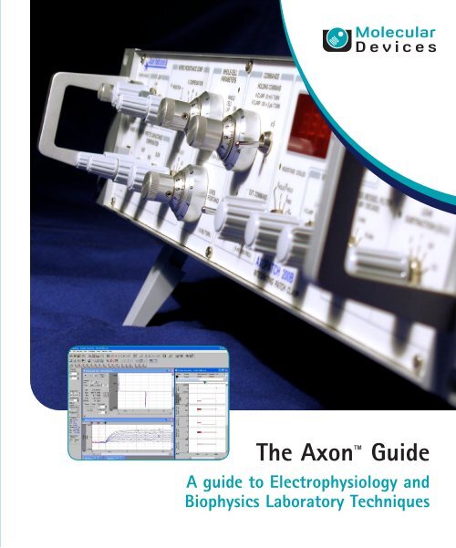
<strong>The</strong> <strong>Axon</strong> <strong>Guide</strong><br />
A guide to Electrophysiology and<br />
Biophysics Laboratory Techniques
<strong>The</strong> <strong>Axon</strong> <strong>Guide</strong><br />
Electrophysiology and Biophysics Laboratory<br />
Techniques<br />
Third Edition<br />
1-2500-0102 D<br />
February 2012
This document is provided to customers who have purchased Molecular Devices, LLC<br />
(“Molecular Devices”) equipment, software, reagents, and consumables to use in the<br />
operation of such Molecular Devices equipment, software, reagents, and<br />
consumables. This document is copyright protected and any reproduction of this<br />
document, in whole or any part, is strictly prohibited, except as Molecular Devices<br />
may authorize in writing.<br />
Software that may be described in this document is furnished under a license<br />
agreement. It is against the law to copy, modify, or distribute the software on any<br />
medium, except as specifically allowed in the license agreement. Furthermore, the<br />
license agreement may prohibit the software from being disassembled, reverse<br />
engineered, or decompiled for any purpose.<br />
Portions of this document may make reference to other manufacturers and/or their<br />
products, which may contain parts whose names are registered as trademarks and/or<br />
function as trademarks of their respective owners. Any such usage is intended only<br />
to designate those manufacturers’ products as supplied by Molecular Devices for<br />
incorporation into its equipment and does not imply any right and/or license to use<br />
or permit others to use such manufacturers’ and/or their product names as<br />
trademarks.<br />
Molecular Devices makes no warranties or representations as to the fitness of this<br />
equipment for any particular purpose and assumes no responsibility or contingent<br />
liability, including indirect or consequential damages, for any use to which the<br />
purchaser may put the equipment described herein, or for any adverse circumstances<br />
arising therefrom.<br />
For research use only. Not for use in diagnostic procedures.<br />
<strong>The</strong> trademarks mentioned herein are the property of Molecular Devices, LLC or their<br />
respective owners. <strong>The</strong>se trademarks may not be used in any type of promotion or<br />
advertising without the prior written permission of Molecular Devices, LLC.<br />
Product manufactured by Molecular Devices, LLC.<br />
1311 Orleans Drive, Sunnyvale, California, United States of America 94089.<br />
Molecular Devices, LLC is ISO 9001 registered.<br />
© 2012 Molecular Devices, LLC.<br />
All rights reserved.
Contents<br />
Preface . . . . . . . . . . . . . . . . . . . . . . . . . . . . . . . . . . . . . . . 9<br />
Introduction . . . . . . . . . . . . . . . . . . . . . . . . . . . . . . . . . . 11<br />
Acknowledgment To Molecular Devices Consultants<br />
And Customers . . . . . . . . . . . . . . . . . . . . . . . . . . . . . . 11<br />
Editorial . . . . . . . . . . . . . . . . . . . . . . . . . . . . . . . . . . . . . 13<br />
Contributors . . . . . . . . . . . . . . . . . . . . . . . . . . . . . . . . . . 15<br />
Chapter 1: Bioelectricity . . . . . . . . . . . . . . . . . . . . . . . . 17<br />
Electrical Potentials . . . . . . . . . . . . . . . . . . . . . . . . . . . 17<br />
Electrical Currents . . . . . . . . . . . . . . . . . . . . . . . . . . . . 17<br />
Resistors And Conductors . . . . . . . . . . . . . . . . . . . . . . . 19<br />
Ohm’s Law . . . . . . . . . . . . . . . . . . . . . . . . . . . . . . . . . 22<br />
<strong>The</strong> Voltage Divider . . . . . . . . . . . . . . . . . . . . . . . . . . . 24<br />
Perfect and Real Electrical Instruments. . . . . . . . . . . . . . 25<br />
Ions in Solutions and Electrodes . . . . . . . . . . . . . . . . . . 26<br />
Capacitors and <strong>The</strong>ir Electrical Fields . . . . . . . . . . . . . . . 29<br />
Currents Through Capacitors . . . . . . . . . . . . . . . . . . . . . 31<br />
Current Clamp and Voltage Clamp . . . . . . . . . . . . . . . . . 33<br />
Glass Microelectrodes and Tight Seals . . . . . . . . . . . . . . 34<br />
Further Reading. . . . . . . . . . . . . . . . . . . . . . . . . . . . . . 37<br />
Chapter 2: <strong>The</strong> Laboratory Setup. . . . . . . . . . . . . . . . . . 39<br />
<strong>The</strong> In Vitro Extracellular Recording Setup . . . . . . . . . . . 39<br />
<strong>The</strong> Single-channel Patch Clamping Setup . . . . . . . . . . . 39<br />
Vibration Isolation Methods. . . . . . . . . . . . . . . . . . . . . . 40<br />
Electrical Isolation Methods . . . . . . . . . . . . . . . . . . . . . . 41<br />
Equipment Placement. . . . . . . . . . . . . . . . . . . . . . . . . . 43<br />
Further Reading. . . . . . . . . . . . . . . . . . . . . . . . . . . . . . 46<br />
1-2500-0102 D 3
Contents<br />
Chapter 3: Instrumentation for Measuring Bioelectric<br />
Signals from Cells . . . . . . . . . . . . . . . . . . . . . . . . . . . . . . 49<br />
Extracellular Recording . . . . . . . . . . . . . . . . . . . . . . . . . 49<br />
Single-Cell Recording . . . . . . . . . . . . . . . . . . . . . . . . . . 50<br />
Multiple-cell Recording . . . . . . . . . . . . . . . . . . . . . . . . . 51<br />
Intracellular Recording Current Clamp. . . . . . . . . . . . . . . 51<br />
Intracellular Recording—Voltage Clamp. . . . . . . . . . . . . . 70<br />
Single-Channel Patch Clamp . . . . . . . . . . . . . . . . . . . . . 99<br />
Current Conventions and Voltage Conventions . . . . . . . . 110<br />
References . . . . . . . . . . . . . . . . . . . . . . . . . . . . . . . . 114<br />
Chapter 4: Microelectrodes and Micropipettes. . . . . . . 117<br />
Electrodes, Microelectrodes, Pipettes, Micropipettes,<br />
and Pipette Solutions . . . . . . . . . . . . . . . . . . . . . . . . . 117<br />
Fabrication of Patch Pipettes . . . . . . . . . . . . . . . . . . . . 119<br />
Pipette Properties for Single-channel vs.<br />
Whole-Cell Recording . . . . . . . . . . . . . . . . . . . . . . . . . 121<br />
Types of Glasses and <strong>The</strong>ir Properties . . . . . . . . . . . . . . 121<br />
<strong>The</strong>rmal Properties of Glasses . . . . . . . . . . . . . . . . . . . 124<br />
Noise Properties of Glasses . . . . . . . . . . . . . . . . . . . . . 125<br />
Leachable Components . . . . . . . . . . . . . . . . . . . . . . . . 127<br />
Further Reading . . . . . . . . . . . . . . . . . . . . . . . . . . . . . 127<br />
Chapter 5: Advanced Methods in Electrophysiology . . 129<br />
Recording from Xenopus Oocytes . . . . . . . . . . . . . . . . . 129<br />
Further Reading . . . . . . . . . . . . . . . . . . . . . . . . . . . . . 134<br />
Patch-Clamp Recording in Brain Slices . . . . . . . . . . . . . 135<br />
Further Reading . . . . . . . . . . . . . . . . . . . . . . . . . . . . . 142<br />
Macropatch and Loose-Patch Recording. . . . . . . . . . . . . 142<br />
<strong>The</strong> Giant Excised Membrane Patch Method . . . . . . . . . . 151<br />
Further Reading . . . . . . . . . . . . . . . . . . . . . . . . . . . . . 154<br />
Recording from Perforated Patches<br />
and Perforated Vesicles . . . . . . . . . . . . . . . . . . . . . . . . 154<br />
Further Reading . . . . . . . . . . . . . . . . . . . . . . . . . . . . . 163<br />
Enhanced Planar Bilayer Techniques for<br />
Single-Channel Recording . . . . . . . . . . . . . . . . . . . . . . 164<br />
Further Reading . . . . . . . . . . . . . . . . . . . . . . . . . . . . . 177<br />
4 1-2500-0102 D
<strong>The</strong> <strong>Axon</strong> <strong>Guide</strong> to Electrophysiology and Biophysics Laboratory Techniques<br />
Chapter 6: Signal Conditioning and<br />
Signal Conditioners . . . . . . . . . . . . . . . . . . . . . . . . . . . 179<br />
Why Should Signals be Filtered? . . . . . . . . . . . . . . . . . 179<br />
Fundamentals of Filtering . . . . . . . . . . . . . . . . . . . . . . 180<br />
Filter Terminology . . . . . . . . . . . . . . . . . . . . . . . . . . . 182<br />
Preparing Signals for A/D Conversion . . . . . . . . . . . . . . 192<br />
Averaging . . . . . . . . . . . . . . . . . . . . . . . . . . . . . . . . . 200<br />
Line-frequency Pick-up (Hum) . . . . . . . . . . . . . . . . . . . 200<br />
Peak-to-Peak and RMS Noise Measurements . . . . . . . . . 200<br />
Blanking . . . . . . . . . . . . . . . . . . . . . . . . . . . . . . . . . . 202<br />
Audio Monitor: Friend or Foe? . . . . . . . . . . . . . . . . . . . 202<br />
Electrode Test . . . . . . . . . . . . . . . . . . . . . . . . . . . . . . 203<br />
Common-Mode Rejection Ratio . . . . . . . . . . . . . . . . . . 203<br />
References . . . . . . . . . . . . . . . . . . . . . . . . . . . . . . . . 205<br />
Further Reading. . . . . . . . . . . . . . . . . . . . . . . . . . . . . 205<br />
Chapter 7: Transducers . . . . . . . . . . . . . . . . . . . . . . . . 207<br />
Temperature Transducers for Physiological<br />
Temperature Measurement . . . . . . . . . . . . . . . . . . . . . 207<br />
Temperature Transducers for Extended<br />
Temperature Ranges . . . . . . . . . . . . . . . . . . . . . . . . . 209<br />
Electrode Resistance and Cabling Affect Noise<br />
and Bandwidth . . . . . . . . . . . . . . . . . . . . . . . . . . . . . 211<br />
High Electrode Impedance Can Produce<br />
Signal Attenuation . . . . . . . . . . . . . . . . . . . . . . . . . . . 211<br />
Unmatched Electrode Impedances Increase<br />
Background Noise and Crosstalk . . . . . . . . . . . . . . . . . 213<br />
High Electrode Impedance Contributes to the<br />
<strong>The</strong>rmal Noise of the System . . . . . . . . . . . . . . . . . . . 214<br />
Cable Capacitance Filters out the High-Frequency<br />
Component of the Signal . . . . . . . . . . . . . . . . . . . . . . 215<br />
EMG, EEG, EKG, and Neural Recording . . . . . . . . . . . . . 215<br />
Metal Microelectrodes . . . . . . . . . . . . . . . . . . . . . . . . . 216<br />
Bridge Design for Pressure and Force Measurements . . . 217<br />
Pressure Measurements . . . . . . . . . . . . . . . . . . . . . . . 218<br />
Force Measurements . . . . . . . . . . . . . . . . . . . . . . . . . 218<br />
Acceleration Measurements. . . . . . . . . . . . . . . . . . . . . 219<br />
Length Measurements . . . . . . . . . . . . . . . . . . . . . . . . 219<br />
1-2500-0102 D 5
Contents<br />
Self-Heating Measurements . . . . . . . . . . . . . . . . . . . . . 219<br />
Isolation Measurements . . . . . . . . . . . . . . . . . . . . . . . 220<br />
Insulation Techniques . . . . . . . . . . . . . . . . . . . . . . . . . 220<br />
Further Reading . . . . . . . . . . . . . . . . . . . . . . . . . . . . . 220<br />
Chapter 8: Laboratory Computer Issues and<br />
Considerations . . . . . . . . . . . . . . . . . . . . . . . . . . . . . . . 223<br />
Select the Software First . . . . . . . . . . . . . . . . . . . . . . . 223<br />
How Much Computer do you Need? . . . . . . . . . . . . . . . 223<br />
Peripherals and Options . . . . . . . . . . . . . . . . . . . . . . . 224<br />
Recommended Computer Configurations . . . . . . . . . . . . 225<br />
Glossary . . . . . . . . . . . . . . . . . . . . . . . . . . . . . . . . . . 225<br />
Chapter 9: Acquisition Hardware . . . . . . . . . . . . . . . . . 231<br />
Fundamentals of Data Conversion . . . . . . . . . . . . . . . . 231<br />
Quantization Error . . . . . . . . . . . . . . . . . . . . . . . . . . . 233<br />
Choosing the Sampling Rate . . . . . . . . . . . . . . . . . . . . 234<br />
Converter Buzzwords . . . . . . . . . . . . . . . . . . . . . . . . . 235<br />
Deglitched DAC Outputs . . . . . . . . . . . . . . . . . . . . . . . 235<br />
Timers . . . . . . . . . . . . . . . . . . . . . . . . . . . . . . . . . . . 236<br />
Digital I/O . . . . . . . . . . . . . . . . . . . . . . . . . . . . . . . . . 237<br />
Optical Isolation . . . . . . . . . . . . . . . . . . . . . . . . . . . . . 237<br />
Operating Under Multi-Tasking Operating Systems . . . . . 238<br />
Software <strong>Support</strong> . . . . . . . . . . . . . . . . . . . . . . . . . . . . 238<br />
Chapter 10: Data Analysis . . . . . . . . . . . . . . . . . . . . . . 239<br />
Choosing Appropriate Acquisition Parameters . . . . . . . . 239<br />
Filtering at Analysis Time . . . . . . . . . . . . . . . . . . . . . . 241<br />
Integrals and Derivatives . . . . . . . . . . . . . . . . . . . . . . 242<br />
Single-Channel Analysis . . . . . . . . . . . . . . . . . . . . . . . 243<br />
Fitting . . . . . . . . . . . . . . . . . . . . . . . . . . . . . . . . . . . . 250<br />
References . . . . . . . . . . . . . . . . . . . . . . . . . . . . . . . . 256<br />
6 1-2500-0102 D
<strong>The</strong> <strong>Axon</strong> <strong>Guide</strong> to Electrophysiology and Biophysics Laboratory Techniques<br />
Chapter 11: Noise in Electrophysiological<br />
Measurements . . . . . . . . . . . . . . . . . . . . . . . . . . . . . . . 259<br />
<strong>The</strong>rmal Noise . . . . . . . . . . . . . . . . . . . . . . . . . . . . . . 260<br />
Shot Noise . . . . . . . . . . . . . . . . . . . . . . . . . . . . . . . . 262<br />
Dielectric Noise . . . . . . . . . . . . . . . . . . . . . . . . . . . . . 263<br />
Excess Noise . . . . . . . . . . . . . . . . . . . . . . . . . . . . . . . 264<br />
Amplifier Noise . . . . . . . . . . . . . . . . . . . . . . . . . . . . . 265<br />
Electrode Noise . . . . . . . . . . . . . . . . . . . . . . . . . . . . . 268<br />
Seal Noise. . . . . . . . . . . . . . . . . . . . . . . . . . . . . . . . . 278<br />
Noise in Whole-cell Voltage Clamping . . . . . . . . . . . . . . 279<br />
External Noise Sources . . . . . . . . . . . . . . . . . . . . . . . . 282<br />
Digitization Noise . . . . . . . . . . . . . . . . . . . . . . . . . . . . 283<br />
Aliasing . . . . . . . . . . . . . . . . . . . . . . . . . . . . . . . . . . 284<br />
Filtering . . . . . . . . . . . . . . . . . . . . . . . . . . . . . . . . . . 287<br />
Summary of Patch-Clamp Noise. . . . . . . . . . . . . . . . . . 291<br />
Limits of Patch-Clamp Noise Performance . . . . . . . . . . . 293<br />
Further Reading. . . . . . . . . . . . . . . . . . . . . . . . . . . . . 293<br />
Appendix A: <strong>Guide</strong> to Interpreting Specifications . . . . 295<br />
General . . . . . . . . . . . . . . . . . . . . . . . . . . . . . . . . . . 295<br />
RMS Versus Peak-to-Peak Noise . . . . . . . . . . . . . . . . . 296<br />
Bandwidth—Time Constant—Rise Time . . . . . . . . . . . . . 296<br />
Filters. . . . . . . . . . . . . . . . . . . . . . . . . . . . . . . . . . . . 296<br />
Microelectrode Amplifiers . . . . . . . . . . . . . . . . . . . . . . 297<br />
Patch-Clamp Amplifiers. . . . . . . . . . . . . . . . . . . . . . . . 298<br />
1-2500-0102 D 7
Contents<br />
8 1-2500-0102 D
Preface<br />
Molecular Devices is pleased to present you with the third edition of the<br />
<strong>Axon</strong> <strong>Guide</strong>, a laboratory guide to electrophysiology and biophysics.<br />
<strong>The</strong> purpose of this guide is to serve as an information and data<br />
resource for electrophysiologists. It covers a broad scope of topics<br />
ranging from the biological basis of bioelectricity and a description of<br />
the basic experimental setup to a discussion of mechanisms of noise<br />
and data analysis.<br />
<strong>The</strong> <strong>Axon</strong> <strong>Guide</strong> third edition is a tool benefiting both the novice and the<br />
expert electrophysiologist. Newcomers to electrophysiology will gain an<br />
appreciation of the intricacies of electrophysiological measurements<br />
and the requirements for setting up a complete recording and analysis<br />
system. For experienced electrophysiologists, we include in-depth<br />
discussions of selected topics, such as advanced methods in<br />
electrophysiology and noise.<br />
This edition is the first major revision of the <strong>Axon</strong> <strong>Guide</strong> since the<br />
original edition was published in 1993. While the fundamentals of<br />
electrophysiology have not changed in that time, changes in<br />
instrumentation and computer technology made a number of the<br />
original chapters interesting historical documents rather than helpful<br />
guides. This third edition is up-to-date with current developments in<br />
technology and instrumentation.<br />
This guide was the product of a collaborative effort of many researchers<br />
in the field of electrophysiology and the staff of Molecular Devices. We<br />
are deeply grateful to these individuals for sharing their knowledge and<br />
devoting significant time and effort to this endeavor.<br />
David Yamane<br />
Director, Electrophysiology Marketing<br />
Molecular Devices<br />
1-2500-0102 D 9
Preface<br />
10 1-2500-0102 D
Introduction<br />
<strong>Axon</strong> Instruments, Inc. was founded in 1983 to design and<br />
manufacture instrumentation and software for electrophysiology and<br />
biophysics. Its products were distinguished by the company’s<br />
innovative design capability, high-quality products, and expert technical<br />
support. Today, the <strong>Axon</strong> Instruments products are part of the<br />
Molecular Devices portfolio of life science and drug discovery products.<br />
<strong>The</strong> <strong>Axon</strong> brand of microelectrode amplifiers, digitizers, and data<br />
acquisition and analysis software provides researchers with<br />
ready-to-use, technologically advanced products which allows them<br />
more time to pursue their primary research goals.<br />
Furthermore, to ensure continued success with our products, we have<br />
staffed our Technical <strong>Support</strong> group with experienced Ph.D.<br />
electrophysiologists.<br />
In addition to the <strong>Axon</strong> product suite, Molecular Devices has developed<br />
three automated electrophysiology platforms. Together with the <strong>Axon</strong><br />
Conventional Electrophysiology products, Molecular Devices spans the<br />
entire drug discovery process from screening to safety assessment.<br />
In recognition of the continuing excitement in ion channel research, as<br />
evidenced by the influx of molecular biologists, biochemists, and<br />
pharmacologists into this field, Molecular Devices is proud to support<br />
the pursuit of electrophysiological and biophysical research with this<br />
laboratory techniques workbook.<br />
Acknowledgment To Molecular Devices Consultants<br />
And Customers<br />
Molecular Devices employs a talented team of engineers and scientists<br />
dedicated to designing instruments and software incorporating the<br />
most advanced technology and the highest quality. Nevertheless, it<br />
would not be possible for us to enhance our products without close<br />
collaborations with members of the scientific community. <strong>The</strong>se<br />
collaborations take many forms.<br />
Some scientists assist Molecular Devices on a regular basis, sharing<br />
their insights on current needs and future directions of scientific<br />
research. Others assist us by virtue of a direct collaboration in the<br />
design of certain products. Many scientists help us by reviewing our<br />
instrument designs and the development versions of various software<br />
products. We are grateful to these scientists for their assistance. We<br />
also receive a significant number of excellent suggestions from the<br />
customers we meet at scientific conferences. To all of you who have<br />
visited us at our booths and shared your thoughts, we extend our<br />
1-2500-0102 D 11
Introduction<br />
sincere thanks. Another source of feedback for us is the information<br />
that we receive from the conveners of the many excellent summer<br />
courses and workshops that we support with equipment loans. Our<br />
gratitude is extended to them for the written assessments they often<br />
send us outlining the strengths and weaknesses of <strong>Axon</strong> Conventional<br />
Electrophysiology products from Molecular Devices.<br />
12 1-2500-0102 D
Editorial<br />
Editor<br />
Rivka Sherman-Gold, Ph.D.<br />
Editorial Committee<br />
Michael J. Delay, Ph.D.<br />
Alan S. Finkel, Ph.D.<br />
Henry A. Lester, Ph.D.1<br />
W. Geoffrey Powell, MBA<br />
Rivka Sherman-Gold, Ph.D.<br />
Layout and Editorial Assistance<br />
Jay Kurtz<br />
Artwork<br />
Elizabeth Brzeski<br />
Editor – 3rd Edition<br />
Warburton H. Maertz<br />
Editorial Committee – 3rd Edition<br />
Scott V. Adams, Ph.D.<br />
S. Clare Chung, Ph.D.<br />
Toni Figl, Ph.D.<br />
Warburton H. Maertz<br />
Damian J. Verdnik, Ph.D.<br />
Layout and Editorial Assistance – 3rd Edition<br />
Simone Andrews<br />
Eveline Pajares<br />
Peter Valenzuela<br />
Damian J. Verdnik, Ph.D.<br />
Alfred Walter, Ph.D.<br />
1-2500-0102 D 13
Editorial<br />
Acknowledgements<br />
<strong>The</strong> valuable inputs and insightful comments of Dr. Bertil Hille, Dr. Joe<br />
Immel, and Dr. Stephen Redman are much appreciated.<br />
14 1-2500-0102 D
Contributors<br />
<strong>The</strong> <strong>Axon</strong> <strong>Guide</strong> is the product of a collaborative effort of Molecular<br />
Devices and researchers from several institutions whose contributions<br />
to the guide are gratefully acknowledged.<br />
John M. Bekkers, Ph.D., Division of Neuroscience, John Curtin School of<br />
Medical Research, Australian National University, Canberra, A.C.T.<br />
Australia.<br />
Richard J. Bookman, Ph.D., Department of Molecular & Cellular<br />
Pharmacology, Miller School of Medicine, University of Miami, Miami,<br />
Florida.<br />
Michael J. Delay, Ph.D.<br />
Alan S. Finkel, Ph.D.<br />
Aaron Fox, Ph.D., Department of Neurobiology, Pharmacology and<br />
Physiology, University of Chicago, Chicago, Illinois.<br />
David Gallegos.<br />
Robert I. Griffiths, Ph.D., Monash Institute of Medical Research, Faculty<br />
of Medicine, Monash University, Clayton, Victoria, Australia.<br />
Donald Hilgemann, Ph.D., Graduate School of Biomedical Sciences,<br />
Southwestern Medical School, Dallas, Texas.<br />
Richard H. Kramer, Department of Molecular & Cell Biology, University<br />
of California Berkeley, Berkeley, California.<br />
Henry A. Lester, Ph.D., Division of Biology, California Institute of<br />
Technology, Pasadena, California.<br />
Richard A. Levis, Ph.D., Department of Molecular Biophysics &<br />
Physiology, Rush-Presbyterian-St. Luke’s Medical College, Chicago,<br />
Illinois.<br />
Edwin S. Levitan, Ph.D., Department of Pharmacology, School of<br />
Medicine, University of Pittsburgh, Pittsburgh, Pennsylvania.<br />
M. Craig McKay, Ph.D., Department of Molecular Endocrinology,<br />
GlaxoSmithKline, Inc., Research Triangle Park, North Carolina.<br />
David J. Perkel, Department of Biology, University of Washington,<br />
Seattle, Washington.<br />
Stuart H. Thompson, Hopkins Marine Station, Stanford University,<br />
Pacific Grove, California.<br />
James L. Rae, Ph.D., Department of Physiology and Biomedical<br />
Engineering, Mayo Clinic, Rochester, Minnesota.<br />
Michael M. White, Ph.D., Department of Pharmacology & Physiology,<br />
Drexel University College of Medicine, Philadelphia, Pennsylvania.<br />
1-2500-0102 D 15
Contributors<br />
William F. Wonderlin, Ph.D., Department of Biochemistry & Molecular<br />
Phamacology, West Virginia University, Morgantown, West Virginia.<br />
16 1-2500-0102 D
Bioelectricity<br />
1<br />
Electrical Potentials<br />
Electrical Currents<br />
Chapter 1 introduces the basic concepts used in making electrical<br />
measurements from cells and the techniques used to make these<br />
measurements.<br />
A cell derives its electrical properties mostly from the electrical<br />
properties of its membrane. A membrane, in turn, acquires its<br />
properties from its lipids and proteins, such as ion channels and<br />
transporters. An electrical potential difference exists between the<br />
interior and exterior of cells. A charged object (ion) gains or loses<br />
energy as it moves between places of different electrical potential, just<br />
as an object with mass moves “up” or “down” between points of<br />
different gravitational potential. Electrical potential differences are<br />
usually denoted as V or V and measured in volts. Potential is also<br />
termed voltage. <strong>The</strong> potential difference across a cell relates the<br />
potential of the cell’s interior to that of the external solution, which,<br />
according to the commonly accepted convention, is zero.<br />
<strong>The</strong> potential difference across a lipid cellular membrane<br />
(“transmembrane potential”) is generated by the “pump” proteins that<br />
harness chemical energy to move ions across the cell membrane. This<br />
separation of charge creates the transmembrane potential. Because the<br />
lipid membrane is a good insulator, the transmembrane potential is<br />
maintained in the absence of open pores or channels that can conduct<br />
ions.<br />
Typical transmembrane potentials amount to less than 0.1 V, usually 30<br />
to 90 mV in most animal cells, but can be as much as 200 mV in plant<br />
cells. Because the salt-rich solutions of the cytoplasm and extracellular<br />
milieu are fairly good conductors, there are usually very small<br />
differences at steady state (rarely more than a few millivolts) between<br />
any two points within a cell’s cytoplasm or within the extracellular<br />
solution. Electrophysiological equipment enables researchers to<br />
measure potential (voltage) differences in biological systems.<br />
Electrophysiological equipment can also measure current, which is the<br />
flow of electrical charge passing a point per unit of time. Current (I) is<br />
measured in amperes (A). Usually, currents measured by<br />
electrophysiological equipment range from picoamperes to<br />
microamperes. For instance, typically, 10 4 Na + ions cross the<br />
membrane each millisecond that a single Na + channel is open. This<br />
1-2500-0102 D 17
Bioelectricity<br />
current equals 1.6 pA (1.6 x 10 -19 C/ion x 10 4 ions/ms x 10 3 ms/s; recall<br />
that 1 A is equal to 1 coulomb (C)/s).<br />
Two handy rules about currents often help to understand<br />
electrophysiological phenomena: 1) current is conserved at a branch<br />
point (Figure 1-1); and 2) current always flows in a complete circuit<br />
(Figure 1-2). In electrophysiological measurements, currents can flow<br />
through capacitors, resistors, ion channels, amplifiers, electrodes and<br />
other entities, but they always flow in complete circuits.<br />
Figure 1-1 Conservation of current. Current is conserved at a branch<br />
point.<br />
18 1-2500-0102 D
<strong>The</strong> <strong>Axon</strong> <strong>Guide</strong> to Electrophysiology and Biophysics Laboratory Techniques<br />
Figure 1-2 A typical electrical circuit. Example of an electrical circuit<br />
with various parts. Current always flows in a complete circuit.<br />
Resistors And Conductors<br />
Currents flow through resistors or conductors. <strong>The</strong> two terms actually<br />
complement one another—the former emphasizes the barriers to<br />
current flow, while the latter emphasizes the pathways for flow. In<br />
quantitative terms, resistance R (units: ohms ()) is the inverse of<br />
conductance G (units: siemens (S)); thus, infinite resistance is zero<br />
conductance. In electrophysiology, it is convenient to discuss currents<br />
in terms of conductance because side-by-side (“parallel”) conductances<br />
simply sum (Figure 1-3). <strong>The</strong> most important application of the parallel<br />
conductances involves ion channels. When several ion channels in a<br />
membrane are open simultaneously, the total conductance is simply the<br />
sum of the conductances of the individual open channels.<br />
1-2500-0102 D 19
Bioelectricity<br />
Figure 1-3 Summation of conductance. Conductances in parallel<br />
summate together, whether they are resistors or channels.<br />
A more accurate representation of an ion channel is a conductor in<br />
series with two additional circuit elements (Figure 1-4):<br />
1. A switch that represents the gate of the channel, which would be<br />
in its conducting position when the gate is open, and<br />
2. A battery that represents the reversal potential of the ionic<br />
current for that channel. <strong>The</strong> reversal potential is defined<br />
operationally as the voltage at which the current changes its<br />
direction. For a perfectly selective channel (for example, a<br />
channel through which only a single type of ion can pass), the<br />
reversal potential equals the Nernst potential for the permeant<br />
ion. <strong>The</strong> Nernst potential for ion A, Ea, can be calculated by the<br />
Nernst equation:<br />
where R is the gas constant (8.314 V C K -1 mol -1 ), T is the<br />
absolute temperature (T = 273° + °C), z A is the charge of ion A,<br />
F is Faraday’s constant (9.648x10 4 C mol -1 ), and [A] o and [A] i<br />
are the concentrations of ion A outside the cell and inside the<br />
20 1-2500-0102 D
<strong>The</strong> <strong>Axon</strong> <strong>Guide</strong> to Electrophysiology and Biophysics Laboratory Techniques<br />
cell, respectively. At 20 °C (“room temperature”), 2.303(RT/z A F)<br />
log 10{[A] o/[A] i} = 58 mV log 10{[A] o/[A] i}for a univalent ion.<br />
Figure 1-4 Equivalent circuit for a single-membrane channel.<br />
A more realistic equivalent circuit for a single-membrane channel.<br />
For instance, at room temperature, a Na + channel facing intracellular<br />
Na + concentration that is ten-fold lower than the extracellular<br />
concentration of this ion would be represented by a battery of +58 mV.<br />
A K + channel, for which the concentration gradient is usually reversed,<br />
would be represented by a battery of -58 mV.<br />
Reversal potentials are not easily predicted for channels that are<br />
permeable to more than one ion. Nonspecific cation channels, such as<br />
nicotinic acetylcholine receptors, usually have reversal potentials near<br />
zero millivolts. Furthermore, many open channels have a nonlinear<br />
relation between current and voltage. Consequently, representing<br />
channels as resistors is only an approximation. Considerable<br />
biophysical research has been devoted to understanding the<br />
current-voltage relations of ion channels and how they are affected by<br />
the properties and concentrations of permeant ions.<br />
<strong>The</strong> transmembrane potential is defined as the potential at the inner<br />
side of the membrane relative to the potential at the outer side of the<br />
membrane. <strong>The</strong> resting membrane potential (E rp ) describes a<br />
steady-state condition with no net flow of electrical current across the<br />
membrane. <strong>The</strong> resting membrane potential is determined by the<br />
intracellular and extracellular concentrations of ions to which the<br />
membrane is permeable and on their permeabilities. If one ionic<br />
conductance is dominant, the resting potential is near the Nernst<br />
1-2500-0102 D 21
Bioelectricity<br />
Ohm’s Law<br />
potential for that ion. Since a typical cell membrane at rest has a much<br />
higher permeability to potassium (P K) than to sodium, calcium or<br />
chloride (P Na , P Ca and P Cl , respectively), the resting membrane potential<br />
is very close to E K , the potassium reversal potential.<br />
For electrophysiology, perhaps the most important law of electricity is<br />
Ohm’s law. <strong>The</strong> potential difference between two points linked by a<br />
current path with a conductance G and a current I (Figure 1-5) is:<br />
Figure 1-5 Figure 1.5: Ohm’s law.<br />
This concept applies to any electrophysiological measurement, as<br />
illustrated by the two following examples:<br />
1. In an extracellular recording experiment: the current I that<br />
flows between parts of a cell through the external resistance R<br />
produces a potential difference V, which is usually less than 1<br />
mV (Figure 1-6). As the impulse propagates, I changes and,<br />
therefore, V changes as well.<br />
22 1-2500-0102 D
<strong>The</strong> <strong>Axon</strong> <strong>Guide</strong> to Electrophysiology and Biophysics Laboratory Techniques<br />
Figure 1-6 IR drop. In extracellular recording, current I that<br />
flows between points of a cell is measured as the potential<br />
difference (“IR drop”) across the resistance R of the fluid<br />
between the two electrodes.<br />
2. In a voltage-clamp experiment: when N channels, each of<br />
conductance , are open, the total conductance is N. <strong>The</strong><br />
electrochemical driving force V (membrane potential minus<br />
reversal potential) produces a current NV. As channels open<br />
and close, N changes and so does the voltage-clamp current I.<br />
Hence, the voltage-clamp current is simply proportional to the<br />
number of open channels at any given time. Each channel can<br />
be considered as a conductance increment.<br />
1-2500-0102 D 23
Bioelectricity<br />
<strong>The</strong> Voltage Divider<br />
Figure 1-7 describes a simple circuit called a voltage divider in which<br />
two resistors are connected in series:<br />
Figure 1-7 A voltage divider.<br />
<strong>The</strong> total potential difference provided by the battery is E; a portion of<br />
this voltage appears across each resistor:<br />
When two resistors are connected in series, the same current passes<br />
through each of them. <strong>The</strong>refore the circuit is described by:<br />
where E is the value of the battery, which equals the total potential<br />
difference across both resistors. As a result, the potential difference is<br />
divided in proportion to the two resistance values.<br />
24 1-2500-0102 D
<strong>The</strong> <strong>Axon</strong> <strong>Guide</strong> to Electrophysiology and Biophysics Laboratory Techniques<br />
Perfect and Real Electrical Instruments<br />
Electrophysiological measurements should satisfy two requirements: 1)<br />
<strong>The</strong>y should accurately measure the parameter of interest, and 2) they<br />
should produce no perturbation of the parameter. <strong>The</strong> first requirement<br />
can be discussed in terms of a voltage divider. <strong>The</strong> second point will be<br />
discussed after addressing electrodes.<br />
<strong>The</strong> best way to measure an electrical potential difference is to use a<br />
voltmeter with infinite resistance. To illustrate this point, consider the<br />
arrangement of Figure 1-8 A, which can be reduced to the equivalent<br />
circuit of Figure 1-8 B.<br />
Figure 1-8 Representative voltmeter with infinite resistance.<br />
Instruments used to measure potentials must have a very high input<br />
resistance R in .<br />
1-2500-0102 D 25
Bioelectricity<br />
Before making the measurement, the cell has a resting potential of E rp ,<br />
which is to be measured with an intracellular electrode of resistance R e.<br />
To understand the effect of the measuring circuit on the measured<br />
parameter, we will pretend that our instrument is a “perfect” voltmeter<br />
(for example, with an infinite resistance) in parallel with a finite<br />
resistance R in , which represents the resistance of a real voltmeter or<br />
the measuring circuit. <strong>The</strong> combination R e and R in forms a voltage<br />
divider, so that only a fraction of E rp appears at the input of the<br />
“perfect” voltmeter; this fraction equals E rp R in /(R in + R e ). <strong>The</strong> larger R in ,<br />
the closer V is to E rp . Clearly the problem gets more serious as the<br />
electrode resistance R e increases, but the best solution is to make R in as<br />
large as possible.<br />
On the other hand, the best way to measure current is to open the path<br />
and insert an ammeter. If the ammeter has zero resistance, it will not<br />
perturb the circuit since there is no IR-drop across it.<br />
Ions in Solutions and Electrodes<br />
Ohm’s law—the linear relation between potential difference and current<br />
flow—applies to aqueous ionic solutions, such as blood, cytoplasm, and<br />
sea water. Complications are introduced by two factors:<br />
1. <strong>The</strong> current is carried by at least two types of ions (one anion<br />
and one cation) and often by many more. For each ion, current<br />
flow in the bulk solution is proportional to the potential<br />
difference. For a first approximation, the conductance of the<br />
whole solution is simply the sum of the conductances<br />
contributed by each ionic species. When the current flows<br />
through ion channels, it is carried selectively by only a subset of<br />
the ions in the solution.<br />
2. At the electrodes, current must be transformed smoothly from a<br />
flow of electrons in the copper wire to a flow of ions in solution.<br />
Many sources of errors (artifacts) are possible. Several types of<br />
electrodes are used in electrophysiological measurements. <strong>The</strong><br />
most common is a silver/silver chloride (Ag/AgCl) interface,<br />
which is a silver wire coated with silver chloride (Figure 1-9). If<br />
electrons flow from the copper wire through the silver wire to<br />
the electrode AgCl pellet, they convert the AgCl to Ag atoms and<br />
the Cl- ions become hydrated and enter the solution. If electrons<br />
flow in the reverse direction, Ag atoms in the silver wire that is<br />
coated with AgCl give up their electrons (one electron per atom)<br />
and combine with Cl- ions that are in the solution to make<br />
insoluble AgCl. This is, therefore, a reversible electrode, for<br />
example, current can flow in both directions.<br />
26 1-2500-0102 D
<strong>The</strong> <strong>Axon</strong> <strong>Guide</strong> to Electrophysiology and Biophysics Laboratory Techniques<br />
<strong>The</strong>re are several points to remember about Ag/AgCl electrodes:<br />
• <strong>The</strong> Ag/AgCl electrode performs well only in solutions<br />
containing chloride ions.<br />
• Because current must flow in a complete circuit, two<br />
electrodes are needed. If the two electrodes face different Cl -<br />
concentrations (for instance, 3 M KCl inside a micropipette 1<br />
and 120 mM NaCl in a bathing solution surrounding the cell),<br />
there will be a difference in the half-cell potentials (the<br />
potential difference between the solution and the electrode)<br />
at the two electrodes, resulting in a large steady potential<br />
difference in the two wires attached to the electrodes. This<br />
steady potential difference, termed liquid junction potential,<br />
can be subtracted electronically and poses few problems as<br />
long as the electrode is used within its reversible limits.<br />
• If the AgCl is exhausted by the current flow, bare silver could<br />
come in contact with the solution. Silver ions leaking from<br />
the wire can poison many proteins. Also, the half-cell<br />
potentials now become dominated by unpredictable, poorly<br />
reversible surface reactions due to other ions in the solution<br />
and trace impurities in the silver, causing electrode<br />
polarization. However, used properly, Ag/AgCl electrodes<br />
possess the advantages of little polarization and predictable<br />
junction potential.<br />
1. A micropipette is a pulled capillary glass into which the Ag/AgC1 electrode is<br />
inserted (see Chapter 4: Microelectrodes and Micropipettes on page 117).<br />
1-2500-0102 D 27
Bioelectricity<br />
Figure 1-9 <strong>The</strong> silver/silver chloride electrode. <strong>The</strong> silver/silver<br />
chloride electrode is reversible but exhaustible.<br />
Another type of electrode, made of platinum (Pt) (Figure 1-10), is<br />
irreversible but not exhaustible. At its surface, Pt catalyzes the<br />
electrolysis of water. <strong>The</strong> gaseous H 2 or O 2 produced, depending on the<br />
direction of current flow, leaves the surface of the electrode. If both<br />
electrodes are Pt electrodes, the hydroxyl ions and protons are<br />
produced in equal numbers; however, local pH changes can still occur.<br />
28 1-2500-0102 D
<strong>The</strong> <strong>Axon</strong> <strong>Guide</strong> to Electrophysiology and Biophysics Laboratory Techniques<br />
Figure 1-10 <strong>The</strong> platinum electrode. A platinum electrode is<br />
irreversible but inexhaustible.<br />
Capacitors and <strong>The</strong>ir Electrical Fields<br />
<strong>The</strong> electrical field is a property of each point in space and is defined as<br />
proportional to the force experienced by a charge placed at that point.<br />
<strong>The</strong> greater the potential difference between two points fixed in space,<br />
the greater the field at each point between them. Formally, the<br />
electrical field is a vector defined as the negative of the spatial<br />
derivative of the potential.<br />
<strong>The</strong> concept of the electrical field is important for understanding<br />
membrane function. Biological membranes are typically less than<br />
10 nm thick. Consequently, a transmembrane resting potential of about<br />
100 mV produces a very sizable electrical field in the membrane of<br />
about 10 5 V/cm. This is close to the value at which most insulators<br />
break down irreversibly because their atoms become ionized. Of<br />
course, typical electrophysiological equipment cannot measure these<br />
fields directly. However, changes in these fields are presumably sensed<br />
by the gating domains of voltage-sensitive ion channels, which<br />
determine the opening and closing of channels, and so the electrical<br />
fields underlie the electrical excitability of membranes.<br />
1-2500-0102 D 29
Bioelectricity<br />
Another consequence of the membrane’s thinness is that it makes an<br />
excellent capacitor. Capacitance (C; measured in farads, F) is the ability<br />
to store charge Q when a voltage V occurs across the two “ends,” so<br />
that:<br />
<strong>The</strong> formal symbol for a capacitor is two parallel lines (Figure 1-12).<br />
This symbol arose because the most effective capacitors are parallel<br />
conducting plates of large area separated by a thin sheet of insulator<br />
(Figure 1-11) an excellent approximation of the lipid bilayer.<br />
<strong>The</strong> capacitance C is proportional to the area and inversely proportional<br />
to the distance separating the two conducting sheets.<br />
Figure 1-11 Capacitance. A charge Q is stored in a capacitor of value<br />
C held at a potential V.<br />
When multiple capacitors are connected in parallel, this is electronically<br />
equivalent to a single large capacitor; that is, the total capacitance is<br />
the sum of their individual capacitance values (Figure 1-12). Thus,<br />
membrane capacitance increases with cell size. Membrane capacitance<br />
is usually expressed as value per unit area; nearly all lipid bilayer<br />
membranes of cells have a capacitance of 1 μF/cm 2 (0.01 pF/μm 2 ).<br />
30 1-2500-0102 D
<strong>The</strong> <strong>Axon</strong> <strong>Guide</strong> to Electrophysiology and Biophysics Laboratory Techniques<br />
Figure 1-12 Capacitors in parallel add their values.<br />
Currents Through Capacitors<br />
<strong>The</strong> following equation shows that charge is stored in a capacitor only<br />
when there is a change in the voltage across the capacitor. <strong>The</strong>refore,<br />
the current flowing through capacitance C is proportional to the voltage<br />
change with time:<br />
Until now, we have been discussing circuits whose properties do not<br />
change with time. As long as the voltage across a membrane remains<br />
constant, one can ignore the effect of the membrane capacitance on<br />
the currents flowing across the membrane through ion channels. While<br />
the voltage changes, there are transient capacitive currents in addition<br />
to the steady-state currents through conductive channels. <strong>The</strong>se<br />
capacitive currents constitute one of the two major influences on the<br />
time-dependent electrical properties of cells (the other is the kinetics of<br />
channel gating). On <strong>Axon</strong> Conventional Electrophysiology<br />
voltage-clamp or patch-clamp amplifiers, several controls are devoted<br />
to handle these capacitive currents. <strong>The</strong>refore it is worth obtaining<br />
some intuitive “feel” for their behavior.<br />
<strong>The</strong> stored charge on the membrane capacitance accompanies the<br />
resting potential, and any change in the voltage across the membrane<br />
is accompanied by a change in this stored charge. Indeed, if a current<br />
is applied to the membrane, either by channels elsewhere in the cell or<br />
by current from the electrode, this current first satisfies the<br />
requirement for charging the membrane capacitance, then it changes<br />
1-2500-0102 D 31
Bioelectricity<br />
the membrane voltage. Formally, this can be shown by representing the<br />
membrane as a resistor of value R in parallel with capacitance C<br />
(Figure 1-13).<br />
Figure 1-13 Membrane behavior compared with an electrical current.<br />
A membrane behaves electrically like a capacitance in parallel with a<br />
resistance.<br />
Now, if we apply a pulse of current to the circuit, the current first<br />
charges up the capacitance, then changes the voltage (Figure 1-14).<br />
Figure 1-14 RC parallel circuit response. Response of an RC parallel<br />
circuit to a step of current.<br />
32 1-2500-0102 D
<strong>The</strong> <strong>Axon</strong> <strong>Guide</strong> to Electrophysiology and Biophysics Laboratory Techniques<br />
<strong>The</strong> voltage V(t) approaches steady state along an exponential time<br />
course:<br />
<strong>The</strong> steady-state value V inƒ (also called the infinite-time or equilibrium<br />
value) does not depend on the capacitance; it is simply determined by<br />
the current I and the membrane resistance R:<br />
This is just Ohm’s law, of course; but when the membrane capacitance<br />
is in the circuit, the voltage is not reached immediately. Instead, it is<br />
approached with the time constant , given by:<br />
Thus, the charging time constant increases when either the membrane<br />
capacitance or the resistance increases. Consequently, large cells, such<br />
as Xenopus oocytes that are frequently used for expression of genes<br />
encoding ion-channel proteins, and cells with extensive membrane<br />
invigorations, such as the T-system in skeletal muscle, have a long<br />
charging phase.<br />
Current Clamp and Voltage Clamp<br />
In a current-clamp experiment, one applies a known constant or timevarying<br />
current and measures the change in membrane potential<br />
caused by the applied current. This type of experiment mimics the<br />
current produced by a synaptic input.<br />
In a voltage clamp experiment one controls the membrane voltage and<br />
measures the transmembrane current required to maintain that<br />
voltage. Despite the fact that voltage clamp does not mimic a process<br />
found in nature, there are three reasons to do such an experiment:<br />
1. Clamping the voltage eliminates the capacitive current, except<br />
for a brief time following a step to a new voltage (Figure 1-15).<br />
<strong>The</strong> brevity of the capacitive current depends on many factors<br />
that are discussed in following chapters.<br />
2. Except for the brief charging time, the currents that flow are<br />
proportional only to the membrane conductance, for example,<br />
to the number of open channels.<br />
3. If channel gating is determined by the transmembrane voltage<br />
alone (and is insensitive to other parameters such as the current<br />
and the history of the voltage), voltage clamp offers control over<br />
the key variable that determines the opening and closing of ion<br />
channels.<br />
1-2500-0102 D 33
Bioelectricity<br />
Figure 1-15 Typical voltage-clamp experiment. A<br />
voltage-clamp experiment on the circuit of Figure 1-13.<br />
<strong>The</strong> patch clamp is a special voltage clamp that allows one to resolve<br />
currents flowing through single ion channels. It also simplified the<br />
measurement of currents flowing through the whole-cell membrane,<br />
particularly in small cells that cannot be easily penetrated with<br />
electrodes. <strong>The</strong> characteristics of a patch clamp are dictated by two<br />
facts:<br />
1. <strong>The</strong> currents measured are very small, on the order of<br />
picoamperes in single-channel recording and usually up to<br />
several nanoamperes in whole-cell recording. Due to the small<br />
currents, particularly in single-channel recording, the electrode<br />
polarizations and nonlinearities are negligible and the Ag/AgCl<br />
electrode can record voltage accurately even while passing<br />
current.<br />
2. <strong>The</strong> electronic ammeter must be carefully designed to avoid<br />
adding appreciable noise to the currents it measures.<br />
Glass Microelectrodes and Tight Seals<br />
Successful electrophysiological measurements depend on two<br />
technologies: the design of electronic instrumentation and the<br />
properties and fabrication of glass micropipettes. Glass pipettes are<br />
used both for intracellular recording and for patch recording; quartz<br />
pipettes have been used for ultra low-noise single-channel recording<br />
34 1-2500-0102 D
<strong>The</strong> <strong>Axon</strong> <strong>Guide</strong> to Electrophysiology and Biophysics Laboratory Techniques<br />
(for a detailed discussion of electrode glasses see Chapter 4:<br />
Microelectrodes and Micropipettes on page 117). Successful patch<br />
recording requires a tight seal between the pipette and the membrane.<br />
Although there is not yet a satisfactory molecular description of this<br />
seal, we can describe its electrical characteristics.<br />
Requirement 2) above (see Perfect and Real Electrical Instruments on<br />
page 25) states that the quality of the measurement depends on<br />
minimizing perturbation of the cells. For the case of voltage recording,<br />
this point can be presented with the voltage divider circuit (Figure 1-<br />
16).<br />
Figure 1-16 Intracellular electrode measurement. This intracellular<br />
electrode is measuring the resting potential of a cell whose membrane<br />
contains only open K+ channels. As the seal resistance R s increases,<br />
the measurement approaches the value of E K .<br />
For the case of patch recording, currents through the seal do not distort<br />
the measured voltage or current, but they do add to the current noise.<br />
Current noise can be analyzed either in terms of the Johnson noise of a<br />
conductor, which is the thermal noise that increases with the<br />
conductance (see Chapter 7: Transducers on page 207 and Chapter 11:<br />
Noise in Electrophysiological Measurements on page 259), or in terms<br />
of simple statistics. <strong>The</strong> latter goes as follows: If a current of N ions/ms<br />
passes through an open channel, then the current will fluctuate from<br />
one millisecond to the next with a standard deviation of N. <strong>The</strong>se<br />
fluctuations produce noise on the single-channel recorded traces. If an<br />
additional current is flowing in parallel through the seal (Figure 1-17), it<br />
causes an increase in the standard deviations. For instance, if the<br />
current through the seal is ten-fold larger than through the channel,<br />
then the statistical fluctuations in current flow produced by the seal are<br />
10 (316%) larger than they would be for a “perfect” seal.<br />
1-2500-0102 D 35
Bioelectricity<br />
Figure 1-17 Good and bad seals. In a patch recording, currents<br />
through the seal also flow through the measuring circuit, increasing the<br />
noise on the measured current.<br />
36 1-2500-0102 D
<strong>The</strong> <strong>Axon</strong> <strong>Guide</strong> to Electrophysiology and Biophysics Laboratory Techniques<br />
Further Reading<br />
Brown, K. T., Flaming, D. G. Advanced Micropipette Techniques for Cell<br />
Physiology. John Wiley & Sons, New York, NY, 1986.<br />
Hille, B., Ionic Channels in Excitable Membranes. Second edition.<br />
Sinauer Associates, Sunderland, MA, 1991.<br />
Horowitz, P., Hill, W. <strong>The</strong> Art of Electronics. Second edition. Cambridge<br />
University Press, Cambridge, UK, 1989.<br />
Miller, C., Ion Channel Reconstitution. C. Miller, Ed., Plenum Press, New<br />
York, NY, 1986.<br />
Sakmann, B., Neher, E. Eds., Single-Channel Recording. Plenum Press,<br />
New York, NY, 1983.<br />
Smith, T. G., Lecar, H., Redman, S. J., Gage, P. W., Eds. Voltage and<br />
Patch Clamping with Microelectrodes. American Physiological Society,<br />
Bethesda, MD, 1985.<br />
Standen, N. B., Gray, P. T. A., Whitaker, M. J., Eds. Microelectrode<br />
Techniques: <strong>The</strong> Plymouth Workshop Handbook. <strong>The</strong> Company of<br />
Biologists Limited, Cambridge, England, 1987.<br />
1-2500-0102 D 37
Bioelectricity<br />
38 1-2500-0102 D
<strong>The</strong> Laboratory Setup<br />
2<br />
Each laboratory setup is different, reflecting the requirements of the<br />
experiment or the foibles of the experimenter. Chapter 2 describes<br />
components and considerations that are common to all setups<br />
dedicated to measure electrical activity in cells.<br />
An electrophysiological setup has four main requirements:<br />
1. Environment: the means of keeping the preparation healthy;<br />
2. Optics: a means of visualizing the preparation;<br />
3. Mechanics: a means of stably positioning the microelectrode;<br />
and<br />
4. Electronics: a means of amplifying and recording the signal.<br />
This guide focuses mainly on the electronics of the electrophysiological<br />
laboratory setup.<br />
To illustrate the practical implications of these requirements, two kinds<br />
of “typical” setups are briefly described, one for in vitro extracellular<br />
recording, the other for single-channel patch clamping.<br />
<strong>The</strong> In Vitro Extracellular Recording Setup<br />
This setup is mainly used for recording field potentials in brain slices.<br />
<strong>The</strong> general objective is to hold a relatively coarse electrode in the<br />
extracellular space of the tissue while mimicking as closely as possible<br />
the environment the tissue experiences in vivo. Thus, a rather complex<br />
chamber that warms, oxygenates and perfuses the tissue is required.<br />
On the other hand, the optical and mechanical requirements are fairly<br />
simple. A low-power dissecting microscope with at least 15 cm working<br />
distance (to allow near-vertical placement of manipulators) is usually<br />
adequate to see laminae or gross morphological features. Since neither<br />
hand vibration during positioning nor exact placement of electrodes is<br />
critical, the micromanipulators can be of the coarse mechanical type.<br />
However, the micromanipulators should not drift or vibrate appreciably<br />
during recording. Finally, the electronic requirements are limited to<br />
low-noise voltage amplification. One is interested in measuring voltage<br />
excursions in the 10 μV to 10 mV range; thus, a low-noise voltage<br />
amplifier with a gain of at least 1,000 is required.<br />
<strong>The</strong> Single-channel Patch Clamping Setup<br />
<strong>The</strong> standard patch clamping setup is in many ways the converse of<br />
that for extracellular recording. Usually very little environmental control<br />
is necessary: experiments are often done in an unperfused culture dish<br />
1-2500-0102 D 39
<strong>The</strong> Laboratory Setup<br />
at room temperature. On the other hand, the optical and mechanical<br />
requirements are dictated by the need to accurately place a patch<br />
electrode on a particular 10 or 20 μm cell. <strong>The</strong> microscope should<br />
magnify up to 300 or 400 fold and be equipped with some kind of<br />
contrast enhancement (Nomarski, Phase or Hoffman). Nomarski (or<br />
Differential Interference Contrast) is best for critical placement of the<br />
electrode because it gives a very crisp image with a narrow depth of<br />
field. Phase contrast is acceptable for less critical applications and<br />
provides better contrast for fine processes. Hoffman presently ranks as<br />
a less expensive, slightly degraded version of Nomarski. Regardless of<br />
the contrast method selected, an inverted microscope is preferable for<br />
two reasons: 1) it usually allows easier top access for the electrode<br />
since the objective lens is underneath the chamber, and 2) it usually<br />
provides a larger, more solid platform upon which to bolt the<br />
micromanipulator. If a top-focusing microscope is the only option, one<br />
should ensure that the focus mechanism moves the objective, not the<br />
stage.<br />
<strong>The</strong> micromanipulator should permit fine, smooth movement down to a<br />
couple of microns per second, at most. <strong>The</strong> vibration and stability<br />
requirements of the micromanipulator depend upon whether one<br />
wishes to record from a cell-attached or a cell-free (inside-out or<br />
outside-out) patch. In the latter case, the micromanipulator needs to<br />
be stable only as long as it takes to form a seal and pull away from the<br />
cell. This usually tales less than a minute.<br />
Finally, the electronic requirements for single-channel recording are<br />
more complex than for extracellular recording. However, excellent<br />
patch clamp amplifiers, such as those of the Axopatch amplifier series<br />
from Molecular Devices, are commercially available.<br />
A recent extension of patch clamping, the patched slice technique,<br />
requires a setup that borrows features from both in vitro extracellular<br />
and conventional patch clamping configurations. For example, this<br />
technique may require a chamber that continuously perfuses and<br />
oxygenates the slice. In most other respects, the setup is similar to the<br />
conventional patch clamping setup, except that the optical<br />
requirements depend upon whether one is using the thick-slice or<br />
thin-slice approach (see Further Reading at the end of this). Whereas a<br />
simple dissecting microscope suffices for the thick-slice method, the<br />
thin-slice approach requires a microscope that provides 300- to<br />
400-fold magnification, preferably top-focusing with contrast<br />
enhancement.<br />
Vibration Isolation Methods<br />
By careful design, it should be possible to avoid resorting to the<br />
traditional electrophysiologist’s refuge from vibration: the basement<br />
room at midnight. <strong>The</strong> important principle here is that prevention is<br />
better than cure; better to spend money on stable, well-designed<br />
micromanipulators than on a complicated air table that tries to<br />
40 1-2500-0102 D
<strong>The</strong> <strong>Axon</strong> <strong>Guide</strong> to Electrophysiology and Biophysics Laboratory Techniques<br />
compensate for a micromanipulator’s inadequacies. A good<br />
micromanipulator is solidly constructed and compact, so that the<br />
moment arm from the tip of the electrode, through the body of the<br />
manipulator, to the cell in the chamber, is as short as possible. Ideally,<br />
the micromanipulator should be attached close to the chamber;<br />
preferably bolted directly to the microscope stage. <strong>The</strong> headstage of<br />
the recording amplifier should, in turn, be bolted directly to the<br />
manipulator (not suspended on a rod), and the electrode should be<br />
short.<br />
For most fine work, such as patch clamping, it is preferable to use<br />
remote-controlled micromanipulators to eliminate hand vibration<br />
(although a fine mechanical manipulator, coupled with a steady hand,<br />
may sometimes be adequate). Currently, there are three main types of<br />
remote-controlled micromanipulators available: motorized,<br />
hydraulic/pneumatic, and piezoelectric. Motorized manipulators tend to<br />
be solid and compact and have excellent long-term stability. However,<br />
they are often slow and clumsy to move into position, and may exhibit<br />
backlash when changing direction. Hydraulic drives are fast,<br />
convenient, and generally backlash-free, but some models may exhibit<br />
slow drift when used in certain configurations. Piezoelectric<br />
manipulators have properties similar to motorized drives, except for<br />
their stepwise advancement.<br />
Anti-vibration tables usually comprise a heavy slab on pneumatic<br />
supports. Tables of varying cost and complexity are commercially<br />
available. However, a homemade table, consisting of a slab resting on<br />
partially-inflated inner tubes, may be adequate, especially if high<br />
quality micromanipulators are used.<br />
Electrical Isolation Methods<br />
Extraneous electrical interference (not intrinsic instrument noise) falls<br />
into three main categories: radiative electrical pickup,<br />
magnetically-induced pickup, and ground-loop noise.<br />
Radiative Electrical Pickup<br />
Examples of radiative electrical pickup include line frequency noise from<br />
lights and power sockets (hum), and high frequency noise from<br />
computers. This type of noise is usually reduced by placing conductive<br />
shields around the chamber and electrode and by using shielded BNC<br />
cables. <strong>The</strong> shields are connected to the signal ground of the<br />
microelectrode amplifier. Traditionally, a Faraday cage is used to shield<br />
the microscope and chamber. Alternatively, the following options can<br />
usually reduce the noise: 1) find the source of noise, using an open<br />
circuit oscilloscope probe, and shield it; 2) use local shielding around<br />
the electrode and parts of the microscope; 3) physically move the<br />
offending source (for example, a computer monitor) away from the<br />
setup; or 4) replace the offending source (for example, a monochrome<br />
1-2500-0102 D 41
<strong>The</strong> Laboratory Setup<br />
monitor is quieter than a color monitor). Note that shielding may bring<br />
its own penalties, such as introducing other kinds of noise or degrading<br />
one’s bandwidth (see Chapter 11: Noise in Electrophysiological<br />
Measurements on page 259). Do not assume that commercial<br />
specifications are accurate. For example, a DC power supply for the<br />
microscope lamp might have considerable AC ripple, and the “shielded”<br />
lead connecting the microelectrode preamplifier to the main amplifier<br />
might need additional shielding. Solution-filled perfusion tubing<br />
entering the bath may act as an antenna and pick up radiated noise. If<br />
this happens, shielding of the tubing may be required. Alternatively, a<br />
drip-feed reservoir, such as is used in intravenous perfusion sets, may<br />
be inserted in series with the tubing to break the electrical continuity of<br />
the perfusion fluid. Never directly ground the solution other than at the<br />
ground wire in the chamber, which is the reference ground for the<br />
amplifier and which may not be the same as the signal ground used for<br />
shielding purposes. Further suggestions are given in Line-frequency<br />
Pick-up (Hum) on page 200.<br />
Magnetically-induced Pickup<br />
Magnetically-induced pickup arises whenever a changing magnetic flux<br />
passes through a loop of wire, thereby inducing a current in the wire. It<br />
most often originates in the vicinity of electromagnets in power<br />
supplies, and is usually identified by its non-sinusoidal shape with a<br />
frequency that is a higher harmonic of the line frequency. This type of<br />
interference is easily reduced by moving power supplies away from<br />
sensitive circuitry. If this is not possible, try twisting the signal wires<br />
together to reduce the area of the loop cut by the flux, or try shielding<br />
the magnetic source with “mu-metal.”<br />
Ground-loop Noise<br />
Ground-loop noise arises when shielding is grounded at more than one<br />
place. Magnetic fields may induce currents in this loop. Moreover, if the<br />
different grounds are at slightly different potentials, a current may flow<br />
through the shielding and introduce noise. In principle, ground loops<br />
are easy to eliminate: simply connect all the shields and then ground<br />
them at one place only. For instance, ground all the connected shields<br />
at the signal ground of the microelectrode amplifier. This signal ground<br />
is, in turn, connected at only one place to the power ground that is<br />
provided by the wall socket. In practice, however, one is usually<br />
frustrated by one’s ignorance of the grounding circuitry inside electronic<br />
apparatuses. For example, the shielding on a BNC cable will generally<br />
be connected to the signal ground of each piece of equipment to which<br />
it is attached. Furthermore, each signal ground may be connected to a<br />
separate power ground (but not on <strong>Axon</strong> Cellular Neuroscience<br />
amplifiers). <strong>The</strong> loop might be broken by lifting off the BNC shielding<br />
and/or disconnecting some power grounds (although this creates<br />
hazards of electrocution!). One could also try different power sockets,<br />
because the mains earth line may have a lower resistance to some<br />
42 1-2500-0102 D
<strong>The</strong> <strong>Axon</strong> <strong>Guide</strong> to Electrophysiology and Biophysics Laboratory Techniques<br />
Equipment Placement<br />
sockets than others. <strong>The</strong> grounds of computers are notorious for noise.<br />
Thus, a large reduction in ground-loop noise might be accomplished by<br />
using optical isolation (see Chapter 9: Acquisition Hardware on<br />
page 231) or by providing the computer with a special power line with<br />
its own ground.<br />
<strong>The</strong> logical approach to reducing noise in the setup is to start with all<br />
equipment switched off and disconnected; only an oscilloscope should<br />
be connected to the microelectrode amplifier. First, measure the noise<br />
when the headstage is wrapped in grounded metal foil. Microelectrode<br />
headstages should be grounded through a low resistance (for instance,<br />
1M), whereas patch-clamp headstages should be left open circuit.<br />
This provides a reference value for the minimum attainable radiative<br />
noise. Next, connect additional pieces of electronic apparatuses while<br />
watching for the appearance of ground loops. Last, install an electrode<br />
and add shielding to minimize radiative pickup. Finally, it should be<br />
admitted that one always begins noise reduction in a mood of optimistic<br />
rationalism, but invariably descends into frustrating empiricism.<br />
While the placement of equipment is directed by personal preferences,<br />
a brief tour of electrophysiologists’ common preferences may be<br />
instructive. Electrophysiologists tend to prefer working alone in the<br />
corners of small rooms. This is partly because their work often involves<br />
bursts of intense, intricate activity when distracting social interactions<br />
are inadmissible. Furthermore, small rooms are often physically quieter<br />
since vibrations and air currents are reduced. Having decided upon a<br />
room, it is usually sensible to first set up the microscope and its<br />
intimate attachments, such as the chamber, the manipulators and the<br />
temperature control system (if installed). <strong>The</strong> rationale here is that<br />
one’s first priority is to keep the cells happy in their quiescent state,<br />
and one’s second priority is to ensure that the act of recording from<br />
them is not consistently fatal. <strong>The</strong> former is assisted by a good<br />
environment, the latter by good optics and mechanics. Working<br />
outward from the microscope, it is clearly prudent to keep such things<br />
as perfusion stopcocks and micromanipulator controllers off the<br />
vibration isolation table. Ideally, these should be placed on small<br />
shelves that extend over the table where they can be accessed without<br />
causing damaging vibrations and are conveniently at hand while<br />
looking through the microscope.<br />
Choice and placement of electronics is again a matter of personal<br />
preference. <strong>The</strong>re are minimalists who make do with just an amplifier<br />
and a computer, and who look forward to the day when even those two<br />
will coalesce. Others insist on a loaded instrument rack. An oscilloscope<br />
is important, because the computer is often insufficiently flexible.<br />
Furthermore, an oscilloscope often reveals unexpected subtleties in the<br />
signal that were not apparent on the computer screen because the<br />
sample interval happened not to have been set exactly right. <strong>The</strong><br />
1-2500-0102 D 43
<strong>The</strong> Laboratory Setup<br />
oscilloscope should be at eye level. Directly above or below should be<br />
the microelectrode amplifier so that adjustments are easily made and<br />
monitored. Last, the computer should be placed as far as possible—but<br />
still within a long arm’s reach—from the microscope. This is necessary<br />
both to reduce the radiative noise from the monitor and to ensure that<br />
one’s elbows will not bump the microscope when hurriedly typing at the<br />
keyboard while recording from the best cell all week.<br />
A final, general piece of advice is perhaps the most difficult to heed:<br />
resist the temptation to mess eternally with getting the setup just right.<br />
As soon as it is halfway possible, do an experiment. Not only will this<br />
provide personal satisfaction, it may also highlight specific problems<br />
with the setup that need to be corrected or, better, indicate that an<br />
anticipated problem is not so pressing after all.<br />
List of Equipment<br />
Table 2-1 Traditional Patch-Clamp Setup<br />
Item<br />
Vibration isolation table<br />
Microscope, inverted<br />
Micromanipulators<br />
hydraulic<br />
motorized<br />
piezoelectric<br />
Patch-clamp amplifiers<br />
Oscilloscopes<br />
Suggested Manufacturers<br />
Newport<br />
Micro-g (Technical Manufacturing Corp.)<br />
Carl Zeiss<br />
Leica Microsystems<br />
Nikon<br />
Olympus<br />
Narishige International USA<br />
Newport<br />
EXFO (Burleigh)<br />
Sutter Instrument<br />
Molecular Devices<br />
(<strong>Axon</strong> Cellular Neuroscience)<br />
Tektronix<br />
44 1-2500-0102 D
<strong>The</strong> <strong>Axon</strong> <strong>Guide</strong> to Electrophysiology and Biophysics Laboratory Techniques<br />
Table 2-1 Traditional Patch-Clamp Setup (cont’d)<br />
Item<br />
Pipette fabrication<br />
glass<br />
pullers<br />
microforges<br />
coaters<br />
hydrophobic coating<br />
Microelectrode holders<br />
Chamber, temperature control<br />
Computers<br />
Suggested Manufacturers<br />
Garner Glass<br />
Friedrich & Dimmock<br />
Sutter Instrument<br />
Sutter Instrument<br />
Narishige International USA<br />
homemade - based on:<br />
Carl Zeiss metallurgical microscope<br />
Olympus CH microscope<br />
Narishige International USA<br />
Dow Corning Sylgard 184<br />
Q-dope<br />
Molecular Devices<br />
(<strong>Axon</strong> Cellular Neuroscience)<br />
E. W. Wright<br />
Narashige International USA<br />
see Chapter 9: Acquisition Hardware on<br />
page 231<br />
Table 2-2 Patch-Slice Setup<br />
Item<br />
Suggested Manufacturers<br />
Microscope, low power<br />
Carl Zeiss<br />
Vibratome<br />
Vibratome<br />
(other requirements as for a traditional patch-clamp setup)<br />
Table 2-3 Extra/Intracellular Microelectrode Setup<br />
Item<br />
Vibration isolation table<br />
Microscope<br />
Suggested Manufacturers<br />
Newport<br />
Micro-g (Technical Manufacturing Corp.)<br />
Carl Zeiss<br />
Leica Microsystems<br />
Nikon<br />
Olympus<br />
1-2500-0102 D 45
<strong>The</strong> Laboratory Setup<br />
Table 2-3 Extra/Intracellular Microelectrode Setup (cont’d)<br />
Item<br />
Micromanipulators<br />
mechanical<br />
hydraulic<br />
piezoelectric<br />
Microelectrode amplifiers<br />
Oscilloscopes<br />
Electrode fabrication<br />
glass<br />
pullers<br />
Microelectrode holders<br />
Chamber, temperature control<br />
Computers<br />
Suggested Manufacturers<br />
Narishige International USA<br />
Stoelting<br />
Sutter Instrument<br />
Narishige International USA<br />
EXFO (Burleigh)<br />
Sutter Instrument<br />
Molecular Devices<br />
(<strong>Axon</strong> Cellular Neuroscience)<br />
Tektronix<br />
Garner Glass<br />
Friedrich & Dimmock<br />
Sutter Instrument<br />
David Kopf<br />
Sutter Instrument<br />
GlasswoRx<br />
Molecular Devices<br />
(<strong>Axon</strong> Cellular Neuroscience)<br />
E. W. Wright<br />
Narishige International USA<br />
see Chapter 9: Acquisition Hardware on<br />
page 231<br />
Table 2-4 Optical Recording Setup<br />
Item<br />
Photomultipliers<br />
Imaging systems<br />
Suggested Manufacturers<br />
Hamamatsu<br />
Molecular Devices<br />
Further Reading<br />
Conventional intra- and extracellular recording from brain<br />
slices<br />
Dingledine, R. Ed. Brain Slices. Plenum Press, New York, NY, 1983.<br />
Geddes, L. A. Electrodes and the Measurement of Bioelectric Events.<br />
Wiley Interscience, 1972.<br />
46 1-2500-0102 D
<strong>The</strong> <strong>Axon</strong> <strong>Guide</strong> to Electrophysiology and Biophysics Laboratory Techniques<br />
Purves, R. D. Microelectrode Methods for Intracellular Recording and<br />
Ionophoresis. Academic Press, San Diego, CA, 1986.<br />
Smith, T. G., Jr., Lecar, H., Redman, S. J., Gage, P. W. Ed. Voltage and<br />
Patch Clamping with Microelectrodes. American Physiological Society,<br />
Bethesda, MD, 1985.<br />
Standen, N. B., Gray, P. T. A., Whitaker, M. J. Ed. Microelectrode<br />
Techniques. <strong>The</strong> Company of Biologists Limited, Cambridge, UK, 1987.<br />
General patch-clamp recording<br />
Hamill, O. P., Marty, A., Neher, E., Sakmann, B., Sigworth, F. J.<br />
Improved patch-clamp techniques for high-resolution current from cells<br />
and cell-free membrane patches. Pflugers Arch. 391: 85–100, 1981.<br />
Sakmann, B. and Neher, E. Ed. Single-Channel Recording. Plenum<br />
Press, New York, NY, 1983.<br />
Smith, T. G., Jr. et al., op. cit.<br />
Standen, N. B. et al., op. cit.<br />
Patch-slice recording<br />
Edwards, F. A., Konnerth, A., Sakmann, B., Takahashi, T. A thin slice<br />
preparation for patch clamp recordings from neurons of the mammalian<br />
central nervous system. Pflugers Arch. 414: 600–612, 1989.<br />
Blanton, M. G., Lo Turco, J. J., Kriegstein, A. Whole cell recording from<br />
neurons in slices of reptilian and mammalian cerebral cortex. J.<br />
Neurosci. Meth. 30: 203–210, 1989.<br />
Vibration isolation methods<br />
Newport Catalog. Newport Corporation, 2006.<br />
Electrical isolation methods<br />
Horowitz, P., Hill, W. <strong>The</strong> Art of Electronics. Cambridge, 1988.<br />
Morrison, R. Grounding and Shielding Techniques in Instrumentation.<br />
John Wiley & Sons, New York, NY, 1967.<br />
1-2500-0102 D 47
<strong>The</strong> Laboratory Setup<br />
48 1-2500-0102 D
Instrumentation for Measuring<br />
Bioelectric Signals from Cells 1<br />
3<br />
<strong>The</strong>re are several recording techniques that are used to measure<br />
bioelectric signals. <strong>The</strong>se techniques range from simple voltage<br />
amplification (extracellular recording) to sophisticated closed-loop<br />
control using negative feedback (voltage clamping). <strong>The</strong> biggest<br />
challenges facing designers of recording instruments are to minimize<br />
the noise and to maximize the speed of response. <strong>The</strong>se tasks are<br />
made difficult by the high electrode resistances and the presence of<br />
stray capacitances 2 . Today, most electrophysiological equipment sport a<br />
bevy of complex controls to compensate electrode and preparation<br />
capacitance and resistance, to eliminate offsets, to inject control<br />
currents and to modify the circuit characteristics in order to produce<br />
low-noise, fast and accurate recordings.<br />
Extracellular Recording<br />
<strong>The</strong> most straight-forward electrophysiological recording situation is<br />
extracellular recording. In this mode, field potentials outside cells are<br />
amplified by an AC-coupled amplifier to levels that are suitable for<br />
recording on a chart recorder or computer. <strong>The</strong> extracellular signals are<br />
very small, arising from the flow of ionic current through extracellular<br />
fluid (see Chapter 1: Bioelectricity on page 17). Since this saline fluid<br />
has low resistivity, and the currents are small, the signals recorded in<br />
the vicinity of the recording electrode are themselves very small,<br />
typically on the order of 10–500 μV.<br />
<strong>The</strong> most important design criterion for extracellular amplifiers is low<br />
instrumentation noise. Noise of less than 10 μV peak-to-peak (μV p-p ) is<br />
desirable in the 10 kHz bandwidth. At the same time, the input bias<br />
current 3 of the amplifier should be low (< 1 nA) so that electrodes do<br />
1. In this chapter “Pipette” has been used for patch-clamp electrodes.<br />
“Micropipette” has been used for intracellular electrodes, except where it is<br />
unconventional, such as single-electrode voltage clamp. “Electrode” has been<br />
used for bath electrodes. “Microelectrode” has been used for extracellular<br />
electrodes.<br />
2. Some level of capacitance exists between all conductive elements in a circuit.<br />
Where this capacitance is unintended in the circuit design, it is referred to as<br />
stray capacitance. A typical example is the capacitance between the<br />
micropipette and its connecting wire to the headstage enclosure and other<br />
proximal metal objects, such as the microscope objective. Stray capacitances<br />
are often of the order of a few picofarads, but much larger or smaller values are<br />
possible. When the stray capacitances couple into high impedance points of the<br />
circuit, such as the micropipette input, they can severely affect the circuit<br />
operation.<br />
1-2500-0102 D 49
Instrumentation for Measuring Bioelectric Signals from Cells<br />
Single-Cell Recording<br />
not polarize. Ideally, the amplifier will have built-in high-pass and<br />
low-pass filters so that the experimenter can focus on the useful signal<br />
bandwidth.<br />
In single-cell extracellular recording, a fine-tipped microelectrode is<br />
advanced into the preparation until a dominant extracellular signal is<br />
detected. This will generally be due to the activity of one cell. <strong>The</strong><br />
microelectrode may be made of metal, for example, glass-insulated<br />
platinum, or it may be a saline-filled glass micropipette.<br />
While not specifically targeted at extracellular recording, the<br />
Axoclamp 900A and the MultiClamp 700B amplifiers are particularly<br />
suitable for single-cell extracellular recording if the extracellular<br />
electrode is a microelectrode of several megohms or more. <strong>The</strong> input<br />
leakage current of these amplifiers is very low and headstages are<br />
designed to directly accommodate the micropipette holder. If<br />
necessary, capacitance compensation can be used to speed up the<br />
response. <strong>The</strong> x100 AC-coupled output signal of the MultiClamp 700B<br />
amplifier, in particular, is useful for measuring small extracellular<br />
signals, because up to 2000x further amplification can be applied to the<br />
signal with the built-in output gain. Thus the total amplification is<br />
200,000x, allowing for easier measurement of extracellular potentials<br />
of less than 1 mV.<br />
<strong>The</strong> <strong>Axon</strong> CyberAmp ® 380 programmable signal conditioner amplifier<br />
has special ultra low-noise probes suitable for extracellular recording.<br />
<strong>The</strong>se probes do not have the special features of the Axoclamp 900A<br />
and the MultiClamp 700B amplifiers, but for low-resistance electrodes,<br />
from tens of ohms up to a few hundred kilohms, these ultra low-noise<br />
probes have superior noise characteristics. <strong>The</strong> AI 402 x50 probe for<br />
the CyberAmp 380 amplifier contributes less noise than the thermal<br />
noise of a 250 resistor. Electrodes can be connected directly to the<br />
CyberAmp 380 amplifier without using a separate low-noise probe. In<br />
this case, the additional noise due to the amplifier will still be very<br />
low—less than the thermal noise of a 5 k resistor. If electrodes are<br />
directly connected to the main instrument, there is always a risk of<br />
picking up line-frequency noise or noise from other equipment. Using a<br />
probe located very close to the electrode greatly reduces the probability<br />
that it will pick up extraneous noise.<br />
3. In the ideal operational amplifier (op amp), no current flows into the inputs.<br />
Similarly, in the ideal transistor, no current flows into the gate. In practice,<br />
however, amplifying devices always have an input current. This current is<br />
commonly known as the input bias current.<br />
50 1-2500-0102 D
<strong>The</strong> <strong>Axon</strong> <strong>Guide</strong> to Electrophysiology and Biophysics Laboratory Techniques<br />
Multiple-cell Recording<br />
In multiple-cell extracellular recording, the goal is to record from many<br />
neurons simultaneously to study their concerted activity. Several<br />
microelectrodes are inserted into one region of the preparation. Each<br />
electrode in the array must have its own amplifier and filters. If tens or<br />
hundreds of microelectrodes are used, special fabrication techniques<br />
are required to produce integrated pre-amplifiers. If recording is<br />
required from up to 16 sites, two CyberAmp 380 amplifiers can be<br />
alternately used with one 16-channel A/D system such as the Digidata ®<br />
1440A digitizer.<br />
Intracellular Recording Current Clamp<br />
VOLTAGE FOLLOWER<br />
<strong>The</strong> traditional method for recording the cell interior potential is the<br />
current-clamp technique, also commonly known as “Bridge” recording,<br />
and occasionally as “voltage-follower” recording. <strong>The</strong> essence of the<br />
technique is the connection of a micropipette to a unity gain buffer<br />
amplifier that has an input resistance many orders of magnitude<br />
greater than that of the micropipette and the input resistance of the cell<br />
(Figure 3-1). <strong>The</strong> output of the buffer amplifier follows the voltage at<br />
the tip of the electrode. <strong>The</strong> ideal buffer amplifier draws no input bias<br />
current, therefore the current through the micropipette is “clamped” at<br />
zero.<br />
Figure 3-1 An ideal micropipette buffer amplifier. In A the buffer (A1)<br />
is represented in block-diagram form as a unity-gain amplifier.<br />
Note: <strong>The</strong> symbol +1 indicates a non-inverting input of gain x1. B<br />
shows how A1 is built from an operational amplifier with unity<br />
feedback.<br />
1-2500-0102 D 51
Instrumentation for Measuring Bioelectric Signals from Cells<br />
If a high-quality current injection circuit (current source) is connected<br />
to the input node, all of the injected current flows down the<br />
micropipette and into the cell (see Figure 3-2). <strong>The</strong> current source can<br />
be used to inject a pulse of current to stimulate the cell, a DC current to<br />
depolarize or hyperpolarize the cell, or any variable waveform that the<br />
user introduces into the control input of the current source.<br />
Figure 3-2 A high-quality current source.<br />
A high-quality current source can be made by adding a second amplifier<br />
to the buffer amplifier circuit. <strong>The</strong> inputs to A2 are a command voltage,<br />
V cmd , and the pipette voltage (V p ) buffered by A1. <strong>The</strong> voltage across<br />
the output resistor, R o, is equal to V cmd regardless of V p. Thus the<br />
current through R o is given exactly by I = V cmd /R o . If stray capacitances<br />
are ignored, all of this current flows through the pipette into the cell,<br />
then out through the cell membrane into the bath grounding electrode.<br />
Bridge Balance<br />
Can the intracellular potential be measured if the current injected down<br />
the micropipette is a variable waveform? Without special compensation<br />
circuitry, the answer is no. <strong>The</strong> variable current waveform causes a<br />
corresponding voltage drop across the micropipette. It is too difficult to<br />
distinguish the intracellular potential from the changing voltage drop<br />
across the micropipette. However, special compensation circuitry can<br />
be used to eliminate the micropipette voltage drop from the recording.<br />
<strong>The</strong> essence of the technique is to generate a signal that is proportional<br />
to the product of the micropipette current and the micropipette<br />
resistance. This signal is then subtracted from the buffer amplifier<br />
output (Figure 3-3). This subtraction technique is commonly known as<br />
52 1-2500-0102 D
<strong>The</strong> <strong>Axon</strong> <strong>Guide</strong> to Electrophysiology and Biophysics Laboratory Techniques<br />
“Bridge Balance” because in the early days of micropipette recording, a<br />
resistive circuit known as a “Wheatstone Bridge” was used to achieve<br />
the subtraction. In all modern micropipette amplifiers, operational<br />
amplifier circuits are used to generate the subtraction, but the name<br />
has persisted.<br />
Figure 3-3 <strong>The</strong> “Bridge Balance” technique.<br />
This technique is used to separate the membrane potential (V m) from<br />
the total potential (V p ) recorded by the micropipette. <strong>The</strong> technique is<br />
schematically represented in A. A differential amplifier is used to<br />
subtract a scaled fraction of the current (I) from V p. <strong>The</strong> scaling factor<br />
is the micropipette resistance (R p ). <strong>The</strong> traces in B illustrate the<br />
operation of the bridge circuit. When the current is stepped to a new<br />
value, there is a rapid voltage step on V p due to the ohmic voltage drop<br />
across the micropipette. Since the micropipette is intracellular, changes<br />
in V m are included in V p . Thus the V p trace shows an exponential rise to<br />
a new potential followed by some membrane potential activity. <strong>The</strong><br />
bridge amplifier removes the instantaneous voltage step, leaving the<br />
V m trace shown.<br />
<strong>The</strong>re are several ways to set the bridge balance. A commonly used<br />
technique is to apply brief repetitive pulses of current to the<br />
micropipette while it is immersed in the preparation bath. <strong>The</strong> Bridge<br />
Balance control is advanced until the steady-state pulse response is<br />
1-2500-0102 D 53
Instrumentation for Measuring Bioelectric Signals from Cells<br />
eliminated (Figure 3-4). At this point, the circuit is balanced and the<br />
micropipette resistance can be read from the calibrated readout of the<br />
Bridge Balance control. <strong>The</strong> same technique can even be used after the<br />
micropipette has penetrated the cell. Details of how to achieve this are<br />
described in the Axoclamp 900A and MultiClamp 700B manuals.<br />
Figure 3-4 Illustration of bridge balancing while micropipette is<br />
extracellular.<br />
All traces show the V m output from the bridge-balance circuit. A 5 nA,<br />
2 ms pulse is applied to a 50 M electrode. No bridge balance is used<br />
in the left trace, revealing the full voltage drop across the micropipette.<br />
In the middle trace, optimum bridge balance is used and the voltage<br />
drop across the micropipette is eliminated from the record. <strong>The</strong><br />
transients at the onset and finish of the step result from the finite<br />
bandwidth of the headstage amplifier. In the right trace, the<br />
bridge-balance control is advanced too far; the voltage drop across the<br />
micropipette is overcompensated and a reverse-polarity step appears.<br />
It is important that the micropipette resistance be constant. Clearly, if<br />
the micropipette resistance varies as the current flows there will be no<br />
unique setting of the Bridge Balance control and the measurement will<br />
include an artifact due to the variable voltage drop across the<br />
micropipette.<br />
Junction Potentials<br />
A second extraneous contributor to the measured voltage at the output<br />
of the micropipette buffer amplifier is the sum of the junction<br />
potentials. Junction potentials occur wherever dissimilar conductors are<br />
in contact. <strong>The</strong> largest junction potentials occur at the liquid-metal<br />
junction formed where the wire from the amplifier input contacts the<br />
electrolyte in the micropipette and at the liquid-liquid junction formed<br />
at the tip of the micropipette. <strong>The</strong> sum of all of the junction potentials<br />
can be eliminated by introducing a single DC potential of the opposite<br />
54 1-2500-0102 D
<strong>The</strong> <strong>Axon</strong> <strong>Guide</strong> to Electrophysiology and Biophysics Laboratory Techniques<br />
polarity. In micropipette experiments, the junction potentials can often<br />
sum to a few hundred millivolts. <strong>The</strong> ±500 mV range of the offset<br />
controls provided by all <strong>Axon</strong> Conventional Electrophysiology<br />
microelectrode amplifiers is more than sufficient to compensate for<br />
even the worst-case junction potentials.<br />
Track<br />
A special form of automatic offset compensation is available in many<br />
patch clamp amplifiers, including the Axopatch-1 and the Axopatch<br />
200 amplifier series. In Axopatch amplifiers this technique is called<br />
“Track.” In some instruments of other manufacturers it is called<br />
“Search.” <strong>The</strong> Track circuit is used only during seal formation. During<br />
seal formation the tip comes in and out of contact with the membrane<br />
and rapid shifts in the tip potential of the headstage sometimes occur.<br />
<strong>The</strong>se potential shifts lead to equivalent rapid shifts in the current<br />
record, which may exceed the dynamic range of the headstage. Since<br />
the headstage itself saturates, AC coupling at the oscilloscope or any<br />
other form of external DC offset removal will not be useful. Instead, the<br />
Track circuit dynamically adjusts the offset potential of the micropipette<br />
itself, so that the current is forced to be zero on average. It is important<br />
that the Track circuit be switched off before recording of ionic currents<br />
commences since the Track circuit introduces the same type of<br />
distortion produced by AC coupling.<br />
Current Monitor<br />
Current-clamp circuits include a monitor output proportional to the<br />
current waveform. In some current-clamp amplifiers, the monitor<br />
output is merely a scaled version of the control voltage driving the<br />
current-clamp circuit. As long as the micropipette resistance is<br />
moderate and the current-clamp circuit output does not exceed its<br />
linear operating range, this technique is accurate. However, if the<br />
micropipette resistance is very high and the current-clamp circuitry<br />
saturates, the actual current through the micropipette will be less than<br />
the expected current. This simple monitor output will not indicate the<br />
failure to pass the expected current. A better technique is to actually<br />
measure the current by monitoring the voltage drop across a resistor in<br />
series with the micropipette (Figure 3-5). This is a superior technique<br />
used in all of <strong>Axon</strong>’s micropipette current-clamp amplifiers, including<br />
the Axoclamp 900A and MultiClamp 700A and 700B.<br />
1-2500-0102 D 55
Instrumentation for Measuring Bioelectric Signals from Cells<br />
Figure 3-5 Series current measurement.<br />
<strong>The</strong> circuit in Figure 3-2 is extended by the addition of a differential<br />
amplifier (A3) to measure the voltage drop across the current setting<br />
resistor, R o . This voltage drop is proportional to the total current (I)<br />
flowing through R o . For high-frequency currents (for example, the<br />
transient current during a step change in the command potential), the<br />
current through R o is not quite identical to the membrane current (I m ).<br />
<strong>The</strong>re is an additional current that flows through the capacitance (C w )<br />
of the wall of the micropipette and another current that flows into the<br />
input capacitance (C in ) of the amplifier. Nevertheless, if precautions are<br />
taken to minimize C w (for example, shallow bath immersion, Sylgard or<br />
mineral oil coating) and C in (for example, bootstrapped buffer amplifier,<br />
capacitance neutralization), the error current in these stray<br />
capacitances can be minimized and the output signal, V I , is a good<br />
representation of the membrane current.<br />
Another way to measure the true micropipette current is to use a<br />
separate circuit called a “virtual ground” (Figure 3-6). Instead of<br />
connecting the bath ground electrode directly to ground, it is connected<br />
to the virtual ground formed at the negative input of an inverting<br />
operational amplifier. <strong>The</strong> negative input in this configuration is called a<br />
“virtual” ground because the operational amplifier endeavors to keep<br />
the negative input equal to the ground potential at the positive input.<br />
To achieve this goal, the operational amplifier output must continuously<br />
pass a current through the feedback resistor (into the node at the<br />
negative input) that is equal and opposite to the bath ground current.<br />
56 1-2500-0102 D
<strong>The</strong> <strong>Axon</strong> <strong>Guide</strong> to Electrophysiology and Biophysics Laboratory Techniques<br />
Thus the voltage at the output of the operational amplifier is directly<br />
proportional to the bath current.<br />
Figure 3-6 Virtual-ground current measurement.<br />
<strong>The</strong> bath grounding electrode is connected to the negative input of<br />
operational amplifier A3. <strong>The</strong> output (V I) continually adjusts to pass<br />
current through the feedback resistor (R f ) equal and opposite to the<br />
total current flowing into the junction at the negative input of A3. <strong>The</strong><br />
potential at the negative input is “virtually” equal to ground at all times.<br />
Most of the current measured by A3 is the membrane current, I m .<br />
However, during rapid potential changes, transient current flows<br />
through the capacitance (C w) of the wall of the micropipette and the<br />
input capacitance (C in) of the amplifier. <strong>The</strong> current (I w) through C w flows<br />
into the bath and is recorded by the virtual-ground circuit. <strong>The</strong> current<br />
(I in ) through C in flows into the system ground and is not recorded by the<br />
virtual-ground circuit.<br />
Since the series current-measurement technique is much more<br />
convenient, it is routinely used in preference to the virtual-ground<br />
current-measurement technique. <strong>The</strong> virtual-ground technique is<br />
inconvenient for several reasons:<br />
1-2500-0102 D 57
Instrumentation for Measuring Bioelectric Signals from Cells<br />
1. <strong>The</strong> input lead to the virtual ground is very sensitive and easily<br />
picks up line-frequency and other interference.<br />
2. It is extremely difficult to use two microelectrode amplifiers in<br />
the same preparation bath if one or both of them uses a<br />
virtual-ground current-measurement circuit. If one amplifier<br />
uses a virtual ground, the individual micropipette currents will<br />
be difficult to identify because the virtual-ground current<br />
monitor measures the summed current from all sources. If two<br />
amplifiers use a virtual ground, large DC-error currents will flow<br />
between the virtual-ground circuits because of the small<br />
differences in offset voltages between the two circuits. This is<br />
equivalent to the problem that would exist if two voltage sources<br />
were connected in parallel.<br />
3. <strong>The</strong> virtual-ground probe is just one more box of electronics that<br />
has to be mounted in the crowded space around the preparation<br />
bath.<br />
Nevertheless, the virtual ground technique is still used in some<br />
circumstances, most commonly with high-voltage two-electrode<br />
voltage-clamp amplifiers, because of the technical difficulty of making a<br />
series current-measurement circuit operate at high voltages.<br />
<strong>The</strong> Bath Error Potentials section below describes an important<br />
variation on the virtual-ground design to eliminate the voltage error<br />
due to current flow through the grounding electrode.<br />
Headstage Current Gain<br />
<strong>The</strong> HS-9A and HS-2A headstages used with the Axoclamp 900A and<br />
Axoclamp 2B amplifiers all have unity voltage gain but come in a<br />
variety of current-passing capabilities. <strong>The</strong> current-passing range and<br />
resolution are determined by the size of a single resistor in the<br />
headstage. This resistor (R o) has two functions. In addition to<br />
determining the size and resolution of the current that can be passed, it<br />
also determines the sensitivity with which current is measured. In the<br />
standard HS-9A headstage, R o is 10 M; this is arbitrarily assigned a<br />
current-passing gain of H = 1. To pass more current, a headstage with<br />
a smaller value of R o is required. <strong>The</strong> headstage with R o = 1 M can<br />
pass ten times as much current as the standard headstage; it is<br />
assigned a current-passing gain of H = 10. <strong>The</strong> headstage designed for<br />
ion-sensitive micropipettes can only pass very tiny currents. <strong>The</strong> value<br />
of R o in this headstage is 100 G; the current-passing gain is<br />
H = 0.0001.<br />
Capacitance Compensation<br />
<strong>The</strong> high-frequency performance of the micropipette amplifier is<br />
compromised by the presence of capacitance at the amplifier input.<br />
This capacitance comes from several sources: the capacitance across<br />
the glass wall (transmural capacitance) of the immersed part of the<br />
58 1-2500-0102 D
<strong>The</strong> <strong>Axon</strong> <strong>Guide</strong> to Electrophysiology and Biophysics Laboratory Techniques<br />
micropipette; the stray capacitance from the rest of the micropipette to<br />
nearby grounded surfaces; the stray capacitance from the micropipette<br />
holder; and the capacitance to ground at the input of the buffer<br />
operational amplifier.<br />
<strong>The</strong> input capacitance and the micropipette form a simple low-pass<br />
filter. That is, high-frequency signals at the tip of the micropipette are<br />
shunted to ground by the input capacitance. <strong>The</strong>re are several ways to<br />
improve the bandwidth of the recording system.<br />
<strong>The</strong> best is to minimize the physical magnitude of the various elements<br />
contributing to the input capacitance. <strong>The</strong> transmural capacitance of<br />
the immersed part of the micropipette can be quite large. Typically, it is<br />
1 pF or more per millimeter of immersion depth. An effective way to<br />
reduce this capacitance is to thicken the wall of the micropipette. This<br />
can be done by coating the micropipette with Sylgard or an equivalent<br />
material (see Chapter 4: Microelectrodes and Micropipettes on<br />
page 117). Other materials have been used, and in extreme cases,<br />
researchers have used a wide micropipette to form a jacket over the<br />
narrower recording micropipette.<br />
<strong>The</strong> other obvious way to reduce the transmural capacitance is to<br />
reduce the fluid level so that the immersion depth is minimal. This is<br />
not always as effective as might be expected, because surface tension<br />
causes the bath solution to creep up the surface of the micropipette.<br />
This generates significant capacitance between the inside of the<br />
micropipette and the film of solution on the outside. Forming this film<br />
can be prevented by making the surface of the micropipette<br />
hydrophobic by dipping the filled micropipette into mineral oil (or<br />
silane) immediately before using it. Note that it is essential to fill the<br />
micropipette first with the aqueous electrolyte pipette solution so that<br />
the aqueous solution prevents the mineral oil from entering the tip.<br />
Finally, one should try not to place the holder or the microelectrode too<br />
close to grounded surfaces (such as the microscope objective).<br />
Once the physical magnitude of the stray capacitance has been<br />
minimized, electrical techniques can be used to reduce the effective<br />
magnitude. <strong>The</strong>re are three such techniques:<br />
1. Some researchers surround the micropipette with a metal shield<br />
that is connected to the unity-gain output of the buffer amplifier.<br />
<strong>The</strong> principle here is to make sure that the stray capacitance<br />
does not have a signal voltage across it. As long as there is no<br />
signal voltage across the capacitance, no high-frequency current<br />
is drawn and the bandwidth is increased. Unfortunately, this<br />
technique can significantly compromise the noise performance<br />
of the amplifier because random noise generated in the buffer<br />
amplifier is coupled back into its input via the shield<br />
capacitance. For this reason, Molecular Devices does not<br />
recommend the use of a driven shield.<br />
2. Unity-gain feedback can be used to reduce the component of<br />
stray capacitance that exists between the amplifier input and its<br />
power supplies and case (Figure 3-7). In a technique known as<br />
1-2500-0102 D 59
Instrumentation for Measuring Bioelectric Signals from Cells<br />
“bootstrapping,” sophisticated circuitry is used to superimpose<br />
the unity-gain output of the buffer amplifier back onto its own<br />
power supplies and the headstage case. This eliminates the<br />
high-frequency current loss through the power supply<br />
capacitance and consequently increases the bandwidth. Since<br />
the power supply capacitance is present whether or not the<br />
power supply is bootstrapped, there is no noise penalty due to<br />
implementing the technique. Bootstrapped power supplies are<br />
implemented in the Axoclamp 900A and MultiClamp 700B<br />
amplifiers and contribute to their excellent noise and bandwidth<br />
characteristics.<br />
Figure 3-7 Bootstrapped power supplies.<br />
Bootstrapped power supplies are used to increase the bandwidth<br />
by minimizing the high-frequency current loss through the<br />
power-supply capacitance. <strong>The</strong> power-supply capacitance is<br />
made up of C + and C - from the positive input of A1 to the<br />
positive and negative power supplies, respectively. All of the<br />
stray capacitances other than C + and C - are lumped together<br />
and called C ext in this figure. <strong>The</strong> power supplies of A1 are<br />
batteries. <strong>The</strong> center point of the batteries is connected to a<br />
buffered version of V p. If the batteries are 10 V, the power<br />
supplies for A1 are (V p + 10 V) and (V p – 10 V). <strong>The</strong> voltages<br />
across C + and C - are constant at +10 V and -10 V, respectively.<br />
Hence, there are no transient currents in neither C + nor C - ,<br />
except at very high frequencies where these simple amplifier<br />
models are not appropriate. To ensure stability, the bandwidth of<br />
A2 must be half or less of the bandwidth of A1. In practical<br />
implementations such as the MultiClamp 700B and Axoclamp<br />
900A microelectrode amplifiers, the batteries are simulated by<br />
electronics.<br />
60 1-2500-0102 D
<strong>The</strong> <strong>Axon</strong> <strong>Guide</strong> to Electrophysiology and Biophysics Laboratory Techniques<br />
3. Finally, but not least, the technique known as “capacitance<br />
compensation” or “negative capacitance” can be used to reduce<br />
the effective value of whatever capacitance remains. An<br />
amplifier at the output of the unity-gain buffer drives a currentinjection<br />
capacitor connected to the input (Figure 3-8). At the<br />
ideal setting of the amplifier gain, the current injected by the<br />
injection capacitor is exactly equal to the current that passes<br />
through the stray capacitance to ground. If the amplifier gain is<br />
increased past the ideal setting, the current injected back into<br />
the input will cause the input signal to overshoot. As the gain is<br />
increased past a certain setting, the circuit will oscillate with<br />
potentially disastrous consequences for the cell.<br />
Figure 3-8 Capacitance neutralization circuit.<br />
Amplifier A2 places a voltage proportional to the pipette potential (V p)<br />
across the capacitance-neutralization capacitor (C n ). At a particular<br />
setting of the gain (k n ) of A2, the current (In) through C n is exactly<br />
equal to the current (I in) through the stray capacitance (C in) at the<br />
input of A1. By conservation of current, the pipette current (I p ) is zero.<br />
In the ideal case, the effective capacitance loading the pipette<br />
resistance (R p) is zero and the bandwidth is therefore very high. In<br />
practice, limitations due to the finite bandwidth of the amplifiers, the<br />
distributed nature of C in and other second-order effects limit the<br />
recording bandwidth.<br />
An important consideration in the design of a capacitance neutralization<br />
circuit is to avoid introducing much additional noise from the circuitry<br />
itself. Low-noise amplifiers must be used and the size of the injection<br />
capacitor must be small. <strong>Axon</strong> Cellular Neuroscience HS-2 series<br />
headstages were produced in two varieties. <strong>The</strong> low-noise “L” series<br />
headstages used a 3 pF injection capacitor. <strong>The</strong> medium-noise “M”<br />
series headstages used a 10 pF injection capacitor. Generally, the M<br />
series headstages were only used for the current-passing micropipette<br />
1-2500-0102 D 61
Instrumentation for Measuring Bioelectric Signals from Cells<br />
(ME2 on an Axoclamp 900A amplifier) in a two-micropipette<br />
voltage-clamp setup. Noise in this micropipette is less important than<br />
noise in the voltage recording micropipette (ME1), and it is sometimes<br />
useful to have a larger compensation range on the current-passing<br />
micropipette.<br />
With the Axoclamp 900A amplifier, the HS-9A headstage series was<br />
introduced. <strong>The</strong>se headstages have an increased capacitance<br />
neutralization range (-10 to 35.5 pF), independent of the gain of the<br />
headstage. <strong>The</strong>se headstages can be used equally effectively as<br />
voltage-recording or current-passing electrodes with the Axoclamp<br />
900A amplifier. <strong>The</strong> choice of HS-9A headstages for TEVC depends on<br />
the size of the typical current to be measured.<br />
A problem often encountered in TEVC is saturation of the amplifier<br />
output, because of the limits imposed by the power supply ultimately<br />
supplying the current to the microelectrode. If the optimum gain of the<br />
capacitance neutralization amplifier is moderately high, (for example,<br />
2), the amplifier may saturate during large signals. In many<br />
experiments, large inputs would be unusual, and even if they occurred<br />
the speed penalty incurred would not be disastrous. However, in<br />
two-electrode voltage clamp experiments, large signal excursions are<br />
common and speed is crucial. To eliminate the saturation of the<br />
capacitance compensation circuit, the Axoclamp 900A amplifier uses<br />
±180 V amplifiers in the capacitance neutralization circuit in<br />
two-electrode voltage clamp mode.<br />
If everything were ideal, the bandwidth of the recording system would<br />
approach the bandwidth of the buffer amplifier alone (typically a few<br />
megahertz). However, in practice the bandwidth is limited by the phase<br />
delays in the capacitance compensation pathway, and by the false<br />
assumption that the stray capacitance can be represented by a lumped<br />
capacitor at the input of the amplifier. <strong>The</strong> capacitance is distributed<br />
along the length of the micropipette. Nevertheless, the techniques<br />
described above can be used to substantially improve the bandwidth. It<br />
is not unusual to achieve bandwidths of 30 kHz with micropipettes that<br />
would have a bandwidth of just one or two kilohertz if the above<br />
techniques were not implemented.<br />
Leakage Current<br />
Ideally, a micropipette headstage should pass zero current into the<br />
micropipette when the current command is at zero. In practice, there is<br />
a leakage current created from two sources. <strong>The</strong> first is the inherent<br />
bias current of the operational amplifier’s input. This is usually of the<br />
order of a few picoamps or less. <strong>The</strong> second is the current that flows<br />
through the current injection resistor (R o in Figure 3-2) because of<br />
voltage offsets in the current-control circuitry. A trim potentiometer can<br />
be used to reduce this offset. In fact, an optimal setting can be found<br />
where a small offset is left across R o so that the resistor current<br />
compensates the operational amplifier bias current and leaves a net<br />
zero current through the micropipette. However, the leakage current<br />
62 1-2500-0102 D
<strong>The</strong> <strong>Axon</strong> <strong>Guide</strong> to Electrophysiology and Biophysics Laboratory Techniques<br />
will quickly grow again to unacceptable levels if the offsets in the<br />
control circuitry change significantly with temperature or time. To avoid<br />
this problem, Molecular Devices uses extremely high-quality, low-drift<br />
operational amplifiers in its <strong>Axon</strong> Conventional Electrophysiology<br />
micropipette amplifiers.<br />
Headstages for Ion-sensitive Microelectrodes<br />
<strong>The</strong> most demanding application in terms of low-leakage current<br />
requirements is the measurement of ion concentration using ionsensitive<br />
microelectrodes (ISMs). In spite of the current popularity<br />
about ion-sensitive intracellular dyes, ISMs are still the best means<br />
available for determining extracellular and, in some cases, intracellular<br />
ion concentrations. <strong>The</strong>se electrodes can be very difficult to fabricate<br />
because of the efforts required to place the highly hydrophobic<br />
ion-sensitive resin at the very tip of the electrode. Due to the<br />
hydrophobic nature of the liquid resin, ISMs are also very sensitive to<br />
current passing into the electrode. Even the tiniest amount of leakage<br />
current from the headstage can cause the resin to move. If the resin<br />
leaks out of the electrode it can contaminate the preparation and<br />
destroy cells in the vicinity of the electrode. If the resin moves up the<br />
barrel of the pipette, the response of the electrode is slowed<br />
dramatically or destroyed.<br />
<strong>The</strong>re are two ways to ensure low-leakage currents for these<br />
microelectrodes: First, use special operational amplifiers that have bias<br />
currents of just a few tens of femtoamps. Second, make the value of R o<br />
very large (100 G) so that the current induced by small offset<br />
voltages across R o is small.<br />
Bath Error Potentials<br />
In most experiments, the bathing solution is grounded by a solid<br />
grounding electrode (such as an Ag/AgCl pellet in the bath, or which<br />
can be connected to the bath via an agar/KCL bridge) and all<br />
measurements are made relative to ground, on the assumption that the<br />
bath is also at ground. This assumption may not be true in experiments<br />
in which the Cl- concentration or temperature of the bathing solution is<br />
significantly changed, or where the membrane current is sufficiently<br />
large to cause a significant voltage drop across the resistance of the<br />
grounding electrode. <strong>The</strong> latter circumstance would normally occur only<br />
when voltage clamping very large cells such as frog oocytes, in which<br />
case the ionic current may be of the order of several microamps or<br />
even several tens of microamps.<br />
Depending upon the grounding method, the resistance of the bath<br />
grounding electrode (R b ) could be as much as 10 k, although with<br />
care it is not difficult to achieve Rb values less than 1 k.<br />
In a simple two-electrode voltage clamp (TEVC) setup, the voltage drop<br />
across R b is indistinguishable from the membrane potential. That is, the<br />
potential (V 1 ) recorded by the voltage-recording micropipette is the<br />
1-2500-0102 D 63
Instrumentation for Measuring Bioelectric Signals from Cells<br />
sum of the transmembrane potential (V m ) and the bath potential (V b ).<br />
Problems arise if the product of the clamp current (I 2) and R b is<br />
significant. For example, for I2 = 5 μA and R b = 2 k, the error voltage<br />
is 10 mV. In some experiments, a worst-case error of this magnitude<br />
might be tolerable; but if the error were to be much greater, the<br />
position of the peak of I-V curves and other responses would be<br />
seriously affected.<br />
To faithfully record V m , either V b must be made equal to or nearly equal<br />
to zero, or the value of V b must be independently measured and<br />
subtracted from the potential recorded by ME1. <strong>The</strong>re are several<br />
methods to choose from:<br />
1. <strong>The</strong> best method is to minimize R b in the first place.<br />
2. Electronically predict and subtract the error voltage. This<br />
technique is known as “series-resistance compensation.”<br />
3. Use an independent electrode to measure V b and subtract this<br />
value from V 1 .<br />
4. Set up an independent clamping circuit to clamp V b , measured<br />
near the surface of the cell, to zero.<br />
5. Set up an independent virtual-ground circuit to clamp V b ,<br />
measured near the surface of the cell, to zero, while<br />
simultaneously measuring the bath current.<br />
Minimizing R b<br />
<strong>The</strong>re are three main contributors to R b:<br />
1. <strong>The</strong> cell access resistance from the membrane surface to the<br />
bath.<br />
2. <strong>The</strong> resistance of an agar bridge, if used.<br />
3. <strong>The</strong> resistance of the grounding wire.<br />
Cell Access Resistance<br />
<strong>The</strong> access resistance to a sphere is given by:<br />
where is the resistivity (typically 80 cm for Ringer’s solution<br />
at 20 °C [Hille, 1984]) and r is the radius of the sphere in<br />
centimeters.<br />
Modeling an oocyte as a 1 mm diameter sphere:<br />
Resistance of an Agar Bridge<br />
64 1-2500-0102 D
<strong>The</strong> <strong>Axon</strong> <strong>Guide</strong> to Electrophysiology and Biophysics Laboratory Techniques<br />
<strong>The</strong> resistance of a cylindrical agar bridge is given by (Hille,<br />
1984):<br />
where length refers to the length of the agar bridge and area is<br />
the cross-sectional area of the agar bridge.<br />
For a 1 cm long agar bridge with a 2 mm internal diameter (ID)<br />
filled with Ringer’s solution:<br />
For an agar bridge 1 cm long x 1 mm ID, filled with Ringer’s<br />
solution:<br />
<strong>The</strong> resistivity of 3 M KCl is approximately 20 times lower than<br />
for Ringer’s solution. Thus, for an agar bridge filled with 3 M KCl,<br />
having the same length as above:<br />
Resistance of Grounding Wire<br />
When immersed in 0.9% saline, the impedance of a 1 mm<br />
diameter Ag/AgCl pellet is in the range of 300–600 ,<br />
depending on how much of the surface is in contact<br />
with the saline. 4<br />
Conclusion<br />
<strong>The</strong> access resistance to the cell is quite small and beyond the<br />
control of the experimenter. <strong>The</strong> other major contributors to R b<br />
are the resistance of the agar bridge and the resistance of the<br />
grounding electrode.<br />
To minimize R b, it would be best to eliminate the agar bridge and<br />
ground the preparation directly with an Ag/AgCl pellet. <strong>The</strong><br />
pellet should be as large as practical, and the area of contact<br />
between the pellet and the solution should be maximized.<br />
4. Measurements made at <strong>Axon</strong> Instruments, Inc. using a 700 Hz signal source.<br />
1-2500-0102 D 65
Instrumentation for Measuring Bioelectric Signals from Cells<br />
However, if the bathing solution is changed during the<br />
experiment, the DC offset of the Ag/AgCl pellet will change with<br />
the chloride activity. Thus, in this case it is essential to either<br />
use an agar bridge to prevent the DC offset of the bath from<br />
changing, or to use a bath-potential recording headstage with a<br />
3 M KCl electrode to follow and remove the shift. Another<br />
advantage of an agar bridge is that it prevents metal ions from<br />
the grounding electrode from entering the bathing solution.<br />
If an agar bridge is used, it is best to fill it with 3 M KCl instead<br />
of Ringer’s solution in order to minimize R b. When the agar<br />
bridge is filled with 3 M KCl, the sum of all components of R b will<br />
be approximately 1–2 k. If leakage of KCl from the agar bridge<br />
is a problem, it may be necessary to fill the agar bridge with<br />
Ringer’s solution. In this case, R b will be several kilohms or<br />
more.<br />
Series-Resistance Compensation<br />
This sophisticated technique is used to minimize the effects of the<br />
electrode resistance in a whole-cell voltage clamp. It is described later<br />
in this chapter and is of paramount importance when there is no<br />
alternative available. However, series-resistance compensation is never<br />
completely effective. <strong>The</strong> other methods discussed in this section are<br />
more reliable and easier to apply when it comes to eliminating the<br />
voltage error across R b .<br />
Measure and Subtract V b<br />
Another technique used to eliminate the effects of the IR voltage drop<br />
across the bath resistance is to actively measure the bath potential<br />
outside the cell.<br />
In this method, the bath is grounded by any preferred method. <strong>The</strong><br />
bath potential is allowed to shift because of solution changes,<br />
temperature changes or current flow. <strong>The</strong> changing values of the bath<br />
potential (V b ) are continuously monitored by a 3 M KCl-filled electrode<br />
placed near the cell. <strong>The</strong> measured value of V b is subtracted from the<br />
potentials measured by the intracellular microelectrodes. For<br />
microelectrodes being used<br />
for voltage recording, this subtraction yields the true transmembrane<br />
potential (V m ). <strong>The</strong> signal provided to all subsequent circuits in the<br />
amplifier is V m.<br />
This method operates well. <strong>The</strong> minor inconveniences are the more<br />
complex electronics, the noise of the bath electrode that is added to the<br />
noise of the intracellular recording micropipette, and some (minor)<br />
potential for instability in a two-electrode voltage clamp if the<br />
bandwidth of the bath potential headstage is greater than the<br />
bandwidth of the intracellular recording micropipette.<br />
66 1-2500-0102 D
<strong>The</strong> <strong>Axon</strong> <strong>Guide</strong> to Electrophysiology and Biophysics Laboratory Techniques<br />
Clamp V b Using a Bath Clamp<br />
Another means to eliminate the effect of the voltage drop across R b is<br />
to actively control the bath potential measured near the outside surface<br />
of the cell. This is achieved using a two-electrode virtual-ground circuit<br />
(Figure 3-9). One electrode (SENSE) is a voltagesensing electrode. This<br />
electrode is placed in the preparation bath near the cell surface. It is<br />
connected to the virtual-ground circuit by an agar bridge or other<br />
means, of resistance R b2 . Since there is no current flow through this<br />
electrode, there is no voltage drop across R b2. <strong>The</strong> other electrode<br />
(I BATH ) is placed in the preparation bath. This electrode carries the ionic<br />
current. <strong>The</strong> feedback action of the operational amplifier ensures that<br />
the potential at the SENSE electrode is equal to the potential at the<br />
negative amplifier input (ignoring the offset voltage at the inputs to the<br />
operational amplifier).<br />
Figure 3-9 Two-electrode virtual-ground circuit.<br />
In this implementation, separate electrodes are used to pass the bath<br />
current and control the bath potential (unlike Figure 3-6 above). <strong>The</strong><br />
basic operation is the same as for the virtual-ground circuit described<br />
earlier, except that instead of connecting the feedback resistor directly<br />
to the negative input of the operational amplifier, it is connected<br />
indirectly via the bath solution. <strong>The</strong> bath potential near the surface of<br />
the cell is recorded by the SENSE electrode (resistance R b2 ) and forced<br />
by the feedback action of the operational amplifier to be near ground.<br />
<strong>The</strong> bath current required to achieve this is passed by the operational<br />
amplifier through the feedback resistor (R f ) and through the bath<br />
current electrode (I BATH ; resistance R b1 ).<br />
Clamp V b Using a Virtual-Ground Current Monitor<br />
This technique is identical to the bath clamp described above, except<br />
that in this case instead of being ignored, the output of the circuit is<br />
used to monitor the current flowing from the micropipettes into the<br />
bath. <strong>The</strong> output (V I ) of this circuit is proportional to the bath current.<br />
1-2500-0102 D 67
Instrumentation for Measuring Bioelectric Signals from Cells<br />
<strong>The</strong>re are several problems with this technique. First, if the output is<br />
used to record the bath current, it is usual to ignore the R b1 term<br />
(because its value is unknown) and to assume that<br />
Thus, there is a small error equal to R b1 /(R b1 + R f ). Second, if there are<br />
fluid-filled tubes connected to the bath, and if some of these run<br />
outside of the Faraday cage, they will act as antennas and conduct a lot<br />
of hum current into the bath that will be recorded by the virtual-ground<br />
circuit. Third, if it is desired to use more than one amplifier with the<br />
same preparation, it is essential that no more than one of them uses a<br />
virtual-ground circuit to clamp the bath potential. This is not a serious<br />
problem, since it is easy to make one virtual-ground serve the needs of<br />
both amplifiers. <strong>The</strong> problem is serious, however, if one of the<br />
amplifiers introduces command signals via the virtual-ground bath<br />
clamp. This is a practice employed by some manufacturers because in<br />
some circumstances it is easier to design the electronics for this mode<br />
of operation. However, if command potentials are introduced via the<br />
bath electrode, it is extremely difficult to perform experiments using<br />
two amplifiers, for example, whole-cell patch clamp of two fused cells.<br />
When used with consideration for the measurement error, and if hum<br />
pick up is avoided, a virtual-ground circuit to measure I BATH offers<br />
excellent performance.<br />
Summary<br />
As a first priority, we recommend that the value of R b be minimized by<br />
the techniques described above. If it is still considered to be important<br />
to compensate for the IR voltage drop, a virtual-ground headstage<br />
(such as the VG-9A series) can be used to clamp the bath potential, or<br />
a unity-gain voltage follower headstage (such as the HS-9A and<br />
HS-2A series) can be used to record and subtract the bath potential.<br />
Cell Penetration: Mechanical Vibration,<br />
Buzz, and Clear<br />
Often when a micropipette is pressed against a cell, the membrane<br />
dimples but the micropipette fails to penetrate. A variety of methods<br />
are used to overcome this problem. <strong>The</strong> oldest and frequently effective<br />
trick is to tap the micromanipulator gently. Since this “method” is<br />
difficult to calibrate and reproduce, two electronic alternatives are<br />
widely used. One is to drive a brief, high-frequency oscillatory current<br />
through the micropipette. This is often done by briefly increasing the<br />
capacitance compensation such that the compensation circuit oscillates.<br />
On the Axoclamp amplifiers, a dedicated feature called "Buzz" is<br />
provided for this purpose. On the Axoclamp 900A amplifier, this feature<br />
has a duration control setting; on the Axoclamp 2B amplifier, buzz<br />
68 1-2500-0102 D
<strong>The</strong> <strong>Axon</strong> <strong>Guide</strong> to Electrophysiology and Biophysics Laboratory Techniques<br />
duration can also be controlled via its remote buzz accessory unit. <strong>The</strong><br />
duration used should be long enough for penetration, but short enough<br />
to prevent damage to the cell. <strong>The</strong> mechanism of penetration is<br />
unknown, but it may involve attraction between the charge at the tip of<br />
the micropipette and bound charges on the inside of the membrane.<br />
<strong>The</strong> second electronic alternative is to drive a large positive or negative<br />
current step into the cell.<br />
In the Axoclamp 900A amplifier, both the duration and height of the<br />
current pulse are adjustable. Again, the mechanism of penetration is<br />
not known. One cannot even predict whether a positive or a negative<br />
pulse will be more effective.<br />
None of the three methods described here can be described as “best.”<br />
For a particular cell type, the researcher must experiment to find the<br />
method that works best with those cells.<br />
Command Generation<br />
In general, current and voltage commands arise from a variety of<br />
sources: internal oscillators in the instrument, internal DC current or<br />
voltage controls, external input from a computer, etc. <strong>The</strong>se commands<br />
are usually summed in the instrument so that, for example, an<br />
externally generated step command can be superimposed on the<br />
internally generated holding potential.<br />
An important consideration when using externally generated commands<br />
is the attenuation provided by the instrument’s command input.<br />
Manufacturers attenuate the external command so that the effects of<br />
noise and offset in the external signal generator are minimized. Noise is<br />
particularly common on the analog output of most computer interfaces.<br />
To illustrate the problem, imagine a micropipette amplifier used for<br />
current injection into cells that has a current passing capacity of<br />
±100 nA maximum. Compare the situation for two different values of<br />
the command input attenuation. In the first case, the amplifier passes<br />
1 nA for each millivolt from the computer. If there is a 2 mV of wideband<br />
digital noise on the output of the D/A converter, this will generate<br />
2 nA of noise in the recording a substantial fraction of the usable range.<br />
Further, if the D/A converter had a 3 mV offset, then instead of a<br />
command of zero generating 0 nA, it would generate 3 nA. In the<br />
second case, the amplifier passes 1 nA for each 100 mV from the<br />
computer. This time, the 2 nA of noise from the D/A converter only<br />
generates 20 pA of noise in the recording, and the 3 mV offset error<br />
only generates a 30 pA current error. <strong>The</strong> full ±100 nA range of the<br />
amplifier can still be controlled, since most D/A converters produce up<br />
to ±10 V.<br />
All of the <strong>Axon</strong> Conventional Electrophysiology current-clamp and<br />
voltage-clamp instruments from Molecular Devices use the maximum<br />
possible attenuation so that the ±10.24 V output from the D/A<br />
converter corresponds to the normal maximum operating limits of the<br />
instrument.<br />
1-2500-0102 D 69
Instrumentation for Measuring Bioelectric Signals from Cells<br />
Another important consideration when using externally generated<br />
commands is the presence or absence of polarity inversion. To minimize<br />
the risk of making mistakes during an experiment, it is best that the<br />
instrument does not invert the command signal. Thus the user should<br />
always know that a positive command from the computer will generate<br />
a positive current or voltage in the instrument.<br />
Intracellular Recording—Voltage Clamp<br />
In the voltage-clamp technique, the membrane potential is held<br />
constant (for example, “clamped”) while the current flowing through<br />
the membrane is measured. Usually, the investigator has no inherent<br />
interest in the membrane current, but is interested in the membrane<br />
conductance, since conductance is directly proportional to the ionchannel<br />
activity. Current is measured because the investigator has no<br />
direct way of measuring conductance. By holding the membrane<br />
potential constant (or at the very least, constant after a rapid step), the<br />
investigator ensures that the current is linearly proportional to the<br />
conductance being studied.<br />
Micropipette voltage-clamp techniques for whole-cell current<br />
measurement traditionally assign the role of potential measurement<br />
and current passing to two different intracellular micropipettes. This is<br />
done using the conventional two-electrode voltage-clamp technique, or<br />
it is simulated by time-division multiplexing (time-sharing) using the<br />
discontinuous single-electrode voltage-clamp technique. Separation of<br />
the micropipette roles by either of these methods avoids the<br />
introduction of errors in the measurement due to unknown and<br />
unstable voltage drops across the series resistance of the currentpassing<br />
micropipette.<br />
<strong>The</strong> so-called “whole-cell patch” voltage clamp is a technique wherein<br />
only one micropipette is used full-time for both voltage recording and<br />
current passing. A method known as series-resistance compensation<br />
(see Series-Resistance Compensation on page 86) attempts to<br />
eliminate the aberrations arising when one micropipette is made to<br />
perform the work of two. For series-resistance compensation to work,<br />
the micropipette resistance must be reasonably low compared to the<br />
impedance of the cell. Since the resistance of intracellular micropipettes<br />
generally is too high, researchers use very blunt micropipettes that can<br />
be tightly sealed to a patch of membrane. <strong>The</strong> patch of membrane is<br />
subsequently ruptured so that the micropipette provides a<br />
low-resistance access to the whole cell, hence the term “whole-cell<br />
patch” clamp.<br />
<strong>The</strong> Ideal Voltage Clamp<br />
<strong>The</strong> ideal voltage clamp simply consists of a battery, a switch, a wire,<br />
the cell and an ammeter (Figure 3-10). Since the wire has zero<br />
resistance when the switch is closed, the membrane potential steps<br />
70 1-2500-0102 D
<strong>The</strong> <strong>Axon</strong> <strong>Guide</strong> to Electrophysiology and Biophysics Laboratory Techniques<br />
instantly to the battery voltage. This generates an impulse of current<br />
that charges the membrane capacitance, followed by a steady-state<br />
current to sustain the voltage across the membrane resistance.<br />
Figure 3-10 <strong>The</strong> ideal voltage clamp.<br />
<strong>The</strong> ideal voltage clamp simply consists of a battery, a switch, a wire,<br />
the cell and an ammeter. When the switch closes, the membrane<br />
potential steps instantly to the battery voltage. (For simplicity, it is<br />
assumed that V m = 0 before the step.) <strong>The</strong>re is an impulse of current<br />
injecting a charge Q = C m V cmd ; the steady-state current is V cmd /R m .<br />
In this experiment, the voltage is the independent variable. Its value is<br />
controlled and equal to the battery value. <strong>The</strong> current is the dependent<br />
variable; its value is measured by an ammeter. For this reason, the<br />
voltage-clamp circuit is sometimes called a “current follower.”<br />
Real Voltage Clamps<br />
<strong>The</strong>re are many reasons for real voltage clamps to be inferior to the<br />
ideal voltage clamp. <strong>The</strong>se include finite resistance of the currentpassing<br />
micropipette, limited bandwidth of the voltage-recording<br />
micropipette, capacitive coupling between the two micropipettes,<br />
limited bandwidth of the clamp amplifier, and non-ideal phase shifts in<br />
the membrane. <strong>The</strong>se phenomena and the minimization of their impact<br />
are discussed in the following sections.<br />
Large Cells—Two-Electrode Voltage Clamp<br />
<strong>The</strong> theory of two-electrode voltage clamping is discussed in detail by<br />
Finkel and Gage (1985). Without attempting to duplicate that<br />
discussion, some of the most important conclusions will be presented<br />
here.<br />
1-2500-0102 D 71
Instrumentation for Measuring Bioelectric Signals from Cells<br />
A conventional two-electrode voltage clamp is shown in Figure . This<br />
figure has been simplified by ignoring several frequency-dependent<br />
components: the input capacitance at the input of micropipette ME1,<br />
the coupling capacitance between the micropipettes, and the special<br />
phase lead and lag circuitry that is often introduced deliberately into<br />
the electronics. Furthermore, series resistance in the membrane and<br />
the bathing solution has been ignored. In the figure, the sole<br />
frequency-dependent component is the membrane capacitance.<br />
Figure 3-11 Conventional two-electrode voltage clamp.<br />
A<br />
B<br />
<strong>The</strong> membrane potential (V m) is recorded by a unity-gain buffer<br />
amplifier (A1) connected to the voltage-recording microelectrode (ME1).<br />
V m is compared to the command potential (V cmd) in a high-gain<br />
differential amplifier (A2; gain = μ). <strong>The</strong> output of A2 is proportional to<br />
the difference (ε) between V m and V cmd. <strong>The</strong> voltage at the output of A2<br />
forces current to flow through the current-passing microelectrode (ME2)<br />
into the cell. <strong>The</strong> polarity of the gain in A2 is such that the current in<br />
ME2 reduces ε.<br />
Unlike the ideal case, there is a finite time required to charge the cell<br />
capacitance. <strong>The</strong> time constant for the current and potential transients<br />
is τ = R p2C m/μ, where R p2 is the resistance of ME2. If was infinite, or if<br />
R p2 was zero, the response would approach the ideal case.<br />
72 1-2500-0102 D
<strong>The</strong> <strong>Axon</strong> <strong>Guide</strong> to Electrophysiology and Biophysics Laboratory Techniques<br />
Error<br />
<strong>The</strong> steady-state membrane potential (V m ) after a step change in the<br />
command voltage (V cmd) is:<br />
where μ is the gain of the clamp amplifier and K is the attenuation of<br />
the clamp amplifier caused by the cell membrane resistance (R m) and<br />
the resistance (R p2 ) of the output micropipette (ME2):<br />
As the product μK becomes very large, the difference between V m and<br />
V cmd becomes very small. Ideally, the error will be very low: just a<br />
fraction of one percent. <strong>The</strong> gain of the clamp μ is typically set by a<br />
front-panel gain control in the range of about 100 to 10,000. If K were<br />
unity, the error would vary from 1 percent down to 0.01 percent.<br />
However, if K is less than unity (as it always is), the error will be worse.<br />
If the output micropipette resistance is 90 M and the membrane<br />
resistance is 10 M, K is 0.1 and the error will be ten times worse than<br />
if K were unity. If the two resistances are equal, K will be 0.5. Thus, as<br />
a rule of thumb it is desirable to use an output micropipette whose<br />
resistance is as low as possible, ideally about the same size or smaller<br />
than the membrane resistance.<br />
Step Response and Bandwidth<br />
After a step command, the membrane potential relaxes exponentially<br />
towards its new value. For μK >> 1, the time constant for the<br />
relaxation is:<br />
Increasing the clamp gain decreases the time constant for the step<br />
response. For example, if R p2 = 10 M, C m = 1000 pF and μ = 100, the<br />
time constant is 100 μs.<br />
Stated differently, increasing the clamp gain also increases the<br />
bandwidth with which V m can follow changes in V cmd . <strong>The</strong> -3 dB<br />
frequency of the bandwidth is:<br />
1-2500-0102 D 73
Instrumentation for Measuring Bioelectric Signals from Cells<br />
Stability<br />
<strong>The</strong> voltage clamp circuit shown in Figure 3-12 is unconditionally<br />
stable. <strong>The</strong> membrane capacitance provides a 90° phase shift, which is<br />
required for stability in all negative feedback circuits. Unfortunately,<br />
other factors combine to make the circuit unstable at high clamp gains.<br />
<strong>The</strong> coupling capacitance (C x ) between the micropipettes is extremely<br />
destabilizing. Values as small as 0.01 pF can lead to oscillation if μ has<br />
a magnitude of several hundred or more. <strong>The</strong>re are several ways to<br />
minimize this coupling capacitance. <strong>The</strong> two best ways are by:<br />
1. Introducing the two micropipettes into the preparation at a wide<br />
angle, preferably greater than 90°.<br />
2. Placing a grounded metal shield between the two micropipettes.<br />
This shield should be large enough to block all line-of-sight<br />
pathways between the two micropipettes and their holders.<br />
Another destabilizing factor is the non-ideal nature of the membrane.<br />
In Figure 3-11, the membrane is simply modeled as a parallel resistor<br />
and capacitor. In practice, a distributed model applies. <strong>The</strong> capacitance<br />
elements are themselves non-ideal; they should be modeled by an<br />
ideal capacitor with a series-resistance component. For real<br />
membranes, the phase shift at high frequencies is less than 90°. In the<br />
Axoclamp 900A amplifier, a phaseshift control is included to allow the<br />
user to empirically introduce a phase lag to the circuit to build the total<br />
high-frequency phase shift up to 90°.<br />
<strong>The</strong> input capacitance of the voltage-recording micropipette (ME1) adds<br />
another frequency-dependent variable into the system that also tends<br />
to decrease the stability. <strong>The</strong> effect of this input capacitance is usually<br />
minimized by carefully adjusting the capacitance neutralization control<br />
to maximize the bandwidth of ME1.<br />
Small instabilities in the voltage clamp usually show up as an overshoot<br />
in the current and voltage responses. Full instability shows up as a<br />
continuous oscillation that can destroy the cell. In some voltage<br />
clamps, special circuits are used to detect oscillations and take<br />
appropriate action to avoid damage to the cell. <strong>The</strong> Axoclamp 2 series<br />
and the GeneClamp ® 500 amplifiers do not include an oscillation guard<br />
circuit. <strong>The</strong> Axoclamp 900A amplifier includes an oscillation detection<br />
feature that automatically reduces the voltage clamp gain, protecting<br />
the cells from harm.<br />
Membrane Conductance Changes<br />
It can be shown that the current response to a step change in the<br />
membrane conductance is identical to the membrane potential<br />
response to a step change in the command voltage. This is fortunate,<br />
because it means that if the voltage clamp is set up optimally by<br />
observing the response to a repetitive command step, it is also<br />
optimally set up for measuring conductance changes. In order for this<br />
equivalence to be maintained, it is essential that the experimenter not<br />
74 1-2500-0102 D
<strong>The</strong> <strong>Axon</strong> <strong>Guide</strong> to Electrophysiology and Biophysics Laboratory Techniques<br />
use tricks such as filtering the command voltage to eliminate an<br />
overshoot in the response. This is a bad practice because it disguises<br />
the dynamic performance of the clamp circuit.<br />
Noise<br />
<strong>The</strong>re are several sources of noise in a two-electrode voltage-clamp<br />
circuit, but the most important is the thermal and excess noise of the<br />
input micropipette, ME1. To the voltage-clamp circuit, this noise<br />
appears as an extra command signal. It is imposed across the<br />
membrane and leads to a noise current. Because of the membrane<br />
capacitance, this noise current increases directly with frequency in the<br />
bandwidths of interest. <strong>The</strong> magnitude of the noise is worst in cells with<br />
large C m values.<br />
Since the current noise increases with frequency, a single-pole filter is<br />
inadequate. An active two-pole filter is required at minimum; while a<br />
four-pole filter is preferred. <strong>The</strong> Axoclamp 900A amplifier includes a<br />
four-pole filter with a corner frequency up to 30 kHz on each output.<br />
<strong>The</strong> user can choose between a Bessel and a Butterworth filter.<br />
Micropipette Selection<br />
Ideally, both micropipettes should have very small resistances. <strong>The</strong><br />
resistance of ME1 should be small to minimize the micropipette noise,<br />
and the resistance of ME2 should be small to maximize the product μK.<br />
However, low-resistance micropipettes are more blunt than<br />
high-resistance micropipettes and do more damage to the cell. In<br />
general, it is more important to use a low resistance micropipette for<br />
ME2. If its value is high, the amplifier’s power supply will saturate at a<br />
lower current, causing a voltage-clamp error because the ME2<br />
headstage cannot inject enough current to hold the membrane voltage<br />
“clamped.” As a result, the recorded data will be of dubious merit. A<br />
relatively high-resistance micropipette for ME1 has its problems—<br />
increased noise and reduced bandwidth—but at least it does not<br />
introduce an error.<br />
Input vs. Output Offset Removal<br />
Certain offset-removal circuits act only on the output signal delivered to<br />
the oscilloscope or data acquisition system. Examples of these are the<br />
AC input of the oscilloscope itself, the DC-offset removal in the<br />
CyberAmp 380 amplifier, and the output offset controls in the Axoclamp<br />
900A. Adjusting these controls does not affect the membrane currents<br />
injected through the micropipette.<br />
On the other hand, some offset-removal circuits act on the input signal<br />
seen by the voltage-clamp circuit. For example, the micropipette-offset<br />
controls alter the potential that is clamped by the voltage-clamp circuit.<br />
Thus the actual membrane potential is affected when these controls are<br />
altered and, consequently, the membrane current is affected. Care<br />
1-2500-0102 D 75
Instrumentation for Measuring Bioelectric Signals from Cells<br />
should be taken not to alter the setting of an input-offset control once<br />
the experiment has begun.<br />
Small Cells—Discontinuous Single-Electrode<br />
Voltage Clamp<br />
In discontinuous single-electrode voltage clamp (dSEVC), the tasks of<br />
voltage recording and current passing are allocated to the same<br />
micropipette. Time-sharing techniques are used to prevent interactions<br />
between the two tasks. <strong>The</strong> principles of operation are discussed in<br />
detail by Finkel and Redman (1985).<br />
A block diagram and a timing diagram illustrating the technique are<br />
shown in Figure 3-12. A single micropipette (ME1) penetrates the cell<br />
and the voltage recorded (V p ) is buffered by a unity-gain headstage<br />
(A1). To begin the discussion, assume that V p is exactly equal to the<br />
instantaneous membrane potential (V m). A sample-and-hold circuit<br />
(SH1) samples V m and holds the recorded value (V ms ) for the rest of the<br />
cycle.<br />
76 1-2500-0102 D
<strong>The</strong> <strong>Axon</strong> <strong>Guide</strong> to Electrophysiology and Biophysics Laboratory Techniques<br />
Figure 3-12 Block diagram and timing waveforms.<br />
V ms is compared with a command voltage (V cmd) in a differential<br />
amplifier (A2). <strong>The</strong> output of this amplifier becomes the input of a<br />
1-2500-0102 D 77
Instrumentation for Measuring Bioelectric Signals from Cells<br />
controlled-current source (CCS) if the switch S1 is in the<br />
current-passing position.<br />
<strong>The</strong> CCS injects a current into the micropipette that is directly<br />
proportional to the voltage at the input of the CCS irrespective of the<br />
resistance of the micropipette. <strong>The</strong> gain of this transconductance circuit<br />
is G T .<br />
<strong>The</strong> period of current injection is illustrated at the start of the timing<br />
waveform. S1 is shown in the current-passing position during which a<br />
square pulse of current is injected into the micropipette, causing a rise<br />
in V p .<br />
<strong>The</strong> rate of rise is limited by the parasitic effects of the capacitance<br />
through the wall of the glass micropipette to the solution and the<br />
capacitance at the input of the buffer amplifier. <strong>The</strong> final value of V p<br />
mostly consists of the IR voltage drop across the micropipette due to<br />
the passage of current Io through the micropipette resistance R p . Only<br />
a tiny fraction of V p consists of the membrane potential (V m ) recorded<br />
at the tip.<br />
S1 then switches to the voltage-recording position. When the input of<br />
the CCS is 0 volts, its output current is zero and V p passively decays.<br />
During the voltage-recording period V p decays asymptotically towards<br />
V m . Sufficient time must be allowed for V p to reach within a millivolt or<br />
less of V m. This requires a period of up to nine micropipette time<br />
constants ( p). At the end of the voltage-recording period, a new<br />
sample of V m is taken and a new cycle begins.<br />
<strong>The</strong> actual voltage used for recording purposes is V ms. As illustrated in<br />
the bottom timing waveform, V ms moves in small increments about the<br />
average value. <strong>The</strong> difference between V ms,avg and V cmd is the<br />
steady-state error () of the clamp that arises because the gain (G T) of<br />
the CCS is finite. <strong>The</strong> error becomes progressively smaller as G T is<br />
increased.<br />
Minimum Sampling Rate and Maximum Gain<br />
If the sampling rate (f s ) is too slow, the dSEVC will become unstable.<br />
This is because the resultant long current-passing period (T i) allows the<br />
membrane potential to charge right through and past the desired<br />
potential before the clamp has had an opportunity to take a new<br />
sample of potential and adjust the current accordingly. <strong>The</strong> larger the<br />
cell membrane capacitance (C m) the larger the value of T i (and hence<br />
the slower the sampling rate) that can be used for a given average gain<br />
(G T ). <strong>The</strong> stability criterion is:<br />
78 1-2500-0102 D
<strong>The</strong> <strong>Axon</strong> <strong>Guide</strong> to Electrophysiology and Biophysics Laboratory Techniques<br />
For optimum response (critical damping) we require:<br />
Thus for a given G T , if C m is small, T i must be small (for example, f s must<br />
be large).<br />
For example, if G T = 1 nA/mV and C m = 100 pF, then Ti must be 100 μs<br />
for optimum response. If T i is greater than 100 μs, the step response<br />
will overshoot and at 200 μs the clamp will oscillate. (Note that in<br />
Axoclamp amplifiers, T i = 100 μs corresponds to a sampling rate of<br />
3 kHz because the duty cycle (discussed later) is 30%.)<br />
If T i in this example cannot be as short as 100 μs because the<br />
micropipette response is too slow, then a lower value of G T will have to<br />
be used to maintain optimum response.<br />
Fastest Step Response<br />
In a critically damped system, that is, where the gain has been turned<br />
up to achieve the fastest possible response with no overshoot, the<br />
clamp can settle to its final value in as little as one or two periods.<br />
Error Number One: Clamping the Micropipette<br />
If the micropipette voltage does not have sufficient time to decay to<br />
zero before a new sample of V m is taken, the sample will include a<br />
micropipette artifact voltage. <strong>The</strong> clamp circuit will be incapable of<br />
distinguishing this micropipette artifact voltage from the membrane<br />
potential and thus it will voltage clamp the sum of the two potentials.<br />
This leads to inaccurate and bizarre data collection. For example, cells<br />
with known rectifiers will generate I-V curves that are almost linear.<br />
This error is easily avoided by carefully observing the monitor output on<br />
the Axoclamp 900A amplifier. It is essential that the micropipette<br />
voltage trace on this output be given sufficient time to decay to zero<br />
before the sample is taken. <strong>The</strong> user can achieve this by adjusting the<br />
sampling frequency and by optimizing the micropipette capacitance<br />
compensation. With the Axoclamp 900A amplifier, the micropipette<br />
voltage monitor is transmitted to the computer through a USB 2.0<br />
connection. When dSEVC is activated, a window displaying the<br />
micropipette voltage is automatically displayed. This facilitates the<br />
experimenter in observing the micropipette voltage and avoiding clamp<br />
errors.<br />
Error Number Two: Steady-State Clamp Error<br />
Even if the sampling is such that there is negligible voltage drop across<br />
the micropipette, there will still be a steady-state error due to the finite<br />
gain, G T, of the clamp. To minimize the error, it is necessary to use as<br />
high a gain setting as possible, consistent with stable operation.<br />
1-2500-0102 D 79
Instrumentation for Measuring Bioelectric Signals from Cells<br />
Ripple in the Membrane Potential<br />
Due to the switching nature of the dSEVC circuit, the actual membrane<br />
potential changes during each cycle. <strong>The</strong> size of the ripple is<br />
proportional to the command voltage; for a clamp at the resting<br />
membrane potential, the ripple is zero. Typically, the ripple is no more<br />
than one or two percent of the command voltage. <strong>The</strong> actual error<br />
between V m and V cmd is worst at the end of each cycle, just when the<br />
sample is taken. <strong>The</strong>refore, the presence of ripple means that the<br />
average value of V m is closer to V cmd than is suggested by the sampled<br />
voltages.<br />
Aliased Noise and the Anti-Aliasing Filter<br />
<strong>The</strong> dSEVC is a sampling system, and thus it suffers from the noise<br />
amplification property called “aliasing” (see Chapter 11: Noise in<br />
Electrophysiological Measurements on page 259). <strong>The</strong> only way to<br />
prevent aliasing is to sample two or more times more rapidly than the<br />
bandwidth of the signal. For example, if the micropipette has a 20 kHz<br />
bandwidth, the sample rate should be 40 kHz or more to prevent<br />
aliasing. Unfortunately, in the dSEVC there is a competing<br />
consideration: the sample period (T) should be at least ten times longer<br />
than the micropipette time constant. For a 20 kHz micropipette, the<br />
time constant is about 8 μs; therefore the sampling period should not<br />
be less than 80 μs, equivalent to a 12.5 kHz sampling rate.<br />
<strong>The</strong> anti-alias filter is a special filter used in the Axoclamp 2-series<br />
amplifiers to reduce the bandwidth of the micropipette. Ordinarily, this<br />
is not desirable, because the sampling rate has to be reduced to suit.<br />
However, some micropipettes have a biphasic decay after a current<br />
pulse (Figure 3-13). <strong>The</strong>re is an initial rapid decay as the micropipette<br />
capacitance is discharged, followed by a smaller, slower decay resulting<br />
from ion redistribution in the tip. For these micropipettes, the<br />
maximum sampling rate is severely restricted by the slow phase of the<br />
micropipette decay. However, the wide-bandwidth noise of the<br />
micropipette is related to the initial fast phase. For these micropipettes,<br />
it makes sense to slightly filter the micropipette response to try to<br />
remove some of the wideband noise as long as this can be achieved<br />
without increasing the ultimate settling time (which is limited by the<br />
slow decay). This filtering is provided by a variable single-pole filter<br />
called the Anti-Alias Filter. With the Axoclamp 900A amplifier, the<br />
anti-alias filter is applied along with the lag filter. This implementation<br />
leads to increased clamp stability and speed.<br />
80 1-2500-0102 D
<strong>The</strong> <strong>Axon</strong> <strong>Guide</strong> to Electrophysiology and Biophysics Laboratory Techniques<br />
Figure 3-13 Biphasic voltage response of a high-resistance<br />
micropipette.<br />
<strong>The</strong> voltage response of a high-resistance micropipette is often<br />
biphasic. <strong>The</strong> initial fast phase reflects the time constant formed by the<br />
micropipette resistance and the stray capacitance. This time constant<br />
sets the cutoff frequency for thermal noise. <strong>The</strong> slow phase is thought<br />
to be due to ion redistribution in the tip. <strong>The</strong> maximum sampling rate<br />
during dSEVC is limited by the time taken for the slow phase to settle.<br />
Capacitance Neutralization Noise<br />
Another source of noise in discontinuous single-electrode voltage<br />
clamps arises from the capacitance neutralization circuitry. A<br />
fundamental property of all capacitance neutralization circuits is that<br />
they introduce noise in excess of what is contributed by the thermal<br />
noise of the recording micropipette and the input noise of the buffer<br />
amplifier. <strong>The</strong> excess noise becomes progressively larger as the amount<br />
of capacitance neutralization is increased to reduce the micropipette<br />
time constant. In discontinuous systems the micropipette time constant<br />
must be reduced more than in continuous systems because of the need<br />
for the micropipette voltage to decay to V m within the time allotted for<br />
passive recording. Compared to a continuous two-electrode voltage<br />
clamp, the dSEVC will always be a factor of three or more times noisier<br />
at similar gains.<br />
Selecting the Duty Cycle<br />
<strong>The</strong> duty cycle is the fraction of each period spent in current passing.<br />
As the duty cycle approaches unity, the clamp fails to operate properly<br />
because insufficient time is left for the micropipette artifact to decay,<br />
unless the sampling period is made unreasonably slow. On the other<br />
hand, if the duty cycle is made very small, the magnitude of the current<br />
pulse becomes very large (to maintain a given average current). Since<br />
there are conflicting requirements on the selection of the duty cycle, a<br />
compromise must be found. It has been shown that the best<br />
1-2500-0102 D 81
Instrumentation for Measuring Bioelectric Signals from Cells<br />
compromise is a duty cycle equal to 0.3. This is the value used in the<br />
Axoclamp amplifiers.<br />
Useful Signal Bandwidth<br />
As a general rule, the useful signal bandwidth is about one tenth of the<br />
switching rate. If the dSEVC is switching at 20 kHz, it is reasonable to<br />
expect to record membrane currents to within a bandwidth of 2 kHz.<br />
Current and Voltage Measurement<br />
Since the system is injecting current through the micropipette during<br />
every cycle, there is no place that a continuous record of the membrane<br />
current and voltage exists. Instead, sample-and-hold amplifiers must<br />
be used to store the values measured at the appropriate times.<br />
For the V m output, the appropriate time to measure the voltage is at the<br />
end of the period of passive decay, just before the next current pulse<br />
begins. This sampled value is held for the duration of the cycle.<br />
For the I m output, the current is sampled during the middle of the<br />
current pulse. <strong>The</strong> value is held until the next current pulse. Since the<br />
user is interested in the average current entering the cell, the output of<br />
the sample and hold is multiplied by the duty cycle before being<br />
presented on the output.<br />
Conditions For Good dSEVC<br />
1. <strong>The</strong> micropipette resistance should be as small as possible.<br />
2. <strong>The</strong> micropipette capacitance should be minimized.<br />
3. <strong>The</strong> membrane time constant must be at least ten times the<br />
micropipette time constant.<br />
Comparison with Two-Electrode Voltage Clamp<br />
<strong>The</strong> TEVC is generally superior to the dSEVC. If the gains are adjusted<br />
for a similar steady-state error, the TEVC will generally have just one<br />
third of the noise of the dSEVC. If the gain of the TEVC is increased so<br />
that the noise of the two clamps is similar, the TEVC will generally settle<br />
three times faster.<br />
It is clear that a dSEVC should not be used in preference to a TEVC. It<br />
should only be used in those cases where it is impractical to implement<br />
a TEVC, either because the cells are not individually discernible or<br />
because they are too small.<br />
Discontinuous Current Clamp (Dcc)<br />
Discontinuous current clamp mode (DCC) uses the same circuitry and<br />
principles as the dSEVC mode. <strong>The</strong> main difference is that in dSEVC the<br />
amplitude of each current pulse depends on the difference between the<br />
membrane potential and the command potential, whereas in DCC the<br />
amplitude is determined by the experimenter. Thus, to inject a constant<br />
82 1-2500-0102 D
<strong>The</strong> <strong>Axon</strong> <strong>Guide</strong> to Electrophysiology and Biophysics Laboratory Techniques<br />
current into the cell, a DC command is presented and successive<br />
current pulses should be identical.<br />
<strong>The</strong> main use of the discontinuous current clamp technique is to aid in<br />
the setup of the instrument before selecting dSEVC. DCC-mode<br />
recording has been used in some experiments in place of Bridge-mode<br />
recording to eliminate the possibility of a badly balanced Bridge<br />
introducing an error in the voltage measurement. However, in general,<br />
the advantage of DCC mode is small because in DCC mode the<br />
capacitance neutralization control plays a similar role to the Bridge<br />
Balance control in continuous current clamping. If the micropipette<br />
response is too slow, the transient after the current pulses will not have<br />
decayed to baseline before the next sample is taken, and a<br />
current-dependent error voltage will be measured. Thus, DCC mode<br />
just replaces one problem with another. At the same time, DCC mode<br />
has more noise and a lower signal-recording bandwidth, so its use as a<br />
substitute for Bridge mode should be undertaken with caution.<br />
Continuous Single-Electrode Voltage Clamp (Whole-Cell<br />
Patch Clamp)<br />
To implement a continuous single-electrode voltage clamp (cSEVC; also<br />
known as a whole-cell patch clamp), a blunt, low-resistance pipette is<br />
sealed by suction to the surface of the membrane. <strong>The</strong> patch of<br />
membrane enclosed within the tip of the pipette is ruptured by one of a<br />
variety of techniques. <strong>The</strong> electrolyte solution in the pipette then forms<br />
an electrical continuity with the interior of the cell. It is equivalent to an<br />
extremely lowresistance (for example, 1–10 M) intracellular<br />
micropipette.<br />
<strong>The</strong> voltage at the top of the pipette is controlled by a voltage-clamp<br />
circuit. It is important to realize that this is quite different from the<br />
situation in TEVC or dSEVC. In both of the latter cases, the voltage at<br />
the tip of the voltage-recording micropipette is controlled (remember,<br />
in dSEVC, time-division multiplexing effectively yields two<br />
micropipettes: the voltage-recording micropipette and the<br />
current-passing micropipette). In cSEVC, the same electrode is used<br />
simultaneously for voltage recording and for current passing. <strong>The</strong><br />
voltage recorded at the top of the pipette is the sum of the membrane<br />
potential V m, which the experimenter wishes to control, and the<br />
current-induced voltage drop across the pipette.<br />
<strong>The</strong> current through the series resistance of the pipette and the<br />
residual resistance of the ruptured patch is often sufficiently large to<br />
introduce significant voltage errors. Techniques exist for compensating<br />
these errors. To get a feeling for the magnitude of the errors, assume<br />
that the maximum compensation is 90%, beyond which the system<br />
oscillates and destroys the cell. Further assume that the access<br />
resistance (R a; the sum of the pipette resistance and the residual<br />
resistance of the ruptured patch) is 5 M. After compensation, the<br />
effective value of R a (R a,eff ) would be just 0.5–1 M. In this case, a<br />
1-2500-0102 D 83
Instrumentation for Measuring Bioelectric Signals from Cells<br />
10 nA current would cause a 50 mV uncompensated voltage error,<br />
reduced to 5 mV by the compensation. Clearly, the cSEVC technique<br />
cannot be used to record large currents. Even for modest whole-cell<br />
currents, care must be taken to compensate for the series resistance<br />
and then correctly interpret the residual error. <strong>The</strong> dSEVC technique<br />
should be considered as an alternative to the cSEVC technique when<br />
the access resistance is too large.<br />
<strong>The</strong> temporal resolution of the whole-cell patch clamp is also affected<br />
by R a,eff . <strong>The</strong> time constant for resolving currents is the product of R a,eff<br />
(assuming that the membrane resistance is much greater) and the<br />
membrane capacitance ( = R a,eff C m ). Thus the technique is also<br />
limited to small cells where this product is small enough for the desired<br />
time resolution to be achieved. Figure 3-14 illustrates the voltage and<br />
temporal errors caused by the presence of R a.<br />
84 1-2500-0102 D
<strong>The</strong> <strong>Axon</strong> <strong>Guide</strong> to Electrophysiology and Biophysics Laboratory Techniques<br />
Figure 3-14 Voltage and temporal errors caused by the presence of<br />
R a .<br />
A<br />
B<br />
<strong>The</strong> cSEVC circuit is simply illustrated as a voltage source (V cmd) in<br />
series with the effective access resistance (R a,eff) and the membrane<br />
(R m, C m). <strong>The</strong> cSEVC circuit ensures that the pipette potential (V p) is<br />
equal to V cmd.<br />
After V cmd steps to V 1, a steady-state current, I m, flows in the circuit. <strong>The</strong><br />
membrane potential is equal to V cmd - I mRa,eff. After the step change in<br />
the command potential, I m and V m settle exponentially to their steadystate<br />
values with τ =[R a,effR m/(R a,eff + R m)]C m, but since in general R m<br />
>> R a,eff, a good approximation is ≈ R a,effC m.<br />
1-2500-0102 D 85
Instrumentation for Measuring Bioelectric Signals from Cells<br />
Series-Resistance Compensation<br />
In the ideal experiment, the resistance of the patch micropipette in<br />
whole-cell experiments would be zero. In this case, the time resolution<br />
for measuring membrane currents and changing the membrane voltage<br />
would be limited only by the speed of the electronics (typically just a<br />
few microseconds).<br />
Positive Feedback (“correction”)<br />
Series-resistance compensation using positive feedback is an attempt<br />
to achieve this ideal electronically. This technique is also called<br />
“correction.” It is the same technique that has been widely used in<br />
conventional two-electrode voltage clamps. Basically, a signal<br />
proportional to the measured current is used to increase the command<br />
potential. This increased command potential compensates in part for<br />
the potential drop across the micropipette (Figure 3-15). <strong>The</strong> amount<br />
of compensation achievable is limited by two considerations.<br />
First, as the compensation level () approaches 100%, the increase in<br />
the command potential hyperbolically approaches infinity. For example,<br />
at 90% compensation, the command potential is transiently increased<br />
by a factor of ten (V cmd /(1 – )). Thus at large compensation levels the<br />
electronic circuits approach saturation. Second, the current feedback is<br />
positive; therefore, the stability of the circuit is degraded by the<br />
feedback and at 100% compensation the circuit becomes an oscillator.<br />
In practice, the oscillation point is much lower than 100% because of<br />
non-ideal phase shifts in the micropipette and the cell membrane.<br />
Figure 3-15 Series resistance correction.<br />
86 1-2500-0102 D
<strong>The</strong> <strong>Axon</strong> <strong>Guide</strong> to Electrophysiology and Biophysics Laboratory Techniques<br />
In this figure, a single pipette is used to voltage-clamp the cell.<br />
Operational amplifier A1 is configured as a current-to-voltage<br />
converter. Differential amplifier A2 subtracts the pipette potential (V p )<br />
to generate the current output (V I ). A fraction () of V I is summed with<br />
the command voltage (V cmd) used to control V p. This causes both a<br />
transient and a steady-state increase in V p compared with V cmd . As a<br />
result, the membrane charges faster, the voltage drop across the<br />
electrode resistance is compensated and the bandwidth is increased.<br />
<strong>The</strong> first problem, saturation of the electronics, could in principle be<br />
reduced by using high-voltage (for example, ±120 V) operational<br />
amplifiers. However, this approach has not been pursued because these<br />
types of operational amplifiers have more noise and worse drift than<br />
good conventional operational amplifiers. <strong>The</strong> second problem, stability,<br />
has been partially reduced by adding a variable low-pass filter in the<br />
current-feedback loop (for example, the “lag” control of the<br />
Axopatch-1D and the Axopatch 200 series amplifiers, and “bandwidth”<br />
in the MultiClamp 700 series amplifiers). By empirically setting the<br />
low-pass filter cutoff frequency, large percentage compensations can be<br />
used, but these only apply to the currents at bandwidths below that of<br />
the filter cutoff. Thus the DC, low- and mid-frequency series resistance<br />
errors can be substantially reduced while the high-frequency errors<br />
remain large.<br />
1-2500-0102 D 87
Instrumentation for Measuring Bioelectric Signals from Cells<br />
Supercharging (“prediction”)<br />
Another technique for speeding up the response to a command step is<br />
the “supercharging” technique, also known as “prediction.” In contrast<br />
to the “correction” method of series-resistance compensation,<br />
“prediction” is an open-loop method. That is, there is no feedback and<br />
there is little risk of oscillations.<br />
Supercharging is accomplished by adding a brief “charging” pulse at the<br />
start and the end of the command voltage pulse. This means that<br />
initially the membrane is charging towards a larger final value than<br />
expected (Figure 3-16). In its crudest form, the charging-pulse<br />
amplitude or duration are adjusted empirically by the investigator so<br />
that the membrane potential does not overshoot. Since the membrane<br />
potential is not directly observable by the user, this is accomplished by<br />
adjusting the controls until the current transient is as brief as possible.<br />
Once the optimum setting for one step size has been found, the size of<br />
the supercharging pulse for all other step sizes can be calculated by the<br />
computer. Since the large, transient supercharging current has to be<br />
carried by the feedback resistor in the headstage, the amount of<br />
supercharging that can be used is limited by saturation of the<br />
current-to-voltage converter in the headstage.<br />
88 1-2500-0102 D
<strong>The</strong> <strong>Axon</strong> <strong>Guide</strong> to Electrophysiology and Biophysics Laboratory Techniques<br />
Figure 3-16 Prediction implemented empirically by the computer.<br />
A<br />
B<br />
A brief charging pulse is added to the start and the end of V cmd.<br />
<strong>The</strong> size and duration of the charging pulse is empirically<br />
determined to give the fastest charging current. V m charges<br />
rapidly to its final value. V cmd and the supercharging pulses are<br />
not part of a positive feedback circuit.<br />
An expanded view of V cmd and V m. Initially, V m increases towards<br />
the amplitude (V pre) of the pre-pulse. However, the pre-pulse is<br />
terminated early, just when V m reaches the desired command<br />
voltage, V step.<br />
1-2500-0102 D 89
Instrumentation for Measuring Bioelectric Signals from Cells<br />
<strong>The</strong> Axopatch 200 amplifier pioneered an alternative technique for<br />
generating the supercharging current. This technique takes advantage<br />
of having full knowledge of the R a and C m values. Fortunately, this<br />
information is generally available on modern whole-cell patch-clamp<br />
amplifiers as part of the technique used to unburden the feedback<br />
resistor (R f ) from the necessity of passing the charging current into the<br />
cell (for example, whole-cell capacitance compensation). Once these<br />
parameters are determined, it is possible to automatically boost<br />
changes in the command voltage to supercharge the cell (Figure 3-17).<br />
This technique has the significant advantage that no empirical<br />
determination of the charging-pulse amplitude or time is needed and<br />
that it works with any command-voltage shape without requiring a<br />
computer to calculate the supercharging pulse.<br />
Figure 3-17 Implementation of prediction based on the knowledge of<br />
cell parameters.<br />
<strong>The</strong> supercharging pulse added to V cmd is a fraction () of a shaped<br />
version of V cmd itself. <strong>The</strong> shaped version, V cmd<br />
(s), is generated in the<br />
whole-cell capacitance compensation circuit described later. Since the<br />
90 1-2500-0102 D
<strong>The</strong> <strong>Axon</strong> <strong>Guide</strong> to Electrophysiology and Biophysics Laboratory Techniques<br />
calculation of the supercharging pulse is determined by the parameters<br />
of the cell and V cmd itself, the system works with all commands (step,<br />
triangle, sine, etc.) without the need for an empirical determination of<br />
the shape and amplitude for each command shape and size.<br />
Supercharging is appropriate for a specific type of experiment, but it is<br />
inappropriate in others and its use should be carefully considered. <strong>The</strong><br />
appropriate application is the measurement of ionic currents that<br />
activate during the time that the membrane potential is normally<br />
changing. If supercharging is applied, it might be possible to clamp the<br />
potential to its final value before the ionic current substantially<br />
activates.<br />
<strong>The</strong>re are two significant shortcomings that the investigator should be<br />
aware of when using the supercharging technique:<br />
1. <strong>The</strong> technique does not correct for the voltage error that occurs<br />
when current flows through the series resistance of the pipette.<br />
2. <strong>The</strong> dynamic response of the circuit is not improved. That is,<br />
there is no improvement in the speed with which changes in<br />
current (and hence, changes in conductance) can be resolved.<br />
Membrane current changes are still filtered by the time constant<br />
of the access resistance and the membrane capacitance. <strong>The</strong><br />
investigator should not be misled by the rapid settling of the<br />
response to a command step into thinking that this settling time<br />
represents the time resolution of the recording system.<br />
Neither of these problems occurs with the correction method of<br />
series-resistance compensation. <strong>The</strong> relative merits of correction<br />
versus prediction are shown in Table 3-1.<br />
Table 3-1 Correction vs. Prediction<br />
Error<br />
Correction<br />
Fast<br />
Response to<br />
V cmd Change<br />
Fast<br />
Response to<br />
R m Change<br />
Stability<br />
Correction Yes Yes Yes Not guaranteed<br />
Prediction No Yes No Guaranteed<br />
Usually, the type of series-resistance compensation provided in a patch<br />
clamp amplifier is a combination of correction and prediction. <strong>The</strong><br />
Axopatch 200 and MultiClamp 700 amplifier series allow the prediction<br />
and correction components of the circuitry to be set independently so<br />
that the researcher can optimize the technique that is most appropriate<br />
for the experiment. Optimum performance is usually achieved by<br />
combining series-resistance “correction” with series-resistance<br />
“prediction.”<br />
1-2500-0102 D 91
Instrumentation for Measuring Bioelectric Signals from Cells<br />
Limitations of Series-Resistance Compensation<br />
Series-resistance compensation is an attempt to electronically reduce<br />
the effect of the pipette resistance. Because of practical limitations, it is<br />
never perfect. Even if 100% compensation could be used with stability,<br />
this would only apply to DC and medium-speed currents. Very fast<br />
currents cannot be fully corrected.<br />
Pipette-Capacitance Compensation<br />
When the command voltage is stepped, a large amount of current flows<br />
into the pipette capacitance during the transition from one potential to<br />
the next. In an intracellular micropipette amplifier, such as the<br />
Axoclamp 900A amplifier, this is reduced by setting the capacitance<br />
neutralization controls, as discussed in the earlier section titled<br />
Capacitance Compensation. In a patch-clamp amplifier, such as the<br />
Axopatch 200 and MultiClamp 700 amplifier series, a variation of the<br />
technique is used for the same purpose—to reduce or even eliminate<br />
the transient. This section discusses pipette capacitance compensation<br />
in a patch clamp.<br />
Compensating the pipette capacitance in a patch clamp has three<br />
purposes. First, many researchers want to remove the transient from<br />
the records for “cosmetic” reasons. Second, during the transient the<br />
potential at the top of the pipette is changing slowly while the pipette<br />
capacitance charges. By rapidly charging the pipette capacitance<br />
through the compensation circuitry, the potential at the top of the<br />
pipette is stepped more rapidly, reducing the likelihood that rapid-onset<br />
ionic currents will be distorted. Third, uncompensated pipette<br />
capacitance has a detrimental effect on the stability of the seriesresistance<br />
correction circuitry. <strong>The</strong> component of the current that flows<br />
into the pipette capacitance is not in series with any resistance. Thus<br />
the series-resistance correction circuit exceeds 100% compensation for<br />
this component of the current as soon as the circuit is switched in.<br />
In the Axopatch and MultiClamp amplifiers, two pairs of pipette<br />
capacitance compensation controls are available. With these controls, it<br />
is often possible to reduce the pipette transients to extremely low<br />
levels. <strong>The</strong> bulk of the transient is reduced by using the fast magnitude<br />
and time constant () controls. <strong>The</strong> magnitude control compensates<br />
the net charge. <strong>The</strong> control adjusts the time constant of the charge<br />
compensation to match the time constant of the command pathway and<br />
to compensate for small non-idealities in the frequency response of the<br />
pipette and electronics.<br />
<strong>The</strong> residual slow component seen in many pipettes is reduced by using<br />
the slow magnitude and controls. A simplified circuit of the fast and<br />
slow compensation circuitry is shown in Figure 3-18.<br />
92 1-2500-0102 D
<strong>The</strong> <strong>Axon</strong> <strong>Guide</strong> to Electrophysiology and Biophysics Laboratory Techniques<br />
Figure 3-18 Pipette capacitance compensation circuit<br />
When the command potential (V p) changes, current I p flows into C p to<br />
charge it to the new potential. If no compensation is used, I p is supplied<br />
by the feedback element (R f ), resulting in a large transient signal on<br />
the output (I).<br />
By properly setting the fast and slow magnitude and controls, a<br />
current (I C1) can be induced in capacitor C1 (connected to the<br />
headstage input) to exactly equal I p . In this case no current needs to be<br />
supplied by R f , and there is no transient on the output.<br />
1-2500-0102 D 93
Instrumentation for Measuring Bioelectric Signals from Cells<br />
Whole-Cell Capacitance Compensation<br />
When the membrane potential is stepped, there is a significant current<br />
transient required to charge the membrane capacitance. This is true<br />
whether the cell is clamped using a two-electrode clamp or a<br />
single-electrode clamp. Since it is impossible to record meaningful<br />
currents during this transient (the membrane potential has not settled),<br />
this transient is ignored in most two-electrode voltage-clamp or<br />
discontinuous single-electrode voltage-clamp experiments. However,<br />
when whole-cell voltage clamping is performed using a patch-clamp<br />
amplifier, it is common to suppress this transient from the recording.<br />
It is important to understand that the transient is not suppressed in the<br />
belief that somehow the data during this period is more meaningful in a<br />
patch-clamp amplifier than it is in a two-electrode voltage clamp. <strong>The</strong><br />
transient is suppressed for technological reasons. In a patch-clamp<br />
amplifier the value of the feedback resistor for whole-cell clamping is<br />
typically 500 M. In an instrument driven from 15 volt power<br />
supplies, the maximum current that can be passed through a 500 M<br />
resistor is less than 30 nA. <strong>The</strong> transient current required to apply a<br />
100 mV step to a cell that is clamped through a 1 M resistor is<br />
typically 100 nA. If series-resistance compensation is used, much<br />
larger currents are required. Since the 500 M resistor cannot pass the<br />
required current, the system would saturate and the time to effect a<br />
step would be prolonged significantly. In order to prevent saturation of<br />
the system, the transient current is injected through a capacitor. Since<br />
the current monitor output on patch clamps is only proportional to the<br />
current through the feedback resistor, the transient current is not seen,<br />
even though in reality it is still being passed through the pipette into<br />
the cell.<br />
<strong>The</strong> settings of the whole-cell capacitance controls to eliminate the<br />
transient are unique to the cell being clamped. <strong>The</strong> values of the cell<br />
membrane capacitance and the access resistance can be directly read<br />
from the controls. Figure 3-19 is a simplified circuit illustrating how the<br />
whole-cell capacitance controls operate.<br />
94 1-2500-0102 D
<strong>The</strong> <strong>Axon</strong> <strong>Guide</strong> to Electrophysiology and Biophysics Laboratory Techniques<br />
Figure 3-19 Whole-cell capacitance compensation circuit.<br />
Assume that the fast and slow electrode compensation controls have<br />
already been set to compensate for C p . By appropriately adjusting the<br />
series-resistance and whole-cell capacitance controls, the current<br />
injected through C2 will supply the transient membrane current (I m).<br />
<strong>The</strong>se adjustments do not alter the time constant for charging the<br />
membrane. <strong>The</strong>ir function is to offload the burden of this task from the<br />
feedback resistor, R f. In many cells, even a small command voltage of a<br />
few tens of millivolts can require such a large current to charge the<br />
membrane that it cannot be supplied by R f . <strong>The</strong> headstage output<br />
saturates for a few hundred microseconds or a few milliseconds, thus<br />
extending the total time necessary to charge the membrane. This<br />
saturation problem is eliminated by the appropriate adjustment of the<br />
series-resistance and whole-cell capacitance controls. This adjustment<br />
is particularly important during series-resistance correction since<br />
series-resistance correction increases the current-passing demands on<br />
R f. By moving the pathway for charging the membrane capacitance<br />
from R f to C2, the series-resistance circuitry can operate without<br />
1-2500-0102 D 95
Instrumentation for Measuring Bioelectric Signals from Cells<br />
causing the headstage input to saturate. <strong>The</strong> effect of transferring the<br />
current-passing burden from R f to C2 is illustrated in Figure 3-20.<br />
Figure 3-20 Using the injection capacitor to charge the membrane<br />
capacitance.<br />
V cmd is the patch-clamp voltage command; V m is the voltage across the<br />
cell membrane; I Rf is the current across the patch-clamp feedback<br />
resistor; I C2 is the current injected across the patch-clamp<br />
compensation capacitor.<br />
<strong>The</strong> traces in this figure were recorded using an Axopatch 200 amplifier<br />
and a model cell. After perfect whole-cell compensation is applied, the<br />
current to charge the membrane capacitor is removed from the I Rf trace<br />
and only the steady-state current remains. All of the transient current<br />
appears in the I C2 trace. (<strong>The</strong> I C2 trace in the figure was recorded using<br />
an oscilloscope probe connected to the internal circuitry). <strong>The</strong> I and V m<br />
outputs on the Axopatch 200 amplifier show the I Rf and V cmd trace<br />
illustrated in Figure 3-20. It is easy to mistakenly think that the time<br />
course for charging the membrane is very fast, but this is clearly not<br />
the case. Use of an independent electrode in the cell would show that<br />
the cell charging rate is not affected by these adjustments.<br />
96 1-2500-0102 D
<strong>The</strong> <strong>Axon</strong> <strong>Guide</strong> to Electrophysiology and Biophysics Laboratory Techniques<br />
Absolute Value<br />
<strong>The</strong> absolute value of the membrane capacitance is displayed on the<br />
whole-cell capacitance dial after the whole-cell current transient has<br />
been eliminated. This value may be used to estimate the surface area<br />
of the cell assuming that the membrane capacitance per unit area is<br />
1 μF/cm 2 .<br />
Rupturing the Patch<br />
To go from the patch to the whole-cell mode, the usual method used to<br />
rupture the membrane under the pipette is to apply a pulse of gentle<br />
suction. This can be done by mouth or by syringe. “Zap” is another<br />
alternative for going whole-cell. In this technique, a large, brief<br />
hyperpolarizing voltage pulse (about 1.5 V) is applied to the cell. This<br />
pulse initiates dielectric breakdown of the membrane patch and allows<br />
access to the interior of the cell. It is important to use the briefest pulse<br />
consistent with low-access resistance. If the pulse is too long, the seal<br />
might be lost. Both the Axopatch 200B and MultiClamp 700B amplifier<br />
allow the Zap pulse to be applied for 0.5 ms or less at the minimum.<br />
With some cells it is difficult to go whole cell without losing the seal. An<br />
alternative is the perforated patch technique, which is discussed in<br />
Chapter 5: Advanced Methods in Electrophysiology on page 129.<br />
Which one Should you Use: dSEVC or cSEVC?<br />
Two methods can be used to implement a whole-cell patch voltage<br />
clamp. <strong>The</strong> first is the discontinuous single-electrode voltage clamp<br />
(dSEVC) method. <strong>The</strong> second is the continuous single-electrode voltage<br />
clamp (cSEVC) method. <strong>The</strong> cSEVC method is more commonly known<br />
as the “whole-cell patch clamp”; but formally, the term cSEVC is more<br />
descriptive of the method. Both methods have their pros and cons. <strong>The</strong><br />
dSEVC is generally superior when the currents being clamped are<br />
modest or large in size ( > 5 nA), leading to a concern that the error<br />
due to the uncompensated series resistance might be significant. In<br />
dSEVC mode, if the sampling rate is correctly chosen and the<br />
capacitance compensation correctly set, there is no error due to series<br />
resistance. For small currents, the dSEVC mode is less attractive<br />
because it is more difficult to set up and because it is noisier. Moreover,<br />
the error due to the uncompensated series resistance can generally be<br />
made negligible for small currents, making the cSEVC mode very<br />
attractive.<br />
<strong>The</strong> cSEVC mode is available on patch-clamp amplifiers (Axopatch 200B<br />
and MultiClamp 700B) as the “whole-cell patch” mode. In a patchclamp<br />
amplifier, the voltage-clamp circuit is a current-to-voltage<br />
converter located in the headstage (described in detail below; see<br />
Figure 3-21). <strong>The</strong> Axoclamp 900A amplifier offers the dSEVC mode,<br />
and the voltage-clamp circuit is located in the main unit. <strong>The</strong>re are<br />
fewer ancillary controls in the Axoclamp 900A amplifiers pertaining to<br />
whole-cell patch clamp as compared to the Axopatch 200B and<br />
1-2500-0102 D 97
Instrumentation for Measuring Bioelectric Signals from Cells<br />
MultiClamp 700B amplifiers. For example, the Axoclamp 900A amplifier<br />
does not have a whole-cell capacitance compensation control.<br />
Space Clamp Considerations<br />
<strong>The</strong>re is one limitation to the performance of the voltage clamp that<br />
cannot be electrically compensated. This is the deviation of the cell<br />
from a sphere centered on the tip of the voltage-recording<br />
micropipette. <strong>The</strong> voltage clamp is maintained at the tip of the<br />
voltage-recording micropipette. If all portions of the cell membrane are<br />
separated from this tip by equal access resistance, then the membrane<br />
will be uniformly voltage clamped. However, many cells have processes<br />
such as axons, dendrites and filopodia attached to the cell body (where<br />
the micropipettes are usually located). <strong>The</strong> membranes of these<br />
processes are separated from the cell body by an axial access<br />
resistance whose value depends on the distance to each portion of the<br />
membrane and the cross section in that region of the cell. Thus there is<br />
a voltage drop across the access resistance that becomes substantial<br />
for distal components of the membrane. Even though the somatic<br />
membrane potential may be well controlled, the axonal or dendritic<br />
membrane potential may be very poorly controlled. In these cases, the<br />
time course of synaptic currents, regenerative currents and<br />
measurements of reversal potentials may be grossly distorted.<br />
As a general rule, the voltage clamp is considered to be acceptable if<br />
the length of the attached axon or dendrites is no more than 1/10 of<br />
the length constants. Even this short length will cause significant<br />
distortion of fast currents (see Figure 7 in Rall and Segev, 1985).<br />
Calculation of the length constant for a cell is complicated since it<br />
depends on the geometry of the particular cell under investigation.<br />
Some of the common ways to avoid the problems of poor space<br />
clamping are as follows:<br />
1. Restrict investigations to spherical cells. Many cultured cells are<br />
convenient.<br />
2. Ligate attached axons. For example, the axon of large molluscan<br />
neurons can be tied off with nylon thread.<br />
3. Use short segments. For example, short segments (100 μm) of<br />
arteriolar syncytia can be separated from the arteriole by careful<br />
cutting with a razor blade.<br />
4. Restrict the range of the clamp to a short segment of the cell.<br />
This is the essence of the “sucrose gap” technique sometimes<br />
used on axons.<br />
5. Restrict the measurement to currents that are generated close<br />
to the micropipettes. For example, the end plate currents in<br />
muscle fibers can be well clamped, even though the bulk<br />
membrane current is very poorly clamped.<br />
6. Restrict the measurement to the current flowing through a large<br />
patch of membrane instead of the whole cell. <strong>The</strong> “macro patch”<br />
98 1-2500-0102 D
<strong>The</strong> <strong>Axon</strong> <strong>Guide</strong> to Electrophysiology and Biophysics Laboratory Techniques<br />
technique is a special case of the single-channel patch-clamp<br />
technique described in Chapter 5: Advanced Methods in<br />
Electrophysiology on page 129, in which there are sufficient<br />
channels for an ensemble current to be recorded.<br />
7. Apply the patch pipette not at the cell body, but out on the<br />
process. This technique has been successfully applied to<br />
dendrites, for example, where electrical access to the cell is<br />
achieved far from the cell body along a dendritic shaft. This<br />
should improve voltage clamp in nearby dendritic spines.<br />
Single-Channel Patch Clamp 5<br />
<strong>The</strong> single-channel patch clamp is a special case of the voltage-clamp<br />
technique. This technique permits the direct measurement of the<br />
dynamic activity of individual membrane-bound channel proteins.<br />
Like the whole-cell patch technique, a blunt pipette is sealed onto a<br />
patch of membrane. If single-channel recording is intended, the<br />
membrane at the tip of the pipette is preserved (for example, not<br />
ruptured). <strong>The</strong> current recorded is then simply the current that flows<br />
through the membrane at the tip of the pipette. Since this membrane<br />
area is very small, there is a good chance that just one or a a few ion<br />
channels are located in the patched membrane. Individual ion-channel<br />
currents can thus be recorded. Previously, the only way to estimate<br />
kinetics or conductance of these channels was the technique of “noise”<br />
or “fluctuation” analysis. Noise analysis yields an estimate of the mean<br />
lifetimes of the channels and the mean amplitudes, but no information<br />
about actual opening and closing transitions nor about the shape of the<br />
conductance changes.<br />
In single-channel recording, the current through the series resistance<br />
of the pipette is negligible, perhaps only a few picoamps flowing<br />
through a series resistance of just 10 M. <strong>The</strong> resulting voltage error is<br />
just a few tens of microvolts and is always ignored.<br />
<strong>The</strong> Importance of a Good Seal<br />
When a heat-polished pipette is pressed against a cell membrane it<br />
may adhere tightly. <strong>The</strong> interior of the pipette is then isolated from the<br />
extracellular solution by the seal that is formed. If the resistance of this<br />
seal is infinite, no current can leak across it.<br />
Current leakage though the seal resistance has a crucial bearing on the<br />
quality of the patch current recording. Firstly, depending on the size of<br />
the seal resistance, a fraction of the current passing through the<br />
5. Parts of this section have been reprinted with permission from Finkel, A. S.,<br />
Progress in Instrumentation Technology for Recording from Single Channels and<br />
Small Cells, in Cellular and Molecular Neurobiology: A Practical Approach,<br />
Oxford University Press, 1991.<br />
1-2500-0102 D 99
Instrumentation for Measuring Bioelectric Signals from Cells<br />
membrane patch will leak out through the seal and will not be<br />
measured. <strong>The</strong> lower the seal resistance the larger the fraction of<br />
undetected current.<br />
Secondly, thermal movement of the charges in the conducting<br />
pathways of the seal constitute the major source of noise in the<br />
recording unless the seal resistance is very high (several gigohms or<br />
more). A high seal resistance is a prerequisite of low-noise recordings.<br />
High-resistance seals, often called “gigaseals,” require that very clean<br />
pipettes be used and that they only be used once. If this is the case,<br />
gigaseals will routinely form after the application of gentle suction.<br />
When good gigaseals are formed, the noise due to the leakage current<br />
is virtually eliminated and other sources of noise remain the dominant<br />
limitations in the resolution of the recording technique. <strong>The</strong>se are the<br />
noise of the electronics, the pipette glass, the input capacitance and the<br />
feedback resistor. Commercial patch clamp amplifiers such as the<br />
Axopatch 200B amplifier have minimized the noise of the electronics<br />
and the feedback element in the headstage. Chapter 4: Microelectrodes<br />
and Micropipettes on page 117 describes the attributes of desirable<br />
glasses and the ways to fabricate low-noise pipettes. It also discusses<br />
the use of Sylgard coatings to minimize pipette capacitance.<br />
Resistor Feedback Technology<br />
Current-to-Voltage Converter<br />
<strong>The</strong> basic concept behind the design of patch-clamp electronics is very<br />
simple Figure 3-21). A sensitive current-to-voltage converter is<br />
fabricated using a high-megohm resistor (R f ) and an operational<br />
amplifier (A1). <strong>The</strong> pipette is connected to the negative input and the<br />
command voltage is connected to the positive input. Since the<br />
operational amplifier has extremely high gain, the potential at the<br />
negative input is forced to follow the potential at the positive input. All<br />
current flowing in the micropipette also flows through R f. This current is<br />
proportional to the voltage across R f which is measured at the output of<br />
the differential amplifier (A2).<br />
100 1-2500-0102 D
<strong>The</strong> <strong>Axon</strong> <strong>Guide</strong> to Electrophysiology and Biophysics Laboratory Techniques<br />
Figure 3-21 Resistive headstage.<br />
Operational amplifier A1 is configured as a current-to-voltage<br />
converter. Differential amplifier A2 subtracts V cmd from the output of A1<br />
to generate a voltage that is purely proportional to the voltage across R f<br />
and hence the feedback current, I f . <strong>The</strong> boost circuit increases the highfrequency<br />
gain to compensate for the narrow bandwidth of the<br />
feedback resistor.<br />
In principle, the patch clamp is equivalent to a conventional<br />
two-electrode voltage clamp in which the output circuit is connected<br />
back to the input pipette. In practice, the patch clamp is better<br />
behaved. <strong>The</strong> patch-clamp system is simpler since all of the gain is in a<br />
single operational amplifier stage. <strong>The</strong> stray capacitance across the<br />
feedback resistor guarantees stability. Furthermore, because the gain<br />
of this operational amplifier is so high, the difference between the<br />
command potential and the potential of the micropipette is negligible.<br />
Remember that the potential that is controlled is the potential at the<br />
top of the micropipette, not the potential at its tip.<br />
Problems<br />
<strong>The</strong> special demands of patch clamping lead to complicating factors in<br />
the design of a good headstage.<br />
1. Integrated circuit operational amplifiers do not have the<br />
required combination of ultra-low noise and sub-picoamp bias<br />
currents. Thus the operational amplifier has to be made from a<br />
composite of low-noise FETs 6 , such as the U430 type, and<br />
conventional operational amplifiers. <strong>The</strong> voltage noise of the<br />
input FETs leads to noise current being injected into the input<br />
capacitance. If this input capacitance is large, this source of<br />
noise becomes dominant; it is proportional to the product of the<br />
input capacitance and the noise of the input FETs.<br />
1-2500-0102 D 101
Instrumentation for Measuring Bioelectric Signals from Cells<br />
2. <strong>The</strong> minimum theoretically achievable current noise is lower for<br />
larger R f values; therefore it is important to choose large Rf<br />
values. <strong>The</strong> largest value typically used is 50 G, since it allows<br />
a reasonable maximum current of more than 200 pA to be<br />
measured. Unfortunately, for reasons that are not well<br />
understood, high-value resistors are several times noisier than<br />
predicted by thermal-noise theory. Since the noise of these<br />
resistors cannot be predicted, various brands of resistors must<br />
be tested until the best one is found.<br />
3. <strong>The</strong> inherent bandwidth of a 50 G resistor is limited by the<br />
stray capacitance across the resistor (C Rf in Figure 3-21). For<br />
example, a 50 G resistor with 0.1 pF stray capacitance has a<br />
5 ms time constant, corresponding to a bandwidth of only<br />
32 Hz. This poor time resolution is unacceptable for measuring<br />
ionic currents. <strong>The</strong>refore, the high-frequency components of the<br />
headstage output signal must be boosted. This is typically<br />
achieved by an analog frequency compensation circuit. This<br />
circuit is made complicated by the fact that R f cannot be<br />
considered as an ideal capacitor in parallel with an ideal resistor;<br />
thus a simple high-pass filter cannot be used. Complex circuits<br />
consisting of up to four poles and three zeros in the transfer<br />
function are commonly used. <strong>The</strong> placement of the poles and<br />
zeros must be carefully set for the particular resistor.<br />
4. <strong>The</strong> current required to charge the input capacitance during a<br />
step voltage command can easily exceed the maximum current<br />
that can be passed by R f from the typical 13 V swing of the<br />
operational amplifier. For example, to linearly charge 5 pF of<br />
input capacitance to 100 mV in 10 μs would require a charging<br />
current of 50 nA. This is well beyond the 260 pA that can be<br />
passed by a 50 G resistor driven from 13 V. Thus, special<br />
circuits are typically added to inject the required charging<br />
current through a capacitor.<br />
5. <strong>The</strong> best high-value resistors seem to be available only in a<br />
miniature “surface mount” form. This means that the headstage<br />
electronics have to be manufactured in a hybrid. This<br />
manufacturing technique has some advantages. First, the U430<br />
input FETs and other FETs typically used to switch between<br />
different values of R f can be used in an unpackaged form. This is<br />
to be preferred, since the sealing glasses used in packaged<br />
transistors typically have a leakage resistance that increases the<br />
noise. Second, it means that the sensitive input components can<br />
be maintained in a hermetically sealed environment that<br />
reduces the rate at which they age due to environmental<br />
contamination.<br />
102 1-2500-0102 D
<strong>The</strong> <strong>Axon</strong> <strong>Guide</strong> to Electrophysiology and Biophysics Laboratory Techniques<br />
If the Probe Output is Slow, How Can Voltage Clamping be<br />
Fast?<br />
In resistive-feedback headstages (but not capacitive-feedback<br />
headstages, discussed below) the current output of the<br />
current-to-voltage (I-V) converter in the probe is slow. <strong>The</strong><br />
high-frequency boost occurs afterwards and cannot influence the<br />
events at the pipette. Thus, one might conclude that the voltage clamp<br />
of the pipette must also be slow.<br />
In fact, despite the slow current output of the I-V converter, the voltage<br />
clamp of the pipette is rapid. <strong>The</strong> pipette is connected to the negative<br />
input (summing junction) of the op amp. <strong>The</strong> command potential is<br />
connected to the positive input of the op amp. <strong>The</strong> operation of the op<br />
amp in this configuration is to force the potential at the summing<br />
junction to rapidly follow the potential at the positive input. If the<br />
command potential is a step, the potential at the summing junction<br />
(and hence the pipette) is also a step. <strong>The</strong> current required to achieve<br />
this step is passed through the feedback resistor (R f) and the<br />
associated stray feedback capacitance (C Rf ) of the I-V converter. <strong>The</strong><br />
output of the I-V converter settles to the final value with time constant<br />
R fC Rf. This relatively slow settling occurs despite the fact that the step<br />
at the summing junction is fast.<br />
In this discussion, we have carefully referred to the fact that it is the<br />
pipette that is rapidly voltage clamped. <strong>The</strong> membrane potential is<br />
voltage clamped to its final value much more slowly. To a reasonable<br />
approximation, the time constant for voltage clamping the membrane is<br />
R p C m , where R p is the pipette resistance and C m is the membrane<br />
capacitance.<br />
Capacitor Feedback Technology<br />
Rationale<br />
A newer technology in single-channel recording is the<br />
capacitor-feedback technique, also known as the integrating headstage<br />
technique. While this technique was talked about for many years, it was<br />
only first attempted and described in 1985 by Offner and Clark.<br />
Practical implementations did not become commercially available until<br />
1989.<br />
In this technique, the feedback resistor in the headstage is replaced by<br />
a capacitor (C f in Figure 3-22). When a capacitor is used instead of a<br />
resistor, the headstage acts as an integrator. That is, for a constant<br />
input current, the output of the headstage is a ramp. <strong>The</strong> slope of the<br />
ramp is proportional to the current. To recover a voltage that is simply<br />
proportional to the input current, the integrator must be followed by a<br />
differentiator (A2).<br />
1-2500-0102 D 103
Instrumentation for Measuring Bioelectric Signals from Cells<br />
Figure 3-22 Capacitive feedback headstage.<br />
<strong>The</strong> feedback element around operational amplifier A1 is capacitor (C f ).<br />
Thus the output of A1 is the integral of the pipette current. <strong>The</strong> actual<br />
current is recovered from the integral by differentiating it in the<br />
differentiator formed by C d , R d and A2.<br />
Compared with resistor-feedback picoamp current-to-voltage<br />
converters (RIV), capacitor feedback current-to-voltage converters<br />
(CIV) exhibit less noise, increased dynamic range, wider bandwidth,<br />
and improved linearity.<br />
Noise, Dynamic Range, Linearity, and Bandwidth<br />
1. <strong>The</strong> noise of the CIV is lower for two reasons. First, the<br />
capacitors do not generate as much thermal noise as resistors<br />
do. (<strong>The</strong>re is an equivalent resistor noise source due to the<br />
differentiator, but with careful design this can be made<br />
negligible.) Second, capacitors are commercially available that<br />
are free of the excess noise sources that plague high-gigaohm<br />
resistors.<br />
<strong>The</strong> noise benefit of the CIV is eliminated if C f is large (10 pF or<br />
more). This is because from the noise point of view, C f sums<br />
with the other sources of input capacitance. <strong>The</strong> voltage noise of<br />
the input FETs leads to a current injection into the total input<br />
capacitance. If this input capacitance is large, this source of<br />
noise becomes dominant.<br />
<strong>The</strong> lower noise of the CIV can be realized only in situations<br />
where all other noise sources are minimized. That is, the<br />
experimenter should use a low-noise glass and pipette holder,<br />
Sylgard coating, and a high-resistance seal. By carefully<br />
removing other sources of noise, the reduced noise of the CIV<br />
can be realized in real patches, not just in theory.<br />
104 1-2500-0102 D
<strong>The</strong> <strong>Axon</strong> <strong>Guide</strong> to Electrophysiology and Biophysics Laboratory Techniques<br />
2. <strong>The</strong> capacitor-feedback headstage has an equivalent transfer<br />
resistance (R T) given by<br />
Because the noise is theoretically independent of the C f value<br />
and the gain of the differentiator, R T, may be kept quite low, for<br />
example, 100 M. At this gain, the maximum current that can<br />
be recorded is 500 times greater than for an RIV using a 50 G<br />
feedback resistor. Thus the CIV potentially has vastly improved<br />
dynamic range.<br />
3. In the section on Resistor Feedback Technology, it was pointed<br />
out that in order to achieve an acceptable bandwidth of, for<br />
example, 20 kHz for single-channel recording, a complex boost<br />
circuit has to be used to correct the frequency response. Even if<br />
the boost box has as many as four poles and three zeros, the<br />
frequency response of R f is not perfectly corrected. <strong>The</strong><br />
imperfections of the correction are most easily seen by<br />
observing the step response of the headstage. Frequently, there<br />
will be overshoots and ripples that can amount to as much as<br />
2% of the response. However, because excellent capacitors are<br />
available, the step response is inherently square with a CIV.<br />
4. <strong>The</strong> bandwidth of a CIV can be very wide. It is maximized by<br />
using a small value of C f and a small value of R d. For practical<br />
values (C f = 1 pF and R d = 150 k), the bandwidth is of the<br />
order of 70 kHz or more. This is considerably better than the<br />
20 kHz of a good RIV using a 50 G feedback resistor.<br />
Whether it is practical to use this increased bandwidth or not is<br />
another matter. <strong>The</strong> noise of both the RIV and the CIV<br />
headstages increases dramatically with frequency.<br />
Problems<br />
<strong>The</strong>re are two major problems that make the capacitor feedback<br />
technology difficult to implement. <strong>The</strong> first problem is that after a<br />
sustained net DC input current the integrator voltage ramps towards<br />
one of the power-supply limits. When this happens, the integrator must<br />
be reset by discharging the capacitor. <strong>The</strong> frequency of the resets<br />
depends on the size of C f and the average input current. For example, if<br />
C f is 1 pF and the input current is 2 pA, the output of the integrator will<br />
ramp at 2 V/s. If the resetting circuitry is triggered when the integrator<br />
voltage exceeds 10 V, resets will occur every five seconds.<br />
<strong>The</strong> reset itself lasts approximately 50 μs. During the reset,<br />
sample and-hold circuits are used to maintain the current output at its<br />
value immediately prior to the start of the reset. If it is acceptable to<br />
lose 0.1% of the data, resets as frequent as every 50 ms would be<br />
1-2500-0102 D 105
Instrumentation for Measuring Bioelectric Signals from Cells<br />
acceptable. In the above example, this corresponds to an input current<br />
of 200 pA. For single-channel recording, this is more than adequate.<br />
Figure 3-23 shows the signal pathway for the capacitor-feedback<br />
headstage. <strong>The</strong> output current (I) of the capacitor-feedback headstage<br />
is normally connected through a switch to the output pre-filter<br />
amplifier, then to the low-pass filter, and finally to the post-filter<br />
amplifier. <strong>The</strong> signal also goes through a low-pass filter to a<br />
sample-and-hold amplifier. During reset, the switch shifts to the RESET<br />
position. Simultaneously, the sample-and-hold amplifier is switched to<br />
the hold mode so that the signal immediately before the reset transient<br />
occurs is presented to the output amplifiers.<br />
Figure 3-23 Signal handling during resets in the capacitor-feedback<br />
headstage.<br />
A capacitor feedback is not practical for whole-cell recordings. Due to<br />
the large whole-cell currents the resets would be too frequent unless<br />
the value of C f was increased. In practice, it is hardly worth it. It is<br />
simpler to switch to a modest-sized resistor (for example, 500 M) and<br />
avoid the problem of resets altogether. Reverting to resistor-feedback<br />
methods for wholecell recordings does not represent a setback in terms<br />
of noise because the 500 M resistor is not the dominant source of<br />
noise.<br />
During the measurement of voltage-activated single-channel currents,<br />
the resets can be made irrelevant. This is done by using a control pulse<br />
to force a reset immediately prior to the voltage step. This guarantees<br />
that C f is in its discharged state at the beginning of the voltage step and<br />
is therefore unlikely to need resetting during the step.<br />
<strong>The</strong> second problem is that during resets transients are injected into<br />
the headstage input by the reset circuitry. If left uncompensated, these<br />
would cause unwanted and possibly damaging current pulses to be<br />
injected down the micropipette into the patch. Special compensation<br />
circuitry needs to be included in the design to exactly balance out these<br />
injected currents.<br />
<strong>The</strong>re are other transient problems that occur during and after the<br />
reset. <strong>The</strong>se include dielectric absorption in the differentiator and<br />
feedback capacitors, and other ill-defined transients that work their<br />
106 1-2500-0102 D
<strong>The</strong> <strong>Axon</strong> <strong>Guide</strong> to Electrophysiology and Biophysics Laboratory Techniques<br />
way into the system. Compensation circuitry must also be included to<br />
compensate for these transients.<br />
Overall, the need to reset and to compensate for the many transients<br />
that occur during reset make the design of an integrating patch clamp<br />
challenging. However, in a well-designed system, the reset transients<br />
measured at the output will be less than 1 pA measured in a 1 kHz<br />
bandwidth.<br />
Special Considerations for Bilayer Experiments<br />
<strong>The</strong> Advantage of Resets in Bilayer Experiments<br />
While they are normally considered to be a nuisance, there is one<br />
application where resets are quite an advantage. This is in voltage-step<br />
experiments in bilayers. During a reset, a low resistance of about<br />
10 k is placed in parallel to the feedback capacitor. This resistor<br />
allows currents up to 1 mA to pass out of the headstage to rapidly<br />
charge the bilayer capacitance. Voltage steps as large as 100 mV are<br />
normally achieved with one or two resets separated by less than a<br />
millisecond.<br />
Noise vs. Access Resistance<br />
In bilayer experiments, the access resistance is quite low, usually<br />
ranging from a few kilohms to several tens of kilohms. <strong>The</strong> access<br />
resistance is in series with the membrane capacitance and produces<br />
voltage noise just as though the headstage had high intrinsic noise.<br />
For bilayer recording done at a low bandwidth (
Instrumentation for Measuring Bioelectric Signals from Cells<br />
Figure 3-24 Typical current noise in bilayer experiments.<br />
How Fast is “fast”?<br />
<strong>The</strong> speed of the observed transitions of single-channel currents is the<br />
same as the response time of the electronics. Whenever recordings are<br />
made at wider bandwidths, the observed transition rates become<br />
shorter. It is not clear that we will ever be able to resolve the actual<br />
transition time using electrical measurements (perhaps optical<br />
techniques will emerge to do the job); but the discovery of new lower<br />
limits to the transition rate will be made possible by the low-noise<br />
wide-bandwidth patch clamps such as the Axopatch 200 series<br />
amplifiers.<br />
Measurement of Changes in Membrane Capacitance<br />
<strong>The</strong> measurement of minute changes in membrane capacitance, such<br />
as the changes that occur during exocytosis, was made practical by the<br />
whole-cell patch-clamp technique. Two methods can be used. <strong>The</strong><br />
simplest and the most traditional way is to use a fitting technique to<br />
find the time constant of the current response to a voltage step. By<br />
assuming a simple cell model, the membrane capacitance can be easily<br />
deduced. This technique is relatively easy to apply, but it has a<br />
resolution no better than 100–200 fF, which is insufficient to resolve the<br />
10–20 fF capacitance increases occurring during fusion of single<br />
granules with the membrane.<br />
108 1-2500-0102 D
<strong>The</strong> <strong>Axon</strong> <strong>Guide</strong> to Electrophysiology and Biophysics Laboratory Techniques<br />
A much more sensitive technique involves the use of a lock-in amplifier<br />
(also known as a phase detector). In this technique, a sinusoidal<br />
command voltage is applied to the cell. <strong>The</strong> magnitude of this voltage<br />
must be small enough so that the cell’s properties are essentially linear.<br />
One measures two outputs that are proportional to the sinusoidal<br />
current response at two orthogonal frequencies. It can be shown that<br />
the magnitudes of these two responses are a function of the access<br />
resistance, the membrane resistance and the cell capacitance. With a<br />
third measurement of the DC current, and assuming that the reversal<br />
potential of the DC currents is known, there is sufficient information to<br />
calculate the value of all three parameters. <strong>The</strong> resolution of this<br />
technique can be as good as 1 fF, with measurements being made up to<br />
one hundred times per second.<br />
Joshi and Fernandez (1988) have described a similar technique where<br />
all of the phase-sensitive measurements and sinusoidal stimulations<br />
are performed by software. This has the great advantage of not<br />
requiring any special equipment other than the patch clamp.<br />
Seal and Pipette Resistance Measurement<br />
<strong>The</strong> micropipette resistance is easily measured with micropipette<br />
amplifiers by passing a pulse of current through the micropipette.<br />
Generally, a convenient pulse size such as 0.1, 1 or 10 nA is used. If<br />
the pulse size is 1 nA, the micropipette potential without any Bridge<br />
Balance is 1 mV/Mof micropipette resistance. For example, if the<br />
pulse response is 55 mV, the micropipette resistance is 55 M.<br />
Alternatively, the Bridge Balance control can be adjusted to null the<br />
pulse response and the micropipette resistance can be measured<br />
directly from the Bridge Balance dial.<br />
In patch clamps, the measurement of the combined pipette and seal<br />
resistance is not quite as straight forward because the patch clamp is<br />
normally used in voltage-clamp mode rather than current-clamp mode.<br />
In this case, a voltage step is applied to the pipette. <strong>The</strong> combination of<br />
the pipette and seal resistance is calculated from the current response<br />
using the formula:<br />
For example, if the test pulse is 1 mV and the current response is 67<br />
pA, the resistance is 15 M.<br />
With the MultiClamp 700B and Axoclamp 900A amplifiers, the<br />
Commander software controlling the amplifier can apply a pulse and<br />
automatically calculate the electrode resistance, whether in current or<br />
voltage clamp mode.<br />
1-2500-0102 D 109
Instrumentation for Measuring Bioelectric Signals from Cells<br />
Micropipette Holders<br />
High-quality micropipette holders are crucial to successful<br />
single-channel recording. Two characteristics of the holder are<br />
important. First, the holder must be mechanically stable. When working<br />
with a cell-attached patch, drift of the micropipette is unacceptable.<br />
Clearly, a low-drift micromanipulator is required (as discussed in<br />
Chapter 2: <strong>The</strong> Laboratory Setup on page 39). But if the holder is loose<br />
in the headstage connector or if the pipette does not seat properly, drift<br />
will occur despite the high quality of the micromanipulator. One<br />
common, exasperating problem associated with loose holders is<br />
movement of the pipette when suction is applied, which either prevents<br />
seal formation or damages the cell. <strong>The</strong> HL-U holders from Molecular<br />
Devices fit all recent <strong>Axon</strong> Conventional Electrophysiology headstages<br />
and have been designed to fit snugly into the headstage connector and<br />
to firmly grip the pipette to prevent drift. Second, the holder must not<br />
introduce noise. To assure this:<br />
1. Select the right holder material. When a holder is connected to<br />
the headstage, even before a pipette is connected, the opencircuit<br />
noise goes up slightly. <strong>The</strong> reasons are not understood.<br />
Empirical tests have shown that when used with glass<br />
micropipettes, polycarbonate adds the least noise. <strong>The</strong> HL-U<br />
holders use polycarbonate for the pipette cap and the body of<br />
the holder. <strong>The</strong> portion that plugs into the headstage is made of<br />
Teflon. Teflon in this position does not add noise, and it<br />
facilitates smooth insertion and removal.<br />
2. Use a small pin for the electrical connection. Large pins have<br />
more capacitance contributed by the surrounding grounded<br />
surfaces.<br />
3. Do not use a shield. Surrounding the holder with a driven or<br />
grounded shield adds to the input capacitance and thus<br />
increases the noise.<br />
Current Conventions and Voltage Conventions<br />
<strong>The</strong> terminology used in this discussion applies to all <strong>Axon</strong> Conventional<br />
Electrophysiology amplifiers manufactured by Molecular Devices.<br />
Definitions<br />
Positive Current<br />
<strong>The</strong> flow of positive ions out of the headstage into the microelectrode<br />
and out of the microelectrode tip into the preparation is termed positive<br />
current.<br />
110 1-2500-0102 D
<strong>The</strong> <strong>Axon</strong> <strong>Guide</strong> to Electrophysiology and Biophysics Laboratory Techniques<br />
Inward Current<br />
Current that flows across the membrane, from the outside surface to<br />
the inside surface, is termed inward current.<br />
Outward Current<br />
Current that flows across the membrane, from the inside surface to the<br />
outside surface, is termed outward current.<br />
Positive Potential<br />
<strong>The</strong> term positive potential means a positive voltage at the headstage<br />
input with respect to ground.<br />
Transmembrane Potential<br />
<strong>The</strong> transmembrane potential (V m ) is the potential at the inside of the<br />
cell minus the potential at the outside. This term is applied equally to<br />
the whole-cell membrane and to membrane patches.<br />
Depolarizing / Hyperpolarizing<br />
<strong>The</strong> resting V m value of most cells is negative. If a positive current flows<br />
into the cell, V m initially becomes less negative. For example, V m might<br />
shift from an initial resting value of -70 mV to a new value of -20 mV.<br />
Since the absolute magnitude of V m is smaller, the current is said to<br />
depolarize the cell (for example, it reduces the “polarizing” voltage<br />
across the membrane). This convention is adhered to even if the<br />
current is so large that the absolute magnitude of V m becomes larger.<br />
For example, a current that causes V m to shift from -70 mV to +90 mV<br />
is still said to depolarize the cell. Stated simply, depolarization is a<br />
positive shift in V m . Conversely, hyperpolarization is a negative shift in<br />
V m .<br />
Whole-Cell Voltage and Current Clamp<br />
Depolarizing / Hyperpolarizing Commands<br />
In whole-cell voltage clamping, whether it is performed by TEVC,<br />
dSEVC, cSEVC or whole-cell patch clamp, a positive shift in the<br />
command voltage causes a positive shift in V m and is said to be<br />
depolarizing. A negative shift in the command voltage causes a<br />
negative shift in V m and is said to be hyperpolarizing.<br />
Transmembrane Potential vs. Command Potential<br />
In whole-cell voltage clamping, the command potential controls the<br />
voltage at the tip of the intracellular voltage-recording microelectrode.<br />
<strong>The</strong> transmembrane potential is thus equal to the command potential.<br />
1-2500-0102 D 111
Instrumentation for Measuring Bioelectric Signals from Cells<br />
Inward / Outward Current<br />
In a cell generating an action potential, depolarization is caused by a<br />
flow of positive sodium or calcium ions into the cell. That is,<br />
depolarization in this case is caused by an inward current.<br />
During intracellular current clamping, a depolarizing current is a<br />
positive current out of the micropipette tip into the interior of the cell.<br />
This current then passes through the membrane out of the cell into the<br />
bathing solution. Thus, in intracellular current clamping, a depolarizing<br />
(positive) current is an outward current.<br />
An inward sodium current flows in some cells after a depolarizing<br />
voltage step. When the cell is voltage clamped, the sodium current is<br />
canceled by an equal and opposite current flowing into the headstage<br />
via the microelectrode. Thus it is a negative current. When<br />
two-electrode voltage clamping was first used in the early 1950’s, the<br />
investigators chose to call the negative current that they measured a<br />
depolarizing current because it corresponded to the depolarizing<br />
sodium current. This choice, while based on sound logic, was<br />
unfortunate because it means that from the recording instrument’s<br />
point of view, a negative current is hyperpolarizing in intracellular<br />
current-clamp experiments but depolarizing in voltage-clamp<br />
experiments.<br />
To prevent confusion, <strong>Axon</strong> Conventional Electrophysiology instruments<br />
always use current and voltage conventions based on the instrument’s<br />
perspective. That is, the current is always unambiguously defined with<br />
respect to the direction of flow into or out of the headstage. Some<br />
instrument designers have put switches into the instruments to reverse<br />
the current and even the command voltage polarities so that the<br />
researcher can switch the polarities depending on the type of<br />
experiment. This approach has been rejected by Molecular Devices<br />
because of the real danger that if the researcher forgets to move the<br />
switch to the preferred position, the data recorded on the computer<br />
could be wrongly interpreted. Molecular Devices believes that the data<br />
should be recorded unambiguously.<br />
Patch Clamp<br />
By design, the patch-clamp command voltage is positive if it increases<br />
the potential inside the micropipette. Whether it is hyperpolarizing or<br />
depolarizing depends upon whether the patch is “cell attached,” “inside<br />
out” or “outside out.” <strong>The</strong> patch-clamp pipette current is positive if it<br />
flows from the headstage through the tip of the micropipette into the<br />
patch membrane.<br />
Cell-Attached Patch<br />
<strong>The</strong> membrane patch is attached to the cell. <strong>The</strong> pipette is connected to<br />
the outside surface of the membrane. A positive command voltage<br />
causes the transmembrane potential to become more negative,<br />
112 1-2500-0102 D
<strong>The</strong> <strong>Axon</strong> <strong>Guide</strong> to Electrophysiology and Biophysics Laboratory Techniques<br />
therefore it is hyperpolarizing. For example, if the intracellular potential<br />
is -70 mV with respect to 0 mV outside, the potential across the patch<br />
is also -70 mV. If the potential inside the pipette is then increased from<br />
0 mV to +20 mV, the transmembrane potential of the patch<br />
hyperpolarizes from -70 mV to -90 mV.<br />
From the examples it can be seen that the transmembrane patch<br />
potential is inversely proportional to the command potential, and<br />
shifted by the resting membrane potential (RMP) of the cell. A positive<br />
pipette current flows through the pipette across the patch membrane<br />
into the cell. <strong>The</strong>refore a positive current is inward.<br />
Inside-Out Patch<br />
<strong>The</strong> membrane patch is detached from the cell. <strong>The</strong> surface that was<br />
originally the inside surface is exposed to the bath solution. Now the<br />
potential on the inside surface is 0 mV (bath potential). <strong>The</strong> pipette is<br />
still connected to the outside surface of the membrane. A positive<br />
command voltage causes the transmembrane potential to become<br />
more negative, therefore it is hyperpolarizing. For example, to<br />
approximate resting membrane conditions of V m = -70 mV, the<br />
potential inside the pipette must be adjusted to +70 mV. If the<br />
potential inside the pipette is increased from +70 mV to +90 mV, the<br />
transmembrane potential of the patch hyperpolarizes from -70 mV to<br />
-90 mV.<br />
From the example it can be seen that the transmembrane patch<br />
potential is inversely proportional to the command potential. A positive<br />
pipette current flows through the pipette across the patch membrane<br />
from the outside surface to the inside surface. <strong>The</strong>refore a positive<br />
current is inward.<br />
Outside-Out Patch<br />
<strong>The</strong> membrane patch is detached from the cell in such a way that the<br />
surface that was originally the outside surface remains exposed to the<br />
bath solution. <strong>The</strong> potential on the outside surface is 0 mV (bath<br />
potential). <strong>The</strong> pipette interior is connected to what was originally the<br />
inside surface of the membrane. A positive command voltage causes<br />
the transmembrane potential to become less negative, therefore it is<br />
depolarizing. For example, to approximate resting membrane<br />
conditions, assuming that V m = -70 mV, the potential inside the pipette<br />
must be adjusted to -70 mV. If the potential inside the pipette<br />
is then increased from -70 mV to -50 mV, the transmembrane potential<br />
of the patch depolarizes from -70 mV to -50 mV.<br />
<strong>The</strong> membrane potential is directly proportional to the command<br />
potential. A positive pipette current flows through the pipette across<br />
the patch membrane from the inside surface to the outside surface.<br />
<strong>The</strong>refore a positive current is outward.<br />
1-2500-0102 D 113
Instrumentation for Measuring Bioelectric Signals from Cells<br />
References<br />
Summary<br />
1. Positive current corresponds to:<br />
Cell-attached patch<br />
patch inward current<br />
Inside-out patch<br />
patch inward current<br />
Outside-out patch<br />
patch outward current<br />
Whole-cell voltage clamp outward membrane current<br />
Whole-cell current clamp outward membrane current<br />
2. A positive shift in the command potential is:<br />
Cell-attached patch<br />
hyperpolarizing<br />
Inside-out patch<br />
hyperpolarizing<br />
Outside-out patch<br />
depolarizing<br />
Whole-cell voltage clamp depolarizing<br />
3. <strong>The</strong> correspondence between the command potential (V cmd) and<br />
the transmembrane potential (V m ) is:<br />
Cell-attached patch<br />
V m = RMP – V cmd<br />
Inside-out patch<br />
V m = – V cmd<br />
Outside-out patch<br />
V m = V cmd<br />
Whole-cell voltage clamp V m = V cmd<br />
Patch Clamp<br />
Rae, J.L. and Levis, R.A. Patch voltage clamp of lens epitheleal cells:<br />
theory and practice. Molec. Physiol, 6, 115–162, 1984.<br />
Hamill, O. P., Marty, A., Neher, E., Sakmann, B., and Sigworth, F.J.<br />
Improved patchclamp techniques for high-resolution current recording<br />
from cells and cell-free membrane patches. Pflugers Archiv., 391,<br />
85–100, 1981.<br />
Sakmann, B. and Neher, E., Eds. Single-Channel Recording. New York:<br />
Plenum Press, 1983.<br />
Joshi, C. & Fernandez, J. M. Capacitance measurements: an analysis of<br />
the phase detector technique used to study excocytosis and<br />
endocytosis. Biophys. J. 53, 885–892, 1988.<br />
Finkel, A. S., Progress in instrumentation technology for recording from<br />
single channels and small cells, in Cellular and Molecular Neurobiology:<br />
A Practical Approach. Oxford University Press, 1991.<br />
114 1-2500-0102 D
<strong>The</strong> <strong>Axon</strong> <strong>Guide</strong> to Electrophysiology and Biophysics Laboratory Techniques<br />
Two-Electrode Voltage Clamp<br />
Finkel, A.S. & Gage, P.W. Conventional voltage clamping with two<br />
intracellular microelectrodes, in Voltage and Patch Clamping with<br />
Microelectrodes. Ed. T. Smith Jr. et al., Baltimore: Williams & Wilkins,<br />
1985.<br />
Single-Electrode Voltage Clamp<br />
Finkel, A.S. & Redman, S.J. <strong>The</strong>ory and operation of a single<br />
microelectrode voltage clamp. J. Neurosci. Meths. 11, 101–127, 1984.<br />
Space-Clamp Considerations<br />
Rall, W. & Segev, I. Space-clamp problems when voltage clamping<br />
branched neurons with intracellular microelectrodes. in Voltage and<br />
Patch Clamping with Microelectrodes. Eds. T. Smith Jr. et al., Baltimore:<br />
Williams & Wilkins, 1985.<br />
Other<br />
Hille, B. Ionic Channels of Excitable Membranes. Sunderland,<br />
Massachusetts: Sinauer Associates. 1984.<br />
1-2500-0102 D 115
Instrumentation for Measuring Bioelectric Signals from Cells<br />
116 1-2500-0102 D
Microelectrodes and Micropipettes<br />
4<br />
Electrodes, Microelectrodes, Pipettes, Micropipettes, and<br />
Pipette Solutions 1<br />
Electrodes convert ionic current in solution into electron current in<br />
wires; they are made of materials that can participate in a reversible<br />
reaction with one of the ions in the solution. <strong>The</strong> most frequently used<br />
electrode material in electrophysiology is a silver (Ag) wire coated with<br />
a composite of Ag and silver-chloride (AgCl). For this electrode, chloride<br />
ions (C1 - ) react with the Ag to produce AgCl plus an electron (e - ), or an<br />
electron reacts with AgCl to produce Ag plus Cl - . Thus, the current<br />
carried by chloride ions in the solution is converted into electrons<br />
according to the following reversible reaction:<br />
<strong>The</strong> electrical potential at one of these electrodes is equal to the<br />
standard electrochemical potential for Ag/AgCl plus RT/F ln(aCl - ),<br />
where R is the gas constant (8.314 V C K -1 mol -1 ), T is the absolute<br />
temperature on the Kelvin scale, F is Faraday’s constant (9.648 x 10 4<br />
Cmol -1 ), and aCl - is the activity (for example, the effective<br />
concentration) of Cl - in solution near the electrode solution interface.<br />
<strong>The</strong> potential difference between a pair of these electrodes should be<br />
zero if they are immersed in connected compartments with equal<br />
chloride concentrations. <strong>The</strong>se concentrations must be kept constant<br />
during the course of an experiment or an offset voltage will occur. An<br />
offset may be encountered when a Ag/AgCl electrode is used as a bath<br />
electrode and the bath Cl - concentration is changed. To prevent this<br />
type of voltage offset, many investigators connect their bath electrode<br />
through an agar bridge that maintains a constant Cl - concentration in<br />
the immediate vicinity of the Ag/AgCl electrode.<br />
Intracellular and patch microelectrodes contain a AgCl-coated Ag wire<br />
placed in a solution-filled glass micropipette. <strong>The</strong> electrolyte solution<br />
provides the fluid connection from the cell to the electrode. Thus, the<br />
glass “electrodes” one pulls are not electrodes at all but simple conduits<br />
of proper geometry to establish this fluid bridge. In recognition of this,<br />
microelectrodes are often referred to as “pipettes” or “micropipettes.”<br />
<strong>The</strong> composition of the pipette solution depends on the type of<br />
measurement being made. Standard intracellular microelectrodes that<br />
are used to penetrate the cell membrane must be filled with a highly<br />
1. Please see footnote 1 in Chapter 3: Instrumentation for Measuring Bioelectric<br />
Signals from Cells on page 49.<br />
1-2500-0102 D 117
Microelectrodes and Micropipettes<br />
concentrated electrolyte solution, usually 2–4 M salt. <strong>The</strong> high<br />
electrolyte concentration reduces the electrode resistance. This<br />
minimizes the rectifying current flow, lowers voltage noise and provides<br />
a wider recording bandwidth. A solution this concentrated dominates<br />
the liquid junction potential formed between the pipette solution and<br />
the cell cytoplasm and results in a final junction potential that depends<br />
primarily on the relative mobilities of the anions and cations in the<br />
filling solution. This is important for the measurement of DC voltages<br />
because the cytoplasm anions are, predominantly, low mobility ions<br />
(mostly negative charges on large proteins), whereas the cations are<br />
small, high-mobility charges. <strong>The</strong> difference in the mobilities of the<br />
cellular anions and cations may result in a positive liquid junction<br />
potential. This positive liquid junction potential would lead to the<br />
measurement of an artificially depolarized membrane resting potential.<br />
<strong>The</strong> concentrated pipette solution is important because it negates the<br />
development of the liquid junction potential, thus preventing an<br />
erroneous measurement of the resting potential. However, there is a<br />
disadvantage in using the concentrated filling solution since it can enter<br />
the cell and produce a hyperosmotic load that would cause the cell to<br />
swell and alter its normal anion and cation content. While pipettes with<br />
very small tip diameters can minimize or prevent concentrated solution<br />
from entering the cell, they do so at the expense of noise, diminishing<br />
current passing ability, and limiting recording bandwidth. <strong>The</strong>se<br />
limitations result from the high resistance of small-tip pipettes. In<br />
practice, the choice of the pipette tip size reflects a compromise<br />
between its ability to prevent the concentrated solution from entering<br />
the cell and the pipette’s resistance.<br />
Patch micropipettes are usually filled with a solution that is iso-osmotic<br />
to cell cytoplasm to allow current measurements in the absence of bulk<br />
water flow. Because of the much larger diameter of the patch pipette<br />
tip (1–2 μm) as compared to the tip diameter of an intracellular<br />
microelectrode (0.01–0.1 μm), the lower ionic-strength filling solution<br />
results in an electrode whose resistance is as low as a few megohms.<br />
<strong>The</strong> specific choice of the filling solution is dictated by the specific<br />
experiment to be done. Care must be taken to minimize<br />
junction-potential artifacts. When the pipette tip is immersed in the<br />
bath, a junction potential is generated. Its magnitude depends on the<br />
concentrations and mobilities of the ions in both the pipette solution<br />
and the bath (see discussions of the Henderson equation in<br />
electrochemistry textbooks). <strong>The</strong> junction potential is usually nulled<br />
while the pipette tip is immersed in the bath. When the seal forms, the<br />
junction potential disappears or changes its magnitude, and,<br />
consequently, the nulling voltage is no longer correct. <strong>The</strong> junction<br />
potential also changes upon going whole cell. <strong>The</strong>se effects must be<br />
taken into account or offset errors will exist in either single-channel or<br />
whole-cell current recordings.<br />
<strong>The</strong> filling solution in a patch pipette can be changed during the course<br />
of an experiment by inserting a fine tube into the back of the pipette<br />
through a special post in the electrode holder. Different solutions can be<br />
118 1-2500-0102 D
<strong>The</strong> <strong>Axon</strong> <strong>Guide</strong> to Electrophysiology and Biophysics Laboratory Techniques<br />
injected into the tube. Since the tube can be placed quite close to the<br />
pipette tip, the new solution can reach the vicinity of the pipette tip<br />
after a short diffusion delay. <strong>The</strong> procedure of draining excess solution<br />
from the pipette depends on the specific setup. This technique cannot<br />
be applied to standard intracellular microelectrodes; due to their small<br />
tip size, the tube cannot be placed close enough to the tip to allow<br />
changing the tip solution within a reasonable time.<br />
<strong>The</strong> pipette is mounted in a holder that contains a Ag/AgCl electrode<br />
and a proper connector to plug into the headstage of the amplifier. <strong>The</strong><br />
holder must be constructed of a low-noise, inert material, which is<br />
often chosen empirically. <strong>Axon</strong> Cellular Neuroscience holders are<br />
constructed from polycarbonate, which yields low noise with several<br />
patch-clamp pipettes. HL-U headstages have an electrode well to<br />
accommodate pipettes with an outside diameter 1.0–1.7 mm. <strong>The</strong><br />
HL-1-17 (for the non-U-type CV-type headstages) and HL-2-17 holders<br />
(for the HS-type and VG-type headstages) have an electrode well that<br />
can accommodate pipettes with an outside diameter of 1.5–1.7 mm.<br />
Pipettes of this size have an internal bore that allows insertion of a 1<br />
mm diameter Ag/AgCl pellet electrode. <strong>The</strong> HL-1-12 (for the non-<br />
U-type CV-type headstages) and HL-2-12 (for the HS-type and VG-type<br />
headstages) holders have a pipette well that can accommodate pipette<br />
sizes of 1.0–1.2 mm diameter. This pipette size requires an electrode<br />
made of small-gauge Ag wire coated with AgCl. A simple yet effective<br />
coating procedure is to clean the silver wire down to bare metal using<br />
fine sand paper and immerse the cleaned wire in bleach (Clorox) for<br />
20–30 minutes, until the wire is uniformly blackened by the Ag/AgCl<br />
coating. Coating of the silver wire must be repeated whenever the AgCl<br />
becomes scraped from the wire. Disruption of the coat can be<br />
minimized either by heat polishing the back of each pipette before<br />
insertion into the holder or by encapsulating the coated portion of the<br />
wire in a perforated Teflon sleeve.<br />
<strong>The</strong> selection of the glass used for making the electrodes is more<br />
important for patch pipettes than for intracellular micropipettes. Most<br />
intracellular micropipettes are made from Corning #7740 Pyrex glass<br />
(Corning Inc., Corning, NY). However, when used for tiny current<br />
measurements as in patch recordings, this glass is too noisy. <strong>The</strong> best<br />
patch pipettes are made from glasses with low loss factors (see<br />
Table 4-1). <strong>The</strong>se glasses generate lower noise, and often have lower<br />
capacitances and fewer time constants to compensate.<br />
Fabrication of Patch Pipettes<br />
Glass tubing selected for the fabrication of patch pipettes should have<br />
walls of substantial thickness (0.2–0.3 mm). A thick wall generates<br />
lower electrical noise and increases bluntness at the tip, thereby<br />
preventing penetration into the cell during seal formation. Most<br />
investigators use glass tubing with a 1.5–2.0 mm outside diameter and<br />
a 1.15–1.2 mm inside diameter.<br />
1-2500-0102 D 119
Microelectrodes and Micropipettes<br />
If not pre-cleaned, the glass tubing should be cleaned prior to the<br />
preparation of the patch pipette. Sonicating the glass in 100% ethanol<br />
in an ultrasonic cleaner is an effective cleaning procedure. After<br />
cleaning, the glass should be placed in an oven at 200°C for 10–30<br />
minutes to achieve complete drying. This heat treatment is necessary<br />
for low-noise recording in environments with very high humidity.<br />
Patch pipettes require much blunter tips than standard intracellular<br />
micropipettes and it is usually impossible to pull them adequately on<br />
single-stage electrode pullers. Many laboratories have modified<br />
standard vertical pipette pullers to operate as multiple-stage pipette<br />
pullers. Pulling pipettes, however, has become remarkably easier with<br />
the advent of microprocessor-driven microelectrode pullers like those<br />
available from the Sutter Instrument Company. With these pullers, it is<br />
possible to implement very complicated multistage pulls of glass<br />
pipettes and to store the parameters required for the desired operation<br />
in the instrument’s memory.<br />
For the lowest noise recordings, patch pipettes must be coated with a<br />
hydrophobic material to prevent the bathing solution from creeping up<br />
the wall of the pipette and, thus, limit a substantial noise source. A<br />
commonly used compound is Sylgard #184 from Dow Corning<br />
(Midland, MI). Coating the glass with Sylgard also improves the<br />
electrical properties of the glass. After preparing the Sylgard solution<br />
per the manufacturer’s instructions, it can be stored in the freezer in<br />
small, well-capped centrifuge tubes for several weeks. When brought to<br />
room temperature for use in painting electrodes, this freezer-stored<br />
Sylgard will last for several hours before it begins to polymerize.<br />
Sylgard is painted on the pipette tip using a small utensil, such as a<br />
piece of capillary tubing pulled over flame to a reasonably fine tip.<br />
Sylgard painting can be done using the magnifications available with<br />
standard dissecting microscopes. <strong>The</strong> pipette is coated to within 100<br />
μm or less from its tip. It is important that the Sylgard be directed<br />
away from the tip by gravity at all times during the painting procedure.<br />
Otherwise, it will flow into the tip and make firepolishing and/or sealing<br />
impossible. After painting, Sylgard can be cured by holding the tip for<br />
5–10 seconds in the hot air stream coming from a standard heat gun of<br />
the type used in electronics fabrication.<br />
To promote gigaohm seals and to reduce the possibility of tip<br />
penetration into the cell during seal formation, pipette tips should be<br />
firepolished. Firepolishing is accomplished by connecting to a<br />
micromanipulator a rod of inert material to which a short loop of<br />
platinum iridium wire has been fastened. <strong>The</strong> ends of this wire must be<br />
soldered to two other pieces of wire that can be connected to a voltage<br />
or current source to allow current passing through the platinum wire.<br />
<strong>The</strong> platinum loop is generally bent into a very fine hairpin so that it<br />
can be brought to within a few microns of the electrode tip under direct<br />
observation using a compound microscope. Magnifications of<br />
600x–1500x are required for adequate visibility. <strong>The</strong> platinum wire is<br />
usually coated with a glass, such as Pyrex or Corning #7056, to<br />
prevent sputtering. After positioning the electrode tip near the<br />
120 1-2500-0102 D
<strong>The</strong> <strong>Axon</strong> <strong>Guide</strong> to Electrophysiology and Biophysics Laboratory Techniques<br />
glass-coated wire, current is passed through the wire to heat it. <strong>The</strong><br />
amount of current required depends on the softening temperature of<br />
the glass from which the pipette is constructed. <strong>The</strong> hot platinum wire<br />
heats the electrode tip and causes sharp corners to round and rough<br />
surfaces to smooth. This polishing is best done under direct observation<br />
at a magnification of 600x–1500x.<br />
Pipette Properties for Single-channel vs.<br />
Whole-Cell Recording<br />
Whereas some properties of pipettes used for single-channel and<br />
whole-cell recordings are similar, other properties are entirely different.<br />
Most importantly, reducing the noise of the pipette is much more crucial<br />
in single-channel recording than in whole-cell recording. <strong>The</strong> dominant<br />
noise source in whole-cell recording is the pipette resistance that is in<br />
series with the capacitance of the entire cell. Hence, the noise<br />
contribution of the pipette is not as important. However, in order to<br />
provide sufficient bandwidth for whole-cell current recording and to<br />
limit command voltage errors, the resistance of a pipette used in<br />
whole-cell recording should not exceed a few megohms. Limiting the<br />
pipette resistance is not required in single-channel recording; nor does<br />
a higher pipette resistance, up to several tens of megohms, result in a<br />
significant increase in the noise (see Chapter 11: Noise in<br />
Electrophysiological Measurements on page 259 for further discussion<br />
of pipette noise). In either single-channel or whole-cell recording,<br />
capacitance currents following voltage steps must be sufficiently small<br />
and simple in time course to allow the researcher to compensate them<br />
by simple circuitry in the patch-clamp amplifier. Additionally, whether<br />
used for single-channel or whole-cell recording, pipettes must be made<br />
of glasses that do not leach compounds from their walls since these<br />
compounds might alter the currents measured from a particular<br />
channel.<br />
Types of Glasses and <strong>The</strong>ir Properties<br />
Patch-clamp pipette glasses can be classified on the basis of the<br />
temperature at which they soften, on the basis of their electrical<br />
properties, or on the basis of their major chemical constituents. Many<br />
of these properties are itemized in specifications provided by the<br />
manufacturers. <strong>The</strong>refore, it is often possible to choose glasses that<br />
should be effective for patch clamping just by considering their<br />
specifications. Table 4-1 lists the properties of a number of glasses that<br />
have been used for patch clamping. Glasses are listed in increasing<br />
order of loss factors.<br />
1-2500-0102 D 121
Microelectrodes and Micropipettes<br />
Table 4-1 Electrical And <strong>The</strong>rmal Properties of Different Glasses.<br />
Glass<br />
Loss<br />
Factor<br />
Log 10<br />
Volume<br />
Resistivity<br />
Dielectric<br />
Constant<br />
Softening<br />
Temp. °C<br />
Description<br />
7940 0.0038 11.8 3.8 1580 Quartz (fused silica)<br />
1724 0.0066 13.8 6.6 926 Aluminosilicate<br />
7070 25 11.2 4.1 — Low loss borosilicate<br />
8161 0.50 12.0 8.3 604 High lead<br />
Sylgard 0.581 13.0 2.9 — #184 Coating compound<br />
7059 0.584 13.1 5.8 844 Barium-borosilicate<br />
7760 0.79 9.4 4.5 780 Borosilicate<br />
7040 1.00 9.6 4.8 700 Kovar seal borosilicate<br />
KG-12 1.00 9.9 6.7 632 High lead<br />
1723 1.00 13.5 6.3 910 Aluminosilicate<br />
0010 1.07 8.9 6.7 625 High lead<br />
EN-1 1.30 9.0 5.1 716 Kovar seal borosilicate<br />
7720 1.30 8.8 4.7 755 Tungsten seal borosilicate<br />
7056 1.50 10.2 5.7 720 Kovar seal borosilicate<br />
3320 1.50 8.6 4.9 780 Tungsten seal borosilicate<br />
7050 1.60 8.8 4.9 705 Series seal borosilicate<br />
KG-33 2.20 7.9 4.6 827 Kimax borosilicate<br />
7740 2.60 8.1 5.1 820 Pyrex borosilicate<br />
1720 2.70 11.4 7.2 915 Aluminosilicate<br />
N-51A 3.70 7.2 5.9 785 Borosilicate<br />
R-6 5.10 6.6 7.3 700 Soda lime<br />
0080 6.50 6.4 7.2 695 Soda lime<br />
Note: <strong>The</strong> electrical properties and the thermal properties of the<br />
glasses are not necessarily related to each other. Note also that<br />
Sylgard has better electrical properties than most glasses shown in<br />
the table. It is, therefore, not surprising that heavy Sylgard coating of<br />
pipettes fabricated from many types of glasses improves their<br />
electrical properties.<br />
122 1-2500-0102 D
<strong>The</strong> <strong>Axon</strong> <strong>Guide</strong> to Electrophysiology and Biophysics Laboratory Techniques<br />
Table 4-2 shows the chemical constituents of many of the same glasses<br />
listed in Table 4-1. This table may be useful in deciding which glasses<br />
are likely to contain leachable components that might affect channel<br />
currents.<br />
Table 4-2 Chemical Compositions of Different Glasses.<br />
1-2500-0102 D 123
Microelectrodes and Micropipettes<br />
<strong>The</strong>rmal Properties of Glasses<br />
Glasses that soften at lower temperatures offer several advantages<br />
over glasses that soften at higher temperatures when fabricating<br />
patch-clamp pipettes. First, they do not impose a burden on the<br />
heating filaments of microelectrode pullers. Since low-filament current<br />
is required to pull these glasses, filaments rarely change their<br />
properties with extended use and do not require replacement even<br />
after one or two years of continued operation. Second, pipettes with<br />
extremely blunt tips can be more readily fabricated from these glasses<br />
than from glasses with high softening temperatures. Blunt tips provide<br />
the lowest access resistance (R a) for whole-cell recording. Furthermore,<br />
the blunt-tapered pipettes are less likely to penetrate into the cell when<br />
pressed against the cell membrane during seal formation.<br />
High-lead glasses pull at the lowest temperatures and are remarkably<br />
amenable to firepolishing. It is possible to pull pipettes at such a low<br />
temperature that their tips are broken and jagged, forming tip openings<br />
with diameters in excess of 50 μm, and yet the pipettes are easily<br />
firepolished into usable patch pipettes. Although the resulting tips are<br />
exceedingly blunt, these pipettes are effective in forming a tight seal<br />
with the cell even when the final pipette resistance is < 0.5 M.<br />
Blunt tips are very beneficial for perforated-patch recording because<br />
they are capable of drawing in large omega-shaped pieces of<br />
membrane when suction is applied. <strong>The</strong> large surface area of the<br />
omega-shaped membrane maximizes the number of parallel channels<br />
created by amphotericin B or nystatin, thus minimizing the final R a that<br />
is achievable (see Chapter 5: Advanced Methods in Electrophysiology<br />
on page 129).<br />
Most of the pipettes used for patch-clamp recording are fabricated from<br />
borosilicate glasses. <strong>The</strong>se glasses soften at temperatures in the<br />
700–850 °C range (see Table 4-1). Although those at the low end of the<br />
range are quite easily pulled and firepolished, they are clearly not as<br />
good as the high-lead glasses in this regard. Compared to high-lead<br />
glasses, the success of firepolishing borosilicate glasses depends more<br />
on the shape of the tip obtained after pulling. However, with the advent<br />
of multi-stage, computerized pipette pullers, one can routinely make<br />
both patch and whole-cell pipettes from almost any glass.<br />
Aluminosilicate glasses are very hard glasses with high softening<br />
temperatures. <strong>The</strong>y produce low-noise single-channel recordings.<br />
However, their low noise comes at a high price. Glasses in this class<br />
soften at temperatures above 900 °C and, therefore, pulling them<br />
wears out the coils and filaments of pipette pullers. Consequently, the<br />
coils change their properties with time and must be replaced or<br />
readjusted frequently. Moreover, pipettes fabricated from<br />
aluminosilicate glasses produce undesirably thin-walled tips after<br />
pulling. This, along with their high softening temperature, makes them<br />
much more difficult to firepolish than softer glasses.<br />
124 1-2500-0102 D
<strong>The</strong> <strong>Axon</strong> <strong>Guide</strong> to Electrophysiology and Biophysics Laboratory Techniques<br />
Noise Properties of Glasses<br />
While there is no simple way to predict the noise that will be generated<br />
from a particular glass when it is used for single-channel patch<br />
clamping, it has been observed that the noise of a patch pipette is<br />
related to its loss factor. <strong>The</strong> loss factor is a parameter used by glass<br />
manufacturers to describe the dielectric properties of a glass.<br />
Figure 4-1 illustrates the dependence of the noise of a patch pipette on<br />
the loss factor of the glass. Patch pipettes fabricated from various<br />
glasses, coated with Sylgard to within 100 μm of the tip and filled with<br />
solution were sealed to Sylgard lining the bottom of a fluid-filled<br />
chamber. <strong>The</strong> rms noise was measured using an rms meter, with the<br />
pipette in air just above the bath and then again with the pipette sealed<br />
to Sylgard. <strong>The</strong> 10 kHz rms noise in this figure was calculated by<br />
subtracting the noise measured in air from the noise measured when<br />
the pipette was sealed to Sylgard.<br />
1-2500-0102 D 125
Microelectrodes and Micropipettes<br />
Figure 4-1 Noise properties of glasses.<br />
<strong>The</strong> dependence of the rms noise of a patch pipette on the loss factor of<br />
the glass.<br />
126 1-2500-0102 D
<strong>The</strong> <strong>Axon</strong> <strong>Guide</strong> to Electrophysiology and Biophysics Laboratory Techniques<br />
As seen in Figure 4-1, glasses with the lowest loss factor exhibit the<br />
lowest noise. Furthermore, noise decreases as the wall thickness<br />
increases (compare 7052 with 7052TW, thin walled). Even greater<br />
improvement in noise can be achieved using quartz (fused silica)<br />
pipettes. <strong>The</strong> noise of pipettes fabricated from Corning 7070 (low loss<br />
electrical) or Corning 1724 (aluminosilicate) glasses could also be<br />
improved if proper techniques of fabricating patch pipettes from these<br />
glasses would be developed. <strong>The</strong> noise values shown in Figure 4-1 are<br />
higher than would be obtained with the Axopatch 200B amplifier in<br />
single-channel recording mode since its capacitive headstage produces<br />
lower noise than is produced by standard resistive headstages. It has<br />
been observed that as the noise of the headstage electronics<br />
decreases, the noise of other components, such as holder and glass, is<br />
reduced as well.<br />
Leachable Components<br />
Further Reading<br />
Glasses are complicated substances made of many different<br />
compounds as shown in Table 4-2. While glasses have major<br />
constituents that lead to their classification as soda lime,<br />
aluminosilicate, borosilicate, for example, they have many trace<br />
compounds whose location in the glass is unknown. Glasses may have<br />
components on their surfaces that can leach into an aqueous<br />
environment with which they are in contact. Leachable components<br />
could be particularly problematic in single-channel and whole-cell<br />
recording due to the close proximity of the channels to the glass. <strong>The</strong><br />
literature contains reports of undesirable effects of the leaching of<br />
constituents from the glass on membrane currents.<br />
Cota, G., Armstrong, C. M. Potassium channel “inactivation” induced by<br />
soft-glass patch pipettes. Methods in Enzymology. Plenum Press. New<br />
York and London, 1983.<br />
Sakmann, B., Neher, E. Geometric parameters of pipettes and<br />
membrane patches. Single- Channel Recording. Sakmann, B., Neher,<br />
E., Eds. pp. 37–51. Plenum Press. New York and London, 1983.<br />
Cota, G., Armstrong, C. M. Potassium channel “inactivation” induced by<br />
soft-glass patch pipettes. Biophys. J. 53, 107–109, 1988.<br />
Corey, D. P., Stevens, C. F. Science and technology of patch-recording<br />
electrodes. Single-Channel Recording. Sakmann, B., Neher, E. Eds. pp.<br />
53–68. Plenum Press, New York and London, 1983.<br />
Hammill, O.P., Marty, A., Neher, E., Sakmann, B., Sigworth, F.J.<br />
Improved patch-clamp techniques for high resolution current-recording<br />
from cells and cell-free membrane patches. Pflugers Arch. 391,<br />
85–100, 1981.<br />
1-2500-0102 D 127
Microelectrodes and Micropipettes<br />
Rae, J. L., Levis, R. A. Patch-clamp recordings from the epithelium of<br />
the lens obtained using glasses selected for low noise and improved<br />
sealing properties. Biophys. J. 45, 144–146, 1984.<br />
Rae, J. L., Levis, R. A. Patch voltage clamp of lens epithelial cells:<br />
theory and practice. Molec. Physiol. 6, 115–162, 1984.<br />
Rae, J. L., Levis, R. A., Eisenberg, R. S. Ion Channels. Narahashi, T. Ed.<br />
pp. 283–327. Plenum Press, New York and London, 1988.<br />
Ogden, D. C., Stanfield, P. R. Microelectrode Techniques: <strong>The</strong> Plymouth<br />
Workshop Handbook. Standen, N. B., Gray, P. T. A., Whitaker, M. J. Eds.<br />
pp. 63–81. <strong>The</strong> Company of Biologists Limited, Cambridge, 1987.<br />
Furman, R. E., Tanaka, J. C. Patch electrode glass composition effects<br />
on ion channel currents. Biophys J. 53, 287–292, 1988.<br />
Rojas, L., Zuazaga, C. Influence of the patch pipette glass on single<br />
acetylcholine channels recorded from Xenopus myocytes. Neurosci.<br />
Lett. 88: 39–44, 1988.<br />
128 1-2500-0102 D
Advanced Methods in Electrophysiology<br />
5<br />
Recording from Xenopus Oocytes<br />
<strong>The</strong> Xenopus oocyte expression system was introduced by Gurdon in<br />
1971 as a means to study various aspects of the control of gene<br />
expression. Injection of exogenous DNA into the oocyte nucleus or<br />
mRNA into the cytoplasm led to the expression of functional proteins by<br />
the oocytes. Early studies dealt with the expression of proteins that are<br />
of little interest to electrophysiologists, such as globin, interferon and<br />
various viral proteins. Consequently, electrophysiologists and<br />
biophysicists paid scant attention to studies of protein expression in<br />
oocytes. However, beginning in 1982, Miledi and co-workers<br />
demonstrated that various types of ion channels and receptors could<br />
also be expressed in oocytes after injection of mRNAs that had been<br />
isolated from the appropriate tissue. For example, injection of Torpedo<br />
electric organ mRNA led to the expression of functional nicotinic<br />
acetylcholine receptors, while injection of rat brain mRNA resulted in<br />
the expression of a large number of different voltage- and ligand-gated<br />
ion channels, among them the voltage-gated Na + channels, NMDA and<br />
non-NMDA subtypes of glutamate receptors, and GABA A receptors.<br />
Since these initial experiments, a number of workers have adopted this<br />
expression system to study various aspects of the structure and<br />
function of ion channels and receptors. Some of the more common<br />
types of studies that have employed expression of exogenous ion<br />
channels and receptors in oocytes include:<br />
1. Analyzing the properties of mutated channels as a means to<br />
understand structure-function relationships in ion channels;<br />
2. Studying the post-translational processing and the assembly of<br />
multisubunit channels and receptors;<br />
3. Comparing the properties of channels from various tissues<br />
expressed in a common environment;<br />
4. Examining the modulation of channel and receptor function by<br />
various second messenger systems;<br />
5. Analyzing various aspects of receptor-effector coupling; and<br />
6. Functional screening of cloned genes that encode channels and<br />
receptors (expression cloning).<br />
Most of these experiments involve electrophysiological recording from<br />
oocytes. This describes the mechanics of recording from oocytes and<br />
points out some oocyte features of which one should be aware in order<br />
to obtain high-quality recordings.<br />
1-2500-0102 D 129
Advanced Methods in Electrophysiology<br />
What is a Xenopus Oocyte?<br />
Xenopus oocytes are egg precursors that, upon proper hormonal<br />
stimulation, pass through the frog’s oviduct and become eggs, which<br />
can then be fertilized to make more frogs. Oocytes are stored in the<br />
abdominal cavity and can be removed surgically for experimental<br />
purposes. <strong>The</strong>y go through six developmental stages (termed stages I<br />
to VI). Most researchers use only the large stage V and VI oocytes,<br />
which can be used interchangeably in electrophysiological experiments.<br />
Oocytes are found in clumps called ovarian lobes, which are made up of<br />
oocytes, connective tissue, blood vessels and follicle cells. Figure 5-1<br />
shows an individual stage V or VI oocyte as found in an ovarian lobe.<br />
Figure 5-1 Stage V or VI oocytes as found in an ovarian lobe.<br />
<strong>The</strong> oocyte is a large cell, with a diameter of approximately 1–1.2 mm.<br />
It is surrounded by the vitelline membrane, which is a glycoprotein<br />
matrix that gives the oocyte some structural rigidity and helps it<br />
maintain a spherical shape. <strong>The</strong> mesh formed by this matrix is rather<br />
large; consequently, small molecules and even small proteins such as<br />
-bungarotoxin (molecular weight 8,000 dalton) can interact with<br />
proteins on the oocyte surface. Since devitellinized oocytes are<br />
extremely fragile, the vitelline membrane is usually removed only for<br />
single-channel recording, where the surface of the oocyte must be<br />
sufficiently clean to allow the formation of a gigaohm seal. A layer of<br />
follicle cells surrounds the vitelline membrane. <strong>The</strong> individual cells in<br />
the follicle cell layer are electrically coupled to each other and to the<br />
oocyte through gap junctions, so that electrical events taking place in<br />
the follicle cells can be detected in the oocyte.<br />
<strong>The</strong> follicle cell layer is a potential source for many problems.<br />
<strong>The</strong>refore, most researchers remove it prior to mRNA injection or<br />
recording by one of two ways: 1) an extensive collagenase treatment to<br />
130 1-2500-0102 D
<strong>The</strong> <strong>Axon</strong> <strong>Guide</strong> to Electrophysiology and Biophysics Laboratory Techniques<br />
completely strip off the layer by immersing the oocyte for 1–3 hours in<br />
a calcium-free saline containing 2 mg/mL collagenase Type IA (Sigma<br />
Chemical Company, St. Louis, MO) until about half of the oocytes have<br />
been released from the ovarian lobe; or 2) a less extensive collagenase<br />
treatment followed by manual removal of the follicular layer with<br />
watchmaker’s forceps. <strong>The</strong> latter is preferred, since oocytes that<br />
undergo the extensive collagenase treatment sometimes do not survive<br />
as long as those treated less extensively. Some of the problems<br />
introduced by the follicle cell layer are rather trivial. For instance, it is<br />
more difficult to impale a follicle-encased oocyte with an injection<br />
needle or a microelectrode. Other complications, however, can create<br />
major problems. <strong>The</strong> electrical coupling between the oocyte proper and<br />
the follicle cells means that one records from both. Numerous examples<br />
of oocyte “endogenous” receptors that turned out to be in the follicle<br />
cell layer were published. In fact, the Xenopus oocyte is a nearly ideal<br />
expression system for ion channels and receptors since it has very few<br />
types of endogenous channels and receptors, an advantage that is<br />
defeated if the follicle cells are left on the oocyte.<br />
One of the striking features of a Xenopus oocyte is its two-toned color<br />
scheme: the animal pole hemisphere is dark colored, while the vegetal<br />
pole is light colored. This polarity is maintained inside the oocyte: the<br />
nucleus is found in the animal pole, and different populations of mRNAs<br />
exist in the hemispheres. In addition, a standing Cl - current flows from<br />
one pole to the other, indicating that the distribution of channels in the<br />
oocyte membrane is not homogeneous. Published reports showed that<br />
ACh receptors expressed from exogenous mRNAs are also unevenly<br />
distributed; although receptors were expressed all over the oocyte,<br />
more receptors were expressed on the vegetal pole. It is not clear if<br />
there is any relation between the site of RNA injection and the location<br />
of the expressed channels on the oocyte membrane. This<br />
non-homogeneity is not important for whole-cell recording, but could<br />
be important for single-channel recording.<br />
Two-Electrode Voltage Clamping of Oocytes<br />
Two factors need to be taken into consideration when voltage clamping<br />
oocytes. First, with an apparent surface area on the order of 10 6 μm 2 ,<br />
there is an enormous amount of membrane that must be charged in<br />
order to clamp the cell. In fact, the situation is even worse than it may<br />
appear since there is considerable invagination of the surface<br />
membrane, thereby doubling or even tripling the surface area<br />
compared to an “ideal” spherical oocyte. Second, one can encounter<br />
currents up to 10 μA or larger after mRNA injection, potentially causing<br />
appreciable series resistance errors. In the case of currents from cloned<br />
K + and Na + channels, which activate rapidly and can give rise to very<br />
large currents, there may be an inadequate control of the voltage<br />
during the initial phases of channel activation.<br />
1-2500-0102 D 131
Advanced Methods in Electrophysiology<br />
<strong>The</strong> response time () of a voltage clamp to a step voltage change is:<br />
where R I is the resistance of the current-passing electrode, C m is the<br />
membrane capacitance, and A is the gain of the command amplifier.<br />
Since not much can be done about C m (Combining Whole-Cell Clamp<br />
with Focal Current Recording on page 144), the only two things one can<br />
do in order to achieve fast clamping of the cell is to use the lowest R I<br />
and largest A possible. One “advantage” of the oocyte’s large size is<br />
that one can use low-resistance electrodes for both the<br />
voltage-recording and current-passing electrodes. (This can hardly be<br />
regarded as an advantage, since the large size of the oocyte is what<br />
caused the problem in the first place). Electrodes that have resistances<br />
of 0.5–2 M when filled with 3 M KCl are used. <strong>The</strong> particular type of<br />
glass is unimportant; both regular and fiber-filled electrode glass have<br />
been used successfully. Since the electrodes have rather large tip<br />
openings, they usually do not clog very often; the same set of<br />
electrodes can be used repeatedly during the course of a day. <strong>The</strong><br />
electrodes’ low resistance eliminates the need in a<br />
negative-capacitance circuit to correct for the frequency response of<br />
the voltage-recording electrode, thereby eliminating this potential<br />
source of clamp instability. To achieve the fastest response, the<br />
electrodes must be shielded in order to reduce capacitive coupling<br />
between them. Two rather simple ways of doing this are 1) Placing a<br />
grounded sheet of metal between the two electrodes (making sure that<br />
it doesn’t make contact with the bath), or 2) wrapping a wire around a<br />
piece of tubing that fits over the voltage electrode like a sleeve, and<br />
then grounding the wire. In the latter case, the shield should extend as<br />
close to the bath as possible.<br />
<strong>The</strong> other parameter that one can control in order to maximize the<br />
speed of the voltage clamp is the command amplifier gain. As with<br />
other types of cell-electrode combinations, one must usually introduce<br />
a frequency response compensation network to ensure clamp stability<br />
at high gains. <strong>The</strong>refore, the absolute gain of the clamp and the<br />
command voltage of the amplifier are less important than the<br />
maximum gain reached before the clamp becomes unstable. <strong>The</strong> LAG<br />
and (AC) GAIN controls of the Axoclamp 900A microelectrode<br />
amplifier allow one to fine-tune the clamp. For oocytes, start with the<br />
LAG at about 0.2 ms and the GAIN set to its lowest position, and<br />
gradually increase it as much as possible until the clamp starts<br />
oscillating. <strong>The</strong> lower the LAG setting, the faster the clamp. Axoclamp<br />
900-series amplifiers include an Oscillation Detection circuit that<br />
automatically reduces the voltage clamp gain when oscillation in the<br />
circuit is detected.<br />
<strong>The</strong> goal is to achieve a fast voltage clamp. But, how fast is fast<br />
enough? <strong>The</strong> actual speed required depends on the type of signal being<br />
measured. For example, when studying a ligand-activated ion channel<br />
132 1-2500-0102 D
<strong>The</strong> <strong>Axon</strong> <strong>Guide</strong> to Electrophysiology and Biophysics Laboratory Techniques<br />
that is activated by bath application of an agonist, it makes no<br />
difference if the clamp settles in 2 ms or 20 ms, since the exchange<br />
time of the chamber is the limiting factor in the rate of activation. On<br />
the other hand, when studying voltage-gated channels, one wants the<br />
fastest clamp attainable. A well-tuned, two-microelectrode voltage<br />
clamp can clamp an oocyte fast enough to study the gating kinetics of<br />
most voltage-gated channels. However, it is probably impossible to<br />
clamp an oocyte fast enough to study the activation kinetics of<br />
fast-gating voltage-gated Na + channels in which the rising phase of the<br />
current can be on the order of 1 ms or shorter. It is still possible to<br />
study slower processes, such as inactivation or pharmacological<br />
properties, even if the clamp is not fast enough to study the activation<br />
process. A faster clamp can be achieved either by recording from<br />
smaller oocytes (stage II or III) whose capacitances are approximately<br />
one-fifth those of the larger stage V and VI oocytes, or applying the<br />
more common “macropatch” technique. Either of these two approaches<br />
provides a sufficiently fast clamp for studying the kinetics of Na +<br />
channel activation.<br />
Electrode penetration is most easily achieved simply by advancing the<br />
electrode into the oocyte until it dimples the membrane and then visibly<br />
pops into the cell. Due to the large size of the oocyte, a coarse,<br />
inexpensive manipulator provides adequate control of movement.<br />
Typical resting potentials in a physiological saline solution (100 mM<br />
NaCl, 2 mM KCl, 1.8 mM CaCl 2 , 1 mM MgCl 2 , 5 mM HEPES, pH 7.6) are<br />
approximately - 40 mV or more negative. <strong>The</strong> voltage clamp is tuned<br />
by applying a small (2 mV) square wave at the resting potential and<br />
then gradually increasing the gain of the clamp. As the gain increases,<br />
the voltage trace “squares up” and the capacitive transient in the<br />
current trace gets faster and faster. Instability in the clamp will first<br />
appear as small oscillations in the traces; the oscillations will increase<br />
as the gain increases. If oscillations do appear, the TIME CONSTANT<br />
control should be adjusted until they disappear. This process should be<br />
continued until one reaches the highest gain possible without any<br />
apparent oscillations. In general, if the electrodes are shielded as<br />
described above, full gain can be achieved.<br />
Patch Clamping Xenopus Oocytes<br />
While oocytes are obviously too large for the whole-cell mode of patch<br />
clamping, they are amenable to all three configurations of<br />
single-channel recording, i.e., cell-attached, inside-out and outside-out.<br />
One of the major requirements for achieving the high-resistance seals<br />
necessary for single-channel recording is a clean membrane surface.<br />
With oocytes, one must remove the vitelline membrane prior to patch<br />
clamping by placing the oocyte in a hypertonic stripping solution<br />
(usually 200 mM K-aspartate, 20 mM KCl, 1 mM MgCl2, 10 mM EGTA,<br />
10 mM HEPES, pH 7.4) and allowing it to shrink. As the oocyte shrinks,<br />
the vitelline membrane detaches from the cell membrane and appears<br />
as a transparent sphere around the oocyte. One can then manually<br />
remove the vitelline membrane using fine watchmaker’s forceps. Since<br />
1-2500-0102 D 133
Advanced Methods in Electrophysiology<br />
Further Reading<br />
devitellinized oocytes will stick to glass or plastic (but not to agarose)<br />
after a few minutes of contact, the shrinking and vitelline membrane<br />
removal is usually carried out in small petri dishes with a layer of 2%<br />
agarose on the bottom. This allows one to prepare a number of oocytes<br />
at once. At this point, the oocytes are extremely fragile. In particular,<br />
stripped oocytes have a propensity for disintegrating when they are at<br />
an air-water interface; therefore, extreme care must be taken when<br />
transferring them to the recording chamber.<br />
Once the vitelline membrane has been removed, gigaohm seals can be<br />
obtained with high success rates. Oocytes, like most cells, contain<br />
stretch-activated channels that can interfere with recording<br />
“interesting” channels in the cell-attached mode. <strong>The</strong>refore, most<br />
researchers prefer to use excised patches for single-channel work,<br />
where the incidence of stretch-activated channel currents is much<br />
lower. <strong>The</strong> actual mechanics of obtaining excised patches is the same<br />
as for any cell type, with the exception that some researchers prefer to<br />
rupture the membrane by applying positive pressure rather than<br />
suction, since suction can sometimes cause yolk platelets to clog the<br />
pipette tip.<br />
Barnard, E.A., Miledi, R., Sumikawa, K. Translation of exogenous<br />
messenger RNA coding for nicotinic acetylcholine receptors induces<br />
functional receptor in Xenopus oocytes. Proc. R. Soc. Lond. B<br />
215:241–246, 1982.<br />
Gurdon, J.B., Lane, C.D., Woodland, H.R., Marbaix, G. Use of frog eggs<br />
and oocytes for the study of messenger RNA and its translation in living<br />
cells. Nature. 233:177–182, 1971.<br />
Krafte D., Lester, H.A. Expression of functional sodium channels in<br />
stage II–III Xenopus oocytes. J. Neurosci. Meth. 26:211–215, 1989.<br />
Methfessel, C., Witzemann, V., Takahashi, T., Mishina, M., Numa, S.,<br />
Sakmann, B. Patch clamp measurements on Xenopus laevis oocytes:<br />
currents through endogenous channels and implanted acetylcholine<br />
receptor and sodium channels. Pflugers Arch. 407:577–588, 1986.<br />
Snutch, T.P. <strong>The</strong> use of Xenopus oocytes to probe synaptic<br />
communication. Trend Neurosci. 11:250–256, 1988.<br />
Stuhmer, W., Methfessel, C., Sakmann, B., Noda, M. Y., Numa, S. Patch<br />
clamp characterization of sodium channels expressed from rat brain<br />
cDNA. Eur. Biophys. J. 14:131–138, 1987.<br />
Stuhmer, W., and Parekh, A.B., Electrophysiological Recordings from<br />
Xenopus Oocytes in Single-Channel Recording, 2nd Ed., Sakmann, B.,<br />
and Neher, E., eds., pp. 341–355, 1995.<br />
134 1-2500-0102 D
<strong>The</strong> <strong>Axon</strong> <strong>Guide</strong> to Electrophysiology and Biophysics Laboratory Techniques<br />
Patch-Clamp Recording in Brain Slices<br />
Although it is commonly accepted that patch-clamp techniques offer<br />
many technical advantages over conventional intracellular<br />
microelectrode recording configurations, one nagging limitation has<br />
been the belief that, in order to allow formation of gigaohm seals with<br />
patch electrodes, cells must be treated in some way that leaves their<br />
membrane “clean.” Such treatment might entail dissociation of tissue<br />
by enzymatic digestion or mechanical disruption, and might or might<br />
not be followed by growth in tissue culture. <strong>The</strong>se techniques invariably<br />
result in significant damage to the cells or severe alteration of their<br />
environment, at least transiently, during the preparation of the cells for<br />
recording. Alternatively, tissue explants or slices can be maintained in<br />
“organotypic” culture, allowing many of the cell-cell interactions to be<br />
maintained and mitigating some of the problems<br />
described above. However, with any of these approaches, the possibility<br />
remains that significant alteration of cellular properties occurs prior to<br />
recording.<br />
<strong>The</strong>refore, it is of great interest to be able to apply patch-recording<br />
techniques to neurons in acute tissue slices. <strong>The</strong> marriage of these two<br />
approaches offers many of the advantages of both, with few of the<br />
limitations:<br />
1. Cells in tissue slices are likely to be much closer to their original<br />
state than cells subjected to the above-mentioned treatments.<br />
No disruption of the normal cellular environment need take<br />
place until the preparation of slices and disruption is limited to<br />
the surface of the slice.<br />
2. <strong>The</strong> increased signal-to-noise ratio and improved voltage-clamp<br />
quality, compared with conventional intracellular recording using<br />
sharp microelectrodes, provide substantial benefits for cellular<br />
electrophysiology. <strong>The</strong>se benefits are particularly helpful when<br />
measuring small or rapid events.<br />
3. <strong>The</strong> ability to control the intracellular environment is greatly<br />
enhanced by whole-cell recording, thus facilitating the study of<br />
interactions between intracellular biochemistry and<br />
electrophysiology.<br />
It should be noted however, that not all problems are solved by<br />
applying whole-cell techniques. “Washout” of intracellular biochemical<br />
machinery may affect electrophysiological properties, and control of<br />
membrane potential in branched or elongated neurons is often<br />
inadequate.<br />
Two strategies have been applied in obtaining patch-clamp recordings<br />
from tissue slices. One approach, the “cleaning” technique, involves<br />
removing overlying neuropil or cellular debris from a visually-identified<br />
cell using a relatively large pipette filled with the same solution used to<br />
superfuse the slice. Recording is then possible in a manner similar to<br />
standard patch recording. In the other approach, called the “blind”<br />
1-2500-0102 D 135
Advanced Methods in Electrophysiology<br />
technique, the recording pipette is lowered into a tissue slice without<br />
high-magnification visual guidance; seal formation is monitored using<br />
electrical measurements.<br />
<strong>The</strong> Cleaning Technique<br />
Tissue preparation for this technique uses procedures slightly modified<br />
from those used for standard slices. <strong>The</strong> slices must be thin enough to<br />
allow good cellular visibility (100–300 μm, depending on the age of the<br />
animal), but thicker slices result in a greater number of undamaged<br />
cells. Generally, vibrating microtomes are preferable to tissue choppers<br />
because their use results in less dead or damaged tissue at the cut<br />
surface, and because they facilitate preparation of thin slices. Special<br />
care should be taken when preparing thin slices to ensure that the<br />
bathing medium remains ice cold in order to maintain the necessary<br />
firm tissue consistency. Following their preparation and prior to<br />
recording, slices are commonly incubated at 37°C for about one hour.<br />
This procedure softens the tissue and facilitates subsequent cleaning of<br />
cells. Various methods may be used for storing slices, but care is<br />
needed to avoid damage to the cut surfaces resulting from procedures<br />
such as placing slices on filter paper. Slices may be stored at room<br />
temperature or warmed to 30–35°C.<br />
<strong>The</strong> only major piece of equipment required for this technique, in<br />
addition to a standard recording setup, is an upright microscope<br />
equipped with a water-immersion objective, most commonly 40x<br />
magnification. Use of an inverted microscope is not feasible since, even<br />
if very thin slices are used, visibility through the tissue to a recording<br />
pipette is significantly impaired. Hoffman or Nomarski<br />
differential-interference-contrast optics can be helpful, but are not<br />
necessary. Unless manipulators are mounted on the stage, it is<br />
extremely helpful to use a fixed-stage microscope, so that changing<br />
focus does not cause movement of the tissue with respect to cleaning<br />
or recording pipettes.<br />
An objective with high numerical aperture is important. A commonly<br />
used apparatus is the Zeiss 40x water-immersion objective, which has<br />
a numerical aperture of 0.75. This objective offers a working distance<br />
of approximately 1.6 mm. <strong>The</strong> pipettes must, therefore, be placed at a<br />
very shallow angle (15° from the horizontal is common). One<br />
consideration in mounting micromanipulators is that the movement in<br />
true horizontal and vertical directions is preferable to a coordinate<br />
system with an axis oriented parallel to the pipette. An advantage of<br />
this arrangement is the ability to lower the recording pipette directly<br />
onto the target cell in a single motion.<br />
<strong>The</strong> recording chamber must allow continuous superfusion of the slice;<br />
it must also permit immobilization of the tissue despite the fluid flow,<br />
and do so using a device that does not interfere with the objective or<br />
the pipettes. One solution to this problem is a net consisting of a<br />
U-shaped piece of flattened platinum wire with a parallel array of very<br />
fine nylon monofilaments, such as stocking fibers, glued across the<br />
136 1-2500-0102 D
<strong>The</strong> <strong>Axon</strong> <strong>Guide</strong> to Electrophysiology and Biophysics Laboratory Techniques<br />
arms of the U-shaped wire (Edwards et al., 1989; Sakmann et al.,<br />
1989). <strong>The</strong> net can be placed on the slice and is usually heavy enough<br />
to prevent movement. Although the filaments may cause some damage<br />
to the surface of the tissue, they may be placed sufficiently far apart to<br />
leave considerable working space (the precise distance depends on the<br />
cellular architecture in the slice). Another solution is to attach the slice<br />
to the bottom of the recording chamber using a plasma clot (Blanton et<br />
al., 1989).<br />
<strong>The</strong> pipette used for cleaning should have a tip much larger in diameter<br />
than a recording pipette, usually in the range of 5–20 μm. <strong>The</strong> optimal<br />
tip size depends on several factors; in general, larger tips are<br />
preferable for larger cells and for deeper cleaning. However, the<br />
consistency of the tissue and cell density may also affect the choice of<br />
tip diameter. For example, with particularly “sticky” tissue, such as<br />
neocortex, small cleaning pipettes may tend to become clogged with<br />
membranous debris. For a given preparation, it is best to arrive<br />
empirically at an appropriate size. Additionally, since debris tends to<br />
accumulate on the jagged edges of broken pipettes, it is usually best to<br />
pull unbroken pipettes to the appropriate size. To minimize any damage<br />
that the process might inflict on the cell, the cleaning pipette should be<br />
filled with the same solution used for the bathing medium. Note that<br />
some researchers, instead of using a separate cleaning electrode,<br />
perform the cleaning with the same recording electrode for the sake of<br />
expediency.<br />
<strong>The</strong> first step in establishing a recording is to locate a healthy cell. It is<br />
usually possible to distinguish cells in good condition by their<br />
appearance of solidity or opacity. In such neurons, it is often possible to<br />
follow dendritic processes for some distance. Some healthy cells may<br />
be observed whose somata are on the surface of the slice and which<br />
display a “clean” membrane suitable for immediate patching with no<br />
further cleaning. Such neurons often have dendrites that can be seen<br />
projecting into the depths of the slice. It seems that cells that have lost<br />
most of their dendrites due to the slicing procedure are often “sick,” as<br />
characterized by a granular and/or transparent appearance of the<br />
soma.<br />
Once a healthy cell has been located, the next step is to remove the<br />
overlying neuropil. Mouth control of the pressure applied to the back of<br />
the pipette permits rapid, highly precise application of pressure and<br />
suction. It is often best to begin by applying positive pressure gently<br />
with the pipette placed just at the surface of the slice and directly<br />
above the targeted cell. Blowing accomplishes two goals. First, it<br />
provides information about the consistency of the tissue, which may be<br />
rather variable from slice to slice and which differs dramatically from<br />
one brain region to another. Second, application of positive pressure<br />
begins the process of disrupting the tissue overlying the desired cell.<br />
When tissue has been visibly disrupted, slight suction helps remove the<br />
loosened debris (Figure 5-2). Repeated application of this blowing and<br />
sucking cycle, with appropriate movement of the pipette to remove<br />
specific chunks of tissue, eventually results in a clean cell.<br />
1-2500-0102 D 137
Advanced Methods in Electrophysiology<br />
Figure 5-2 <strong>The</strong> Cleaning technique. Use of a cleaning pipette to<br />
remove tissue overlying a cell in tissue slices. Reproduced with<br />
permission according to Edwards et al., (1989).<br />
138 1-2500-0102 D
<strong>The</strong> <strong>Axon</strong> <strong>Guide</strong> to Electrophysiology and Biophysics Laboratory Techniques<br />
At first, this procedure may take a long time. It is almost inevitable that<br />
an inexperienced experimenter, who has not yet developed a feel for<br />
the tissue, will kill cells by applying pressure or suction that is too<br />
vigorous. With practice, however, one can clean even deep cells quite<br />
rapidly. Careful and repeated adjustment of the focus aids greatly in<br />
determining the degree of cleaning required at each stage. Depending<br />
on the particular type of tissue, a few tricks to facilitate and accelerate<br />
the process of producing clean cells may be applied. For example, in<br />
regions of densely packed somata, such as the CA1 pyramidal cell layer<br />
of the hippocampus, it is possible to remove tissue at the slice surface<br />
over a relatively large length of the cell layer by applying continuous<br />
suction and moving the pipette along the layer as if using a vacuum<br />
cleaner to remove dead tissue. Inspection of the underlying neurons<br />
often reveals at least one clean pyramidal cell.<br />
After completing the cleaning procedure, patch-clamp recordings of<br />
cleaned cells in slices are carried out following essentially the same<br />
procedures that would be applied to cells in culture. All four major<br />
patch-recording configurations (cell-attached, inside-out, outside-out<br />
and whole-cell) are possible, as well as the nystatin-perforated patch<br />
technique (see Recording from Perforated Patches and Perforated<br />
Vesicles on page 154).<br />
<strong>The</strong> Blind Technique<br />
Apart from a microelectrode holder with the appropriate diameter for<br />
patch pipettes, and with a side port for pressure application, no special<br />
equipment is necessary to adapt a standard slice setup equipped for<br />
intracellular recording to one suitable for blind-patch recording. A<br />
dissecting microscope is sufficient. <strong>The</strong> recording pipette should have a<br />
resistance in the bath of approximately 3–5 M; the optimal resistance<br />
depends on the size and type of cell being recorded.<br />
<strong>The</strong> procedure for establishing gigaohm seals is very simple (Figure 5-<br />
3). It is possible to use either the current-clamp or voltage-clamp<br />
recording mode. A small repetitive current or voltage pulse is applied to<br />
the electrode at relatively high frequency (for example, 10 Hz) and the<br />
voltage or current response is monitored with an oscilloscope.<br />
Continuous, slight pressure is applied to the back of the pipette, for<br />
example, by blowing gently into the tubing connected to the pipette<br />
and closing a stopcock to retain the pressure. <strong>The</strong> pipette is then slowly<br />
lowered into the tissue in the region where the desired cells are found.<br />
It is advanced slowly (1–5 steps per second, each step of 1–2 μm) into<br />
the tissue until a sudden increase in resistance is detected at the<br />
pipette tip, indicating contact with a cell. Often a further advance of<br />
1–2 μm causes a further increase in the resistance. Release of the<br />
positive pressure should result in another approximate doubling of the<br />
resistance if the electrode is indeed contacting a cell. Although gigaohm<br />
seals sometimes form spontaneously at this stage, the next step is to<br />
apply light suction to cause the seal to form. This may occur rapidly, in<br />
less than one second, or slowly, up to several seconds, requiring<br />
1-2500-0102 D 139
Advanced Methods in Electrophysiology<br />
constant, gentle suction. <strong>The</strong> patch is then ruptured by additional<br />
suction creating electrical continuity between the pipette and cell<br />
interior.<br />
Figure 5-3 <strong>The</strong> Blind technique. Obtaining a recording using the blind<br />
technique by advancing the patch pipette through a tissue slice.<br />
According to the method of Blanton et al., (1989).<br />
Although this technique can be applied successfully, achieving a good<br />
recording on nearly every attempt, it is almost inevitable that at some<br />
point one will encounter some difficulty either in generating seals or<br />
140 1-2500-0102 D
<strong>The</strong> <strong>Axon</strong> <strong>Guide</strong> to Electrophysiology and Biophysics Laboratory Techniques<br />
establishing whole-cell recordings. Through experience, it is possible to<br />
identify the problem-causing aspects of the procedure. Often, changing<br />
the pipette size helps. A general rule is to use smaller tips to improve<br />
sealing, and larger ones to facilitate the transition to the whole-cell<br />
mode. One should be prepared to use a number of pipettes, since the<br />
probability of obtaining a recording on the second attempt to apply<br />
suction to the same pipette is very small. Another critical variable,<br />
which applies to all patch-clamp recording, is the intracellular solution<br />
whose osmolarity should be verified routinely, particularly when<br />
difficulties are encountered.<br />
Advantages and Disadvantages of the two Methods of<br />
Patch Clamping Brain Slices<br />
It is impossible to give a complete discussion of the relative merits of<br />
the cleaning and blind techniques here. However, some of the<br />
important considerations are outlined in the following table.<br />
Table 5-1 Brain Slice Techniques.<br />
Cleaning Technique<br />
Need upright microscope and water<br />
immersion objective.<br />
Vibrating microtome almost<br />
essential.<br />
Maximum slice thickness limited by<br />
visibility.<br />
Once a cell is located, additional<br />
time is invested in cleaning.<br />
Cell is visually identified<br />
Can see neighboring cells and<br />
apply local stimuli.<br />
All four standard patch-clamp<br />
configurations possible, plus the<br />
perforated patch technique.<br />
Recording quality similar to<br />
standard patch clamp<br />
techniques.<br />
Blind Technique<br />
Need no special equipment beyond that<br />
for intracellular recording in slices.<br />
Vibrating microtome strongly<br />
recommended, essential for some<br />
tissues.<br />
Maximum slice thickness limited by<br />
viability.<br />
Less preparation time for each<br />
penetration, but no certainty of cell<br />
health.<br />
Need post-hoc identification of cell<br />
type unless electrophysiological<br />
identification is possible or a<br />
homogeneous cell population is<br />
used.<br />
Visually-guided local stimulation is<br />
not possible.<br />
All four standard patch-clamp<br />
configurations possible, plus the<br />
perforated patch technique<br />
High or unstable access resistance is<br />
often a problem with whole-cell<br />
mode (possibly due to debris in the<br />
pipette tip).<br />
1-2500-0102 D 141
Advanced Methods in Electrophysiology<br />
Further Reading<br />
Blanton, M.G., Loturco, J.J., Kriegstein, A.R., Whole cell recording from<br />
neurons in slices of reptilian and mammalian cerebral cortex. J.<br />
Neurosci. Methods. 30:203–210, 1989.<br />
Coleman, P.A., Miller, R.F., Measurements of passive membrane<br />
parameters with wholecell recording from neurons in the intact<br />
amphibian retina. J. Neurophysiol. 61:218–230, 1989.<br />
Edwards, F.A., Konnerth, A., Sakmann, B., Takahashi, T., A thin slice<br />
preparation for patch clamp recordings from neurones of the<br />
mammalian central nervous system. Pflugers Arch. 414:600–612,<br />
1989.<br />
Gahwiler, B.H., Organotypic cultures of neural tissue. Trends Neurosci.<br />
11:484–489, 1988.<br />
Hestrin, S., Nicoll, R.A., Perkel, D.J., Sah, P., Analysis of excitatory<br />
synaptic action in the rat hippocampus using whole cell recording from<br />
thin slices. J. Physiol., 422, 203–225, 1990.<br />
Rall, W., Segev, I., Space clamp problems when voltage clamping<br />
branched neurons with intracellular microelectrodes. T.G.J. Smith, H.<br />
Lecar, S.J. Redman, P.W. Gage (Eds.), Voltage and Patch Clamping With<br />
Microelectrodes. Bethesda: American Physiological Society, pp.<br />
191–215, 1985.<br />
Sakmann, B., Edwards, F., Konnerth, A., Takahashi, T., Patch clamp<br />
techniques used for studying synaptic transmission in slices of<br />
mammalian brain. Q J Exp Physiol 74: 1107–18, 1989.<br />
Sakmann, B., and Stuart, G., Patch-pipette Recordings from the Soma,<br />
Dendrites, and <strong>Axon</strong> of Neurons in Brain Slices in Single-Channel<br />
Recording, 2nd Ed., Sakmann, B., and Neher, E., eds. pp. 199–211,<br />
1995.<br />
Macropatch and Loose-Patch Recording<br />
Measuring the local density of a specific type of ion channel in different<br />
regions of a cell provides valuable information. This kind of information<br />
is helpful for understanding the specialized functions of axons, cell<br />
bodies and dendrites in neurons, as well as for studying mechanisms of<br />
sensory transduction, the spatial distribution of ion channels near the<br />
muscle end-plate, and localized changes in channel density or channel<br />
properties resulting from single-transduction events. To efficiently<br />
address these areas of research, one needs a method that is<br />
intermediate in scale between single-channel patch clamp and<br />
whole-cell voltage clamp. Macropatch recording methods fulfill this<br />
need.<br />
Two approaches are used to record ionic currents from patches of<br />
membrane containing tens to hundreds of ion channels. <strong>The</strong> first<br />
applies standard gigaohm-seal (gigaseal) patch-clamp methods<br />
142 1-2500-0102 D
<strong>The</strong> <strong>Axon</strong> <strong>Guide</strong> to Electrophysiology and Biophysics Laboratory Techniques<br />
employing large-diameter patch pipettes (5–10 μm tip diameter) and is<br />
used to sample currents from much larger areas than are commonly<br />
used in single-channel recording (which is done with 0.5–2 μm tip<br />
diameter pipettes). <strong>The</strong> second method of macropatch recording has<br />
been termed loose-patch recording. This method, which enables one to<br />
gather data that could not be obtained with the gigaseal method,<br />
differs from single-channel recording in three ways:<br />
1. <strong>The</strong> tip of the loose-patch pipette is usually much larger than<br />
the conventional single-channel patch pipette;<br />
2. <strong>The</strong> loose-patch pipette does not form a gigaseal with the<br />
membrane, whereas gigaohm seals are required for<br />
single-channel recording; and<br />
3. <strong>The</strong> loose-patch pipette can be reused and repositioned to map<br />
the spatial distribution of channel subtypes on individual cells<br />
(Thompson and Coombs, 1988).<br />
Gigaseal-Macropatch Voltage Clamp<br />
Macroscopic patch currents can be recorded using standard<br />
patch-clamp instrumentation and a large patch pipette having a<br />
resistance of a few hundred kilohms. A gigaseal is formed using<br />
suction. This method can be applied to cell-attached and excised<br />
patches. Data analysis uses the same methods that are applied in<br />
whole-cell clamp experiments. Consequently, data concerning the<br />
kinetic behavior and local amplitude of ionic currents can be rapidly<br />
acquired. This method has been used to record macroscopic currents in<br />
Xenopus oocytes (Hoshi et al., 1990), which are particularly amenable<br />
to this method of recording because channel expression can be<br />
controlled, to a certain degree, by the experimental conditions.<br />
<strong>The</strong>re are a few problems associated with this method that have to be<br />
considered in the experimental design. <strong>The</strong> patch-clamp amplifier must<br />
be able to fully compensate for the greater capacitance of larger<br />
patches. When voltage pulses are applied to macropatches in the<br />
cell-attached configuration, current flowing in the patch will change the<br />
cell voltage; this change can be significant. For example, with a<br />
macropatch current of 100 pA and a cell input resistance of 100 M,<br />
the cell voltage will change by 10 mV. Dynamic currentdependent<br />
errors of this type are difficult to correct and are most severe in small<br />
cells. In some preparations it may be impossible to use cell-attached<br />
macropatches to obtain accurate I/V curves, especially for rapidly<br />
changing currents. One can eliminate this source of error by studying<br />
excised patches.<br />
Milton & Caldwell (1990) identified a more subtle error associated with<br />
macropatch recording. <strong>The</strong>y observed that the suction used to form the<br />
seal between the patch pipette and the membrane can cause a change<br />
in ion channel density in the patch. With large patch pipettes, a<br />
noticeable membrane bleb may form in the orifice of the pipette during<br />
suction. This membrane bleb can be visualized under a microscope. It<br />
1-2500-0102 D 143
Advanced Methods in Electrophysiology<br />
appears that the ion-channel density decreases during bleb formation,<br />
possibly because of a flow of lipid into the bleb causing membrane<br />
rearrangement and changes in channel distribution. <strong>The</strong>refore, the<br />
gigaseal-macropatch method may be unsuitable for mapping<br />
ion-channel densities because the procedure modifies the channel<br />
density before the measurement is made.<br />
Loose-Patch Voltage Clamp<br />
In loose-patch recording it is not necessary to form a tight seal<br />
between the patch pipette and the plasma membrane. <strong>The</strong>refore,<br />
strong suction is not needed. This has at least two advantages:<br />
1. <strong>The</strong> pipette can be repositioned to sample currents from a<br />
number of patches on the same cell; and<br />
2. <strong>The</strong> loose-patch method can be used to measure local currents<br />
in voltage-clamped preparations where establishing gigaseals is<br />
impossible due to the presence of adherent connective tissue,<br />
basement membrane or glial cells. Note that loose-patch<br />
recording is done only in the cell-attached configuration.<br />
<strong>The</strong> low seal resistance between the pipette and the membrane<br />
presents the principal technical difficulty one must overcome in<br />
applying loose-patch methods. Low seal resistance creates two types of<br />
problems. First, the current flowing through the seal resistance and<br />
capacitance introduces noise in the current record, thus limiting the<br />
resolution (see <strong>The</strong> Importance of a Good Seal on page 99). A more<br />
significant problem arises from the fact that membrane current flowing<br />
through the seal resistance is lost.<br />
<strong>The</strong> following discussion will focus on the errors introduced by low seal<br />
resistance and the procedures used to limit errors to acceptable levels.<br />
Three designs are available to combat the problems associated with low<br />
seal resistance in loose-patch recording. <strong>The</strong> choice of the most<br />
appropriate method depends on the nature of the preparation. <strong>The</strong><br />
original literature should be consulted for further details.<br />
Combining Whole-Cell Clamp with Focal Current Recording<br />
<strong>The</strong> first method to be introduced combines whole-cell voltage<br />
clamping with loose-patch current recording. This is the preferred<br />
method for many applications. <strong>The</strong> cell is voltage clamped by a<br />
microelectrode or a whole-cell patch pipette using standard<br />
microelectrode voltage-clamp or patch-clamp instrumentation, while<br />
current is recorded from a restricted area of membrane with a<br />
loose-patch pipette using patch-clamp instrumentation. Voltage-clamp<br />
pulses are applied via the whole-cell clamp while the loose-patch<br />
pipette is maintained at the bath potential to ensure that no current<br />
flows across the seal resistance. This method has been used to record<br />
local currents in large molluscan neurons (Neher and Lux, 1969;<br />
Johnson and Thompson, 1989; Thompson and Coombs 1989; Premack<br />
et al., 1990) and in muscle cells (Almers et al., 1983). <strong>The</strong> method is<br />
144 1-2500-0102 D
<strong>The</strong> <strong>Axon</strong> <strong>Guide</strong> to Electrophysiology and Biophysics Laboratory Techniques<br />
particularly well suited for measuring the kinetics and current/voltage<br />
relationships of macroscopic currents expressed in Xenopus oocytes<br />
where a whole-cell voltage clamp is severely limited by the large<br />
capacitance of the cell and high intracellular series resistance. <strong>The</strong><br />
electrode arrangement is shown in Figure 5-4.<br />
Figure 5-4 Combining whole-cell voltage clamping with loose-patch<br />
current recording.<br />
Schematic diagram of a cell with voltage clamp electrodes and the large<br />
patch pipette in place. <strong>The</strong> symbols used in the figure are as follows: A,<br />
voltage clamp open loop gain; C c , coupling capacitance between<br />
voltage clamp current electrode and patch electrode; C mp , capacitance<br />
of membrane patch; I c, current error; I m, membrane current supplied<br />
by voltage clamp; I p , patch membrane current supplied by patch clamp<br />
amplifier; Rei, resistance of voltage clamp current electrode; R p , axial<br />
resistance of patch electrode; R sl, resistance of seal between patch<br />
pipette and cell membrane; R sr , series resistance between the outside<br />
of the patch electrode and the bath clamp amplifier; V c , voltage clamp<br />
command voltage; V o, voltage clamp output voltage. Reproduced with<br />
permission according to Johnson and Thompson (1989).<br />
A reusable loose-patch pipette (5–20 μm in diameter; axial resistance<br />
(R p ) of 100–500 k) is used in cell-attached configuration. <strong>The</strong><br />
resistance of the seal is monitored by applying test voltage steps via<br />
the patch amplifier and measuring the amplitude of the resulting<br />
1-2500-0102 D 145
Advanced Methods in Electrophysiology<br />
current steps. After contacting the cell, gentle suction is applied until<br />
the sum of the seal resistance (R sl) and the axial resistance of the<br />
pipette (R p ) increases by at least 10 fold; increases greater than<br />
100-fold are common. <strong>The</strong> suction is released to avoid pulling a “finger”<br />
of membrane and cytoplasm into the pipette since this would increase<br />
the series resistance and lead to errors in voltage control and current<br />
measurement. Since the lumen of the loose-patch pipette is held at the<br />
bath potential and R p is low compared to R sl, there is no need to<br />
compensate for the finite value of R sl .<br />
This method allows one to simultaneously measure whole-cell and<br />
patch currents as well as whole-cell and patch capacitances. <strong>The</strong> areas<br />
of the cell and of the patch can be estimated by measuring the<br />
whole-cell and patch capacitances. A convenient way to do this is to<br />
apply a triangle-wave voltage command to the whole-cell voltage clamp<br />
and record the resulting whole-cell and patch current waveforms. <strong>The</strong><br />
patch capacitance (c) is calculated from the change in the time<br />
derivative of the voltage (dV/dt) at a vertex of the triangle wave and<br />
the corresponding jump in patch current (I), using the following<br />
equation (Neher, 1971; Palti, 1971):<br />
Because the voltage of the patch pipette is equal to the bath voltage<br />
and only the voltage across the membrane patch covered by the<br />
pipette is changing, the capacitance of the patch pipette itself is<br />
rejected. <strong>The</strong> capacitance of the whole cell is measured in a similar<br />
fashion. However, the interpretation is complicated by current<br />
contributed from poorly clamped regions (see Johnson and Thompson<br />
(1989) for further discussion).<br />
<strong>The</strong>re are several issues to consider when applying this combined<br />
voltage-clamp method. <strong>The</strong> most important requirement is that the<br />
patch current must be measured from a well-clamped region of<br />
membrane, for example, from a region that is “space clamped” by the<br />
whole-cell voltage clamp amplifier. In order to accurately measure<br />
current-voltage relationships and limit the capacitive current flowing<br />
across the patch during voltage steps, the loose-patch pipette should<br />
be positioned close to the point where the whole-cell voltage is<br />
controlled. Cell geometry and electrode positioning should be<br />
considered when designing the physical layout of the experiment.<br />
<strong>The</strong>se considerations limit the kinds of preparations that can be studied<br />
with the method. It is reasonable to use the loose-patch pipette to<br />
record local current densities at different points on a well-clamped<br />
spherical cell body. However, it would be inappropriate to record patch<br />
currents from a thin dendrite when the voltage clamp is applied to the<br />
soma.<br />
<strong>The</strong> most significant error results from current flowing in the seal<br />
resistance between the loose-patch pipette and the bath. This error<br />
146 1-2500-0102 D
<strong>The</strong> <strong>Axon</strong> <strong>Guide</strong> to Electrophysiology and Biophysics Laboratory Techniques<br />
current (I e ), which is not measured by the patch-clamp amplifier, is<br />
given by:<br />
where R p is the resistance of the loose-patch pipette and R sl is the seal<br />
resistance. If (R p + R sl ) is ten times larger than R p the error in the<br />
measured current is 10%. To minimize the error, one can reduce R p by<br />
using a patch pipette with a blunt taper and increase R sl by using<br />
suction to improve the seal with the membrane. Enzyme treatment<br />
with dispase, pronase or trypsin can be used to clean the cell surface<br />
and improve the seal resistance.<br />
Two kinds of errors caused by series resistance should be considered:<br />
1. <strong>The</strong> voltage drop that results from macroscopic patch currents<br />
flowing through the axial resistance of the patch pipette (R p ) will<br />
produce an insignificant error in most cases. For example, if the<br />
value of R p is 300 k and the maximal patch current is 500 pA,<br />
the resulting voltage error is 0.15 mV.<br />
2. A more troublesome error occurs when the bath voltage outside<br />
the patch pipette changes due to large whole-cell currents<br />
flowing in the series resistance (R sr ) between the cell membrane<br />
and the bath ground or virtual ground.<br />
<strong>The</strong> error current is given by:<br />
where R sr is the sum of the series resistances of the bath, the agar<br />
bridges and the liquidsilver chloride junction at the bath electrode; it<br />
typically has a value of several kilohms.<br />
Bath series resistance creates an insidious problem when membrane<br />
currents are large. However, the error can be reduced by measuring the<br />
bath voltage with a separate electrode and using it as the reference<br />
voltage at the patch-clamp amplifier. Good practice dictates that the<br />
bath voltage be used to correct the membrane voltage signal applied to<br />
the whole-cell clamp amplifier as well. Instability can result, however, if<br />
the bath voltage is measured with a greater bandwidth than the<br />
membrane voltage. To remedy this, one can limit the frequency<br />
response of the bath voltage follower, but this degrades performance<br />
and causes other errors. A better solution is to eliminate the need to<br />
subtract the bath voltage at the other circuits by voltage clamping the<br />
bath to ground potential. This can be accomplished using an<br />
independent voltage-clamp amplifier operating at a high gain and<br />
bandwidth. Two separate agar bridges and silver-chloride electrodes<br />
can be used to measure the bath voltage and pass current to the bath.<br />
1-2500-0102 D 147
Advanced Methods in Electrophysiology<br />
Because of the low resistance of these electrodes, a simple voltageclamp<br />
circuit employing a single operational amplifier is adequate for<br />
clamping the bath to ground potential. This arrangement ensures that<br />
the bath voltage does not change during clamp steps. <strong>The</strong> output of the<br />
circuit provides the record of whole-cell current.<br />
Other problems can be overcome by a careful design of the<br />
experimental layout. Since this method uses several voltage-clamp<br />
amplifiers working simultaneously, it is necessary to limit capacitive<br />
coupling between electrodes. This can be achieved by inserting a<br />
grounded shield between the whole-cell current electrode and the<br />
macropatch pipette, and by lowering the volume of saline to the<br />
minimum required for covering the cell. To reduce high frequency noise<br />
in the patch recording, the submerged tip of the pipette should be<br />
coated with Sylgard (Dow Corning, Midland, MI) and only the tip should<br />
be filled with saline. Johnson and Thompson (1989) provide a thorough<br />
analysis of the problems and the approaches used to reduce errors to<br />
acceptable levels.<br />
Voltage Pulses Applied to the Loose-Patch Pipette<br />
Ionic currents can also be studied by applying voltage pulses directly to<br />
the loose-patch pipette, thus eliminating the need for an independent<br />
whole-cell clamp. Errors resulting from the finite seal resistance and<br />
the axial resistance of the pipette are more severe in this case and<br />
require more elaborate compensation. <strong>The</strong> advantage of this method is<br />
that membrane currents can be recorded locally without the need to<br />
establish an overall space clamp. This loose-patch method has been<br />
very successful in studies of acetylcholine (ACh) receptor and<br />
Na + -channel distribution near muscle end-plates (Almers et al.,<br />
1983a,b).<br />
Although extra effort is required with this method, it may be the best<br />
approach for some preparations. It is important to remember that<br />
current flowing across a macropatch can cause a significant change in<br />
cell voltage. In its simplest form, this method uses a large diameter<br />
patch pipette in a cell-attached configuration and a conventional<br />
patch-clamp amplifier (Almers et al., 1983a,b; Stuhmer et al., 1983).<br />
In this configuration, the axial resistance of the pipette (R p ) and the<br />
seal resistance (R sl) act as a voltage divider. When a voltage command<br />
pulse is applied to the pipette (V p ), it will be attenuated at the pipette<br />
tip. <strong>The</strong> voltage at the pipette tip (V t ) is given by (Stuhmer et al.,<br />
1983):<br />
When R sl is large, as is the case in gigaseal recordings, the error is<br />
insignificant. In loosepatch applications, however, R p and R sl are<br />
approximately equal and a large fraction of the current delivered to the<br />
pipette will flow through the seal resistance. It is necessary to correct<br />
148 1-2500-0102 D
<strong>The</strong> <strong>Axon</strong> <strong>Guide</strong> to Electrophysiology and Biophysics Laboratory Techniques<br />
the amplitude of command-voltage steps by a factor equal to<br />
R sl/(R p+R sl). To do so, R p is measured before the pipette contacts the<br />
cell and Rp + Rsl is monitored continually throughout the experiment.<br />
Almers et al., (1983a,b) used a computer to calculate a correction<br />
factor A = R sl/(R p+R sl) before each voltage step. <strong>The</strong> correction factor<br />
was used to scale command-voltage steps applied by the computer by<br />
1/A to ensure that the intended voltage appeared at the pipette tip.<br />
Recorded currents were divided by A to correct for current (often<br />
termed leakage current) flowing through the seal resistance. This<br />
digital approach to seal resistance compensation is technically sound.<br />
To implement it, the user has to develop a software program for<br />
continuously updating the parameters needed for scaling the amplitude<br />
of the command pulses and correct for leakage current; an extra effort<br />
that is well justified in some applications.<br />
Seal resistance compensation can also be achieved with an analog<br />
circuit. Stuhmer et al., (1983) pioneered a method that uses an active<br />
voltage divider and potentiometers to compensate for measured values<br />
of R p and R sl . <strong>The</strong>ir circuit allows one to null R p and limit the series<br />
resistance errors to acceptable values. When correctly adjusted, their<br />
design ensures that the input impedance of the patch pipette is near<br />
zero so that there is minimal loss of membrane current across the seal.<br />
A disadvantage of this method is that it requires the user to build a<br />
specialized patch-clamp headstage and other circuitry following the<br />
example provided by Stuhmer et al., (1983).<br />
With both digital and analog seal-resistance compensation, accuracy<br />
depends on one’s ability to measure R p and (R sl + R p ) and on the<br />
assumption that these resistances remain constant during voltage<br />
steps that activate conductances in the patch. <strong>The</strong> error increases as<br />
the amplitude of the voltage step and the conductance of the<br />
membrane patch increase. It is not certain that R p and R sl are<br />
independent of the current amplitude. In practice, one often observes<br />
an unexpected flattening of the current-voltage relationship for inward<br />
or outward current activation when the voltage step or the patch<br />
current become large. Because most of the uncertainty in current<br />
measurement depends on R sl , one should use suction and/or enzyme<br />
treatment to maximize the seal. It also helps to limit the diameter of<br />
the patch pipette to about 20 μm or less. R p is minimized by using<br />
pipettes with a blunt taper.<br />
Loose-Patch Clamp with Concentric Patch Pipettes<br />
Almers et al., (1984) introduced a modification of the loose-patch<br />
method that employs a two-chambered concentric patch electrode. <strong>The</strong><br />
electrode is fashioned from two glass capillaries, one inside the other,<br />
with the inner capillary held in place by support rods during the pull.<br />
<strong>The</strong> concentric electrode is pushed against the cell surface where it<br />
separates a central membrane patch from an annular surround. <strong>The</strong><br />
voltage-clamp amplifier is designed to clamp both the center patch and<br />
the surround to the command potential. Membrane current is measured<br />
in the central barrel of the electrode while the outer annulus acts as a<br />
1-2500-0102 D 149
Advanced Methods in Electrophysiology<br />
guard. Since the inner and outer barrels are at the same potential<br />
during the command steps, current loss from the central membrane<br />
region to ground is minimized and R sl takes a value that is effectively<br />
infinite. This approach is historically related to vaseline-gap and<br />
sucrose-gap voltage-clamp methods. It has been used successfully to<br />
voltage clamp mammalian skeletal muscle. However, the<br />
implementation is somewhat difficult since it requires the construction<br />
of an elaborate concentric electrode and some specialized<br />
instrumentation. It may still be the best approach in some cases and<br />
the original literature should be consulted for further details.<br />
References<br />
Almers, W., Stanfield, P.R., Stuhmer, W., Lateral distribution of sodium<br />
and potassium channels in frog skeletal muscle: measurements with a<br />
patch-clamp technique. J.Physiol. 336, 261–284, 1983a.<br />
Almers, W., Stanfield, P.R., Stuhmer, W., Slow changes in currents<br />
through sodium channels in frog muscle membrane. J. Physiol. 339,<br />
253–271, 1983b.<br />
Almers, W., Roberts, W.M., Ruff, R.L., Voltage clamp of rat and human<br />
skeletal muscle: measurements with an improved loose-patch<br />
technique. J.Physiol. 347, 751–768, 1984.<br />
Hoshi, T., Zagotta, W.N., Aldrich R.W., Biophysical and molecular<br />
mechanisms of Shaker potassium channel inactivation. Science 250,<br />
533–538, 1990.<br />
Johnson, J.W, Thompson, S., Measurement of non-uniform current<br />
density and current kinetics in Aplysia neurons using a large patch<br />
method. Biophys. J. 55, 299–308, 1989.<br />
Milton, R.L., Caldwell, J.H., Na current in membrane blebs: Implications<br />
for channel mobility and patch clamp recording. J. Neurosci. 10,<br />
885–893, 1990.<br />
Neher, E., Two fast transient current components during voltage clamp<br />
on snail neurons. J.Gen.Physiol. 58, 36–53, 1971.<br />
Neher, E., Lux, H.D., Properties of somatic membrane patches of snail<br />
neurons under voltage clamp. Pflugers Arch. 322, 35–38, 1971.<br />
Palti, Y., Varying potential control voltage clamp on axons. in Biophysics<br />
and Physiology of Excitable Membranes. W.F. Adelman, Jr., Ed. Von<br />
Nostrand Reinhold Co. pp. 194–205, 1971.<br />
Premack, B.A., Thompson, S., Coombs-Hahn, J., Clustered distribution<br />
and variability in kinetics of transient K channels in molluscan neuron<br />
cell bodies. J. Neurosci. 9, 4089–4099, 1990.<br />
Stuhmer, W., Roberts, W.M., Almers, W., <strong>The</strong> loose patch clamp. in<br />
Single-Channel Recording. Sakmann, B., Neher, E., Eds. pp. 123–132,<br />
1983.<br />
150 1-2500-0102 D
<strong>The</strong> <strong>Axon</strong> <strong>Guide</strong> to Electrophysiology and Biophysics Laboratory Techniques<br />
Thompson, S. and Coombs, J., Spatial distribution of Ca currents in<br />
molluscan neuron cell bodies and regional differences in the strength of<br />
inactivation. J. Neurosci. 8, 1929–1939, 1988.<br />
<strong>The</strong> Giant Excised Membrane Patch Method<br />
Many physiologically important ion pumps, exchangers and<br />
co-transporters are electrogenic. <strong>The</strong> giant excised membrane patch<br />
method has been developed to improve electrophysiological studies of<br />
such mechanisms. Although whole-cell recording techniques are often<br />
employed in transport studies, a method offering free access to the<br />
cytoplasmic membrane surface and faster voltage clamping was<br />
strongly desirable. While the excised membrane patch method was an<br />
attractive alternative, the conventional patch clamp techniques were<br />
not useful. First, the single turnover rates of transporters (1–10,000<br />
per second) are far too small to expect resolution of the current<br />
generated by a single transporter. Second, the magnitudes of<br />
“macroscopic” currents expected in excised patches are difficult to<br />
measure because they are much smaller than the patch leak currents.<br />
<strong>The</strong> giant excised membrane patch technique enables one to achieve<br />
1–10 gigaohm seals routinely, using pipette tips with inner diameters of<br />
12–40 μm. <strong>The</strong> giant membrane patch produced by this method has<br />
2–15 pF membrane capacitance, representing a 100-fold increase of<br />
membrane area over the area of conventional, single-channel patch<br />
membrane. Pipette diameters of > 50% of the cell diameter can be<br />
employed when recording from small, spherical cells.<br />
<strong>The</strong> giant patch method has proven to be advantageous for fast<br />
macroscopic current recording whenever free access to a large surface<br />
area of the cytoplasmic membrane is desired. To date, the giant patch<br />
method has been applied successfully to cardiac, skeletal and tracheal<br />
myocytes, pancreas acinus cells, a Jurkat human T-cell line, Sf9 cells<br />
and Xenopus oocytes. <strong>The</strong> method should be useful for a wide range of<br />
studies of electrogenic mechanisms involving native proteins and<br />
proteins expressed from cloned genes. Promising applications include<br />
studies of macroscopic current modulation by intracellular compound<br />
(for example, enzymes or ions acting from the cytoplasmic side of the<br />
membrane), studies of charge movement generated during channel<br />
gating, and studies of partial reactions of transporters.<br />
Pre-treatment of Muscle Cells<br />
Cell-surface invaginations of muscle cells limit the ability to form<br />
large-diameter seals. To promote sealing, cells can be pretreated to<br />
induce separation of the surface membrane from the underlying cell<br />
structure or produce large-scale surface membrane “blebbing.” In some<br />
cases both separation and blebbing occur. To accomplish this, digested<br />
pieces of tissue are placed in a “storage solution” consisting of<br />
60–150 mM KCl, 10 mM EGTA, 1–5 mM MgCl2, 20 mM dextrose, 15 mM<br />
1-2500-0102 D 151
Advanced Methods in Electrophysiology<br />
HEPES, pH 7, for 1–12 hours. This treatment is similar to the treatment<br />
previously used to induce blebbing in skeletal muscle (Standen et al.,<br />
1984). Disruption of volume regulation and the Donnan equilibrium is<br />
clearly important for the blebbing process. Large-scale (20–50 μm)<br />
membrane bleb formation or apparent lifting of sarcolemma away from<br />
myofilaments are routinely obtained after 2 h treatment. <strong>The</strong> treatment<br />
has been found effective in inducing blebbing in many different cell<br />
types. However, blebbing is not necessarily conducive to large-diameter<br />
seal formation in all cell types, and other methods may be required. For<br />
instance, giant patches have not been obtained on the blebbed<br />
membrane of oocytes, although they are routinely obtained on<br />
hypertonically shrunken oocytes after mechanical removal of the<br />
vitelline layer.<br />
Pipette Fabrication<br />
Large-diameter (1.5–1.8 mm), thin-walled glass pipettes are used to<br />
fabricate pipettes with large-diameter tips and relatively steep descents<br />
at their tips. <strong>The</strong> type of glass is unimportant. A conventional<br />
double-pull technique using standard patch-pipette pullers can be<br />
employed. For work with myocytes, light fire polishing with barely<br />
visible effects on the tip favors the spontaneous formation of inside-out<br />
patches upon membrane excision, rather than vesicles that must be<br />
disrupted mechanically. Pipette A in Figure 5-5 shows a typical pipette<br />
tip with a 17 μm inner diameter.<br />
Figure 5-5 Pipette fabrication for giant excised patch method.<br />
A<br />
B<br />
Oocyte electrode before fire polish.<br />
Oocyte electrode after fire polish.<br />
152 1-2500-0102 D
<strong>The</strong> <strong>Axon</strong> <strong>Guide</strong> to Electrophysiology and Biophysics Laboratory Techniques<br />
Figure 5-5 Pipette fabrication for giant excised patch method. (cont’d)<br />
C<br />
D<br />
Myocyte electrode after cutting and fire polish.<br />
Myocyte electrode after fire polish (no cutting).<br />
Alternatively, pipettes can be pulled to smaller diameters and then<br />
scratched gently on the edge of a soft glass bead to the desired<br />
diameter, leaving a rough edge (pipette B in Figure 5-5, 17 μm<br />
diameter). When such electrodes are employed without fire polishing,<br />
the membrane usually remains localized at the pipette opening during<br />
sealing, and vesicles are obtained in most cases upon membrane<br />
excision. For very large cells, particularly for oocytes, a different<br />
procedure is employed. Pipettes are first scratched to a 30–50 μm<br />
diameter (pipette C in Figure 5-5) and then melted to a diameter of<br />
18–35 μm (pipette D Figure 5-5) close to a glass bead placed on a<br />
microforge. <strong>The</strong> resulting bullet-shaped pipette tips are advantageous<br />
for fast voltage clamping, and the oocyte membrane remains localized<br />
to the rim of the pipettes. We have had good success preparing<br />
electrodes by melting the tip into a bead of soft glass, followed by<br />
cooling and “cutting” by a small pull.<br />
A number of hydrocarbon mixtures consisting of oils, waxes, and<br />
Parafilm (American National Can Co.) allow large-diameter,<br />
high-resistance seal formation. Viscous oils can be used by simply<br />
dipping the dry pipette tip into the oil of choice followed by washing<br />
with the desired filling solution. <strong>The</strong> most consistent success has been<br />
obtained with acetyltocopherol (Sigma Chemical Co.). Silicone oils,<br />
phospholipids, and plasticizers are usually not useful, and seals formed<br />
with oils alone are generally unstable. Seal stability can be improved<br />
with a highly viscous mixture of Parafilm, acetyltocopherol (or light<br />
mineral oil), and heavy mineral oil. <strong>The</strong> standard mixture is prepared<br />
by mixing approximately three parts by weight of finely shredded<br />
Parafilm with each of the two oils and vigorously stirring over heat for<br />
30–60 minutes. When a thin mixture is prepared, pipette tips can<br />
simply be dipped. With a thick mixture, a peanut-sized amount of the<br />
mixture is smeared with a plastic rod on a clean surface, and a thin film<br />
of the mixture is drawn into air by lifting the rod. <strong>The</strong> pipette tip is then<br />
passed quickly through the film, and the procedure is repeated once<br />
more. In order to reduce the pipette capacitance, a thick mixture is<br />
applied as close as possible to the tip. <strong>The</strong> pipette is then backfilled and<br />
a brief, strong, positive-pressure pulse is applied with a syringe to clear<br />
the tip. <strong>The</strong> pipette tip usually appears clean and is visually<br />
indistinguishable from that of an untreated electrode.<br />
Seal Formation<br />
Seals are formed in the usual manner by applying gentle negative<br />
pressure at the cell surface. Maintenance of positive pressure up to the<br />
time of sealing is essential both to keep the electrode tip clean and to<br />
1-2500-0102 D 153
Advanced Methods in Electrophysiology<br />
Further Reading<br />
prevent contamination of the pipette tip with bath solution constituents.<br />
When working with myocytes, membrane vesicles rather than insideout<br />
patches are often obtained upon membrane excision, as indicated<br />
by the absence of the characteristic currents. Although membrane<br />
position in the pipette tip can usually be visualized, membrane patches<br />
cannot be visually distinguished from vesicles. <strong>The</strong> method of choice to<br />
disrupt vesicles is to touch the tip of the pipette against an air bubble or<br />
a bead of hydrocarbon placed on the wall of the patch chamber.<br />
Success rate is about 50%.<br />
To date, the best success with oocytes has been obtained by shrinking<br />
oocytes with 200 mM K-aspartate rather than sucrose. Patches from<br />
oocytes are excised by gently moving the pipette tip from side to side<br />
in gradually increasing distances. Dark, granular cytoplasmic material is<br />
often excised with the oocyte patches, and vesicles are obtained only<br />
rarely. <strong>The</strong> cytoplasmic material can be washed away by rapid pulses of<br />
solution directed at the patch.<br />
Collins, A., Somlyo, A. V., Hilgemann, D. W., <strong>The</strong> giant cardiac<br />
membrane patch method: Simulation of outward Na/Ca exchange<br />
current by MgATP. J. Physiol. 454, 27–57, 1992.<br />
Hilgemann, D. W., Giant excised cardiac sarcolemmal membrane<br />
patches: sodium and sodium-calcium exchange currents. Pflugers<br />
Archiv. 415, 247–249, 1989.<br />
Hilgemann, D.W., <strong>The</strong> Giant Membrane Patch in Single-Channel<br />
Recording, 2nd Ed., Sakmann, B., Neher, E., eds. pp. 307–326, 1995.<br />
Hilgemann, D. W., Regulation and deregulation of cardiac Na-Ca<br />
exchange in giant excised sarcolemmal membrane patches. Nature<br />
344, 242–245, 1990.<br />
Standen, N. B., Stanfield, P. R., Ward, T. A., Wilson, S. W. A new<br />
preparation for recording single-channel currents from skeletal muscle.<br />
Proceedings of the Royal Society of London (Biology) B221, 455–464,<br />
1984.<br />
Recording from Perforated Patches<br />
and Perforated Vesicles<br />
<strong>The</strong> study of ion channel function and modulation by neurotransmitters<br />
and hormones advanced dramatically with the advent of patch<br />
clamping in the mid-1970’s. Approximately a decade later, a variation<br />
of the single-channel patch-clamp recording method was introduced<br />
and termed the perforated-patch recording method. Perforated-patch<br />
recording techniques allow the measurement of whole-cell currents,<br />
154 1-2500-0102 D
<strong>The</strong> <strong>Axon</strong> <strong>Guide</strong> to Electrophysiology and Biophysics Laboratory Techniques<br />
single-channel currents, and transmembrane voltages much less<br />
invasively than do standard patch-clamp or microelectrode approaches.<br />
Properties of Amphotericin B and Nystatin<br />
Perforated-patch recording, as presently implemented, uses either<br />
nystatin or amphotericin B to gain electrical access to the cell’s interior.<br />
<strong>The</strong>se polyene antibiotics form channels in cholesterol- or<br />
ergosterol-containing membranes. <strong>The</strong> channels formed by both<br />
perforating compounds are permeable to monovalent cations and Cl -<br />
but exclude multivalent ions such as Ca 2+ or Mg 2+ . <strong>The</strong> monovalent<br />
cations are nine times more permeant than Cl - through the channels<br />
formed by either perforating compound. When measured in molar salt<br />
concentrations, the single-channel conductance of amphotericin<br />
channels is twice that of nystatin channels.<br />
Stock Solutions and Pipette Filling<br />
Because of their limited water solubility, amphotericin and nystatin<br />
stock solutions of 30 mg/mL are prepared in DMSO. <strong>The</strong>se stock<br />
solutions lose activity upon prolonged storage and freezing. <strong>The</strong>refore,<br />
it is best to prepare them freshly before use and use within 1 hour. A<br />
convenient approach is to weigh 6 mg of the antibiotic powder into each<br />
of 40–50 microcentrifuge tubes and store them indefinitely in the<br />
freezer without loss of activity. At the time of use, the powder is<br />
solubilized by pipetting 100 μL fresh DMSO into the tube. Solubilization<br />
is enhanced by sonication or vortex mixing (but this is apparently not<br />
required). Stock solution can be added directly to the desired pipette<br />
filling solution to a final concentration of 120–350 μg/mL. Since<br />
120 μg/mL appears to be a saturating concentration, it is not clear if<br />
higher concentrations offer any advantage. Vortex mixing for a few<br />
seconds or sonication for a minute is recommended for maximum<br />
solubilization of the antibiotic in the pipette filling solution. When the<br />
antibiotic is in excess, some of it settles out of solution and adheres<br />
quite well to the wall of a plastic tube or syringe. <strong>The</strong>refore, the<br />
saturated solution can be used without adding much particulate matter<br />
to the pipette interior. <strong>The</strong> filling solution can be filtered after adding<br />
the antibiotics, but care must be taken since some filters might<br />
inactivate these compounds. With either antibiotic, the final solution is<br />
yellow in color and slightly turbid. After filtration with either a 0.22 or<br />
0.45 micron filter, the final solution is perfectly clear. With nystatin, it is<br />
possible to make up larger volumes of the stock solution and adjust the<br />
pH to neutrality before use. Under these conditions, no precipitate<br />
forms when the final filling solution is made. This approach has not<br />
been used with amphotericin B because of its higher cost. <strong>The</strong><br />
perforating activity of either compound begins to decrease about<br />
3 hours after preparing the stock solution. Since whole-cell recording<br />
experiments can easily last for 1–3 hours employing the perforatedpatch<br />
approach, it is advisable to use stock and antibiotic-containing<br />
filling solutions within one hour of preparation.<br />
1-2500-0102 D 155
Advanced Methods in Electrophysiology<br />
Both amphotericin and nystatin seem to interfere with seal formation.<br />
Although it is possible to seal some cells when either compound is<br />
present in the pipette filling solution, the success rate is lower than<br />
when an antibiotic-free filling solution is used with the same cells.<br />
<strong>The</strong>refore, it is best to fill the tip of the pipette with an antibiotic-free<br />
solution and then to backfill with the antibiotic-containing solution. <strong>The</strong><br />
tip-filling process can be accomplished by simply dipping the tip into<br />
the desired solution. With borosilicate glasses, 1–5 seconds of tip<br />
immersion suffices, whereas with the more hydrophobic<br />
lead-containing glasses, 30–60 seconds may be required. With<br />
high-lead glasses, gentle suction for a second or so may be beneficial.<br />
With any glass, it is important that the antibiotic-free solution not<br />
exceed 500 μm distance from the tip; otherwise, the time required for<br />
the diffusion of the antibiotic from the backfilling solution to the tip will<br />
be excessive. For example, if the pipette is filled up to 1.0 mm from the<br />
tip with an antibiotic-free solution, more than an hour will be required<br />
for the antibiotic to reach a tip-concentration comparable to that in the<br />
backfilling solution. Ideally, the tip should be filled to a distance that<br />
allows the antibiotic to reach the tip within a few seconds following a<br />
gigaohm seal formation. Since the time required for seal formation<br />
depends on the cells and the setup, tip filling distances should be<br />
optimized by each investigator. Consistently optimal tip filling is<br />
routinely possible by first overfilling the tip and then forcing the excess<br />
solution out by applying positive pressure to the back of the pipette<br />
while observing it under a dissecting microscope.<br />
Properties of Antibiotic Partitioning<br />
Following seal formation, the antibiotic slowly diffuses to the tip where<br />
it contacts the membrane and begins to partition and form channels.<br />
While most of the time delay can be attributed to diffusion, some<br />
investigators reported that incorporating a mild detergent like Pluronic<br />
F-127 into the filling solution decreases the perforation time. Since it is<br />
unlikely that this detergent could affect the diffusion time of the<br />
antibiotics, it seems that, at least in some cells, the membrane<br />
partitioning (channel formation) time of the antibiotics is appreciable<br />
and that mild detergents decrease this time.<br />
In a real experiment, one can observe the partitioning by applying a<br />
voltage pulse to the pipette at frequent intervals. As perforation occurs,<br />
the size of the capacity transient increases and the time constant of the<br />
transient decreases. <strong>The</strong> time constant to charge the cell capacitance is<br />
approximately R aC m, where R a is the access resistance and C m is the cell<br />
capacitance. When R a is large, a brief voltage pulse will not charge the<br />
capacitance fully before the pulse turns off. <strong>The</strong>refore, as partitioning<br />
continues and R a decreases, the capacity transient gets larger since an<br />
increasing fraction of the cell capacity gets charged during the brief<br />
pulse. <strong>The</strong> decaying phase of the transient gets shorter as R a<br />
decreases. In most cells, a properly filled pipette will produce<br />
minimized R a by 20–30 minutes after seal formation. R a of less than<br />
156 1-2500-0102 D
<strong>The</strong> <strong>Axon</strong> <strong>Guide</strong> to Electrophysiology and Biophysics Laboratory Techniques<br />
20 M is often achieved in about 5 minutes. Some investigators begin<br />
recording at this time. However, R a continues to fall and finally becomes<br />
stable after 20–30 minutes. Once R a stabilizes, it remains stable for<br />
2.5–3 hours before beginning to increase (presumably due to loss of<br />
activity of the antibiotic). <strong>The</strong> remarkable stability of R a is one of the<br />
main virtues of the technique. For example, using amphotericin, one<br />
investigator reported R a of 3.4 M that did not change by more than<br />
100 k for three hours.<br />
<strong>The</strong> Advantages of <strong>The</strong> Perforated-Patch Technique<br />
<strong>The</strong> perforated-patch technique has several advantages over<br />
conventional whole-cell patch-clamp approaches:<br />
1. <strong>The</strong> channels formed by either amphotericin or nystatin are<br />
impermeable to molecules as large as or larger than glucose.<br />
<strong>The</strong>refore, whole-cell recordings can be done without dialyzing<br />
important substances from the cell’s cytoplasm. Currents run<br />
down significantly slower; physiologically relevant<br />
second-messenger cascades and mechanisms important to cell<br />
signaling and channel regulation remain operative.<br />
2. Intracellular multivalent ions are not affected by the<br />
pipette-filling solution since the channels formed by the<br />
antibiotics are not permeated by these ions. <strong>The</strong>refore,<br />
intracellular Ca 2+ , for example, can be measured by optical<br />
techniques simultaneously with recording whole-cell currents.<br />
3. With carefully fabricated pipettes and the use of appropriate<br />
composition of the filling solution, the perforation technique is<br />
less damaging to cells than are intracellular microelectrodes or<br />
standard whole-cell patch clamping. It is not uncommon to be<br />
able to record from a single cell for 3 hours. In some instances,<br />
freshly dissociated cells plate out on the bottom of the recording<br />
chamber while their whole-cell currents are being measured.<br />
<strong>The</strong> ability to plate out is indicative of the cells’ viability.<br />
4. Unlike the frequently experienced loss of seal when suction or<br />
voltage pulses are applied to disrupt the membrane patch (to<br />
“go whole cell”), seals are rarely lost with the perforating-patch<br />
approach.<br />
5. <strong>The</strong> R a achieved with the perforated-patch technique is as low or<br />
lower than that obtained by standard approaches. Moreover,<br />
once achieved, the low R a of the perforated patch is usually<br />
considerably more stable than that produced with standard<br />
techniques.<br />
6. In many cells, whole-cell capacitance is easier to compensate<br />
when using perforated patches. It appears that the equivalent<br />
circuit of the perforated patch is often better approximated by a<br />
single RC than is the access resistance and capacitance in a<br />
standard whole-cell recording.<br />
1-2500-0102 D 157
Advanced Methods in Electrophysiology<br />
<strong>The</strong> Limitations of the Perforated-Patch Technique<br />
<strong>The</strong> perforated-patch technique has some disadvantages when<br />
compared to the conventional whole-cell patch-clamp approaches:<br />
1. <strong>The</strong> perforated-patch approach does not allow one to dialyze the<br />
cell’s cytoplasm and replace its content with compounds other<br />
than the small ions included in the filling solution, as is possible<br />
with conventional methods. <strong>The</strong>refore, the effects of such<br />
compounds on intercellular mechanisms cannot be studied.<br />
2. <strong>The</strong> pipette-filling solution and the cell cytoplasm exist in<br />
Donnan Equilibrium. Thus, the pipette must contain an<br />
impermeant anion and a Cl - concentration that match the cell<br />
interior. Failure to do so results in a Cl - flux either into or out of<br />
the cell (depending on the direction of the mismatch). A charge<br />
balancing cation and water will follow, resulting in changes in<br />
cell volume. Due to the low Cl - permeability of the antibiotic<br />
channels as compared to their cation permeability, it may take<br />
approximately 30 minutes to reach this equilibrium. It is not yet<br />
clear if there is a way to alleviate this problem since it is difficult<br />
to find an impermeant anion that matches the effective valence<br />
of the intracellular impermeant anions. Recently, it has been<br />
reported that including 20–25 mM Cl - and 125–130 mM<br />
methanesulfonate in the pipette solution appears to keep cell<br />
volume reasonably stable.<br />
On the other hand, this disadvantage can be exploited for<br />
studies of volume-activated currents by purposefully<br />
mismatching the pipette and cytoplasmic Cl - concentrations<br />
causing swelling and shrinking of cells. To date, this approach<br />
has not been employed.<br />
In addition to the influx and efflux of ions described above, the<br />
cells that are voltage-clamped by the perforated-patch<br />
technique are very susceptible to volume changes caused by<br />
external perturbations. In a standard whole-cell recording<br />
configuration, cell volume is kept reasonably constant by the<br />
large volume of the pipette-filling solution and the high hydraulic<br />
conductivity of the pipette tip. In contrast, when applying the<br />
perforated-patch method, the access to a cell is provided by a<br />
million or so tiny parallel channels. While these channels can<br />
provide electrical access comparable to that obtained by a<br />
standard patch pipette, their composite hydraulic conductivity is<br />
significantly lower than that of a single orifice with the same<br />
total electrical resistance.<br />
3. <strong>The</strong> perforation process requires considerably longer time for<br />
achieving access into the cell interior, as determined by the low<br />
R a, than do suction or voltage pulses. When rapid<br />
measurements from a large number of cells is important,<br />
standard whole-cell patch-clamp approaches would be more<br />
productive.<br />
158 1-2500-0102 D
<strong>The</strong> <strong>Axon</strong> <strong>Guide</strong> to Electrophysiology and Biophysics Laboratory Techniques<br />
4. While the relative Na + , K + and Cl - permeabilities of nystatin and<br />
amphotericin channels are known, the permeation by other<br />
anions and cations, particularly multivalent ions that are<br />
generally considered impermeant, needs further study.<br />
Suggested Ways to Minimize the Access Resistance<br />
Clearly, the access resistance (R a ) cannot be lower than the resistance<br />
of the pipette itself. <strong>The</strong> resistance of the patch of membrane to be<br />
perforated is in series with the electrode tip and, therefore, the total<br />
resistance is the sum of the two. <strong>The</strong> final patch resistance depends<br />
upon the total number of antibiotic channels formed (partitioning) and<br />
the single-channel conductance of each channel. <strong>The</strong> total number of<br />
channels equals the number of channels formed in a unit surface area<br />
times the patch area. <strong>The</strong>se simple considerations suggest the<br />
following ways to minimize R a :<br />
1. <strong>The</strong> largest pipette tips compatible with seal formation should<br />
be used to minimize the pipette resistance.<br />
2. Pipette geometry should maximize the size of the<br />
omega-shaped membrane patch that is drawn into the tip during<br />
seal formation. This maximizes the surface area available for<br />
channel insertion.<br />
3. <strong>The</strong> perforating compound that produces the highest total<br />
conductance should be used. It may not be simple to predict the<br />
conductance-producing ability of a compound since it requires<br />
both the partitioning (number of channels formed) and<br />
single-channel conductance to be maximal. To date,<br />
amphotericin B has produced the lowest R a . This can be<br />
anticipated since the single-channel conductance of<br />
amphotericin channels is twice that of nystatin, at least in<br />
measurements made in salts at molar concentration. However,<br />
the relative conductance of the two types of channels in lower<br />
salt concentrations (for example, 150 mM) is unknown. In<br />
addition, there are no relevant studies of the relative<br />
partitioning of the two compounds when presented to the<br />
membrane in saturating concentrations. To date, the lowest<br />
patch resistance obtained by perforating with amphotericin is<br />
about 2 M. <strong>The</strong> lowest resistance pipette that is capable of<br />
forming gigaohm seals is approximately 0.5 M. Thus, at<br />
present, an R a value of approximately 2.5 M appears to be the<br />
lowest one can achieve. Lower R a values will require either<br />
perforating compounds with higher single-channel conductance<br />
or finding ways to enhance the partitioning of the existing<br />
antibiotics. It is unlikely that larger pipettes than those<br />
presently used would be successful in forming seals.<br />
1-2500-0102 D 159
Advanced Methods in Electrophysiology<br />
Other Uses for Perforated Patches<br />
Cellular Voltage Measurements<br />
In addition to whole-cell recording using patch-clamp amplifiers like the<br />
Axopatch amplifier in the voltage-clamp or current-clamp mode, the<br />
perforated-patch method is useful for making cellular voltage<br />
measurements, such as for resting and action potentials. <strong>The</strong>se can be<br />
done either with patch clamp amplifiers operating in current-clamp<br />
mode or with standard microelectrode amplifiers like the Axoclamp<br />
900A or MultiClamp 200B amplifiers. In any cellular voltage<br />
measurement, two main factors affect the accuracy of the<br />
measurement:<br />
1. <strong>The</strong> extent to which the cell’s voltage is altered by current flow<br />
through the shunt resistance along the electrode’s outer surface<br />
at either the penetration or seal sites; and<br />
2. <strong>The</strong> extent to which the pipette-filling solution alters the<br />
electrolyte concentrations in the cell cytoplasm. High shunt<br />
resistances and minimal alteration of the cell content are<br />
desirable.<br />
Patch electrodes, with their tens to hundreds of gigaohm seal<br />
resistances, clearly have much higher shunt resistances than do<br />
intracellular microelectrodes. On the other hand, patch electrodes<br />
sealed to cells cause rapid changes in the concentrations of the cellular<br />
electrolytes if the pipette-filling solution contains different electrolyte<br />
concentrations than the cytoplasm. Intracellular microelectrodes, which<br />
are usually filled with molar concentrations of potassium salts, also<br />
alter cellular electrolytes but not as rapidly as do patch electrodes.<br />
<strong>The</strong>refore, the perforated-patch technique provides both the advantage<br />
of the higher shunt resistance than intracellular microelectrodes<br />
without the significant change in cytoplasmic content caused by<br />
conventional patch electrodes.<br />
Single-Channel Recording in Outside-Out Vesicles<br />
<strong>The</strong> outside-out patch configuration has been particularly useful<br />
because it allows direct application of agents to the extracellular side of<br />
the membrane while maintaining complete voltage control during single<br />
channel recording. <strong>The</strong>se patches have been required for studying<br />
ligand-gated ion channels such as the nicotinic acetylcholine, GABAa,<br />
glycine and glutamate receptors. Likewise, transmitters that regulate<br />
channels through nondiffusible second messengers must be examined<br />
with outside-out patches. For example, activation of atrial potassium<br />
channels is not seen with cell-attached patches when muscarinic<br />
agonists are applied to the bath, but is seen with direct application of<br />
transmitter to an outside-out patch.<br />
Unfortunately, conventional outside-out patches have a drawback. <strong>The</strong><br />
formation of these cell-free patches requires replacement of cytoplasm<br />
with artificial pipette solutions. In many cases, this “washout” is<br />
160 1-2500-0102 D
<strong>The</strong> <strong>Axon</strong> <strong>Guide</strong> to Electrophysiology and Biophysics Laboratory Techniques<br />
accompanied by a loss of ion channel activity and/or modulation. For<br />
example, in GH3 pituitary tumor cells, the neuropeptide thyrotropin<br />
releasing hormone (TRH) triggers mobilization of intracellular calcium<br />
that in turn regulates calcium and potassium channel activity. With<br />
outside-out patches, TRH fails to regulate potassium channels.<br />
Furthermore, calcium channel activity runs down within 15 minutes.<br />
<strong>The</strong>se problems had prevented direct detection of transmitter-induced<br />
inhibition of single calcium channels until the development of a new<br />
patch clamp configuration, termed the perforated vesicle, that results<br />
in the formation of outside-out patches that retain cytoplasmic factors<br />
and organelles.<br />
Developments in single-channel recording combine the outside-out<br />
patch recording method with the perforated-patch technique. In this<br />
application, a perforating antibiotic is used to produce an outside-out<br />
patch, termed perforated vesicle, that retains the cytoplasmic content<br />
of the cell forming the equivalent to tiny single-channel cells. Thus, the<br />
perforated vesicle is an extension of the nystatin-perforated patch<br />
technique.<br />
Similar to the protocol described above, nystatin stock solution<br />
(25 mg/mL) is prepared by dissolving 2 mg nystatin in 80 μL DMSO.<br />
Since this pore-forming agent is susceptible to oxidation, the stock<br />
solution can be frozen and used within two days. Simple pipette<br />
solutions can be used (for example, 150 mM KCl, 5 mM MgCl2, 10 mM<br />
KHepes, pH 7.1) because nucleotides, proteins and calcium buffers<br />
cannot permeate through nystatin perforations. Four (4) μL of nystatin<br />
stock solution is pipetted into a glass tube and then 1 mL of filtered<br />
pipette solution is added. After covering the tube and vortexing, the<br />
slightly cloudy solution is vigorously sonicated for 30 seconds in a<br />
cylindrical bath sonicator to completely dissolve the nystatin, giving a<br />
final concentration of 100 μg/mL. This aqueous solution can be used for<br />
only 3 hours.<br />
Conventional patch pipettes are fabricated. When filled with pipette<br />
solution, their resistance is 2–4 M. Smaller pipettes minimize the<br />
surface area of the perforated patch and, thus, give larger series<br />
resistances (R s ). On the other hand, larger pipette tips were found to<br />
form fragile patches that could spontaneously break forming the<br />
conventional whole-cell configuration. Pipettes are coated with Sylgard<br />
to reduce their capacitance. This not only lowers noise for<br />
single-channel recording, but also permits accurate series resistance<br />
measurements.<br />
Pipettes are momentarily dipped in nystatin-free pipette solution and<br />
then backfilled with nystatin-containing solution. A cell-attached seal is<br />
then formed. After compensating, electrode capacitance (for example,<br />
3pF), R s is monitored by examining the unfiltered current response to<br />
a 10 mV voltage step from -70 mV (holding potential). <strong>The</strong> height of<br />
the capacitance transient is equal to the voltage step divided by R s .<br />
Series resistance is monitored for 5–20 minutes to ensure that it<br />
1-2500-0102 D 161
Advanced Methods in Electrophysiology<br />
decreases smoothly to a value less than 50 M. Cells that show an<br />
abrupt decrease in R s should be discarded.<br />
<strong>The</strong> patch-clamp amplifier is switched from voltage-clamp (V-clamp)<br />
mode to current-clamp (I-clamp) mode with zero resting current and<br />
the pipette is withdrawn from the cell to form a perforated vesicle<br />
(Figure 5-6). Switching to I-clamp ensures that no large changes in<br />
current could pass through the patch and disrupt the resealing of the<br />
membrane that forms the vesicle. <strong>The</strong> patch-clamp amplifier is then<br />
switched to V-clamp mode to record single-channel activity.<br />
Figure 5-6 Forming a cell-attached perforated patch and a perforated<br />
vesicle.<br />
<strong>The</strong> formation of a perforated vesicle can be verified by many methods.<br />
In some cases, vesicles can be seen with 400x magnification. <strong>The</strong><br />
presence of cytoplasm can be confirmed by preloading the cells with a<br />
fluorescent dye. For example, cells may be treated with 40 μM<br />
carboxyfluorescein diacetate, a membrane-permeant nonfluorescent<br />
compound. After crossing the membrane, this agent is hydrolyzed to<br />
yield water-soluble carboxyfluorescein. <strong>The</strong> presence of organelles can<br />
also be verified with specific dyes. Rhodamine 123 specifically partitions<br />
into functioning mitochondria and produces a characteristic punctate<br />
staining that can be detected in GH3 cells and perforated vesicles.<br />
Channel activity can also verify the presence of nystatin perforations.<br />
With normal vesicles, channel currents appear attenuated and rounded.<br />
Furthermore, channels in the inner face of the vesicle are not accessible<br />
to bath-applied hydrophilic inhibitors and have opposite voltage<br />
dependence to channels in the vesicle surface that is in contact with the<br />
bath. In contrast, perforated-vesicle channel activity can be recorded<br />
only from the portion of the vesicle in contact with the bath. Through<br />
the use of fluorescent dyes and channel recordings, the perforated<br />
162 1-2500-0102 D
<strong>The</strong> <strong>Axon</strong> <strong>Guide</strong> to Electrophysiology and Biophysics Laboratory Techniques<br />
Further Reading<br />
vesicle can be differentiated from normal excised vesicles and<br />
outside-out patches.<br />
<strong>The</strong> nystatin-perforated vesicle method allows the study of channel<br />
activity and modulation that is lost with conventional outside-out<br />
patches. Initial studies indicate that cyclic AMP-dependent protein<br />
kinase, phosphodiesterase, phosphatase, G-proteins, phospholipase C,<br />
and intracellular calcium stores can modulate ion channels in perforated<br />
vesicles excised from GH3 cells. Two channel types that quickly “wash<br />
out” of conventional patches from these cells, L-type calcium channels<br />
and voltage-gated potassium channels, survive for extended periods<br />
with perforated vesicles. Perforated vesicles were excised successfully<br />
from a variety of cell lines and primary cultured pituitary cells. Thus, it<br />
appears likely that this patch clamp configuration will aid the study of<br />
ion channel modulation in many systems.<br />
Falke, L.C., Gillis, K.D., Pressel, D.M., Misler, S., Perforated patch<br />
recording allows longterm monitoring of metabolite-induced electrical<br />
activity and voltage-dependent Ca 2+ currents in pancreatic islet B-cells.<br />
FEBS Lett., 251: 167–172, 1989.<br />
Finkelstein, A., Water movement through lipid bilayers, pores, and<br />
plasma membranes: theory and reality., Vol. 4, Wiley-Interscience,<br />
New York, p. 127, 1987.<br />
Holz, R., Finkelstein, A., <strong>The</strong> water and nonelectrolyte permeability<br />
induced in thin lipid membranes by the polyene antibiotics nystatin and<br />
amphotericin B. J. Gen. Physiol., 56: 125–145, 1970.<br />
Horn, R., Diffusion of nystatin in plasma membrane is inhibited by a<br />
glass-membrane seal. Biophys. J., 60: 329–333, 1991.<br />
Horn, R., Marty, A., Muscarinic activation of ionic currents measured by<br />
a new whole-cell recording method. J. Gen. Physiol., 92: 145–159,<br />
1988.<br />
Korn, S.J., Bolden, A., Horn, R., Control of action potential duration and<br />
calcium influx by the calcium-dependent chloride current in AtT-20<br />
pituitary cells. J. Physiol. (Lond), 439: 423–437, 1991.<br />
Korn, S.J., Horn, R., Influence of sodium-calcium exchange on calcium<br />
current rundown and the duration of calcium-dependent chloride<br />
currents in pituitary cells, studied with whole cell and perforated patch<br />
recording. J. Gen. Physiol., 94: 789–812, 1989.<br />
Korn, S.J., Marty, A., Connor, J.A., Horn, R., Perforated patch recording.<br />
Methods in Neuroscience 4: 264–373, 1991.<br />
Kurachi, Y., Asano, Y., Takikawa, R., Sugimoto, T., Cardiac Ca current<br />
does not run down and is very sensitive to isoprenaline in the nystatinmethod<br />
of whole cell recording. Archiv. Pharm., 340: 219–222, 1990.<br />
1-2500-0102 D 163
Advanced Methods in Electrophysiology<br />
Levitan, E.S., Kramer, R.H., Neuropeptide modulation of single calcium<br />
and potassium channels detected with a new patch clamp<br />
configuration. Nature, 348: 545–547, 1990.<br />
Lindau, M., Fernandez, J.M., IgE-mediated degranulation of mast cells<br />
does not require opening of ion channels. Nature, 319: 150–153, 1986.<br />
Lucero, M.T., Pappone, P.A., Membrane responses to norepinephrine in<br />
cultured brown fat cells. J. Gen. Physiol., 95: 523–544, 1990.<br />
Rae, J., Cooper, K., Gates, G., Watsky, M., Low access resistance<br />
perforated patch recordings using amphotericin B. J. Neurosci.<br />
Methods., 37: 15–26, 1991.<br />
Enhanced Planar Bilayer Techniques for<br />
Single-Channel Recording<br />
<strong>The</strong> planar bilayer recording technique, in which ion channels are<br />
incorporated into an artificial, planar lipid bilayer either by fusion of<br />
vesicles containing the channels with the bilayer or by direct insertion<br />
of water-soluble channels, provides an unique approach for studying<br />
single ion channels, enabling experimental designs that are impractical<br />
or impossible using standard patch-clamp methods. For example, the<br />
effects of membrane composition on channel function can be rigorously<br />
studied, changes in solution composition on either side of the<br />
membrane can be made easily, and channels can be incorporated from<br />
membranes that are normally inaccessible to patch-clamp methods (for<br />
example, cytoplasmic vesicles, sarcoplasmic reticulum) (Wonderlin et<br />
al., 1990; Wonderlin et al., 1991). <strong>The</strong> utility of the planar bilayer<br />
method for studying single ion channels, however, is often limited by a<br />
low recording bandwidth, primarily due to the high background current<br />
noise, and poor resolution of voltage step activation of ion channel<br />
activity due to the large, slow capacitive current transient associated<br />
with a voltage step applied to a bilayer. <strong>The</strong>se limitations may be<br />
especially problematic in investigations of small-conductance, rapidly<br />
gating channels, the kinetics of channel-blocking drugs, or<br />
voltage-gated ion channels that must be activated by voltage steps<br />
because of steady-state inactivation. <strong>The</strong> purpose of this brief tutorial is<br />
to describe a combination of equipment and methods that partially<br />
overcome these limitations and improve the quality of planar bilayer<br />
recordings. With this approach, it is reasonable to attempt to record<br />
single-channel activity with low background noise (< 0.35 pA rms at<br />
5 kHz) and rapid resolution (
<strong>The</strong> <strong>Axon</strong> <strong>Guide</strong> to Electrophysiology and Biophysics Laboratory Techniques<br />
Solving the Problems of High Resolution<br />
and Voltage Steps<br />
Minimizing the Background Current Noise<br />
Bilayer recording systems are prone to high background current noise<br />
which can easily obscure single-channel gating and often may require<br />
low-pass filtering of the current record to a bandwidth of a few hundred<br />
hertz or less. This heavy filtering may obscure rapid gating fluctuations,<br />
and it badly distorts the current response to a voltage step. Unlike<br />
gigaohm-seal (gigaseal) patch-clamp recording, thermal noise currents<br />
in either the large value (for example, 50 G) feedback resistor of a<br />
resistive headstage amplifier or in the seal resistance are generally<br />
insignificant for bilayer recordings. Rather, the majority of the<br />
background noise in a planar bilayer recording system results from<br />
“voltage clamping” across the bilayer capacitance (C b ) voltage noise<br />
present in the following sources:<br />
1. <strong>The</strong> FET input of the patch amplifier,<br />
2. <strong>The</strong> voltage command circuitry, and<br />
3. <strong>The</strong> access resistance R a, for example the resistance of the<br />
solutions and electrodes in series with the bilayer.<br />
<strong>The</strong> spectral density of the current noise S 2 i(f ) produced by voltage<br />
clamping a noise voltage in either the FET input or the voltage<br />
command across the bilayer capacitance is given by Ohm’s law:<br />
where e n is the rms noise voltage (V/ Hz) and X c is the capacitive<br />
reactance of the bilayer. <strong>The</strong> capacitive reactance is 1/(2C bf), where f<br />
is the frequency and C b is the bilayer capacitance. <strong>The</strong>refore:<br />
<strong>The</strong> spectral density of the current noise produced by voltage clamping<br />
the thermal voltage noise in the access resistance R a across the bilayer<br />
capacitance is:<br />
where Re{Y(f)} is the real part of the admittance of R a in series with C b ,<br />
and is given by<br />
1-2500-0102 D 165
Advanced Methods in Electrophysiology<br />
with = 2fR a C b . For a constant bandwidth, at the lower limit of R a<br />
(small ), Re{Y(f)} = R a (2C b f) 2 , and equations (7) and (8) have a<br />
similar form. At the upper limit of R a (large ), Re{Y(f)} = 1/R a. <strong>The</strong><br />
transition from a linear to a reciprocal dependence on R a produces<br />
maxima in plots of this noise component against R a (see Figure 2A of<br />
Wonderlin et al., 1990).<br />
Strategies for Minimizing the Background Current Noise<br />
Reduce the Bilayer Capacitance. <strong>The</strong> single most effective approach<br />
to reducing the background current noise is to reduce the bilayer<br />
diameter and hence its capacitance. This assumes that the access<br />
resistance is not increased (see Wonderlin et al., 1990 and “Choosing a<br />
Recording Configuration” on page 137) for a discussion of open<br />
chamber versus pipette/bilayer configurations). <strong>The</strong>re is, however, a<br />
practical limit to the extent of reduction as the bilayer diameter must<br />
be large enough both to allow easy visualization and physical<br />
manipulation and to ensure an acceptable rate of channel<br />
incorporation. Bilayer diameters in the 40–60 μm range (having<br />
10–25 pF Cb at 0.8 μF/cm2) represent a reasonable compromise<br />
between the competing needs of reducing the capacitance versus ease<br />
of channel incorporation and manipulation. A very good method for<br />
making these small bilayers is the shaved-aperture method, which is<br />
described in detail (see Assembling a Bilayer Setup for High-Resolution<br />
Recordings on page 171).<br />
Use an Amplifier with Low Internal Noise Voltage. Noise voltages<br />
are present in the FET input of the headstage and in the circuitry with<br />
which voltage command inputs are buffered and applied to the bilayer.<br />
Reduction of the noise voltage in these internal sources is difficult for<br />
the investigator to control without being involved in the design and<br />
construction of the amplifier. However, when selecting an amplifier, the<br />
magnitude of the internal noise voltage sources can be quickly but<br />
qualitatively determined by comparing the noise in the current output<br />
before and after connection of the amplifier input through a bilayersized<br />
capacitor to ground. This test should be done without the addition<br />
of any resistance in series with the test capacitor to avoid confusing an<br />
internal noise voltage source with voltage noise in the access<br />
resistance. It should be noted that amplifiers which exhibit low noise<br />
when used for patch recording (where the input capacitance is small<br />
and the background noise is dominated by thermal noise currents in<br />
the feedback resistor and seal resistance) can perform poorly when the<br />
input capacitance is increased from patch to bilayer values, because of<br />
high internal voltage noise.<br />
Use Low-Noise Voltage Command Sources. Although many<br />
commercially available amplifiers are equipped with internal voltage<br />
command generators, voltage-step protocols will usually require the<br />
generation of the voltage command from an external source, usually a<br />
computer-driven digital-to-analog (D/A) converter. This can present a<br />
problem if the D/A source has a significant noise voltage. This is very<br />
likely if the board containing the D/A is mounted internally in a<br />
166 1-2500-0102 D
<strong>The</strong> <strong>Axon</strong> <strong>Guide</strong> to Electrophysiology and Biophysics Laboratory Techniques<br />
computer, using the computer’s power supply. <strong>The</strong> best configuration is<br />
an outboard D/A unit, with optically isolated digital connections and its<br />
own, isolated power supply. Once again, a D/A source that provides<br />
perfectly suitable voltage commands for patch clamping can fail<br />
miserably when used to provide voltage commands for bilayer<br />
recordings, because of the much greater sensitivity to voltage noise in<br />
the voltage command source. One should also be careful about using<br />
digital function generators for producing command voltages such as<br />
voltage ramps, as they can introduce considerably more noise than<br />
analog function generators.<br />
Minimize the Access Resistance. Even with perfect, noise-free<br />
electronics, considerable background current noise can be generated<br />
when the thermal voltage noise in the access resistance is voltage<br />
clamped across the bilayer capacitance. This source can account for as<br />
much as one half the noise under many bilayer recording conditions<br />
(Wonderlin et al., 1990). <strong>The</strong> access resistance responsible for<br />
generating the voltage noise is composed primarily of the resistance of<br />
the solution within the aperture in which the bilayer is formed and the<br />
convergence resistances between each end of the aperture and the<br />
bulk solutions (see Wonderlin et al., 1990). Because the resistance of<br />
these solutions is inversely related to the salt concentration, it may be<br />
possible to reduce the intra-aperture and convergence resistances by<br />
increasing the salt concentration, within the limits of the experimental<br />
design. Furthermore, the shaved-aperture method can be used to<br />
minimize the length of the aperture and, therefore, its resistance. <strong>The</strong><br />
resistance of the bulk solution should not contribute appreciably to the<br />
access resistance when an open chamber design is used; but it can be<br />
an important factor if large bilayers are made on the tips of glass<br />
pipettes (Wonderlin et al., 1990). Finally, if salt bridges are used to<br />
connect the Ag/AgCl pellets to each bath, they should be designed with<br />
a low resistance in mind, such as by using relatively large diameter (for<br />
example, 2 mm) glass capillaries filled with a high-salt solution such as<br />
3M KCl.<br />
Choice of Chamber Construction Materials. When selecting a type<br />
of plastic from which to fabricate a recording chamber, one should<br />
consider, in addition to factors such as machinability, durability and<br />
solvent resistance, the fact that some plastics generate excess current<br />
noise when a hole in the plastic is filled with a salt solution and<br />
connected to the input of a patch amplifier. For example, when an<br />
Axopatch-1B amplifier was connected via an Ag/AgCl electrode to a<br />
0.5 mL well filled with 3 M KCl, the following relative rms noise levels<br />
(30 Hz–5 kHz) were measured: high density polyethylene (1.0),<br />
polystyrene (1.0), polycarbonate (1.0), Teflon (1.0), acrylic (1.4) and<br />
nylon (1.5) (Wonderlin et al., 1990). Although the absolute amount of<br />
this excess noise is small relative to other sources listed above, this<br />
source of noise can be eliminated easily by selecting an appropriate<br />
plastic.<br />
Type of Solvent in a Painted Bilayer. We have found that the type of<br />
solvent present in a painted bilayer can affect the background current<br />
1-2500-0102 D 167
Advanced Methods in Electrophysiology<br />
noise. If a bilayer is painted from phospholipids dissolved in decane,<br />
the background noise can often be reduced by rewiping the bilayer with<br />
hexadecane alone. <strong>The</strong> reason for the improvement is not known and<br />
appears somewhat paradoxical, since it is usually associated with a<br />
slightly larger bilayer capacitance.<br />
Maximizing the Bandwidth<br />
Minimization of the background noise currents by one or more of the<br />
methods described above may help increase the usable bandwidth by<br />
decreasing the amount of low-pass filtering required to clearly discern<br />
single channel gating. If the low-pass filter cutoff (-3 dB) frequency is<br />
increased to several kilohertz or more, a second, less obvious source of<br />
bandwidth limitation may come into play. This limitation is the<br />
decreased efficiency of the high-frequency boost circuit used in<br />
resistive-headstage amplifiers due to loading the headstage input with<br />
large bilayer capacitance. <strong>The</strong> boost circuit is designed to restore then<br />
bandwidth that is lost because of low-pass filtering (< 100 Hz) by the<br />
parallel combination of the feedback resistor and its stray capacitance.<br />
<strong>The</strong> high-frequency boost circuit adds a scaled derivative of the<br />
headstage current to the raw headstage current, thereby increasing the<br />
headstage bandwidth. For patch recording, a boost circuit can work well<br />
because of the dominant role of fixed electrical elements, such as the<br />
feedback resistance and stray feedback capacitance, whose properties<br />
can be optimally compensated in the selection of circuit components.<br />
When a resistive headstage amplifier is used for bilayer recordings,<br />
however, the loss of bandwidth due to the variable, and frequently<br />
large, bilayer capacitance must also be restored. This is much more<br />
difficult because the optimal amount of high-frequency boost that is<br />
required will change in parallel with any change in bilayer capacitance.<br />
<strong>The</strong>refore, it is difficult to select components in the design of the<br />
amplifier that enable optimal boosting across a large range of bilayer<br />
capacitance values. Furthermore, because the bilayer capacitance will<br />
often vary by 50% within a day’s experiments, it may be very difficult<br />
to maintain optimal boost adjustment.<br />
Strategies for Maximizing the Bandwidth<br />
Use a Capacitive-Headstage Patch Amplifier. An integratingheadstage<br />
patch-clamp amplifier (for example, the Axopatch 200B)<br />
measures the patch current as the integral of the current flow across a<br />
feedback capacitor, rather than as the voltage drop across a feedback<br />
resistor. A boost circuit is not required for an integrating-headstage<br />
amplifier because the feedback capacitor does not produce any<br />
significant low-pass filtering of the patch current. Integrating<br />
headstage amplifiers therefore provide a broader recording bandwidth<br />
that is also independent of the size of the bilayer capacitance.<br />
168 1-2500-0102 D
<strong>The</strong> <strong>Axon</strong> <strong>Guide</strong> to Electrophysiology and Biophysics Laboratory Techniques<br />
Resolving Voltage Steps Across Bilayers<br />
A change of voltage across a bilayer of capacitance C b by an amount<br />
V step requires the movement across the bilayer of electrical charge Q b<br />
where:<br />
For a resistive-headstage amplifier, the amount of time required to move<br />
Q b is:<br />
For example, a headstage with a 50 G feedback resistor and a V max of<br />
10 V can inject charge at a maximum rate of 200 pA, requiring 25 ms<br />
to change the voltage across a 100 pF bilayer by 50 mV. During this<br />
25 ms period, the output of the amplifier will be saturated (at V max) and<br />
the bilayer will not be voltage clamped. This example illustrates that<br />
the primary difficulty associated with applying voltage steps to a bilayer<br />
is the requirement for rapid movement of a large amount of charge<br />
across the bilayer. For traditional resistive-headstage patch amplifiers<br />
this is a nearly insurmountable problem because of the very slow rate<br />
at which charge can be delivered to the bilayer capacitance through the<br />
large feedback resistance.<br />
A secondary problem is that planar bilayers do not behave as ideal<br />
capacitors. <strong>The</strong> application of a voltage step to a planar bilayer<br />
compresses the bilayer (for example, causes electrostriction). This<br />
electrostrictive force can change the relative areas of the bilayer and<br />
surrounding annulus, producing a concomitant change in the<br />
capacitance of the bilayer. During the period of time that the bilayer<br />
capacitance is changing, a capacitive current will flow across the<br />
bilayer. Electrostrictive changes in bilayer capacitance are relatively<br />
slow (milliseconds to seconds) and rarely large enough to produce<br />
capacitive currents that obscure single-channel gating. However, the<br />
electrostrictive capacitive current tends to be variable in magnitude and<br />
kinetics and it can change significantly as a bilayer “ages” during the<br />
course of an experiment. This variability can make it difficult to subtract<br />
the electrostrictive component of the capacitive current from the<br />
current record.<br />
Strategies for Resolving Voltage Steps Across Bilayers<br />
Reduce the Size of the Bilayer and Annulus. An essential first step<br />
is to make small bilayers, because reducing the bilayer capacitance<br />
proportionally decreases the charging time. Unfortunately, reducing the<br />
bilayer diameter into the 40–60 μm range (10–25 pF) may still result in<br />
an excessive saturation time (several milliseconds). Because the<br />
magnitude of the electrostrictive change in capacitance depends on the<br />
area of the annulus, decreasing the bilayer diameter also reduces this<br />
1-2500-0102 D 169
Advanced Methods in Electrophysiology<br />
component. <strong>The</strong> width of the annulus (for example, in the plane of the<br />
bilayer) is also roughly proportional to the thickness of the margin of<br />
the aperture. A substantial, further reduction in the electrostrictive<br />
component can be achieved by using the shaved-aperture method,<br />
because it produces a margin of the aperture that is very thin (a few<br />
micrometers). Other techniques, such as drilling or punching apertures,<br />
produce a margin that is much thicker. Finally, any manipulation of the<br />
bilayer that produces a “bulkier” annulus, such as addition of excess<br />
solvent or repeated wiping, can greatly increase the electrostrictive<br />
component and should be avoided.<br />
Use Capacitance Compensation. <strong>The</strong> capacitance compensation<br />
circuits in most commercially available patch clamps are only slightly<br />
effective in reducing the duration of saturation because they are<br />
designed to compensate the relatively small capacitance of a patch<br />
pipette rather than the much larger capacitance of a planar bilayer.<br />
Special circuits capable of compensating the larger bilayer capacitance<br />
can be added to the headstage, but they require the injection of<br />
compensating current into the amplifier input through a large capacitor,<br />
which increases the total input capacitance and degrades the noise<br />
performance. Sometimes, the electrostrictive capacitive current can be<br />
partially subtracted by adjustment of a capacitance compensation<br />
circuit with a slow time constant. <strong>The</strong> BL type of CV-7 headstages (such<br />
as the CV-7B/BL) in use with MultiClamp 700 amplifiers has an<br />
increased fast pipette capacitance compensation range that allows<br />
compensation of up to 300 pF of pipette capacitance.<br />
Decrease the Feedback Resistance. <strong>The</strong> charging time can be<br />
reduced by decreasing the value of R f , thereby increasing the rate at<br />
which charge can be delivered to the bilayer. However, the price for this<br />
improvement is a proportional increase in the background thermal<br />
noise currents in R f and, therefore, a concomitant decrease in usable<br />
bandwidth. Although this may work for large conductance channels, it<br />
is hardly a general solution. A better solution is to include both a large<br />
(50 G) and a small (50 M) feedback resistor in the headstage with<br />
logic-controlled circuitry that can switch between the two. This<br />
approach was taken in the CV-4B headstage for the Axopatch 1 series<br />
of <strong>Axon</strong> Conventional Electrophysiology amplifiers. During a voltage<br />
step, the bilayer capacitance is rapidly charged through the small R f .<br />
Immediately after the bilayer capacitance is charged, the feedback<br />
pathway is switched to the large R f , enabling high resolution recording<br />
of single-channel activity. This approach was used successfully to study<br />
voltage-dependent activation of K + channels from squid (Wonderlin et<br />
al., 1990; Wonderlin et al., 1991).<br />
Use a Capacitive-Headstage Patch Amplifier. Capacitive-headstage<br />
patch clamps can very rapidly charge the bilayer capacitance with a<br />
maximum instantaneously injected charge of:<br />
170 1-2500-0102 D
<strong>The</strong> <strong>Axon</strong> <strong>Guide</strong> to Electrophysiology and Biophysics Laboratory Techniques<br />
where C f is the feedback capacitance. With a V max of 10 V and C f equal<br />
to 1 pF, 10 pC of charge can be delivered nearly instantly. This charge<br />
is sufficient to change the voltage across a 100 pF bilayer by 100 mV<br />
without saturation of the headstage output. Because integration of a<br />
constant patch current will eventually saturate C f, an integrating<br />
headstage requires a reset circuit that periodically discharges the<br />
feedback capacitor; this is usually a logic-controlled switch that shunts<br />
the feedback capacitor. In some designs, this same reset switch can be<br />
activated during a voltage step. Such a “forced reset” allows rapid<br />
charging of the bilayer capacitance.<br />
Assembling a Bilayer Setup for<br />
High-Resolution Recordings<br />
Choosing an Amplifier<br />
Traditionally, resistive headstage patch amplifiers have been used in<br />
bilayer recording systems. <strong>The</strong>se amplifiers are widely commercially<br />
available or they can be built rather inexpensively because of the<br />
simple design of the circuitry. Integrating-headstage amplifiers are<br />
commercially available.<br />
How should one choose among these amplifiers? <strong>The</strong> ideal amplifier for<br />
high-resolution bilayer recordings should have:<br />
1. Low intrinsic current and voltage noise,<br />
2. A wide bandwidth, and<br />
3. <strong>The</strong> ability to rapidly charge the bilayer capacitance.<br />
Although integrating headstages have less intrinsic current noise than<br />
resistive headstages, there may be little difference in noise<br />
performance during actual bilayer recordings due to the dominance in<br />
bilayer recordings of current noise produced by the interaction of the<br />
bilayer capacitance with various noise voltage sources. With regard to<br />
bandwidth, the integrating headstage is a clear favorite because it does<br />
not require a high-frequency boost circuit. Adjustment of the<br />
high-frequency boost of a resistive headstage with a large, variable<br />
bilayer capacitance at the input can be very frustrating. On the other<br />
hand, within the 5–10 kHz bandwidth that one might hope to achieve<br />
with bilayer recordings, the bandwidth of an integrating headstage<br />
should not be affected by the bilayer capacitance. Finally, the<br />
integrating headstage is the better choice for rapid charging of the<br />
bilayer capacitance during voltage steps, although a<br />
switching-feedback resistive headstage (such as the CV-4B headstage<br />
for the old Axopatch 1 series of amplifiers) can perform nearly as well<br />
(Wonderlin et al., 1990; Wonderlin et al., 1991), since it also provides a<br />
low-impedance pathway for charging the bilayer capacitance. It should<br />
be emphasized that the rapid charging by either an integrating<br />
headstage amplifier or a switching-resistive headstage amplifier does<br />
not decrease the slow, electrostrictive component of the capacitive<br />
1-2500-0102 D 171
Advanced Methods in Electrophysiology<br />
current, whose time course is determined only by the dynamic<br />
properties of the bilayer/annulus structure.<br />
When all of the requirements for an amplifier are considered, it is clear<br />
that anyone attempting high-resolution recording from bilayers with<br />
voltage steps should seriously consider using a capacitive-headstage<br />
amplifier.<br />
Choosing a Recording Configuration<br />
<strong>The</strong>re are two primary recording configurations for planar bilayers:<br />
1. A bilayer formed over an aperture in a partition separating two<br />
open bath chambers (open-chamber configuration); and<br />
2. A bilayer formed on the tip of a polished glass pipette<br />
(pipette/bilayer configuration), either by “punching” a<br />
preformed planar bilayer or by dipping the tip through a lipid<br />
monolayer.<br />
<strong>The</strong> relative merits of these configurations have been discussed in<br />
(Wonderlin et al., 1990). Briefly, for bilayer capacitance values larger<br />
than a few picofarads, the open-chamber configuration with a shaved<br />
aperture will generate less background current noise than the<br />
bilayer/pipette configuration, due to the much higher access resistance<br />
of the interior of the pipette compared to the open chamber. <strong>The</strong>refore,<br />
if bilayers must be made large enough to permit incorporation of<br />
channels by fusion of vesicles with the bilayer, the open chamber<br />
configuration should be used. If channels can be incorporated during<br />
the formation of the bilayer (“tip-dip”) so that bilayers with a very small<br />
area (less than a few picofarads capacitance) can be used, then the<br />
pipette/bilayer configuration can be used. Finally, the open chamber<br />
configuration offers greater flexibility with regard to manipulation of the<br />
bilayer and bath solutions.<br />
Making Small Apertures<br />
It is fundamentally important to be able to work with small bilayers,<br />
usually with diameters in the range of about 40–60 μm. This size range<br />
represents a trade-off between the need to reduce the area (and<br />
capacitance) while maintaining a large enough area to ensure adequate<br />
incorporation by fusion of vesicles. Among the many techniques<br />
available for making apertures in plastic partitions, the shaved-aperture<br />
method is especially well suited for making small apertures. This<br />
method has been previously described in detail for making small<br />
apertures in plastic cups (Wonderlin et al., 1990).<br />
172 1-2500-0102 D
<strong>The</strong> <strong>Axon</strong> <strong>Guide</strong> to Electrophysiology and Biophysics Laboratory Techniques<br />
Figure 5-7 Aperture formation in a plastic cup (not drawn to scale).<br />
A<br />
B<br />
A metal stylus is warmed and pressed against the inner surface of<br />
the cup, producing a cone-shaped depression.<br />
After cooling, the stylus is retracted and the plastic shaved from the<br />
outer surface, leaving a thin-edged aperture where the cut<br />
intersects the depression in the wall of the cup, as in C. See text for<br />
additional details. Reproduced with permission from Wonderlin et<br />
al., 1990.<br />
Briefly, a conical metal stylus is warmed and pressed against the inner<br />
surface of a plastic cup, forming a cone-shaped depression extending<br />
part way across the cup (Figure 5-7). <strong>The</strong> stylus is then removed and<br />
the plastic shaved away from the outer surface, using a disposable<br />
microtome blade, until the depression is intersected. <strong>The</strong> size of the<br />
aperture is controlled by varying the depth of shaving. Apertures made<br />
using this method have a thin margin beyond which the plastic rapidly<br />
increases in thickness, thereby decreasing the stray capacitance across<br />
the partition and providing good mechanical strength. <strong>The</strong> thin margin<br />
of the shaved aperture is very important in reducing the size of the<br />
annulus and, therefore, the electrostrictive change in capacitance<br />
following voltage steps. Following are the essential steps in<br />
implementing the method:<br />
1. Making the stylus. <strong>The</strong> key to successfully using the shavedaperture<br />
method is the manufacture of a high-quality stylus.<br />
Using a lathe and dissecting microscope, stainless steel darning<br />
needles can be ground to a very fine tip (< 5 μm diameter) and<br />
polished to a very smooth finish using ultra-fine abrasive paper<br />
(Thomas Scientific, Swedesboro, NJ). Softer metals can be<br />
substituted, but it is more difficult to produce the highly polished<br />
tip. <strong>The</strong> stylus should be examined under high magnification<br />
(400x) to ensure that the tip is very smooth.<br />
1-2500-0102 D 173
Advanced Methods in Electrophysiology<br />
2. Selecting the plastic. <strong>The</strong> original description of the shavedaperture<br />
technique (Wonderlin et al., 1990) recommended<br />
polystyrene as a good plastic for recording cups. Since that<br />
time, repeated difficulties have been encountered with crazing<br />
of the margin of apertures made in polystyrene cups, which<br />
sometimes appears to make the cups electrically leaky. More<br />
recently, shaved apertures in cups were made from Ultra-Clear<br />
centrifuge tubes (Beckman, Colombia, MD). Also, if a flat<br />
partition rather than a cup is preferred, shaved apertures have<br />
been formed in plastic coverslips (Fisher Scientific, Pittsburgh,<br />
PA). <strong>The</strong> margin of these apertures does not craze and bilayers<br />
formed on apertures shaved in these plastics are very stable,<br />
lasting several hours. Other plastics can probably be<br />
substituted, with the basic requirement being that they cut<br />
cleanly so that the margin of the aperture is very smooth.<br />
3. Melting the plastic. <strong>The</strong>re are many choices of mechanical<br />
apparatus for manipulating and heating the stylus. A rather<br />
simple method is to mount the stylus in a hole in the tip of a<br />
variable temperature soldering iron, and then to mount the<br />
soldering iron on a manipulator so that the tip can be carefully<br />
pushed into the plastic. Control of the heat is important to<br />
ensure replicability and to avoid overheating, which may<br />
damage the plastic and, perhaps, the stylus.<br />
4. Shaving the aperture. It is easiest to shave the apertures<br />
while observing with a dissecting microscope. <strong>The</strong> shaved<br />
apertures should be examined under high magnification (400x)<br />
before use to ensure they are free from deformations that might<br />
interfere with bilayer stability.<br />
Viewing with a Microscope<br />
It is not practical to attempt to work with small bilayers without<br />
observing them with a microscope. Bilayers are usually observed under<br />
reflected light because the disappearance of reflected light provides a<br />
good monitor of bilayer thinning. Quite often, the bilayer is viewed<br />
through one eyepiece of a dissecting microscope, while the bilayer is<br />
illuminated by light focused from a fiber bundle through the second<br />
eyepiece. This method is not useful for small bilayers because of the<br />
lack of detail visible with reflected light and the relatively low<br />
magnification (40–80x) of most dissecting microscopes. Using a<br />
horizontally mounted compound microscope (Nikon Inc., Garden City,<br />
NY) with a long working-distance objective (Nikon, E-Plan 10x) and a<br />
bilayer recording chamber designed for transillumination provides very<br />
good resolution of detail in the bilayer and annulus at a magnification of<br />
100x.<br />
Testing the System<br />
<strong>The</strong> performance of the recording system should be tested using a<br />
dummy bilayer, especially if voltage steps are to be used. <strong>The</strong> dummy<br />
bilayer should use a capacitor with a value close to that expected for<br />
the bilayer capacitance. It may also be useful to add a resistor in series<br />
174 1-2500-0102 D
<strong>The</strong> <strong>Axon</strong> <strong>Guide</strong> to Electrophysiology and Biophysics Laboratory Techniques<br />
with the capacitor to mimic the access resistance. In choosing a<br />
capacitor, polystyrene capacitors are preferred. Some other types of<br />
capacitors, such as ceramic capacitors, can exhibit very non-ideal<br />
properties, with odd relaxations similar to the electrostrictive relaxation<br />
of planar bilayers. By simulating a bilayer recording using the dummy<br />
bilayer, it is possible to determine the dependence of the background<br />
noise and voltage-step response on electrical components with ideal<br />
electrical properties, as opposed to the non-ideal electrical properties of<br />
components used during experiments, such as the bilayer composition<br />
and salt solutions. It also permits optimization of the timing of voltage<br />
steps relative to logic-controlled switching between different feedback<br />
resistors in a resistive headstage amplifier or the timing of a forced<br />
reset in a capacitive headstage amplifier.<br />
Making the Recordings<br />
An example of the voltage-step activation of a delayed rectifier K+<br />
channel from squid is shown in Figure 5-8. <strong>The</strong> ultimate success of<br />
high-resolution recording in planar bilayers depends not only on the<br />
various details described above, but also on the ease of fusion of<br />
certain populations of membrane vesicles. Rates of incorporation are<br />
likely to be lower for small bilayers than are typical for larger bilayers,<br />
but the difference may be reduced by methods such as pressure<br />
application of vesicles from a pipette onto the bilayer. Although the<br />
high-resolution recording method may increase the difficulty beyond<br />
that of traditional bilayer recording techniques, it opens exciting<br />
opportunities for new experimental designs and the investigation of a<br />
broader range of ion channels.<br />
1-2500-0102 D 175
Advanced Methods in Electrophysiology<br />
Figure 5-8 Voltage-step activation of a K-channel from squid giant<br />
axon axoplasm.<br />
A<br />
B<br />
A single, unsubtracted current record showing the response to a<br />
voltage step. In A–C, the current is actually inward, but is shown<br />
inverted to be consistent with the usual orientation of K-current<br />
records. <strong>The</strong> gain was switched to low 1 ms before the voltage step,<br />
producing a small current transient (logic pulse is shown in trace E).<br />
<strong>The</strong> gain was switched back to high 25 μs after the voltage step.<br />
Current record from which the capacitive current and switching artifact<br />
have been digitally subtracted. Blank traces (7 traces in which the<br />
channel was not active) were averaged and subtracted from the active<br />
trace. Subtraction is very effective in removing the step artifact, and<br />
high-resolution recording is established within 1ms after the voltage<br />
step.<br />
C An average current record simulating a macroscopic response. 240<br />
current traces were averaged, from which the average of the blank<br />
records was subtracted, as in B.<br />
D<br />
E<br />
Voltage command trace.<br />
Logic pulse used to switch the headstage gain. <strong>The</strong> headstage was<br />
switched to low gain during the rectangular pulse. Reproduced with<br />
permission from Wonderlin et al., 1990.<br />
176 1-2500-0102 D
<strong>The</strong> <strong>Axon</strong> <strong>Guide</strong> to Electrophysiology and Biophysics Laboratory Techniques<br />
Further Reading<br />
Wonderlin, W.F., Finkel, A., French, R.J., Optimizing planar lipid bilayer<br />
single-channel recordings for high resolution with rapid voltage steps.<br />
Biophys. J., 58: 289–297, 1990.<br />
Wonderlin, W.F., French, R.J., Ion channels in transit: voltage-gated Na<br />
and K channels in axoplasmic organelles of the squid Loligo pealei.<br />
Proc. Natl. Acad. Sci. USA, 88: 4391–4395, 1991.<br />
Hamill, O.P., Marty, A., Neher, E., Sakmann, B., Sigworth, F.J. Improved<br />
patch-clamp techniques for high-resolution current recording from cells<br />
and cell-free membrane patches. Pflugers Arch. Eur. J. Physiol.<br />
391:85–100, 1981.<br />
Sigworth, F.J., Electronic design of the patch clamp. in Single-Channel<br />
Recording, 2nd Edition, pp. 95–126, Sakmann, B., Neher, E. Eds.<br />
Plenum Press, New York, 1995.<br />
1-2500-0102 D 177
Advanced Methods in Electrophysiology<br />
178 1-2500-0102 D
Signal Conditioning and<br />
Signal Conditioners<br />
6<br />
It is rare for biological, physiological, chemical, electrical, or physical<br />
signals to be measured in the appropriate format for recording and<br />
interpretation. Usually, a signal must be “massaged” to optimize it for<br />
both of these functions. For example, storage of recorded data is more<br />
accurate if the data are amplified before digitization so that they occupy<br />
the whole dynamic range of the A/D converter, and interpretation is<br />
enhanced if extraneous noise and signals above the bandwidth of<br />
interest are eliminated by a low-pass filter.<br />
Why Should Signals be Filtered?<br />
A filter is a circuit that removes selected frequencies from the signal.<br />
Filtering is most often performed to remove unwanted signals and noise<br />
from the data. <strong>The</strong> most common form of filtering is low-pass filtering,<br />
which limits the bandwidth of the data by eliminating signals and noise<br />
above the corner frequency of the filter (Figure 6-1). <strong>The</strong> importance of<br />
low-pass filtering is apparent when measuring currents from single-ion<br />
channels. For example, channel openings that are obscured by noise in<br />
a 10 kHz bandwidth may become easily distinguishable if the<br />
bandwidth is limited to 1 kHz.<br />
High-pass filtering is required when the main source of noise is below<br />
the frequency range of the signals of interest. This is most commonly<br />
encountered when making intracellular recordings from nerve cells in<br />
the central nervous system. <strong>The</strong>re are low-frequency fluctuations in the<br />
membrane potential due to a variety of mechanisms, including the<br />
summation of synaptic inputs. <strong>The</strong> small, excitatory synaptic potentials<br />
that the user might be interested in are often smaller than the<br />
low-frequency fluctuations. Since excitatory synaptic potentials are<br />
often quite brief, the low-frequency fluctuations can be safely<br />
eliminated by using a high-pass filter. High-pass filtering is also referred<br />
to as AC coupling.<br />
Another type of filter that is often used in biological recording is the<br />
notch filter. This is a special filter designed to eliminate one<br />
fundamental frequency and very little else. Notch filters are most<br />
commonly used at 50 or 60 Hz to eliminate line-frequency pickup.<br />
1-2500-0102 D 179
Signal Conditioning and Signal Conditioners<br />
Fundamentals of Filtering<br />
Filters are distinguished by a number of important features. <strong>The</strong>se are:<br />
-3 dB Frequency<br />
<strong>The</strong> -3 dB frequency (f -3) is the frequency at which the signal voltage at<br />
the output of the filter falls to 1/2, for example, 0.7071, of the<br />
amplitude of the input signal. Equivalently, f -3 is the frequency at which<br />
the signal power at the output of the filter falls to half of the power of<br />
the input signal. (See Decibels (dB) on page 184.)<br />
Type: High-Pass, Low-Pass, Band-pass or<br />
Band-Reject (Notch)<br />
A low-pass filter rejects signals in high frequencies and passes signals<br />
in frequencies below the -3 dB frequency. A high-pass filter rejects<br />
signals in low frequencies and passes signals in frequencies above the<br />
-3 dB frequency. A band-pass filter rejects signals outside a certain<br />
frequency range (bandwidth) and passes signals inside the bandwidth<br />
defined by the high and the low -3 dB frequencies. A band-pass filter<br />
can simply be thought of as a series cascade of high-pass and low-pass<br />
filters. A band-reject filter, often referred to as a notch filter, rejects<br />
signals inside a certain range and passes signals outside the bandwidth<br />
defined by the high and the low -3 dB frequencies. A band-reject filter<br />
can simply be thought of as a parallel combination of a high-pass and a<br />
low-pass filter.<br />
Order<br />
A simple filter made from one resistor and one capacitor is known as a<br />
first-order filter. Electrical engineers call it a single-pole filter. Each<br />
capacitor in an active filter is usually associated with one pole. <strong>The</strong><br />
higher the order of a filter, the more completely out-of-band signals are<br />
rejected. In a first-order filter, the attenuation of signals above f-3<br />
increases at 6 dB/octave, which is equal to 20 dB/decade. In linear<br />
terminology, this attenuation rate can be re-stated as a voltage<br />
attenuation increasing by 2 for each doubling of frequency, or by 10 for<br />
each ten-fold increase in frequency.<br />
Implementation: Active, Passive or Digital<br />
Active filters are usually made from resistors, capacitors and<br />
operational amplifiers. Passive filters use resistors, capacitors and<br />
inductors. Passive filters are more difficult to make and design because<br />
inductors are relatively expensive, bulky and available in fewer<br />
varieties and values than capacitors. Active filters have the further<br />
virtue of not presenting a significant load to the source. Digital filters<br />
are implemented in software. <strong>The</strong>y consist of a series of mathematical<br />
calculations that process digitized data.<br />
180 1-2500-0102 D
<strong>The</strong> <strong>Axon</strong> <strong>Guide</strong> to Electrophysiology and Biophysics Laboratory Techniques<br />
Filter Function<br />
<strong>The</strong>re are many possible transfer functions that can be implemented by<br />
active filters. <strong>The</strong> most common filters are: Elliptic, Cauer, Chebyshev,<br />
Bessel and Butterworth. <strong>The</strong> frequency responses of the latter two are<br />
illustrated in Figure 6-1. Any of these filter transfer functions can be<br />
adapted to implement a high-pass, low-pass, band-pass or band-reject<br />
filter. All of these filter transfer functions and more can be implemented<br />
by digital filter algorithms. Digital filters can even do the seemingly<br />
impossible: since all of the data may be present when the filtering<br />
begins, some digital filters use data that arrive after the current<br />
data. This is clearly impossible in an analog filter because the future<br />
cannot be predicted. Typical digital filters are the box-car smoothing<br />
filter and the Gaussian filter.<br />
Figure 6-1 Frequency response of 4th-order Bessel and Butterworth<br />
filters. <strong>The</strong> spectra have been normalized so that the signal magnitude<br />
in the pass band is 0 dB. <strong>The</strong> -3 dB frequency has been normalized to<br />
unity.<br />
1-2500-0102 D 181
Signal Conditioning and Signal Conditioners<br />
Filter Terminology<br />
<strong>The</strong> terminology in this section is defined and illustrated in terms of a<br />
low-pass filter. <strong>The</strong> definitions can easily be modified to describe<br />
high-pass and band-pass filters. Many of these terms are illustrated in<br />
Figure 6-2.<br />
- 3 dB Frequency<br />
f -3, defined previously, is sometimes called the cutoff frequency or the<br />
corner frequency. While most engineers and physiologists specify a<br />
filter’s bandwidth in terms of the -3 dB frequency, for obscure reasons<br />
some manufacturers label the filter frequency on the front panel of<br />
their instruments based on a frequency calculated from the intersection<br />
frequency of the pass-band and the stop-band asymptotes. This is very<br />
confusing. To make sure that the filter is calibrated in terms of the<br />
-3 dB frequency, a sine wave generator can be used to find the -3 dB<br />
frequency.<br />
Attenuation<br />
Attenuation is the reciprocal of gain. For example, if the gain is 0.1 the<br />
attenuation is 10. <strong>The</strong> term attenuation is often preferred to gain when<br />
describing the amplitude response of filters since many filters have a<br />
maximum gain of unity. (For accurate measurements, note that even<br />
filters with a stated gain of unity can differ from 1.00 by a few percent.)<br />
Pass Band<br />
Pass band is the frequency region below f -3. In the ideal low-pass filter<br />
there would be no attenuation in the pass band. In practice, the gain of<br />
the filter gradually reduces from unity to 0.7071 (for example, -3 dB)<br />
as the signal frequencies increase from DC to f -3.<br />
Stop Band<br />
Stop band is the frequency region above f -3 . In the ideal low-pass filter,<br />
the attenuation of signals in the stop band would be infinite. In<br />
practice, the gain of the filter gradually reduces from 0.7071 to a<br />
filter-function and implementation-dependent minimum as the signal<br />
frequencies increase from f -3 to the maximum frequencies in the<br />
system.<br />
Phase Shift<br />
<strong>The</strong> phase of sinusoidal components of the input signal is shifted by the<br />
filter. If the phase shift in the pass band is linearly dependent on the<br />
frequency of the sinusoidal components, the distortion of the signal<br />
waveform is minimal.<br />
182 1-2500-0102 D
<strong>The</strong> <strong>Axon</strong> <strong>Guide</strong> to Electrophysiology and Biophysics Laboratory Techniques<br />
Overshoot<br />
When the phase shift in the pass band is not linearly dependent on the<br />
frequency of the sinusoidal component, the filtered signal generally<br />
exhibits overshoot. That is, the response to a step transiently exceeds<br />
the final value.<br />
Octave<br />
An octave is a range of frequencies where the largest frequency is<br />
double the lowest frequency.<br />
Decade<br />
A decade is a range of frequencies where the largest frequency is ten<br />
times the lowest frequency.<br />
Figure 6-2 Illustration of filter terminology. A number of the terms<br />
used to describe a filter are illustrated in the context of a single-pole,<br />
low-pass filter.<br />
1-2500-0102 D 183
Signal Conditioning and Signal Conditioners<br />
Decibels (dB)<br />
Since filters span many orders of magnitude of frequency and<br />
amplitude, it is common to describe filter characteristics using<br />
logarithmic terminology. Decibels provide the means of stating ratios of<br />
two voltages or two powers.<br />
Voltage:<br />
Thus, 20 dB corresponds to a tenfold increase in the voltage.<br />
-3 dB corresponds to a 2 decrease in the voltage.<br />
Power:<br />
Thus, 10 dB corresponds to a tenfold increase of the power.<br />
-3 dB corresponds to a halving of the power.<br />
Some useful values to remember:<br />
Decibels Voltage Ratio Power Ratio<br />
3 dB 1.414:1 2:1<br />
6 dB 2:1 4:1<br />
20 dB 10:1 100:1<br />
40 dB 100:1 10,000:1<br />
60 dB 1,000:1 1,000,000:1<br />
66 dB 2,000:1 4,000,000:1<br />
72 dB 4,000:1 16,000,000:1<br />
80 dB 10,000:1 10 8 :1<br />
100 dB 100,000:1 10 10 :1<br />
120 dB 1,000,000:1 10 12 :1<br />
184 1-2500-0102 D
<strong>The</strong> <strong>Axon</strong> <strong>Guide</strong> to Electrophysiology and Biophysics Laboratory Techniques<br />
Order<br />
As mentioned above, the order of a filter describes the number of<br />
poles. <strong>The</strong> order is often described as the slope of the attenuation in<br />
the stop band, well above f -3 , so that the slope of the attenuation has<br />
approached its asymptotic value (Figure 6-2). Each row in the following<br />
table contains different descriptions of the same order filter.<br />
Pole Order Slopes<br />
1 pole 1st order 6 dB/octave 20 dB/decade<br />
2 poles 2nd order 12 dB/octave 40 dB/decade<br />
4 poles 4th order 24 dB/octave 80 dB/decade<br />
8 poles 8th order 48 dB/octave 160 dB/decade<br />
Typically, the higher the order of the filter, the less the attenuation in<br />
the pass band. That is, the slope of the filter in the pass band is flatter<br />
for higher order filters (Figure 6-3).<br />
Figure 6-3 Difference between a 4th- and 8th-order transfer function.<br />
1-2500-0102 D 185
Signal Conditioning and Signal Conditioners<br />
10-90% RISE TIME<br />
<strong>The</strong> 10-90% rise time (t 10-90 ) is the time it takes for a signal to rise<br />
from 10% of its final value to 90% of its final value. For a signal passing<br />
through a low-pass filter, t 10-90 increases as the -3 dB frequency of the<br />
filter is lowered. Generally, when a signal containing a step change<br />
passes through a high-order filter, the rise time of the emerging signal<br />
is given by:<br />
For example, if f -3 is 1 kHz, t10-90 is approximately 300 μs.<br />
As a general rule, when a signal with t 10-90 = t s is passed through a filter<br />
with t 10-90 = t f , the rise time of the filtered signal is approximately:<br />
Filtering for Time-Domain Analysis<br />
Time-domain analysis refers to the analysis of signals that are<br />
represented the same way they would appear on a conventional<br />
oscilloscope. That is, steps appear as steps and sine waves appear as<br />
sine waves. For this type of analysis, it is important that the filter<br />
contributes minimal distortion to the time course of the signal. It would<br />
not be very helpful to have a filter that was very effective at eliminating<br />
high-frequency noise if it caused 15% overshoot in the pulses. Yet this is<br />
what many kinds of filters do.<br />
In general, the best filters to use for time-domain analysis are Bessel<br />
filters because they add less than 1% overshoot to pulses. <strong>The</strong> Bessel<br />
filter is sometimes called a “linear-phase” or “constant delay” filter. All<br />
filters alter the phase of sinusoidal components of the signal. In a<br />
Bessel filter, the change of phase with respect to frequency is linear. Put<br />
differently, the amount of signal delay is constant in the pass band. This<br />
means that pulses are minimally distorted. Butterworth filters add<br />
considerable overshoot: 10.8% for a fourth-order filter; 16.3% for an<br />
eighth-order filter. <strong>The</strong> step response of the Bessel and Butterworth<br />
filters is compared in Figure 6-4.<br />
186 1-2500-0102 D
<strong>The</strong> <strong>Axon</strong> <strong>Guide</strong> to Electrophysiology and Biophysics Laboratory Techniques<br />
Figure 6-4 Step response comparison between bessel and butterworth<br />
filters. <strong>The</strong> overshoots of fourth-order Bessel and Butterworth filters<br />
are compared.<br />
In many experiments in biology and physiology (for example,<br />
voltage-and patch-clamp experiments), the signal noise increases<br />
rapidly with bandwidth. <strong>The</strong>refore, a single-pole filter is inadequate. A<br />
four-pole Bessel filter is usually sufficient, but eight-pole filters are not<br />
uncommon. In experiments where the noise power spectral density is<br />
constant with bandwidth (for example, recording from a strain gauge),<br />
a single-pole filter is sometimes considered to be adequate.<br />
In the time domain, a notch filter must be used with caution. If the<br />
recording bandwidth encompasses the notch filter frequency, signals<br />
that include a sinusoidal component at the notch frequency will be<br />
grossly distorted, as shown in Figure 6-5. On the other hand, if the<br />
notch filter is in series with a low-pass or high-pass filter that excludes<br />
the notch frequency, distortions will be prevented. For example, notch<br />
filters are often used in electromyogram (EMG) recording in which the<br />
line-frequency pickup is sometimes much larger than the signal. <strong>The</strong> 50<br />
or 60 Hz notch filter is typically followed by a 300 Hz high-pass filter.<br />
<strong>The</strong> notch filter is required because the high-pass filter does not<br />
adequately reject the 50 or 60 Hz hum (Figure 6-5).<br />
1-2500-0102 D 187
Signal Conditioning and Signal Conditioners<br />
Figure 6-5 Use of a notch filter: inappropriately and appropriately.<br />
A<br />
B<br />
Shows an inappropriate use of the notch filter. <strong>The</strong> notch filter is tuned<br />
for 50 Hz. <strong>The</strong> input to the notch filter is a 10 ms wide pulse. This pulse<br />
has a strong component at 50 Hz that is almost eliminated by the notch<br />
filter. Thus, the output is grossly distorted.<br />
Shows an appropriate use of the notch filter. An EKG signal is corrupted<br />
by a large 60 Hz component that is completely eliminated by the notch<br />
filter.<br />
188 1-2500-0102 D
<strong>The</strong> <strong>Axon</strong> <strong>Guide</strong> to Electrophysiology and Biophysics Laboratory Techniques<br />
Filtering for Frequency-Domain Analysis<br />
Frequency-domain analysis refers to the analysis of signals that are<br />
viewed to make a power spectrum after they are transformed into the<br />
frequency domain. This is typically achieved using a fast Fourier<br />
transform (FFT). After transformation, sine waves appear as thin peaks<br />
in the spectrum and square waves consist of their component sine<br />
waves. For this type of analysis it does not matter if the filter distorts<br />
the time-domain signal. <strong>The</strong> most important requirement is to have a<br />
sharp filter cutoff so that noise above the -3 dB frequency does not get<br />
folded back into the frequency of interest by the aliasing phenomenon<br />
(see Chapter 11: Noise in Electrophysiological Measurements on<br />
page 259).<br />
<strong>The</strong> simplest and most commonly used filter for frequency-domain<br />
analysis in biological applications is the Butterworth filter. This filter<br />
type has “maximally flat amplitude” characteristics in the pass band. All<br />
low-pass filters progressively attenuate sinusoidal components of the<br />
signal as the -3 dB frequency is approached from DC. In a Butterworth<br />
filter, the attenuation in the pass band is as flat as possible without<br />
having pass-band ripple. This means that the frequency spectrum is<br />
minimally distorted.<br />
Usually, notch filters can be safely used in conjunction with<br />
frequency-domain analysis since they simply remove a narrow section<br />
of the power spectrum. Nevertheless, they are not universally used this<br />
way because many experimenters prefer to record the data “as is” and<br />
then remove the offending frequency component from the power<br />
spectrum digitally.<br />
Sampling Rate<br />
If one intends to keep the data in the time domain, sufficient samples<br />
must be taken so that transients and pulses are adequately sampled.<br />
<strong>The</strong> Nyquist Sampling <strong>The</strong>orem states that a bare minimum sampling<br />
rate is twice the signal bandwidth; that is, the -3 dB frequency of the<br />
low-pass filter must be set at one half the sampling rate or lower.<br />
<strong>The</strong>refore, if the filter -3 dB frequency is 1 kHz, the sampling theorem<br />
requires a minimum sampling rate of 2 kHz. In practice, a significantly<br />
higher sampling rate is often used because it is not practical to<br />
implement the reconstruction filters that would be required to<br />
reconstruct time-domain data acquired at the minimum sampling rate.<br />
A sampling rate of five or more times the -3 dB frequency of the filter is<br />
common.<br />
If you wish to make peak-to-peak measurements of your data for<br />
high-frequency signals, you must consider the sampling rate closely.<br />
<strong>The</strong> largest errors occur when samples are equally spaced around the<br />
true peak in the record. This gives the worst estimate of the peak<br />
value. To illustrate the magnitude of this problem, assume that the<br />
signal is a sine wave and that the following number of samples are<br />
1-2500-0102 D 189
Signal Conditioning and Signal Conditioners<br />
taken per cycle of the sine wave. <strong>The</strong> maximum errors that one could<br />
get are then as follows:<br />
5 samples/cycle 19.0% error<br />
10 samples/cycle 4.9% error<br />
20 samples/cycle 1.2% error<br />
<strong>The</strong> sampling of the peak values varies between no error and the<br />
maximum as stated above.<br />
If one intends to transform the data into the frequency domain,<br />
Butterworth or Elliptic filters are more suitable than the Bessel filter.<br />
<strong>The</strong>se filters have a sharper cutoff near the -3 dB frequency than the<br />
Bessel filter, and thus better prevent the phenomenon known as<br />
aliasing. With a fourth-order or higher Butterworth filter, it is usual to<br />
set f -3 to about 40% of the sampling rate. Frequently, researchers do<br />
not have a Butterworth filter handy. If a Bessel filter is available, it can<br />
be used instead, but f -3 would normally be set to about 25–30% of the<br />
sampling rate. This is because the Bessel filter attenuation is not as<br />
sharp near the -3 dB frequency as that of a Butterworth filter.<br />
Filtering Patch-Clamp Data<br />
<strong>The</strong> analog filters that are typically used with a patch clamp are the<br />
Bessel and Butterworth types, in either 4-pole or 8-pole versions. If<br />
simply regarded from the noise point of view, the Butterworth filter is<br />
the best. Empirical measurements carried out by Molecular Devices<br />
reveal that the rms noise passed by the filters (relative to a 4-pole<br />
Bessel filter) when used with an Axopatch 200 amplifier in singlechannel<br />
patch mode is as follows:<br />
Noise<br />
4-Pole<br />
Bessel<br />
8-Pole<br />
Bessel<br />
4-Pole<br />
Butterworth<br />
8-Pole<br />
Butterworth<br />
1 kHz 1.0 0.97 0.95 0.93<br />
5 kHz 1.0 0.97 0.85 0.82<br />
10 kHz 1.0 0.94 0.83 0.80<br />
<strong>The</strong> table shows that the main reduction in noise is gained by using a<br />
Butterworth filter instead of a Bessel filter. <strong>The</strong> improvement achieved<br />
by going from a 4-pole filter to an 8-pole filter of the same kind is<br />
small.<br />
However, for the reasons previously discussed above, the Butterworth<br />
filter cannot be used for time-domain analysis. Since this is the most<br />
common kind of analysis performed on patch-clamp data, Bessel filters<br />
are almost invariably used.<br />
190 1-2500-0102 D
<strong>The</strong> <strong>Axon</strong> <strong>Guide</strong> to Electrophysiology and Biophysics Laboratory Techniques<br />
Digital Filters<br />
Some researchers prefer to record data at wide bandwidths to prevent<br />
the loss of potentially important information. An analog filter is used to<br />
provide anti-alias filtering, followed by a digital filter implemented at<br />
various lower -3 dB frequencies during the analysis. <strong>The</strong>re are many<br />
types of digital filters. Two types are described here:<br />
Nonrecursive Filter<br />
<strong>The</strong> output of a nonrecursive filter depends only on the input data.<br />
<strong>The</strong>re is no dependence on the history of previous outputs. An example<br />
is the smoothing-by-3’s filter:<br />
where y n and x n are the output and input samples at sample interval n.<br />
Nonrecursive filters are also known as “finite impulse response” filters<br />
(FIR) because their response to a single impulse endures only as long<br />
as the newest sample included in the formula (for example, x n+1 in the<br />
smoothing-by-3’s filter).<br />
Another example of a nonrecursive digital filter is the Gaussian filter. It<br />
has a similar form to the smoothing-by-3’s filter described above,<br />
except that typically there are more terms and the magnitudes of the<br />
coefficients lie on the bell-shaped Gaussian curve.<br />
<strong>The</strong>se types of filters have the advantage of not altering the phase of<br />
the signal. That is, the mid-point for the rise time of a step occurs at<br />
the same time both before and after filtering. In contrast, analog filters<br />
always introduce a delay into the filtered signal.<br />
A problem with digital filters is that values near the beginning and end<br />
of the data cannot be properly computed. For example, in the formula<br />
above, if the sample is the first point in the data, x n-1 does not exist.<br />
This may not be a problem for a long sequence of data points; however,<br />
the end effects can be serious for a short sequence. <strong>The</strong>re is no good<br />
solution other than to use short filters (for example, few terms). Adding<br />
values outside the sequence of data is arbitrary and can lead to<br />
misleading results.<br />
Recursive Filter<br />
<strong>The</strong> output of a recursive filter depends not only on the inputs, but on<br />
the previous outputs as well. That is, the filter has some<br />
time-dependent “memory.” Recursive filters are also known as “infinite<br />
impulse response” filters (IIR) because their response to a single<br />
impulse extends indefinitely into the future (subject to computer<br />
processing limitations).<br />
Digital-filter implementations of analog filters such as the Bessel,<br />
Butterworth, and RC filters are recursive.<br />
1-2500-0102 D 191
Signal Conditioning and Signal Conditioners<br />
Correcting for Filter Delay<br />
<strong>The</strong> delay introduced by analog filters necessarily makes recorded<br />
events appear to occur later than they actually occurred. If it is not<br />
accounted for, this added delay can introduce an error in subsequent<br />
data analysis. <strong>The</strong> effect of the delay can be illustrated by considering<br />
two common questions: How long after a stimulus did an event occur<br />
(latency measurement)? And, what was the initial value of an<br />
exponentially decaying process (extrapolation back to the zero time of<br />
a fitted curve)? If the computer program was instructed to record 50<br />
points of baseline and then apply a stimulus, it would be natural, but<br />
incorrect, to assume in the subsequent analysis that zero time begins<br />
somewhere between points 50 and 51 of the recorded data, since zero<br />
time may actually be at point 52 or later.<br />
<strong>The</strong> delay can be seen in Figure 6-4. <strong>The</strong> times to the 50% rise or fall<br />
point of a step signal (delay) are approximately 0.33/f -3 for a<br />
fourth-order low-pass Bessel filter, and 0.51/f -3 for an eighth-order<br />
Bessel filter. In practice, when using a fourth-order Bessel filter with<br />
f -3 = 1 kHz and sampling the trace at 6 kHz, the filter delay is 330 μs.<br />
So, the whole record will have to be shifted by 2.0 points with respect<br />
to the stimulus events that were programmed.<br />
Preparing Signals for A/D Conversion<br />
Analog-to-Digital (A/D) converters have a fixed resolution and measure<br />
signals in a grainy manner. This means that all signals lying between<br />
certain levels are converted as if they are the same value. To minimize<br />
the impact of this undesirable “quantization” effect, it is important to<br />
amplify the signal prior to presenting it to the A/D converter. Ideally,<br />
the gain should be chosen so that the biggest signals of interest occupy<br />
the full range of the A/D converter but do not exceed it. In most<br />
laboratory systems, the full range of the A/D converter is 10 V, but<br />
other ranges such as 5 V and 1.25 V are often used in industrial<br />
applications.<br />
Where to Amplify<br />
Amplification is possible at a number of different points along the signal<br />
pathway. Since the amplification, filtering and offset circuits can<br />
themselves introduce noise, the location of the amplification circuitry<br />
must be carefully considered. <strong>The</strong>re are several options:<br />
Inside the Recording Instrument<br />
<strong>The</strong> ideal place to amplify the signal is inside the instrument that<br />
records the signal. For example, the Axopatch 200B, MultiClamp 700B,<br />
and Axoclamp 900A amplifiers from Molecular Devices contain a<br />
variable gain control in the output section that can be used to provide<br />
low-noise amplification of the pipette current or membrane potential.<br />
192 1-2500-0102 D
<strong>The</strong> <strong>Axon</strong> <strong>Guide</strong> to Electrophysiology and Biophysics Laboratory Techniques<br />
<strong>The</strong> advantage of placing the amplification inside the recording<br />
instrument is that the amount of circuitry between the low-level signal<br />
and the amplifying circuitry can be minimized, thereby reducing<br />
extraneous noise contributions.<br />
Between the Recording Instrument and the A/D Converter<br />
A good place to amplify the signal is after it emerges from the recording<br />
instrument, before it is sent to the A/D converter. <strong>The</strong> CyberAmp ® 380<br />
amplifier from Molecular Devices can be used for this purpose. For the<br />
best noise performance, a small amount of initial amplification should<br />
be provided in the recording instrument if the signal levels are low. <strong>The</strong><br />
main advantage of using a multi-channel amplifier such as the<br />
CyberAmp is that the gain of each recording pathway can be<br />
independently set by the computer. An instrument such as the<br />
CyberAmp amplifier has more gain choices than are usually available in<br />
a recording instrument.<br />
In either of the examples discussed so far, anti-aliasing filtering is<br />
conveniently provided on a per-channel basis after the gain<br />
amplification.<br />
After the Channel Multiplexor on the A/D Converter Board<br />
A common place to provide amplification is in a programmable gain<br />
amplifier (PGA) located after the channel multiplexor on the A/D<br />
converter board. Briefly, in the A/D converter board, many channels are<br />
typically digitized by a single A/D converter. <strong>The</strong> signals are<br />
sequentially presented to the A/D converter by a multiplexor circuit. A<br />
PGA located after the multiplexor is very economical, since only one<br />
PGA is required regardless of the number of channels.<br />
<strong>The</strong> main advantage of locating a PGA after the multiplexor on the A/D<br />
board is that it is inexpensive. However, there are significant<br />
disadvantages:<br />
1. <strong>The</strong> PGA has to have extremely wide bandwidth since it must be<br />
able to settle within the resolution of the A/D converter at the<br />
multiplexing rate. Depending on the number of channels being<br />
sampled, this could mean that the bandwidth of the PGA has to<br />
be ten to several hundred times greater than the bandwidth of<br />
the analog signals. Such fast amplifiers are difficult to design,<br />
but in many cases the required speed can be achieved. <strong>The</strong> less<br />
obvious problem is that every amplifier introduces intrinsic<br />
noise, and the amount of noise observed on the amplifier output<br />
increases with bandwidth. Since the PGA may have several<br />
hundred times more bandwidth than the analog signal, it is<br />
likely to contribute more noise to the recording than is inherent<br />
in the signal. This problem cannot be eliminated or even<br />
reduced by filtering because filtering would lengthen the settling<br />
time of the PGA. This serious problem limits the usefulness of a<br />
shared PGA. This problem does not exist in the previously<br />
1-2500-0102 D 193
Signal Conditioning and Signal Conditioners<br />
discussed systems in which there is an amplifier for each<br />
channel.<br />
2. If the PGA is located on the A/D board, the low-level signals<br />
must be brought by cables to the A/D board. Typically, these<br />
cables are a couple of meters or more in length and provide<br />
ample opportunity for pick up of hum, radio-frequency<br />
interference, and cross-talk between signals. With careful<br />
attention to shielding and grounding, these undesirable effects<br />
can be minimized. In the alternative approaches, in which the<br />
signals are amplified before they are sent to the A/D converter,<br />
the relative impact of these undesirable interferences are<br />
reduced in proportion to the amount of early amplification.<br />
Systems that include a PGA on the A/D board are useful when the<br />
signals require only a small amount of amplification (ten fold or less) or<br />
when the user cannot afford or has not invested in external amplifiers.<br />
Pre-Filter vs. Post-Filter Gain<br />
When low-level signals are recorded, it is essential that the first-stage<br />
amplification be sufficient to make the noise contributed by succeeding<br />
stages irrelevant. For example, in a microphone amplifier the tiny<br />
output from the microphone is first coupled into an extremely low-noise<br />
pre-amplifier. After this first-stage amplification, circuits with more<br />
modest noise characteristics are used for further amplification and to<br />
introduce treble and bass filtering.<br />
If the only rule was to maximize the early gain, all of the gain would be<br />
implemented before any low-pass filter, notch filter or offset stages.<br />
However, with some signals, too much gain in front of the low-pass<br />
filter can introduce a different problem. This occurs when the signal is<br />
much smaller than the out-of-band noise.<br />
This problem is illustrated by the signal in Figure 6-6. Panel A shows an<br />
input signal consisting of a pair of 1 pA current pulses significantly<br />
corrupted by instrumentation noise. When the noisy signal is first<br />
amplified by x100 and then low-pass filtered, the amplified noisy signal<br />
saturates the electronics and the noise is clipped. (Note that for clarity,<br />
these traces are not drawn to scale.) After low-pass filtering, the signal<br />
looks clean, but its amplitude is less than x100 the original signal<br />
because the low-pass filter extracts a signal that is the average of the<br />
non-symmetrical, clipped noise. When the noisy signal is first amplified<br />
by x10, then low-pass filtered before a further amplification by x10,<br />
saturation is avoided and the amplitude of the filtered signal is x100 the<br />
original input. Panel B shows the two outputs superimposed to illustrate<br />
the loss of magnitude in the signal that was amplified too much before<br />
it was low-pass filtered.<br />
194 1-2500-0102 D
<strong>The</strong> <strong>Axon</strong> <strong>Guide</strong> to Electrophysiology and Biophysics Laboratory Techniques<br />
Figure 6-6 Distortion of signal caused by high amplification prior to<br />
filtering.<br />
Offset Control<br />
In some cases, it is necessary to add an offset to the signal before it is<br />
amplified. This is necessary if the gain required to amplify the signal of<br />
interest would amplify the DC offset of the signal to the point that it<br />
would cause the gain amplifier to saturate.<br />
An example is provided by an electronic thermometer. A typical<br />
sensitivity of an electronic thermometer is 10 mV/°C, with zero volts<br />
corresponding to 0°C. A 12-bit A/D converter can measure with an<br />
approximate resolution of 5 mV, corresponding to 0.5°C in this<br />
example. If the data are to be analyzed at 0.01°C resolution,<br />
amplification by a factor of at least x50, and probably x100, would be<br />
necessary. If the temperatures of interest are between 30°C and 40°C<br />
and are amplified by x100, these temperatures will correspond to<br />
voltages between 30 V and 40 V values well beyond the range of the<br />
A/D converter. <strong>The</strong> solution is to introduce an offset of -350 mV before<br />
any amplification, so that zero volts will correspond to 35°C. Now when<br />
the signal is amplified by x100, the 30–40°C temperature range will<br />
correspond to voltages between -5 V and +5 V.<br />
1-2500-0102 D 195
Signal Conditioning and Signal Conditioners<br />
AC Coupling and Autozeroing<br />
AC coupling is used to continuously remove DC offsets from the input<br />
signal. Signals below the -3 dB frequency of the AC coupling circuit are<br />
rejected. Signals above this frequency are passed. For this reason, AC<br />
coupling circuits are more formally known as high-pass filters. In most<br />
instruments with AC coupling, the AC coupling circuit is a first-order<br />
filter. That is, the attenuation below the -3 dB frequency increases at<br />
20 dB/decade.<br />
When a signal is AC coupled, the DC component of the signal is<br />
eliminated and the low-frequency content is filtered out. This causes<br />
significant distortion of the signal, as shown in Figure 6-7.<br />
Figure 6-7 Distortion of signal caused by AC-coupling at high<br />
frequencies.<br />
A 1 Hz square wave is AC-coupled at three different frequencies. <strong>The</strong><br />
distortion is progressively reduced as the AC-coupling frequency is<br />
reduced from 10 Hz to 0.1 Hz. In all cases, the DC content of the signal<br />
is removed. <strong>The</strong> problem when using the lowest AC-coupling<br />
frequencies is that slow shifts in the baseline may not be rejected and<br />
transient shifts in the baseline might take a long time to recover.<br />
Since there is less distortion when the AC-coupling frequency is lower, it<br />
is tempting to suggest that the AC-coupling should always be set to<br />
196 1-2500-0102 D
<strong>The</strong> <strong>Axon</strong> <strong>Guide</strong> to Electrophysiology and Biophysics Laboratory Techniques<br />
very low frequencies, such as 0.1 Hz. However, this is often<br />
unacceptable because some shifts in the baseline are relatively rapid<br />
and need to be eliminated quickly.<br />
A significant difference between using an AC coupling circuit and setting<br />
a fixed DC offset is the way the two circuits handle ongoing drift in the<br />
signal. With fixed DC offset removal, the ongoing drift in the signal is<br />
recorded. With AC coupling, the drift is removed continuously. Whether<br />
this is an advantage or a disadvantage depends on whether the drift<br />
has meaning.<br />
For very slow signals, even the lowest AC-coupling frequency causes<br />
significant distortion of the signal. For these signals, an alternative<br />
technique known as autozeroing can be used. This technique is<br />
available in some signal conditioners, such as the CyberAmp 380<br />
amplifier, as well as in some amplifiers, such as the Axopatch-1<br />
amplifier. In this technique, the signal is DC-coupled and a sample of<br />
the signal is taken during a baseline period. This DC sample value is<br />
stored and continuously subtracted by the instrument from the signal<br />
until the next sample is taken. In early instruments, the sample was<br />
taken using an analog sample-and-hold circuit. <strong>The</strong>se circuits exhibit<br />
the problem known as “droop.” That is, the sampled value drifts with<br />
time. Later systems, such as the ones used in the CyberAmp 380<br />
amplifier, use droop-free, digital sample-and-hold circuits.<br />
<strong>The</strong> efficacy of the technique is illustrated in Figure 6-8.<br />
1-2500-0102 D 197
Signal Conditioning and Signal Conditioners<br />
Figure 6-8 Comparison of autozeroing to AC-coupling.<br />
<strong>The</strong> top trace shows an EKG signal that is sitting on a 5 mV offset<br />
resulting from electrode junction potentials. At AC-coupling frequencies<br />
of 10 Hz and 1 Hz, the signal is distorted. In the bottom trace, an<br />
Autozero command is issued to the signal conditioner (the CyberAmp<br />
380 amplifier) at the time indicated by the arrow. <strong>The</strong> DC component is<br />
immediately removed, but the transients are unaffected. Autozeroing<br />
should be restricted to cases where the time of occurrence of signals is<br />
known, so that the Autozero command can be issued during the<br />
baseline recording period preceding the signal.<br />
Time Constant<br />
<strong>The</strong> AC-coupling frequency is related to the time constant of decay, <br />
(Figure 6-7):<br />
198 1-2500-0102 D
<strong>The</strong> <strong>Axon</strong> <strong>Guide</strong> to Electrophysiology and Biophysics Laboratory Techniques<br />
<strong>The</strong> time constants for some common AC-coupling frequencies are:<br />
f -3 (Hz)<br />
In one time constant, the signal decays to approximately 37% of its<br />
initial value. It takes approximately 2.3 time constants for the signal to<br />
decay to 10% of the initial value.<br />
Saturation<br />
(ms)<br />
100.0 1.6<br />
30.0 5.3<br />
10.0 16.0<br />
3.0 53.0<br />
1.0 160.0<br />
0.1 1,600.0<br />
<strong>The</strong> AC-coupling circuit is the first circuit in most signal conditioning<br />
pathways. If a large step is applied to the AC-coupled inputs, the AC<br />
coupling rejects the step voltage with a time constant determined by<br />
the AC-coupling frequency. If the amplifiers are set for high gain, the<br />
output might be saturated for a considerable time. For example, if the<br />
gain is x100, the AC coupling is 1 Hz and the step amplitude is 1 V, the<br />
output will be saturated until the voltage at the output of the<br />
AC-coupling circuit falls to 100 mV from its initial peak of 1 V. This will<br />
take about 2.3 time constants. Since the time constant is 160 ms, the<br />
output will be saturated for at least 370 ms. For the next several time<br />
constants, the output will settle towards zero.<br />
Overload Detection<br />
An amplified signal may exceed the 10 V acceptable operating range<br />
inside the instrument in two places: at the input of the various filters<br />
and at the output of the final amplifier stage. An example of an<br />
overload condition at the input of an internal low-pass filter is shown in<br />
Figure 6-6.<br />
It is common practice to place overload-detection circuitry inside the<br />
instrument at these points. <strong>The</strong> overload-detection circuitry is activated<br />
whenever the signal exceeds an upper or lower threshold such as<br />
11 V. <strong>The</strong> limit of 11 V exceeds the usual 10 V recommended<br />
operating range in order to provide some headroom. <strong>The</strong>re is no difficulty<br />
in providing this headroom since the internal amplifiers in most<br />
instruments operate linearly for signal levels up to about 12 V.<br />
Normally, the overload circuitry simply indicates the overload condition<br />
by flashing a light on the front panel. In more sophisticated instruments<br />
1-2500-0102 D 199
Signal Conditioning and Signal Conditioners<br />
such as the CyberAmp amplifier, the host computer can interrogate the<br />
CyberAmp amplifier to determine if an overload has occurred.<br />
Averaging<br />
Averaging is a way to increase the signal-to-noise ratio in those cases<br />
where the frequency spectrum of the noise and the signal overlap. In<br />
these cases, conventional filtering does not help because if the -3 dB<br />
frequency of the filter is set to reject the noise, it also rejects the<br />
signal.<br />
Averaging is applicable only to repetitive signals, when many sweeps of<br />
data are collected along with precise timing information to keep track of<br />
the exact moment that the signal commences or crosses a threshold.<br />
All of these sweeps are summed, then divided by the total number of<br />
sweeps (N) to form the average. Before the final division, the amplitude<br />
of the signal in the accumulated total will have increased by N. Because<br />
the noise in each sweep is uncorrelated with the noise in any of the<br />
other sweeps, the amplitude of the noise in the accumulated signal will<br />
only have increased by N. After the division, the signal will have a<br />
magnitude of unity, whereas the noise will have a magnitude of 1/ N.<br />
Thus, the signal-to-noise ratio increases by N.<br />
Line-frequency Pick-up (Hum)<br />
An important consideration when measuring small biological signals is<br />
to minimize the amount of line-frequency pickup, often referred to as<br />
hum. Procedures to achieve this goal by minimizing the hum at its<br />
source are discussed in Chapter 2: <strong>The</strong> Laboratory Setup on page 39.<br />
Hum can be further minimized by using a notch filter, as discussed<br />
above, or by differential amplification. To implement the latter<br />
technique, the input amplifier of the data acquisition system is<br />
configured as a differential amplifier. <strong>The</strong> signal from the measurement<br />
instrument is connected to the positive input of the differential<br />
amplifier, while the ground from the measurement instrument is<br />
connected to the negative input. If, as is often the case, the hum signal<br />
has corrupted the ground and the signal equally, the hum signal will be<br />
eliminated by the differential measurement.<br />
Peak-to-Peak and RMS Noise Measurements<br />
Noise is a crucially important parameter in instruments designed for<br />
measuring low-level signals.<br />
Invariably, engineers quote noise specifications as root-mean-square<br />
(rms) values, whereas users measure noise as peak-to-peak (p-p)<br />
values. Users’ preference for peak-to-peak values arises from the fact<br />
200 1-2500-0102 D
<strong>The</strong> <strong>Axon</strong> <strong>Guide</strong> to Electrophysiology and Biophysics Laboratory Techniques<br />
that this corresponds directly to what they see on the oscilloscope<br />
screen or data acquisition monitor.<br />
Engineers prefer to quote rms values because these can be measured<br />
consistently. <strong>The</strong> rms is a parameter that can be evaluated easily. In<br />
statistical terms, it is the standard deviation of the noise. True rms<br />
meters and measurement software are commonly available and the<br />
values measured are completely consistent.<br />
On the other hand, peak-to-peak measurements are poorly defined and<br />
no instruments or measurement software are available for their<br />
determination. Depending on the interpreter, estimates of the<br />
peak-to-peak value of Gaussian noise range from four to eight times<br />
the rms value. This is because some observers focus on the “extremes”<br />
of the noise excursions (hence, the x8 factor), while others focus on the<br />
“reasonable” excursions (x6 factor) or the “bulk” of the noise (x4<br />
factor).<br />
Molecular Devices has developed the pCLAMP ® software to measure the<br />
rms and the peak-to-peak noise simultaneously. <strong>The</strong> peak-to-peak<br />
noise is calculated as the threshold level that would encompass a<br />
certain percentage of all of the acquired data. For white noise, the<br />
corresponding values are:<br />
Percentage of Data<br />
Encompassed<br />
Peak-To-Peak<br />
Thresholds<br />
95.0% 3.5–4 times rms value<br />
99.0% 5–6 times rms value<br />
99.9% 7–8 times rms value<br />
<strong>The</strong>se empirical measurements can be confirmed by analysis of the<br />
Gaussian probability distribution function.<br />
In this guide and various <strong>Axon</strong> Conventional Electrophysiology product<br />
specifications, noise measurements are usually quoted in both rms and<br />
peak-to-peak values. Since there is no commonly accepted definition of<br />
peak-to-peak values, Molecular Devices usually uses a factor of about 6<br />
to calculate the peak-to-peak values from the measured rms values.<br />
In an article from Bell Telephone Laboratories (1970), the authors<br />
define the peak factor as “the ratio of the value exceeded by the noise a<br />
certain percentage of the time to the rms noise value. This percentage<br />
of time is commonly chosen to be 0.01 per cent.” This ratio, from tables<br />
listing the area under the Gaussian distribution, turns out to be 7.78<br />
times the rms value. Thus, according to this article, the appropriate<br />
factor to calculate the peak-to-peak values from the measured rms<br />
values is closer to x8.<br />
1-2500-0102 D 201
Signal Conditioning and Signal Conditioners<br />
Blanking<br />
In certain experiments, relatively huge transients are superimposed on<br />
the signal and corrupt the recording. This problem commonly occurs<br />
during extracellular recording of nerve impulses evoked by a<br />
high-voltage stimulator. If the isolation of the stimulator is not perfect<br />
(it never is), there is some coupling of the stimulus into the<br />
micropipette input. This relatively large artifact can sometimes cause<br />
the coupling capacitors in subsequent AC amplifiers to saturate. <strong>The</strong>re<br />
might be some time lost while these capacitors recover from saturation,<br />
and thus valuable data might be wasted.<br />
If it is not possible to prevent the stimulus coupling, the next best thing<br />
to do is to suppress the artifact before it feeds into the AC-coupled<br />
amplifiers. This is made possible in the Axoclamp 900A amplifier by<br />
providing sample-and-hold amplifiers early in the signal pathway. <strong>The</strong><br />
user provides a logic-level pulse that encompasses the period of the<br />
transient. This logic-level pulse forces the sample-and-hold amplifiers<br />
into the “hold” mode. In this mode, signals at the input of the<br />
sample-and-hold amplifier are ignored. Instead, the output of the<br />
sample-and-hold amplifier is kept equal to the signal that existed at the<br />
moment the logic-level pulse was applied.<br />
Audio Monitor: Friend or Foe?<br />
When monitoring data, experimenters need not be limited to their<br />
sense of sight. <strong>The</strong> data may also be monitored with great sensitivity<br />
using the sense of hearing. In such cases, the data are input to an<br />
audio monitor and fed to a loudspeaker or a set of headphones.<br />
Two types of audio monitor are used. <strong>The</strong> first is a power amplifier that<br />
applies the signal directly to the speaker, thereby allowing signals in the<br />
audio bandwidth to be heard. This type of audio monitor is frequently<br />
used in EMG monitoring or in central nervous system recording. Each<br />
spike is heard as an audible click, and the rate and volume of clicking is<br />
a good indicator of the muscle or nerve activity. This type of audio<br />
monitor is either called an AM (amplitude modulated) audio monitor, or<br />
it is said to be operating in “click” mode.<br />
<strong>The</strong> second type of audio monitor is a tone generator whose frequency<br />
depends on the amplitude of the input signal. Usually, the oscillator<br />
frequency increases with increasingly positive signals. This type of<br />
audio monitor is often used for intracellular recording. It provides an<br />
extremely sensitive measure of the DC level in the cell. This type of<br />
audio monitor is either called an FM (frequency modulated) audio<br />
monitor, or it is said to be operating in “tone” mode.<br />
202 1-2500-0102 D
<strong>The</strong> <strong>Axon</strong> <strong>Guide</strong> to Electrophysiology and Biophysics Laboratory Techniques<br />
Electrode Test<br />
Most researchers have a very strong opinion about audio monitors;<br />
they either love them or hate them. Those who love audio monitors<br />
appreciate the supplementary “view” of the data. Those who hate audio<br />
monitors have probably suffered the annoyance of having to listen for<br />
interminable lengths of time to the audio output from another rig in the<br />
same room. A constant tone can be irritating to listen to, especially if it<br />
is high pitched, unless it has an important message for you, such as<br />
“my cell is maintaining its potential admirably.”<br />
Audio monitors are standard features in <strong>Axon</strong> Conventional<br />
Electrophysiology computer controlled amplifiers. To minimize the<br />
potential for aggravation, we include a headphone jack so that users<br />
can listen to the audio output without testing the patience of their<br />
colleagues.<br />
It is useful to be able to measure the electrode resistance for two<br />
reasons. <strong>The</strong> first reason is to establish the basic continuity of the<br />
electrode circuit. Sometimes, electrode leads can break, leaving an<br />
open-circuit input and, consequently, no incoming data. <strong>The</strong> second<br />
reason is to verify that the electrode is acceptably attached. For<br />
example, it may be that to achieve the low-noise recording levels<br />
needed in an EMG recording, the electrode resistance must be less than<br />
5 k.<br />
Some transducer amplifiers allow the electrode impedance to be easily<br />
measured. For example, the CyberAmp amplifiers have an Electrode<br />
Test facility. When this is activated, an approximate 1 μA p-p , 10 Hz<br />
square wave is connected to every input via individual 1 M resistors.<br />
<strong>The</strong> electrode resistance can be directly determined from the amplitude<br />
of the voltage response.<br />
Common-Mode Rejection Ratio<br />
In general, the information that the researcher wants to record is the<br />
difference between two signals connected to the positive and the<br />
negative amplifier inputs. Often, these signals contain an additional<br />
component that is common to both, but that does not contain relevant<br />
information. For example, both outputs of a strain gauge might include<br />
a DC potential of 2.5 V that arises from the excitation voltage.<br />
However, the strain on the gauge generates a microvolt-size difference<br />
between the two outputs, but does not affect the 2.5 V<br />
“common-mode” voltage. Another example is often seen when<br />
recording EMG signals from an animal. Both the positive and the<br />
negative electrodes pick up line-frequency hum that has coupled into<br />
the animal. <strong>The</strong> hum picked up by the electrodes may be as large as<br />
10 mV, but it is identical on both electrodes. <strong>The</strong> EMG signal of interest<br />
1-2500-0102 D 203
Signal Conditioning and Signal Conditioners<br />
is the small difference between the potentials on the two electrodes; it<br />
may be as small as a few tens of microvolts.<br />
To prevent the common-mode signal from swamping the much smaller<br />
differential signal, the positive and negative gains of the amplifier must<br />
be nearly identical. In the above EMG example, if the amplifier inputs<br />
are exactly unity, the 10 mV of hum that appears equally on both<br />
electrodes does not show up at all on the amplifier output. <strong>The</strong> only<br />
signal to appear on the output is the small signal proportional to the<br />
EMG potential generated between the two electrodes.<br />
In practice, the positive and negative inputs of the amplifier are never<br />
exactly equal. <strong>The</strong> quality of their matching is measured by the<br />
common-mode rejection ratio (CMRR). This is normally quoted in dB,<br />
where 20 dB corresponds to a factor of ten. Returning to the EMG<br />
example, if the amplifier operates at unity gain with a CMRR of 60 dB<br />
(for example, one part in a thousand), the 10 mV of common-mode<br />
hum results in 10 μV of hum appearing on the amplifier output. This is<br />
small, but still significant compared with the smallest EMG signals, so<br />
an amplifier with higher CMRR, for example, 80 dB, may be desirable.<br />
<strong>The</strong> CMRR of an amplifier varies with frequency. It is best at very low<br />
frequencies, while above a certain frequency it diminishes steadily as<br />
the frequency of the common-mode signal increases. It is therefore<br />
important to verify the CMRR of the amplifier at a frequency that<br />
exceeds the expected frequency of the common-mode signal.<br />
<strong>The</strong> CMRR of the recording system is adversely affected by imbalances<br />
in the source resistances of the recording electrodes. This is because<br />
the source resistance of each electrode forms a voltage divider with the<br />
input resistance of the amplifier. If the source resistances of the two<br />
electrodes are not identical, the voltage dividers at the positive and<br />
negative inputs of the amplifier are not equal. Returning to the EMG<br />
example, if the resistance of one electrode is 9 k, the resistance of<br />
the other is 10 k, and the amplifier input resistances total 1 M, then<br />
the gain for one electrode is 0.9901 instead of unity, while the gain for<br />
the other electrode is 0.9911. <strong>The</strong> difference is 0.001. Thus, even<br />
though the amplifier may have a CMRR of 80 dB or more, the system<br />
CMRR is only 60 dB. In some cases, 60 dB is acceptable, but in others it<br />
is not. <strong>The</strong> solution to this problem is to use an amplifier that has very<br />
high input resistances of 100 M or more.<br />
If there is a large common-mode signal and a source imbalance of<br />
more than a few kilohms, a high input resistance amplifier probe should<br />
be used. Several AI 400 series probes are available from Molecular<br />
Devices that have input resistances of 10 gigohms (1010 ) or more.<br />
<strong>The</strong>se probes are distinguished on the basis of noise, cost and size.<br />
204 1-2500-0102 D
<strong>The</strong> <strong>Axon</strong> <strong>Guide</strong> to Electrophysiology and Biophysics Laboratory Techniques<br />
References<br />
Further Reading<br />
Bell Telephone Laboratories. Transmission Systems for<br />
Communications. By the members of the technical staff at Bell<br />
Telephone Laboratories. Western Electric Company, Inc., Technical<br />
Publications. Winston-Salem, North Carolina, 1970.<br />
Hamming, R. W., Digital Filters. Prentice-Hall, Inc., Englewood Cliffs,<br />
New Jersey, 1977.<br />
Tietze, U., Schenk, Ch., Advanced Electronic Circuits. Springer-Verlag,<br />
Berlin, 1978.<br />
1-2500-0102 D 205
Signal Conditioning and Signal Conditioners<br />
206 1-2500-0102 D
Transducers<br />
7<br />
Transducers are devices that convert a signal of one form to another. In<br />
physiological applications this usually means converting variations of a<br />
physical signal, such as temperature or pressure, into variations in<br />
voltage that can then be recorded or manipulated as necessary.<br />
<strong>The</strong>re are many different types of transducers available for<br />
physiological recording and they range in their requirements for<br />
excitation voltages, amplification, and recording techniques as well as<br />
mechanical connections. In the past this has required a different<br />
amplifier for each transducer type. <strong>The</strong> introduction of the CyberAmp ®<br />
380 amplifier (Molecular Devices), a computer-controlled signalconditioning<br />
amplifier, heralded a new approach to interfacing<br />
transducers. A small 15-pin socket on each channel of the CyberAmp<br />
380 amplifier provides the excitation voltage, the differential inputs,<br />
the offset correction and the filtering required by a wide variety of<br />
transducers. This eliminates the need for a dedicated amplifier to suit<br />
the transducer type. <strong>The</strong> AI 400 series probes and adapters can plug<br />
into any socket on the CyberAmp 380 amplifier. Within the 15-pin probe<br />
connector is a small memory device called an EEPROM (electrically<br />
erasable programmable read only memory) that stores such<br />
information as the transducer type, its scale factor and the offset.<br />
When the transducer is plugged into the CyberAmp 380 amplifier, this<br />
information may be automatically loaded by the acquisition software,<br />
thereby making the transducer ready for immediate use.<br />
Temperature Transducers for Physiological Temperature<br />
Measurement<br />
Three types of transducers are suitable for physiological temperature<br />
measurement: thermistors, integrated circuit (IC) temperature<br />
transducers that produce an output current proportional to absolute<br />
temperature, and IC temperature transducers that produce an output<br />
voltage proportional to absolute temperature.<br />
<strong>The</strong>rmistors<br />
<strong>The</strong>rmistors are resistors whose resistance drops significantly as<br />
temperature increases. <strong>The</strong>y are composed of a compressed and<br />
sintered mixture of metallic oxides of manganese, nickel, cobalt,<br />
copper, magnesium, titanium and other metals. <strong>The</strong>rmistors exhibit<br />
temperature coefficients many times greater than those of pure metals.<br />
For most of them, the resistance falls by 4–6% per °C with increasing<br />
temperature.<br />
1-2500-0102 D 207
Transducers<br />
<strong>The</strong> response is exponential with temperature, but can be considered to<br />
be linear over a range of a few tens of degrees Celsius. By combining<br />
multiple thermistors and some resistors, the useful linear range can be<br />
extended to span more than 100°C. <strong>The</strong> Omega Engineering, Inc.<br />
(Stamford, CT) thermistor composites 44018 and 44019 each contain a<br />
matched pair of thermistors. When connected by precision resistors to<br />
an accurate DC excitation voltage, these probes are interchangeable<br />
within 0.15°C over the recommended range of -30 to +100°C. <strong>The</strong>se<br />
probes come in a wide range of shapes and sizes for different<br />
applications such as esophageal, rectal and surface body temperature<br />
measurements, or for immersion in hostile chemical environments.<br />
Omega Engineering Inc. produces many other thermistor types as do<br />
other manufacturers. <strong>The</strong>se other types can also be interfaced to the<br />
CyberAmp 380 amplifier via the AI 490 Connector and AI 491 Cable<br />
Kits. Several different interface circuits are suggested in the CyberAmp<br />
380 manual.<br />
IC Temperature Transducers That Produce an Output<br />
Current Proportional to Absolute Temperature<br />
<strong>The</strong> most notable of these temperature-dependent current sources are<br />
the Analog Devices, Inc. (Norwood, MA) AD590 with a temperature<br />
range of -55 to +150°C and the lowercost AD592 with a temperature<br />
range of -25 to +105°C. <strong>The</strong>se are not available in the wide range of<br />
probe configurations provided by thermistor manufacturers. Most often<br />
they are supplied in 0.23” (5.8 mm) diameter metal cans or in 0.25” x<br />
0.093” (6.4 mm x 2.4 mm) ceramic flat packs. <strong>The</strong>se need to be<br />
connected to wires and must be insulated for special purposes. <strong>The</strong><br />
only temperature probe available from Analog Devices is the AC2626,<br />
which includes an AD590 enclosed in a 4” (102 mm) or 6” (153 mm)<br />
long stainless steel sheath with a 3/16” (4.8 mm) outside diameter.<br />
When connected to a DC voltage source (for example, the +5 V<br />
excitation voltage in the CyberAmp 380 amplifier) these transducers<br />
force the current that flows in the circuit to be equal to 1 μA/°K. <strong>The</strong><br />
external circuitry required to use the AD590 is very simple and it is<br />
easy to configure for minimum and average temperature<br />
measurements by placing transducers in series or in parallel,<br />
respectively. <strong>The</strong> absolute accuracy and interchangeability of these<br />
probes is inferior to the Omega Engineering probes described above. In<br />
their best and most expensive grade, the interchangeability is 0.5°C<br />
at 25°C. <strong>The</strong> main advantages of the AD590 temperature probes are<br />
that the external support circuitry is very simple and that they are<br />
suitable for remote sensing applications. Because the output is<br />
insensitive to voltage drops, the devices can be used with twisted-pair<br />
cabling hundreds of feet in length.<br />
<strong>The</strong> Analog Devices semiconductor temperature probes can be<br />
interfaced to the CyberAmp 380 amplifier via the AI 490 Connector and<br />
AI 491 Cable Kits, with the user providing the interface design based on<br />
208 1-2500-0102 D
<strong>The</strong> <strong>Axon</strong> <strong>Guide</strong> to Electrophysiology and Biophysics Laboratory Techniques<br />
the range of application circuits provided by Analog Devices and in the<br />
manual for the CyberAmp 380 amplifier.<br />
IC Temperature Transducers That Produce an Output<br />
Voltage Proportional to Absolute Temperature<br />
Two of the most suitable temperature-dependent voltage sources are<br />
the LM35A and the LM135A sensors from National Semiconductor<br />
Corporation (Santa Clara, CA). <strong>The</strong>se have absolute accuracies<br />
(interchangeability) of 0.5°C and 1.0°C, respectively, at 25°C. <strong>The</strong><br />
LM35A has a zero voltage output at 0 °C and a sensitivity of 10 mV/°C.<br />
<strong>The</strong> LM135A behaves as a zener diode with a zero voltage output at 0°C<br />
and a sensitivity of 10 mV/°C. <strong>The</strong>se devices are generally cheaper<br />
than the AD590 transducers. <strong>The</strong>y are supplied in approximately 0.2”<br />
(5 mm) metal cans or plastic packages, but they are not available in<br />
ready-to-use probes.<br />
<strong>The</strong> National Semiconductor temperature probes can be interfaced to<br />
the CyberAmp 380 amplifier via the AI 490 Connector and AI 491 Cable<br />
Kits with the user providing the interface design using the range of<br />
application circuits provided by National Semiconductor and in the<br />
manual for the CyberAmp 380 amplifier.<br />
Temperature Transducers for Extended<br />
Temperature Ranges<br />
Two types of transducers are commonly used for temperature<br />
measurements beyond the physiological range: thermocouples and<br />
resistance temperature detectors.<br />
<strong>The</strong>rmocouples<br />
<strong>The</strong>rmocouples are economical and rugged transducers, having the<br />
advantage of small size with a very fast response time and a wide<br />
temperature range. <strong>The</strong>rmocouples consist of two dissimilar metals in<br />
contact. <strong>The</strong> offset potential between the metals is proportional to<br />
temperature (see Table 7-1). <strong>The</strong> low sensitivity and broad operating<br />
range generally make thermocouples more suitable for industrial<br />
applications than for physiological ones. Because of their greater<br />
sensitivity, thermistors are more popular than thermocouples for most<br />
physiological applications. On the other hand, thermocouples can be<br />
fabricated in remarkably small sizes, creating the opportunity for some<br />
unusual biological applications. For example, micron-dimension<br />
thermocouples that can be inserted into single living cells have been<br />
described (Cain & Welch, 1974). Commercially available thermocouples<br />
can be obtained with diameters as low as 25 μm and thermal time<br />
constants as short as 2 ms (Omega Engineering, Inc., Stamford, CT).<br />
1-2500-0102 D 209
Transducers<br />
Table 7-1 Common <strong>The</strong>rmocouple Materials.<br />
ANSI<br />
Type<br />
Min<br />
Value °C<br />
Max<br />
Value °C<br />
Sensitivity at<br />
20°C μV/°C<br />
<strong>The</strong>rmocouples require a reference temperature that traditionally was<br />
an ice bath but is more commonly provided by a compensation circuit.<br />
Complete signal-conditioning modules, such as the Analog Devices<br />
AD594 and 3B47 signal conditioners, exist. <strong>The</strong>y contain the differential<br />
amplifiers and the temperature compensation circuitry. Alternatively,<br />
voltage-output temperature transducers, such as the LM35A, can be<br />
used to derive a compensation voltage to apply to the negative input of<br />
a CyberAmp 380 amplifier. To interface thermocouples to the CyberAmp<br />
380 amplifier, the user must provide an interface circuit using a<br />
temperature transducer for compensation or using the Analog Devices<br />
or equivalent conditioning modules. Circuit examples are given in the<br />
CyberAmp manuals.<br />
Resistance Temperature Detectors<br />
Material<br />
E -200 900 60.48 chromel/constantan<br />
J -200 750 51.45 iron/constantan<br />
K -200 1250 40.28 chromel/alumel<br />
R 0 1450 5.80 platinum/Pt-13%<br />
rhodium<br />
S 0 1450 5.88 platinum/Pt-10%<br />
rhodium<br />
T -200 350 40.28 copper-constantan<br />
Resistance thermometers can be made of metals or ceramic-like<br />
mixtures. By convention, resistance thermometers made of metals are<br />
called Resistance Temperature Detectors (RTD), while the ceramic-like<br />
resistance thermometers are called thermistors.<br />
<strong>The</strong> resistance of most metals increases with increasing temperature.<br />
<strong>The</strong> sensitivity is small, less than 0.4% per °C. <strong>The</strong> most commonly<br />
used metal is platinum because of its wide linear<br />
resistance-to-temperature relationship.<br />
Platinum RTD’s offer better stability and linearity than thermocouples,<br />
but are limited to temperatures below 850 °C. Most platinum RTD’s<br />
have a resistance of 100 at 0°C and a positive temperature<br />
coefficient of 0.385% per °C at 0°C.<br />
<strong>The</strong> simplest way to configure a platinum RTD is to excite it with a<br />
small, accurate DC current. Currents of 1 mA or less are used so as to<br />
minimize self-heating. A differential amplifier is used to measure the<br />
voltage across the RTD. If the excitation current is 1 mA, the sensitivity<br />
210 1-2500-0102 D
<strong>The</strong> <strong>Axon</strong> <strong>Guide</strong> to Electrophysiology and Biophysics Laboratory Techniques<br />
is 385 μV per °C. To interface RTD’s to the CyberAmp 380 amplifier, the<br />
user must provide an interface circuit. Circuit examples are given in the<br />
CyberAmp manuals.<br />
Electrode Resistance and Cabling Affect Noise<br />
and Bandwidth<br />
<strong>The</strong> electrode impedance (resistance), the capacitance of the cable to<br />
the electrode, and the amplifier input impedance combine to produce<br />
resistive dividers and filters (Figure 7-1) that can substantially degrade<br />
the quality of a recorded signal. <strong>The</strong> easiest solution to these potential<br />
problems is to use high-impedance probes (for example, AI 401, AI<br />
402) and place them as close as possible to the electrodes.<br />
Figure 7-1 Electrode resistance and cabling can degrade a signal.<br />
<strong>The</strong> electrode impedances (resistances) R E combine with the amplifier<br />
input impedances R Z to produce resistive dividers. R Z also combines<br />
with the capacitance of the cable to the electrodes C C to produce<br />
low-pass filters. V S and V O are the source and output voltages.<br />
High Electrode Impedance Can Produce<br />
Signal Attenuation<br />
<strong>The</strong> electrode resistance in series with the amplifier input impedance<br />
acts as a voltage divider (Figure 7-2). <strong>The</strong> CyberAmp 380 amplifier has<br />
input impedances of 1 M and if the electrode resistance is 1 k this<br />
reduces the signal by only 0.1%, since:<br />
1-2500-0102 D 211
Transducers<br />
However, if the electrode resistance is 100 k, the signal would be<br />
reduced by about 9.1%, since:<br />
<strong>The</strong> solution to high electrode resistance is to use a high-input<br />
impedance amplifier. If using a CyberAmp 380 amplifier, then you<br />
should use a high-impedance probe between it and the electrodes. <strong>The</strong><br />
AI 401 x10 Low-Noise Differential amplifier probe and the AI 402 X50<br />
Ultra Low-Noise Differential amplifier probe both have input<br />
impedances measured in the teraohms (10 12 ).<br />
Figure 7-2 High electrode resistance can attenuate a signal and<br />
increase crosstalk.<br />
<strong>The</strong> resistance of each electrode in series with the amplifier input<br />
impedance acts as a pair of voltage dividers. This can reduce the<br />
recorded amplitude of the signal. If the electrode impedances are not<br />
matched, it will reduce the CMRR leading to amplification of common<br />
background signals. R E and R Z are the electrode and amplifier<br />
impedances. V S and V I are the source voltage and input voltage to the<br />
amplifier.<br />
212 1-2500-0102 D
<strong>The</strong> <strong>Axon</strong> <strong>Guide</strong> to Electrophysiology and Biophysics Laboratory Techniques<br />
Unmatched Electrode Impedances Increase<br />
Background Noise and Crosstalk<br />
When making differential measurements such as EMG, EKG and EEG it<br />
is important that both electrodes have similar properties. As we have<br />
seen above, high electrode impedance can produce signal attenuation.<br />
If we apply Figure 7-2 to both inputs of Figure 7-1, we see that we<br />
could easily get greater attenuation of the signal at one electrode than<br />
at the other. This can substantially degrade the common mode rejection<br />
ratio (CMRR) and result in amplification of common background signals<br />
that would otherwise be rejected.<br />
For a differential amplifier, the CMRR is the ratio of the response for a<br />
signal at just one input to the response for a common mode signal<br />
(applied to both inputs) of the same amplitude. <strong>The</strong> CMRR of the<br />
CyberAmp 380 amplifier is 100 dB (at high gain). That is, the output<br />
voltage with a signal applied to just one input is 100,000 times greater<br />
than the output voltage with the same signal applied to both inputs.<br />
(<strong>The</strong> ratio of amplitudes is measured in decibels where<br />
dB = 20 log 10(V out/V in). A ten-fold increase in the ratio equals a 20 dB<br />
increase (see Chapter 6: Signal Conditioning and Signal Conditioners<br />
on page 179).<br />
If the electrode impedance of one electrode connected to a CyberAmp<br />
380 amplifier is 1 k and that of the other electrode 10 k then the<br />
CMRR of the system as a whole is reduced 1,000 fold to 41 dB.<br />
Applied to a single channel of the CyberAmp 380 amplifier:<br />
<strong>The</strong> difference equals 0.009:<br />
As for the problem of the signal attenuation, the solution to a mismatch<br />
of electrode resistances is to use an amplifier probe with very high<br />
input resistance such as the AI 401 and the AI 402 probes. With each of<br />
these probes, the electrode impedances used in the example are too<br />
small to have any influence on the CMRR of the probes.<br />
If high-input impedance probes are not available, the CyberAmp 380<br />
amplifier has a built-in capacity to measure electrode impedances and<br />
this should be carried out prior to each recording session to ensure a<br />
1-2500-0102 D 213
Transducers<br />
match. This is a good idea anyway as it can reveal the development of<br />
faults such as breaks in the electrodes.<br />
Crosstalk from other body signals can occur for additional reasons,<br />
although infrequently. For example, if invasive EMG leads are routed<br />
subcutaneously near the heart, the EKG can capacitively couple into the<br />
EMG leads. This problem is eliminated by recording differentially using<br />
lead pairs that are twisted together, or by not routing the EMG leads<br />
near the heart.<br />
High Electrode Impedance Contributes to the<br />
<strong>The</strong>rmal Noise of the System<br />
With an active amplifier the only noise sources encountered are the<br />
thermal noise of the electrode, the noise provided by the amplifier<br />
itself, and crosstalk from other body signals.<br />
<strong>The</strong>rmal noise, also called Johnson noise, is a voltage produced across<br />
the terminals of all resistive elements (including electrodes) due to the<br />
random motion of charge carriers within the element. <strong>The</strong>rmal noise is<br />
proportional to the resistance (R) and absolute temperature (T) of the<br />
resistive element (see Chapter 11: Noise in Electrophysiological<br />
Measurements on page 259). To minimize the thermal noise<br />
contribution, the electrode resistance must be minimized. This is<br />
usually accomplished by maximizing the surface area of the electrode<br />
and ensuring good electrical contact.<br />
<strong>The</strong> noise contribution to the signal from the amplifiers is negligible if<br />
the amplifier noise is less than the thermal noise of the electrodes. All<br />
<strong>Axon</strong> Conventional Electrophysiology amplifiers have very low noise<br />
levels. <strong>The</strong> lowest noise amplifier, the AI 402 x50 differential amplifier<br />
probe, has extremely low noise of just 1.1 μV p-p in the DC-10 kHz<br />
bandwidth (0.18 μV rms). This is approximately equivalent to the<br />
thermal noise of a 250 resistor. This means that for electrodes whose<br />
resistance exceeds 250 , the thermal noise of the electrode exceeds<br />
the noise of the amplifier.<br />
214 1-2500-0102 D
<strong>The</strong> <strong>Axon</strong> <strong>Guide</strong> to Electrophysiology and Biophysics Laboratory Techniques<br />
Cable Capacitance Filters out the High-Frequency<br />
Component of the Signal<br />
<strong>The</strong> source resistance, together with the capacitance of the cable to the<br />
amplifier, act as a simple RC low-pass filter (Figure 7-1). Electrical<br />
cables can provide 30–100 pF capacitance per foot (100–300 pF per<br />
meter). If a cable has a 1,000 pF capacitance and the electrode<br />
resistance is 10 k, then the cut-off frequency is about 16 kHz. A<br />
100 k electrode resistance with the same cable would filter the signal<br />
at 1,600 Hz. <strong>The</strong> appropriate solution is to use an active probe as close<br />
to the signal source as possible.<br />
EMG, EEG, EKG, and Neural Recording<br />
EMG<br />
Electromyograms (EMG) can be recorded in many ways, each with its<br />
own special requirements. Surface EMG electrodes have the advantage<br />
of being non-invasive but suffer from artifacts or even total loss of<br />
signal during movements. <strong>The</strong>y are also not as selective as implanted<br />
electrodes.<br />
Implanted electrodes must be capable of remaining in the same<br />
location and must be of an appropriate size and separation. Bipolar<br />
electrodes should be placed in parallel with the muscle fibers to record<br />
the maximum signal with an electrode spacing of 2–10 mm appropriate<br />
for most mammalian muscles. This close spacing of electrodes reduces<br />
crosstalk from other muscle sites and is therefore appropriate for<br />
selective recording from local areas. Low-frequency components of<br />
electrical signals propagate for larger distances in body tissues than do<br />
high-frequency components. <strong>The</strong> bipolar electrode configuration acts as<br />
a high-pass filter whose cut-off frequency is determined by electrode<br />
spacing. Close spacing results in a high cut-off frequency, thus filtering<br />
out some of the remaining low-frequency components from distant<br />
muscle activity. However, close electrode spacing also reduces the<br />
amplitude of the signal in a non-linear fashion. Larger electrodes<br />
reduce the impedance (see Noise Arising From Distributed Pipette<br />
Resistance and Capacitance on page 272) but are less selective<br />
regarding the site of muscle activity.<br />
<strong>The</strong> frequency range of the EMG is 10–2,000 Hz, although the electrode<br />
configuration and separation will have considerable influence on what<br />
frequencies are recorded. <strong>The</strong> signal size ranges from 5 μV–20 mV for<br />
surface recording and from 50–1,000 μV for invasive recording.<br />
Several probes are available for recording EMG with the CyberAmp 380<br />
amplifier. <strong>The</strong>se include the AI 401, 402, and 405 active amplifier<br />
probes, the AI 417 passive adapter and direct user connection.<br />
1-2500-0102 D 215
Transducers<br />
EKG<br />
<strong>The</strong>re are probably more books available on electrocardiogram (EKG)<br />
recording and analysis than any other electrophysiological topic. <strong>The</strong><br />
signals are usually of large amplitudes and readily recorded without the<br />
need for any amplifier probes. As electrode spacing is reduced, there is<br />
an increasing possibility of recording unwanted EMG signals from<br />
neighboring muscles. Consequently, the traditional electrode sites are<br />
worth considering. <strong>The</strong> AI 417 passive 2 mm adapter provides a single<br />
differential EKG channel and plugs directly into the CyberAmp 380<br />
amplifier.<br />
<strong>The</strong> normal frequency range of the mammalian EKG is 0.2–100 Hz; its<br />
amplitude size is up to 2–3 mV.<br />
EEG<br />
<strong>The</strong> electroencephalogram (EEG) ranges from 10–300 μV in amplitude<br />
and has a frequency range from 0.2–50 Hz. A single differential EEG<br />
channel is best recorded with the assistance of one of the low-noise AI<br />
400 series active probes.<br />
Nerve Cuffs<br />
Metal Microelectrodes<br />
Nerve-cuff recordings have a frequency response to 10 kHz and an<br />
amplitude in the low microvolt range. <strong>The</strong> AI 402, x50 Ultra Low-Noise<br />
Differential amplifier probe is designed for this application, with 10 kHz<br />
noise of less than 0.18 μV rms (1.1 μV p-p ) in the 0.1–10 kHz bandwidth.<br />
<strong>The</strong> nerve cuffs themselves are very simple to make by running bare<br />
stainless steel fine wires through a small length of silicone tubing split<br />
longitudinally. Any bared wires that are in contact with the outer<br />
surface of the cuff can be insulated with silicone adhesive.<br />
<strong>The</strong> impedance of metal microelectrodes may be several hundred<br />
kilohms or more. All of the low-noise AI 400 series active probes are<br />
suitable for recording from high-resistance microelectrodes. Since the<br />
input capacitance of the AI 401 differential amplifier probe is<br />
approximately 5 pF, the largest electrode resistance consistent with<br />
maintaining a 10 kHz bandwidth is about 3 M.<br />
216 1-2500-0102 D
<strong>The</strong> <strong>Axon</strong> <strong>Guide</strong> to Electrophysiology and Biophysics Laboratory Techniques<br />
Bridge Design for Pressure and Force Measurements<br />
Pressure and force transducers are often constructed using strain<br />
gauges connected in a Wheatstone bridge circuit. <strong>The</strong> basic circuit is<br />
shown in Figure 7-3.<br />
<strong>The</strong> Wheatstone bridge should be considered as two pairs of resistive<br />
dividers with the output voltage equaling the difference between the<br />
voltages at (A) and (B). <strong>The</strong> bridge circuit is good for detecting small<br />
changes in resistance, and the output is linear in the region of balance,<br />
for example, when the voltage difference is small.<br />
Figure 7-3 Wheatstone Bridge circuit with amplifier.<br />
<strong>The</strong> largest output comes from having strain gauges for all four<br />
resistive elements, although physical constraints may require the user<br />
to have only two or even one “active” element. For applications where<br />
only two gauges can be used, they should be placed in positions R1 and<br />
R2 and the gauges should be located such that one increases in<br />
resistance and the other decreases in resistance during the applied<br />
pressure or force. This is usually achieved by placing the elements on<br />
opposites sides of the beam under strain. When choosing strain gauges,<br />
keep in mind that semiconductor types have outputs ten times higher<br />
than metal-film types.<br />
1-2500-0102 D 217
Transducers<br />
<strong>The</strong> sensitivity of a bridge circuit decreases with increasing temperature<br />
and some applications may therefore require the addition of a<br />
temperature compensation circuit that should be placed as close as<br />
possible to the bridge in order to experience the same temperature<br />
changes.<br />
Pressure Measurements<br />
Force Measurements<br />
In the past, high-quality pressure transducers were very expensive and<br />
suffered from significant temperature sensitivity. <strong>The</strong> introduction of<br />
semiconductor pressure transducers has greatly reduced the cost,<br />
decreased the temperature sensitivity and enhanced the range of<br />
transducer devices available.<br />
An important point when measuring very low pressures is to set the<br />
height of the pressure transducer to that of the sense location in order<br />
to avoid hydrostatic errors introduced by fluid-filled catheters. Pressure<br />
transducers should also be kept out of the range of heat sources such<br />
as heat lamps to avoid temperature-induced increases in the signal<br />
output.<br />
Major producers of semiconductor pressure sensors include Honeywell,<br />
Inc. (Minneapolis, MN). Blood pressure transducers from Cobe<br />
Laboratories, Inc. (Lakewood, CO) have a usable range of -50 to<br />
+ 300 mmHg. All pressure transducers can be interfaced to the <strong>Axon</strong><br />
CyberAmp 380 amplifier via the AI 490 Connector and AI 491 Cable<br />
Kits.<br />
<strong>The</strong> requirements for force measurements are so diverse that many<br />
people need to make their own force transducer, although some are<br />
commercially available (for example, Grass Instruments Company,<br />
Quincy, MA). Force transducers have some compliance and you must<br />
consider this when choosing transducers. For example, with the Grass<br />
Instruments FT10, a force of 100 N will stretch the device by 1 mm.<br />
For user-designed force transducers, the two main choices are between<br />
resistive or semiconductor strain gauges. Resistive gauges are cheaper<br />
and generally more rugged while semiconductor strain gauges are<br />
smaller and about 10 times more sensitive. Strain gauges are available<br />
from Measurement Specialties, Inc. (Hampton, VA).<br />
User-designed force transducers can be interfaced to the CyberAmp<br />
380 amplifier via the AI 490 Connector and AI 491 Cable Kits.<br />
218 1-2500-0102 D
<strong>The</strong> <strong>Axon</strong> <strong>Guide</strong> to Electrophysiology and Biophysics Laboratory Techniques<br />
Acceleration Measurements<br />
Length Measurements<br />
Accelerometers can consist of piezoelectric pressure sensors attached<br />
to a test mass, small enough for physiological experiments.<br />
Piezoelectric accelerometers can have a wide frequency range (1 Hz to<br />
25 kHz) and a wide dynamic range (0.01–2,500 g). Accelerometers can<br />
also consist of strain gauges attached to a mass. Alternative systems<br />
use feedback to prevent the test mass from being displaced; the<br />
amount of the applied feedback force represents the output signal.<br />
Implantable Length Gauges<br />
Length can be measured inside an animal using mercury-filled or<br />
saline-filled silicone tubing length gauges (Lemon and Prochazka,<br />
1984). As the tubing is stretched, the impedance of the transducer<br />
increases. To avoid the generation of bubbles in the tubing, a<br />
high-frequency AC signal is used with an AC bridge circuit. In practice,<br />
these gauges have many difficulties and may not be much better than<br />
cinematography techniques using markers on the limb joints for<br />
measuring muscle lengths.<br />
Linear Potentiometers<br />
Linear potentiometers are inexpensive and very simple to use but must<br />
be perfectly aligned to avoid internal damage. Linear potentiometers<br />
can be interfaced to the Cyber-Amp 380 amplifier via the AI 490<br />
Connector and AI 491 Cable Kits.<br />
Linear Variable Differential Transformers (Lvdt)<br />
Linear variable differential transformers are used for accurate and<br />
stable measurement of linear position and displacement. Rotary<br />
variable differential transformers (RVDT) are similarly used to measure<br />
rotation. <strong>The</strong>se devices are very rugged and since there is no contact<br />
between the core and the body of the LVDT (or RVDT), there is no<br />
friction or wear, providing essentially infinite life. Linearity of 0.25%<br />
over the full range is typical (ADS Transicoil, Inc., Collegeville, PA).<br />
Self-Heating Measurements<br />
As a result of applying an excitation current or voltage to the<br />
temperature measurement sensor, power is dissipated in the sensor.<br />
This power dissipation causes the temperature of the sensor to rise<br />
above the ambient temperature that is being measured. This<br />
phenomenon is referred to as “self heating.” For most of the<br />
temperature sensors used in physiology, it is necessary to keep the<br />
1-2500-0102 D 219
Transducers<br />
power dissipation to a few milliwatts or less. If this condition is met,<br />
temperature changes of less than 0.01°C can be measured.<br />
Other transducers, such as strain gauges, are temperature-sensitive;<br />
therefore it is important not to allow self heating in these transducers<br />
to cause a temperature rise sufficient to affect the measurement.<br />
Isolation Measurements<br />
Insulation Techniques<br />
Further Reading<br />
In industrial environments, it is common for signal leads from<br />
transducers to be run over long distances past machinery that can<br />
induce large voltages in the leads. To protect the measurement<br />
instrument, isolation amplifiers should be used for each transducer.<br />
Induced voltages are not commonly a problem in animal physiology<br />
applications and, therefore, isolation is not required. Note that if<br />
measurements are to be made from human subjects, isolated probes<br />
that are approved for human use must be used.<br />
Electrodes that are implanted for long-term recording eventually fail<br />
either due to the rupture of a solder joint, breakage of wires at points<br />
of repeated flexion, or moisture penetration through the insulation.<br />
Although teflon-insulated wire is commonly used, when it is connected<br />
to the leads of the transducer it is difficult to join the teflon insulation to<br />
the insulation on the transducer leads. A solution is to etch the teflon so<br />
that it will adhere to the adhesive used to join the two insulations. In<br />
practice it is often faster and better to use PVC-insulated wires because<br />
it is much easier to make a strong adhesion, even though PVC is more<br />
permeable to water over the long term. Also, Araldite AV138M<br />
(CIBAGEIGY) and Epoxylite #6001 (Epoxylite Corporation, Anaheim,<br />
CA) both provide excellent water-resistant insulation for rigid<br />
applications.<br />
Special attention should be paid to providing strain relief where wires,<br />
and particularly connections, are repeatedly flexed. Sliding a length of<br />
silicon tubing over the stress region often reduces this problem.<br />
Cain, C., Welch, A. J., Thin-film temperature sensors for biological<br />
measurement. IEEE Trans. Biomed. Eng. BME-21(5), 421–423, 1974.<br />
Cooke, I.R., Brodecky, V., Becker, P.J., Easily implantable electrodes for<br />
chronic recording of electromyogram activity in small fetuses. J.<br />
Neurosci. Meth. 33, 51–54, 1990.<br />
Geddes, L. A., Baker, L. E., Principles of Applied Biomedical<br />
Instrumentation. John Wiley & Sons, New York, 1989.<br />
220 1-2500-0102 D
<strong>The</strong> <strong>Axon</strong> <strong>Guide</strong> to Electrophysiology and Biophysics Laboratory Techniques<br />
Horowitz, P., Hill, W., Measurements and signal processing. <strong>The</strong> Art of<br />
Electronics, 14. Cambridge University Press, Cambridge, 1986.<br />
Lemon, R., Prochazka, A. Eds. Methods for neuronal recording in<br />
conscious animals. IBRO Handbook Series: Methods in the<br />
Neurosciences, Vol. 4. John Wiley & Sons, Chichester, 1984.<br />
Loeb, G. E., Gans, C., Electromyography for Experimentalists.<br />
University of Chicago Press, Chicago, 1986.<br />
1-2500-0102 D 221
Transducers<br />
222 1-2500-0102 D
Laboratory Computer Issues and<br />
Considerations<br />
8<br />
Chapter 8 provides an overview of personal computers. <strong>The</strong> earlier<br />
sections give more detailed information on computer options and the<br />
issue of price vs. performance. <strong>The</strong> last section provides a glossary of<br />
computer terms that you may come across when researching<br />
computers.<br />
Select the Software First<br />
<strong>The</strong> first consideration when setting up a personal computer is to<br />
determine which software packages will be used, and what kind of<br />
computer that software will run on. For specialized applications, such as<br />
scientific analysis packages, it is not safe to assume that comparable<br />
packages are available for both Macintosh and Windows computers. If<br />
one needs a software package for the acquisition and analysis of<br />
microelectrode voltage-clamp and current-clamp data, the <strong>Axon</strong><br />
pCLAMP ® software suite is available for Windows.<br />
How Much Computer do you Need?<br />
<strong>The</strong> Machine Spectrum: Capability vs. Price<br />
Once you have determined the type of computer you are going to buy,<br />
you need to decide how much you are willing to spend for performance<br />
and convenience. <strong>The</strong> main components of the computer that you need<br />
to consider are discussed in the following sections. Although very<br />
powerful computers have become relatively cheap in recent years, it is<br />
still worth considering the motivations for upgrading the various system<br />
components.<br />
Memory<br />
RAM (Random Access Memory) is the physical device used to store<br />
programs and data while the computer is running. When the computer<br />
power is off, RAM cannot hold any information, so it is referred to as<br />
“volatile” memory. It is used because instructions and data can be read<br />
from, and written to, RAM much faster than any other type of memory<br />
device. How much RAM you need and can use depends on the type of<br />
computer you buy as well as the capabilities of the programs you use.<br />
<strong>The</strong> usefulness of extra memory depends on the type of applications<br />
that you will be running. Acquiring and analyzing very large datasets in<br />
general requires more memory than acquiring and analyzing smaller<br />
amounts of data.<br />
1-2500-0102 D 223
Laboratory Computer Issues and Considerations<br />
Hard Drive<br />
At the time of writing, 500 GB hard drives are not uncommon, and hard<br />
drives with capacity of a terabyte or more are now widely available to<br />
consumers.<br />
For traditional electrophysiology experiments, these hard drives provide<br />
more than enough storage. For applications that include live cell video<br />
at high frame rates, a terabyte of storage is not enough for long-term<br />
archiving of data. <strong>The</strong> good news is that hard drives using USB,<br />
Firewire, or SATA can be stacked externally, and are cheap enough to<br />
be able to keep up with the increasing amounts of data.<br />
For data acquisition, hard disk drive performance may be important. To<br />
record long segments of unbroken data, direct acquisition to disk<br />
(rather than memory) is necessary, and the speed of acquisition is<br />
limited by the drive performance. High hard drive speeds of 5,400 RPM<br />
are standard, and for a small price premium 7,200 RPM drives are<br />
readily available.<br />
Graphics Card<br />
Recent versions of the Windows and Macintosh operating systems have<br />
numerous extraneous “eye candy” features that can consume a large<br />
proportion of a system’s resources without any tangible benefit. To free<br />
up the main system processor, dedicated high-powered graphics<br />
processors are now included in most modern computer systems.<br />
Computations required to render high-speed or 3D graphics are<br />
performed on the graphics card, freeing up the system processor for<br />
other tasks. <strong>The</strong> purchase of a high-end graphics card can therefore<br />
increase overall system performance.<br />
Peripherals and Options<br />
One of the design principles of personal computers has been the<br />
modularity of design to accommodate user-specific functionality. <strong>The</strong><br />
descriptions below discuss the peripherals most pertinent to laboratory<br />
setups.<br />
Data Backup<br />
As common and everyday as personal computers have become, hard<br />
disks are complex mechanical devices requiring precise tolerances in<br />
their operation, and are subject to mechanical failure. <strong>The</strong> lifetime of<br />
hard disks is usually several years, but manufacturing defects, physical<br />
shocks, or too many power-on cycles can make your hard disk ready to<br />
fail at any moment. It is essential that a disk full of programs and data<br />
be backed up regularly.<br />
<strong>The</strong>re are two common methods of backing up data: you can backup to<br />
another hard drive, or back up to an optical medium such as CD or<br />
224 1-2500-0102 D
<strong>The</strong> <strong>Axon</strong> <strong>Guide</strong> to Electrophysiology and Biophysics Laboratory Techniques<br />
DVD. With the advent of rewritable optical disk drives, it is possible to<br />
store 680 MB of data on a CD, and 4.4 GB on a single-layer DVD.<br />
Although outstripped by hard drives for storage capacity, optical<br />
storage has the advantage of being compact for small amounts of data<br />
and electrically stable. Another clear advantage of optical storage is<br />
that once a CD/DVD has been closed, there is a very low chance of<br />
overwriting data. With a hard drive, there is a much higher chance of<br />
losing data through accidental overwrite unless special precautions are<br />
taken.<br />
On the other hand, hard drives allow data to be recovered and<br />
reorganized relatively easily. Optical disks can be almost impossible to<br />
recover once they become scratched, and reorganizing data requires<br />
data transfer and re-burning.<br />
Recommended Computer Configurations<br />
Glossary<br />
Because computer state-of-the-art changes so quickly, please consult<br />
the Molecular Devices web site for the latest recommended computer<br />
specifications.<br />
A/D converter<br />
An Analog-to-Digital converter (ADC) maps analog measurements to<br />
digital numbers that the computer can store and use in calculations.<br />
See Analog and Digital.<br />
Analog<br />
Real signals in the physical world are described as analog, meaning that<br />
the values change in a continuous manner. A mercury thermometer is<br />
an analog measurement device; the mercury rises smoothly in<br />
response to changing temperature. However, the computer requires<br />
discrete digital numbers to describe measurements of the data. See<br />
A/D converter and Digital.<br />
ASCII<br />
American Standard Code for Information Interchange. A standardized<br />
7-bit code for exchanging information between computer systems. <strong>The</strong><br />
128-character set consists of the alpha-numeric and punctuation<br />
characters, as well as control codes such as Escape and Carriage<br />
Return. Files on a computer are often stored in a simple ASCII format,<br />
so that they can be easily transferred and read by different systems<br />
and programs.<br />
1-2500-0102 D 225
Laboratory Computer Issues and Considerations<br />
Bit<br />
Information is stored in computer memory as a series of bits, which are<br />
binary switches (numbers) that can either be on (1) or off (0). A group<br />
of eight bits is called a byte, which is a common unit for addressing and<br />
storing information.<br />
Boot<br />
To boot a computer refers to the sequence of events that occurs when a<br />
computer is switched on. When the computer is turned on, its built-in<br />
software instructs the computer to load the operating system from disk<br />
into memory, and turns over control of the computer to the operating<br />
system.<br />
Byte<br />
A byte is a series of eight bits of data. Since a bit can have two values<br />
(zero and one), a byte can have 28 (256) values. A character on an IBM<br />
text screen is stored in a byte of memory, so there are 256 distinct<br />
characters available. A byte is a typical unit for storing data and<br />
computer instructions.<br />
Cache<br />
A cache is a place to store often-accessed data or computer<br />
instructions, and is designed to provide quicker access for increased<br />
performance. A memory cache is faster-than-usual RAM for storing<br />
data that the microprocessor requires often from the normal RAM<br />
storage area. A disk cache is a RAM storage area for data that is stored<br />
on disk but is being read frequently.<br />
CPU<br />
Central Processing Unit. See Microprocessor.<br />
D/A converter<br />
A Digital-to-Analog converter (DAC) transforms digital information from<br />
the computer into analog voltages to drive real world devices. See<br />
Analog and Digital.<br />
Data acquisition<br />
In the context of computers, data acquisition refers to the<br />
analog-to-digital conversion of measurements for storage and analysis<br />
by the computer.<br />
226 1-2500-0102 D
<strong>The</strong> <strong>Axon</strong> <strong>Guide</strong> to Electrophysiology and Biophysics Laboratory Techniques<br />
Device driver<br />
Software that acts as an extension to the operating system, usually for<br />
supporting a hardware device. Mouse pointing devices and network<br />
cards are common devices that require a device driver to be loaded<br />
when booting the system up. On PC computers, device drivers are files<br />
that traditionally have a .SYS filename extension.<br />
Digital<br />
Data represented in discrete, discontinuous form is digital, as opposed<br />
to the smooth representation of data as measured by an analog device,<br />
such as a pen chart recorder. Computers require discrete digital<br />
numbers for storage and processing. See A/D converter and Analog.<br />
DPI<br />
Dots Per Inch is a measure of resolution on a printed page or computer<br />
screen. <strong>The</strong> more dots per inch, the sharper that text or a graphic<br />
image will appear. A laser printer typically has a resolution of 300 DPI.<br />
EEPROM<br />
Electrically Erasable Programmable Read-Only Memory. Allows ROM<br />
memory chips to be reprogrammed with new information by users in<br />
the field. See ROM.<br />
I/O Interfaces<br />
An I/O interface defines the physical interface and communication<br />
method for devices attached to the computer, such as the display,<br />
printer, mouse and disk storage device. For devices attached internally,<br />
such as the display adapter, the physical I/O interface is the computer’s<br />
internal bus. For external devices, for example, the mouse or printer,<br />
there are several interfaces implemented on Windows and Macintosh<br />
computers.<br />
PARALLEL PORT<br />
<strong>The</strong> parallel port interface, which exists primarily in the Windows world,<br />
allows eight bits of data to be transferred at a time (“in parallel”). It<br />
offers high speed, but its design allows mostly one-way<br />
communication; the external device is very limited in the signals it can<br />
send back to the computer. As a result the only devices designed to use<br />
the parallel port are those that mostly receive data, the most common<br />
example being the printer. While most consumer computers implement<br />
this port as a 25-pin connector, commercial printers often use a 36-pin<br />
Centronics connector.<br />
Parallel ports are becoming uncommon with the advent of USB printers.<br />
1-2500-0102 D 227
Laboratory Computer Issues and Considerations<br />
SERIAL PORT<br />
<strong>The</strong> RS-232 serial port is a general communication interface that allows<br />
full, two-way communication, thereby making it much more versatile<br />
than the parallel port. It exists on many types of computer systems.<br />
Common devices which attach to a serial port include the mouse and<br />
the keyboard, but which used dedicated 6-pin PS/2 connectors vs. the<br />
general-use 9-pin DB connector.<br />
Serial ports have already been phased out for consumer devices, but<br />
because communication using the serial interface is relatively<br />
straightforward, serial ports remain useful for low-level device<br />
communication. For device development, therefore, one or two serial<br />
ports is essential.<br />
When buying a new PC computer for use in a laboratory, you should<br />
ensure that it has a serial port.<br />
USB PORT<br />
In just a few years, USB has lived up to its name and become the<br />
Universal Serial Bus. Devices such as cameras, phones, printers,<br />
scanners, external hard drives, flash drives, keyboards, mice and other<br />
consumer products now come with USB as the standard interface. USB<br />
is much faster than older protocols such as serial, and so it is suitable<br />
for applications that require large amounts of data to be transferred at<br />
high speed. Modern scientific instruments such as the MultiClamp<br />
700B and Axoclamp 900A amplifiers are now being made with USB<br />
interfaces, and this trend is likely to continue.<br />
<strong>The</strong> original implentation of USB 1 has been supplanted by a<br />
high-speed version known as USB 2. USB 2 allows high-speed devices,<br />
such as the Digidata 1440A digitizer, to be easily connected to either<br />
desktop or laptop computers.<br />
KB<br />
A kilobyte is approximately 1,000 bytes. As a computer’s natural<br />
system of numbers is in base two, a kilobyte actually refers to 210<br />
(1,024) bytes. It is abbreviated with a capital K to distinguish it from<br />
the abbreviation for 1,000, as in kHz.<br />
MB<br />
A megabyte is 1,024 kilobytes, or 1,048,576 bytes. See KB.<br />
Memory<br />
See RAM and ROM.<br />
228 1-2500-0102 D
<strong>The</strong> <strong>Axon</strong> <strong>Guide</strong> to Electrophysiology and Biophysics Laboratory Techniques<br />
Microprocessor<br />
A microprocessor is a single integrated circuit chip that combines<br />
functions for doing logical and mathematical operations, managing<br />
memory and communicating with external devices. It is the main<br />
control center of a microcomputer.<br />
Operating system<br />
An operating system manages the hardware in a computer and<br />
provides a set of services for an application program, such as writing<br />
data to a disk drive.<br />
Parallel port<br />
A type of communication interface that transfers data across several<br />
parallel lines. Communication is mostly one-way, and the length of the<br />
connecting cable is limited. In the PC, the parallel port is used mainly<br />
for connecting printers.<br />
PC<br />
Originally referred to the IBM PC, but now refers to any computer<br />
compatible with the IBM PC designs based upon Intel CPUs.<br />
RAM<br />
Random Access Memory is composed of specialized integrated circuits<br />
designed for storing programs and data. RAM can be quickly read from<br />
and written to, so programs execute quickly.<br />
However, when the computer power is off, RAM cannot hold any<br />
information.<br />
Resolution<br />
<strong>The</strong> “sharpness” of images and characters on the computer screen or<br />
on printer output. Most accurately described in units of dots per inch.<br />
ROM<br />
Read Only Memory resides on integrated circuits that hold information<br />
which is not lost when power is turned off. <strong>The</strong> contents of the ROM are<br />
usually loaded by the manufacturer and are not alterable by the user.<br />
RS-232<br />
A common, standard protocol for communicating over serial ports. It is<br />
a general two-way communication interface that transmits data on a<br />
single line and receives data on a single line.<br />
1-2500-0102 D 229
Laboratory Computer Issues and Considerations<br />
SCSI<br />
Small Computer System Interface. This is a type of parallel interface<br />
that provides for higher speeds than were possible in the older serial or<br />
parallel interfaces, while allowing multiple devices to be connected to a<br />
single port.<br />
Serial port<br />
A legacy standard connection, which provides a general two-way<br />
communication interface that transmits data on a single line and<br />
receives data on a single line. <strong>The</strong> serial interface no longer exists in<br />
most types of computers. Also known as a RS-232 serial port.<br />
Throughput<br />
A measure of performance describing how much data per unit of time<br />
can be transferred from device to device in a computer system.<br />
Video display card<br />
A specialize circuit card to handle the complexity of graphics display.<br />
High-end graphics cards can be added to computers to upgrade their<br />
standard display speeds, resolution, and number of colors.<br />
230 1-2500-0102 D
Acquisition Hardware<br />
9<br />
Scientists are always seeking better methods to record and store the<br />
growing amounts of data generated in experiments. In<br />
electrophysiological experiments, the data are most often in the form of<br />
voltage waveforms whose magnitudes vary with time. Data in this form<br />
are appropriate for displaying on an oscilloscope or chart recorder, but<br />
entirely inappropriate for storing on a computer disk. Since computers<br />
can only store discrete numbers, a process of “analog-to-digital”<br />
conversion must be undertaken to convert the analog data into a<br />
compatible format for the computer.<br />
Fundamentals of Data Conversion<br />
<strong>The</strong> analog-to-digital (A/D) conversion process can be illustrated by<br />
recording temperature during the course of a day, using pencil and<br />
paper. One way to do this is to look at the mercury level in a glass<br />
thermometer every 30 minutes and write the temperature in a column<br />
on a piece of paper. <strong>The</strong> result is a sequence of numbers, recorded at<br />
uniform time intervals, which describes the variations in the<br />
temperature during the day. This process illustrates the execution of an<br />
analog-to-digital conversion and the creation of an array of data that<br />
could be typed into the computer and stored on disk.<br />
Table 9-1 Example Temperature Data.<br />
Time Digitized Temperature °C<br />
6:00 AM 10<br />
6:30 AM 11<br />
7:00 AM 11<br />
7:30 AM 12<br />
8:00 AM 13<br />
8:30 AM 13<br />
9:00 AM 14<br />
9:30 AM 14<br />
10:00 AM 14<br />
10:30 AM 15<br />
11:00 AM 15<br />
11:30 AM 15<br />
1-2500-0102 D 231
Acquisition Hardware<br />
Table 9-1 Example Temperature Data.<br />
Time Digitized Temperature °C<br />
12:00 PM 17<br />
12:30 PM 18<br />
1:00 PM 19<br />
1:30 PM 19<br />
2:00 PM 19<br />
2:30 PM 18<br />
3:00 PM 16<br />
3:30 PM 15<br />
Figure 9-1 Analog-to-digital conversion.<br />
This figure illustrates the analog and the digital representations of the<br />
temperature data presented in Table 9-1.<br />
A much easier way to accomplish the same task would be to use a data<br />
acquisition board in the computer to directly record the temperature<br />
every 30 minutes. In this case the thermometer must report the<br />
temperature in a form that the analog-to-digital converter (ADC) can<br />
read. Nearly all ADCs require that the data be presented as a voltage.<br />
To do so, one uses a different type of thermometer, such as a<br />
temperature-dependent resistor in an appropriate circuit, that<br />
generates an analog voltage proportional to the analog temperature.<br />
<strong>The</strong> computer is instructed to initiate an analog-to-digital conversion<br />
every 30 minutes and store the results on disk.<br />
Sometimes the computer is required to generate an analog waveform<br />
that will be proportional to a list of numbers that are held in memory or<br />
on disk. In this case, a digital-to-analog (D/A) converter (DAC) is used.<br />
232 1-2500-0102 D
<strong>The</strong> <strong>Axon</strong> <strong>Guide</strong> to Electrophysiology and Biophysics Laboratory Techniques<br />
Quantization Error<br />
<strong>The</strong> principles of operation of a DAC are similar to those of an ADC, but<br />
the operation is the reverse.<br />
<strong>The</strong> ADC converter generates binary numbers that have finite<br />
resolution. That is, a small range of analog values will all produce the<br />
same binary number after conversion. Returning to the<br />
temperature-measurement example above, the hypothetical ADC<br />
rounded off all of the temperature values to the nearest degree Celsius<br />
reading. That is, only numbers between 0 and 99°C in steps of 1°C<br />
were allowed. Clearly, the temperature changes continuously between<br />
each 30-minutes reading, not in 1 °C jumps. <strong>The</strong> range of analog<br />
values that are recorded as the same digital number is the quantization<br />
error.<br />
In an ADC, the total measurement range (for example, 0–100°C) is<br />
divided into a fixed number of possible values. <strong>The</strong> number of values is<br />
a power of two, often referred to as the number of “bits.” Commonly,<br />
these values are:<br />
8 bit = 2 8 = 256 values<br />
12 bit = 2 12 = 4,096 values<br />
16 bit = 2 16 = 65,536 values<br />
To illustrate the impact on the resolution of using an 8-bit, 12-bit or<br />
16-bit ADC, consider the temperature-measurement example where<br />
the electronic thermometer circuit generates an analog output<br />
from-10 V to +10 V for temperatures in the range -100°C to +100°C.<br />
In this case, the resolutions are:<br />
8 bit = 78.4 mV = 0.784°C<br />
12 bit = 4.88 mV = 0.0488°C<br />
16 bit = 0.305 mV = 0.00305°C<br />
If the ADC is preceded by a programmable-gain amplifier, signals that<br />
occupy only a small range, for example, 1 V, can be amplified before<br />
conversion so that the full resolution of the ADC can be utilized.<br />
<strong>The</strong> resolution of a 12-bit ADC with an input range of 10 V is 4.88 mV.<br />
Although this resolution does not represent a difficult number for a<br />
computer-based system to use, some researchers preferred a round<br />
number such as 5.00 mV. This could easily be achieved by setting the<br />
span of the ADC to 10.24 V instead of 10 V. Now, many ADC<br />
systems are now designed with the 10.24 V span (for example, the<br />
<strong>Axon</strong> Digidata ® 1440A data acquisition system).<br />
1-2500-0102 D 233
Acquisition Hardware<br />
Choosing the Sampling Rate<br />
<strong>The</strong>re is a well-known theorem known as the sampling theorem or the<br />
Nyquist theorem stating that data should be sampled at a frequency<br />
equal to twice the bandwidth of the signal or faster in order to prevent<br />
an artifactual increase in the noise, due to a phenomenon known as<br />
aliasing (see Chapter 11: Noise in Electrophysiological Measurements<br />
on page 259), and guarantee that the analog signal can be<br />
reconstructed unambiguously from the digital samples.<br />
In practice, it is common to sample at a rate significantly faster than<br />
the minimum rate specified by the sampling theorem. This is known as<br />
oversampling. Exactly how much oversampling should be used depends<br />
upon the type of experiment.<br />
For experiments where the data are analyzed in the frequency domain<br />
(for example, noise analysis, impedance analysis), it is common to<br />
oversample only modestly. <strong>The</strong> main concern is to prevent aliasing. An<br />
anti-aliasing filter is introduced between the signal source and the<br />
analog-to-digital converter to control the bandwidth of the data.<br />
<strong>The</strong> factor of twice the analog bandwidth required by the sampling<br />
theorem is only applicable if the anti-aliasing filter is ideal, for example,<br />
the gain is unity in the pass band and it abruptly changes to zero in the<br />
stop band. Ideal filters cannot be realized, although they can be closely<br />
approximated. For frequency-domain analysis it is common to use<br />
sharp cutoff filters such as Butterworth or Chebyshev realizations.<br />
Sampling is typically performed at 2.5 times the filter bandwidth. For<br />
example, if the data are filtered at 10 kHz they should be sampled at<br />
about 25 kHz. Slower sampling rates are unacceptable. Faster sampling<br />
rates are acceptable, but offer little advantage and increase the storage<br />
and analysis requirements.<br />
For experiments where the data are analyzed in the time domain (for<br />
example, pulse analysis, I-V curves), greater oversampling is required<br />
because reconstructing the analog signal requires not only an ideal<br />
anti-aliasing filter but also an ideal reconstruction filter. <strong>The</strong> simplest<br />
and most common reconstruction filter is to join each sample by a<br />
straight line. Other techniques, such as cubic-spline interpolation, can<br />
be used, but due to their much more demanding computational<br />
requirements they are used infrequently.<br />
<strong>The</strong> reconstruction problem is discussed in the Sampling Rate section in<br />
Chapter 11: Noise in Electrophysiological Measurements on page 259.<br />
<strong>The</strong>re is no commonly accepted rule regarding the sampling rate of<br />
data for time-domain analysis. In general, five times the data<br />
bandwidth is a common sampling rate, and ten times is considered<br />
good. Sampling at 20 times is excessive and is rarely used.<br />
234 1-2500-0102 D
<strong>The</strong> <strong>Axon</strong> <strong>Guide</strong> to Electrophysiology and Biophysics Laboratory Techniques<br />
Converter Buzzwords<br />
<strong>The</strong> terms in this section are defined for a D/A converter. With simple<br />
reorganization, these definitions can be re-cast for an A/D converter.<br />
Only the more esoteric terms have been defined in this section. It is<br />
assumed that the reader already understands conventional terms such<br />
as “voltage offset.”<br />
Gain Accuracy<br />
Gain accuracy is the closeness with which the output of the D/A<br />
converter corresponds to the specified full-scale value when the digital<br />
input to the D/A converter is the maximum value.<br />
Linearity Error<br />
Linearity error is the maximum deviation of the D/A output from a<br />
straight line drawn between the minimum and the maximum output.<br />
Differential Nonlinearity<br />
When the digital control word changes by a minimal step, for example,<br />
one least-significant bit (LSB), the output value should change by 2 -n of<br />
the full-scale value, where n is the number of bits in the converter. Any<br />
deviation from this ideal change is called the differential nonlinearity<br />
error. It is expressed in multiples of LSBs.<br />
Least Significant Bit (LSB)<br />
LSB is the value of the smallest possible digital increment.<br />
Monotonicity<br />
Monotonic behavior means that for every increase in the digital input to<br />
the D/A converter there will be an increase in the analog output. This<br />
requires that the differential nonlinearity error be less than 1 LSB.<br />
Deglitched DAC Outputs<br />
When the input to a digital-to-analog converter changes value, the<br />
analog output ideally moves rapidly and monotonically towards its new<br />
value. While usually this is the case, for certain changes in value a<br />
transient is superimposed on the changing output. This transient is<br />
known as a “glitch.”<br />
Glitches occur when several bits in the control word change. Each bit is<br />
connected to a set of internal digital logic and analog switches. <strong>The</strong>re is<br />
variability in the response time of each part of the circuit, and therefore<br />
the response to the new control word does not happen at the same<br />
instant for each bit. In addition, charge is injected from the digital logic<br />
1-2500-0102 D 235
Acquisition Hardware<br />
circuitry into the analog switches. Glitches are worst when a large<br />
number of the bits of the digital input to the D/A are changed. For<br />
example, when the analog output is required to go from negative to<br />
positive, the digital input bits can change from 1111 1111 1111 to 0000<br />
0000 0000. That is, every single bit changes. This change generates<br />
the worst glitch.<br />
Glitches can be prevented by including a sample-and-hold circuit at the<br />
output of the D/A converter. <strong>The</strong> sample-and-hold is normally in the<br />
“sample” mode so that the output of the D/A converter feeds straight<br />
through. However, for one or two microseconds, perhaps less, after the<br />
digital input to the D/A converter is updated, “hold” mode is selected so<br />
that the glitch, if present, is blocked.<br />
In modern integrated-circuit D/A converters, efforts are made in the<br />
design of the chip to match the propagation and activation delays of<br />
each bit in the circuit and to minimize the charge injection so that<br />
glitches are small in the first place. It is thus becoming increasingly less<br />
common for a deglitching circuit to be included. On the other hand, the<br />
high-level of integration in modern D/A converters sometimes<br />
introduces another problem, called feedthrough noise. This problem<br />
occurs when the digital latches that contain the word for the D/A<br />
converter are integrated into the same chip. It may happen that<br />
because of the proximity of the digital circuits to the D/A converter,<br />
digital noise couples into the D/A converter circuit. <strong>The</strong> coupled signal<br />
manifests itself as a pulse on the output of the D/A converter circuit<br />
each time the digital word is updated. Strangely, this feedthrough noise<br />
often appears even if the D/A value is being held constant. This is<br />
because, for simplicity, most software that simultaneously performs<br />
A/D and D/A conversions updates the D/A word once per A/D sample,<br />
even if the D/A value is not changing. <strong>The</strong> best solution is to eliminate<br />
the feedthrough noise at its source by using separate integrated circuits<br />
for the D/A converter latches. This is the approach used in the Digidata<br />
1440A digitizer.<br />
Timers<br />
Most ADC systems have several timers available for a multitude of<br />
tasks. <strong>The</strong> most essential is the provision of a regular clock signal to<br />
initiate each A/D conversion. In most systems, one D/A conversion is<br />
performed for each A/D conversion, but this is not required and some<br />
systems provide for running the D/A converter at a different clock rate<br />
to the A/D converter.<br />
Additional timers are used to implement “gear shifting.” This is a<br />
technique wherein the acquisition rate is rapidly changed (gear shifted)<br />
during acquisition from a low to a high rate or vice versa, without<br />
stopping the acquisition.<br />
Uncommitted timers are generally provided for use in frequency<br />
measurement, event counting or interval timing. For example, the<br />
236 1-2500-0102 D
<strong>The</strong> <strong>Axon</strong> <strong>Guide</strong> to Electrophysiology and Biophysics Laboratory Techniques<br />
Digital I/O<br />
Optical Isolation<br />
frequency of nerve spikes can be counted if they are first detected by<br />
an event detector that puts out a digital pulse for every spike. An<br />
example of event counting is the measurement of the number of<br />
photons collected by a photomultiplier tube in a fixed interval. An<br />
example of interval timing is measurement of the period of a spinning<br />
optical filter wheel so that the optical data collected can be normalized<br />
against fluctuations in the rotational speed of the wheel.<br />
Digital outputs are used during experiments to control external<br />
equipment. For example, an oscilloscope can be triggered before the<br />
acquisition commences, or a flash lamp or an isolated stimulator could<br />
be activated during the acquisition. In other experiments, several<br />
solenoids can be sequentially activated before, during and after the<br />
acquisition.<br />
Digital inputs are used routinely in industrial control applications for<br />
monitoring the state of solenoids and other apparatuses, but they are<br />
rarely used in electrophysiological experiments. <strong>The</strong> main application<br />
for digital inputs in electrophysiology is for triggering acquisitions and<br />
for indicating that a tag should be attached to the data.<br />
Computers provide quite hostile environments for plug-in data<br />
acquisition boards. <strong>The</strong> rapid switching activity of the digital logic<br />
circuits causes electronic noise to radiate directly into plug-in boards<br />
installed by the user and to be introduced into the ground reference<br />
used by the plug-in boards.<br />
Nevertheless, it is usually possible to design data acquisition systems<br />
that do not suffer from extraneous noise pickup. This is achieved by<br />
careful layout and good grounding. Grounding and shielding, if applied<br />
with great skill and diligence, can sometimes be made to work<br />
acceptably for the A/D converter, especially when combined with<br />
differential recording. However, it is very difficult to achieve acceptable<br />
noise levels on the DAC outputs. <strong>The</strong> following approach is often<br />
invoked for 16-bit systems.<br />
First, move the A/D converter off the plug-in board in the computer to a<br />
separate board housed in an external box. This eliminates the problems<br />
due to direct pickup of noise generated inside the computer housing.<br />
However, it does not eliminate the noise introduced through the system<br />
ground. <strong>The</strong> noise in the system ground can be eliminated by using<br />
optical isolation. In this technique, the digital signals to and from the<br />
computer are not connected directly. Instead, an optical coupler is<br />
interposed in each digital line. A separate power supply is provided for<br />
the A/D and D/A side of the optical couplers. This technique allows a<br />
1-2500-0102 D 237
Acquisition Hardware<br />
low-noise ground to be maintained for the A/D and D/A converters and<br />
for the experimental setup.<br />
Operating Under Multi-Tasking Operating Systems<br />
Software <strong>Support</strong><br />
Data acquisition systems are not naturally compatible with<br />
multi-tasking operating systems. As a general rule, data acquisition<br />
systems are designed to use all of the resources of the computer (for<br />
example, display and disk) and they use these resources on demand,<br />
rather than when the computer makes them available.<br />
Interrupt handling represents an acute problem for data acquisition in<br />
multi-tasking operating systems. Usually, when an interrupt occurs an<br />
immediate response is required. If the multi-tasking operating system<br />
is busy updating a screen for a word processor, an immediate response<br />
will clearly not be forthcoming. <strong>The</strong>refore, high performance clearly<br />
requires that the data acquisition task runs as the foreground<br />
application. Another problem is that modern computers generally have<br />
long interrupt latencies of several hundred microseconds or more. One<br />
way to minimize the problem of excessive interrupt latency is to ensure<br />
that the data acquisition routine has the highest priority and, in many<br />
cases, exclusive control over the computer. Another way to minimize<br />
excessive interrupt latency is to provide hardware support in the<br />
acquisition system. For example, a memory-buffered data acquisition<br />
system can be configured by the acquisition software and left in a<br />
“primed” state. When it “sees” an external trigger it can generate an<br />
interrupt, then commence the acquisition before it even receives a<br />
response from the host.<br />
Nowadays fewer researchers are writing their own data acquisition and<br />
analysis software using low-level languages and device drivers. Most<br />
researchers are using one of two types of commercially available<br />
software. <strong>The</strong> first type is a rich development environment such as<br />
MATLAB or LabVIEW. This environment allows researchers to design<br />
their own software without having to become experts in the low-level<br />
control of the data-acquisition hardware. <strong>The</strong> second type of software is<br />
a turn-key package, such as pCLAMP ® software. <strong>The</strong>se packages<br />
provide sophisticated acquisition and analysis, but only limited ability to<br />
customize them. Nevertheless, they are extremely popular because<br />
they perform well and are easy to learn.<br />
When choosing hardware and software for a data acquisition system,<br />
we recommend that you choose the software first and then purchase<br />
the hardware recommended by the software applications that you have<br />
selected.<br />
238 1-2500-0102 D
Data Analysis<br />
10<br />
Chapter 10 provides guidelines to selecting certain key data acquisition<br />
parameters, and discusses current methods used in analysis of<br />
biological data, including fitting models to data and analyzing single<br />
ion-channel recordings. <strong>The</strong> scope of this discussion is limited to brief,<br />
practical introductions with references to more detailed discussion in<br />
this guide and in the literature. <strong>The</strong> manuals for the various software<br />
programs can be used for more specific step-by-step instructions, and<br />
the text by Dempster (1993) is a useful collection of standard<br />
techniques in this field.<br />
Choosing Appropriate Acquisition Parameters<br />
Successful analysis of a data set requires that the signals be recorded<br />
using appropriate data acquisition parameters. Following are guidelines<br />
to selecting several critical recording parameters.<br />
Gain and Offset<br />
Sufficient gain and offset should be maintained on all transducers and<br />
amplifiers during data acquisition, because data review and analysis<br />
software can usually provide only limited amounts of additional<br />
(“software”) offset and gain. More than 10x of software gain usually<br />
results in excessively jagged traces; this is because the digitized signal<br />
does not vary smoothly but is quantized (see Chapter 9: Acquisition<br />
Hardware on page 231), and too much amplification allows the<br />
difference between one quantization level and the next to become<br />
visible. Software offset is limited to the full range of the analog-todigital<br />
converter (ADC) input, which is usually equivalent to about<br />
10 V referred to the input of the ADC. Signal resolution is best<br />
preserved when the signal fills this voltage range without exceeding it.<br />
Sampling Rate<br />
<strong>The</strong> sampling rate used should be selected considering the particular<br />
experiment. Use of excessively high sampling rates wastes disk space<br />
and will increase the time required for analysis. Furthermore, a higher<br />
sampling rate is usually associated with a higher filtering frequency,<br />
which in turn allows a larger amount of noise to contaminate the signal.<br />
Subsequent analysis of the data may therefore require noise reduction<br />
using analysis-time filtering, which can be time-consuming. <strong>Guide</strong>lines<br />
to choosing the correct sampling rate are discussed in the following<br />
paragraphs (Colquhoun and Sigworth, 1983, and Ogden, 1987).<br />
1-2500-0102 D 239
Data Analysis<br />
Biological signals are most commonly analyzed in the time domain. This<br />
means that the time dependence of the signals is examined, for<br />
example, to characterize the membrane response to a voltage-clamp<br />
pulse. <strong>The</strong> usual rule for time-domain signals is that each channel<br />
should be sampled at a frequency between 5 and 10 times its data<br />
bandwidth. Knowing the value of the data bandwidth is required in<br />
order to set the filter cut-off frequency during acquisition and analysis.<br />
For a sinusoidal waveform, the data bandwidth is the frequency of the<br />
sine itself. For most biological signals, the data bandwidth is the highest<br />
frequency of biological information of interest present in the recorded<br />
signal. This can be determined directly by examining a power spectrum<br />
of rapidly sampled unfiltered data, though this is rarely done.<br />
Alternatively, one can estimate the number of points per time interval<br />
required to give a data record whose points can be easily “connected”<br />
by eye and calculate the sampling rate directly. <strong>The</strong> data bandwidth and<br />
filter frequency can then be calculated from the sampling rate. For<br />
example, if a fast action potential (1 ms to peak) is to be recorded, 25<br />
samples on the rising phase would yield a reasonably good 40 μs<br />
resolution, requiring a sampling rate of 25 kHz and an approximate<br />
data bandwidth of 5 kHz.<br />
<strong>The</strong> rules are more straightforward in some special cases.<br />
Single-channel recording is discussed below. For signals with<br />
exponential relaxation phases, the sampling rate needed to estimate a<br />
time constant depends on the amount of noise present; for moderately<br />
noise-free data, at least 15 points should be taken per time constant<br />
over a period of 4 to 5 time constants. Many fitting routines will fail if<br />
sampling is performed over only 3 time constants, since the waveform<br />
does not relax sufficiently far towards the baseline. For a sum of<br />
multiple exponentials, the sampling rate is determined in this way from<br />
the fastest phase; sampling must extend to 4 time constants of the<br />
slowest phase. If this would result in too many samples, a split clock<br />
(as in the Clampex program of the <strong>Axon</strong> pCLAMP ® suite) or other<br />
methods of slowing the acquisition rate during the acquisition, could be<br />
employed as long as at least 15 points are taken over each time<br />
constant.<br />
When a set of several channels is recorded (for example, channels 0<br />
through 3), most data acquisition systems sample the channels<br />
sequentially rather than simultaneously. This is because the system<br />
usually has only one analog-to-digital converter circuit that must be<br />
shared among the channels in the set. For example, if four channels are<br />
sampled at 10 kHz per channel, one might expect that they would be<br />
sampled simultaneously at 0 μs, 100 μs, 200 μs, etc. Instead, channel<br />
0 is sampled at 0 μs, channel 1 at 25 μs, channel 2 at 50 μs, channel 3<br />
at 75 μs, channel 0 again at 100 μs, channel 1 again at 125 μs, etc.<br />
<strong>The</strong>re is therefore a small time skew between the channels; if this<br />
causes difficulties in analysis or interpretation, a higher sampling rate<br />
can be used to minimize the time skew (but this may cause problems<br />
associated with high sampling rates, as mentioned above).<br />
240 1-2500-0102 D
<strong>The</strong> <strong>Axon</strong> <strong>Guide</strong> to Electrophysiology and Biophysics Laboratory Techniques<br />
An additional consideration arises from the fact that on many data<br />
acquisition systems, including the Digidata ® 1440A digitizer from MDS<br />
Analytical Technologies, the digital-to-analog converter (DAC) is<br />
updated whenever the ADC is read, even if there is no change in the<br />
DAC output. This means that the DAC is updated only at the sample<br />
rate over all channels. For example, if a stimulus is a 0 to 150 mV ramp<br />
and 50 samples are acquired from one channel at a sampling interval of<br />
25 μs, the DAC output will appear as a series of steps each 25 μs long<br />
followed by an upward jump of 150 mV/50 = 3 mV, which may be too<br />
large for some electrophysiological applications. <strong>The</strong>refore, if a rapidly<br />
changing continuous waveform is applied while acquiring slowly, the<br />
output waveform should be checked with an oscilloscope and, if<br />
necessary, the sampling interval should be increased. <strong>The</strong> computer<br />
preview of a waveform cannot be relied upon for this purpose because<br />
it does not account for the effect of sampling. Note, however, that since<br />
most users acquire significantly more samples per sweep than 50, this<br />
problem will not occur except in very unusual situations.<br />
Filtering<br />
<strong>The</strong> signal should be filtered using an analog filter device before it<br />
arrives at the ADC. As discussed in Chapter 6: Signal Conditioning and<br />
Signal Conditioners on page 179 and in Colquhoun and Sigworth, 1983<br />
and Ogden, 1987, this is done to prevent aliasing (folding) of<br />
high-frequency signal and noise components to the lower frequencies<br />
of biological relevance.<br />
Acquisition-time filtering of time-domain signals is usually performed<br />
using a Bessel filter with the cut-off frequency (-3 dB point; see<br />
Chapter 6: Signal Conditioning and Signal Conditioners on page 179)<br />
set to the desired value of the data bandwidth. A 4-pole filter is usually<br />
sufficient unless excessive higher frequency noise requires the 6- or<br />
8-pole version. <strong>The</strong> Bessel filter minimizes both the overshoot (ringing)<br />
and the dependence of response lag on frequency. <strong>The</strong> latter two<br />
effects are properties of the Chebyshev and Butterworth filters (see<br />
Chapter 6: Signal Conditioning and Signal Conditioners on page 179<br />
and Chapter 11: Noise in Electrophysiological Measurements on<br />
page 259, or Ogden, 1987), which are less appropriate for time-domain<br />
analysis.<br />
Filtering at Analysis Time<br />
It is sometimes reasonable to sample data at higher rates than seems<br />
necessary, for example, when a greater bandwidth might be required<br />
during analysis. If excessive sample rates are used, the filter frequency<br />
must be set to a higher value. Since there is more noise at higher<br />
frequency, more noise is likely to contaminate the signal. <strong>The</strong>refore, the<br />
data must be filtered further during analysis in order to reduce the<br />
noise and avoid aliasing of high-frequency signal content.<br />
1-2500-0102 D 241
Data Analysis<br />
This analysis-time filtering is performed using filters implemented in<br />
software. <strong>The</strong> Gaussian filter is most commonly used for this purpose<br />
because of its execution speed, though the Bessel filter is employed as<br />
well. If one wants to write a computer program for a filter, the Gaussian<br />
is easier to implement than the Bessel filter (see program example in<br />
Colquhoun and Sigworth, 1983). All filters alter the waveform; for<br />
example, a Gaussian-filtered step function deviates from the baseline<br />
before the time of transition of the unfiltered signal. <strong>The</strong> user can<br />
examine data records filtered at different frequencies to make sure that<br />
there is no significant distortion of the quantities of interest, such as<br />
time of transition or time to peak.<br />
Another common software filter is smoothing, the replacement of a<br />
data point by a simply weighted average of neighboring points, used to<br />
improve the smoothness of the data in a record or graph. In contrast to<br />
the smoothing filter, the Bessel and Gaussian types have well-known<br />
filtering (transfer) functions, so that (i) it is easy to specify an effective<br />
cut-off frequency, and (ii) the effect of the filter may be compensated<br />
for by a mathematical procedure analogous to a high-frequency boost<br />
circuit in a voltage-clamp amplifier (Sachs, 1983). <strong>The</strong>se advantages<br />
are important if the frequency properties must be known throughout<br />
the analysis. If not, the smoothing filter is much faster to execute,<br />
especially on long data records, and easier to implement if one writes<br />
one’s own software.<br />
Integrals and Derivatives<br />
<strong>The</strong> integral function is used to calculate the area under a digitized data<br />
trace. Applications include measuring the total charge transfer from<br />
records of membrane current, and measuring the area under a<br />
miniature endplate potential. <strong>The</strong> integral is generally calculated by<br />
direct summation of y(x i )xi between two cursors placed along the x<br />
axis. For a data record, y(x i) is the amplitude at time point x i, and x i<br />
is the sampling interval at that time. For a histogram, y(xi) is the<br />
amplitude of the bin at location x i , and x i is the bin width of bin i (this<br />
allows for nonuniform bin width). <strong>The</strong> y values must be corrected for<br />
any superfluous baseline before integration using a fixed or slanting<br />
baseline, as appropriate. If the sample rate were low compared to the<br />
rate of change of the signal, so that y values show large changes from<br />
one xi to the next, the integral is considerably less accurate than if<br />
significantly more samples were taken. Large inaccuracies can also<br />
occur if the signal does not return to baseline by the end of the data set<br />
or if part of the data is corrupted by an extraneous signal. In some<br />
cases, errors in the integral can be reduced by first fitting a smooth<br />
curve to the available data and then using the formula and parameters<br />
of the best fit to calculate the integral.<br />
<strong>The</strong> derivative function is used to determine the rates of change of a<br />
digitized signal. This can be used to help in peak location (where the<br />
derivative will be near zero), transition detection (large positive or<br />
242 1-2500-0102 D
<strong>The</strong> <strong>Axon</strong> <strong>Guide</strong> to Electrophysiology and Biophysics Laboratory Techniques<br />
negative derivative), sudden departure from baseline, etc. <strong>The</strong><br />
derivative will, however, amplify noise, making it difficult to determine<br />
trends. <strong>The</strong> signal should therefore be filtered beforehand or fit to a<br />
function which can then be used to obtain a noise-free form of the<br />
derivative. However, the worse the fit, the greater the error in the<br />
derivative.<br />
Single-Channel Analysis<br />
Goals and Methods<br />
<strong>The</strong> goal of single-channel current analysis is to reconstruct the<br />
idealized current waveforms from which information about the<br />
mechanisms of channel function is derived. Specific information can be<br />
deduced with respect to channel state diagrams, kinetics of channel<br />
opening, closing and gating, channel barrier models, and the effects of<br />
channel blocking agents and membrane constituent on channel<br />
function.<br />
Articles that present approaches and methods used in single-channel<br />
current analysis include Colquhoun and Hawkes, 1983; Colquhoun and<br />
Sigworth, 1983; McManus, Blatz and Magleby, 1987; Sigworth and<br />
Sine, 1987; and French et al., 1990.<br />
<strong>The</strong> current from a single ion channel is idealized as a rectangular<br />
waveform, with a baseline current of zero (closed-channel), an<br />
open-channel current dependent on the conductance and membrane<br />
potential, and rapid transitions between these current levels. In<br />
practice, this idealized current is distorted by the limited bandwidth of<br />
the apparatus and contaminated by noise. Shifts in the baseline may<br />
occur during the recordings, and capacitative current spikes are present<br />
in sweeps in which voltage changes are applied across the membrane.<br />
<strong>The</strong> current signal is further altered by filtering and by periodic<br />
sampling. <strong>The</strong>se effects can impede making confident inferences from<br />
data regarding channel behavior.<br />
Sampling at Acquisition Time<br />
In order to adequately reconstruct the transitions, the sampling rate<br />
should be no less than 5 times higher than the cut-off frequency of the<br />
filter (Colquhoun and Sigworth, 1983; French et al., 1990).<br />
Filtering at Acquisition Time<br />
As discussed above, the input signal should be filtered with a Bessel<br />
filter during acquisition. <strong>The</strong> cut-off frequency must usually be set<br />
sufficiently low to prevent unacceptably frequent occurrences of noise,<br />
which cause false closings and false brief open-close events during the<br />
construction of idealized channel currents. French et al., (1990) discuss<br />
how to decide on the cut-off frequency for a particular situation: open<br />
1-2500-0102 D 243
Data Analysis<br />
events of longer duration are more likely to be falsely closed by noise,<br />
so it is convenient to specify the longest duration d max that can be<br />
recorded with less than 1% occurrence of these false closings. Once<br />
d max has been specified, the required cut-off frequency f c can be<br />
calculated using the following combination of equations (18) and (19)<br />
in French et al., (1990):<br />
Here FTC * is the observed rate of false threshold crossings measured<br />
using recordings made with an arbitrary cut-off frequency f c<br />
*. FTC * can<br />
be measured from idealized single-channel records generated using a<br />
threshold set on the side of the baseline opposite to where transitions<br />
are observed. This analysis can be achieved using pCLAMP software.<br />
Analysis-Time Filtering<br />
<strong>The</strong> digital Gaussian filter can be used for additional analysis-time<br />
filtering of single-channel records. This introduces symmetrical time<br />
delays at both opening and closing and can therefore be used for the<br />
unbiased estimation of latencies using the 50% criterion (Setting the<br />
Threshold for a Transition on page 244).<br />
Generating the Events List<br />
<strong>The</strong> first step during analysis of single-channel current records is to<br />
idealize the current records to a series of noise-free open and closed<br />
states having infinitely short (or at least shorter than the sample<br />
interval) transition times. This analysis, which can be performed by<br />
pCLAMP software, results in a list of durations and amplitudes, called<br />
the events list. Several effects that tend to complicate this<br />
reconstruction are briefly discussed below; more thorough discussions<br />
are presented in the cited literature.<br />
Setting the Threshold for a Transition<br />
A transition between states of a channel occurs when the current<br />
amplitude passes through a threshold between two levels. <strong>The</strong> most<br />
commonly used threshold is 50% of the difference between the levels.<br />
<strong>The</strong> advantage of this threshold setting is that the event durations are<br />
not biased because the values of the threshold are the same for both<br />
opening and closing transitions.<br />
Baseline Definition<br />
<strong>The</strong> accuracy of the threshold method for transition detection depends<br />
on the stability of the baseline (for example, closed-channel) current or,<br />
if the baseline is unstable, on the ability of an automated procedure to<br />
correctly identify the baseline as it changes. A number of ways have<br />
244 1-2500-0102 D
<strong>The</strong> <strong>Axon</strong> <strong>Guide</strong> to Electrophysiology and Biophysics Laboratory Techniques<br />
been devised to track a moving baseline, including 1) averaging the<br />
baseline current level to get the new baseline; 2) defining the new<br />
baseline as that level which maximizes the number of times that the<br />
current signal crosses it during the closed state (“zero crossings”<br />
method; Sachs, 1983); and 3) defining the new baseline at the peak of<br />
the histogram of the baseline current amplitude (for example, G.<br />
Yellen, quoted in Sachs, 1983). pCLAMP software uses a hybrid<br />
approach in which the average of the most recent closed channel<br />
current level is averaged with the old baseline level, weighted by a<br />
selectable factor. Regardless of the method used, the user must<br />
carefully monitor the baseline to ensure that any automatic procedure<br />
does not lose track of the baseline value.<br />
Missed Events<br />
Events will be missed if their true durations are shorter than the dead<br />
time of the system, which is determined by the filter cut-off frequencies<br />
used during acquisition and analysis. Events will also be missed if their<br />
superthreshold portions happen to miss the times when the signal is<br />
sampled even if their durations are longer than this dead time. <strong>The</strong><br />
resulting error is minimal if the fastest time constant in the system is<br />
much longer than the sampling interval because few events will be<br />
shorter than the sampling interval. If this is not the case, the sampling<br />
rate must be increased with respect to the filter cut-off frequency (see<br />
Section 10.4.2., “Sampling at Acquisition Time”).<br />
False Events<br />
<strong>The</strong> probability of detecting false events depends on the amount and<br />
spectrum of noise in the system, the filter characteristics and the rate<br />
of sampling (French et al., 1990).<br />
Multiple Channels<br />
<strong>The</strong> presence of multiple channels complicates the determination of the<br />
kinetic behavior of a channel. If a record shows a transition from two<br />
open channels to one open, it cannot be determined if the transition<br />
was due to the closing of the first or the second channel. A number of<br />
methods have been proposed to deal with this ambiguity (French et al.,<br />
1990). As a precaution, the amplitude histogram of the raw data can be<br />
inspected to determine if multiple channels are present.<br />
Analyzing the Events List<br />
<strong>The</strong> second step in the analysis procedure is the extraction of modelspecific<br />
information from the events list. Because the number of events<br />
is usually large, it is often convenient to sort the event data into bins.<br />
<strong>The</strong> binned data may then be fit to a mathematical function whose<br />
parameters are related to a state diagram of the channel.<br />
1-2500-0102 D 245
Data Analysis<br />
<strong>The</strong> most common histograms include 1) the dwell time histogram, in<br />
which the duration of events is binned and which can be related to state<br />
models; 2) the first latency histogram, in which the period of time from<br />
stimulus onset to first opening is binned and which is used to extract<br />
kinetic information; and 3) the amplitude histogram, in which the<br />
amplitudes of the levels or of all points in the records are binned and<br />
yield information about the conductance, the system noise and the<br />
incidence of multiple channels.<br />
Histograms<br />
<strong>The</strong> simplest histogram is one in which a series of bins are defined,<br />
each of which has an associated upper and lower limit for the quantity<br />
of interest, for example, dwell time. Each dwell time will then fall into<br />
one of the bins, and the count in the appropriate bin is incremented by<br />
one. In the cumulative histogram, a bin contains the number of<br />
observations whose values are less than or equal to the upper limit for<br />
the bin.<br />
Histogram Abscissa Scaling<br />
Histograms may be scaled and displayed in a number of ways. <strong>The</strong><br />
most common histogram has constant bin width, a linearly scaled<br />
abscissa (x axis), and counts displayed on the ordinate (y axis). A<br />
number of alternatives have been developed to improve the treatment<br />
of exponential distributions, in which low-time bins have many counts<br />
and high-time bins have few, yielding histograms that are difficult to<br />
visualize (for example, McManus et al., 1987; Sigworth and Sine,<br />
1987). One solution is to increase the bin width logarithmically, so that<br />
for a single exponential, a constant number of events is expected per<br />
bin. This has the disadvantage that the presence of multiple time<br />
constants is not evident from the plot if the ordinate is not rescaled.<br />
<strong>The</strong>se issues are discussed in the next section.<br />
Histogram Ordinate Scaling<br />
If a constant bin width histogram is used, linear scaling of the ordinate<br />
is the most common (Figure 10-1 A). If a logarithmic bin width is used,<br />
several methods are common.<br />
In one of the methods, the number of counts in each bin is divided by<br />
the bin width and plotted on a logarithmic axis, in which case multiple<br />
time constants are evident (Figure 10-1 B). <strong>The</strong> main difficulty with this<br />
scaling is that the bins do not have the same weight during the fit<br />
process (see Fitting to Histograms on page 250), because the variance<br />
of the bin depends on the number of counts it contains. This can be<br />
compensated for using the transformation of Sigworth and Sine (1987).<br />
In this transformation the bin width is constant and the square root of<br />
the number of counts per bin is plotted on the ordinate, with time<br />
plotted logarithmically on the abscissa Figure 10-1 C).<br />
246 1-2500-0102 D
<strong>The</strong> <strong>Axon</strong> <strong>Guide</strong> to Electrophysiology and Biophysics Laboratory Techniques<br />
Figure 10-1 Histogram scaling.<br />
Three representations of a dwell-time distribution with two exponential<br />
components. 5,120 random numbers were generated according to a<br />
distribution with time constants of 10 ms (70% of the events) and 100 ms<br />
(30%) and binned for display as histograms in the lower panel of each part of<br />
the figure. Superimposed are the theoretical probability density functions for<br />
each component (dashed curves) and their sum (continuous curve). In each<br />
part of the figure the upper panel plots the absolute value of the deviation of<br />
the height of each bin from the theoretical curve, with dashed curves showing<br />
the expectation value of the standard deviation for each bin. <strong>The</strong> upper panels<br />
were plotted with vertical expansion factors of 2.1, 5.4, and 3.1 respectively.<br />
1-2500-0102 D 247
Data Analysis<br />
Figure 10-1 Histogram scaling. (cont’d)<br />
A<br />
B<br />
C<br />
Linear histogram. Events are collected into bins of 1 ms width and<br />
plotted on a linear scale. <strong>The</strong> 100-ms component has a very small<br />
amplitude in this plot.<br />
Log-log display with variable-width (logarithmic) binning. <strong>The</strong> number<br />
of entries in each bin is divided by the bin width to obtain a probability<br />
density in events/s which is plotted on the ordinate.<br />
Square-root ordinate display of a logarithmic histogram. Note that the<br />
scatter about the theoretical curve is constant throughout the display<br />
(reproduced with permission from Sigworth and Sine, 1987).<br />
Errors Resulting from Histogramming Events Data<br />
Problems may appear as a result of the histogram process. <strong>The</strong> first<br />
problem may occur in either amplitude or time histograms if the bin<br />
size is not an integral multiple of the resolution of the signal. For a<br />
dwell time, the resolution is the interval between successive samplings<br />
of the ADC channel; for an amplitude, the resolution is determined by<br />
the gain and the ADC. <strong>The</strong> symptom is that occasional or periodic peaks<br />
or valleys appear artifactually in the histogram. This problem is less<br />
severe if the bin width corresponds to many resolution intervals (for<br />
example, 10). If bin widths are variable, the smallest bin width must<br />
likewise include many resolution intervals.<br />
A second problem is called sampling promotion error. Sampling<br />
promotion error (Sine and Steinbach, 1986) occurs because data are<br />
sampled periodically. Suppose data were acquired with 1 ms sampling<br />
rate and dwell times binned into a histogram with bin width equal to the<br />
sampling rate (1 ms) and centered around 3 ms, 4 ms, 5 ms, etc. <strong>The</strong><br />
4 ms bin would therefore contain events whose true dwell times lie<br />
between 3 and 5 ms (Figure 10-2). If the dwell times fall exponentially<br />
with increasing times, the 4 ms bin would contain more events from 3<br />
to 4 ms than from 4 to 5. <strong>The</strong> subsequent fit would treat all these<br />
events as if they had occurred at 4 ms (the midpoint), thereby resulting<br />
in an error. In a single exponential fit, this affects only the intercept and<br />
not the time constant; but there may be more subtle effects in a<br />
multi-exponential fit. <strong>The</strong> error is small when the bin width is much<br />
smaller (perhaps by a factor of 5) than the fastest time constant<br />
present.<br />
248 1-2500-0102 D
<strong>The</strong> <strong>Axon</strong> <strong>Guide</strong> to Electrophysiology and Biophysics Laboratory Techniques<br />
Figure 10-2 Sampling error.<br />
Figure 10-2 illustrates how a 1 ms sampling rate can result in an<br />
apparent 4 ms open time for events of between 3 and 5 ms. <strong>The</strong> dots in<br />
the top part of the figure represent times when the signal is sampled.<br />
<strong>The</strong> lower part of the figure shows two events whose waveforms are<br />
high at 4 sample times.<br />
A third problem is termed binning promotion error. In an exponentially<br />
falling dwell time distribution, a bin is likely to contain more events<br />
whose true dwell times are at the left side of the bin than are at the<br />
right side. <strong>The</strong> average dwell time of the events in that bin is therefore<br />
less than the midpoint time t. <strong>The</strong> error occurs when a fit procedure<br />
assumes that all the bin events are concentrated at a single point<br />
located at the midpoint t, instead of at the average, which is less than t.<br />
Binning promotion error can occur in addition to sampling promotion<br />
error because binning takes place in a different step. Both of these<br />
errors are due to the asymmetric distribution of true dwell times about<br />
the bin midpoint. A correction procedure has been proposed by<br />
McManus et al., (1987).<br />
<strong>The</strong> degree of these bin-width-related errors may be reduced<br />
independently of the corrective procedures mentioned above, if the fit<br />
procedure explicitly uses the fact that a bin actually represents an area<br />
instead of an amplitude. Some errors may be eliminated if the<br />
individual data points are used without ever using a histogram, as in<br />
the maximum likelihood fitting method.<br />
1-2500-0102 D 249
Data Analysis<br />
Lastly, as discussed in the section on missed events, short events may<br />
not be detected by the 50% threshold criterion. This can give rise to a<br />
number of errors in the extracted fit parameters, which relate<br />
specifically to state models. For further details, consult the references<br />
cited in French et al., 1990.<br />
Amplitude Histogram<br />
Amplitude histograms can be used to define the conductances of states<br />
of single channels. Two kinds of amplitude histograms are common:<br />
point histograms and level histograms. <strong>The</strong> former use all the acquired<br />
data points; they are useful mainly for examining how “well-behaved”<br />
is a data set. Abnormally wide distributions may result from high noise<br />
if the signal were over-filtered, or if baseline drift had been significant.<br />
Similarly, the number of peaks will indicate the presence of multiple<br />
channels or subconductance states. <strong>The</strong> all-points histogram will<br />
probably not be useful for determining the conductances unless<br />
baseline drift is small. Level histograms use only the mean baselinecorrected<br />
amplitudes associated with each event in the events list.<br />
Such histograms can be fitted to the sum of one or more Gaussian<br />
functions in order to estimate conductances.<br />
Fitting to Histograms<br />
Amplitude and dwell time histograms can be fitted by appropriate<br />
functions, usually sums of several Gaussian functions for the former<br />
and sums of exponentials for the latter (see <strong>The</strong> Chi-Square Function<br />
(Least-Squares Method) on page 253). <strong>The</strong> time constants and<br />
amplitudes can be related to the parameters of several single-channel<br />
models, but this will not be described here. Histogram bins containing<br />
zero counts should usually be excluded from a fit because the chisquare<br />
function (see below) is not defined when a bin i contains N i = 0<br />
counts and therefore has i = 0. Alternatively, adjacent bins can be<br />
combined to yield a nonzero content.<br />
Fitting<br />
Reasons for Fitting<br />
Fitting a function to a set of data points, such as a histogram or a time<br />
series, may be done for any of the following reasons:<br />
1. A function could be fitted to a data set in order to describe its<br />
shape or behavior, without ascribing any “biophysical” meaning<br />
to the function or its parameters. This is done when a smooth<br />
curve is useful to guide the eye through the data or if a function<br />
is required to find the behavior of some data in the presence of<br />
noise.<br />
250 1-2500-0102 D
<strong>The</strong> <strong>Axon</strong> <strong>Guide</strong> to Electrophysiology and Biophysics Laboratory Techniques<br />
2. A theoretical function may be known to describe the data, such<br />
as a probability density function consisting of an exponential,<br />
and the fit is made only to extract the parameters, (for example,<br />
a time constant). Estimates of the confidence limits on the<br />
derived time constant may be needed in order to compare data<br />
sets.<br />
3. One or more hypothetical functions might be tested with respect<br />
to the data, for example, to decide how well the data were<br />
followed by the best fit function.<br />
<strong>The</strong> fitting procedure begins by choosing a suitable function to describe<br />
the data. This function has a number of free parameters whose values<br />
are chosen in order to optimize the fit between the function and the<br />
data points. <strong>The</strong> set of parameters that gives the best fit is said to<br />
describe the data, as long as the final fit function adequately describes<br />
the behavior of the data. Fitting is best performed by software<br />
programs; the software follows an iterative procedure to successively<br />
refine the parameter estimates until no further improvement is found<br />
and the procedure is terminated. Feedback about the quality of the fit<br />
allows the model or initial parameter estimates to be adjusted manually<br />
before restarting the iterative procedure. Fitting by pure manual<br />
adjustment of the parameters (the so-called “chi by eye”) may be<br />
effective in simple cases but is usually difficult and untrustworthy in<br />
more complex situations.<br />
<strong>The</strong> two following topics will be briefly discussed below: statistics, i.e.,<br />
how good is the fit and how confident is the knowledge of the<br />
parameters, and optimization, for example, how to find the best fit<br />
parameters. <strong>The</strong> statistical aspects are well discussed in Eadie et al.,<br />
(1971); Colquhoun and Sigworth (1983) provide examples relevant to<br />
the electrophysiologist. A number of aspects of optimization are<br />
presented in Press et al., (1988).<br />
Statistical Aspects of Fitting<br />
Statistics deals with the probability of occurrence of events. Probability<br />
is difficult to define; there are two ways in which the word is used:<br />
1. Direct probability: If we observe that N 1 of N single-channel<br />
events have open channel durations between 10 and 20 ms, we<br />
say that the probability p1 of this occurring is N 1 /N, as long as N<br />
is very large. <strong>The</strong> probability density function (pdf) is an<br />
algebraic expression that when summed or integrated between<br />
10 and 20 ms gives the value of p1.<br />
2. Inverse probability: If you are told by your physician that you<br />
have one of three possible diseases D 1 , D 2 , or D 3 , and you ask<br />
what the probability is that you have D 2, the physician might<br />
say, “It’s either 0 or 1,” meaning that either you already have D 2<br />
or you do not have it, but the physician cannot yet determine<br />
which of these two situations exists. To be helpful, your<br />
physician might give an inverse probability of 0.6, meaning that<br />
1-2500-0102 D 251
Data Analysis<br />
if you had D 2 , the probability that your particular set of<br />
symptoms would have been observed is 0.6.<br />
<strong>The</strong> Likelihood Function<br />
Inverse probability is more appropriate for the scientist who may have<br />
N measurements of open channel durations and wants to know which<br />
time constant best describes their exponential distribution. For a<br />
particular time constant, 1, one can calculate the direct probability of<br />
getting those N observed durations by first calculating the probability of<br />
observing each duration and then multiplying these individual<br />
probabilities together. <strong>The</strong> resulting number is called the likelihood of<br />
the time constant 1. One could calculate the likelihoods for many such<br />
values of and plot the logarithm of these likelihoods versus their<br />
respective values (Figure 10-3). If N is sufficiently large, this curve<br />
will usually be Gaussian. <strong>The</strong> value * at the peak of the function is<br />
called the maximum likelihood value of . <strong>The</strong> root-mean-square<br />
spread of the function about is known as the standard deviation of<br />
*, though this is not the same as a standard deviation of a set of<br />
numbers in the direct probability case.<br />
Figure 10-3 <strong>The</strong> likelihood function.<br />
<strong>The</strong> logarithm of the likelihood presented as a function of variable τ;<br />
the maximum likelihood of the function occurs at *. For large<br />
numbers of samples, the shape of this curve approaches a Gaussian.<br />
It turns out that * reliably converges to the true time constant if the<br />
number of events N is sufficiently large. For this reason it is important<br />
to either collect a large enough number of data points or repeat the<br />
252 1-2500-0102 D
<strong>The</strong> <strong>Axon</strong> <strong>Guide</strong> to Electrophysiology and Biophysics Laboratory Techniques<br />
experiment several times so as to reduce the variation of the<br />
parameters obtained over the data sets to an acceptable level. If the<br />
noise is large or there are sources of significant variability in the signal,<br />
the data may be useless except in a qualitative way because of large<br />
variations of the best fit parameters between the runs. If one long run<br />
is taken instead of several smaller ones, the run can be broken up into<br />
segments and the analysis results of the segments compared with each<br />
other to assure that convergence is near.<br />
Although the maximum likelihood method is the most reliable, the time<br />
requested for the calculations may be prohibitively long. <strong>The</strong> chi-square<br />
method, described below, is an alternative that requires less time.<br />
<strong>The</strong> Chi-Square Function (Least-Squares Method)<br />
Suppose that a set of p measurements are made at the times x 1 , x 2 , ...,<br />
x p , and that the values measured are y 1 , ..., y p . If each y i is measured<br />
with a measurement error distributed as a Gaussian with standard<br />
deviation 1, ..., p, the maximum likelihood method is equivalent to<br />
minimizing the chi-square function:<br />
where y* i is the fit value corresponding to y i . Minimizing chi-square is<br />
also called the least-squares method. If the fit is made to a data sweep,<br />
each y i is the value measured at the time x i , and each i is the<br />
standard deviation or uncertainty of that value. In the typical case<br />
when all the i ’s are equal (for example, the uncertainty in the data<br />
does not depend on the time), the iʼs can be ignored while performing<br />
the search for the best fit parameters, but must be specified if the<br />
goodness of fit is to be calculated. If the fit is made to a histogram,<br />
each y i is the numbers of events N i in bin i, and each i is N i.<br />
It is much easier to maximize the chi-square function than to minimize<br />
the likelihood function, whether for data or for many mathematical<br />
functions used as models.<br />
Since the use of the chi-square function is equivalent to the use of the<br />
likelihood function only if the uncertainties in the data (for example,<br />
noise) are distributed as Gaussians, the correctness of a least-squares<br />
fit can depend on the characteristics of this uncertainty.<br />
<strong>The</strong> Goodness of Fit<br />
After a best fit has been obtained, the user might wish to know if the fit<br />
was good. If the fit to p data points employed M free parameters, the<br />
probability of obtaining a chi-square value greater than that of the best<br />
fit, given p–M degrees of freedom, can be read from a table of<br />
probabilities of chi-square and compared to a chosen significance level.<br />
1-2500-0102 D 253
Data Analysis<br />
Often several models are fitted to a single set of data so as to define<br />
the best-fit model. Horn (1987) discussed choosing between certain<br />
types of models using their respective chi-square values (with the F<br />
test) or the logarithm of the ratio of the likelihoods, which follows the<br />
chi-square distribution. <strong>The</strong>se tests can help decide whether the model<br />
contains irrelevant parameters, for example, if a three-exponential<br />
function was fitted to a set of data containing only two exponentials.<br />
Confidence Limits for the Parameters<br />
An estimate of the confidence limits for each of the parameters is often<br />
useful. For a model with one parameter a, some fit programs will derive<br />
the standard deviation from the dependence of the likelihood or<br />
chi-square on the parameter. One then says that the 68.3% confidence<br />
limits are within a distance a from a*, the chosen value for a. This<br />
does not mean that 68.3% of the time the true value of a will fall<br />
between a* + a and a* – a . It does mean that we will be right<br />
68.3% of the time if we assert that these limits include the true a. In<br />
the limit of a large number of data sets, a* will tend to converge to the<br />
true value of a.<br />
Suppose there are two parameters a and b to be fit. One can make a<br />
two-dimensional contour plot of the likelihood or chi-square function,<br />
with each axis corresponding to one of the parameters, showing the<br />
probability of the limits including the true parameter values (Figure<br />
10.4). <strong>The</strong> resultant ellipse is usually inclined at an angle to the<br />
parameter axes due to the correlation of the two parameters. <strong>The</strong>se<br />
correlations tend to increase the confidence limits, which are indicated<br />
by the dotted lines which project to the respective axes. <strong>The</strong> probability<br />
that the confidence limits for the best fit a includes the true a is 0.683,<br />
but this does not specify anything about b, as indicated by the shaded<br />
region. If, in addition, limits b are specified for the best fit b, the<br />
joint probability that both sets of confidence limits include the true a<br />
and b has an upper limit of (0.683) 2 or 0.393, for example, the<br />
probability content of the area inside the ellipse. If one has a five<br />
exponential fit with an offset, the analogous cumulative joint probability<br />
will be (0.683) 11 , or 0.015, which is quite small.<br />
254 1-2500-0102 D
<strong>The</strong> <strong>Axon</strong> <strong>Guide</strong> to Electrophysiology and Biophysics Laboratory Techniques<br />
Figure 10-4 Confidence limits.<br />
Two-dimensional contour plot of a likelihood or chi-square function vs.<br />
two parameters a and b around the function minimum, with the two<br />
sets of dashed lines indicating the respective confidence limits.<br />
Statistical Tests of Significance for Parameters<br />
If one wishes to compare, for example, two time constants from two<br />
data sets obtained under different conditions, one must first obtain the<br />
standard deviation of the time constant, usually derived from the fitting<br />
procedure. Conventional statistical tests, such as the chi-square table,<br />
the F test or Student’s t test, can then be applied to determine<br />
significance.<br />
Methods of Optimization<br />
Optimization methods are concerned with finding the minimum of a<br />
function (for example, the chi-square) by adjusting the parameters. A<br />
global minimum, for example, the absolute minimum, is clearly<br />
preferred. Since it is difficult to know whether one has the absolute<br />
minimum, most methods settle for a local minimum, for example, the<br />
minimum within a neighborhood of parameter values. A number of<br />
algorithms have been developed to find such a minimum. For example,<br />
to find time constants and coefficients in an exponential fit, pCLAMP<br />
software allows the user to choose between the following:<br />
1-2500-0102 D 255
Data Analysis<br />
References<br />
• Minimizing the chi-square using the Levenberg-Marquardt<br />
method.<br />
• Minimizing the chi-square using the Simplex method.<br />
• Maximizing the likelihood using the variable metric or Simplex<br />
method.<br />
Of the three methods, the Simplex method is relatively insensitive to<br />
shallow local minima. Though it will reliably find the region of the global<br />
minimum or maximum, it may not find the precise location of the<br />
minimum or maximum if the function is rather flat in that vicinity. <strong>The</strong><br />
Levenberg-Marquardt method is more easily trapped in local minima of<br />
the function, but it can provide better fits than the Simplex because it<br />
uses the mathematical characteristics of the function being minimized<br />
to find the precise location of the minimum or maximum, within the<br />
numerical resolution of the computer. This method also provides<br />
statistical information sufficient to find the confidence limits.<br />
<strong>The</strong>se methods are iterative, for example, they continue refining<br />
parameter values until the function stops changing within a certain<br />
convergence criterion. <strong>The</strong>y also require reasonable starting estimates<br />
for the parameters, so that the function to be minimized or maximized<br />
is not too far away from its optimum value; a poor starting set can lead<br />
some fit programs to a dead end in a shallow local minimum.<br />
For Boltzmann and exponential fits, the <strong>Axon</strong> Conventional<br />
Electrophysiology analysis program (Clampfit in the pCLAMP software<br />
suite) provides a non-iterative method, in which the data points and<br />
function to be fit are transformed using a set of orthogonal Chebyshev<br />
polynomials, and the fit function coefficients are quickly calculated<br />
using these transformed numbers in a linear regression. This method is<br />
very fast and requires no initial guesses, though the parameters may<br />
be slightly different than those found by the methods listed above<br />
because the underlying algorithm minimizes a quantity other than the<br />
sum of squared differences between fit and data.<br />
Colquhoun, D. and Sigworth, F.J. Fitting and Statistical Analysis of<br />
Single-Channel Records. in Single-Channel Recording. Sakmann, B. and<br />
Neher, E., Eds. Plenum Press, New York, 1983.<br />
Colquhoun, D. and Hawkes, A.G. <strong>The</strong> Principles of the Stochastic<br />
Interpretation of Ion-Channel Mechanisms. in Single-Channel<br />
Recording. Sakmann, B. and Neher, E., Eds. Plenum Press, New York,<br />
1983.<br />
Dempster, J. Computer Analysis of Electrophysiological Signals.<br />
Academic Press, London, 1993.<br />
Eadie, W.T., Drijard, D., James, F.E., Roos, M, Sadoulet, B. Statistical<br />
Methods in Experimental Physics. North-Holland Publishing Co.,<br />
Amsterdam, 1971.<br />
256 1-2500-0102 D
<strong>The</strong> <strong>Axon</strong> <strong>Guide</strong> to Electrophysiology and Biophysics Laboratory Techniques<br />
Horn, R. Statistical methods for model discrimination. Biophysical<br />
Journal. 51:255–263, 1987.<br />
McManus, O.B., Blatz, A.L. and Magleby, K.L. Sampling, log binning,<br />
fitting, and plotting distributions of open and shut intervals from single<br />
channels and the effects of noise. Pflugers Arch, 410:530–553, 1987.<br />
Ogden, D.C. Microelectrode electronics. in Microelectrode Techniques.<br />
<strong>The</strong> Plymouth Workshop Handbook. Standen, N.B., Gray, P.T.A. and<br />
Whitaker, M.J., eds.<br />
<strong>The</strong> Company of Biologists Ltd., Cambridge, 1987.<br />
Press, W.H., Flannery, B.P., Teukolsky, S.A. and Vetterling, W.T.<br />
Numerical Recipes in C. Cambridge University Press, Cambridge, 1988.<br />
Sachs, F. Automated Analysis of Single-Channel Records. in Single-<br />
Channel Recording. Sakmann, B. and Neher, E., Eds. Plenum Press,<br />
New York, 1983.<br />
Sigworth, F.J. and Sine, S.M. Data transformations for improved display<br />
and fitting of single-channel dwell time histograms. Biophysical Journal,<br />
52:1047–1054, 1987.<br />
Sine, S.M. and Steinbach, J.H. Activation of acetylcholine receptors on<br />
clonal mammalian BC3H-1 cells by low concentrations of agonist.<br />
Journal of Physiology (London), 373:129–162, 1986.<br />
Wonderlin, W.F., French, R.J. and Arispe, N.J. Recording and analysis of<br />
currents from single ion channels. in Neurophysiological Techniques.<br />
Basic Methods and Concepts. Boulton, A.A., Baker, G.B. and<br />
Vanderwolf, C.H., Eds. Humana Press, Clifton, N.J., 1990.<br />
1-2500-0102 D 257
Data Analysis<br />
258 1-2500-0102 D
Noise in Electrophysiological<br />
Measurements<br />
11<br />
In the most general sense, noise can be defined as any disturbance<br />
that interferes with the measurement of the desired signal. In<br />
electrophysiological measurements, such disturbances can arise from<br />
the preparation itself, the electrodes that couple the preparation to the<br />
measurement instrument, the electronic instrumentation (for example,<br />
voltage-clamp amplifier, patch-clamp amplifier), interference from<br />
external sources for example, electrostatic and electromagnetic<br />
coupling between the circuitry and 50 or 60 Hz power lines, fluorescent<br />
lights, video monitors, acoustic noise, and noise associated with<br />
mechanical vibrations), and, if the data is digitized (as is usually the<br />
case), from the digitization process itself (for example, quantizing<br />
noise, aliasing).<br />
We will begin by discussing the basic noise mechanisms that arise from<br />
the physics of the materials and devices that comprise the electrical<br />
system. Interference from external sources and noise associated with<br />
digitization will be considered later.<br />
<strong>The</strong> main fundamental types of noise are: thermal noise, shot noise,<br />
dielectric noise, and “excess” noise (for example, 1/f noise). <strong>The</strong>se<br />
types of noise form the basis for a discussion of amplifier noise and<br />
electrode noise.<br />
All the fundamental types of noise are completely random in nature.<br />
<strong>The</strong>ir average properties can be measured, but their actual values at<br />
any particular instant in time cannot be predicted. <strong>The</strong> most convenient<br />
measure of the amplitude of noise is its root-mean-square (rms) value.<br />
Many noise processes have a Gaussian distribution of instantaneous<br />
amplitudes versus time. <strong>The</strong> area under the Gaussian distribution<br />
represents the probability that a noise event of a particular amplitude<br />
will occur (the total area is unity). <strong>The</strong> probability that a noise peak will<br />
exceed plus or minus one times its rms value is 0.32; the probability of<br />
a particular noise peak exceeding plus or minus three times its rms<br />
value is 0.003. It is common engineering practice to define<br />
peak-to-peak noise as 6 times the rms value; if the noise process has<br />
Gaussian distribution, the noise will remain within this bound 99.7% of<br />
the time. However, peak-to-peak noise is not as clearly defined as is<br />
rms noise, and it is not uncommon to find peak-to-peak noise<br />
operationally defined as anything from 5 times to 8 times rms.<br />
When considering noise in the time domain, it is important to know the<br />
bandwidth over which the noise process is observed. Noise is made up<br />
of many frequency components, frequently extending from DC to many<br />
megahertz or gigahertz. Some noise processes are naturally restricted<br />
in bandwidth, but most require the appropriate use of filtering to<br />
restrict the bandwidth of the noise while allowing adequate resolution<br />
of the signal. When a noise amplitude (rms or peak-to-peak) is<br />
1-2500-0102 D 259
Noise in Electrophysiological Measurements<br />
discussed, it is appropriate to also note the bandwidth over which the<br />
noise is observed and the type of filter that has been used to restrict<br />
the bandwidth.<br />
Due to the random nature of noise, uncorrelated noise sources will add<br />
in an rms fashion. Thus if E 1 , E 2 , and E 3 are the rms values of three<br />
uncorrelated noise sources, the total rms noise, E T , of these sources<br />
together is given by:<br />
<strong>The</strong>rmal Noise<br />
Because of this relationship, the largest individual source of noise will<br />
tend to dominate the total noise. For example, if two noise sources with<br />
rms values of 100 μV and 20 μV are added together, the resulting noise<br />
will have an amplitude of {(100 μV) 2 + (20 μV) 2 } 1/2 =102 μV rms.<br />
<strong>The</strong>rmal noise results from random motion of thermally excited charge<br />
carriers in a conductor. It is often referred to as Johnson noise or<br />
Nyquist noise. For a resistor, thermal noise can be represented as a<br />
series voltage noise source or a parallel current noise source as<br />
illustrated in Figure 11-1. <strong>The</strong>se two representations are equivalent.<br />
Figure 11-1 Noise equivalent circuits of a resistor.<br />
<strong>The</strong> power spectral density (PSD) of thermal noise is white, for<br />
example, it does not vary with frequency. Its value, S 2 thV, is given as a<br />
voltage noise by:<br />
260 1-2500-0102 D
<strong>The</strong> <strong>Axon</strong> <strong>Guide</strong> to Electrophysiology and Biophysics Laboratory Techniques<br />
or, equivalently, as a current noise PSD, S 2 thI, by:<br />
where k is Boltzmann’s Constant (1.38 x 10 -23 J/ °K), T is the absolute<br />
temperature in degrees Kelvin (°K = °C + 273), and R is the resistance<br />
in ohms. It should be noted that spectral densities are also often<br />
expressed in units of Volt/Hz and Amp/Hz. <strong>The</strong> variance (also called<br />
the noise power) over a bandwidth B (units: Hz) is then:<br />
or<br />
<strong>The</strong> rms noise over a bandwidth B is given by:<br />
or<br />
Spectral densities, rms current and voltage noise for bandwidths of 1<br />
and 10 kHz are listed in Table 11-1 for resistances of 100 to 100 G.<br />
While thermal voltage noise increases with increasing resistance,<br />
thermal current noise decreases as the resistor’s value increases.<br />
Table 11-1 <strong>The</strong>rmal Noise of Resistors.<br />
Resistance<br />
Value<br />
1 kHz<br />
V noise<br />
(μV rms)<br />
10 kHz<br />
V noise<br />
(μV rms)<br />
1 kHz<br />
I noise<br />
(pA rms)<br />
10 kHz<br />
I noise<br />
(pA rms)<br />
Voltage<br />
Density<br />
(nV/ Hz)<br />
Current<br />
Density<br />
(pA/ Hz)<br />
100 0.040 0.126 400 1260 1.26 12.6<br />
1 k 0.126 0.40 126 400 4.0 4.0<br />
10 k 0.4 1.26 40 126 12.6 1.26<br />
100 k 1.26 4.0 12.6 40 40 0.4<br />
1 M 4.0 12.6 4.0 12.6 126 0.126<br />
10 M 12.6 40 1.26 4.0 400 0.040<br />
1-2500-0102 D 261
Noise in Electrophysiological Measurements<br />
Table 11-1 <strong>The</strong>rmal Noise of Resistors. (cont’d)<br />
Resistance<br />
Value<br />
100 M 40 126 0.40 1.26 1260 0.0126<br />
1 G 126 400 0.126 0.40 4000 0.004<br />
10 G 400 1260 0 0.04 0.126 12600 0.0013<br />
100 G 1260 4000 0.013 0.040 40000 0.0004<br />
Shot Noise<br />
1 kHz<br />
V noise<br />
(μV rms)<br />
10 kHz<br />
V noise<br />
(μV rms)<br />
1 kHz<br />
I noise<br />
(pA rms)<br />
10 kHz<br />
I noise<br />
(pA rms)<br />
Voltage<br />
Density<br />
(nV/ Hz)<br />
Current<br />
Density<br />
(pA/ Hz)<br />
Shot noise arises when current flows across a potential barrier, for<br />
example, a p-n junction in a semiconductor device. Since potential<br />
barriers are not present in simple resistive elements, resistors do not<br />
display shot noise. Over a bandwidth B, the rms value of the shot noise<br />
current is given by:<br />
where q is the charge of the elementary charge carrier (for electrons<br />
q = 1.6 x 10 -19 coulomb) and I is the DC current in Amps.<br />
An important example of shot noise is the equivalent input current<br />
noise of an operational amplifier that arises from its input bias current<br />
I b (also referred to as gate current for FET 1 input devices). For bipolar<br />
transistor input devices, I b is typically in the range of 1 nA to 1 μA; for<br />
FET input operational amplifiers, Ib (at room temperature) is typically<br />
in the range of 1 to 10 pA, and can be less than 0.5 pA for special<br />
devices. <strong>The</strong> rms value of shot noise for operational amplifiers with I b<br />
ranging from 0.1 pA to 1 μA in a 1 and 10 kHz bandwidth are listed in<br />
Table 11-2. In capacitive-feedback patch voltage-clamp amplifiers (for<br />
example, the Axopatch 200 series amplifiers), shot noise current<br />
from the headstage amplifier sets the noise floor at low-to-moderate<br />
frequencies. By design these devices display very low levels of shot<br />
noise; selected units can have gate currents as low as 0.2 pA, resulting<br />
in low-frequency current noise less than that of a 250 G resistor.<br />
1. FET is Field Effect Transistor.<br />
262 1-2500-0102 D
<strong>The</strong> <strong>Axon</strong> <strong>Guide</strong> to Electrophysiology and Biophysics Laboratory Techniques<br />
Table 11-2 Shot Noise.<br />
Op Amp Bias<br />
Current<br />
1 kHz Shot Noise<br />
(pA rms)<br />
10 kHz Shot Noise<br />
(pA rms)<br />
1 μA 18 57<br />
100 nA 5.7 18<br />
10 nA 1.8 5.7<br />
1 nA 0.57 1.8<br />
100 pA 0.18 0.57<br />
10 pA 0.057 0.18<br />
1 pA 0.018 0.057<br />
0.1 pA 0.0057 0.018<br />
Dielectric Noise<br />
An ideal lossless capacitor does not produce thermal noise. However, all<br />
real dielectric materials display some loss that results in the generation<br />
of thermal noise. For dielectrics with relatively low losses, the spectral<br />
density of this noise S D<br />
2 can be described in terms of the dissipation<br />
factor D and the capacitance CD of the dielectric:<br />
Where f is the frequency in Hz. <strong>The</strong> PSD of dielectric noise<br />
characteristically rises linearly with increasing frequency. <strong>The</strong><br />
dissipation factor is often listed as tan e where e is the loss angle<br />
(note that for small e, tan e ≈ e); the “quality factor” Q is the<br />
inverse of D (Q = 1/D). For a bandwidth extending from DC to an<br />
uppermost cut-off frequency B, the rms noise current I D , resulting from<br />
a lossy dielectric, is given by:<br />
For the best solid dielectrics (for example, quartz, sapphire and some<br />
ceramics), D is on the order of 10-5 to 10-4. For poorer (lossier)<br />
dielectrics D can be much higher; for example, some thermosetting<br />
plastics have D in the range of 0.01 to 0.1. For some glasses used to<br />
fabricate patch pipettes, D is at least 0.01. <strong>The</strong> dissipation factor has<br />
some frequency dependence, although in the important range from<br />
about 1 kHz to 100 kHz it is usually reasonable to approximate it as a<br />
constant. D also shows some temperature dependence (typically<br />
decreasing with lower temperatures), although again in the usual range<br />
of temperatures it may be considered constant.<br />
1-2500-0102 D 263
Noise in Electrophysiological Measurements<br />
Excess Noise<br />
Dielectric noise is one consideration in the selection of feedback and<br />
compensation capacitors in capacitive-feedback headstages for patch<br />
clamping. It must also be considered in the packaging materials,<br />
electrode holders, etc. for any high-sensitivity current or charge<br />
amplification device. By using high-quality capacitors (D 0.0001) the<br />
dielectric noise of the feedback and compensation capacitor can be<br />
made a relatively insignificant component of the total noise of present<br />
day capacitive-feedback headstages. For example, using equation (10)<br />
it can be estimated that for D = 0.0001 and CD = 2 pF (1 pF feedback<br />
capacitor and 1 pF compensation capacitor), the dielectric noise of<br />
these components in a 10 kHz bandwidth is only about 0.032 pA rms.<br />
Dielectric noise associated with packaging (for example, with the input<br />
gate lead of the patch-clamp headstage) can be somewhat higher. <strong>The</strong><br />
most important source of dielectric noise in common<br />
electrophysiological measurements is the dielectric noise of the glass<br />
used to fabricate pipettes for patch voltage clamping. This subject will<br />
be considered in greater detail below (see Electrode Noise on<br />
page 268). It is worth noting that a dielectric does not actually have to<br />
be in contact with the input of a sensitive current amplifier, such as a<br />
patch-clamp headstage, in order to produce noise. For example, a 1 pF<br />
capacitor formed by a high-loss dielectric with D = 0.1 that is coupled<br />
through air to the input by a capacitance of 1 pF can result in<br />
approximately 0.35 pA rms in a 10 kHz bandwidth. Thus it is a good<br />
idea both in the design of headstage electronics and of an experimental<br />
patch clamp set-up not to have exposed, high-loss dielectrics in the<br />
immediate vicinity of the input.<br />
Excess noise can broadly be defined as any noise that is present in a<br />
circuit element in addition to the fundamental noise mechanisms<br />
already described. For example, in the presence of direct current all<br />
resistors exhibit low-frequency noise whose power spectral density<br />
varies inversely with frequency. This noise is present in addition to the<br />
thermal noise of the resistor and is usually referred to as “1/f noise.”<br />
Different resistor types display different amounts of 1/f noise, with<br />
metal film and wirewound types showing the least excess<br />
low-frequency noise and carbon composition resistors showing the<br />
most. <strong>The</strong> PSD (in V 2 /Hz) of this excess noise rises with decreasing<br />
frequency essentially as 1/f. Its magnitude is directly proportional to<br />
the DC current flowing through the resistor. Semiconductor devices also<br />
exhibit 1/f noise.<br />
High-value (gigaohm range) resistors, such as those used as the<br />
feedback element in resistive-feedback patch-clamp headstages, also<br />
display a form of excess noise that rises with increasing frequency.<br />
Generally, such resistors only achieve their thermal noise level (which is<br />
the minimum possible) in the frequency range from about 10 Hz to<br />
1 kHz. At low frequencies, 1/f noise is observed, and at high<br />
264 1-2500-0102 D
<strong>The</strong> <strong>Axon</strong> <strong>Guide</strong> to Electrophysiology and Biophysics Laboratory Techniques<br />
Amplifier Noise<br />
frequencies the noise PSD typically rises as f , where is usually in<br />
the range of 1 to 2. This noise can result in rms noise from a resistor<br />
having several times the predicted thermal noise value in a 10 kHz<br />
bandwidth. <strong>The</strong> elimination of this noise source is one of the<br />
motivations behind using capacitive-feedback in patch clamps such as<br />
the Axopatch 200 series amplifiers.<br />
<strong>The</strong> intrinsic noise of an operational amplifier can be described by an<br />
equivalent input voltage noise E n in series with the negative (-) input<br />
and an equivalent input current noise In between the positive (+) and<br />
negative (-) inputs (see Figure 11-2). <strong>The</strong>se noise sources have been<br />
referred to the input for convenience of analysis. It should be noted,<br />
however, that they are measured at the output. For example, in an<br />
open-loop situation (such as shown in (Figure 11-2), if the inputs are<br />
grounded, the voltage at the negative (-) input will be zero (ground). E n<br />
must be inferred by measuring the output noise voltage and dividing it<br />
by the open-loop gain (both are frequency dependent) of the amplifier.<br />
On the other hand, in a closed-loop situation (which is always the case<br />
when using operational amplifiers, as illustrated in Figure 11-3), E n will<br />
actually appear at the negative (-) input due to the action of the<br />
feedback loop.<br />
Figure 11-2 Operational amplifier noise model.<br />
1-2500-0102 D 265
Noise in Electrophysiological Measurements<br />
Figure 11-3 Noise model of a simplifier current-to-voltage converter.<br />
When analyzing the noise of an operational circuit it is convenient to<br />
consider the noise sources in terms of their power spectral densities. In<br />
this case the noise sources will be denoted by lower case symbols, for<br />
example, e n(f) (units: V/ Hz) or e n 2 (f) (units: V 2 /Hz). As a useful<br />
example of noise analysis in an operational circuit, consider the<br />
simplified current-to-voltage converter illustrated in Figure 11-3. In this<br />
circuit e n and i n are the input voltage and current noise PSDs,<br />
respectively, and e f is the PSD of the noise (thermal and excess) of the<br />
feedback resistor R f . <strong>The</strong> current to be measured is introduced at the<br />
terminal labeled “input.” For simplicity, the positive (+) input is shown<br />
to be grounded. C g is any capacitance between the negative (-) input<br />
and ground; this includes the input capacitance of the amplifier plus<br />
strays and is usually about 15 pF. <strong>The</strong> noise PSD at the output of the IV<br />
converter, S out 2 (f), can be shown to be:<br />
It is useful to present this result in terms of current so it can be<br />
compared directly with the current signal being measured. This is<br />
accomplished by dividing the above expression by R f<br />
2, thus referring<br />
the noise to the input. <strong>The</strong> input referred PSD, S in 2 (f), is then given by:<br />
In general, i n , e n and e f are all functions of frequency: i n is the shot<br />
noise of the input gate current of the amplifier but usually displays<br />
266 1-2500-0102 D
<strong>The</strong> <strong>Axon</strong> <strong>Guide</strong> to Electrophysiology and Biophysics Laboratory Techniques<br />
some 1/f behavior at low frequencies; e f is the thermal and excess<br />
noise of R f; and e n also displays 1/f behavior at low-to-moderate<br />
frequencies. Nevertheless, it is convenient to assume that each of these<br />
terms is independent of frequency so that equation (12) can be easily<br />
integrated over a frequency band (for instance, DC to some uppermost<br />
bandwidth denoted by B). <strong>The</strong> square root of this result is then the rms<br />
noise for the bandwidth B:<br />
It should be noted that if it is assumed that e f is only the thermal noise<br />
of the feedback resistor, then the term e f<br />
2/R f<br />
2 is simply 4kT/R f (see<br />
equation (3) above).<br />
It is instructive to consider the relative magnitudes of the various terms<br />
in equation (12) or (13). In the case of a patch voltage clamp, the<br />
value of R f is selected to be very high, both to provide adequate gain to<br />
resolve tiny (pA) currents and to minimize the noise contribution of this<br />
resistor; 50 Gis common for single-channel recording. <strong>The</strong> PSD of<br />
the thermal noise of an ideal 50 G resistor is 3.2 x 10 -31 Amp 2 /Hz and<br />
the rms noise associated with such a component over a bandwidth of<br />
10 kHz is 0.057 pA rms (although as already noted, excess<br />
high-frequency noise can increase this value several fold). <strong>The</strong> shot<br />
noise, which accounts for in, associated with an input gate current of<br />
0.2 pA would, by itself, produce noise of about 0.025 pA rms in a<br />
10 kHz bandwidth. For a typical high-quality patch-clamp headstage, e n<br />
is about 2–3 nV/ Hz at frequencies above about 1 kHz; C g is usually<br />
about 15–20 pF. With these values, the term<br />
amounts<br />
to roughly 0.15 pA rms in a 10 kHz bandwidth. <strong>The</strong>se three terms<br />
should be uncorrelated; therefore, they sum in an rms fashion so that<br />
the total predicted noise is (0.057 2 + 0.025 2 + 0.15 2 ) 1/2 = 0.162 pA<br />
rms. For a resistive-feedback headstage, actual noise in a 10 kHz<br />
bandwidth is normally substantially higher (about 0.25–0.30 pA rms)<br />
both because of excess noise from the feedback resistor and because of<br />
the characteristics of the low-pass filters that do not abruptly cut off<br />
noise at the -3dB bandwidth (Filtering on page 287). Nevertheless, it is<br />
important to note that for bandwidths above a few kilohertz, the term<br />
arising from e n and C g dominates total noise. To intuitively understand<br />
the origin of this term, it is only required to remember that the current<br />
through a capacitor is directly proportional to the rate of change of<br />
voltage across the capacitor (I = C(dV/dt)). <strong>The</strong>refore, the voltage<br />
noise at the negative (-) input of the amplifier produces current noise<br />
through the capacitor C g . This noise increases with increasing frequency<br />
simply because higher frequency components of the voltage noise<br />
involve more rapidly changing voltages. <strong>The</strong> noise current through the<br />
1-2500-0102 D 267
Noise in Electrophysiological Measurements<br />
Electrode Noise<br />
capacitor is supplied by the feedback resistor and therefore appears as<br />
noise at the amplifier output.<br />
Noise associated with recording electrodes is significant and often<br />
dominant in most electrophysiological measurements. In<br />
microelectrode voltage clamps and the whole-cell variant of the patch<br />
voltage-clamp technique, the thermal voltage noise of the electrode is<br />
of greatest importance since this noise will exist across the cell<br />
membrane and produce large levels of current noise in conjunction with<br />
the membrane capacitance. In patch single-channel measurements<br />
using the patch clamp, the voltage noise of the electrode is also<br />
important; but here the situation is further complicated by such factors<br />
as the dielectric noise of the pipette that produces a major source to<br />
overall noise. We will consider noise associated with the electrode in<br />
single-channel recording and whole-cell patch recording separately,<br />
beginning with the single-channel recording.<br />
Electrode Noise in Single-Channel<br />
Patch Voltage Clamping<br />
<strong>The</strong>re are a variety of mechanisms by which the patch pipette<br />
contributes noise to the measured current in the patch voltage-clamp<br />
technique. <strong>The</strong> first mechanism we will consider here is the fact that<br />
the holder plus pipette add a significant amount of capacitance to the<br />
headstage input. This capacitance reacts with the input voltage noise,<br />
e n , of the headstage amplifier to produce current noise with a PSD that<br />
rises with frequency in essentially the same fashion as the PSD of the<br />
open-circuit headstage. In fact, this noise is perfectly correlated with<br />
the noise resulting from the intrinsic capacitance, C in , associated with<br />
the headstage input. Note that C in consists of the input capacitance of<br />
the JFET (C iss = C gs + C gd , where C gs is the gate-source capacitance and<br />
C gd is the gate-drain capacitance), plus any compensation capacitors<br />
connected to the input, plus, for a capacitive-feedback headstage, the<br />
feedback capacitor (1 pF for the Axopatch 200 amplifier) and<br />
capacitance associated with the reset switch, plus about 1–2 pF of stray<br />
capacitance: in total, C in is usually about 15 pF. Denoting the holder<br />
plus electrode capacitance as C he, the PSD of the current noise arising<br />
from en is given by 4 2 e n<br />
2(C in +C he )f 2 . <strong>The</strong> capacitance of the holder<br />
can vary from about 1–5 pF; larger capacitances are associated with<br />
holders with metallic shielding. <strong>The</strong> pipette capacitance depends on the<br />
depth of immersion of the pipette, the type of glass used, the thickness<br />
of the pipette wall, and the use of Sylgard coating. <strong>The</strong> total<br />
capacitance of the pipette will generally be in the range of 1–5 pF.<br />
Thus, C he can range from 2–10 pF. With C he = 2 pF, the increment in<br />
wideband noise from this mechanism above that of the open-circuited<br />
headstage should not be much more than 10%. However, with<br />
C he = 10 pF the noise increment could be more than 50% from this<br />
268 1-2500-0102 D
<strong>The</strong> <strong>Axon</strong> <strong>Guide</strong> to Electrophysiology and Biophysics Laboratory Techniques<br />
mechanism alone. Obviously, from the point of view of noise, it is<br />
important to minimize the capacitance of the holder and pipette.<br />
Dielectric Noise<br />
<strong>The</strong> basic characteristics of dielectric noise have already been described<br />
above (see equations (9) and (10)). Dielectric noise of the pipette can<br />
be a major contributor to total noise in patch voltage clamping; in some<br />
situations it can be the dominant noise source. <strong>The</strong> dielectric noise<br />
arising from the pipette depends on the dissipation factor D of the glass<br />
used to fabricate the pipette, on the pipette capacitance (C D in<br />
equations (9) and (10)), and on the presence of Sylgard coating. <strong>The</strong><br />
dissipation factor D of glasses other than quartz that have been<br />
successfully used for patch pipettes generally falls in the ranges of<br />
0.001–0.01. <strong>The</strong> dissipation factor for quartz is variously reported to be<br />
10-5 to as much as 4 x 10 -4 . For Amersil T-08 quartz, which has been<br />
used in all of the preliminary tests of quartz pipettes described below,<br />
the reported dissipation factor is 10 -4 . For uncoated pipettes the value<br />
of C D is determined by the dielectric constant of the glass, the ratio<br />
of the outer diameter (OD) to the inner diameter (ID) of the pipette,<br />
the depth of immersion of the pipette into the bath, and to some extent<br />
by the pulling characteristics of the glass and the geometry near its tip.<br />
<strong>The</strong> dielectric constant ranges from as little as 3.8 for quartz to more<br />
than 9 for some high-lead glasses. Typical glass capillaries used in the<br />
fabrication of patch pipettes have an OD of 1.65 mm and an ID of<br />
1.15 mm; thus OD/ID ≈ 1.43. If it is assumed that these proportions<br />
(OD/ID = 1.43) are maintained as the glass is drawn out in pulling,<br />
then for uncoated pipettes the value of C D will be approximately<br />
0.15 pF per mm of immersion into the bath. For thick-walled glass<br />
with OD/ID = 2, this value would fall to 0.08 pF per mm of immersion;<br />
while for thin-walled glass with OD/ID = 1.2 the capacitance would<br />
increase to about 0.30 pF per mm.<br />
Actual measurements with uncoated or lightly Sylgard-coated pipettes<br />
fabricated from glasses with known and immersed to a depth of<br />
2–3 mm indicate that these values often underestimate pipette<br />
capacitance; therefore, the dielectric noise is also underestimated. This<br />
is probably due to non-uniform thinning near the tip and to some<br />
uncertainty as to the true depth of immersion (due to a meniscus of<br />
solution around the pipette). For example, using the popular Corning<br />
7052 glass, which has = 4.9 and with OD/ID ≈ 1.43, it is not<br />
uncommon to measure a pipette capacitance as high as ≈ 3pF, for<br />
example, about twice the predicted value, when an uncoated pipette or<br />
a pipette very lightly coated with Sylgard is immersed at a depth of<br />
2mm.<br />
Despite this precautionary note, it is clear that, all else being equal, the<br />
value of C D varies linearly with the dielectric constant ε of the glass.<br />
Equation (10) indicates that if the depth of immersion and the OD/ID<br />
ratio are constant, the rms noise for a given bandwidth arising from the<br />
lossy dielectric characteristics of pipettes fabricated from various<br />
1-2500-0102 D 269
Noise in Electrophysiological Measurements<br />
glasses will be proportional to (D) 1/2 . Thus the lowest noise glasses<br />
are expected to be those that minimize the product of the dissipation<br />
factor and the dielectric constant. This relationship has been born out<br />
experimentally in tests of some 20 different types of glass used to<br />
fabricate patch pipettes (Rae and Levis, Personal Communication).<br />
To illustrate the range of results expected for different glass types, we<br />
will consider three pipettes with identical geometry fabricated from<br />
three different glasses. It will be assumed that OD/ID ≈ 1.4,<br />
C D =0.2pF per mm of immersion, the immersion depth is 2 mm, the<br />
pipettes are uncoated or are coated very lightly with Sylgard (so that<br />
the coating does not reduce the dielectric noise significantly), and the<br />
rms noise is measured at a 10 kHz bandwidth (-3 dB, 8-pole Bessel<br />
filter). Corning 7052 glass (D ≈ 0.003 and ≈ 5) represents<br />
reasonably low-loss glasses used to fabricate patch pipettes. Under the<br />
above stated conditions, the C D value of this pipette is 2 pF and the<br />
dielectric noise contribution predicted by equation 10 is about 0.17 pA<br />
rms, or about 0.2 pA rms if the characteristics of an 8-pole Bessel filter<br />
are taken into account. On the other hand, 0080 soda lime glass<br />
(D ≈ 0.01, ≈ 7) represents high-loss glasses, which were commonly<br />
used in the early days of patch clamping. Its C D = 2.8 pF and dielectric<br />
noise of about 0.44 pA rms is predicted. Finally, Corning 7760 is a very<br />
low-loss glass with D ≈ 0.0017 and ≈ 4.5. With C D = 1.8 pF, a<br />
dielectric noise of slightly less than 0.15 pA rms is predicted. <strong>The</strong>se<br />
figures are in reasonable agreement with experimental findings that<br />
have attempted to separate the components of total noise arising from<br />
the dielectric noise of pipettes fabricated from various glasses.<br />
Since the capacitance CD increases approximately linearly with<br />
increasing depth of immersion, the dielectric noise for any particular<br />
type of glass and OD/ID ratio should vary approximately as the square<br />
root of the depth immersion. From the point of view of noise reduction,<br />
it is clearly useful with excised patches to withdraw the pipette tip as<br />
close to the surface of the bath as possible. If the patch cannot be<br />
excised, then the bath should be as shallow as possible. For example,<br />
for a pipette fabricated from Corning 7760, as in the previous example,<br />
dielectric noise is expected to decrease from about 0.15 pA rms in a<br />
10 kHz bandwidth for a 2 mm immersion depth to roughly 0.05 pA rms<br />
for a 0.2 mm immersion depth. <strong>The</strong> effect of immersion depth on<br />
pipette noise has, at least qualitatively, been verified experimentally. A<br />
method employing Silicone fluid to minimize the effective pipette<br />
immersion depth was introduced by Rae and Levis (1992) and is<br />
described below.<br />
<strong>The</strong> above discussion dealt with expected behavior for uncoated<br />
pipettes. However, it is common practice (and highly recommended for<br />
low-noise measurements) to apply a coating of Sylgard #184 covering<br />
the entire tapered region of the pipettes (for example, approx.<br />
5–10 mm) and extending to within roughly 100 μm of the tip (see<br />
Chapter 4: Microelectrodes and Micropipettes on page 117). Coating<br />
with a hydrophobic substance such as Sylgard is necessary to prevent<br />
270 1-2500-0102 D
<strong>The</strong> <strong>Axon</strong> <strong>Guide</strong> to Electrophysiology and Biophysics Laboratory Techniques<br />
the formation of a thin film of solution that will creep up the outer wall<br />
of an uncoated pipette. Such a film can produce very large amounts of<br />
noise in uncoated pipettes. Sylgard coating not only virtually eliminates<br />
this noise source but also thickens the wall of the pipette thereby<br />
reducing its capacitance. <strong>The</strong> dielectric constant of Sylgard #184 is 2.9<br />
and its dissipation factor is ≈ 0.002, which is lower than that of most<br />
glasses that have been used for patch pipette fabrication. Thus, coating<br />
with Sylgard will reduce dielectric noise of patch pipettes. It is expected<br />
that the improvement in noise associated with Sylgard coating will be<br />
greatest for glasses with a high D product (for example, soda lime<br />
glass); this has been confirmed experimentally. Improvement of noise<br />
should be less for glasses with very low values of D, but coating with<br />
Sylgard will reduce the dielectric noise of all glasses. <strong>The</strong> effects of<br />
Sylgard coating on noise are, however, difficult to quantify theoretically<br />
primarily because the thickness of the coating is usually not uniform. In<br />
particular, it is difficult to achieve a very thick coating very near the tip.<br />
Experimental tests of the noise properties of patch pipettes fabricated<br />
from 19 different kinds of glass (see Chapter 11: Noise in<br />
Electrophysiological Measurements on page 259) have confirmed the<br />
general conclusions described above. With few exceptions, the noise<br />
attributable to the pipette is inversely correlated with the D product of<br />
the glass. In addition, thicker-walled pipettes and shallow depths of<br />
immersion reduce noise for any particular glass type. Sylgard coating<br />
has its greatest effect on the glasses with the poorest inherent<br />
electrical properties, but it is important to remember that such coating<br />
is necessary for all types of glass.<br />
It has been obvious for some time that pipettes fabricated from quartz<br />
should produce only very small amounts of dielectric noise due to the<br />
low dielectric constant of quartz ( = 3.8) and, more importantly, its<br />
extremely low dissipation factor (D ≈ 10 -5 – 10 -4 ). However, due to the<br />
high melting point of quartz (≈ 1600 °C), only with the advent of<br />
automated laser pullers has it become practical to pull patch pipettes<br />
from quartz tubing. A quartz pipette with D = 10 -4 that is immersed to a<br />
depth of 2 mm (again assuming 0.2 pF per mm of immersion) would<br />
be predicted to produce only about 0.03 pA rms of dielectric noise in a<br />
bandwidth of 10 kHz (-3 dB, 8-pole Bessel filter); for D = 10 -5 this<br />
value would fall to 0.01 pA rms. Preliminary measurements using<br />
Amersil T-08 quartz suggest that the amount of dielectric noise in this<br />
situation is closer to 0.04–0.05 pA rms. A more detailed discussion of<br />
preliminary estimates of the noise properties of quartz pipettes is<br />
provided below.<br />
Dielectric noise is probably the largest source of noise for pipettes<br />
fabricated from all glasses other than quartz. For pipettes fabricated<br />
from quartz, due to its very low dissipation factor, sources of noise<br />
other than dielectric noise are expected to dominate total pipette noise<br />
(See Noise Properties of Quartz Patch Pipettes on page 275).<br />
To summarize, dielectric noise can be minimized by using thick-walled<br />
glasses with low values of D and coating the pipette with Sylgard #184.<br />
1-2500-0102 D 271
Noise in Electrophysiological Measurements<br />
<strong>The</strong> effects of Sylgard coating are greatest for glasses with relatively<br />
poor electrical properties. For excised patches, dielectric noise can be<br />
minimized by withdrawing the tip of the pipette as close as possible to<br />
the surface of the bath.<br />
It should be noted that dielectric noise will also contribute to the noise<br />
associated with the holder. For an Axopatch 200A amplifier with an<br />
open circuit noise of 0.06 pA rms in a 5 kHz bandwidth, total noise<br />
should not increase to more than about 0.07 pA rms in this bandwidth<br />
when the <strong>Axon</strong>-supplied polycarbonate holder is attached. Part of this<br />
noise increment is due to the fact that the holder adds about 1–1.5 pF<br />
of capacitance to the headstage input. <strong>The</strong> rest of the increment in<br />
noise associated with the holder is presumably dielectric noise, which<br />
can be estimated to account for roughly 0.03 pA rms in a 5 kHz<br />
bandwidth.<br />
Noise Arising From Distributed Pipette Resistance and<br />
Capacitance<br />
Most of the resistance of a patch pipette resides at or very near its tip.<br />
On the other hand, the capacitance of an uncoated pipette can be<br />
expected to vary linearly with its depth of immersion into the bath.<br />
<strong>The</strong>refore, it has sometimes been assumed that current noise arising<br />
from the thermal voltage noise of the pipette resistance in conjunction<br />
with the pipette capacitance can be assumed to be negligible in<br />
comparison with other noise sources. However, significant resistance<br />
resides in regions of the pipette that are further removed from the tip.<br />
<strong>The</strong> thermal voltage noise of this resistance will greatly exceed the<br />
input voltage noise of the headstage itself, with which it is in series. <strong>The</strong><br />
actual situation is complicated because both the pipette resistance and<br />
capacitance are distributed. In this section we will consider the pipette<br />
capacitance to be lossless, and initially the effects of Sylgard coating<br />
will not be considered. In order to estimate the noise arising from the<br />
distributed pipette resistance and capacitance, we will consider rather<br />
idealized pipette geometry.<br />
<strong>The</strong> pipette has been modeled as a shank and a conical region<br />
approaching the tip; the angle of the cone is 11.4° and the tip opening<br />
is 1 μm in diameter. With this cone angle the ID of the pipette increases<br />
to 100 μm at a distance of 0.5 mm from the tip, 200 μm at a distance<br />
of 1 mm from the tip, 300 μm at a distance 1.5 mm from the tip, etc.<br />
When filled with a solution with a resistivity of 50 cm the pipette will<br />
have a total resistance of about 3.2 M. About 2.1 M of this total<br />
resistance resides in the first 10 μm from the tip; slightly more than<br />
3M occurs in the first 100 μm from the tip. However, about 80 k<br />
resides in the region from 100–200 μm from the tip, an additional<br />
27 kresides in the region from 200–300 μm; and about 37 k occurs<br />
in the region from 300 μm to 1 mm from the tip. <strong>The</strong> region from<br />
1–4 mm from the tip adds another 12 k. It has been assumed that<br />
the capacitance is uniformly distributed along the pipette with a value<br />
of 1 pF/mm of immersion. An Ag/AgCl wire extends into the pipette<br />
272 1-2500-0102 D
<strong>The</strong> <strong>Axon</strong> <strong>Guide</strong> to Electrophysiology and Biophysics Laboratory Techniques<br />
coming to within 4 mm of the tip and it is assumed that the resistance<br />
of the pipette is negligible beyond the tip of the wire (due to the<br />
shunting effect of the wire). A noise equivalent circuit of the pipette can<br />
be created by lumping the resistance and capacitance of each small<br />
segment (for example, 20–50 μm) of pipette. <strong>The</strong> approximate circuit<br />
is then a ladder network formed of a series of resistors and their<br />
equivalent thermal voltage noise sources with a capacitor to ground at<br />
each node representing a portion of the pipette immersed in the bath.<br />
Rough calculations with such an equivalent circuit indicate that for a<br />
depth of immersion of 2 mm the noise arising from this mechanism will<br />
be about 0.13 pA rms in a 10 kHz bandwidth. For a 1 mm depth of<br />
immersion this value would fall to about 0.10 pA rms. For an idealized<br />
pipette identical to the one described here but with a cone angle of<br />
22.6° (total resistance is about 1.6 M) these values fall to about<br />
0.07 pA rms for a 2 mm depth of immersion and about 0.05 pA rms for<br />
a 1 mm depth of immersion, both for a 10 kHz bandwidth. Over the<br />
frequency range of interest to patch voltage clamping, the PSD<br />
(Amp 2 /Hz) of this noise should rise with increasing frequency as f 2 .<br />
<strong>The</strong>se calculated values are highly approximate and the assumed<br />
geometry is obviously an over-simplification. A variety of factors could<br />
increase the noise arising from this mechanism. For example, the noise<br />
should increase if there is an extended region behind the tip where the<br />
angle of taper is quite shallow, resulting in increased resistance in this<br />
region. Noise will also increase if the wire does not protrude as close to<br />
the tip as assumed above. In addition, some glasses tend to thin near<br />
the pipette tip, resulting in increased capacitance in this region. It<br />
should also be noted that shallow depths of immersion would not<br />
decrease this noise quite as much as might be expected since this<br />
would decrease the pipette capacitance, but not its resistance.<br />
<strong>The</strong> noise from this mechanism can be reduced in several ways. From<br />
the above estimates it seems that noise arising from the distributed<br />
resistance and capacitance of the pipette should be smaller than the<br />
dielectric noise of pipettes fabricated from even the best known<br />
glasses. Nevertheless, with better (lower loss) glasses, particularly<br />
quartz, this mechanism could be the dominant source of noise from the<br />
pipette itself, and its reduction may become important. First, it is<br />
obvious that the geometry of the first few millimeters of the pipette will<br />
be an important determinant of the magnitude of this noise; therefore,<br />
when possible, pipettes should be pulled to minimize the resistance<br />
distal to the tip. Anything that reduces the pipette capacitance per unit<br />
length will also reduce this noise. Thus, thick-walled pipettes and<br />
glasses with low dielectric constants should provide the best results in<br />
terms of noise. Perhaps of more practical importance, coating the<br />
pipette with Sylgard #184 can significantly reduce the pipette<br />
capacitance, and consequently reduce noise of the type considered<br />
here. However, as already noted, it is more difficult to build up a thick<br />
coat of Sylgard in the region within the first few hundred micrometers<br />
from the tip than in more distant regions. As was the case with<br />
dielectric noise, shallow depths of immersion will also reduce the noise<br />
1-2500-0102 D 273
Noise in Electrophysiological Measurements<br />
arising from pipette resistance and capacitance. Finally, it should also<br />
be possible to reduce this noise regardless of the immersion depth by<br />
using a fine wire (Ag/AgCl or platinized Ag/AgCl possibly sharpened at<br />
the tip) that protrudes as far as possible toward the tip of the pipette.<br />
Such a wire will, in effect, short out (in a frequency-dependent fashion)<br />
the resistance of the pipette-filling solution in the region into which it<br />
extends; thus, while the pipette capacitance will be unaltered for any<br />
given depth of immersion, its resistance up to the end of the wire would<br />
be significantly reduced.<br />
In 1992, Rae and Levis have introduced a technique that has been<br />
shown to minimize pipette noise arising from all of the mechanisms<br />
discussed above. In this technique, the surface of the bathing solution<br />
was covered with a layer of Silicone oil (#200 Fluid from Dow Corning,<br />
Midland, MI). With excised patches it was then possible to raise the tip<br />
of the electrode to within a few micrometers of the saline-oil interface;<br />
a sharp line of demarcation could be observed along the pipette at this<br />
interface. <strong>The</strong> electrical characteristics of this oil are apparently quite<br />
good 2 , so that only the tip of the pipette that remains in saline appears<br />
to contribute to total noise. If 10 μm of the pipette tip remain in saline,<br />
it would be expected that the capacitance associated with this length<br />
should be roughly 0.01 pF. Dielectric noise associated with the<br />
immersed portion of the pipette should be about one tenth of the<br />
values predicted for a 1 mm depth of immersion. It should be<br />
reasonable to approximate distributed R-C noise in this situation by a<br />
lumped resistance equal to the resistance of the pipette other than that<br />
arising from the tip itself is series with approximately 0.01 pF. If this<br />
resistance is taken to be 2.5 M, the expected noise is only about 7 fA<br />
rms in a 10 kHz bandwidth; even for 10 M it is less that 15 fA rms in<br />
this bandwidth. <strong>The</strong> noise increment associated with the pipette<br />
capacitance and the headstage input voltage noise is also minimized in<br />
this arrangement since the capacitance remains essentially the same as<br />
that with the electrode in air. It should also be noted that even when<br />
patches cannot be excised, this approach can still be effective if the<br />
bath is designed so that the solution level can be lowered, thereby<br />
bringing the layer of silicone fluid very close to the cell surface.<br />
Using quartz pipettes with this technique has resulted in noise levels in<br />
actual recording situations as low as 0.08 pA rms in a 5 kHz bandwidth<br />
(4-pole Butterworth filter). For these experiments, the Axopatch 200A<br />
amplifier was used with an open-circuit headstage noise of 0.057 pA<br />
rms in a 5 kHz bandwidth; with the holder and electrode attached, but<br />
with the electrode tip in air, noise typically increased to about 0.07 pA<br />
rms in this bandwidth.<br />
2. Although the dielectric characteristics of the specific Silicone fluid used in these<br />
experiments is not available, Table 2.5 of Electronics Designers' Handbook<br />
(1977, L. Giacoleto ed., McGraw-Hill) lists a dissipation factor (D) of 8.5x10 -5 at<br />
1 kHz and 2x10 -5 at 100 kHz for Silicone fluid (methyl- or ethyl-siloxane<br />
polymer); the dielectric constant is 2.68.<br />
274 1-2500-0102 D
<strong>The</strong> <strong>Axon</strong> <strong>Guide</strong> to Electrophysiology and Biophysics Laboratory Techniques<br />
In several experiments it was shown that the total noise closely<br />
approximated the rms sum of the noise of the headstage plus<br />
holder/electrode (electrode in air) and the thermal current noise of the<br />
measured DC seal resistance. This implies that dielectric noise and<br />
“distributed R-C noise” of the pipette contributed only a negligible<br />
amount to total noise in this situation. Rae and Levis (1993) showed<br />
that quartz pipettes can be fabricated with the Sutter Instrument P-200<br />
laser-based puller to yield lower noise than they previously achieved,<br />
obviating the need for Silicone oil.<br />
Preliminary tests have also been made using this technique with<br />
pipettes fabricated from two other types of glass (Corning 7760, Kimble<br />
KG-12). Although the noise reduction was significant, results were<br />
never as good as those achieved with quartz. For example, the best<br />
results achieved with KG-12 were slightly more that 0.10 pA rms in a<br />
5 kHz bandwidth, even when seal resistances in the range of<br />
50–100 G were obtained. For a total noise of 0.105 pA rms, and<br />
assuming that the headstage plus holder/electrode in air produced<br />
0.075 pA rms and that a 50 G seal produces 0.04 pA rms, all in a<br />
5 kHz bandwidth, the pipette would be estimated to contribute about<br />
0.06 pA rms in this bandwidth. On the basis of the above discussion,<br />
this is several times the amount of noise expected for a pipette<br />
fabricated from this glass with 10 μm of its tip in saline. One possible<br />
explanation of at least part of this discrepancy would be to assume that<br />
the glass had thinned excessively near the tip. It is also possible that<br />
the noise attributed to the seal in arriving at this estimate is too low.<br />
More work will be needed to clarify this issue.<br />
Noise Properties of Quartz Patch Pipettes<br />
Since 1991, it is now possible to routinely fabricate patch pipettes from<br />
quartz using the Model P-2000 puller from Sutter Instrument Company<br />
(Novato, CA). This puller uses a laser to overcome the extremely high<br />
melting temperatures needed to draw pipettes from quartz tubing.<br />
Preliminary results were made using Amersil T-08 quartz pipettes<br />
although, it was difficult to pull small-tipped blunt pipettes with<br />
1.65 mm OD 1.15 mm ID tubing. Pipettes fabricated from this tubing<br />
typically had a rather long and relatively narrow shank. This geometry<br />
is not ideal for the lowest possible noise. Even so, these pipettes<br />
produced significantly less noise than pipettes fabricated from the best<br />
glasses used previously (for example, 7760, 8161). In actual recording<br />
situations with pipettes immersed about 3 mm into the bath and with seal<br />
resistances 50 G, rms noise in a 5 kHz bandwidth was typically<br />
about 0.12–0.13 pA rms using an Axopatch 200A amplifier. Estimates<br />
of the contribution of the pipette to this noise were in the range of<br />
0.075–0.09 pA rms in this bandwidth. With the new technique<br />
described above (Rae & Levis, 1992), which allows the pipette tip to be<br />
positioned within a few micrometers of a layer of Silicone oil covering<br />
the surface of the bath, background noise levels as low as 0.08 pA rms<br />
in a 5 kHz bandwidth have been achieved in actual single-channel<br />
1-2500-0102 D 275
Noise in Electrophysiological Measurements<br />
recording situations with quartz pipettes. In such cases the contribution<br />
of the dielectric noise and distributed R-C noise of the pipette appears<br />
to be negligible in comparison to other noise sources.<br />
More accurate estimates of the noise attributable to the pipette could<br />
be made by sealing quartz pipettes to Sylgard. A typical result for<br />
approximately a 3 hmm depth of immersion is 0.115 pA rms in a 5 kHz<br />
bandwidth for pipettes coated with a thin layer of Sylgard to within<br />
about 100–200 μm of the tip. <strong>The</strong> power spectral density (PSD) of this<br />
noise was also measured. An estimate of the PSD attributable to the<br />
dielectric noise and distributed R-C noise of the pipette was obtained by<br />
subtracting from the total PSD the PSD of the headstage with the<br />
electrode in air with a correction made for the effects of the additional<br />
capacitance of the immersed pipette (about 2.6 pF) in conjunction with<br />
the headstage input voltage noise. <strong>The</strong> thermal current noise level<br />
associated with the DC seal was also subtracted. <strong>The</strong> resulting PSD was<br />
taken to be the best estimate of the noise attributable to the pipette<br />
per se. This PSD (Amp 2 /Hz) increased with frequency with a slope of<br />
approximately f 1.85 in the frequency range from 2–20 kHz. From the<br />
PSD it could be estimated that the pipette accounted for about 0.07 pA<br />
rms of noise in a 5 kHz band width and about 0.17 pA rms in a 10 kHz<br />
bandwidth. If it is assumed that the slope of f 1.85 was composed of a<br />
component with a slope of f attributable to the dielectric noise of the<br />
pipette and a component with a slope of f 2 , attributed to distributed<br />
resistance-capacitance noise of the pipette, it can be estimated that<br />
dielectric noise would account for slightly less than 0.06 pA rms in a<br />
10 kHz bandwidth. Distributed R-C noise would then account for about<br />
0.16 pA rms at this bandwidth. <strong>The</strong> estimated value of dielectric noise<br />
is somewhat higher than would be expected from the value of D listed<br />
for Amersil TO-8 quartz (0.0001 at 1 MHz) and the measured value of<br />
C D (2.6 pF); with these values, equation 10 predicts 0.04 pA rms of<br />
dielectric noise in a 10 kHz bandwidth. If the above parsing of the noise<br />
components is correct, then it can be estimated that the actual value of<br />
D for the quartz used was closer to 0.00025. Even if this value is correct,<br />
the D product for this quartz is still about an order of magnitude lower<br />
than that of any other glass used to date for the fabrication of patch<br />
pipettes. At the time of this writing, neither different grades (for<br />
example, higher purity) of quartz from the same supplier nor quartz<br />
from other manufacturers have been investigated. It should also be<br />
noted that the noise component attributed to distributed R-C noise<br />
probably could have been reduced if the pipettes tested had a more<br />
favorable geometry.<br />
When the same procedure was used to estimate the pipette noise when<br />
the depth of immersion was decreased such that the increment in<br />
capacitance was about 0.7 pF above that measured with the pipette tip<br />
just above the solution, it was found that the noise attributable to the<br />
pipette was roughly a factor of two lower than the figures reported<br />
above. In this case the estimated pipette noise PSD increased with<br />
increasing frequency approximately as f 1.9 in the range from 2–20 kHz.<br />
276 1-2500-0102 D
<strong>The</strong> <strong>Axon</strong> <strong>Guide</strong> to Electrophysiology and Biophysics Laboratory Techniques<br />
This is consistent with the prediction that the relative decrease in<br />
dielectric noise will be somewhat greater than that of distributed R-C<br />
noise as the depth of immersion decreases.<br />
It should be noted that the results for quartz pipettes are qualitatively<br />
as well as quantitatively different from those that have been obtained<br />
previously from pipettes fabricated with other types of glass. Estimates<br />
of pipette noise based on procedures like those described above have<br />
been performed for many types of glass (for example, Figure 11.4). Even<br />
for the lowest Dproduct glasses (for example, 7760, 8161, 7740), the<br />
estimated pipette noise PSD rises with increasing frequency as f 1.1 – f 1.3<br />
(frequency range 2–20 kHz, immersion depth 2 mm). This indicates<br />
that dielectric noise dominates total pipette noise for these glasses.<br />
However, for quartz not only is the pipette noise significantly less for a<br />
given depth of immersion, but the estimated pipette noise PSD rises<br />
much more steeply with increasing frequency (f 1.8 – f 1.9 in the 2–20 kHz<br />
frequency range) 3 . This indicates that dielectric noise is no longer the<br />
dominant source of noise for patch pipettes fabricated from quartz. <strong>The</strong><br />
data described above are consistent with the interpretation that for<br />
quartz pipettes “distributed R-C noise” has become the dominant noise<br />
mechanism due to the greatly reduced contribution of dielectric noise.<br />
As more data from quartz pipettes becomes available these estimates<br />
and conclusions will doubtlessly be refined.<br />
Noise Arising From Pipette Resistance and Patch<br />
Capacitance<br />
<strong>The</strong> capacitance, C p , of the patch is in series with the entire pipette<br />
resistance, Re. <strong>The</strong> thermal voltage noise of R e will produce current<br />
noise in conjunction with the patch capacitance. <strong>The</strong> PSD of this noise,<br />
S 2 p (f), is given by:<br />
where e 2 e = 4kTR e is the thermal voltage noise of the pipette. <strong>The</strong> rms<br />
noise arising from this mechanism from DC to a bandwidth B is then<br />
3. Note, however, that the PSD for quartz pipettes is lower than that of pipettes<br />
made from other glasses at all frequencies measured. At sufficiently high<br />
frequencies distributed R-C noise should dominate the total noise for pipettes<br />
fabricated from any type of glass. However, for quartz pipettes the PSD of<br />
distributed R-C noise should exceed that of dielectric noise by 1 kHz; for pipettes<br />
made from 7760, distributed R-C noise should not exceed dielectric noise up to<br />
10–20 kHz, and for soda lime (for example, 0080) pipettes this frequency might<br />
be closer to 100 kHz or more. Distributed R-C noise depends on the pipette<br />
geometry and a variety of other factors already described; the only influence of<br />
the glass type itself—accept in so far as it effects the geometry which can be<br />
pulled—is its dielectric constant.<br />
1-2500-0102 D 277
Noise in Electrophysiological Measurements<br />
. In general, this noise should be quite small, but it can<br />
become significant when the patch area is large; for example, when a<br />
large “bleb” of membrane is sucked into the pipette tip. Sakmann and<br />
Neher (1983, in Single-Channel Recording, Sakmann, B. and Neher E.<br />
eds. pp. 37–51) measured patch capacitance for a large number of<br />
pipettes with resistances ranging from about 1–10 M. <strong>The</strong>y found that<br />
the value of C p varied from as little as 0.01 pF to as much as 0.25 pF.<br />
Despite a very large amount of scatter, they found that C p and R e were<br />
correlated, with C p increasing as Re decreases. <strong>The</strong> best fit to their data<br />
was C p = 0.126 pF(1/R + 0.018), where R is the pipette resistance in<br />
M. Using this average relationship it can be predicted that for a<br />
“typical” 10 Mpipette (C p = 0.015 pF) the noise in a 10 kHz<br />
bandwidth (8-pole Bessel filter) arising from this mechanism will be<br />
about 0.03 pA rms, while for a 1 M pipette (typical C p = 0.128 pF) this<br />
value will increase to 0.08 pA rms. Sakmann and Neher’s results<br />
indicate that in the most favorable situations in terms of “R e -C p ” noise,<br />
the rms noise from this mechanism can be as low as 0.01 pA rms in a<br />
10 kHz bandwidth. However, in the least favorable situation (R e ≈ 2<br />
M, C p ≈ 0.25 pF) the noise from this mechanism can be as large as<br />
0.23 pA rms in a 10 kHz bandwidth (8-pole Bessel filter).<br />
Seal Noise<br />
<strong>The</strong> membrane-to-glass seal that is essential to the patch voltageclamp<br />
technique is the final source of noise associated with the pipette<br />
that will be considered here. Seal noise is perhaps the least understood<br />
source of noise in the patch clamp technique. <strong>The</strong> power spectral,<br />
S 2 sh(f), of the noise arising from the seal for zero applied voltage is<br />
given by:<br />
where Re{Y sh } is the real part of the seal admittance Y sh . <strong>The</strong> minimum<br />
estimate of seal noise results from the assumption that<br />
Re{Y sh }=1/R sh , where R sh is the DC seal resistance. If this assumption<br />
is correct, then for a 10 kHz bandwidth, seal resistances of 1, 10 and<br />
100 G would produce noise of 0.4, 0.13 and 0.04 pA rms,<br />
respectively. Since values of R sh in the range of 100–200 G are not<br />
uncommon, this would imply that the noise associated with a very tight<br />
seal would be small in comparison with other sources of patch-clamp<br />
noise. However, it is possible that the PSD of seal noise may rise above<br />
the minimum level given by 4kT/R sh as frequency increases (for<br />
example, the real part of the seal admittance may increase with<br />
frequency) due to the capacitance of the glass and the membrane<br />
which make up the wall of the seal. Unfortunately, we have no good<br />
theoretical basis upon which to estimate Y sh since the precise nature of<br />
the membrane-glass seal is not known.<br />
278 1-2500-0102 D
<strong>The</strong> <strong>Axon</strong> <strong>Guide</strong> to Electrophysiology and Biophysics Laboratory Techniques<br />
It is also very difficult to empirically dissect out the noise associated<br />
with the seal from total patch clamp noise. We believe that previous<br />
attempts to do this have overestimated this noise. For example, as<br />
shown in Figure 1-11 of Sigworth (1983), data from F. Sachs and E.<br />
Neher indicate a frequency-dependent seal noise PSD (R sh ≈ 20 G)<br />
that would amount to at least 0.13 pA rms in a bandwidth from DC to<br />
5 kHz. However, with an integrating headstage (for example, the CV<br />
203BU of the Axopatch 200B amplifier), total noise levels (for example,<br />
including the noise of the headstage, holder, pipette, seal, etc.) lower<br />
than this value have often been achieved in the same 5 kHz bandwidth<br />
in actual recording situations.<br />
Rae and Levis have reported in 1992 measurements using an Axopatch<br />
200A amplifier with quartz pipettes and a novel technique involving<br />
placing a layer of Silicone oil on the surface of the bathing solution<br />
containing the cells to be patched. This technique allows excised<br />
patches to be brought within a few micrometers of the oil-water<br />
interface, thereby minimizing the noise contribution of the pipette. With<br />
this approach, total noise levels as low as ≈ 0.08 pA rms in a 5 kHz<br />
bandwidth were achieved. In several experiments with excised patches,<br />
the DC seal resistance was measured (range 40–60 G) and the noise<br />
was measured with the tip just beneath the layer of oil and again with<br />
the tip withdrawn into the oil. <strong>The</strong> rms difference of these two noise<br />
measurements should be dominated by the seal noise plus the small<br />
amount of noise arising from the pipette resistance in series with the<br />
patch capacitance. In all cases, it was found that this rms difference for<br />
a bandwidth of 5 kHz was close to the thermal current noise predicted<br />
from a measured seal resistance (for example, (4kTB/R sh ) 1/2 ).<br />
Nevertheless, it is well known to anyone who has struggled to achieve<br />
the lowest possible noise in patch-clamp measurements that there is<br />
considerable variability in the total noise even among patches with very<br />
high seal resistances and with all other conditions seemingly identical.<br />
Despite the conclusions drawn above, it seems reasonable to guess<br />
that some of this variability arises from the seal. Noise associated with<br />
the membrane-glass seal represents one of the fundamental limitations<br />
of the patch clamp technique, but it now seems clear that under<br />
favorable conditions such noise can be as low as 0.03 pA rms in a 5 kHz<br />
bandwidth. Rae and Levis (1993) achieved ultra low-noise recording<br />
without silicone oil by improving the fabrication of the quartz pipettes.<br />
Noise in Whole-cell Voltage Clamping<br />
<strong>The</strong> noise associated with the whole-cell variant of the patch voltageclamp<br />
technique at moderate to high frequencies will almost always be<br />
dominated by current noise arising from the series (pipette) resistance<br />
R s, in conjunction with the membrane capacitance, C m, of the cell. It<br />
should be noted that the measured value of R s is often 2 or even 3<br />
times higher than the resistance of the pipette prior to achieving a<br />
whole-cell recording. Due to the filtering effect of the access resistance<br />
1-2500-0102 D 279
Noise in Electrophysiological Measurements<br />
and cell capacitance, series-resistance compensation is required to<br />
increase the actual bandwidth of current measurement beyond<br />
1/2R s C m . <strong>The</strong> level of series-resistance compensation is an important<br />
determinant of whole-cell voltage clamp noise as well as of the<br />
bandwidth and fidelity of recording (see Series-Resistance<br />
Compensation on page 86). <strong>The</strong> voltage noise PSD of R s is<br />
approximated by 4kTRs (V 2 /Hz). It should be noted, however, that in<br />
addition to thermal noise a 1/f component is expected, particularly<br />
when relatively large currents are passed through the pipette. <strong>The</strong> PSD,<br />
S 2 wc (f), of the current noise arising from R s and C m is given by:<br />
where e 2 s = 4kTR s and sr = R srC m and R sr is the residual (i.e.,<br />
uncompensated) series resistance, for example, if R s = 10 M then for<br />
series-resistance compensation levels of 50%, 70% and 90%, R sr will<br />
be 5 M, 3 M and 1 M, respectively. It should be noted that for<br />
100% series-resistance compensation, equation (16) reduces to<br />
. This emphasizes that series-resistance compensation (if it is<br />
properly designed) only restores the noise to the level that would have<br />
resulted from the thermal voltage noise of R s in series with C m in the<br />
absence of the filtering effect of R s mentioned above. <strong>The</strong> rms noise<br />
arising from R s and C m over a bandwidth from DC to B Hz can be<br />
obtained by integrating equation (16) over f from 0 to B.<br />
Patch clamps commonly use (switch in) a 500 M feedback resistor for<br />
whole-cell voltage clamping. <strong>The</strong> noise of this resistor dominates opencircuit<br />
headstage noise up to bandwidths of about 10 kHz. However, for<br />
typical cells the noise arising from R s and C m will be larger than that of<br />
the headstage for all bandwidths above a few hundred Hz. Even for<br />
relatively ideal situations in terms of noise (for example, a small cell<br />
with C m = 5 pF voltage clamped through R s = 5 M), the noise of a<br />
500 Mfeedback resistor will not dominate total noise for bandwidths<br />
above about 1 kHz. Figure 11-4 shows the noise PSD and rms noise for<br />
a typical whole-cell voltage clamp situation with R s = 10 M and<br />
C m = 33 pF (these are the values used in the Model Cell provided with<br />
the Axopatch-1 and the Axopatch 200 series of patch clamp amplifiers).<br />
<strong>The</strong> top panel shows the noise PSD (as computed from equation 16 plus<br />
headstage noise) for series-resistance compensation levels of 0%,<br />
50%, 70% and 90%; the noise PSD of an open-circuit headstage alone<br />
is shown for comparison. It is instructive to derive an expression for the<br />
high frequency plateau of these PSDs (from equation 16). If the<br />
fraction of series-resistance compensation is defined as and<br />
=1– (for example, for 80% compensation = 0.2; also note<br />
that R sr = R s), then as f , S 2 wc(f) 4kT/( 2 R). Even without<br />
series-resistance compensation ( = 1) the high frequency plateau is<br />
4kT/R s ; for example, 50 times larger than that of a 500 M resistor.<br />
280 1-2500-0102 D
<strong>The</strong> <strong>Axon</strong> <strong>Guide</strong> to Electrophysiology and Biophysics Laboratory Techniques<br />
With 90% compensation ( = 0.1) the plateau level is 4kT/(0.01R s ),<br />
which, in this case, is equivalent to the thermal current noise of a<br />
100 kresistor.<br />
Figure 11-4 Noise in whole-cell voltage clamping.<br />
A<br />
Power spectral density as a function of frequency for a whole-cell<br />
voltage clamp. Cell membrane capacitance is 33 pF and series<br />
resistance is 10 M; cell input resistance has been assumed to<br />
be much larger than 500 M. Series-resistance compensation<br />
levels of 50%, 70% and 90% are shown. <strong>The</strong> lower curve<br />
(system noise) approximates the open circuit headstage noise in<br />
whole-cell mode with a feedback resistor of 500 M.<br />
1-2500-0102 D 281
Noise in Electrophysiological Measurements<br />
Figure 11-4 Noise in whole-cell voltage clamping. (cont’d)<br />
B<br />
RMS noise as a function of bandwidth for the same whole-cell<br />
voltage clamp situation illustrated in A. <strong>The</strong> headstage noise<br />
dominates total noise in this situation only at bandwidths less<br />
than ≈ 100 Hz.<br />
<strong>The</strong> lower panel of Figure 11-4 shows the total rms noise as a function<br />
of bandwidth for the same cell parameters with R s compensation levels<br />
of 0%, 50%, 70% and 90%; the rms noise of the open circuit<br />
headstage is also shown. A bandwidth of only about 200 Hz the total<br />
noise is twice that of the headstage alone; therefore, even if a<br />
headstage with negligible noise had been used, at this bandwidth total<br />
noise would only decline to about 70% of the value shown. As the<br />
bandwidth of current measurement increases, the noise of the<br />
headstage itself becomes progressively more negligible; with 90%<br />
compensation at a bandwidth of 1 kHz, a headstage with no noise<br />
would have reduced total noise by less than 1% (recall that the noise of<br />
the headstage and the R s -C m noise considered here are not correlated<br />
and, therefore, add in an rms fashion). It should also be pointed out<br />
that the bandwidths in this figure refer to the setting of an external<br />
filter (a perfect “brick-wall” filter has been assumed, see Filtering on<br />
page 287). But it is important to realize that the actual bandwidth of<br />
current measurement is limited to 1/2R srC m (1 pole RC filter). Without<br />
series-resistance compensation, in this example the bandwidth is only<br />
about 480 Hz and the use of external filter bandwidths much above this<br />
will only add noise, not new signal information. <strong>The</strong> effective bandwidth<br />
increases with increasing series-resistance compensation, reaching<br />
nearly 5 kHz with 90% compensation.<br />
External Noise Sources<br />
Interference from external sources can be almost completely<br />
eliminated in a well-designed system, but can become the dominant<br />
source of noise if proper precautions are not taken. <strong>The</strong> most familiar<br />
form of interference is line-frequency pickup (50 or 60 Hz and<br />
harmonics) from power supplies, fluorescent lights, etc. Well-designed<br />
instruments will not introduce significant amounts of interference from<br />
their internal power supplies. However, a typical laboratory<br />
environment is full of potential sources of interference from sources<br />
external to the electronic instrumentation involved in a particular<br />
measurement. In addition to line-frequency pickup, other potential<br />
sources of interference include nearby motors, elevators, radio and<br />
television stations, and the video monitor of your computer (which<br />
produces an annoying timing signal at 16 kHz or higher frequency).<br />
High impedance measurements, such as patch clamping or work with<br />
intracellular microelectrodes, are particularly sensitive to such external<br />
interference.<br />
282 1-2500-0102 D
<strong>The</strong> <strong>Axon</strong> <strong>Guide</strong> to Electrophysiology and Biophysics Laboratory Techniques<br />
Digitization Noise<br />
In most cases such noise sources can be controlled by careful<br />
grounding, shielding and filtering. (For a detailed discussion of these<br />
techniques see Chapter 2: <strong>The</strong> Laboratory Setup on page 39). In some<br />
situations, however, shielding can actually increase noise. An example<br />
is metal shielding of the pipette holder used in the patch clamp. Such<br />
shielding inevitably increases the capacitance at the input of the<br />
amplifier (C g in the Amplifier Noise on page 265 equations) by several<br />
picofarads. Even if the mean voltage on the shield is precisely the same<br />
as that of the negative input of the amplifier, the noise voltages will<br />
differ and lead to increased high frequency noise. For this reason<br />
Molecular Devices does not offer or recommend such shielded holders;<br />
it is our experience that grounding of nearby metal objects, such as the<br />
microscope, usually provides adequate shielding. Vibration, either<br />
transmitted through the floor or through the air, is another source of<br />
external interference and adequate vibration isolation is almost always<br />
required for sensitive electrophysiological measurements.<br />
Noise arising from digitization is often ignored. Sometimes this is<br />
justified since such noise can be negligible with respect to other<br />
sources of noise. However, in some situations this potential source of<br />
noise can become significant. In order to ensure that this noise remains<br />
negligible one needs to understand the types of noise that can arise<br />
from digitization and to use analog-to-digital converters, preamplifiers,<br />
filters, etc. that are appropriate for the measurement of the signal.<br />
Quantization is the approximation of each value of a signal by an<br />
integer multiple of an elementary quantity , which is the quantizing<br />
step. For a 12-bit analog-to-digital converter (ADC) with full-scale<br />
range (FSR) of ±10 V, = 20 V/2 12 = 4.88 mV; for a 16-bit converter<br />
with the same FSR, = 305 μV. This approximation leads to the<br />
addition of a noise signal, called quantizing noise, to the original signal.<br />
When the signal being digitized is reasonably large relative to the<br />
quantizing step , the power of the quantizing noise can usually be<br />
approximated by:<br />
and the rms value of the quantizing noise is therefore:<br />
For a 12-bit ADC with a 20 V FSR this noise value is 1.41 mV rms or<br />
about 8.5 mV peak-to-peak.<br />
1-2500-0102 D 283
Noise in Electrophysiological Measurements<br />
Aliasing<br />
In the process of analog-to-digital conversion, the signal is sampled as<br />
well as quantized. <strong>The</strong> sampling frequency is denoted by f s; for<br />
example, when converting at 1 point every 10 μs, f s is 100 kHz. In this<br />
case all of the quantizing noise power in the ADC output will appear in<br />
the frequency band from DC to f s/2. <strong>The</strong> PSD is usually white (for<br />
example, constant over the frequency band) and has a value of 2 /6f s .<br />
It is obvious that the quantizing step should be small relative to the<br />
signal being measured. This is easily accomplished in most situations<br />
by the use of appropriate preamplification to scale the desired signal<br />
such that it fills a reasonable portion of the dynamic range of the ADC.<br />
Difficulties can arise, however, if you need to measure small changes<br />
embedded in large signals. Analog instruments can often have a<br />
dynamic range that considerably exceeds that of ADCs. Again, using<br />
the capacitive-feedback patch clamp as an example, noise levels as low<br />
as 0.02 pA rms can be achieved at a bandwidth of 1 kHz; moreover,<br />
such an instrument can achieve noise this low even with a gain as low<br />
as 100 μV/pA. This would amount to an rms noise of only 2 μV. In order<br />
to utilize the full dynamic range of such an instrument at this<br />
bandwidth, an ADC with 22-bit resolution (and capable of sampling at<br />
2–5 kHz) would be required. To the best of our knowledge, this much<br />
dynamic range is not required for electrophysiological measurements<br />
and 12- to 16-bit resolution in conjunction with a variable amount of<br />
preamplification is quite adequate (see Chapter 9: Acquisition<br />
Hardware on page 231 for further discussion).<br />
A signal can be determined completely by a set of regularly spaced<br />
samples at intervals T = 1/f s only if it does not contain components with<br />
a frequency equal to or greater than f s /2. This is basically a statement<br />
of the sampling theorem; f s is just the sampling frequency mentioned<br />
above. <strong>The</strong> term f s/2 will be denoted by f n and is often called the<br />
Nyquist frequency or folding frequency for reasons that will be<br />
described below. Another way of putting this is that for sampled data,<br />
frequency is only defined over the range from 0 to f n; components with<br />
frequencies higher than f n cannot be described (at least 2 points per<br />
cycle are needed to uniquely define a sine wave). Obviously there is<br />
nothing (other than good sense) that will stop one from digitizing a<br />
signal with frequency components extending many times beyond f n.<br />
However, digitizing frequency components of the signal that lie above f n<br />
will result in “folding back” of these higher frequency components into<br />
the frequency range from 0 to f n, consequently producing aliases.<br />
<strong>The</strong> term f n is referred to as the folding frequency because the<br />
frequency axis of the power spectral density will fold around f n in a<br />
manner similar to folding a road map or a carpenter’s scale. This folding<br />
effect is illustrated in Figure 11-5; frequency components above f n are<br />
shifted to lower frequencies (in the range 0 to f n). If f x is the frequency<br />
284 1-2500-0102 D
<strong>The</strong> <strong>Axon</strong> <strong>Guide</strong> to Electrophysiology and Biophysics Laboratory Techniques<br />
of a signal component (desired or noise) above f n , then the frequency<br />
of its alias, f a, is given by:<br />
where the brackets, , indicate absolute value, f s is the sampling<br />
frequency, and k is a positive integer taking on whatever value is<br />
required so that f a falls in the range 0 f a f n . For example, with<br />
f s =20kHz (f n = 10 kHz), a frequency component at 18 kHz will alias<br />
to a component at 2 kHz (f a = 18 kHz – 1 x 20 kHz = 2 kHz) in the<br />
digitized waveform. Similarly, as shown inFigure 11-5, frequency<br />
components at 22 kHz, 38 kHz, 58 kHz, 62 kHz, etc., will all alias to<br />
2 kHz in the sampled data.<br />
Figure 11-5 Folding of the frequency axis.<br />
In this folding of the frequency axis for f s = 20 kHz (f n = 10 kHz), all of<br />
the round points shown (18 kHz, 22 kHz, ...) alias to 2 kHz.<br />
As an example of aliasing and the problems it can produce, consider<br />
white noise extending from DC to 10 MHz. To be specific we will assume<br />
that the PSD of this noise is 10 -14 V 2 /Hz (100 nV/Hz); the total noise in<br />
the 10 MHz bandwidth is then 316 μV rms (about 2 mV peak-to-peak).<br />
1-2500-0102 D 285
Noise in Electrophysiological Measurements<br />
If this noise is sampled at f s = 20 kHz without the use of an<br />
anti-aliasing filter, it should be obvious that the rms value of the<br />
sampled points will be the same as that of the original data, for<br />
example, 316 μV. However, the sampled data cannot describe any<br />
frequency component greater than f n, here 10 kHz. If a smooth curve is<br />
fitted through the sampled points (for example, using a cubic spline),<br />
you will find that the noise appears to be bandlimited from DC to<br />
10 kHz and that its amplitude is the same as the original data; its<br />
power spectral density will be 10 -11 V 2 /Hz, for example, 1,000 times<br />
higher than that of the original data because the sampling has folded<br />
over the original spectrum 1,000 times. <strong>The</strong> frequency components<br />
above 10 kHz have all been aliased into the frequency band extending<br />
from DC to f n . Clearly, aliasing has not increased the total amount of<br />
noise, but it has shifted it from higher to lower frequencies. It is worth<br />
considering what will happen if the sampled data is subsequently<br />
digitally filtered with a cutoff frequency of 1 kHz. This will result in<br />
reducing the noise from 316 μV rms to 100 μV rms. However, if the<br />
original noise signal had been passed through an analog filter with the<br />
same cutoff frequency (1 kHz), the noise amplitude would have been<br />
reduced to only 3.16 μV rms. Once aliasing has occurred it cannot be<br />
undone by any digital operation. <strong>The</strong> solution here is either to sample<br />
much faster (> 20 MHz in this example) or, if a 20 kHz sample rate is<br />
required, to use an analog anti-aliasing filter to adequately reduce the<br />
amplitude of all frequency components above 10 kHz.<br />
If the PSD of the noise is not white but instead rises as f 2 with<br />
increasing frequency—as is the case for high frequency noise from a<br />
patch voltage clamp—the consequences of aliasing can be even more<br />
severe. As an extreme example, consider a voltage noise of<br />
100 nV/ Hz in series with a 10 pF capacitor (see equation (13) and<br />
related discussion); in a 10 MHz bandwidth this would produce a<br />
current noise of 115 nA rms. Again, assuming a digitization rate of<br />
20 kHz with no anti-aliasing filter, all of this noise would be aliased into<br />
the frequency band from DC to 10 kHz, even though the noise in a<br />
bandwidth of 10 kHz would have only been about 3.6 pA rms.<br />
Moreover, subsequent digital filtering of the digitized noise with a<br />
cut-off frequency of 1 kHz, as in the previous example, would only<br />
reduce the noise amplitude to about 35 nA rms, whereas an analog<br />
filter set to a 1 kHz bandwidth would have reduced the noise to only<br />
0.115 pA rms—300,000 times less than achieved by digitally filtering<br />
the aliased noise. Of course patch clamps do not have a bandwidth of<br />
10 MHz; even with more realistic bandwidths, failure to use proper<br />
anti-aliasing filters can greatly increase noise beyond that existing in<br />
the bandwidth resolved by the digitization process (for example, f n ) and<br />
can reduce the effectiveness of subsequent digital filtering of the data.<br />
It should be noted that in the above examples it has been assumed that<br />
the filter used had an abrupt cut-off at its -3 dB bandwidth. This will<br />
normally not be the case when measuring signals in the time domain.<br />
Filters with Gaussian or Bessel characteristics (as are used most<br />
286 1-2500-0102 D
<strong>The</strong> <strong>Axon</strong> <strong>Guide</strong> to Electrophysiology and Biophysics Laboratory Techniques<br />
frequently for electrophysiological measurements) roll off quite<br />
gradually beyond their -3 dB bandwidth (f c) and it is therefore not<br />
appropriate when using such filters to eliminate aliasing to set f c = f n .<br />
Recall that the requirement to avoid significant aliasing is that all<br />
frequency components above f n must be adequately attenuated. When<br />
using a Bessel filter (typically 4- or 8-pole) for anti-aliasing, the choice<br />
of the cut-off frequency relative to f n depends on the characteristics of<br />
the noise and how much aliasing can be tolerated. We advise the use of<br />
f c 0.4–0.5f n (0.2–0.25f s ) as a useful and generally reasonable<br />
practice.<br />
Filtering<br />
<strong>The</strong> above discussion naturally leads to a brief discussion of filtering. In<br />
general, the bandwidth of a filter is selected to reduce noise to<br />
acceptable levels so that the desired signal can be adequately<br />
observed. As described above, filtering prior to digitization is also<br />
necessary to prevent aliasing. If the signal to be measured is large in<br />
comparison with the background noise, then the filter bandwidth—and<br />
the appropriate digitization rate—can be chosen on the basis of the<br />
desired time resolution; wider bandwidths allow more rapid events to<br />
be resolved. However, in many electrophysiological measurements very<br />
wide bandwidths will result in background noise levels that will obscure<br />
the signals of interest. In such situations it is necessary to make<br />
compromises between the amount of noise that can be tolerated vs.<br />
the time resolution that is achieved after filtering.<br />
<strong>The</strong>re is an interesting—although perhaps unfortunate—relationship<br />
between a function and its Fourier transform: as the function gets<br />
narrower its transform becomes wider. <strong>The</strong> impulse response of a filter<br />
(time domain; note that the integral of the impulse response is the step<br />
response) and its transfer function (frequency domain) are a Fourier<br />
transform pair. <strong>The</strong>re are limits on the degree to which signals can be<br />
simultaneously “concentrated” in both the time and the frequency<br />
domain (in fact, if stated formally, this is the uncertainty principle in the<br />
units we are using). In practical terms this means that a filter with a<br />
narrow impulse response, and therefore a rapid rise time, will have a<br />
rather gradual roll-off in the frequency domain. Conversely, a filter with<br />
a sharp cut-off in the frequency domain will have a more spread-out<br />
impulse response and slower rise time.<br />
Among commonly used filters, those that provide the best resolution<br />
with little or no overshoot and ringing of the step response are the<br />
Gaussian and Bessel filters (as the order of a Bessel filter increases it<br />
more closely approximates a Gaussian filter). A true Gaussian filter is<br />
easy to implement digitally, but is not often produced as an analog filter<br />
(although passive filters that are a good approximation of this type can<br />
be constructed). Bessel filters are more commonly used in analog<br />
applications. <strong>The</strong> basic characteristics of both filter types are quite<br />
similar. A Gaussian filter has an impulse response with the shape of the<br />
1-2500-0102 D 287
Noise in Electrophysiological Measurements<br />
Gaussian distribution; its step response has no overshoot. <strong>The</strong> 10–90%<br />
rise time of the step response of a Gaussian filter is approximately<br />
0.34/f c , where f c is the -3 dB bandwidth in hertz. However, in the<br />
frequency domain the roll-off of this filter is quite gradual. Denoting the<br />
transfer function by H(f), H(f c) = 0.707 (-3 dB),<br />
H(2f c ) = 0.25 (-12 dB), and H(3f c ) = 0.044 (-27 dB). An 8th-order<br />
Bessel filter closely approximates this response in both the time domain<br />
and the frequency domain. Clearly, in terms of noise reduction, a filter<br />
whose transfer function rolls off much more quickly after it reaches f c<br />
would appear to be desirable. Analog filters with such characteristics<br />
are readily available (for example, Elliptical and Chebyshev types); in<br />
fact, you can buy sharp cut-off filters with H(f) = 0.01 (-40 dB) when<br />
f = 1.06f c . Digital filters can achieve even sharper cutoffs.<br />
Unfortunately, however, sharp cut-off filters are characterized by severe<br />
overshoot and prolonged ringing in their step responses. Additionally,<br />
for a given value of f c , their rise times can be nearly twice that of a<br />
Gaussian or Bessel filter. Because of this, very sharp cutoff filters are<br />
desirable for frequency domain measurements but quite undesirable for<br />
measurements in the time domain. In fact, in order to achieve the<br />
same time resolution with a sharp cut-off filter that is achieved with a<br />
Gaussian or Bessel filter it is necessary to use a higher value of f c. In<br />
this case, the sharp cut-off filter (with its higher f c) will pass as much or<br />
even more noise (depending on the spectral characteristics of the<br />
noise) as the more gradual roll-off Gaussian or Bessel filter when the<br />
two have been set to provide essentially equivalent time resolution (as<br />
judged by step response rise time; see next paragraph). Some “exotic”<br />
filter types can produce the same rise time as the Gaussian filter with<br />
minimal overshoot and reduce the noise by a small amount; however,<br />
unless the noise PSD rises very steeply with increasing frequency, the<br />
improvement is only a few percent.<br />
It is instructive to quantitatively compare the performance of a<br />
Gaussian or Bessel filter with a very sharp cut-off filter for time-domain<br />
measurements. When the underlying signal to be resolved is a square<br />
pulse, as is the case with single-channel currents, it is reasonable to<br />
relate the time resolutions of the filter to its 10–90% rise time. For<br />
single-channel measurements time resolution is often thought of in<br />
terms of the minimum detectable event duration. To some extent, of<br />
course, such a minimum duration is dependent on the detection<br />
algorithm used. Even so, it is reasonable to approximate the minimum<br />
detectable duration in terms of the filter bandwidth as 1/T r , where T r is<br />
the 10–90% rise time of the filter. With such an operational definition of<br />
time resolution—or minimum detectable duration—it is possible to<br />
compare the performances of different filter types. As already noted,<br />
for a Gaussian or Bessel filter (8th order) T r ≈ 0.34/f c, where f c is the<br />
-3 dB bandwidth in hertz. A 10th-order Chebyshev filter<br />
(0.1 dB pass-band ripple) is a reasonable selection to approximate the<br />
“brick wall” characteristic mentioned above; for this filter<br />
H(1.09f c ) = 0.1 (-20 dB), H(1.22f c ) = 0.01 (-40 dB), and<br />
H(2f c ) = -95 dB. However, for this filter T r ≈ 0.58/f c , for example,<br />
approximately 1.7 times the rise time of a Gaussian or Bessel filter with<br />
288 1-2500-0102 D
<strong>The</strong> <strong>Axon</strong> <strong>Guide</strong> to Electrophysiology and Biophysics Laboratory Techniques<br />
the same f c . Moreover the step response is characterized by a peak<br />
overshoot of about 20% and sustained ringing which is noticeable up to<br />
about 10/f c . In order to achieve the same 10–90% rise time with the<br />
10th-order Chebyshev filter that is achieved with a Gaussian or 8thorder<br />
Bessel filter, it is necessary to select the -3 dB bandwidth of the<br />
Chebyshev filter to be about 1.7 times higher than the -3 dB bandwidth<br />
of the Gaussian or Bessel filter. For example, the rise time of a<br />
Chebyshev filter with f c = 17 kHz will be approximately the same as<br />
that of a Gaussian or Bessel filter with f c = 10 kHz; it should be noted<br />
that the step response of the Chebyshev filter will ring severely. Even if<br />
the ringing of its step and impulse response is ignored so that it is<br />
assumed that the 10th-order Chebyshev filter and the Gaussian or<br />
Bessel filter have the same time resolution provided that f c<br />
(Chebyshev) ≈ 1.7f c (Gaussian), it is found that the Gaussian or Bessel<br />
filter significantly out-performs the Chebyshev filter in terms of noise in<br />
the filtered signal.<br />
For white noise it would be found that the Chebyshev filter would pass<br />
approximately 1.3x the noise passed by the Gaussian or Bessel filter,<br />
provided, of course, that both filters had the same 10–90% rise time.<br />
For noise with a PSD that rises as f 2 , the Chebyshev filter would actually<br />
pass about 1.5x the rms noise passed by the Gaussian or Bessel filter,<br />
again provided that the filters’ corner frequencies were set to produce<br />
the same rise time. Obviously, when time resolution is considered,<br />
extremely sharp cut-off filters are not the best selection. Sharp cut-off<br />
filters should only be used when frequency domain data (for example,<br />
power spectral density) is being collected.<br />
Finally, as an illustration of the distinction between the -3 dB bandwidth<br />
and the uppermost frequency component of noise that can pass<br />
through a Gaussian or Bessel filter, consider a noise process (e 2 (f))<br />
whose PSD (V 2 /Hz) rises as f 2 with increasing frequency. On linear<br />
scales such noise is shown in Figure 11-6 along with the transfer<br />
function of a Gaussian filter (squared, H 2 (f)) and the noise PSD that will<br />
be observed at the filter output (for example, H 2 (f)e 2 (f)). <strong>The</strong> square<br />
root of the integral of the filtered noise PSD (for example, the square<br />
root of the area under the curve) will be the rms noise. <strong>The</strong> PSD of the<br />
filtered noise does not fall sharply at f c . In fact, the filtered PSD reaches<br />
its peak at about 1.2 f c and does not fall to negligible levels until more<br />
than 3 f c . This must be remembered when selecting a digitizing rate for<br />
any particular -3 dB filter setting if aliasing is to be avoided. In<br />
comparison with a filter with a “brick wall” roll-off at the same f c, the<br />
Gaussian filter will have somewhat more than 40% more noise at its<br />
output. But recall that the sharp cutoff filter will have a much poorer<br />
time-domain response. In fact, as described above, if the -3 dB<br />
bandwidth of the sharp cut-off filter is selected to produce essentially<br />
the same time resolution as the Gaussian or Bessel filter, it will pass<br />
even more noise in this situation.<br />
1-2500-0102 D 289
Noise in Electrophysiological Measurements<br />
Figure 11-6 Squared transfer function of a Gaussian filter.<br />
Illustrated are squared transfer function of a Gaussian filter (H 2 (f)),<br />
noise process with PSD that arises as f 2 with increasing frequency<br />
(e 2 (f)), and the same noise process PSD after passing through the<br />
Gaussian filter (e 2 (f)H 2 (f)). <strong>The</strong> scales are linear; frequency is<br />
normalized to the -3 dB bandwidth (f c ) of the filter; PSD scale is<br />
arbitrary. Note that due to the gradual roll-off of the Gaussian filter, the<br />
filtered noise still has significant power well beyond f c .<br />
A useful alternative to adjusting the analog filter bandwidth to attempt<br />
to achieve an optimum signal-to-noise ratio is to digitize the data at a<br />
rapid rate through a Bessel filter with its -3 dB bandwidth set to<br />
prevent aliasing. <strong>The</strong> wideband digitized data may then be filtered<br />
digitally to achieve an acceptable signal to noise level. <strong>The</strong> digital filter<br />
should once again generally be of the Gaussian or Bessel type. In such<br />
situations more than one filter are used in series; if these are all of the<br />
290 1-2500-0102 D
<strong>The</strong> <strong>Axon</strong> <strong>Guide</strong> to Electrophysiology and Biophysics Laboratory Techniques<br />
Gaussian or Bessel type, then the final -3 dB bandwidth, f cF , is<br />
approximately given by:<br />
where f 1 , f 2 , f 3 ... are the -3 dB bandwidths of the individual filters in<br />
series. Of course, the same expression holds for a series combination of<br />
analog filters. <strong>The</strong> disadvantage to this general approach is that initial<br />
data storage will be increased due to the high digitization rate; once the<br />
data has been digitally filtered it can also be decimated if desired.<br />
Summary of Patch-Clamp Noise<br />
Headstage<br />
At the time of publishing this guide, the best capacitive-feedback<br />
headstage (the CV 203BU of the Axopatch 200B amplifier) has an<br />
open-circuit noise of about 0.13 pA rms in a 10 kHz bandwidth (-3 dB,<br />
8-pole Bessel filter). Resistive-feedback headstages have noise of about<br />
0.25–0.30 pA rms in this bandwidth. It is possible that developing<br />
capacitive-feedback technology could yield further improvements in<br />
noise. In fact, in theory, it should be possible to produce a<br />
capacitive-feedback headstage with noise that is only about half of that<br />
achieved presently.<br />
Holder<br />
<strong>The</strong> pipette holder contributes noise by increasing the capacitance at<br />
the headstage input; some dielectric noise must also be expected. <strong>The</strong><br />
unshielded polycarbonate holders provided by Molecular Devices by<br />
themselves will only increase the headstage noise by about 10% above<br />
its open circuit value even for the lowest noise capacitive-feedback<br />
devices. For example, for the Axopatch 200B amplifier with an<br />
open-circuit noise of 0.060 pA rms in a 5 kHz bandwidth (as shown on<br />
the panel meter), the total noise with the holder attached should not<br />
increase beyond 0.070 pA rms (note that larger noise increments<br />
probably mean that the holder needs to be cleaned), and will often be<br />
as low as 0.065 pA rms. Inserting a saline-filled electrode into the<br />
holder will further increase noise even while the electrode tip is in air.<br />
With a saline-filled electrode, the noise of the Axopatch 200B with an<br />
open-circuit noise of 0.060 pA in a 5 kHz bandwidth will generally<br />
increase to about 0.075 pA rms.<br />
1-2500-0102 D 291
Noise in Electrophysiological Measurements<br />
Electrode<br />
<strong>The</strong> noise associated with the patch pipette is determined by the type<br />
of glass used, the wall thickness of the glass, the pipette geometry, the<br />
use of Sylgard coating, and the depth of immersion into the bath.<br />
Dielectric noise is probably the dominant source of noise associated<br />
with the electrode for all glasses except quartz. For glasses with lower<br />
D products—most notably quartz—it can be expected to become a<br />
much smaller component of overall noise. Additional noise will arise<br />
from the distributed resistance and capacitance of the pipette. For the<br />
best glasses presently in common use, this source of noise is probably<br />
somewhat less than the dielectric noise of the glass; for very low-loss<br />
glasses, such as quartz, it could be the dominant source of pipette<br />
noise. A small amount of noise will also result from the thermal noise of<br />
the pipette resistance in series with the patch capacitance; only under<br />
certain situations (a large patch area) will this “R e -C p ” noise become<br />
significant.<br />
<strong>The</strong>oretical and experimental results indicate that a pipette fabricated<br />
from the lowest-noise glasses (other than quartz) used to date<br />
(Corning #7760, #8161) with a moderate coating of Sylgard will<br />
produce noise of about 0.2 pA rms in a 10 kHz bandwidth (8-pole<br />
Bessel filter) for an immersion depth of about 2 mm. Under favorable<br />
circumstances, this value can be cut at least in half when the tip of the<br />
pipette is withdrawn to within 100–200 μm of the surface of the bath.<br />
For pipettes fabricated from quartz, preliminary results indicate that for<br />
a 1 mm depth of immersion, noise of somewhat less than 0.1 pA rms<br />
can be expected in a bandwidth of 10 kHz; with the very small<br />
immersion depth possible (10 μm or less) with the Silicone-fluid<br />
technique described above, it can be estimated that the noise<br />
contributed by a quartz pipette falls to less than half of this value.<br />
Results obtained for quartz have involved pipettes with relatively long<br />
narrow shanks and resistances of roughly 10 M. Such a geometry is<br />
not ideal for achieving the lowest possible noise. As techniques for<br />
pulling quartz pipettes improve, and as other grades and suppliers of<br />
quartz are investigated, further improvements are likely.<br />
Seal<br />
<strong>The</strong> noise associated with the seal is less easily predicted. Certainly a<br />
minimum estimate of this noise in a bandwidth B is given by<br />
(4kTB/R sh ) 1/2 , for example, the thermal current noise of the DC seal<br />
resistance, R sh. For excellent seals in the range of 50–200 G this<br />
would mean that the minimum noise attributable to the seal is in the<br />
range of 0.03–0.06 pA rms in a 10 kHz bandwidth. Recent data<br />
suggests that under favorable circumstances seal noise may be as low<br />
as these predicted values. However, experience indicates that there is a<br />
good deal of variability in the noise of patches even when the seal<br />
resistances are very high and all other precautions necessary to<br />
292 1-2500-0102 D
<strong>The</strong> <strong>Axon</strong> <strong>Guide</strong> to Electrophysiology and Biophysics Laboratory Techniques<br />
minimize noise have been strictly observed. Some of this variability<br />
may well arise from the seal.<br />
Limits of Patch-Clamp Noise Performance<br />
Further Reading<br />
A capacitive-feedback headstage, such as the CV 203BU of the<br />
Axopatch 200B amplifier, has an open circuit noise of less than 0.13 pA<br />
rms in a 10 kHz bandwidth, (bandwidths are the -3 dB frequency of an<br />
8-pole Bessel filter). <strong>The</strong> total noise is due to contributions from the<br />
headstage, holder, pipette, and seal. However, peltier cooling has<br />
removed the thermal noise from this headstage, reducing total noise<br />
from 0.16 to about 0.13–0.14 pA rms in a 10 kHz bandwidth. Of<br />
course, the noise associated with the seal is a fundamental limitation of<br />
the patch clamp technique. An absolute minimum estimate for seal<br />
noise in a 10 kHz bandwidth is about 0.03–0.04 pA rms; this assumes a<br />
100–200 G seal producing only thermal noise. An estimate of the<br />
minimum noise associated with an improved holder, quartz pipette and<br />
seal in a 10 kHz bandwidth is then about 0.06 pA rms. <strong>The</strong>se figures<br />
suggest that as holder and pipette technology continues to improve, it<br />
will be worthwhile to continue to seek methods to reduce the noise of<br />
the headstage itself. It is reasonable to expect that total noise levels<br />
roughly two thirds of the best values achieved to date will become<br />
possible. However, to achieve such noise levels, careful attention to<br />
every aspect of the patch-clamp technique will be important.<br />
Rae, J.L., Levis, R.A., A method for exceptionally low noise singlechannel<br />
recording. Pflugers Arch. 420, 618–620, 1992.<br />
Rae, J.L., Levis, R.A., Quartz pipettes for single-channel recording.<br />
Biophys. 64, A201 (P394), 1993.<br />
Sakmann, B. and Neher, E., Eds. Single-Channel Recording. New York:<br />
Plenum Press, 1983.<br />
1-2500-0102 D 293
Noise in Electrophysiological Measurements<br />
294 1-2500-0102 D
<strong>Guide</strong> to Interpreting Specifications<br />
A<br />
General<br />
Following are terms commonly used in electrophysiological<br />
measurements.<br />
<strong>The</strong>rmal Noise<br />
All resistors generate thermal noise. <strong>The</strong>rmal noise can be represented<br />
by a mean square voltage generator (e 2 n) in series with a noiseless<br />
resistor:<br />
where k = Boltzmann’s constant (1.138 x 10 -23 VC/°K)<br />
T = temperature in °K<br />
R = resistor value in ohms<br />
B = bandwidth in Hertz<br />
<strong>The</strong> noise specified by this equation has a constant power spectral<br />
density within the bandwidth and is zero outside the bandwidth. When<br />
noise is measured through a low-pass filter having -3 dB bandwidth f -3,<br />
the noise will be greater than the amount predicted because real filters<br />
continue to pass signals at frequencies > f -3 .<br />
<strong>The</strong> noise of a 10 Mresistor measured through various filters is<br />
shown in Table A-1.<br />
Table A-1 Noise of a 10 MResistor in Microvolts rms.<br />
Bandwidth<br />
1-Pole RC<br />
2-Pole<br />
Butterworth<br />
4-Pole<br />
Butterworth<br />
DC-100 Hz 5.07 4.27 4.11 4.06<br />
DC-1 kHz 16.0 13.5 13.0 12.8<br />
DC-10 kHz 50.7 42.8 41.1 40.6<br />
DC-100 kHz 160 135 130 128<br />
Infinitely<br />
Sharp<br />
1-2500-0102 D 295
<strong>Guide</strong> to Interpreting Specifications<br />
RMS Versus Peak-to-Peak Noise<br />
It is equally valid to specify noise as rms (root mean square) or<br />
peak-to-peak. <strong>The</strong> ratio of the zero-to-peak value to the rms value is<br />
called the crest factor. <strong>The</strong> value of the crest factor depends on the type<br />
of noise. For white noise the crest factor is about 4. Thus the<br />
peak-to-peak noise is about 8 times the rms noise.<br />
Bandwidth—Time Constant—Rise Time<br />
For an exponential response, there are simple relationships between<br />
the -3 dB bandwidth (f -3 , Hertz), the time constant (, seconds) and<br />
the 10–90% rise time (t 10–90; seconds):<br />
Filters<br />
Low-pass filters are commonly used in electrophysiology to reduce<br />
noise. <strong>The</strong> important parameters of a low-pass filter are the -3 dB<br />
frequency, the type and the order.<br />
<strong>The</strong> -3 dB frequency (f -3 ) is the frequency where the voltage response<br />
falls to 2 (0.707). Some manufacturers specify the “cutoff ” or<br />
“bandwidth” based on the phase response or an asymptotic<br />
approximation to the high and low frequency amplitude response.<br />
<strong>The</strong>refore when using a new filter you should check that the specified<br />
settings refer to the -3 dB frequency.<br />
<strong>The</strong> three types of low-pass filters commonly used in electrophysiology<br />
are the Bessel, Butterworth, and multiple coincident pole (RC) types.<br />
<strong>The</strong> most important differences are:<br />
1. When driven by a step voltage, the RC filter has no overshoot,<br />
the Bessel filter has < 1% overshoot and the Butterworth filter<br />
has around 10% overshoot.<br />
2. When driven by a noisy source, RC filters show the least<br />
rejection of the noise at frequencies above f -3 , Bessel filters<br />
show moderate rejection and Butterworth filters show the most.<br />
<strong>The</strong> order of a filter refers to the number of poles. Each<br />
resistor-capacitor section contributes one pole (inductors are rarely<br />
used for bandwidths < 100 kHz). Thus 4th-order, 4-pole, and 4 RC<br />
sections all refer to the same thing.<br />
296 1-2500-0102 D
<strong>The</strong> <strong>Axon</strong> <strong>Guide</strong> to Electrophysiology and Biophysics Laboratory Techniques<br />
<strong>The</strong> attenuation of high-frequency noise increases with order. At<br />
frequencies well above f -3, the attenuation increases at 20 dB/decade<br />
(6 dB/octave) for each pole. Thus a 4-pole filter rolls off at<br />
80 dB/decade.<br />
Note: 20 dB attenuation corresponds to a drop in voltage amplitude<br />
to one tenth.<br />
Microelectrode Amplifiers<br />
Noise magnitude is very sensitive to the measurement technique.<br />
Specifications should state:<br />
1. -3 dB bandwidth and order of the filter used in the<br />
measurement circuit. Typically the noise will look 20–30% better<br />
if a fourth-order low-pass filter is used instead of a first-order<br />
low-pass filter.<br />
2. Microelectrode bandwidth. <strong>The</strong> capacitance neutralization should<br />
be adjusted so that the -3 dB bandwidth of the microelectrode is<br />
the same as the -3 dB bandwidth of the measurement circuit.<br />
Voltage-Clamp Noise<br />
Almost impossible to specify. Depends on cell mode, electrode model,<br />
capacitance neutralization setting, electrode interactions, electrode<br />
resistances, current measurement techniques, bandwidth of<br />
measurement circuit and clamp gain.<br />
10–90% Rise Time (t 10–90 )<br />
<strong>The</strong> time for the response to go from 10% to 90% of the final value.<br />
If a voltage step is applied to a microelectrode headstage via a resistor,<br />
the resistor should have a very low stray capacitance. Otherwise the<br />
stray capacitance couples the step directly into the headstage input and<br />
artificially fast rise times are measured. A better estimate of the rise<br />
time can be made by passing a current step out of the headstage into a<br />
load resistor.<br />
For consistent comparisons, rise times should be measured with the<br />
capacitance neutralization adjusted for zero overshoot.<br />
1% Settling Time (t 1 )<br />
When microelectrodes are to be used in a discontinuous (switching)<br />
single-electrode voltage clamp, the more relevant specification is t 1, the<br />
time taken for the response to go from 0% to 99% of the final value.<br />
1-2500-0102 D 297
<strong>Guide</strong> to Interpreting Specifications<br />
Input Capacitance<br />
In a circuit using capacitance neutralization and other compensation<br />
techniques, it is meaningless to specify a value for the input<br />
capacitance. <strong>The</strong> efficacy of the neutralization circuit depends on the<br />
magnitude of the electrode resistance and the measurement technique.<br />
For an electrode with resistance R and an exponential response to a<br />
step input, the effective input capacitance can be estimated from:<br />
For example, if t 10-90 = 10 μs and R = 10 M, then C in = 0.45 pF.<br />
Input Leakage Current<br />
Both well designed and badly designed headstages are normally<br />
adjustable to zero leakage current. This ability in itself is not an<br />
important test of quality. <strong>The</strong> correct measure of quality is the<br />
sensitivity of the input leakage current to temperature changes which<br />
occur after the adjustment is made. Changes of 10°C can be<br />
expected in a typical laboratory environment.<br />
Patch-Clamp Amplifiers<br />
Bandwidth<br />
<strong>The</strong> bandwidth of a patch-clamp headstage is usually measured by<br />
injecting current into the headstage input through a small capacitor. No<br />
holder or electrode is attached to the input.<br />
<strong>The</strong> bandwidth during an experiment is usually much lower. It is<br />
typically limited by the electrode resistance and membrane<br />
capacitance, as well as by stray capacitances. For example, if a 5 M<br />
electrode is used to clamp a whole cell having 20 pF of membrane<br />
capacitance, the time constant for measuring membrane currents is<br />
100 μs (5 M x 20 pF). This corresponds to a -3 dB bandwidth of<br />
1.6 kHz, far below the 100 kHz that may be specified. 80%<br />
series-resistance compensation would improve this to 8 kHz.<br />
Nevertheless, it is still better to have a headstage with very wide<br />
bandwidth because in some cases series-resistance compensation may<br />
work better.<br />
298 1-2500-0102 D
<strong>The</strong> <strong>Axon</strong> <strong>Guide</strong> to Electrophysiology and Biophysics Laboratory Techniques<br />
Noise<br />
<strong>The</strong> ideal RMS current noise in selected bandwidths is shown in<br />
Table A-2 for various feedback resistors. <strong>The</strong>se figures assume ideal<br />
resistors (which only have thermal noise) and noiseless electronics.<br />
Table A-2 rms Current in Picoamps.<br />
BANDWIDTH 500 M 1 G 5 G 10 G 50 G<br />
DC-1 kHz 0.182 0.128 0.057 0.041 0.018<br />
DC-3 kHz 0.331 0.234 0.099 0.074 0.033<br />
DC-10 kHz 0.574 0.406 0.181 0.128 0.057<br />
No patch-clamp amplifier can meet these ideal specifications. <strong>The</strong><br />
difference between a value in the table and the actual value is the<br />
excess noise. <strong>The</strong> excess noise is always proportionately worse in the<br />
wider bandwidths.<br />
Note: <strong>The</strong> specified noise in a 10 kHz bandwidth will be misleadingly<br />
low if the headstage does not have a 10 kHz bandwidth. If the<br />
manufacturer only specifies the 10–90% rise time or the time<br />
constant, use the conversion factors given in the General section to<br />
verify that the bandwidth is sufficient to make the noise specification<br />
meaningful.<br />
1-2500-0102 D 299
<strong>Guide</strong> to Interpreting Specifications<br />
300 1-2500-0102 D
Contact Us<br />
Phone: +1-800-635-5577<br />
Web: www.MolecularDevices.com<br />
Email: info@moldev.com<br />
Check our web site for a current listing of<br />
worldwide distributors.<br />
Regional Offices<br />
USA and Canada +1-800-635-5577<br />
Brazil +55-11-3616-6607<br />
China (Beijing) +86-10-6410-8669<br />
China (Shanghai) +86-21-3372-1088<br />
Germany +49-89/96-05-88-0<br />
Japan (Osaka) +81-6-7174-8831<br />
Japan (Tokyo) +81-3-6362-5260<br />
South Korea +82-2-3471-9531<br />
United Kingdom +44-118-944-8000<br />
FOR RESEARCH USE ONLY. NOT FOR USE IN DIAGNOSTIC PROCEDURES.<br />
<strong>The</strong> trademarks mentioned herein are the property of Molecular Devices, LLC or their respective owners. ©2012 Molecular Devices, LLC.