Solid-state - Wageningen NMR Centre
Solid-state - Wageningen NMR Centre
Solid-state - Wageningen NMR Centre

Create successful ePaper yourself
Turn your PDF publications into a flip-book with our unique Google optimized e-Paper software.
SOLID-STATE 3'P <strong>NMR</strong> SPECTROSCOPY<br />
OF BACTERIOPHAGE MI3 AND<br />
TOBACCO MOSAIC VIRUS<br />
Pieter Magusin
SOLID-STATE 31 P <strong>NMR</strong> SPECTROSCOPY<br />
OF BACTERIOPHAGE MI3 AND<br />
TOBACCO MOSAIC VIRUS
Promotor:<br />
dr. T.J. Schaafsma<br />
hoogleraar in de moleculaire fysica<br />
Co-promotor:<br />
dr. M.A. Hemminga<br />
universitair hoofddocent bij de vakgroep Moleculaire Fysica
Pieter Magusin<br />
SOLID-STATE 31 P <strong>NMR</strong> SPECTROSCOPY<br />
OF BACTERIOPHAGE MI3 AND<br />
TOBACCO MOSAIC VIRUS<br />
Proefschrift<br />
ter verkrijging van de graad van doctor<br />
in de landbouw- en milieuwetenschappen<br />
op gezag van de rector magnificus,<br />
dr. C.M. Karssen,<br />
in het openbaar te verdedigen<br />
op woensdag 8 maart 1995<br />
des namiddags te vier uur in de Aula<br />
van de Landbouwuniversiteit te <strong>Wageningen</strong>
CIP-GEGEVENS KONINKLUKE BIBLIOTHEEK, DEN HAAG<br />
Magusin, Pieter<br />
<strong>Solid</strong>-<strong>state</strong> 3 1P <strong>NMR</strong> spectroscopy of bacteriophage M13 and<br />
tobacco mosaic virus / Pieter Magusin. - [S.l. : s.n.1 -<br />
Ill.<br />
Proefschrift Landbouw Universiteit <strong>Wageningen</strong>. - Met lit.<br />
opg. - Met samenvatting in het Nederlands.<br />
ISBN 90-5485-355-7 geb.<br />
Trefw.: vaste-stof kernspinresonantie / spectroscopic /<br />
virussen.
If you cannot - in the long run - tell everyone<br />
what you have been doing, your doing has been worthless.<br />
Erwin Schrodinger<br />
Aan Jiska en mijn ouders
cover figure: the left first-order sideband in a magic-angle-spinning phase-sensitive twodimensional<br />
exchange phosphorus nuclear-magnetic-resonance spectrum of 60% (wlw) M13 recorded<br />
at a resonance frequency of 202.5 MHz using a mixing time of 1 ms (unpublished). Inhomogeneous<br />
broadening causes the ridge-like shape of the sideband. Its approximately gaussian shape presumably<br />
reflects a continuous distribution of phosphodiester conformations (Chapter 4).
Voorwoord<br />
In dit proefschrift breng ik verslag uit van het onderzoek dat ik in het kader van<br />
een door de Stichting van Biofysica gefinancierd project heb uitgevoerd bij de<br />
vakgroep Moleculaire Fysica. Verschillende medewerkers van de vakgroep waren<br />
met raad en daad bij het project betrokken. Graag wil ik op deze plaats een aantal<br />
van hen met name bedanken: Tjeerd Schaafsma voor zijn bereidheid op te treden als<br />
mijn promotor, en Marcus Hemminga, mijn co-promotor, voor zijn geduldige<br />
begeleiding in al die jaren. Hartelijk dank ik Ruud Spruijt en Cor Wolfs voor de<br />
isolatie van de benodigde grote hoeveelheden virus-materiaal, Adri de ~ager voor<br />
assistentie op het technische vlak en Gerrit Polder, Cornelis Schillemans en Frank<br />
Vergeldt voor hun hulp bij het oplossen van computerproblemen. Verder wil ik<br />
Arno Kentgens en Gerda Nachtegaal van de SON-faciliteit in Nijmegen bedanken<br />
voor hun hulp bij de vaak intensieve meetsessies aldaar. Verschillende studenten<br />
hebben in het kader van hun vakgroep-stage een waardevolle bijdrage geleverd aan<br />
mijn onderzoek: Leon ter Beek, Lucie van der Steeg en Jan-Jaap ter Horst, dank<br />
jullie well Mijn kamergenote op kamer 127 in het Transitorium, Marinette van<br />
der Graaf, dank ik voor haar prettige gezelschap. Bedankt ook Rik Leenders,<br />
Werner Stolle en Henk Franssen voor de talrijke keren dat ik met jullie mee kon<br />
rijden naar <strong>Wageningen</strong> en terug. Tenslotte wil ik mijn ouders en Jiska, mijn<br />
vrouw, bedanken voor hun morele steun in de verschillende stadia van mijn<br />
promotie-onderzoek en de afronding ewan in de vorm van dit proefschrift.<br />
Pieter
CONTENTS<br />
Chapter 1<br />
General Introduction<br />
Chapter 2 12<br />
A theoretical study of rotational diffusion models for rod-shaped<br />
viruses. The influence of motion on 3lP nuclear magnetic resonance<br />
lineshapes and transversal relaxation.<br />
(published in 1993, Biophys. J. 64 , 1851-1860)<br />
Chapter 3<br />
Analysis of 31 P nuclear magnetic resonance lineshapes and<br />
transversal relaxation of bacteriophage MI3 and tobacco mosaic<br />
virus<br />
(published in 1993, Biophys. J. 64 , 1861-1868)<br />
Chapter 4 3 2<br />
Analysis of 31P MAS <strong>NMR</strong> spectra and transversal relaxation of<br />
bacteriophage M13 and tobacco mosaic virus<br />
(published in 1994, Biophys. J. 66 , 1197-1208)<br />
Chapter 5 4 5<br />
2D-Exchange 31 P <strong>NMR</strong> spectroscopy of bacteriophage MI3 and<br />
tobacco mosaic virus<br />
(published in 1995, Biophys. J. 68, final manuscript version)<br />
Appendix<br />
Naming of the binding sites in TMV<br />
Summary<br />
Samenvatting<br />
Curriculum<br />
Vitae
CHAPTER I<br />
General<br />
Introduction
General<br />
Introduction<br />
TMV: infection, structure and assembly<br />
Although a series of related plant viruses is actually<br />
classified as tobacco mosaic viruses or tobamoviruses,<br />
the name tobacco mosaic virus (TMV) is generally used<br />
to refer specifically to the common and most investigated<br />
Vulgare (or U1) sbrain (Eiutler, 1979). In this thesis, this<br />
common practice will be followed. TMV is a singlestranded<br />
RNA virus, which can be isolated with relatively<br />
high yield from infected plants (1-5 g~kg) (Hwang et al.,<br />
1994). Littie is known about the early events in infection<br />
of tobacco plants with TMV (Wilson, 1985). This is<br />
largely due to the fact that plant infection is a complex,<br />
slow and asynchronous process. Some form of wounding<br />
of the plant protoplasts is required for initiating a<br />
synchronous infection in a population of host plants<br />
(Hwang et al., 1994), indicating that TMV infects by<br />
direct penetration via local, transient wounding of the<br />
plasma membrane (Burgess et al., 1973% Burgess et al.,<br />
1973b; Kassanis et al., 1977). However, another<br />
mechanism similar to the disassembly of bacteriophage<br />
M13 in the bacterial membrane has also been suggested<br />
(Durham, 1978): upon membrane attachment, the W<br />
virion would become destabilized by protein-lipid<br />
interactions and the release of ca2+ from the virion core.<br />
The uncoating of the destabilized virus would then be<br />
driven by the translation of the viral RNA by the<br />
ribosomes, as the RNA enters the host cell. In contrast to<br />
M13 coat protein, however, TMV coat protein does not<br />
insert into the membrane (Datema, 1987). The viral RNA<br />
encodes for four proteins (Goelet et al., 1982; Ohno et<br />
al., 1984). Two of these proteins are involved in the<br />
replication of the RNA (Ishikawa et al., 1986), a third<br />
one plays a role in the cell-to-cell movement of W to<br />
adjacent cells through the plasmodesmata (Tomenius et<br />
al., 1987), and the fourth is the coat protein necessary for<br />
the formation of virus particles, which are probably also<br />
involved in the long-distance movement of TMV between<br />
distant parts of the infected plant (Saito et al., 1996)).<br />
After synthesis in the cytoplasma, the coat protein<br />
TMV<br />
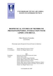
MI3<br />
FIGURE 1: Schematic drawing of a virus particle of TMV (left) and M13 (right) (from Butler, 1984, and Marvin. 1990, respectively). TMV: ?he<br />
protein subunits are arranged in a one start helix of 49 subunits per three tums. Also indicated ic part of the RNA molecule, which is bound with<br />
three nucleotides per protein subunit. M13:The a-helical coat proteins, drawn as gently-curved rods almost parallel to the virion axis, axe<br />
arranged in a five-start helix overlapping each other as scales of a fish.
Chapter 1<br />
General Introduction<br />
FIGURE 2: Possible scheme for nucleation and elongation in TMV assem ~bly (see text) (from Butler, 1984).<br />
accumulates in the chloroplasts and interacts with the<br />
light-harvesting photosystems (especially PSI I) in<br />
thylakoids, giving rise to the disease phenotype after<br />
which the virus is named (Reinero and Beachy, 1989).<br />
TMV particles have the shape of a rod with a length<br />
of 300 nm and an outerdiameter of 18 nm (Fig. la). The<br />
protein coat is formed by 2,130 subunits (M=17,500 Da)<br />
arranged in a helix with 49 subunits per three turns. The<br />
structure of the RNA molecule, which consists of 6,395<br />
nucleotides, has been well determined. It is buried within<br />
the coat between layers of subunits, following the protein<br />
helix with three nucleotides per protein subunit and the<br />
phosphates at a radius of - 4 nm (Stubbs et d., 1977).<br />
Various sorts of binding between RNA and protein<br />
subunits inside TMV have been suggested: electrostatic<br />
interactions between phosphodiesters and arginine<br />
residues, hydrophobic interactions between the nucleotide<br />
bases and the left radial a-helix of the subunits, and<br />
hydrogen bridges (Stubbs and Stauffacher, 1981). As a<br />
consequence of the strong correlation between the<br />
geometry of the protein coat and the encapsulated RNA,<br />
three types of phosphates can be distinguished in the<br />
nucleic acid backbone. One of these three phosphate types<br />
has a relatively high level of disorder (Cross et al.,<br />
1983a). Indeed, in RNA-protein interaction models for<br />
TMV, two phosphates are generally interacting closely<br />
with arginine residues, whereas no arginine is close to a<br />
third one (see Appendix).<br />
Dissociated coat subunits and RNA of TMV<br />
spontaneously reassemble under suitable conditions into<br />
virus particles (Fraenkl-Conrat and Williams, 1955).<br />
Numerous in vitro studies have resulted in a detailed<br />
model for TMV self-assembly in vivo (Fig. 2). At<br />
physiological pH, TMV coat proteins form disc-shaped<br />
aggregates with two ring-like layers of 17 proteins each<br />
(Butler and Klug, 1978). The subunits in the protein disc<br />
largely have a well-defined structure including four a-<br />
helices connected by a strip of P-sheet (Bloomer et d.,<br />
1978). A stretch of 24 amino residues, however, is<br />
highly mobile (Jardetzky et al., 1978; de Wit et al.,<br />
1979), forming a flexible loop located within a radius of<br />
4.0 nm in the central core of the protein disc. The<br />
assembly of a virion is initiated by selective interaction<br />
of a protein disc with a specific part of the viral RNA<br />
molecule, the origin-of-assembly sequence (OAS). The<br />
uncoated OAS region probably forms a RNA hairpin,<br />
which can insert into the central hole of the protein disc.<br />
The loop of the hairpin then intercalates between the two<br />
layers of the protein disc. Among other nucleoprotein<br />
interactions, the residues kg90 and Arg92 in the flexible<br />
loops of the protein units make salt-bridges with the<br />
RNA phosphates. As a result, the flexible loops of the<br />
protein units in the disc become immobilized forming the
Chapter 1<br />
General Introduction<br />
so-called V-columns ("V" for vertical) (Stubbs et al.,<br />
1977). The close interactions between V-columns of<br />
adjoining subunits probably force the disc to transform<br />
into a helix of two turns, the so-called lock washer. At<br />
this stage, both the 3' and 5'-RNA tail protrude from the<br />
same side of the lock washer. A second protein disk<br />
approaches from the opposite side and interacts with the<br />
RNA. It pulls up the 5'-tail and transforms into a lock<br />
washer, as well. Additional disks then add to the<br />
nucleoprotein complex in a similar way, and the complex<br />
elongates to a specific length. The lock washers stack on<br />
top of each other, forming a continuous protein helix of<br />
several turns. This elongation process is repeated until<br />
the 5'-tail of the RNA has been completely encapsulated.<br />
Meanwhile, the 3'-tail becomes covered much more<br />
slowly, perhaps by the binding of smaller aggregates of<br />
coat proteins (Butler, 1979).<br />
Filamentous bacteriophages: life cycle<br />
and structure<br />
Filamentous bacteriophages (Inovirus) form a large<br />
group of related virus strains with similar morphology<br />
and life cycle (Rasched and Oberer, 1986). They infect<br />
gram-negative bacteria by adsorbing to the tip of specific<br />
bacterial pili. Some of the strains, like M13, fd, fl, Ike,<br />
12-2 and Ifl, share Eschen'chia coli as a host, but differ in<br />
the specificity for its pili. For instance, M13, fd and fl<br />
infect E. coli via its F-pili. Other examples are Pfl, Pf2<br />
and Pf3, which infect Pseudomonas aeruginosa, and Xf, a<br />
virus of the plant pathogen Xanthomonas oryzae. Upon<br />
adsorption, the protein coat of the Inovirus virion<br />
dissolves into the inner cell membrane (Marvin, 1989)'<br />
and the circular, single-stranded DNA consisting of<br />
- 6500 nucleotides (* 10%) (Day et al., 1988) enters the<br />
host cell, while being converted into the double-stranded<br />
replicative form. This DNA synthesis probably drives the<br />
disassembly of the virion (Marco et al., 1974). Inside the<br />
host cell, new single-stranded DNA is synthesized off the<br />
replicative form by the "rolling circle" mechanism<br />
(Gilbert and Dressier, 1968). The new progeny viral DNA<br />
is covered by the DNA-binding protein g5p'encoded by<br />
the viral gene-5, resulting in an intra-cellular rod-shaped<br />
nucleoprotein complex, morphologically similar to the<br />
virion outside the cell, but less stable. In contrast to<br />
most bacterial viruses, which are released from the host<br />
by cell lysis, the filamentous bacteriophages are<br />
continuously assembled and extruded through the bacterial<br />
membrane without killing the host cell. During this<br />
phage extrusion, g5p is exchanged by the major coat<br />
protein encoded by the viral gene 8, also called gene-8<br />
protein (g8p). The negative charge of the headgroups of<br />
the lipid molecules in the bacterial membrane may play a<br />
role in the release of g5p (Butler, 1979). A model for<br />
assembly of the virion at membrane adhesions has been<br />
suggested on the basis of the "telescoping" behaviour of<br />
the virions in certain organic solutions in vitro (Marvin,<br />
1989).<br />
Outside the host cell, Inovirus exists as rod-shaped<br />
nucleoprotein particles with a diameter of - 6 to 9 nm<br />
and a length between - 700 and 2000 nm, depending on<br />
the strain and relative humidity (Day et al., 1988; Dunker<br />
et al., 1974). Their protein coat mainly consists of<br />
several thousand (2000 - 7000) copies of g8p, a protein<br />
of - 50 amino acids (Day et al., 1988). Although the<br />
actual amino acid sequence of g8p differs among various<br />
Inovirus strains, its geometry is essentially a slightly<br />
curved a-helix for all of them. Strong homology also<br />
exists at the level of charge distribution along the<br />
protein: there is a collection of acidic and basic residues<br />
near the N-terminus and C-terminus, respectively, and a<br />
hydrophopic stretch between the two charged ends.<br />
Subunits in the protein coat of Inovirus virions are<br />
oriented with their helical axis roughly parallel to the<br />
virion axis, thereby partly overlapping one another as<br />
scales of a fish (Fig. lb) (Marvin, 1990). They form a<br />
tubular protein shell with the acidic N-termini at the<br />
outside and the basic C- termini at the inside of the tube.<br />
X-ray diffraction studies have established two different<br />
classes on the basis of the protein coat structure. The<br />
symmetry of the protein coat of Class I phages, which<br />
include the strains Ff (fd, fl and M13), If1 and Ike,<br />
involves a 5-fold rotation axis combined with a 2-fold<br />
screw axis of pitch 32 A (Fig. lb) (Banner et al., 1981;<br />
Marvin, 1990; Marvin et al., 1994). In Class 11 phages<br />
(Pfl, Pf3 and Xf), the subunits of the protein coat are<br />
arranged according to a one-start helix with 15 A pitch<br />
and 22 units per five turns (Marvin et al., 1974; Marvin,<br />
1990). Protein products of other genes are located at the<br />
two ends of the virion. The virion end, which emerges<br />
first from the host cell, contains the gene-7 and gene-9<br />
proteins, without which virus particles are hardly formed<br />
(Russel, 1991). The four or five copies of gene-3 protein<br />
and gene4 protein at the other virion end, are required,<br />
respectively, for adsorption and penetration, and for<br />
locking the tube into place (Russel, 1991).<br />
In contrast to the structure of the protein coat, little is<br />
known about the geometry of the DNA molecule<br />
encapsulated in the virion, other than that the DNA<br />
molecule seems to be located inside the virion within a<br />
core of less than 2.5 nm wide (Banner et al., 1981;<br />
Russel, 1991). where it is immobilized by interactions<br />
with the protein coat (DiVerdi and Opella, 1981b; Cross<br />
et al., 1983b). For Class I viruses, these nucleoprotein<br />
interactions are predominantly of a electrostatic, nonspecific<br />
nature. Each g8p subunit has four positively<br />
charged residues at conserved positions in the C-terminal<br />
facing the inside of the protein shell which are able to<br />
neuaalize the negative charges of the phosphodiesters in<br />
the encapsulated DNA. Replacement of one positively<br />
charged residue (Lys48) by a neutral residue results in<br />
mutant virions that are 33% longer than wild-type virions<br />
(Hunter et al., 1987). There are no indications for any<br />
interaction between aromatic g8p residues and the bases<br />
of the encapsulated DNA. In Ff-virions, the nucleotides<br />
interact among themselves by base stacking ay, 1973),<br />
but the estimated interbase distance of 3.4 b" is difficult<br />
to reconcile with the mean axial rise of 2.7 A per<br />
nucleotide expected for homogeneous distribution of<br />
DNA along the inside of the protein coat, as commonly<br />
assumed (Day et al., 1979; Banner et al., 1981).<br />
Although nucleoside sugar pucker and glycosyl torsion<br />
are similar as for A-DNA, only 20% of the viral DNA at<br />
most seems to have the regular phosphodiester backbone<br />
found in A-DNA (Thomas et al., 1988). In fact, the DNA<br />
backbone mainly has phosphodiesters with B-type<br />
conformation (Fritzsche et al., I981), or may even be
Chapter 1<br />
General Introduction<br />
largely disordered (Cross et al., 1983b). Based on the<br />
generally non-integral ratio between the number of<br />
subunits and nucleotides, e.g, 1 : 2.4 for Ff-viruses,<br />
helical double-stranded DNA models have been suggested<br />
for Class-I viruses with the bases directed inward, but<br />
not necessarily forming (canonical) base-pairs. In these<br />
models, the symmetry of the DNA-helix and the protein<br />
coat differ, so that consecutive phosphodiesters in the<br />
nucleic acid backbone interact differently with the protein<br />
coat. Such complex phosphodiester inhomogeneity could<br />
perhaps explain the lack of experimental evidence for any<br />
regular geometry of the encapsulated DNA molecule. A<br />
similar DNA-model with the bases facing inward has also<br />
been proposed for the Class-I I virus Xf. For Pfl and<br />
Pf3, other members of Class-I I, "bases-out" models<br />
have been suggested with the nucleotide bases of the<br />
encapsulated DNA interacting with the tyrosine residues<br />
of the coat protein subunits (Day et al., 1979; Kosaikis<br />
et al., 1994). In Pfl, the DNA structure would be<br />
sufficiently open, for lysine and arginine side chains to<br />
reach into the virion core and neutralize the negative<br />
charge of the nucleic acid phosphates located at a 2.5-81<br />
distance from the virion axis (Liu and Day, 1994; Day et<br />
al., 1988; Day et al., 1979). In Pf3, the negative charge<br />
of the DNA backbone is perhaps balanced by the presence<br />
of metal cations, like Ca2+ or Mg2+, in the virion core.<br />
In this thesis, we have studied bacteriophage M13, wellknown<br />
for its use as a DNA-packaging vehicle in<br />
biochemistry, as an example for Inovirus in general, and<br />
Ff-viruses, more specifically.<br />
3lP <strong>NMR</strong> studies of nucleic acids and<br />
nucleoprotein complexes<br />
Phosphorus (31P) nuclear magnetic resonance (<strong>NMR</strong>)<br />
spectroscopy is a powerful technique for obtaining<br />
information about structure and dynamics of nucleic acids<br />
and nucleic acid complexes in solution or gel. At present,<br />
it is commonly accepted that conformational<br />
heterogeneity exists in nucleic acid structures. Structure<br />
variations in long DNA duplexes have largely been<br />
established by x-ray crystallography. <strong>NMR</strong> spectroscopy<br />
has especially provided detailed knowledge about local<br />
structures in oligonucleotides, such as e.g. hairpins and<br />
pseudo-knots. Novel two-dimensional (2D) and threedimensional<br />
(3D) 1H/31P <strong>NMR</strong> methods have made it<br />
possible to assign the 3lP <strong>NMR</strong> spectrum of<br />
oligonucleotides with a length up to 14 basepairs<br />
(Schrdder et d., 1987), so that specific information on<br />
the conformation of each of the phosphodiesters in the<br />
nucleic acid backbone can be obtained in these molecules.<br />
Relatively large variations in phosphodiester<br />
conformation are found (van de Ven and Hilbers, 1988).<br />
Moreover, molecular dynamics calculations suggest that<br />
the structures arising from x-ray and <strong>NMR</strong> studies still<br />
represent time-averaged structures only and that an even<br />
larger variety of conformations temporarily exist at a<br />
time-scale of picoseconds (Scheek, 1994).<br />
Because the electron distribution surrounding a<br />
phosphorus nucleus in a nucleic acid molecule is far from<br />
spherical, its 3lP chemical shift measured by <strong>NMR</strong><br />
spectroscopy, in principle, depends on the molecular<br />
orientation with respect to the external magnetic field.<br />
95 105 5.0 3.5 2.0<br />
0-P-0 bondangle (O)<br />
3i P chemical shift<br />
(PPW<br />
FIGURE 3: Dependence of 3 1 chemical ~ shift on conformational and stereo-electronic factors. a: correlation plot for 3 1 chemical ~ shift of<br />
$osphodiesters versus the smallest 0-PO bondangle. b: correlation between the const~~~t JH3.-P for scalar coupling between 3 1 and ~ 1~31 and<br />
IP chemical shift in oligonucleotides. The solid curve in the figure represents the theoretically derived Karplus relation between JH3I-p and the<br />
CO-torsion angle E, which in turn, in a large variety of structures is correlated to the PO-torsionangle {. By variable scaling of the coupled E and<br />
I; axes, the Karplus curve can be made to fit to the data points. In this way, a coxrelation between the chemical shift and the two coupled<br />
tonionangles is established. c: definition of the various torsionangles (from Gorenstein, 1994).
Chapter 1<br />
General Introduction<br />
200 ppm<br />
FIGURE 4: 61-MHz31p <strong>NMR</strong> spectra of B-form DNA at various temperr<br />
This chemical shift anisotropy (CSA) can be described in<br />
terms of a shielding tensor with three principal<br />
components all, a22 and 033. In (dilute) solutions,<br />
however, fast tumbling of oligonucleotides and segmental<br />
motion of larger nucleic acid molecules tend to average<br />
the shift, leaving only the isotropic chemical shift ais0<br />
to be measured. A number of attempts has been made to<br />
calculate the 31P chemical shift tensor and especially<br />
aiso, for phosphodiesters in nucleic acids and model<br />
compounds (Prado et al., 1979; Giessner-Prettre et al.,<br />
1984). These calculations point out that many factors,<br />
including electron negativity, bondangle and n-electron<br />
overlap, can affect the 31P chemical shift, indicating that<br />
no single factor can unequivocally explain an observed<br />
range or change of 31P shift. Empirically, a correlation<br />
between 31P chemical shifts and the smallest 0-P-0<br />
angle has been found for a wide variety of<br />
alkylphosphates and oligonucleotides. At increasing<br />
bondangle between 95' and 108', oiso increases by as<br />
much as -35 ppm (Fig. 3a) (Gorenstein, 1981). At<br />
further increasing bondangle above 108'. a;,, decreases<br />
again (Gorenstein, 1994). Another correlation is<br />
experimentally observed between oiso and the scalar<br />
coupling constant JH~V-p for the three-bond coupling<br />
constant between H31 and P (Fig. 3b). Because JH~I-p can<br />
be interpreted in terms of the CO torsionangle E (Fig. 3c)<br />
via a Karplus relationship (Lankhorst et al., 1984), and a<br />
linear correlation between E and the PO torsionangle 6 is<br />
found in a variety of duplex structures (Gorenstein,<br />
1994), the correlation between Oiso and JH~-p actually<br />
reflects the influence of the stereo-electronic factors on<br />
the 31P chemical shift. This semi-empirical correlation<br />
between oiso and the coupled torsionangles E and C,<br />
should be distinguished from the separate correlations<br />
between ais0 and the P-0 torsionangles 6 and a, and<br />
between oiso and the C-0 torsionangles E and P,<br />
previously suggested on a theoretical basis (Gorenstein,<br />
1981; Giessner-Prettre et al., 1984). Environmental<br />
effects on 31P chemical shifts are generally smaller than<br />
the intrinsic structural effects. A shift change of about 3<br />
ppm is observed upon changing the solvent from 100%<br />
H20 to 70% DMSO (Lerner and Kearns, 1980), or the<br />
salt concentration from 8 to 5 M (Costello et al., 1976).<br />
The bases with their ring-current in the double helical<br />
nucleic acids have only small effect on the chemical shift<br />
(c 0.1 ppm) (Gorenstein et al., 1988).<br />
Because the range of 31P chemical shifts for<br />
individual phosphates in duplex fragments in solution is<br />
typically c 0.9 ppm, resonance lines can generally not be<br />
resolved individually in 3lP <strong>NMR</strong> spectra of nucleic acids<br />
longer than 100 basepairs. For this reason, most of the<br />
ltures indicating different mobility (from DiVedi, and Opella. 1981a.).<br />
3lP <strong>NMR</strong> work on DNA fragments with a length of a<br />
few hundred basepairs has concentrated on 3lP relaxation.<br />
The measured longitudinal relaxation time (TI),<br />
transversal relaxation time (T2), and nuclear overhauser<br />
effect (NOE) are usually interpreted in terms of models,<br />
which necessarily represent simplifications as compared<br />
to the complicated nature of the flexible DNA. A more<br />
realistic description of the complex backbone dynamics<br />
would require more variables to be fitted, than justified by<br />
the amount and quality of the experimental data. Some<br />
authors, for instance, assign all relaxation effects to<br />
dipolar relaxation between 31P and the nearest 3', 5' and<br />
5"-protons, assuming the 31P - 1H internuclear distances<br />
to be invariant and neglecting the relaxation by the 31P<br />
chemical shift anisotropy (Hogan and Jardetzky, 1980;<br />
Bolton and James, 1979). Internal motion of the 31P - 1H<br />
internuclear vector has been simplified to two-site<br />
exchange (Hogan and Jardetzky, 1980), jumping (Keepers<br />
and James, 1982), rotation about a single axis (Bolton<br />
and James, 1979) and wobbling (Lipari and Szabo, 1981).<br />
Long range bending of the nucleic acids is sometimes<br />
approximately described as isotropic motion (Bolton and<br />
James, 1979; Opella et al., 1981). Relaxation data are<br />
also analyzed by use of a rigid-rod model for the DNA<br />
duplex fragment, regarded as a stiff helix (Shindo, 1980).<br />
Other authors incorporate collective torsional motion<br />
along the DNA duplex into their model (Allison et al.,<br />
1982). The large variety of models in the literature,<br />
illustrates the lack of unanimity about the details of the<br />
motions that mainly cause the observed 3lP <strong>NMR</strong><br />
relaxation. Several assumptions about the local molecular<br />
structure and the predominant relaxation mechanism must<br />
usually be made to interpret relaxation in a system of<br />
dipolar coupled spins. For 31P <strong>NMR</strong> relaxation, CSArelaxation<br />
should also be taken into account. Given the<br />
small amount of experimental data, different models,<br />
varying in nature between "model-freely" simple and<br />
realistically detailed, are equally acceptable. Additional<br />
information is necessary to decide between them.<br />
In contrast to the 31P <strong>NMR</strong> spectra of DNA duplex<br />
fragments in solution, 31P <strong>NMR</strong> spectra of dehydrated<br />
DNA or large nucleoprotein complexes, such as viruses,<br />
potentially offer a wealth of additional mobility<br />
information. 31P <strong>NMR</strong> spectra of concentrated gels of<br />
these large systems generally contain a single broad<br />
resonance line reflecting the average 31P chemical shift<br />
anisotropy (CSA) typical for phosphates in DNA or<br />
RNA (Fig. 4) (DiVerdi and Opella, 1981a; DiVerdi and<br />
Opella, 1981b). In most cases, the observed lineshape<br />
also shows signs of motional narrowing and therefore<br />
contains information about the underlying motion. It is<br />
important to note, that motional distortion of the 31P
Chapter 1<br />
General Introduction<br />
lineshapes not merely represents yet another single-valued<br />
input for motional analysis, just like T1 and T2 values<br />
measured for DNA fragments in solution. Partially<br />
averaged 31P <strong>NMR</strong> lineshapes are typically 200 ppm<br />
wide and are therefore defined in the spectrum by a series<br />
of data points, each of which carries information about<br />
the motions involved. Given the large variety of<br />
relaxation models for DNA fragments in solution and the<br />
lack of sufficient experimental data to decide between<br />
them, it is quite surprising that until this thesis, no<br />
studies had been published about quantitatively analyzing<br />
or simulating 31P <strong>NMR</strong> lineshapes of concentrated<br />
nucleic acid or nucleoprotein gels, except in a superficial<br />
way (Fig. 5) (Tsang and Opella, 1986). Most authors<br />
restrict themselves to qualitative remarks about the<br />
motion involved (DiVerdi and Opella, 1981b; Mai et al.,<br />
1983). It should be mentioned in this respect, that the<br />
literature does contain publications about 2H <strong>NMR</strong><br />
lineshape simulations for 2H-labeled nucleic acids<br />
(Brandes and Kearns, 1988; Alam and Drobny, 1991) and<br />
31P <strong>NMR</strong> lineshape simulations for phospholipids<br />
(Dufourc et al., 1992).<br />
concentrated solutions or viscous gels, such as TBSV,<br />
the rod-shaped tobacco mosaic virus (TMV) and the<br />
bacteriophages Pfl and fd (Munowitz et al., 1980; Cross<br />
et al., 1983a; Tsang and Opella, 1986; DiVerdi and<br />
Opella, 1981b; Cross et al., 1983b). From qualitative<br />
lineshape analyses, conclusions were drawn about<br />
motional frequencies and amplitudes, which agree with<br />
the general picture evolved from dilute solution studies:<br />
nucleic acids inside virions do not undergo large<br />
amplitude motions at frequencies higher than 104 Hz and<br />
motional lineshape effects observed in dilute solutions<br />
can be explained by overall motion of the viral particle as<br />
a rigid body.<br />
i 00 pprn<br />
FIGURE 6: 31~<br />
<strong>NMR</strong> spectra of solid fd (left) and 10% (wt/wt) TMV<br />
(right). upper: nonspinning; lower: magic angle spmmg (from<br />
DiVerdi and Opella, 1981b, and Cmss et al., 1983b).<br />
static powder pattern<br />
100 pprn<br />
rotationally averaged<br />
powder pattern<br />
FIGURE 5: 3 1 <strong>NMR</strong> ~ lineshape of bacteriophage Pfl at different pH<br />
values. The theoretical lineshapes were calculated, assumlng<br />
either the complete absence of motion, or complete averaging by<br />
fast motion of the rod-shaped virions about their length axis (fmm<br />
Tsang and Opella, 1986).<br />
In large nucleoprotein complexes, such as viruses,<br />
31P nuclei represent natural <strong>NMR</strong> labels for studying<br />
structural and dynamic properties of the nucleic acid<br />
backbone selectively, even when the complex mainly<br />
consists of proteins. Indeed, during the last fifteen years<br />
31P <strong>NMR</strong> studies of various viruses have appeared in the<br />
literature. In one class of studies, rapidly tumbling,<br />
mostly spherical viruses, such as the plant viruses alfalfa<br />
mosaic virus (AlMV), cowpea mosaic virus (CpMV),<br />
tomato bushy stunt virus (TBSV) and the bacteriophages<br />
QP and MS2, have been investigated in dilute solution by<br />
use of high resolution <strong>NMR</strong> spectroscopy (Kan et al.,<br />
1987; Virudachalam et al., 1985; Munowitz et al., 1980;<br />
Bolton et al., 1982). In these studies conclusions about<br />
the dynamic behaviour of the nucleic acids inside the<br />
virions have been drawn from linewidths and relaxation<br />
times. In other studies, solid <strong>state</strong> <strong>NMR</strong> techniques have<br />
been used to record 3lP spectra of viruses in more<br />
CSA-broadening tends to mask the small differences<br />
among the phosphates of the nucleic acid encapsulated in<br />
a slowly tumbling virus (Fig. 6). Such phosphodiester<br />
inhomogeneity, indicating e.g. inequivalence among<br />
binding sites, is best studied using magic angle spinning<br />
(MAS) <strong>NMR</strong> spectroscopy. MAS breaks up the broad<br />
31P <strong>NMR</strong> powder lineshape into a sharp centerband at the<br />
isotropic chemical shift position flanked by rotational<br />
sidebands (Herzfeld and Berger, 1980). Because for<br />
phosphodiester compounds, the width of the centerband<br />
and the sidebands is typically a few ppm, resonances are<br />
more easily resolved and resolved sideband patterns may<br />
be assigned to specific phosphates on the basis of their<br />
conformation (see above) (Gorenstein, 1994). 31P MAS<br />
<strong>NMR</strong> spectra of TMV solutions (Cross et al., 1983a) and<br />
dried TMV pellets (Hemminga et al., 1987) show two<br />
resolved sideband patterns with an overall intensity ratio<br />
of approximately 2, which have been assigned by<br />
comparing torsion angle values for the three types of<br />
phosphodiesters in TMV (Hemminga et al., 1987). MAS<br />
<strong>NMR</strong> spectra of bacteriophage fd, which is closely related<br />
to M13, only contain a single, broad centerband flanked<br />
by sidebands (DiVerdi and Opella, 1981b), indicating that<br />
a continuous distribution of phosphodiester conformations<br />
is present in the phage, rather than a distinguishable<br />
few (Fig. 6).
Chapter 1<br />
General Introduction<br />
Research history and environment of<br />
the project<br />
During the past two decades, the molecularbiophysical<br />
aspects of viruses have been studied at the<br />
Department of Molecular Physics. A number of workers<br />
have participated in this research line. Jan de Wit<br />
investigated TMV and its coat protein using 1H and 13C<br />
<strong>NMR</strong> spectroscopy (de Wit, 1978). He concluded that a<br />
specific part of the coat protein, which is mobile in<br />
dissociated protein subunits and in empty capsids,<br />
becomes immobilized by the interaction with RNA.<br />
Indeed, transformation of the so-called flexible loop into<br />
the V-column seems to be an essential step in the<br />
assembly of TMV (see Fig. 3 and remarks about TMV<br />
above). Gert Vriend studied the interaction of the coat<br />
protein of cowpea chlorotic mottle virus (CCMV) with<br />
RNA using 1H and 13C <strong>NMR</strong>, and ESR spectroscopy<br />
(Vriend, 1983). He developed a tentative model for the<br />
binding of protein dimers to RNA. In this model, the<br />
conformation of the N-terminal protein part changes upon<br />
interacting with the RNA from a flexible random-coil<br />
conformation into an a-helical conformation, thereby<br />
pulling the coat protein and RNA towards each other. The<br />
model was tested by Marinette van der Graaf (van der<br />
Graaf, 1992), who investigated the conformation of a<br />
synthetic oligopeptide, containing the first 25 amino<br />
acids in the N-terminal part, in absence and presence of<br />
oligonucleotides and oligophosphates. For this purpose,<br />
she used UV/Vis and two-dimensional 1H <strong>NMR</strong><br />
spectroscopy and carried out molecular dynamics<br />
calculations. Her conclusions confirmed and refined the<br />
"snatch-pull" model.<br />
Klaas Pieter Datema employed various spectroscopic<br />
techniques (circular dichroism, time-resolved fluorescence,<br />
ESR, 31P <strong>NMR</strong>, and 2H <strong>NMR</strong>) to study the interaction<br />
of TMV, CCMV, BMV, SBMV and M13, as well as<br />
their coat proteins, with membranes (Datema, 1987). He<br />
concluded that M13 coat protein readily inserts into lipid<br />
bilayers, where it forms aggregates, whereas TMV coat<br />
protein only interacts with the negatively charged<br />
phospholipid headgroups without actually being inserted<br />
into the membrane. Johan Sanders continued this research<br />
(Sanders, 1992). He established the conformation of two<br />
forms of M13 coat protein in lipid bilayers using several<br />
spectroscopic techniques (circular dichroism, raman,<br />
fourier transform infra-red, ESR, 31P <strong>NMR</strong>, and 2H<br />
<strong>NMR</strong>) and molecular dynamics calculations. He<br />
concluded that one of the two proteins forms was very<br />
similar to the coat protein in M13 virion and was<br />
therefore likely to be present in the membrane of infected<br />
E. coli. His conclusions were supported by the work of<br />
Ruud Spruijt and Cor Wolfs (Spruijt et al., 1989).<br />
In 1981, solid-<strong>state</strong> <strong>NMR</strong> spectroscopy was<br />
introduced to study TMV by Marcus Hemminga and<br />
Wiebren Veeman (Hemminga et al., 1981). This line of<br />
research was continued in 1985 by Jaap Kriise and Rolf<br />
Lamerichs who employed solid-<strong>state</strong> MAS 13C and 31P<br />
<strong>NMR</strong> spectroscopy to study lyophilized or air-dried<br />
pellets of intact TMV and CCMV virions (Hemminga et<br />
al., 1987). The results of these pilot-experiments gave<br />
rise to the project described in this thesis, which started<br />
in December 1987.<br />
Course of the project and outline of<br />
the thesis<br />
The objective of the project presented in this thesis<br />
was in the first place to obtain information about the<br />
protein-nucleic acid interaction in intact virions of M13<br />
and TMV using solid-<strong>state</strong> 3lP <strong>NMR</strong> spectroscopy. At<br />
the project start, the experience within the Department of<br />
Molecular Physics with this type of spectroscopy was<br />
still limited. A secondary objective was therefore to find<br />
out the practical use of solid-<strong>state</strong> <strong>NMR</strong> spectroscopy for<br />
studying intact viruses. It was decided to mainly focus on<br />
31P <strong>NMR</strong>, because phosphates are probably involved in<br />
the protein-nucleic acid interactions and 31P nuclei are<br />
sensitive probes for studying this interactions selectively.<br />
Gels of M13 and TMV are difficult samples as compared<br />
to polymers or crystaline powders, which are more often<br />
studied by use of solid-<strong>state</strong> <strong>NMR</strong> spectroscopy. Under<br />
"physiological" conditions (0-40 OC, > 50% (w/w)<br />
water), the signal-to-noise ratio is low (e.g. 40% M13 w<br />
-4 mglml phage DNA). The high water content in the<br />
<strong>NMR</strong> samples lowers the Q-factor of the probe at the 1Hfrequency,<br />
so that maximal proton power must be used<br />
with the risk of probe sparking, In addition, 1H - 31P<br />
cross-polarization is relatively ineffective, stable magic<br />
angle spinning is difficult to achieve, and the<br />
nonspinning T2 is short (-0.5 ms). For this reason,<br />
many of the "fancy" <strong>NMR</strong> techniques appearing in the<br />
literature, could not be successfully applied to the<br />
viruses, although some of them were tried. It may be<br />
illustrative to briefly present some of these unsuccessful<br />
ideas and pilot-experiments.<br />
For instance, to be able to detect the weak dipolar<br />
couplings between 31P and its nearest protons Hg*, Hy<br />
and Hyt (-2.6 A in DNA, < 2.5 &) and protons in the<br />
presumed salt-bridge between phosphates and basic<br />
residues (-3 A in TMV, < 1.5 kHz), we have tried to<br />
suppress the much stronger 1H homonuclear coupling<br />
using off-resonance and multiple-pulse decoupling<br />
(Slichter, 1978). However, preliminary results were<br />
insufficiently convincing to continue this line of<br />
research. An idea to measure the weak 31P - 15N dipolar<br />
coupling in the presumed salt-bridges between phosphates<br />
and 1sN-labeled lysine residues in M13 could not be<br />
carried out because isotope-labeling was still in an early<br />
stage in our lab (scrambling problems occurred) and the<br />
necessary 1H - 31P - 15N triple-resonance equipment was<br />
not available. Besides, the small size of the 31P - 15N<br />
coupling, presumed to be < 400 Hz on the basis of the<br />
3-A distance suggested for TMV, was not very<br />
motivating. If nonspinning separated local field spectra<br />
would be recorded to measure 31P - 15N dipolar coupling<br />
(DiVerdi and Opella, 1982), T2 broadening would<br />
probably dominate over coupling effects. In onedimensional<br />
rotary resonance recoupling MAS spectra<br />
(Levitt et al., 1988), the coupling effect would likely be<br />
some minor extra broadening of the centerband and the<br />
sidebands already 700-1200 Hz wide at a 31P resonance<br />
frequency of 202.5 MHz.<br />
An attempt was also made to investigate the<br />
orientational distribution of phosphodiesters within M13<br />
using a rotor-synchronized 2D MAS <strong>NMR</strong> method<br />
introduced for oriented polymers (Harbison, 1986). For
Chapter 1<br />
General Introduction<br />
this purpose, we tried to freeze oriented M13 solutions in<br />
the (nonspinning) MAS rotor. As carbonyl groups are<br />
strongly oriented within M13, the carbonyl resonance<br />
lineshape in the 13C <strong>NMR</strong> spectrum of M13 is an<br />
indicator for the orientation of the virions with respect to<br />
the magnetic field. Indeed, a narrow carbonyl lineshape<br />
showed up in spectra of dilute M13 solutions, indicating<br />
orientation (if not motion) of the virus particles in the<br />
10-T field of the AM500 spectrometer. Unfortunately,<br />
upon freezing, the carbonyl lineshape changed into the<br />
typical lineshape of an isotropic powder, reflecting the<br />
randomizing effect of the ice-formation. Attempts to use<br />
MAS 2D-exchange 31P <strong>NMR</strong> experiments (Kentgens,<br />
1987) to study slow motion in M13 gels were<br />
unsuccessful, because highly concentrated or frozen M13<br />
gels contained too little motion to cause meaningful offdiagonal<br />
intensity in the spectra, whereas more dilute gels<br />
at room temperature could not spin in a sufficiently<br />
stable manner.<br />
Thus, despite much effort, the 31P <strong>NMR</strong> results<br />
obtained for M13 and TMV during the first three years of<br />
the project were little spectacular and rather<br />
disappointing: featureless powder lineshapes in<br />
nonspinning 31P <strong>NMR</strong> spectra, different T2-values<br />
depending on the sample preparation, no resolution<br />
improvement in MAS spectra of 30% TMV as compared<br />
to earlier published spectra of air-dried TMV, and<br />
featureless lineshapes with various linewidths in MAS<br />
spectra of M13. A turning point was reached when a<br />
pattern was recognized: the featurelessness of the powder<br />
lineshapes and the sample preparation effects were<br />
actually highly indicative for the presence of 31P motion!<br />
Various models were developed to simulate the effect of<br />
different types of motion on 31P <strong>NMR</strong> spectra and<br />
transversal relaxation. The quantitative analysis of the<br />
experimental data by use of these models, is the main<br />
subject of this thesis.<br />
Chapter 2 describes the theoretical background of the<br />
three models that will be used in Chapter 3 to simulate<br />
the experimental data. An isotropic rotational diffusion<br />
model is set up for mobile nucleic acids that are loosely<br />
or partially bound to the protein coat. A rigid-rod model<br />
is worked out to represent the mobility of rigidly bound<br />
phosphates in a rod-shaped virion which rotates about its<br />
length axis. In addition, a combined diffusion model is<br />
presented, in which fast restricted nucleic acid backbone<br />
motions are superimposed on a slow rotation of the<br />
virion about its length axis.<br />
Chapter 3 compares the experimental 31P <strong>NMR</strong><br />
lineshapes and transversal relaxation decays with the<br />
outcome of the simulations by use of the three rotational<br />
diffusion models developed in Chapter 2. It is concluded<br />
that neither isotropic diffusion, nor (restricted) rigid-rod<br />
diffusion offers a consistent explanation for the<br />
experimental data. The combined diffusion model is<br />
successful for M13. For TMV, the model indicates that<br />
one of the three binding sites is more mobile than the<br />
other two.<br />
In Chapter 4, the combined diffusion model developed<br />
in Chapter 2 and tested in Chapter 3 is extrapolated for<br />
MAS experiments. Comparing theoretical and<br />
experimental MAS spectra, we conclude that backbone<br />
motions influence the sideband intensities as observed.<br />
Backbone motions also seem to cause the decrease of<br />
inhomogeneous linewidth in the MAS spectrum and the<br />
msversal relaxation measured at spinning rates of 4 kHz<br />
or higher. At spinning rates below 2 kHz, transversal<br />
relaxation is significantly faster. This effect is assigned to<br />
slow, overall rotation of the rod-shaped M13 - phage - about<br />
its length axis.<br />
Chapter 5 finally presents a detailed study of the slow<br />
overall motion of MI3 and TMV using two-dimensional<br />
exchange 3lP <strong>NMR</strong> spectroscopy. The combined<br />
diffusion model of Chapters 2 and 3 is now extended for<br />
this type of experiments. It is found that TMV undergoes<br />
much slower rotational diffusion than expected on the<br />
basis of the analysis in Chapter 3. For M13, the<br />
quantitative analysis indicates heterogeneity in the overall<br />
motion throughout the gel. The average overall mobility,<br />
however, is consistent with the outcome of the analysis<br />
in Chapter 3.<br />
REFERENCES<br />
Alam, T. M., and G. P. Drobny. 1991. <strong>Solid</strong>-<strong>state</strong> <strong>NMR</strong> studies of<br />
DNA structure and dynamics. Chem. Rev. 91:1545-1590.<br />
Allison, S. A,, J. H. Shibata, J. Wilcoxon and J. M. Schurr. 1982.<br />
<strong>NMR</strong> relaxation in DNA. I. The dbntribution of torsional<br />
deformation modes of the elastic filament. Biopolymcrs. 21:729-<br />
762.<br />
Banner, D. W., C. Nave, and D. A. Marvin. 1981. Structure of the<br />
rotein and DNA in fd filamenous bacterial virus. Nature.<br />
589:814-816.<br />
Bloomer, A. C., J. N. Champness, G. Bricogne, S. R., and A. Klug.<br />
1978. Protein disk of tobacco mosaic virus at 2.8 A resolution<br />
showing the interaction within and between the subunits. Nature.<br />
276:362-368.<br />
Bolton, .P. H., G. Clawson, V. J. Basus, and T. L. James. 1982.<br />
Comparison of ribonucleic acid rotein interactions in messenger<br />
ribonucleoprotcins, ribosomes, &2 virus, and Q$ virus examined<br />
via hosphorus-31 nuclear magnetic resonance relaxation.<br />
~iochmistr~. 21 :6073-6081.<br />
Bolton, P. H., and T. L. James. 1979. Molecular motions in RNA and<br />
DNA investigated by phos horus 31 and carbon-13 <strong>NMR</strong><br />
relaxation. J. Phw. Chem. 83:&59-3366.<br />
Brandes, R., and D. R. Kearns. 1988. 2~ <strong>NMR</strong> of DNA liquid<br />
crystals: structural and dynamical aspects. J. Phys. Chem.<br />
926836-6841.<br />
Burgess, J., R. Motoyoshi, and E. N. Fleming. 1973a. Effect of poly-Lomithine<br />
on isolated tobacco mesophyl rotoplasts: evidence<br />
against stimulated pinocytosis. Planto. 111:lb-208<br />
Burgess, J., R. Motoyoshi, and E. N. Fleming. 1973b. The mechanism<br />
of infection of plant protoplasts by viruses. Planla. 112:323-332<br />
Butler, P. J. G. 1979. Assembly of regular viruses. In Chemistry of<br />
Macromolecules IIB. R. E. Offord, Eds. University Park Press,<br />
Baltimore. 205-237.<br />
Butler, P. J. G. 1984. The current icture of the structure and<br />
assembly of tobacco mosaic virus. Jf)~en.<br />
Virol. 65:253-279.<br />
Butler, P. J. G., and A. Klug. 1978. The assembly of a virus. Sci.<br />
Amer. 239:6249.<br />
Costello, A. J, R., T, Glonek, and J. R. van Wazer. 1976. Phosphorus-<br />
31 chemical shift variations with countercation and ionic strength<br />
for the various ethyl os hates. J. Inorg. Chem. Soc. 15:972-974.<br />
Cross, 7'. A., S. J. Opt, 8. Stubbs, and D. L. D. Caspar. 1983a.<br />
Phosphorus-31 nuclear magnetic resonance of the RNA in<br />
tobacco mosaic virus. J. Mol. Biol. 170:1037-1043.<br />
Cross, T. A., P. Tsang, and S. J. Opella. 1983b. Comparison of protein<br />
and deoxyribonucleic acid backbone structures in fd and Pfl<br />
bacteriophages. B iochemistry. 22721 -726.<br />
Datema, K. P. 1987. Virus-membrane interactions. (Ph.D. Thesis).<br />
A ricultural University <strong>Wageningen</strong>, The Netherlands.<br />
Day, & A. 1973. Circular dichroism and ultraviolet absorption of a<br />
deoxyribonucleic acid binding protein of filamentous<br />
bacteriophage. Biochemistry. 125329-5339.<br />
Day, L. A,, C. J. Manec, S. A. Reisberg, and A. Casadevall. 1988.<br />
DNA packing in filamentous bacteriophages. Ann. Rev. Bwphys.<br />
Biophys. Chem. 17:509-539.<br />
Day, L. A., R. Wiseman L., and C. J. Manec. 1979. Structure models<br />
for DNA in filamentous viruses with phosphates near the center.<br />
Nucleic Acids Res. 7: 1393-1403.<br />
de Wit, J. L. 1978. <strong>NMR</strong> of TMV. (Ph.D. Thesis). Agricultural<br />
University <strong>Wageningen</strong>, The Netherlands.
Chapter 1<br />
General Introduction<br />
de Wit, J. L., N. C. M. Alma, T. Trienekens, M. A. Hemminga, and T.<br />
J. Schaafsma. 1979. Nuclear magnetic resonance of tobacco<br />
mosaic virus. In Magnetic resonance and related phenomena<br />
(20th Ampere Congress). K. E. e. al., Eds. Springer - - Verlag,<br />
Berlin. 3%.<br />
DiVerdi, J. A., and S. J. Opella. 1981a. Dynamics of B-DNA in the<br />
solid <strong>state</strong>. J. Mol. Bwl. 149:307-311.<br />
DiVerdi, J. A,, and S. J. Opella. 1981b. Phos horus 31 nuclear<br />
magnetic resonance of fd vuus. Biochemistry. %:280-284.<br />
DiVerdi, J. A., and S. J. Opella. 1982. N-H bond lengths in DNA. J.<br />
Am. Chem. Soc. 104:1761-1762.<br />
Dufourc, E. J., C. Mayer, J. Stohrer, G. Althoff, and G. Kothe. 1992.<br />
Dynamics of phosphate head groups in biomembranes.<br />
Comprehensive analysis using phosphorus-31 nuclear magnetic<br />
resonance lineshape and relaxahon tune measurements. Biophys,<br />
J. 61:42-57.<br />
Dunker, A. K., R. D. Klausner, D. A. Marvin, and R L. Wiseman.<br />
1974. Filamentous bacterial viruses X. X-ray diffraction studies of<br />
theR4A roteinrnutan~J.Mol.Biol.81:115-117.<br />
Durham, A. C? H. 1978. The roles of small ions, especially calcium, in<br />
virus disassembly, take-over and transformation. Biomedicine.<br />
28:307-314.<br />
Fraenkl-Conrat, H., and R. C. Williams. 1955. Reconstitution of active<br />
tobacco mosaic virus from inactive protein and nucleic acid<br />
components. Proc. Natl. Acad. Sci. USA. 41:690695.<br />
Fritzsche, H., T. A. Cross, S. J. Opella, and N. R. Kallenbach. 1981.<br />
Structure and architecture of the bacterial virus fd. An infrared<br />
linear dichroism study. Biophys. Chem. 283-291.<br />
Giessner-Prettre, C., B. Pullman, F. R. Prado, D. M. Cheng, V. Iuomo,<br />
and P. 0. P. Ts'o. 1984. Contributions of the PO Ester and CO<br />
torsion angles of the phosphate group to 31P-nuclear magnetic<br />
shielding. Biopolymers. 23:377-388.<br />
Gilbert, W., and D. Dressler. 1968. DNA replication: the rolling circle<br />
model. In Cold Spring Harbor Symp. Quant. Biol. 33 (Replication<br />
of DNA in micm organisms):473-484. New York<br />
Goelet, P., G. P. Lomonossoff, P. J. P. Butler, M. E. Akam, M. J. Gait,<br />
and J. Kam. 1982. Nucleotide sequence of tobacco mosaic virus<br />
RNA. Proc. Natl. Acad. Sci. USA. 79:5818-5822.<br />
Gorenstein, D. G. 1981. Nucleotide conformational analysis by 31P<br />
nuclear magnetic resonance spectroscopy. Ann. Rev. Biophys.<br />
Bioeng. 10:355-386.<br />
Gorenstein, D. G. 1994. Conformation and dynamics of DNA and<br />
protein-DNA complexes by 31P <strong>NMR</strong>. Chem. Rev. 94:1315-1338.<br />
Gorenstein, D. G., S. A. Schrijder, J. M. Fu, J. T. Metz, V. A. Roontga,<br />
and C. R. Jones. 1988. Assignments of 3 1 <strong>NMR</strong> ~ resonances in<br />
oligodwxyribonucleotides: origin of sequence-specific varia$?ns<br />
in the deoxy phosphate backbone conformation and the "P<br />
chemical sMts of double helical nucleic acids. Biochemistry.<br />
27:7223-7237.<br />
Harbison, G. S. 1986. Two-dimensional magic-an le spinning <strong>NMR</strong><br />
of partially ordered systems. Chem. Phys. Lett. f2i128-134.<br />
Hemminga, M. A., P. A. De Jager, J. K~se, and R. M. J. N.<br />
Lamerichs. 1987. Magic-Angle-Spinning <strong>NMR</strong> on <strong>Solid</strong> Biological<br />
Systems. Analysis of the Origin of the Spectral Linewidths. J.<br />
Magn. Reson. 71 :446-460.<br />
Hemminga, M. A,, W. S. Veeman, H. W. M. Hilhorst, and T. J.<br />
Schaafsma. 1981. Magic angle spinning carbon-13 <strong>NMR</strong> of<br />
tobacco mosaic virus. Bbphys. J. 35436-470.<br />
Herzfeld, J., and A. E. Berger. 1980. Sideband intensities in <strong>NMR</strong><br />
s ctra of samples spinning at the magic angle. J. Chem. Phys.<br />
.p" 3:6021-6030.<br />
Hogan, M. E., and 0. J. Jardetzky. 1980. Internal motions in<br />
dwxyribonucleic acid 11. J. Am. Chem. Soc. 19:3460-3468.<br />
Hunter, G. J., D. H. Rowitch, and R. N. Perham. 1987. Interaction<br />
between DNA and coat protein in the structure and assembly of<br />
filamentous bacteriophage fd. Natwe. 327:252-254.<br />
Hwang, D.-J., I. M. Roberts, and T. M. A. Wilson. 1994. Assembly of<br />
tobacco mosaic virus and TMV-like seudovirun particles in<br />
Escherichia cdi. Arch. Virol. Suppl. 954%-558.<br />
Ishikawa, M., T. Meshi, F. Motoyoshi, N. Takamatsu, and Y. Okada.<br />
1986. In vitro mutagenesis of the putative replicase genes of<br />
tobacco mosaic virus. Nucleic Acid Res. 14:8291-8305.<br />
Jardetzky, O., K. Akasaka, D. Vogel, S. Morris, and K. C. Holmes.<br />
1978. Unusual segment flexibility in a region of tobacco mosaic<br />
virus protein. Nature. 273:564-566.<br />
Kan, J. H., A. F. M. Cremers, C. A. G. Haasnoot, and C. W. Hilbers.<br />
1987. 'Ihe dynamical stru.cture of the RNA in alfalfa mosaic virus<br />
studied by phosphorus-31 nuclear magnetic resonance. Eur. J.<br />
Biochem. 168:635-639.<br />
Kassanis, B., R. F. White, T. R.H., and R. D. Woods. 1977. The<br />
mechanism of virus entry during infection of tobacco protoplasts<br />
with TMV. Phytopoth. Z. 88:215-228.<br />
Keepers, J. W., and T. L. James. 1982. Models for DNA backbone<br />
motions: an interpretation of <strong>NMR</strong> relaxation experiments. J. Am.<br />
Chem. Soc. 104:929-939.<br />
Kentgens, A. P. M. 1987. Two-dimensional solid <strong>state</strong> <strong>NMR</strong>. (Ph.D.<br />
Thesis). Catholic University Nijmegen, The Netherlands.<br />
Kostrilds, L. G., D. J. Liu, and L. Day A. 1994. Ultraviolet absorbance<br />
and circular dichmism of Pfl virus: nucleotidelsubunit ratio of<br />
unity, hrperchranic tymsines and DNA bases, and high helicity in<br />
the subunits. BiochemiPlry. 33:1694-1703.<br />
Lpnkhorst, P. P., C. A. G. Haasnoot, C. Erkelenz, and C. J. Altona.<br />
1984. Carbon-13 <strong>NMR</strong> in conformational anal sis of nucleic acid<br />
fragments. 2 A reparametrization of the KarpLs uation forthe<br />
vicinal <strong>NMR</strong> coupling constants in CCOP and H ~ fragments. P<br />
Biml. Struct. Dynam. 1:1387-1405.<br />
Lcmer, D. B., and D. R. Keams. 1980. Observation of large solvent<br />
effects on the 3 1 <strong>NMR</strong> ~ chemical shifts of nucleotides. J. Am.<br />
Chem. Soc. 1027611-7612.<br />
Levitt, M. H., T. G. Oas, and R. G. Griffin. 1988. Rotary resonance<br />
rccou ling in heteronuclear spin pair systems. Isr. J. Chem.<br />
28:271)-282.<br />
Lipari, G., and A. Szabo. 1981. Nuclear magnetic resonance<br />
relaxation in nucleic acid fragments: models for internal motion.<br />
Biochemistry. 20:6250-6256.<br />
Liu, D. J., and L. A. Day. 1994. Pfl vims structure: helical coat<br />
pmtein and DNA with paraxial hosphates. Science. 265:671674.<br />
Mai, M. T., D. E. Wemmer, anlo. Jardetzky. 1983. Effects of<br />
h dration on the dynamics of deoxyribonucleic acid. J. Am.<br />
C! hem. Soc. 1057149-7152.<br />
Marco, R., S. M. Jazwinski, and A. Komberg. 1974. Binding, eclipse<br />
and penetration of the fdamntous bacteriophage M13 in intact and<br />
disru ed cells. Virology. 62:209-223.<br />
Marvin, g A. 1989. Dynamics of telescoping Inovirus: a mechanism<br />
for assembly at membrane adhesions. Int. J. Biol. Macromol.<br />
11:159-164.-<br />
Marvin, D. A. 1990. Model-building studies of Inovirus: genetic<br />
variations on a geometric theme. Int. J. Biol. Macromol. 12:125-<br />
1397<br />
~ a z D. , A., R. D. Hale, and C. Nave. 1994. Molecular models and<br />
structural comparisons of native and mutant class I filamentous<br />
bacteriophages. Ff (fd, fl, M13), Ifl, Ike. J. Mol. Biol. 235260-<br />
796<br />
M~GX D. A,, R. L. Wiseman, and E. J. Wachtel. 1974. Filamentous<br />
bacterial viruses XI. Molecular architecture of the class 11 (Pfl,<br />
Xf) virion. J. Mol. Bwl. 82:121-138.<br />
Munowitz, M. G., C. M. Dobson, R. G. Griffin, and S. C. Hanison.<br />
1980. On the rigidity of RNA in tanato bushy stunt virus. J. Mol.<br />
Bwl. 141:327-333.<br />
Ohno, T., M. Aoyagi, Y. Yamanashi, H. Saito, S. Ikawa, T. Meshi,<br />
and Y. Okada. 1984. Nucleotide sequence of the tobacco mosaic<br />
virus (tomato strain) genome and comparison with the common<br />
strain genome. J. Bkhem. %:1915-1923.<br />
Opella, S. J., W. B. Wise, and J. A. DiVerdi. 1981. Dwxyribmucleic<br />
acid dynamics from phosphorus91 nuclear magnetic resonance.<br />
Biochemistry. 20:284-290.<br />
Prado, F. R., C. Giessner-Pretre, B. Pullman, and J.-P. Daudley. 1979.<br />
Ab initio quantum mechanical calculations of the magnetic<br />
shielding teisor of hos horus-31 of the phosphate group. J. Am.<br />
Chem. SOC. 101:173~-1&2.<br />
Rasched. I.. and E. Oberer. 1986. Ff Coliohaaes: Structural and<br />
Functional Relationships. Microbiol. Rev. 50:461-427.<br />
Reinem, A., and R. N. Beachy. 1989. Reduced photosystem II activity<br />
and accumulation of viral coat protein in chloroplasts of leaves<br />
infected with tobacco mosaic virus. Plant. Physwl. 89:lll-116.<br />
Russel, M. 1991. Filamentous phage assembly. Mol. Microbiol.<br />
5:1607-1613.<br />
Saito, T., K. Yamanaka, and Y. Okada. 1990. Long-distance<br />
movement and viral assembly of tobacco mosaic virus mutants.<br />
Virol. 176329-336.<br />
Sanders, J. C. 1992. The interaction of M13 coat pmtein with lipid<br />
bilayers: a spectroscopic study (Ph.D. Thesrs). Agricultural<br />
University <strong>Wageningen</strong>, The Netherlands.<br />
Scheek, R. M. 1994. The best approach to functional protein<br />
dynamics. (Meeting of the department of molecular and cellular<br />
biophysics of the association for biophysics and the foundation for<br />
bi0sciences):Lunteren<br />
Schriider, S. A,, J. M. Fu, C. R. Jones, and D. G. Gorenstein. 1987.<br />
Assignment of phosphorus-31 and nonexchangeable proton<br />
resonances in a symmetrical 14 base air lac pseudooperator<br />
DNA fragment. Biochemirtry. 26:3812-%821.<br />
Shindo, H. 1980. <strong>NMR</strong> relaxation processes of phosphorus-31 in<br />
macromolecules. Biopolymers. 19:509-522.<br />
Slichter, C. P. 1978. Principles of Magnetic Resonance. Springer-<br />
Verlag. Berlin. 397 pp.<br />
Spruijt, R. B., C. J. A. M. Wolfs, and M. A. Hemminga. 1989.<br />
Aggregation-related conformational change of the mimbraneassociated<br />
coat protein of bacteriophage .* - M13. Biochemistry.<br />
28:9158-9165. -<br />
Stubbs, G., and C. Stauffacher. 1981. Structure of the RNA in tobacco<br />
mosaic virus. J. Md. Biol. 152387-3%.
Chapter 1<br />
General Introduction<br />
Stubbs, G., S. Warren, and K. Holmes. 1977. Structure of RNA and<br />
RNA binding site in tobacco mosaic virus from 4-A map<br />
calculated from x-ray fiber diagrams. Nature. 267:216-221.<br />
Thomas, G. J., Jr., B. Prescott, S. J. Opella, and L. A. Day. 1988.<br />
Sugar pucker and phosphodiester conformations in viral genomes<br />
of filamentous bacterio hages: fd, Ifl, IKe, Pfl, Xf, and Pf3.<br />
Biochemistry. 27:4350-4b7.<br />
Tmenius, K., D. Cla ham, and T. Meshi. 1987. Localization by<br />
immunogold cYtocKemistry of the virus-coded 30K protein in<br />
plasrnodesmata of leaves infected with tobacco mosaic virus.<br />
Virology. 160363-371.<br />
Tsang, P., and S. J. Opella. 1986. Pfl virus particle dynamics.<br />
Biopolymers. 25: 1859-1 864.<br />
van de Ven, F. J. M., and C. W. Hilben. 1988. Nucleic acids and<br />
nuclear magnetic resonance. Ew. J. Biochem. 178:l-38.<br />
van der Graaf, M. 1992. Conformation of the RNA-binding N-<br />
terminus of the coat protein of cowpea chlorotic mottle virus.<br />
(Ph,D. Thesis). Agncultural University <strong>Wageningen</strong>, The<br />
Netherlands.<br />
Virudachalam, R., M. Harrington, J. E. Johnson, and J. L. Markley.<br />
1985. Proton, carbon-13, and phosphorus-31 nuclear magnetic<br />
resonance studies of cowpea mosaic virus: detection and<br />
exchange of polyamines and dynamics of the RNA. Virology.<br />
141:43-50.<br />
Vriend, G. 1983. Molecular interactions during the assembly of<br />
cowpea chlorotic mottle virus (Ph.D. Thesis). Agricultural<br />
University Wagenin en, The Netherlands.<br />
Wilson, T. M. A. 198? Nucleoca sid disassembly and early gene<br />
expression by positive-stran8 RNA viruses. J. Cen. Virol.<br />
66:1201-1207.
CHAPTER 2<br />
A theoretical study of rotational diffusion models<br />
for<br />
rod-shaped viruses.<br />
The influence of motion on 3 1 nuclear ~ magnetic resonance<br />
lineshapes and transversal<br />
relaxation.<br />
(published in 1993, Biophys. J. 64 , 1851-1860)
Biophysical Journal Volume 64 June 1993 1851 -1 860<br />
A theoretical study of rotational difFusion models<br />
for rod-shaped viruses<br />
The influence of motion on 3 1 nuclear ~ magnetic resonance lineshapes<br />
and transversal relaxation<br />
Pieter C. M. M. Magusin and Marcus A. Hemminga<br />
Department of Molecular Physics, Agricultural University, Dreijenlaan 3, 6703 HA <strong>Wageningen</strong>, The Netherlands<br />
ABSTRACT Information about the interaction between nucleic acids and coat proteins in intact virus particles may be obtained by<br />
studying the restricted backbone dynamics of the incapsulated nucleic acids using 31P nuclear magnetic resonance (<strong>NMR</strong>) spectroscopy.<br />
In this article, simulations are carried out to investigate how reorientation of a rod-shaped virus particle as a whole and isolated<br />
nucleic acid motions within the virion influence the 3'P <strong>NMR</strong> lineshape and transversal relaxation dominated by the phosphorus chemical<br />
shift anisotropy. Two opposite cases are considered on a theoretical level. First, isotropic rotational diffusion is used as a model for<br />
mobile nucleic acids that are loosely or partially bound to the protein coat. The effect of this type of diffusion on lineshape and transversal<br />
relaxation is calculated by solving the stochastic Liouville equation by an expansion in spherical functions. Next, uniaxial rotational<br />
diffusion is assumed to represent the mobility of phosphorus in a virion that rotates as a rigid rod about its length axis. This type of<br />
diffusion is approximated by an exchange process among discrete sites. As turns out from these simulations, the amplitude and the<br />
frequency of the motion can only be unequivocally determined from experimental data by a combined analysis of the lineshape and the<br />
transversal relaxation. In the fast motional region both the isotropic and the uniaxial diffusion model predict the same transversal<br />
relaxation as the Redfield theory. For very slow motion, transversal relaxation resembles the nonexponential relaxation as observed for<br />
water molecules undergoing translational diffusion in a magnetic field gradient. In this frequency region T,, is inversely proportional to the<br />
cube root of the diffusion coefficient. In addition to the isotropic and uniaxial diffusion models, a third model is presented, in which fast<br />
restricted nucleic acid backbone motions dominating the lineshape are superimposed on a slow rotation of the virion about its length<br />
axis, dominating transversal relaxation. In an accompanying article the models are applied to the 31P <strong>NMR</strong> results obtained for bacteriophage<br />
MI3 and tobacco mosaic virus.<br />
INTRODUCTION<br />
Phosphorus nuclear magnetic resonance (<strong>NMR</strong>) spectroscopy<br />
is a powerful technique for obtaining information<br />
about structure and dynamics of the nucleic acid<br />
backbone in intact bacteriophages and plant viruses. As<br />
all phosphorus nuclei belong to the viral genome, information<br />
about the nucleic acid backbone can be obtained<br />
selectively, even though the virus particles largely consist<br />
of proteins. Indeed, during the last 15 years 3'P <strong>NMR</strong><br />
studies of various viruses have appeared in the literature.<br />
In one class of studies, rapidly tumbling, mostly spherical<br />
viruses, such as the plant viruses alfalfa mosaic virus,<br />
cowpea mosaic virus, tomato bushy stunt virus, and the<br />
bacteriophages QP and MS2, have been investigated in<br />
dilute solution by use of high resolution <strong>NMR</strong> spectroscopy<br />
( 1-4). In these studies conclusions about the dynamic<br />
behavior of the nucleic acids inside the virions<br />
have been drawn from linewidths and relaxation times.<br />
In other studies, solid-<strong>state</strong> <strong>NMR</strong> techniques have been<br />
used to record 31P spectra of viruses in more concentrated<br />
solutions or viscous gels, such as tomato bushy<br />
stunt virus and the rod-shaped tobacco mosaic virus<br />
(TMV) and the bacteriophages Pfl and fd (3, 5-8).<br />
From qualitative lineshape analyses conclusions were<br />
drawn about motional frequencies and amplitudes,<br />
which agree with the general picture evolved from dilute<br />
Address correspondence to M. A. Hemminga, Department of<br />
Molecular Physics, Agricultural University, P.O. Box 8 178. 6700 ET<br />
<strong>Wageningen</strong>. The Netherlands.<br />
solution studies: nucleic acids inside virions do not undergo<br />
large amplitude motions at frequencies higher<br />
than lo4 Hz and motional lineshape effects observed in<br />
dilute solutions can be explained by overall motion of<br />
the viral particle as a rigid body.<br />
To interpret our "P <strong>NMR</strong> results for bacteriophage<br />
M13 and plant virus TMV in more detail, we have<br />
camed out simulations of the 3'P lineshape and transversal<br />
relaxation for various types of diffusion with intermediate<br />
motional frequencies and amplitudes. As for phosphorus<br />
nuclei in biomolecular systems such simulations<br />
have been carried out for phospholipid membrane systems<br />
(9, lo), but not for nucleic acids encapsulated in<br />
viruses. Several diffusion models can be constructed to<br />
explain the motional effects observed by <strong>NMR</strong> spectroscopy.<br />
On the one hand, as in general a virion is a complex<br />
structure of a nucleic acid molecule situated within<br />
a protein coat, the observed 31P lineshape and transversal<br />
relaxation may actually reflect a superposition of many<br />
types of motion, such as overall rotation of the virus<br />
particle as a whole and isolated backbone motions of the<br />
nucleic acid inside. All these motions together may influence<br />
the lineshape and relaxation decay in a way roughly<br />
comparable with random rotational diffusion in a viscous<br />
solution. Similar assumptions were made in 31P<br />
<strong>NMR</strong> studies of DNA in solution ( 1 1. 12). In this article,<br />
an isotropic diffusion model is set up and the effects<br />
of this type of diffusion on the lineshape and transversal<br />
relaxation will be presented. On the other hand, one may
Magusin and Hemminga<br />
Rotational Diffusion Effects in 31 P <strong>NMR</strong><br />
try to interpret the motional effects observed in 31P<br />
<strong>NMR</strong> spectra and transversal relaxation in terms of rigid<br />
body motion. For rod-shaped viruses of the size of filamentous<br />
phages (-- 1 pm length and 9 nm diameter) in<br />
water, diffision coefficients in the order of lo4 and 10'<br />
Hz can be calculated for diffusion about the length axis<br />
and of the length axis itself ( 13). Rigid body rotation of<br />
these rod-shaped viruses is thus well approximated by<br />
uniaxial diffusion about the length axis. To show the<br />
effect of this type of diffusion, we construct a uniaxial<br />
diffusion model. In addition, we use alternative simulation<br />
methods to check the limiting behavior of the isotropic<br />
and the uniaxial diffusion model for fast and very<br />
slow diffusion. In both diffision models one single type<br />
of motion is assumed to influence both the lineshape and<br />
transversal relaxation. In general, the observed lineshape<br />
and transversal relaxation may be dominated by different<br />
motions. To study such a case we will test a simple<br />
model, which combines slow motion of the virion as a<br />
whole with fast motion of the phosphodiesters inside.<br />
The application ofthese simulation models to the experimental<br />
data will be treated in an accompanying article<br />
( 14). In this article we present the theory of the simulations<br />
and discuss the outcome and trends therein in a<br />
general manner.<br />
THEORY<br />
In the presence of only Zeeman interaction, anisotropic<br />
chemical shift, and rotational diffision, 31P lineshapes<br />
and transversal relaxation can be described by the positive<br />
and negative-helicity components p,(fl, t)I, of the<br />
spin density operator p(Q, t), where Q denotes the orientation<br />
of the principal axis system of the chemical shift<br />
tensor in a laboratory frame with the z-axis parallel to the<br />
magnetic field, t represents the time, and I+ and I- are<br />
the raising and lowering operators for a spin-'/2 nucleus.<br />
By assumption, the detected <strong>NMR</strong> signal is proportional<br />
to the positive-helicity component integrated over all<br />
shift tensor orientations<br />
The positive and negative-helicity components obey part<br />
of the stochastic Liouville equation ( 10, 15)<br />
dP,(Q, t)<br />
dt<br />
= ( riw(Q) + I',)p,(Q, t),<br />
where o(Q) denotes the combined Zeeman and orientation-dependent<br />
chemical shift interaction and r, is the<br />
stochastic operator representing a specific type of rotational<br />
diffusion. Combining Eq. 1 with the formal solution<br />
of Eq. 2 and assuming isotropic spin density at t = 0,<br />
p+ ( Q, 0) = p, (e.g., after a nonselective pulse), it follows<br />
that the normalized free induction decay (FID) S(t) is<br />
given by<br />
As a n pulse interchanges the positive and negative-helicity<br />
component, an echo produced by a n pulse at time 7<br />
is given at a time 27 + t by<br />
( 16). A "powder average" relaxation decay, i.e., the spatial<br />
average relaxation curve of all orientations, is defined<br />
for a series of T values by setting t = 0 in Eq. 4.<br />
Although Eqs. 3 and 4 are formally correct, an appropriate<br />
method for dealing with the exponential operators<br />
therein should be used to actually calculate free induction<br />
and relaxation decays. However, already from these<br />
formal solutions it can be derived for many types of motion,<br />
including the ones discussed in this article, that motion<br />
does not change the second moment of the lineshape<br />
and causes nonexponential transversal relaxation,<br />
which contradicts what is often assumed ( 17- 19). This<br />
contradiction becomes clear when Eqs. 3 and 4 are expanded<br />
as Taylor series in t and T, respectively.<br />
For types of diffision which are nonorienting, i.e.,<br />
lead to an isotropic distribution of spin density, and<br />
which cannot create or annihilate net spin density, although<br />
they can, of course, change "local" spin density,<br />
the diffusion operator I?, satisfies the mathematical conditions<br />
r,l = 0, where 1 denotes the isotropic distribution,<br />
and (I?, f(Q)) = 0 for any distribution function<br />
f ( fl) . For example, for restricted diffusion, the latter<br />
condition ensures that no loss of spin density occurs at<br />
the boundaries. Any type of motion that fulfils both conditions<br />
may be shown to change only third and higher<br />
order terms in the Taylor series of the calculated free<br />
induction decays and relaxation decays, as derived from<br />
Eqs. 3 and 4<br />
and<br />
with<br />
where So(t) denotes the FID in the absence of diffusion.<br />
The constant B in Eqs. 5a, 5b, and 5c is positive, so that
Magusin and Hemminga<br />
Rotational Diffusion Effects in 3lP <strong>NMR</strong><br />
relaxation curves actually decay (sufficiently close to t =<br />
0) and lineshapes are narrowed by motion. Eqs. 5a and<br />
5b further indicate that diffusion does not change the<br />
second moment of the lineshape and transversal relaxation<br />
is nonexponential. Obviously, this does not agree<br />
with the common, experimentally confirmed assumption<br />
that fast motion modulates the second moment of a<br />
lineshape and causes exponential relaxation. However,<br />
higher order terms in the Taylor series become dominant<br />
at larger values of t or 7, especially for fast motions,<br />
which may mask this disagreement and reduce it to a<br />
purely theoretical detail. Although Taylor series provide<br />
some information about lineshape moments and relaxation<br />
decays close to t = 0, they are not easily applicable<br />
to simulate motional effects completely. Instead, we<br />
have used different approaches, which will be explained<br />
in more detail below.<br />
Isotropic diffusion<br />
The effect of isotropic diffusion on ,'P lineshapes and<br />
relaxation decays can be calculated by introducing the<br />
spin density operator p(O, t) as a function of the chemical<br />
shift tensor orientation O = ((a, 0, y) relative to the<br />
laboratory frame and calculating this density as a function<br />
of orientation and time. The specific form of Eq. 2<br />
for isotropic diffusion is<br />
with the combined Zeeman and orientation dependent<br />
chemical shift interaction expressed in terms of the<br />
Wigner functions D$,,(a/3y) = e~p(im'y)d$~,(/3) X<br />
exp(ima) (20,21) as<br />
where w, = yB, is the Zeeman angular frequency, a. =<br />
(a, , + a,z + a,,)/ 3 is the isotropic shift, Fo = ( u,, - uo) is<br />
the anisotropy parameter, and F, = ( a,, - a,, )/ d6 characterizes<br />
the asymmetry of the chemical shift tensor with<br />
Cartesian components a, , , a2,, and a,, . Isotropic diffusion<br />
is represented in Eq. 3 by the differential operator<br />
DV~<br />
~a D a2<br />
=-(sin@$)<br />
sin ~ d b +--<br />
sin2 @ dr2 '<br />
(8)<br />
where D denotes the diffusion coefficient. Utilizing the<br />
method of Freed (9, 15) Eq. 6 is solved by making an<br />
expansion of the two transversal spin density components<br />
in Wigner functions DL,, ( O ) being eigenfunctions<br />
of the diffusion operator. In contrast with the usual<br />
procedure, however, we carry out our simulations in the<br />
time domain, because the calculation of free induction<br />
decays can be more easily extended to the calculation of<br />
echoes and transversal relaxation decays.<br />
As follows from Eqs. 7 and 8, the chemical shift and<br />
the diffusion operator are independent of (a and invari-<br />
ant under the rotations r -P T & y and /3 -+ x - 0.<br />
Therefore, if initial spin density is homogeneously distributed<br />
over all tensor orientations, ,u,(O, 0) =<br />
,u,D&(a), spin density bears the same symmetry<br />
at all times and may thus be expanded more com-<br />
pactly in terms of the normalized eigenfunctions<br />
v(4/+ 1)/8rZ(~Zf,,,o(0) + D:!,,lo(0)). Neglecting<br />
terms with I larger than a specific value L and substituting<br />
the expansion in Eq. 6, this equation reduces to a<br />
matrix equation of the order N = (L + I )( L + 2)/ 2, the<br />
formal solution of which is<br />
and<br />
where p+(t) and p-(t) have been redefined as vectors<br />
containing the N expansion components of the positive<br />
and negative-helicity component, respectively, the matrix<br />
M contains the couplings among,the coefficients in<br />
p+ ( t) , and M * is the complex conjugate of M . To calculate<br />
the FID the positive-helicity component must be<br />
integrated over all tensor orientations (Eq. I ). All expansion<br />
terms vanish under this integration, except for<br />
the first, isotropic term. If initial spin density is homogeneously<br />
distributed over all tensor orientations, as assumed<br />
earlier, the normalized FID S(t) is proportional<br />
to the "upper-left" element of the exponential matrix<br />
(see Eq. 3) where the exponential matrix is calculated by<br />
diagonalizing M. It follows from Eqs. 9a and 9b that a x<br />
pulse, which interchanges the positive and negative-helicity<br />
component at time 7, produces an echo given at<br />
time 27 + t by<br />
(see Eq. 4). Fourier transformation of a calculated FID<br />
or echo produces the corresponding lineshape. A powder<br />
average decay can be calculated for a series of 7 values<br />
using Eq. 11 by setting t = 0. Note that in this case the<br />
product matrix of the two exponential matrices is hermitian,<br />
so that its diagonal elements are real and the transversal<br />
decay is purely absorptive.<br />
Uniaxial diffusion<br />
In the uniaxial diffusion model the orientation of the<br />
principal axis system ofthe chemical shift tensor is specified<br />
by the Euler angles O' = (a, /3, y) in a coordinate<br />
system fixed in a rotor, representing the virion. The orientation<br />
of this rotor axis system in the laboratory frame,<br />
in turn, is given by O" = (d, 0, $). In an isotropic powder,<br />
the rotors are randomly oriented with respect to the magnetic<br />
field. The number of relative chemical shift tensor<br />
orientations within the virion may vary from a single one
Magusin and Hemminga<br />
Rotational Diffusion Effects in 3lP <strong>NMR</strong><br />
in Pfl or three in TMV to a large value representing a<br />
nucleic acid backbone without structural correlation to<br />
the viral coat geometry, like bacteriophage fd (5, 8). In<br />
this model it is assumed that there is an isotropic distribution<br />
of relative chemical shift tensor orientations inside<br />
the virion. By introducing the spin density operator<br />
p(QV, Q ', t), the specific form of Eq. 2 for rotor diffusion<br />
is<br />
combined with the equations for the boundaries<br />
with the combined Zeeman and chemical shift interaction<br />
where the parameters are explained in Eq. 7. For uniaxial<br />
diffusion restricted to angles + E [+, - A, +, + XI, the<br />
diffusion operator I', in Eq. 12 is defined on this interval<br />
as<br />
For free rotor diffusion we take X = T and connect the<br />
boundaries by defining exchange between +N and $, . By<br />
combination of the Ncomponents in the vector p+(O,9,<br />
t) and the shift and the diffusion factors in the symmetric<br />
matrix M Eq. 12 may be rewritten as a matrix equation,<br />
the formal solution of which is<br />
Also here the analogous solution for the negative-helicity<br />
component is simply<br />
where o(d2/dq2) denotes the free uniaxial diffusion operator<br />
( 10) and B,(+, , A) is a boundary operator, which<br />
vanishes everywhere except at the edges and ensures that<br />
I', fulfils the conditions I',l = 0 and (I'+f(Q)) = 0 (see<br />
above). It follows from the symmetry properties of Eqs.<br />
12, 13, and 14 that, if spin density is homogeneously<br />
distributed over all rotor orientations and the relative<br />
tensor orientations at time t = 0, p,(Qr', Q ', t) are independent<br />
of 4 at any time. Furthermore, as a and + are<br />
rotation angles about the same axis, there is no way to<br />
distinguish between a-diffusion, $-diffusion, or a combination<br />
of both and p,(Q", Q', t) should be a function of<br />
rC/ + a rather than of $ and a separately. By redefining the<br />
average angle $, as ($ + a) and the fluctuating angle $<br />
as + + a - +, and collecting the nonfluctuating angles in<br />
O = (8, +, , P, 7) the notation for the spin density components<br />
can be changed to p, (0, +, t).<br />
Following the procedure of Dufourc et al. (lo), we<br />
solve Eq. 12 by using a finite-difference approximation<br />
of the diffusion operator. To simulate the effect of uniaxial<br />
diffusion restricted by f X on the positive-helicity component,<br />
N discrete values +, are taken from the interval<br />
[-A, +A] with a constant difference 6 between subsequent<br />
values. Exchange among these N orientations is<br />
calculated from the coupled equations for 2 < n < N - 1<br />
Because the FID is proportional to the positive-helicity<br />
component, Eq. 16a must be summed over all orientations<br />
(Eq. l ) . If at t = 0 all orientations have the same<br />
spin density, i.e., p+(O, 0) = po(l, 1, . . . , 1, l), the<br />
normalized contribution S(O, t) to the FID by N diffusion-coupled<br />
orientations for a specific combination of<br />
static angles O is<br />
where the exponential matrix is calculated by diagonalizing<br />
M (see Appendix). A T pulse at time T produces<br />
an echo given at time 27 + t by<br />
A way to calculate Eq. 18 efficiently is discussed in the<br />
Appendix. To simulate a FID, an echo or an average<br />
transversal decay curve of a powder of randomly oriented<br />
rotors, Eqs. 17 and 18 should be integrated for all<br />
rotor orientations (8, +,) and summed over all internal<br />
chemical shift tensor orientations (P, y ). Fourier transformation<br />
of a simulated FID or echo produces the<br />
corresponding lineshape. Like for isotropic diffusion the
Magusin and Hemminga<br />
Rotational Diffusion Effects in 31P <strong>NMR</strong><br />
decay of transversal coherence is purely absorptive due<br />
to the herrnitian character of the product matrix of the<br />
two exponential matrices in Eq. 18.<br />
Ultra-slow and fast diffusion<br />
For very slow and fast diffusion matrix calculations as<br />
discussed above are not necessary and faster simulation<br />
procedures can be used instead. Here we discuss such<br />
alternative procedures, because they provide independent<br />
checks for the limiting behavior of the isotropic and<br />
uniaxial diffusion models. For very slow diffusion with a<br />
diffusion coefficient D much smaller than the inhomogeneous<br />
linewidth (wo(a3,- a,,) 1, the lineshape is<br />
hardly influenced by motion and can well be calculated<br />
following one of the standard procedures for simulating<br />
static lineshapes (21). If the diffusion coefficient is even<br />
smaller than the average motion-induced homogeneous<br />
linewidth, transversal relaxation may also be calculated<br />
in an alternative way. In this case it may be assumed that<br />
within a time that the echo intensity decays to typically<br />
e-I of its initial value, spin density mainly spreads out to<br />
orientations close to its initial orientation. As a consequence<br />
the variation of chemical shift as a function of<br />
orientation is well approximated by a "local" linear or<br />
bilinear field gradient. It has been derived earlier that the<br />
Hahn echo in a linear field gradient G decays in a nonexponential<br />
way as exp(-oG2t3/ 12) ( 19, 22). Similarly<br />
we find for ultra-slow uniaxial diffusion (neglecting<br />
boundary effects in the case of restricted diffusion)<br />
and for ultra-slow isotropic diffusion<br />
with a linewidth 1 /(nT2). In contrast, fast uniaxial diffusion<br />
does not completely average the chemical shift<br />
anisotropy. Replacing the complex exponentials in Eq.<br />
13 by their average value exp { imt($ + a) ) sinc(mlX) on<br />
the interval ($ + a - A, $ + a + A), where sinc(mlX)<br />
denotes sin(mlX)/(m'X), one obtains the average chemical<br />
shift expression for fast rotor diffusion with restriction<br />
half-angle X as<br />
From Eq. 20 the lineshape may be simulated numerically<br />
by calculating (o) for a large number of combinations<br />
(cos 19, $ + a, cos p, 7).<br />
In the limit of short correlation times, T2, may be calculated<br />
using the Redfield approximation, which relates<br />
T2, to the spectral density. The general equation for the<br />
transversal relaxation for a chemical shift tensor undergoing<br />
axially symmetric diffusion is<br />
where ci(Q) are geometric parameters relating the principle<br />
axis systems of the diffusion and the chemical shift<br />
tensors, and Ji( w ) are the spectral density functions 27,/<br />
( 1 + o27?), with correlation times T~ depending on the<br />
diffusion model (23). In the case of isotropic diffusion<br />
70 = 7, = 72 = 1 /(6~)<br />
and (co(Q) + c,(Q) + c2(Q)) =<br />
( 1 + 82/ 3 ), SO that for correlation times shorter than T2,<br />
but larger than I /w,, the general equation simply reduces<br />
to<br />
=<br />
J dQ exp(-~{(dw/dp)~ + (sin @)-'(dw/dr)')t3/ 12)<br />
P<br />
Eqs. 19a and 19b show that the transversal decay E(t, 0)<br />
can generally be described as F(Dt3), where F(x) is<br />
some nonexponential, decaying function independent of<br />
D. For this decay of the echo produced by a single n<br />
pulse, a transversal relaxation time T2, may be empirically<br />
defined by fitting a single exponential to the curve.<br />
If F(x) reaches the value e-I for a specific value of x,<br />
defined as x,, T2, should be approximately (x0/~)'l3.<br />
Obviously, in the ultra-slow motion region T,, is inversely<br />
proportional to the cube root of D.<br />
For fast diffusion the lineshape may be calculated<br />
from the time-averaged expression (w) for w. Under fast<br />
isotropic diffusion chemical shift anisotropy is averaged<br />
to ( w) = o0 + woa0 and the lineshape becomes lorentzian<br />
where M, is the second moment of the static lineshape:<br />
( 1 / 5 )(w, F,) ( 1 + 2/ 3 ). For uniaxial diffusion of a rotor<br />
containing many randomly oriented shift tensors inside,<br />
Jo(o) = 0, T, = 1 /D, 72 = 1 /(4~), and (c, (0)) =<br />
(c,(Q)) = (2/5)(1 + 772/3). This may be substituted<br />
into Eq. 2 1 to show that for D
Magusin and Hemminga<br />
Rotational Diffusion Effects in 31P <strong>NMR</strong><br />
well with the simulation results for fast diffusion, which<br />
proves that the isotropic diffusion model is consistent<br />
with Redfield theory.<br />
h (rad)<br />
FIGURE 2 Fourier transformed echoes at 27 = 60 ps for the three types<br />
of uniaxial diffusion with various coefficients and restriction angles:<br />
(a) combined diffusion, (6) fast diffusion, and (c) intermediate diffusion.<br />
(see Fig. 3 a). In general, for fast motions Redfield<br />
theory predicts T2 to become longer at increasing mobility<br />
and the opposite trend is proposed for slow motions<br />
( 19 ). Indeed, our calculations show that for an increasing<br />
diffusion coefficient D, T2, first decreases as a linear<br />
function of the cube root of D, and then passes through a<br />
minimum for D = 5 kHz and finally becomes proportional<br />
to D. A similar trend has been calculated by<br />
Woessner et al. (25). Instead of the cube root dependence<br />
in the slow motion region, Pauls et al. ( 17) have<br />
suggested that for a process of large rotational jumps<br />
among discrete sites T,, would be inversely proportional<br />
to the jumping rate (=D). However, in their jumping<br />
model only spin density leaving the sites is taken into<br />
account and the contribution of spin density aniving at<br />
the sites is neglected, so that the corresponding jumping<br />
operator I' does not fulfill the condition rl = 0 (Eqs. 5a<br />
and 5b). In an attempt to find an empirical formula for<br />
T2, as a function of D, it was found that the curves in Fig.<br />
3 a fit well to T2, = {(uD-"~)' + (b~)'} I/', where parameters<br />
a and b determine the limiting behavior for slow<br />
and fast motion, respectively, and parameter c defines<br />
the "sharpness" of the transition between the two limits.<br />
As a consequence of this hyperbolic correlation, an experimentally<br />
observed T,, above the minimum value<br />
can be interpreted in two ways and a lineshape analysis is<br />
necessary to decide between the two. For slow diffusion<br />
up to 1 kHz plots of echo decays E(t, 0) versus ~t~ are<br />
independent of D, as expected from the linear field gradient<br />
approximation (Eq. 19b). This also explains the<br />
cube root dependence of T2, on D in the slow-motion<br />
region. In the fast-motion region chemical shift anisot-<br />
ropy relaxation can be calculated alternatively from<br />
T, = 6~/iM, (Eq. 22a). As shown in Fig. 3 a this agrees<br />
Uniaxial diffusion<br />
The influence of uniaxial diffusion on )'P lineshapes and<br />
relaxation curves has been calculated for various restriction<br />
half-angles and diffusion coefficients (Figs. 2 c and 3<br />
d). Hardly any lineshape effect is observed for rotor diffusion<br />
with coefficients below lo4 Hz or restriction halfangles<br />
below 0.5 rad. For diffusion coefficients larger<br />
than 10' Hz, the lineshape does not change upon a further<br />
increase and above a half-angle of .rr rad; the lineshapes<br />
are indistinguishable from free rotor diffusion<br />
lineshapes at the same diffusion coefficients. In contrast<br />
with the simulated lineshape for isotropic diffusion, the<br />
lineshapes of Fourier transformed free induction decays<br />
differ only slightly from the corresponding echoes at 60<br />
ps, probably as a consequence of the random orientation<br />
of shift tensors with respect to the rotor axis in this<br />
model.<br />
Powder average T2, values have been obtained by fitting<br />
the simulated echo decays to a single exponential. In<br />
Fig. 3 b these T, values are plotted versus the diffusion<br />
D (kHz)<br />
FIGURE 3 T,, values as a function of the diffusion coefficients for (a)<br />
isotropic diffusion, (b) uniaxial diffision, (c) combined diffusion and<br />
(d) fast diffusion. For isotropic diffusion (a) T?e values obtained by<br />
fitting a single exponential to the powder average decay up to a time of<br />
0.8 ms (W) and 1.6 ms (X) are shown in the same plot for comparison.<br />
For the uniaxial and combined diffusion model the presented T?, values<br />
are obtained by fitting up to 0.8 and 1.6 ms, respectively, for various<br />
restriction half-angles: 0.6 rad (O), 0.9 rad (+), 1.3 rad (Ci), 1.5 rad (O),<br />
and x rad (A). For the fast diffusion model T,, was calculated from Eq.<br />
25. The broken lines in a and b represent the T2, values calculated using<br />
the Redfield approximation for isotropic diffusion (Eq. 23a) and free<br />
uniaxial diffision (Eq. 22b), respectively.
Magusin and Hemminga<br />
Rotational Diffusion Effects in 3lP <strong>NMR</strong><br />
coefficients for a number of restriction angles. Similarly<br />
as for isotropic diffusion, the curves fit well to T2, =<br />
{(a~-'/~)" + (b~)") 'Ic. Thus, for slow motion T2, is<br />
inversely proportional to the cube root of D, reaches a<br />
minimum value in the intermediate region, and is proportional<br />
to D for fast motions. Again we find for slow<br />
diffusion that plots of echo decays E(t, 0) versus ~t~ are<br />
independent of D and that Eq. 19a provides a good approximation,<br />
which explains the cube root dependence<br />
of T2, on D in the slow-motion region. For fast, unrestricted<br />
diffusion about a single axis, T2, may be calculated<br />
alternatively from T2, = ~D/M, (Eq. 22b). This<br />
agrees well with the outcome of the simulations for fast<br />
diffusion with restriction half-angle n (Fig. 3 b) .<br />
Fig. 3 b also shows the effects of motional amplitude<br />
on T2,. For a given diffusion coefficient, T2, decreases as<br />
the restriction angle becomes larger, but for coefficients<br />
below 1 kHz the curves in Fig. 3 b start to coincide.<br />
Apparently, for ultraslow diffusion the fraction of spin<br />
density that actually reaches the boundaries within a<br />
time comparable to T2, is neglectable. In contrast, for<br />
fast motion the curves are well resolved. Relaxation<br />
curves simulated for a restriction half-angle of n are indistinguishable<br />
from free rotor relaxation curves.<br />
The presence of the restriction half-angle X as another<br />
fitting parameter beside the diffusion coefficient D complicates<br />
the interpretation of experimental data. A continuous<br />
set of parameter pairs (D, A) share the same T2,<br />
value (Fig. 3 b), so that the observed transversal relaxation<br />
can be interpreted in many ways, especially when<br />
noise hides detailed features ofthe relaxation curve. Likewise,<br />
a continuous set of (D, X) lineshapes can be simulated<br />
that fit to the experimental line equally well. Even<br />
the theoretical lineshape simulated for free diffusion at<br />
20 kHz is hard to distinguish from the one simulated for<br />
D = 50 kHz and X = 1.5 rad (Fig. 2 c). Obviously, only a<br />
combined analysis of lineshape and transversal relaxation<br />
must be used to unequivocally interpret the data.<br />
Fast uniaxial diffusion<br />
Fast diffusion lineshapes, calculated from Eq. 20, correspond<br />
well to the lineshapes simulated for fast diffusion<br />
using the uniaxial diffusion model (Fig. 2 b). Clearly, the<br />
neglect of the time dependent part of w from which Eq.<br />
20 was derived is allowed in the fast frequency region. In<br />
contrast, T,, values calculated from Eq. 25 do not match<br />
the values simulated for uniaxial diffusion. The order of<br />
magnitude, however, is .the same and the tendency of<br />
increasing T2, with decreasing X is comparable. Given<br />
the earlier discussed inconsistency between Eqs. 5a and<br />
23 on a theoretical basis, the similarity between the outcome<br />
of the two methods is striking. The alternative variant<br />
of Eq. 25, derived by taking T = 5/(8~), does not<br />
provide a better approximation, except for X > 1 rad. In<br />
the analysis of experimental data the fast diffusion<br />
model can be useful to make a quick estimation of the<br />
motional amplitude and frequency.<br />
Combined diffusion<br />
Utilizing the combined diffusion model, lineshapes and<br />
relaxation decays have been simulated for several combinations<br />
of internal restriction half-angles X and overall<br />
diffusion coefficients D. For coefficients below 5 kHz lineshapes<br />
are hardly influenced by slow overall diffusion,<br />
but only depend on the internal restriction angles (Fig. 2<br />
a). Slow overall diffusion does, however, influence<br />
transversal relaxation (Fig. 3 d). In the frequency region<br />
up to 5 kHz investigated, T2, is inversely proportional to<br />
the cube root of D, as expected for ultraslow diffusion. In<br />
contrast to the T2, trend observed in the uniaxial diffusion<br />
simulations, where T2, shortens as the restriction<br />
half-angle X increases, the combined diffusion model<br />
predicts that T2, becomes larger at increasing A, because<br />
internal motions with larger amplitudes average the shift<br />
anisotropy to a larger extend and thereby reduce the<br />
chemical shift fluctuations caused by overall diffusion<br />
more. The curves for X = 0.6 and 0.9 rad in Fig. 3 c<br />
almost coincide, indicating that very fast, internal motion<br />
with amplitudes below 1 rad hardly influence T,,.<br />
Clearly, under these circumstances transversal relaxation<br />
is dominated by the overall diffusion alone.<br />
In the strategy for interpreting experimental results in<br />
terms of combined diffusion, the first step is the determination<br />
of the internal restriction angle from the lineshape<br />
using the fast diffusion model. Then, for this restriction<br />
angle the overall diffusion coefficient can be<br />
obtained from an analysis of the relaxation curve using<br />
the combined diffusion model. This procedure of extracting<br />
X from the experimental lineshape first and then<br />
D from the observed relaxation curve is much less time<br />
consuming than a "two-dimensional" searching procedure<br />
necessary to unequivocally determine X and D using<br />
the uniaxial diffusion model discussed above.<br />
CONCLUSION<br />
In summarizing the simulation results discussed above,<br />
it is convenient to divide motional amplitudes into three<br />
ranges. Small amplitude motions do not cause detectable<br />
lineshape effects; e.g., uniaxial diffusion with a restriction<br />
half-angle < 0.5 rad ( 30" ) hardly influences the<br />
lineshape. Obviously, one should be careful to conclude<br />
the absence of motion from the absence of motional<br />
narrowing alone. In this range motional information can<br />
best be obtained from relaxation studies. Motions with<br />
larger amplitudes can significantly alter the lineshape.<br />
An intermediate amplitude range may be distinguished<br />
between 0.5 and 1.5 rad, where lineshape and transversal<br />
relaxation are sensitive to small changes in the amplitude.<br />
The third range consists of still larger amplitudes.
Magusin and Hemminga<br />
Rotational Diffusion Effects in 31P <strong>NMR</strong><br />
In this region lineshapes and transversal relaxation are<br />
insensitive to amplitude changes and restricted motion<br />
hardly can be distinguished from unrestricted motion.<br />
A similar division can be made for motional frequencies<br />
as well. The slow-motion range contains low frequencies<br />
as compared with static linewidth and correlation<br />
times in the order of the transversal relaxation time<br />
T,, or longer. In this region diffusion does not alter the<br />
lineshape in a detectable way and causes nonexponential<br />
relaxation. Typically the powder average decay of the<br />
echo E(t, 0) is a function of ~t~ and T,, is inversely<br />
proportional to the cube root of the diffusion coefficient.<br />
In contrast, in the fast-motion region lineshapes are generally<br />
strongly narrowed and transversal relaxation is exponential.<br />
Within this frequency region the lineshape is<br />
dominated by the time-independent part of the chemical<br />
shift interaction and transversal relaxation is well described<br />
by the Redfield theory. In the intermediate region<br />
the time-dependent part of the chemical shift interaction<br />
cannot be neglected in lineshape calculations and<br />
the Redfield theory is not applicable. Other methods are<br />
necessary to simulate lineshapes and transversal relaxation.<br />
The diffusion models discussed in this article serve to<br />
interpret the 31P lineshapes and powder average decays<br />
measured for large virions in viscous gels. The application<br />
of these models on our <strong>NMR</strong> results obtained for<br />
M 13 and TMV will be treated in a following article.<br />
APPENDIX<br />
Speeding up uniaxial diffusion<br />
simulations<br />
An echo or free induction decay can be calculated from Eq. 18 for a<br />
series oft or T values, respectively. After diagonalizing M = DAD-' the<br />
calculation may proceed in a straightforward way<br />
C (eM(r+l)e"*~<br />
)ij<br />
i.j<br />
N N N N N<br />
= x 2 2 2 2 Dikea"('+')D-'<br />
i=-I J-I k=l 1-1 m-I<br />
km D* m~ e"ilr(~i')*. (A1 )<br />
Due to the symmetry properties of matrix M, matrix D can be orthogonalized<br />
by normalizing its columns, so that the inverse matrix D-'<br />
can quickly be obtained by simple transposition. Still, however, the<br />
multiple sum in Eq. A1 for every t or T value separately takes much<br />
computer time, which increases as the fifth power of the number of<br />
sites. Furthermore Eq. Al also should be summed over many rotor<br />
orientations (8, $,) and internal chemical shift tensor orientations (0,<br />
y). Therefore, speeding up the simulation is therefore essential for a<br />
practical use. A more efficient way is to calculate in a first step<br />
and in the second step<br />
To simulate an echo, B,,,(T, I) is calculated for a specific value of T and a<br />
series oft values and the echo is obtained from<br />
To simulate a decay, B,,,(T, 0) is calculated for a series of T values and<br />
the decay follows from<br />
Assuming that the rotors are randomly oriented in an isotropic powder<br />
and that the chemical shift tensors are also randomly oriented within<br />
every rotor separately (as may be the case for M13), Eqs. A4 and A5<br />
still have to be summed over a large variety of rotor orientations (8, $,)<br />
and internal chemical shift tensor orientations (P, y). It is well known<br />
that, for simulating static powder lineshapes, only part of the orientations<br />
within an isotropic powder has to be taken into account due to the<br />
symmetry of the chemical shift anisotropy (21 ). Likewise, symmetric<br />
properties of Eq. 13 reduce the number of rotor and tensor orientations<br />
that are actually needed in rotor diffusion simulations. Using the properties<br />
dk,,(P) = (-I )m-m'd?m-m,(p) and d2mm1(fl) = (-1)"' X<br />
dim,(* - 8) and combining the terms tm', the anisotropic part of Eq.<br />
13 may be rewritten as<br />
where rp = $ + a. Eq. A6 shows that the chemical shift is invariant<br />
under the rotations (0, rp) + (a - 0, a + rp), (p, p) -t (a - 13, n + rp),<br />
y -+ a + y, and (y, p) -+ (a - y, 2~ - rp). The diffusion operator (Eq.<br />
14) is invariant under these rotations as well. Thus, if for rotor diffusion<br />
simulations rp is sampled from the whole interval (0. 27r), the<br />
lineshape is sufficiently defined by 0, p, and y taken from (0, a/2),<br />
which only represent one-sixteenth of all possible orientations.<br />
This research was supported by the Netherlands Foundation of Biophysics<br />
with financial aid of the Netherlands Organization for Scientific<br />
Research (NWO).<br />
Received for ptcblication 30 November 1992 and in final form<br />
23 February 1993.<br />
REFERENCES<br />
I. Kan, J. H., A. F. M. Cremers, C. A. G. Haasnoot. and C. W.<br />
Hilbers. 1987. The dynamical structure of the RNA in alfalfa<br />
mosaic virus studied by phosphorus-3 1 nuclear magnetic resonance.<br />
Ettr. J. Biockem. 168:635-639.<br />
2. Virudachalam, R., M. Hamngton. J. E. Johnson. and J. L. Markley.<br />
1985. Proton, carbon-13, and phosphorus-31 nuclear magnetic<br />
resonance studies of cowpea mosaic virus: detection and<br />
exchange of polyamines and dynamics of the RNA. Viro1og.v.<br />
14 1:43-50.<br />
3. Munowitz, M. G.. C. M. Dobson. R. G. Griffin, and S. C. Hamson.<br />
1980. On the rigidity of RNA in tomato bushy stunt virus.<br />
J. .%fol. Biol. 141:327-333.
Magusin and Hemminga<br />
Rotational Diffusion Effects in 3lP <strong>NMR</strong><br />
4. Bolton, P. H., G. Clawson, V. J. Basus, and T. L. James. 1982.<br />
Comparison of ribonucleic acid-protein interactions in messenger<br />
ribonucleoproteins, ribosomes, MS2 virus, and QP virus examined<br />
via phosphorus-3 1 nuclear magnetic resonance relaxation.<br />
Biochei??istrjj. 21:6073-608 1.<br />
5. Cross, T. A,, S. J. Opella, G. Stubbs, and D. L. D. Caspar. 1983.<br />
Phosphorus-3 1 nuclear magnetic resonance of the RNA in tobacco<br />
mosaic virus. J. Mol. Biol. 170: 1037-1043.<br />
6. Tsang, P., and S. J. Opella. 1986. Pfl virus particle dynamics.<br />
Biopolyrners. 25: 1859- 1864.<br />
7. DiVerdi, J. A., and S. J. Opella. 198 1. Phosphorus-3 I nuclear magnetic<br />
resonance of fd virus. Biochemistry. 20:280-284.<br />
8. Cross, T. A., P. Tsang, and S. J. Opella. 1983. Comparison of<br />
protein and deoxyribonucleic acid backbone structures in fd and<br />
Pfl bacteriophages. Biochetnistrjl. 22:721-726.<br />
9. Campbell, R. F., E. Meirovitch, and J. H. Freed. 1979. Slow-motional<br />
<strong>NMR</strong> line shapes for very anisotropic rotational diffusion.<br />
Phosphorus-31 <strong>NMR</strong> of phospholipids. J. Pl~ys. C/?em.<br />
83:525-533.<br />
10. Dufourc, E. J., C. Mayer, J. Stohrer, G. Althoff, and G. Kothe.<br />
1992. Dynamics of phosphate head groups in biomembranes.<br />
Comprehensive analysis using phosphorus-3 1 nuclear magnetic<br />
resonance lineshape and relaxation time measurements.<br />
Biophja. J. 6 1:42-57.<br />
1 1. Bolton, P. H., and T. L. James. 1979. Molecular motions in RNA<br />
and DNA investigated by phosphorus-3 1 and carbon- 13 <strong>NMR</strong><br />
relaxation. J. Phys. Chem. 83:3359-3366.<br />
12. Opella, S. J., W. B. Wise, and J. A. DiVerdi. 1981. Deoxyribonucleic<br />
acid dynamics from phosphorus-3 I nuclear magnetic resonance.<br />
Biochemistrj]. 20:284-290.<br />
13. Edsall, J. T. 1953. Size, shape and hydration ofprotein molecules.<br />
It? The Proteins. H. Neurath and K. Bailey, editors. Academic<br />
Press Inc., New York. 549-686.<br />
14. Magusin, P. C. M. M., and M. A. Hemminga. 1993. Analysis of<br />
3 IP <strong>NMR</strong> lineshapes and transversal relaxation of bacteriophage<br />
MI3 and Tobacco Mosaic Virus. Biophja. J. 64: 1861-<br />
1868.<br />
15. Freed, J. H., G. V. Bruno, and C. F. Polnaszek. 1971. Electron spin<br />
resonance line shapes and saturation in the slow motional region.<br />
J. Phys. Chmn. 75:3385-3399.<br />
16. Spiess, H. W., and H. Sillescu. 1981. <strong>Solid</strong> echoes in the slow-motion<br />
region. J. Magn. Reson. 42:381-389.<br />
17. Pauls, K. P., A. L. MacKay, 0. Soederman, M. Bloom, A. K.<br />
Tanjea, and R. S. Hodges. 1985. Dynamic properties of the<br />
backbone of an integral membrane polypeptide measured by<br />
deuteron <strong>NMR</strong>. Eur. Biophjls. J. 12: 1-1 1.<br />
18. Alam, T. M., and G. P. Drobny. 1991. <strong>Solid</strong>-<strong>state</strong> <strong>NMR</strong> studies of<br />
DNA structure and dynamics. Chein. Rev. 91: 1545-1590.<br />
19. Abragam, A. 1961. The Principles of Nuclear Magnetism. Oxford<br />
University Press, London. 599 pp.<br />
20. Edmonds, A. R. 1960. Angular Momentum in Quantum Mechan-<br />
'ics. Princeton University Press, Princeton, NJ. 146 pp.<br />
21. Haeberlen, U. 1976. High Resolution <strong>NMR</strong> in <strong>Solid</strong>s: Selective<br />
Averaging. Adv. Magn. Reson. S1ippl. 1 (J. S. Waugh, editor).<br />
Academic Press, New York. 208 pp.<br />
22. Slichter, C. P. 1978. Principles of Magnetic Resonance. Springer-<br />
Verlag, Berlin. 397 pp.<br />
23. Hull, W. E., and B. D. Sykes. 1975. Fluorotyrosine alkaline phosphatase:<br />
internal mobility of individual tyrosines and the role of<br />
chemical shift anisotropy as a 19F nuclear spin relaxation mechanism<br />
in proteins. J. Mol. Biol. 98:121-153.<br />
24. Wittebort, R. J., and A. J. Szabo. 1978. Theory of <strong>NMR</strong> relaxation<br />
in macromolecules: restricted diffusion and jump models for<br />
multiple internal rotations in amino acid side chains. J. Chem.<br />
P/?J)S. 69: 1722-1736.<br />
25. Woessner, D. E., B. S. Snowden, Jr., and G. H. Meyer. 1969. Calculation<br />
of N.M.R. free induction signals for nuclei of molecules<br />
in a highly viscous medium or a solid-liquid system. J. C/zem.<br />
P/zj~s. 51:2968-2976.
CHAPTER 3<br />
Analysis of 31 P nuclear magnetic resonance<br />
lineshapes and transversal relaxation of<br />
bacteriophage MI3 and tobacco mosaic virus<br />
(published in 1993, Biophys. J. 64 , 1861-1868)
Biophysical Journal Volume 64 June 1 993 1861 -1 868<br />
Analysis of 3'P nuclear magnetic resonance lineshapes and<br />
transversal relaxation of bacteriophage MI 3 and tobacco mosaic<br />
virus<br />
Pieter C. M. M. Magusin and Marcus A. Hemminga<br />
Department of Molecular Physics, Agricultural University, DreJienlaan 3, 6703 HA <strong>Wageningen</strong>, The Netherlands<br />
ABSTRACT The experimentally observed 3'P lineshapes and transversal relaxation of 15% (wt/wt) M13, 30% M13, and 30% tobacco<br />
mosaic virus (TMV) are compared with lineshapes and relaxation curves that are simulated for various types of rotational diffusion using<br />
the models discussed previously (Magusin, P. C. M. M., and M. A. Hemminga. 1993. Biophys. J. 64:1851-1860). It is found that isotropic<br />
diffusion cannot explain the observed lineshape effects. A rigid rod diffusion model is only successful in describing the experimental data<br />
obtained for 15% M13. For 30% MI3 the experimental lineshape and relaxation curve cannot be interpreted consistently and the TMV<br />
lineshape cannot even be simulated alone, indicating that the rigid rod diffusion model does not generally apply. A combined diffusion<br />
model with fast isolated motions of the encapsulated nucleic acid dominating the lineshape and a slow overall rotation of the virion as a<br />
whole, which mainly is reflected in the transversal relaxation, is able to provide a consistent picture for the 15 and 30% MI3 samples, but<br />
not for TMV. Strongly improved lineshape fits for TMV are obtained assuming that there are three binding sites with different mobilities.<br />
The presence of three binding sites is consistent with previous models of TMV. The best lineshapes are simulated for a combination of<br />
one mobile and two static sites. Although less markedly, the assumption that two fractions of DNA with different mobilities exist within<br />
MI3 also improves the simulated lineshapes. The possible existence of two 3'P fractions in MI3 sheds new light on the nonintegral ratio<br />
2.4:l between the number of nucleotides and protein coat subunits in the phage: 83% of the viral DNA is less mobile, suggesting that the<br />
binding of the DNA molecule to the protein coat actually occurs at the integral ratio of two nucleotides per protein subunit.<br />
INTRODUCTION<br />
Bacteriophage M13 has a cylindrical shape with a length<br />
of - 900 nm and a varying outer diameter between 6 and<br />
10 nm depending on the relative humidity ( 1, 2). The<br />
molecular mass of the phage is - 16 MD. Its genome is a<br />
circular single-stranded DNA molecule of 6,407 nucleotides<br />
( 3 ) protected by a protein coat mainly consisting of<br />
-2,700 copies of the gene-8 protein. The DNA molecule<br />
is situated along the virion in a core of
Magusin and Hemminga<br />
31P <strong>NMR</strong> of MI3 and TMV<br />
As a consequence of the regular RNA geometry, three<br />
types of phosphorus nuclei can be distinguished in <strong>NMR</strong><br />
spectra of oriented TMV samples and in magic angle<br />
spinning (MAS) <strong>NMR</strong> spectra ofTMV. The three phosphorus<br />
resonances in the spectrum of oriented TMV<br />
samples have different linewidths that have been assigned<br />
to varying degrees of disorder in the orientation of<br />
the phosphates in the various binding sites of the coat<br />
protein ( 14). In MAS spectra of TMV, two lines strongly<br />
overlap indicating similar electronic environments for<br />
two of three binding sites ( 14, 15).<br />
To gain information about the dynamic behavior of<br />
the nucleic acid backbone in M 13 and TMV, we used 31P<br />
solid <strong>state</strong> <strong>NMR</strong> spectroscopy. In a previous report we<br />
demonstrated how isotropic diffusion and various types<br />
of uniaxial diffusion influence jlP <strong>NMR</strong> lineshapes and<br />
transversal relaxation theoretically ( 16). Here we will<br />
compare the experimental <strong>NMR</strong> results with the outcome<br />
of these simulations.<br />
MATERIALS AND METHODS<br />
Preparation and purification of MI3<br />
and TMV<br />
M 13 bacteriophage was grown and purified as described by Spruijt et<br />
al. ( 17) except for 10 mM Tris, 0.2 mM EDTA being used as the final<br />
dialyzing and storage buffer. TMV was prepared as described previously<br />
( 15). Gels containing 30% (wt/wt) TMV or up to 20% MI3 were<br />
directly obtained from the storage suspensions by centrifugating for 4 h<br />
in a Ti-75 rotor (model Ti-75: Beckman Instruments. Inc., Pa10 Alto,<br />
CA) at 55,000 rpm. To prepare gels containing more M13, wet M 13<br />
pellets were concentrated further by drying. Finally, the wet pellets<br />
were spread out on the inside of the centrifuge tube and the material<br />
was dried under a flow of air during 2-8 h at slightly below room<br />
temperature. For maximal homogeneity ofhumidity within the pellets,<br />
the material was repelleted at low speed and spread out again on the<br />
inside of the centrifuge tube several times during the drying procedure.<br />
The change ofwater content in the drying pellets was roughly followed<br />
by weighing and finally determined by measuring the phage concentration<br />
spectrophotometrically using A,,, = 3.80 (0.1% wt/vol. I-cm path<br />
length ) for M 13 and A?,, = 3.00 for TMV. Samples were prepared r 12.<br />
h before the <strong>NMR</strong> experiments and were stored at 4°C.<br />
<strong>NMR</strong> methods<br />
<strong>NMR</strong> spectra were recorded on a spectrometer (model CXP300:<br />
Bruker Instruments, Inc.. Billerica, MA) operating at a "P <strong>NMR</strong> frequency<br />
of 12 1.5 MHz. Because of the dielectric properties of wet M 13<br />
and TMV gels, the r/2 pulse was carefully set to 5 ps on the weak "P<br />
signal of the sample itself. For cross-polarization experiments, the<br />
Hartmann-Hahn condition was found by measuring the 'H r/2 pulse<br />
length directly on the water signal and setting it equal to the "P r/2<br />
pulse length. In all experiments a CYCLOPS phase alternation was<br />
used to remove the effects of pulse imperfections and high power proton<br />
decoupling was on during refocussing delays and acquisition time.<br />
Sample tubes were sealed with glue to keep the water content in the<br />
gel constant. This was checked by comparing the sample weight before<br />
and after the experiments. Temperature was controlled by a temperature<br />
unit (Bruker Instruments. Inc.) connected to a thermocouple positioned<br />
at a 3-cm distance from the sample coil. This system was calibrated<br />
using an 1010 fluoroptic thermometer (model 1010: ASEA.<br />
VasterBs. Sweden). The glass fiber detection probe of this instrument<br />
does not contain electronic parts and therefore functions correctly in<br />
strong electromagnetic fields. so that the temperature can be measured<br />
inside the sample during the actual pulse experiment. The fluoroptic<br />
thermometer could only be used for calibration, because sample tubes<br />
could not be sealed completely with the temperaturz fiber penetrating<br />
through the tube cap. Substantial differences were observed between<br />
the indication ofthe fluoroptic thermometer and the Bruker unit, especially<br />
below room temperature, because a temperature gradient existed<br />
within the probehead between the sample directly cooled by a flow of<br />
nitrogen and the probewall heated by the surrounding shimming coils.<br />
The fluoroptic thermometer also showed that the immediate heating<br />
effect by high power decoupling was much stronger than estimated<br />
from the steady <strong>state</strong> indication ofthe temperature unit (Bruker Instruments,<br />
Inc.).<br />
Spectra were recorded using cross-polarization and a Hahn echo<br />
pulse sequence to remove the effect of probe ringing on the weak signal.<br />
For recording standard spectra the echo was made to occur 60 ps after<br />
excitation. Transversal relaxation was studied by acquiring the Hahn<br />
echoes for a series of refocussing delays and analyzing them in a powder<br />
average way by fitting an exponential curve to the echo decay directly.<br />
Longitudinal relaxation was determined using the direct inverse recovery<br />
pulse sequence, followed by curve fitting applied to the spectra after<br />
Fourier transformation. Because in M 13 gels "P longitudinal relaxation<br />
is much slower than transversal relaxation, it was possible to<br />
develop a TI-filtered T, experiment by combining the Carr-Purcell-<br />
Meiboom-Gill (CPMG) experiment for the T, measurement ( 18) and<br />
the progressive saturation method for the TI measurement. This enabled<br />
us to investigate a possible correlation between longitudinal and<br />
transversal relaxation in MI3 gels ( 19, 20). The pulse spacing in the<br />
CPMG sequence was set to 650 ps, because spinlocking effects occurred<br />
at smaller pulse interspacing. At the end of the acquisition time<br />
of 20 ms an additional n/2 pulse was given to reduce longitudinal<br />
magnetization effects due to pulse imperfections. The repetition time<br />
was varied from 2 to 24 s.<br />
Simulation procedures<br />
Phosphorus <strong>NMR</strong> lineshapes and powder average transversal relaxation<br />
decays at 121.5 MHz have been simulated for isotropic. uniaxial,<br />
and combined diffusion using self-made Fortran programs as described<br />
previously ( 16). For "P <strong>NMR</strong> in M13, relative chemical shift tensor<br />
values a,, - a, = 77 ppm, a, - a, = 18 ppm, and a, - a, = -95 ppm<br />
were taken (where a, is the isotropic shift), as obtained from analysis of<br />
sideband intensities in several MAS spectra of dehydrated and frozen<br />
samples recorded at 702.5 MHz. These values correspond within experimental<br />
error to the values reported for bacteriophage fd. which is<br />
closely related to M 13 ( 10). For TMV the values 83, 25. and - 108<br />
ppm were taken from the literature ( 14). In simulations for M 13 it was<br />
assumed that a random distribution of shift tensor orientations exists<br />
within the phage. much like the random orientations of microcrystallites<br />
within a MAS spinner. For TMV the orientation of3'P shift tensor<br />
for the three different binding sites with respect to the rod-shaped TMV<br />
was approximately derived from the "P atomic coordinates ( l3), assuming<br />
that in phosphodiesters the a, lies in the direction of the bisector<br />
of the 0-P-0 angle (as opposed to the RO-P-OR'angle) and that a,<br />
lies in the 0-P-0 plane. as well (21). From these approximate tensor<br />
orientations (Pi, yi) the chemical shift values of the three "P <strong>NMR</strong><br />
lines in the spectrum of an oriented sample of TMV were calculated.<br />
By comparing these shift values with the ones calculated earlier ( l4),<br />
we found that sites 1.2. and 3 as labeled by Stubbs and Stauffacher ( 13)<br />
probably correspond to the lines A. C. and B. respectively, according to<br />
the labeling by Cross et al. ( 14). Because the calculated shift values<br />
differ from the experimentally observed ones ( 14). we slightly modified<br />
the angles pi such that the three "P lines shifted to the observed<br />
positions in the spectrum of oriented TMV without changing their<br />
frequency order. These modified angles Pi and the unchanged angles yi<br />
were used to define the orientation of 31P tensors within TMV (Table<br />
I ). The best fits to the experimental lineshapes of M 13 and TMV were<br />
found by a least-square fitting, allowing height, baseline. and isotropic
Magusin and Hemminga<br />
31 P <strong>NMR</strong> of MI3 and TMV<br />
TAM 1 Labeling of the t hm binding sites in TMV in dinennrt papen and the orientation of the t hm shift t enm with mapact to the<br />
axis of TMV as calculated tmm model coordinates' and refined tmm the shift in the oriented swctmm*<br />
Label<br />
Ref. 13 Ref. 12 Ref. 14 Calculated 0 Calculated y Calculated a Observed at Refined 0<br />
rad PPm rad<br />
1<br />
2<br />
2<br />
I<br />
A<br />
C<br />
0.85<br />
0.63<br />
1.21<br />
0.8 1<br />
-29.2<br />
-51.9<br />
+27<br />
0<br />
1.03<br />
0.93<br />
3 3 B 0.48 1.02 -76.6 -25 0.75<br />
* Reference 13.<br />
* Reference 14.<br />
shifi of simulated lineshapes to vary. Simulated transversal relaxation laxation times T,, of 0.39,0.79, and 1.5 ms are found for<br />
curves were fitted to the experimental relaxation decays with only 3lp in 15, and 30% 3 and 30% TMV gels, respectively.<br />
height as a variable.<br />
The results above have been obtained using cross-polarization,<br />
but no significant differences in lineshapes or re-<br />
RESULTS<br />
laxation rates are observed when direct excitation is<br />
used.<br />
The IH decOu~led <strong>NMR</strong> spectra of and TO gain more infomation about the mechanism taus-<br />
30' (wlw) ' and 30% (wlw) TMV at ' (Fig' ing transversal relaxation, M 13 gels have been studied at<br />
and at 202'5 MHz (not shown) show the character-<br />
istic lineshape caused by chemical shift anisotropy<br />
(CSA). From these spectra the difference between thk<br />
largest and the smallest CSA tensorvalue I a, - a,, I can<br />
be directly estimated as 170 ppm for M 13 and 190 ppm<br />
for TMV. Lineshape discontinuities are only partially<br />
defined in the spectra of M13 and TMV gels. The<br />
powder average decays of 31P <strong>NMR</strong> Hahn echoes fit well<br />
to single exponentials for the two M 13 samples and reasoriably<br />
for TMV (Fig. 2 a). In this way, transversal re-<br />
various frequencies, concentrations, and temperatures.<br />
15% MI3 30°/o M13 30% TMV<br />
* - -<br />
r----<br />
-( PP~)<br />
RGURE I Experimental and simulated "P lineshapes obtained for<br />
15% (wt/wt) M I3 (leji; 2,560 scans), 30% MI3 (middle: 768 scans),<br />
and 30% TMV (right: 512 scans). Experimental spectra (a) are obtained<br />
by Fourier transformation of the echo at 60 ps created by a T<br />
pulse of I0 ps centered at 30 ps; best-fitting lineshapes by Fourier transfontlation<br />
of echoes (b) and free induction decays (c) simulated for<br />
isotropic diffusion; best echo simulated for rigid rod diffusion (d): best<br />
echo simulated for combined diffusion (e); best echo simulated for<br />
combined diffusion with two (leji and ceniral columns) or three (righ<br />
cslumn) internal components; for diffusion coefficients and restriction<br />
halfangles see text.<br />
0.0 0.8 1.6<br />
22 (ms) ---+<br />
RGURE 2 Experimental (a) and simulated (b-f) "P transversal relaxation<br />
decays for 15% M13 (lefi) 30% MI3 (middle), and 30% TMV<br />
(right); for 15% M 1332 echoes were measured at subsequent multiples<br />
of 25 ps starting at 25 ps, including the finite pulse length of 10 ps (leji.<br />
a); for 30% M 13 32 echoes were measured at subsequent multiples of<br />
50 ps starting at 50 ps (center, a); for 30% TMV 32 echoes were measured<br />
at subsequent multiples of 100 ps starting at 200 ps (right. a);<br />
decay for best Fourier-transformed echo (b) and LID (c) simulated for<br />
isotropic diffusion; best decay simulated for rigid rod diffusion (d),<br />
best decay simulated for combined diffusion (e), best decay simulated<br />
for combined diffusion with two (lejt and middle, f) or three (right,!)<br />
internal components; for diffusion coefficients and restriction halfangles<br />
see text.
Magusin and Hemminga<br />
31P <strong>NMR</strong> of MI3 and TMV<br />
In these studies it was generally observed that T2, times,<br />
obtained by single-exponential fitting, decrease with<br />
magnetic field strength and increase with decreasing temperature<br />
and hydration, i.e., with increasing sample viscosity.<br />
Proton-decoupled T2, values measured for gels of<br />
M13 in D20, are shorter than the corresponding values<br />
measured for equally concentrated gels in H20. This is<br />
presumably due to static 2H-31P dipolar interactions,<br />
which could not be eliminated in our experiments. In<br />
contrast to T2,, the longitudinal relaxation time T, does<br />
not vary significantly for M 13 concentrations between<br />
10 and 40% (wt/wt) and temperatures between 10 and<br />
40°C. In this range of concentrations and temperatures,<br />
an average TI value of 4.0 k 0.4 s is measured at 121.5<br />
MHz. Only at low hydration TI becomes larger, e.g., 8 s<br />
for 80% M13. The large difference between TI and T2<br />
makes it possible to carry out hybrid relaxation experiments.<br />
It is found in these experiments that the transversal<br />
relaxation time TfP, measured from the decay of<br />
CPMG echoes, is roughly independent of the repetition<br />
rate and that the longitudinal relaxation time is approximately<br />
the same for all the CPMG echoes. Thus, no T,<br />
filtering effect on TfP, nor TfP filtering effect on TI is<br />
detectable. Transversal relaxation rates as obtained from<br />
the decay of CPMG echoes in our hybrid experiments do<br />
not differ significantly from the rates observed in the<br />
Hahn echo decay for the pulse interspacing (650 ps)<br />
used. If the pulse interspacing is decreased, however, TFP<br />
becomes longer.<br />
DISCUSSION<br />
The width of the observed 31P <strong>NMR</strong> line and its proportionality<br />
to the magnetic field provide strong evidence<br />
for CSA being the dominant line-broadening factor in<br />
31P spectra of MI 3 and TMV gels. Because line discontinuities<br />
characteristic for static CSA are only partially<br />
defined in the experimental spectra, other influences on<br />
the lineshape should be considered as well. As no protons<br />
are directly bound to 31P in the nucleic acid backbone,<br />
the high power proton decoupling used is sufficient<br />
to remove heteronuclear dipolar broadening from<br />
the spectra completely. The large distance between subsequent<br />
phosphorus nuclei of about 6-7 A in TMV and<br />
presumably in M 13 as well ( 13, 22) and the low gyromagnetic<br />
ratio of phosphorus nuclei, rule out the possibility<br />
that homonuclear dipolar coupling has a strong<br />
effect on the 31P resonance line. This finally leaves motional<br />
narrowing as the most likely explanation for the<br />
absence of sharp line discontinuities. Indeed, in M13<br />
spectra the narrowing effect appears more pronounced<br />
at high hydration (Fig. 1 ).<br />
In contrast to their effect on the lineshape, hydration<br />
and temperature strongly influence transversal relaxation<br />
of phosphorus nuclei in M 13. For example, as hydration<br />
decreases from 85 to 70%, T2, doubles from 0.39<br />
to 0.79 ms. Because at higher hydration and temperature<br />
more motion is probably present in M13 gels, T2, aptrend<br />
expected from Redfield theory. The value of T2, of<br />
1.5 ms found for TMV suggests that 31P mobility in 30%<br />
TMV gels is less than in M 13 gels at a comparable hydration<br />
percentage. Several transversal relaxation mechanisms<br />
can be considered to explain this effect. High<br />
power decoupling, as used in our experiments, is sufficient<br />
to remove static 1H-31P dipolar coupling effects<br />
from the transversal relaxation decay, as was tested by<br />
varying the decoupling power. It does not, however, eliminate<br />
the relaxating influence of highly mobile water protons<br />
in the vicinity of 31P. T2 experiments in D20 to<br />
evaluate this solvent effect suffer from the difficulty that<br />
deuterons exchange with protons in the virion and<br />
thereby cause static 2H-31P dipolar effects, which cannot<br />
be removed unless deuterons are decoupled at a third<br />
frequency. In principle, the observed T2, increase at decreasing<br />
hydration could indeed result from the removal<br />
of mobile water protons close to 31P. For motions with<br />
such short correlation times, however, T2 should increase<br />
with increasing mobility, which is inconsistent<br />
with the observed decrease of T2, at increasing temperature.<br />
This indicates that the relaxating influence of water<br />
close to 31P is relatively unimportant. Neither can 31P-<br />
31P dipolar coupling account for the observed relaxation,<br />
because for bacteriophage fd this type of coupling seems<br />
to cause the echo intensity to drop to e-I of its initial<br />
value after 2.6 ms (22), which is long compared with the<br />
T2, values found in our experiments. However, chemical<br />
shift fluctuations caused by motions can also spoil the<br />
refocussing of transversal coherence by n pulses and<br />
thereby shorten T2, (23). For very slow motions such a<br />
mechanism would explain the observed T2, trend ( 16 ) .<br />
In the fast motion region, where T2, increases as the motional<br />
frequency increases, the observed T2, trend can<br />
only be the consequence of an increase of both motional<br />
amplitude and frequency together.<br />
As the dynamic behavior of "naked" nucleic acids is<br />
already complex with a variety of internal motions taking<br />
place in a wide frequency range (24), the assumption<br />
of one single type of motion explaining all experimental<br />
31P data obtained for M13 and TMV is probably too<br />
simple. In fact, the absence of correlation between longitudinal<br />
and transversal relaxation as observed for M13<br />
in our hybrid relaxation experiments and their different<br />
dependence on hydration suggest that TI and T2, are<br />
governed by different types of independent motions.<br />
The relative similarity between spectra of 15 and 30%<br />
M13 gels in contrast with their large T2, difference also<br />
indicates that lineshapes and transversal relaxation<br />
could well be dominated by different motions. A fact<br />
that further complicates the analysis of experimental results<br />
is that motional variations probably occur because<br />
of local viscosity variations within the gel and differences<br />
in local 31P environments within the virion. Nevertheless,<br />
we have used the simple diffusion models for phosphorus<br />
mobility in M13 and TMV described in the pre-
Magusin and Hemminga<br />
31 P <strong>NMR</strong> of MI3 and TMV<br />
vious paper (16) to interpret the experimental lineshapes<br />
and transversal relaxation decays in a more quantitative<br />
manner.<br />
Isotropic diffusion<br />
The combined effect of uncorrelated internal backbone<br />
motions and overall virion motions on 3'P <strong>NMR</strong> lineshapes<br />
and relaxation decays can be assumed to be<br />
roughly comparable to the effect of random 3'P diffusion<br />
in a viscous solution. This hypothesis was tested using<br />
the isotropic diffusion model discussed previously ( 16).<br />
For this type of diffusion, however, no echo lineshapes<br />
can be simulated that fit well to the experimental lineshapes<br />
of M13 and TMV. The relatively best, leastsquare<br />
fits for 15 and 30% M 13 and 30% TMV are found<br />
for a diffusion coefficient of 6, 5, and 5 kHz, respectively<br />
(Fig. 1 b), but for these diffusion constants the simulated<br />
transversal relaxation is much too fast (Fig. 2 6). Clearly,<br />
the observed motional effects cannot be described by unrestricted,<br />
isotropic diffusion.<br />
In the case of restricted motion, boundary effects become<br />
more significant as time increases. As a consequence,<br />
the isotropic diffusion model may provide a better<br />
description for the lineshape, defined by the quickly<br />
decaying free-induction decay, than for the relatively<br />
slow transversal relaxation. Therefore, neglecting T,, anisotropy,<br />
we have also compared Fourier-transformed<br />
free-induction decays with the observed j'P lines. Again,<br />
no lineshapes can be calculated that fit well to the experimental<br />
lineshapes. For TMV the simulated lineshape<br />
that relatively fits the best is the static lineshape (Fig. 1<br />
c). Using the equation for ultraslow isotropic diffusion<br />
discussed previously ( 16), the observed relaxation curve<br />
can be reasonably simulated for a diffusion constant as<br />
small as 0.25 Hz (Fig. 2 c). For 15 and 30% M 13 the<br />
relatively best fit is found for 3 and 4 kHz (Fig. 1 c).<br />
Once more, for these diffusion constants the simulated<br />
transversal relaxation is too fast (Fig. 2 c). Obviously,<br />
the isotropic diffusion model does not agree with the<br />
experimental data.<br />
Rigid rod diffusion<br />
By use of a uniaxial diffusion model, the hypothesis is<br />
tested that free or restricted rotation of the virions as<br />
rigid rods about their length axis explains both the observed<br />
motional lineshape narrowing and transversal relaxation.<br />
For several pairs of the diffision coefficients D<br />
and restriction halfangles A, lineshapes can be simulated<br />
that fit to the experimental lineshape of 15% M13<br />
equally well. A good fit is produced by, for example, free<br />
rotor diffision with a coefficient of 20 kHz, which seems<br />
to confirm an earlier suggestion ( lo), but the simulated<br />
T2, (0.10 ms) for this case is lower than the experimental<br />
one (0.39 ms). The best fit to both the lineshape and the<br />
relaxation curve is found for a diffusion coefficient of 50<br />
kHz and a restriction halfangle of 1.25 rad. For 30% M 13<br />
the experimental lineshape and transversal relaxation<br />
can hardly be interpreted consistently: the lineshape calculated<br />
for 0.75 rad and 200 kHz fits the best, but in this<br />
case the simulated T2, value, 3 ms, is much too high. A<br />
reasonable fit to both the lineshape and the relaxation<br />
curve is produced by a diffusion coefficient of 40 kHz<br />
combined with a restriction halfangle of 1.0 rad. For<br />
TMV even the experimental lineshape alone cannot be<br />
simulated by use of the uniaxial diffusion model because<br />
of the sharp top in combination with the large linewidth<br />
in the lower part of the line. The most reasonable fit to<br />
both the lineshape and the relaxation curve is found for<br />
D = 25 kHz and X = 0.7 rad.<br />
Although in the highly viscous gels that we studied, the<br />
magnetic field is probably unable to orient the rodshaped<br />
virus particles, we have also added partial orientation<br />
to the rigid rod model in an attempt to account for<br />
the inconsistency between lineshape effects and transversal<br />
relaxation. Indeed, as rotation about the magnetic<br />
field axis does not change the chemical shift, the net relaxation<br />
becomes slower for given D and A, if the weight<br />
of parallel rod orientations is increased compared with<br />
an isotropic gel. For the same reason, however, motional<br />
narrowing also becomes less pronounced in the simulated<br />
spectrum. As a consequence, the experimental lineshapes<br />
and transversal relaxation decays still cannot be<br />
interpreted consistently in terms of rigid rod motion.<br />
For rigid cylinders of the same dimensions as M 13 and<br />
TMV, constants of 55 and 20 kHz can be calculated for<br />
unrestricted diffusion about the cylinder axis in pure<br />
water (25). These values almost equal the diffusion constants<br />
50 and 25 kHz found above for 15% M 13 and 30%<br />
TMV. This finding puzzled us, because gels of M 13 and<br />
especially TMV are highly viscous: are translational and<br />
rotational diffusion in these concentrated gels governed<br />
by different viscosity parameters or do the high values of<br />
the simulated constants indicate collective motions of<br />
segments of the virus rather than rotation of the virus as<br />
a whole? The latter interpretation, however, is difficult to<br />
reconcile with the outcome of curvature studies, which<br />
show that M 13 behaves as a rigid rod and not as a string<br />
of beats (26). Thus, although-more successful than the<br />
isotropic diffusion model, the rigid rotor model fails to<br />
provide an unambiguous fit for 30% M 13 and TMV, and<br />
the best diffusion constants that follow from the simulations<br />
are much higher than expected for viscous gels.<br />
Combined diffusion<br />
Instead of regarding M 13 and TMV as rigid rods without<br />
internal mobility, we slso considered the possibility that<br />
the nucleic acid backbone within the viruses undergoes<br />
restricted motion, superimposed on rotation of the virus<br />
particles as a whole. Such a picture is supported by the<br />
observation that motional narrowing is still prominently<br />
present in spectra of oriented solutions of bacteriophage<br />
fd, which is closely related to M 13 ( 1 1 ). In the combined<br />
diffusion model used here. the internal motion is simpli-
Magusin and Hemminga<br />
31P <strong>NMR</strong> of MI3 and TMV<br />
fied to fast, restricted rotation about the length axis of the<br />
virus and the overall rotation is assumed to be unrestricted<br />
and very slow. Under these conditions the lineshape<br />
is dominated by the amplitude of the internal motion<br />
only, whereas transversal relaxation also reflects the<br />
influence of the overall diffusion ( 16). A good fit to the<br />
lineshape of 30% M 13 is calculated for internal rotation<br />
with a restriction halfangle X of 0.75 rad (Fig. 1 e). For X<br />
kept at this value, the best fit to the experimental relaxation<br />
curve is calculated for an overall diffusion coefficient<br />
of D = 50 Hz (Fig. 2 e). Assuming first that hydration<br />
only influences the overall rotation of the virion, an<br />
attempt has been made to simulate the 15% M13 lineshape<br />
for X = 0.75 rad as well. A good fit to the lineshape<br />
is obtained for D = 5 kHz, but the corresponding T2,<br />
(0.16 ms) is too small. When Xis left free to vary, the best<br />
fit to both the lineshape and the relaxation curve of 15%<br />
M13 is obtained for X = 0.9 rad and D = 400 Hz. For<br />
TMV the observed lineshape cannot be simulated well<br />
by use of the combined diffusion model. The relatively<br />
best fit to both the lineshape and the relaxation curve is<br />
found for X = 0.6 rad and D = 3 Hz.<br />
Despite this success in the simulation of M13 lineshapes,<br />
the treatment of the complicated backbone dynamics<br />
as simple uniaxial rotation is bound to be too<br />
simple. Diffusion models involving more angle fluctuations<br />
would probably result in smaller restriction angles<br />
per fluctuating angle than the X values found above. This<br />
should be borne in mide when comparing the halfangles<br />
between 0.6 and 0.9 rad with, for example, backbone<br />
fluctuations of +27" in the GHz range reported earlier<br />
for DNA (27). Interestingly, unoriented fd solutions<br />
show more motional narrowing than oriented ones at<br />
the same or even lower concentration ( 10, 1 1 ) , which<br />
suggests that rotation about the length axis of fd does<br />
contribute a significant part to internal motion. That hydration<br />
influences both motional frequencies and amplitudes,<br />
as observed for M 13, has been reported for DNA<br />
as well (24) and could be related somehow to the swelling<br />
of M13 upon hydration (2).<br />
The low coefficients for overall diffusion as interpreted<br />
from the observed transversal relaxation better<br />
reflect the macroscopic viscosity of the virus gels than<br />
the values that followed from the rigid rod analysis<br />
above. Although the simulated decays fit well (Fig. 2 e),<br />
the observed decays still fit better to single exponentials<br />
(Fig. 2 a). This may be a consequence of the fact that the<br />
combined diffusion model neglects the exponential contribution<br />
of fast internal motions to transversal relaxation.<br />
In the T,, region of interest such a contribution<br />
could only be significant for internal motions with frequencies<br />
< 10 Hz. Alternatively, the observed exponential<br />
deviation from the theoretical decays could be explained<br />
by local viscosity variations within the virus gels<br />
because of local hydration differences: contributions of<br />
more rapidly rotating virus particles would mask the<br />
nonexponential character of transversal relaxation close<br />
to t = 0.<br />
Internal mobility variations<br />
The impossibility to produce lineshapes that fit well the<br />
observed TMV line by use of the isotropic, rotor, or<br />
combined diffusion model has led us to include possible<br />
mobility differences among the three binding sites in<br />
TMV in our simulations. Using the fast diffusion model<br />
( 16 ), a set of 10 lineshapes has been calculated for restriction<br />
halfangles X being multiples of 0.15 rad up to 1.5 rad<br />
for every of the three sites separately. From the three sets<br />
1,000 spectra were generated by making triple combinations.<br />
Comparing these combined spectra with the observed<br />
TMV spectrum, we generally found that a combination<br />
of two static sites and one mobile site greatly improved<br />
the lineshape fit. The best fit was produced by a X<br />
value of 0.15 rad for site 1, 1.5 rad for site 2, and 0.3 rad<br />
for site 3 (as labeled by Stubbs and Stauffacher [I 31 ; Fig.<br />
1 e) . Keeping this optimal combination of three halfangles<br />
X fixed, the best fitting T2 decay was calculated for<br />
overall diffusion with a coefficient of 3 Hz (Fig. 2 e). The<br />
model that sites 1 and 3 undergo only small amplitude<br />
motion, whereas site 2 is relatively mobile, is confirmed<br />
by <strong>NMR</strong> spectra of oriented TMV samples, in which the<br />
central resonance line is much broader than the other<br />
two ( 11 ) and by an early TMV model, on the basis of<br />
x-ray results, in which site 1 and 3 (labeled there as 2 and<br />
3) are in close contact with arginine residues ( 12). A<br />
recently refined model with two arginines binding site 1<br />
and site 2 (28) would favor a combination of halfangles<br />
0.15,O. 15, and 0.9 rad for sites l,2, and 3, respectively,<br />
which fits to the observed lineshape almost equally well<br />
within experimental error.<br />
Because of a slight, but systematic mismatch at the top<br />
between the experimental lineshape of 30% M13 and the<br />
best-fitting simulated lineshapes, we have tried to improve<br />
the lineshape fit for M13 assuming two 3'P fractions<br />
with different mobilities. Using the fast diffusion<br />
model, we calculated 10 spectra for X values being multiples<br />
of 0.15 rad up to 1.5 rad and combined them in<br />
hundred pairs. For every pair the best bilinear fit to the<br />
experimental lineshape was calculated and the statistical<br />
variance was compared with the other bilinear fits. The<br />
best bilinear fit of all pairs to the lineshape of 30% M 13,<br />
produced by a combination of 83% X = 0.6 rad and 17%<br />
X = 1.5 rad, reproduces the experimental lineshape very<br />
well indeed (Fig. 1 e). Keeping the fraction ratio fixed,<br />
we also find for 15% M 13 that the lineshape is better<br />
described by a combination of 83% X = 0.9 rad and 17%<br />
X = 1.5 rad than by 100% 0.9 rad (Fig. 1 e and d, respectively).<br />
For these combinations of halfangles the best fits<br />
to the decays of 30 and 15% M13 are obtained for overall<br />
diffusion coefficients of 70 and 550 Hz, respectively<br />
(Fig. 2 e), which are slightly larger than the coefficients<br />
found for the single component model above.
Magusin and Hemminga<br />
31P <strong>NMR</strong> of MI3 and TMV<br />
When, in analogy with TMV, the immobile phosphorus<br />
fraction in M 13 is associated with the phosphodiesters<br />
that interact most strongly with coat proteins, the<br />
percentage of 83% would indicate that per 10 monomers<br />
forming a unit cell ofthe protein coat, 20 out of 24 phosphodiesters<br />
are actually rigidly bound at a ratio of two<br />
nucleotides per monomer. The same stochiometry is<br />
suggested by the observation that during the extrusion of<br />
a completed virion out of the host cell every gene-5 protein<br />
molecule, which binds four nucleotides, is replaced<br />
by two gen::-8 protein molecules (29). Interestingly,<br />
viral DNA that is twisted into a form resembling a double-stranded<br />
B-DNA helix with a pitch of 34 A, a diameter<br />
of -.,20 A and 10 nucleotides per strand per turn,<br />
would fit in the hollow tube formed by the coat proteins<br />
with approximately the same protein/nucleotide ratio<br />
1:2 and with the axial rise of about 3.4 A per nucleotide<br />
observed in x-ray studies (4). Such a model seems to be<br />
confirmed by infrared linear dichroism results indicating<br />
a B-type backbone conformation as we11 (9). Perhaps<br />
the indications for maximal 20% A-DNA backbone in<br />
Raman spectra (8) and some regular DNA structure<br />
with a pitch of 27 A in diffraction patterns (4) are related<br />
to the mobile fraction of 17% DNA found in our experiments.<br />
Obviously, details of the tentative model of 83% viral<br />
DNA twisted into a sort of double-stranded B-helix<br />
should be specified in agreement with the experimental<br />
data reported until now. Raman spectroscopy has shown<br />
the sugar pucker to be C3'-endo, a feature of doublestranded<br />
A-DNA, rather than C2'-endo, typical for double-stranded<br />
B-DNA ( 8), but the absence of basepairing<br />
in the viral DNA could possibly allow such an A-type<br />
sugar puckering in combination with a B-type backbone.<br />
The different symmetries of the DNA helix and the protein<br />
coat would cause binding to distort the DNA back-<br />
bone and destroy the equivalence among the phosphodiesters.<br />
which could offer a Dossible ex~lanation for the<br />
absence of B-DNA backbone characteristics in diffraction<br />
patterns, Raman spectra. and <strong>NMR</strong> spectra. This<br />
would also explain the characteristics of a disordered<br />
DNA backbone found in 31P <strong>NMR</strong> spectra of oriented<br />
and MAS samples in contrast to the case of, for example,<br />
TMV, where the RNA molecule and the protein share<br />
the same symmetry and as a consequence the 3'P <strong>NMR</strong><br />
resonances of the three binding sites can be resolved.<br />
Backbone motions could further strengthen these features<br />
of disorder, especially under the high hydration<br />
conditions used for sample orientation, when the motional<br />
amplitudes tend to be large (see above).<br />
CONCLUSION<br />
In this paper we have shown by simulations that motional<br />
effects on 31P <strong>NMR</strong> lineshapes and transversal re-<br />
laxation of M 13 and TMV cannot be described in terms<br />
of simple isotropic diffusion. The model for M 13 diffusing<br />
as a rigid rod about its length axis is more successful<br />
for interpreting the <strong>NMR</strong> results obtained for 15% M13,<br />
although the diffusion coefficient, determined from the<br />
simulations, seems much higher than expected from the<br />
high viscosity of the gel. For 30% M13, however, the<br />
Iineshape and transversal relaxation cannot be interpreted<br />
consistently and for 30% TMV the observed lineshape<br />
cannot even be simulated, indicating that the rigid<br />
rod model does not actually apply. Using a combined<br />
diffusion model, which assumes the lineshape to be dominated<br />
by fast internal DNA motions and transversal relaxation<br />
to reflect slow rotation of the vim; as a whole,<br />
we can interpret the <strong>NMR</strong> results obtained for M 13 in a<br />
consistent way. The diffusion coefficients for the slow<br />
overall diffusion are found to be low, as expected from<br />
the high sample viscosity. The restriction halfangle<br />
found for internal DNA motion indicates that the encapsulated<br />
DNA has substantial motional freedom. In contrast<br />
to simulations for M 13, combined diffusion simulations<br />
fail to produce the experimental TMV lineshape.<br />
However, significantly improved lineshape fits are obtained<br />
for TMV under the assumption that the three<br />
binding sites have different mobilities. The finding that<br />
the best lineshape fits consist of one mobile and two<br />
static phosphodiesters seems to be confirmed by TMV<br />
models deduced from x-ray studies. Combined diffusion<br />
simulations with two components also improve the calculated<br />
lineshapes for M 13. About 83% of the viral DNA<br />
appears to be rigidly bound at a protein/nucleotide ratio<br />
of 1:2. Together with the structural parameters of the<br />
protein coat this ratio suggestb that despite the sugar<br />
pucker typical for A-DNA found by Raman spectroscopy,<br />
the backbone structure of 83% of the encapsulated<br />
DNA molecule may be roughly comparable to the backbone<br />
structure of double-stranded B-DNA, although<br />
binding to the protein coat probably induces significant<br />
distortions.<br />
In this paper, parameters for 31P mobility in M13 and<br />
TMV have been estimated by assuming motion and<br />
chemical shift anisotropy to be the only factors influencing<br />
the lineshape and transversal relaxation. Especially<br />
in the case of TMV, however, homonuclear dipolar coupling<br />
cannot be excluded as a significant contributor to<br />
the observed transversal relaxation (22). If this relaxation<br />
mechanism would be included in our simulation<br />
models, the resulting diffusion coefficients would be<br />
lower, but our limited experimental data do not allow<br />
such a refinement. Therefore, values ofthe diffusion coefficients<br />
presented in paper are safest when regarded as<br />
upper limits. As for the method of extracting motional<br />
parameters from experimental data by comparison with<br />
simulated data, our experience is that it is risky to analyze<br />
a <strong>NMR</strong> lineshape or a transversal relaxation decay<br />
alone. Both the lineshape and transversal relaxation
Magusin and Hemminga<br />
31P <strong>NMR</strong> of MI3 and TMV<br />
should be used to check motional models and obtain<br />
better estimations for the motional parameters.<br />
We are grateful to Ruud Spruijt for isolation and purification of M13<br />
and TMV, to Willem Stolp for lending us the fluoroptic thermometer,<br />
to Adrie de Jager and Lucie van de Steeg for experimental assistance,<br />
and to Ben Harmsen and Ruud Konings for helpful discussions.<br />
This research was supported by the Netherlands Foundation of Biophysics<br />
with financial aid of the Netherlands Organization for Scientific<br />
Research (NWO).<br />
Received for pilblication 30 November 1992 and in final form<br />
I I February 1993.<br />
REFERENCES<br />
1. Rasched, I., and E. Oberer. 1986. Ff coliphages: structural and<br />
functional relationships. Microbiol. Rev. 50:401-427.<br />
2. Dunker, A. K., R. D. Klausner, D. A. Marvin, and R. L. Wiseman.<br />
1974. Filamentous bacterial viruses X. X-ray diffraction studies<br />
of the R4 A-protein mutant. J. Mol. Biol. 8 1: 1 15-1 17.<br />
3. Van Weezenbeek, P. M. G. F., T. J. M. Hulsebos, and J. G. G.<br />
Schoenmakers. 1980. Nucleotide sequence of the filamentous<br />
bacteriophage M13 DNA genome: comparison with phage fd.<br />
Gene. 11:129-148.<br />
4. Banner, D. W., C. Nave, and D. A. Marvin. 198 1. Structure of the<br />
protein and DNA in fd filamenous bacterial virus. Nature<br />
(Lond.). 2892314-8 16.<br />
5. Marvin, D. A., R. L. Wiseman, and E. J. Wachtel. 1974. Filamentous<br />
bacterial viruses XI. Molecular architecture of the class I1<br />
(Pfl, Xf) virion. J. Mol. Biol. 82: 121-138.<br />
6. Day, L. A. 1973. Circular dichroism and ultraviolet absorption of a<br />
deoxyribonucleic acid binding protein of filamentous bacteriophage.<br />
Biochemistry. 12:5329-5339.<br />
7. Day, L. A., R. Wiseman L., and C. J. Marzec. 1979. Structure<br />
models for DNA in filamentous viruses with phosphates near<br />
the center. Nzicleic Acids Res. 7: 1393-1 403.<br />
8. Thomas, G. J., Jr., B. Prescott, S. J. Opella, and L. A. Day. 1988.<br />
Sugar pucker and phosphodiester conformations in viral genomes<br />
of filamentous bacteriophages: fd, Ifl, IKe, Pfl, Xf, and<br />
Pf3. Biochemistry. 27:4350-4357.<br />
9. Fritzsche, H., T. A. Cross, S. J. Opella, and N. R. Kallenbach.<br />
1981. Structure and architecture of the bacterial virus fd. An<br />
infrared linear dichroism study. Biophys. Chem. 283-29 1.<br />
10. DiVerdi, J. A., and S. J. Opella. 198 1. Phosphorus-3 1 nuclear magnetic<br />
resonance of fd virus. Biochemistry. 20:280-284.<br />
I I. Cross, T. A., P. Tsang, and S. J. Opella. 1983. Comparison of<br />
protein and deoxyribonucleic acid backbone structures in fd and<br />
Pfl bacteriophages. Biochemistry. 22:721-726.<br />
12. Stubbs, G., S. Warren, and K. Holmes. 1977. Structure of RNA<br />
and RNA binding site in tobacco mosaic virus from 4 A map<br />
calculated from x-ray fiber diagrams. Nature (Lond.). 267:216-<br />
221.<br />
13. Stubbs, G., and C. Stauffacher. 1981. Structure of the RNA in<br />
tobacco mosaic virus. J. Mol. Biol. 152:387-396.<br />
14. Cross, T. A., S. J. Opella, G. Stubbs, and D. L. D. Caspar. 1983.<br />
Phosphorus-3 1 nuclear magnetic resonance of the RNA in tobacco<br />
mosaic virus. J. Mol. Biol. 170:1037-1043.<br />
15. Hemminga, M. A., P. A. De Jager, J. Kruse, and R. M. J. N.<br />
Lamerichs. 1987. Magic-angle-spinning <strong>NMR</strong> on solid biological<br />
systems. Analysis of the origin of the spectral linewidths. J.<br />
Magn. Reson. 7 1 :446-460.<br />
16. Magusin, P. C. M. M., and M. A. Hemminga. 1993. A theoretical<br />
study of rotational diffusion models for rod-shaped viruses: the<br />
influence of motion on 3 1P <strong>NMR</strong> lineshapes and transversal<br />
relaxation. Biophys. J. 64: 185 1-1860.<br />
17. Spruijt, R. B., C. J. A. M. Wolfs, and M. A. Hemminga. 1989.<br />
Aggregation-related conformational change of the membraneassociated<br />
coat protein of bacteriophage M13. Biochemistry.<br />
28:9158-9165.<br />
18. Slichter, C. P. 1978. Principles of Magnetic Resonance. Springer-<br />
Verlag. Berlin. 397 pp.<br />
19. Eisenstadt, M. 1980. <strong>NMR</strong> hybrid relaxation methods of studying<br />
chemical, physical, and spin exchange. I. General theory and<br />
experimental methods. J. Magn. Reson. 38:507-527.<br />
20. Snaar, J. E. M., and H. E. Van As. 1990. Discrimination of different<br />
types of motion by modified stimulated-echo <strong>NMR</strong>. J.<br />
Magn. Reson. 87: 132-140.<br />
21. Herzfeld, J., R. G. Griffin, and R. A. Haberkorn. 1978. Phosphorus-31<br />
chemical-shift tensors in barium diethyl phosphate and<br />
urea-phosphoric acid: model compounds for phospholipid<br />
head-group studies. J. Am. Chem. Soc. 17:27 1 1-27 18.<br />
22. Opella, S. J., and J. A. DiVerdi. 1982. Properties of the phosphodiester<br />
backbone of duplex DNA and filamentous bacteriophage<br />
DNA. In Biochemical Structure Determination by <strong>NMR</strong>. A. A.<br />
Bothner-By, J. Glickson, and B. D. Sykes, editors. Marcel<br />
Dekker, New York. 149-168.<br />
23. Abragam, A. 1961. The Principles of Nuclear Magnetism. Oxford<br />
University Press, London. 599 pp.<br />
24. Alam, T. M., and G. P. Drobny. 1991. <strong>Solid</strong>-<strong>state</strong> <strong>NMR</strong> studies of<br />
DNA structure and dynamics. Chem. Rev. 9 1: 1545-1 590.<br />
25. Edsall, J. T. 1953. Size, shape and hydration of protein molecules.<br />
In The Proteins. H. Neurath, and K. Bailey, editors. Academic<br />
Press, Inc., New York. 549-686.<br />
26. Song, L., U.-S. Kim, J. Wilcoxon, and J. M. Schurr. 1991. Dynamic<br />
light scattering from weakly bending rods: estimation of<br />
the dynamic bending rigidity of the MI3 virus. Biopolymers.<br />
31:547-569.<br />
27. Hogan, M. E., and 0. J. Jardetzky. 1980. Internal motions in deoxyribonucleic<br />
acid 11. J. Am. Chem. Soc. 19:3460-3468.<br />
28. Namba, K., R. Pattanayek, and G. Stubbs. 1989. Visualization of<br />
protein-nucleic acid interactions in a-virus. Refined structure of<br />
intact tobacco mosaic virus at 2.9 A resolution by x-ray fiber<br />
diffraction. J. Mol. Biol. 208:307-325.<br />
29. Marvin, D. A,, W. J. Pigram, R. L. Wiseman, E. J. Wachtel, and<br />
F. J. Marvin. 1974. Filamentous bacterial viruses XII. Molecular<br />
architecture of the class I (fd, lfl, 1Ke) Virion. J. Mol. Biol.<br />
88:58 1-600.
CHAPTER 4<br />
Analysis of 31P MAS <strong>NMR</strong> spectra and transversal<br />
relaxation of bacteriophage MI3<br />
and tobacco mosaic virus<br />
(published in 1994, Biophys. J. 66 , 1197-1208)
Biophysical Journal Volume 66 April 1994 11 97-1 208<br />
Analysis of 3'P MAS <strong>NMR</strong> Spectra and Transversal Relaxation of<br />
Bacteriophage MI3 and Tobacco Mosaic Virus<br />
Pieter C. M. M. Magusin and Marcus A. Hemminga<br />
From the Department of Molecular Physics, Agricultural University, Dreijenlaan 3, 6703 HA <strong>Wageningen</strong>, The Netherlands<br />
ABSTRACT Phosphorus magic angle spinning nuclear magnetic resonance (<strong>NMR</strong>) spectra and transversal relaxation of MI 3<br />
and TMV are analyzed by use of a model, which includes both local backbone motions of the encapsulated nucleic acid molecules<br />
and overall rotational diffusion of the rod-shaped virions about their length axis. Backbone motions influence the sideband<br />
intensities by causing a fast restricted reorientation of the phosphodiesters. To evaluate their influence on the observed sideband<br />
patterns, we extend the model that we used previously to analyze nonspinning 31P <strong>NMR</strong> lineshapes (Magusin, P.C.M.M., and<br />
M. A. Hemminga. 1993a. Biophys. J, 64:1861-1868) to magic angle spinning <strong>NMR</strong> experiments. Backbone motions also<br />
influence the conformation of the phosphodiesters, causing conformational averaging of the isotropic chemical shift, which offers<br />
a possible explanation for the various linewidths of the centerband and the sidebands observed for MI3 gels under various<br />
conditions. The change of the experimental lineshape of MI3 as a function of temperature and hydration is interpreted in terms<br />
of fast restricted fluctuation of the dihedral angles between the POC and the OCH planes on both sides of the 31P nucleus in<br />
the nucleic acid backbone. Backbone motions also seem to be the main cause of transversal relaxation measured at spinning<br />
rates of 4 kHz or higher. At spinning rates less than 2 kHz, transversal relaxation is significantly faster. This effect is assigned<br />
to slow, overall rotation of the rod-shaped MI3 phage about its length axis. Equations are derived to simulate the observed<br />
dependence of T, on the spinning rate.<br />
INTRODUCTION<br />
Although the protein coats of both bacteriophage M13 and<br />
tobacco mosaic virus (TMV) are highly regular cylindrical<br />
arrays of rigidly fixed protein monomers, the packing of the<br />
viral genome within the virions is completely different. From<br />
x-ray studies, much information is obtained about the structure<br />
and the interactions of the single-stranded RNA molecule<br />
in TMV, which is buried within the coat between layers<br />
of subunits, following the protein helix with three nucleoti-<br />
des per protein subunit at a radius of about 40 A(~tubbs et al.,<br />
1977). In contrast, much less is known about the geometry<br />
of the circular, single-stranded DNA molecule in M13, which<br />
is probably located along the inside of the tubular protein<br />
coat. Phosphorus nuclear magnetic resonance (<strong>NMR</strong>) studies<br />
have indicated that the DNA molecule is immobilized<br />
(DiVerdi and Opella, 1981), presumably due to electrostatic<br />
interaction between the negatively charged phosphodiesters<br />
of the nucleic acid backbone and positively charged lysine<br />
residues of the protein coat. The interaction with the highly<br />
regular protein coat, however, does not seem to force the<br />
DNA backbone into some regular structure, because a large<br />
variety of crystallographic and spectroscopic techniques<br />
have been unable to reveal such regularity (Marvin et al.,<br />
1974; Banner et al., 1981; Thomas et al., 1988; Cross et al.,<br />
1983b). Because phosphorus nuclei in MI3 and TMV are<br />
only present in the phdsphodiesters of the encapsulated<br />
nucleic acid molecules, 3 1 ~ <strong>NMR</strong> spectroscopy can be used<br />
Received for publication 13 April 1993 and in final form 25 Jan14ary 1994.<br />
Address reprint requests to Dr. Hemminga, the Department of Molecular<br />
Physics, Agricultural University, P.O. Box 8128,6700 ET <strong>Wageningen</strong>, The<br />
Netherlands.<br />
O 1994 by the Biophysical Society<br />
0006-3495/94/04/1197/12 $2.00<br />
to study structure and dynamics of the nucleic acid backbone<br />
selectively. Motional lineshape narrowing and transversal<br />
relaxation observed in 31P <strong>NMR</strong> studies of virus gels can<br />
neither be explained by (restricted) overall rotation of the<br />
virus particles as rigid rods about their length axis, nor by<br />
isotropic motion of the phosphodiesters inside (Magusin and<br />
Hemminga, 1993a). Instead, a combined diffusion model<br />
with fast restricted motions of the encapsulated nucleic acid<br />
dominating the spectrum superimposed on slow overall rotation<br />
of the virions affecting transversal relaxation provides<br />
a consistent picture. Simulation of the nonspinning 3 1 <strong>NMR</strong> ~<br />
lineshape of TMV is greatly improved by the assumption that<br />
one of the three binding sites is more mobile than the other<br />
two. However, unambiguous assignment of motional amplitudes<br />
to specific binding sites is not possible, because their<br />
powder lineshapes strongly overlap.<br />
The problem of overlapping resonances may be solved by<br />
employing magic angle spinning (MAS), which breaks up a<br />
broad 31P <strong>NMR</strong> powder lineshape into a sharp centerband at<br />
the isotropic chemical shift position flanked by rotational<br />
sidebands. The relative intensities of these sidebands have<br />
been calculated for the "ideal" case that the orientations of<br />
the chemical shift tensors in the sample are rigidly fixed with<br />
respect to the rotor (Herzfeld and Berger, 1980). Motions,<br />
however, may cause "anomalous" MAS behavior and, in<br />
fact, motional information may be extracted from anomalous<br />
features (Schmidt et al., 1986; Schmidt and Vega, 1987).<br />
Resonances are more easily resolved and specific sideband<br />
patterns, including their motional information, may be assigned<br />
to specific isotropic chemical shifts, because for phosphodiester<br />
compounds, the width of the centerband and the<br />
sidebands is typically a few parts per million. Assignment of<br />
isotropic chemical shifts, in turn, is facilitated by the strong
Magusin and Hemminga<br />
31 P MAS <strong>NMR</strong> of MI3 and TMV<br />
dependence of the isotropic chemical shift on the conformation<br />
of the phosphodiester group (Gorenstein, 1981;<br />
Giessner-Prettre et al., 1984). Resonances of inequivalent<br />
phosphodiesters in a nucleic acid molecule packed within a<br />
virion may therefore be resolved in MAS <strong>NMR</strong> spectra, if<br />
their conformations are sufficiently different. Indeed, 31P<br />
MAS <strong>NMR</strong> spectra of TMV solutions (Cross et al., 1983a)<br />
and dried TMV pellets (Hemminga et al., 1987) show two<br />
resolved sideband patterns with an overall intensity ratio of<br />
approximately 2, which have been assigned by comparing<br />
torsion angle values for the three types of phosphodiesters in<br />
TMV (Hemminga et al., 1987). MAS <strong>NMR</strong> spectra of bacteriophage<br />
fd, which is closely related to M13, only contain<br />
a single, broad centerband flanked by sidebands (DiVerdi<br />
and Opella, 1981), indicating that a continuous distribution<br />
of phosphodiester conformations is present in the phage,<br />
rather than a distinguishable few.<br />
In this paper, we will present the results of 3 1 MAS ~ <strong>NMR</strong><br />
experiments applied to MI3 and TMV. Anomalous sideband<br />
patterns of M13 and TMV will be analyzed in terms of restricted<br />
reorientation of the phosphodiester groups which is<br />
caused presumably by fast nucleic acid backbone motions.<br />
It will be demonstrated that the same type of restricted phos-<br />
~hodiestereorientation can also ex~lain transversal 31P relaxation<br />
measured at a spinning rate of 4 kHz or more. Especially<br />
for M13, a model is developed to interpret the<br />
lineshape of the centerband and the sidebands in terms of a<br />
continuous distribution of phosphodiester conformations and<br />
to calculate the narrowing effect of specific conformational<br />
fluctuations on the lineshape of the centerband and sidebands.<br />
The spinning rate dependence of the T2, values measured<br />
for MI3 at low spinning rates will be explained by slow,<br />
overall rotation of the rod-shaped virions about their length<br />
axis.<br />
31P in various nucleic acids, generally occur within the ranges -105 +. 10<br />
ppm and -25 +. 2 ppm, respectively (Magusin and Hemminga, 1993a;<br />
Hemminga et al., 1987), indicating that conformationally induced changes<br />
of Fo and FZ are relatively small. For MAS at an angular frequency or, the<br />
angles ( and 6 in Eq. 1 are set to arccos(l/d3) and, o,t, respectively. All<br />
virion orientations 0' with respect to the MAS rotor equally occur in an<br />
isotropic powder and the number of relative chemical shift tensor orientations<br />
R" within the virion may vary from a single one in Pfl (Cross et al.,<br />
1983b), for example, or three in TMV (Cross et al., 1983a), for instance, to<br />
a large value representing a nucleic acid backbone without structural correlation<br />
to the viral coat geometry, such as bacteriophage fd (Cross et al.,<br />
1983b), so that the number of angles actually involved in the calculation can<br />
become quite large. Due to the symmetry properties of Eq. 1, however, only<br />
6, p, and y values between 0 and 7r/2 need to be taken into account, as long<br />
as 4 and vary between 0 and 2m, and a may be chosen 0 without loss of<br />
generality.<br />
In our previous analysis of static 31P <strong>NMR</strong> lineshapes of both viruses at<br />
121.5 MHz, we characterized the net effect of the probably complex dynamics<br />
of the encapsulated nucleic acid molecule by a single "cumulative<br />
motional amplitude" using a simple model, in which the phosphodiesters<br />
undergo fast restricted rotation about the length axis of the virions. Extending<br />
this model to MAS <strong>NMR</strong>, we assume that the phosphodiesters<br />
undergo uniaxial diffusion restricted to angles a [a,- A, a, + A], fast<br />
enough to allow us to replace D;,.(W)in Eq. 1 by the average value<br />
Dim.(R1') sinc (mlA),where R" is redefined as (a,, P, y) and sinc(mfA) denotes<br />
the function sin(m'A)/m'A. After this substitution, the relative sideband<br />
intensities in the MAS <strong>NMR</strong> spectrum may be calculated by Fourier<br />
transformation of the free induction decay<br />
with<br />
THEORY<br />
Sideband intensities<br />
To calculate the combined effect of both MAS and rotational diffusion on<br />
<strong>NMR</strong> spectra and transversal relaxation of ,'P in virions, the orientation of<br />
a 31P chemical shift tensor is specified most conveniently by a sequence of<br />
three rotation steps, which relates the laboratory frame via subsequent axis<br />
systems fixed to the rotor and the virus particle to the principle axis system<br />
of the chemical shift tensor given by the Euler angles R = (x, (,o, R' =<br />
(4, 6, $1, and R" = (a, p, y), respectively. Consequently, the precession<br />
frequency o caused by the combined Zeeman and orientation-dependent<br />
chemical shift interaction may be expressed in terms of the Wigner functionsD;,,(apy)<br />
= exp(im'y)d;,,(/3)exp(inta) (Edmonds, 1960; Haeberlen,<br />
1976) as<br />
where o, = yB, is the Zeeman angular frequency, a, = (all + azz + a3,)/3,<br />
Fo = (a3, - a,) and F2 = (az2- ~ ~ ~ ) for / a dchemical 6 shift tensor with<br />
principle values all, a,,, and a,,. It is assumed that, although conformational<br />
motions may cause a, to fluctuate, Fo and F2 are constant in time.<br />
This is a reasonable assumption, because the Fo and Fz values measured for<br />
As Eq. 2 shows, a fast restricted motion reduces the apparent chemical shift<br />
anisotropy and thereby influences the sideband pattern produced by MAS.<br />
Transversal relaxation<br />
Slow, unrestricted rotational diffusion of the rod-shaped virions about their<br />
length axes superimposed on the fast restricted nucleic acid motions inside<br />
is incorporated in our model by adding a diffusion term D(d2/d@) to the<br />
so-called stochastic Liouville equation (SLE) (Dufourc et al., 1992). For the<br />
positive and negative-helicity components pL(R1, R", t) I, of the spin density<br />
operator p(R1, R", t), where I+ and I- are the raising and lowering<br />
operators for a spin-112 nucleus, the relevant part of the SLE becomes<br />
where the notation of w((, or, t, R', A, R", t) in Eq. 2 has been simplified<br />
to o(R1, R", t). Unfortunately, Eq. 3 cannot be solved analytically and some<br />
approximation must be made. Theoretical investigation of the influence of<br />
rotational diffusion on transversal relaxationunder nonspinning conditions,<br />
has shown that for very slow diffusion the relaxation rate may be much larger<br />
than the diffusion coefficient (Magusin and Hemminga, 1993b). In such<br />
cases, most spin density stays close to its initial orientation within times<br />
comparable with the transversal relaxation time. This allows local, linear<br />
approximations to be made for the chemical shift as a function of the ori-
Magusin and Hemminga<br />
31P MAS <strong>NMR</strong> of MI3 and TMV<br />
entation, which greatly facilitates the calculation of transversal relaxation.<br />
We have employed the same method to approximately solve Eq. 3 for very<br />
slow diffusion. For every orientation C& = (4, 0, $,), w(R1, R", t) is locally<br />
t) (4- &),where w1(Ci;, W, t)<br />
approximated as o(R& a", t) + w1(R;, a",<br />
denotes the derivative awlarl, for JI = $,. With this linear approximation,<br />
the two spin density components can be obtained analytically from Eq. 3.<br />
Similar to the derivation of transversal relaxation for spins diffusing in a<br />
static field gradient (Slichter, 1978), it follows that a a pulse at t = T<br />
produces an echo at t = 27 given by<br />
where R' has been redefined as RL. When the refocussing a pulse is given<br />
at t = ?IT,, i.e., after an integral number of spinner rotations, the oscillating<br />
terms in the argument of the exponential function in Eq. 4 vanish and only<br />
terms linear in t remain<br />
where<br />
with a short correlation time r (Pauls et al., 1985), we previously derived<br />
for fast restricted uniaxial diffusion about the length axis of the virus particle<br />
an approximate contribution l/TZ:A to the transversal relaxation rate under<br />
nonspinning conditions as (Magusin and Hemminga, 1993b)<br />
where the diffusion coefficient D' may be regarded as an effective measure<br />
of the rate of backbone motions. If the backbone motions are much faster<br />
than the spinning rate (lo3 Hz), their influence on transversal relaxation is<br />
hardly modified by MAS, and Eq. 7 is still approximately valid under spinning<br />
conditions. In contrast to TzfR, T$:A increases as the motion becomes<br />
faster and decreases as the amplitude A becomes larger. This is a consequence<br />
of the fact that the backbone motions are assumed to be fast, as<br />
compared with the nonspinning linewidth in the absence of backbone motion,<br />
and that the portion of the second moment that is modulated by the<br />
backbone motion increases as the motional amplitudes become larger. Similar<br />
to Tz,OR, TZ:A is inversely proportional to the square of the resonance<br />
frequency w,. In our previous analysis of transversal 31P relaxation of nonspinning<br />
samples (Magusin and Hemminga, 1993a), we have assumed that<br />
T$zA >> Tz? SO that it may be neglected. In contrast, it will be shown in<br />
this paper that TifR is negligible at spinning rates of 4 kHz or higher. At<br />
intermediate spinning rates, the total transversal relaxation time TZe is given<br />
by<br />
Lineshape of the centerband and the sidebands<br />
Eq. 5 can be used to calculate the decay of echoes numerically. In practice,<br />
however, this multiexponential relaxation is hard to distinguish from a single<br />
exponential. An apparent relaxation time TzF due to the slow overall rotation<br />
(SOR) of the virions, may be defined from the apparent derivative of<br />
Eq. 5 at nT, = 0, which can be derived by use of the orthogonal properties<br />
of the second rank Wigner functions as<br />
where M2 denotes the second moment of the nonspinning lineshape in the<br />
absence of backbone motions, given by wi(Fi + 2Fi)/5. For extremely<br />
slow motion, a similar equation may be derived for T2 effects caused by<br />
homonuclear dipolar fluctuations in MAS experiments (Mehring, 1983). As<br />
follows from Eq. 6, on the one hand, T2:,OR decreases as the slow overall<br />
rotation becomes faster and as M2 becomes larger at increasing magnetic<br />
field strength. On the other hand, TzfR increases at increasing amplitude A<br />
of the backbone motions and at increasing spinning rate w,. At high spinning<br />
rates, therefore, other relaxation mechanisms, which are independent of the<br />
spinning rate, become more important. The same fast backbone motions that<br />
modify the sideband intensities (Eq. 2) and suppress the relaxation effect<br />
caused by slow overall rotation (Eq. 6), also induce transversal relaxation<br />
by causing local field fluctuations due to the chemical shift anisotropy<br />
(CSA) of the 31P nuclei. From the general formula 1/T2, .=& T, which<br />
relates TZe to the portion of the second moment AM2 modulated by motion<br />
To simulate a finite linewidth for the centerband and the sidebands, Eq. 2<br />
must still be multiplied by some slowly decaying function representing the<br />
effect of inhomogeneous and homogeneous line broadening. For this purpose,<br />
one could use a simple gaussian or exponential decay, selected by<br />
fitting calculated spectra to the experimental spectrum. Such a procedure,<br />
however, does not explain the observed lineshape of the centerband and the<br />
sidebands, and changes thereof under various conditions. Therefore, a model<br />
is set up here, especially for M13, which relates the lineshape to a continuous<br />
distribution of phosphodiester conformations and transversal relaxation. It<br />
further demonstrates how conformational fluctuations may influence the<br />
linewidth.<br />
A strong correlation has been found between the isotropic chemical shift<br />
a, and the RO-P-OR' bond angle @, which typically varies in the range<br />
102" ? 3" for different nucleic acid molecules (Giessner-Prettre et al.,<br />
1984). Because the backbone of MI3 DNA is probably disordered, values<br />
of @ in our model are distributed in a range around some central value<br />
@, which could be, e.g., 102", according to the density function<br />
P,,.(@ - a,) = exp[-{(@ - @,)/w)~], where w determines the distribution<br />
width. In this range a. is a linear function of @, a,(@) = G (@ - @,)with<br />
G .= -2 ppm per degree (Gorenstein, 1981). Ab initio calculations have<br />
indicated that the isotropic chemical shift uo of the 31P nucleus in the dimethyl<br />
phosphate anion also strongly depends on the two smallest dihedral<br />
angles 0, and 0, on both sides of the 31P nucleus between the respective POC<br />
and OCH planes (Giessner-Prettre et al., 1984). Effects on a, caused by<br />
simultaneous variation of both angles are simply additive. The graphic representation<br />
of a, versus 0, or O2 shows a 120"-periodic, roughly sinusoidal<br />
dependence on each angle, whereby a,, shifts approximately 3.5 ppm upfield<br />
as or 0, increases from 0" to 60'. We therefore assume in our model for<br />
the phosphodiesters in the DNA molecule encapsulated in M13, that the<br />
static contribution of the two dihedral angles to the isotropic 31P shift is<br />
b sin{3(0, - 0,)) each, where 0, = 30" and b is a fitting parameter roughly<br />
equal to -1.75 ppm. In addition, because the absolute values of 0, and 0,<br />
typically vary in a range 35" ? 20" for different nucleic acid helices<br />
(Giessner-Prettre et al., 1984) and the geometry of the MI3 DNA backbone<br />
seems to be irregular, we take values for and B2 symmetrically distributed<br />
according to the density function P,(0, - 0,) = exp[-((0, - 0,)/v)"],<br />
where 0, = 30" (causing symmetric lineshapes) and v = 20" (as a realistic
&<br />
&<br />
Magusin and Hemminga<br />
SIP MAS <strong>NMR</strong> of MI3 and TMV<br />
distribution width). To include the effect of conformational fluctuations into<br />
the model as well, we further assume that the dihedral angles 8, are not<br />
rigidly fixed, but fluctuate very rapidly between a lower value (8,) - p and<br />
an upper value (8,) + p, so that the contribution to uo is scaled to the<br />
time-average b sin{3((0,) - 8,)) sinc(3p), where the sinc function is as<br />
defined before Eq. 2. Taking both bond angle and dihedral angle effects into<br />
account and adding to this the transversal relaxation caused by slow overall<br />
motion and fast backbone motion (Eq. 8), we calculate the lineshape of the<br />
centerband and the sidebands by Fourier transformation of the free induction<br />
decay defined at integer multiples of the spinner rotation time by<br />
versa1 relaxation was studied by acquiring the Hahn echoes at even multiples<br />
of the spinner rotation time. To suppress noise, the echoes were Fourier<br />
transformed, and only the relevant parts of the spectrum were integrated.<br />
Sideband patterns, lineshapes, and transversal relaxation at high and low<br />
spinning rates were simulated utilizing Fortran and Igor programs based on<br />
the equations described under Theory. For 31P <strong>NMR</strong> in~13<br />
relative chemical<br />
shift tensor values ull - uo = 77 ppm, u2, - uo = 18 ppm, and<br />
u3, - u, = -95 pprn were taken (where uo is the isotropic shift). For TMV<br />
the values 83,25, and -108 pprn were taken from the literature (Cross et al.,<br />
1983a). In simulations for MI3 it was assumed that a random distribution<br />
of chemical shift tensor orientations exists within the phage. For TMV the<br />
orientation of 31P chemical shift tensor for the three different binding sites<br />
with respect to the rod-shaped TMV was approximately derived from the<br />
31P atomic coordinates as described earlier (Magusin and Hemminga,<br />
1993a). The best fits to the experimental sideband patterns and lineshapes<br />
of MI3 and TMV were found by least square fitting, allowing height, baseline,<br />
and isotropic shift of simulated sideband patterns and lineshapes to<br />
vary. Simulated transversal relaxation curves were fitted to the experimental<br />
relaxation decays with only height as a variable.<br />
where @, 01, and O2 have been redefined as @ - a, (01) - O, and<br />
(02) - Oo, respectively. As Eq. 9 indicates, there is dual effect of nucleic RESULTS<br />
acid backbone motions on the lineshape. On the one hand, the fluctuation<br />
of the dihedral angles caused by them tends to narrow the centerband and MAS <strong>NMR</strong> spectra of 40% and 70% (w/w) MI3 consist of<br />
the sidebands in 31P MAS <strong>NMR</strong> spectra. On the other hand, backbone a gaussian-shaped centerband surrounded by several similar<br />
motions also cause transversal relaxation (Eq. 7), which broadens the line-<br />
(Fig. I), in agreement with results reported for<br />
shape. In principle, the isotropic chemical shift fluctuations caused by conbacteriophage<br />
fd (DiVerdi and Opella, 1981). The linewidth<br />
formational changes also contribute to transversal relaxation. This contribution,<br />
however, is presumably small as compared with relaxation by<br />
Avllz of the centerband and the sidebands, defined as the full<br />
phosphodiester reorientation, because the width of the isotropic chemical width at half-height, depends on the hydration of the gel and<br />
shift range of a few parts per million associated with conformational in- the temperature: at a 31P <strong>NMR</strong> frequency of 202.5 MHz<br />
homogeneity is much smaller than the "size" a33 - ull of the chemical linewidths of 3.9,3.2,3.9 0.2 ppm found for 25%, 40%,<br />
shift anisotropy being 180 pprn for 31P nuclei in phosphodiesters. The relaxation<br />
effect caused by isotropic chemical shift fluctuations is therefore<br />
assumed to be negligible.<br />
MATERIAL AND METHODS<br />
MI3 and TMV were grown, purified, and concentrated as described previously<br />
(Magusin and Hemminga, 1993a). <strong>NMR</strong> spectra were recorded on<br />
a Bruker AM500 and a CXP300 spectrometer operating at a 31P frequency<br />
of 202.5 and 121.5 MHz, respectively. The MAS experiments were conducted<br />
in standard 7-mm Bruker VTMAS-DAB probes. For MI3 and TMV<br />
gels at room temperature special care had to be taken to let the samples spin.<br />
The following spinner preparation was the most successful. Zirconia spinners<br />
were carefully filled with the sticky viral material, packed by use of<br />
an Eppendorf lab centrifuge to remove air bubbles from the samples. Tightly<br />
fitting Kel-F caps were used to close the spinners, and an ink marker was<br />
used for sealing. Leakage of material or evaporation of water from the<br />
samples was checked by comparing the spinner weights before and after<br />
every experiment. It was important to clean the outside of the spinner thoroughly<br />
with alcohol to remove all traces of material. After this preparation<br />
spinners could sometimes attain spinning speeds up to 5 kHz. Stable spinning<br />
of dilute viral gels was almost impossible at concentrations less than<br />
200 mglml or at temperatures more than 40°C, probably due to the high<br />
fluidity of the sample under these conditions.<br />
Because the tuning properties of the wet virus gels were quite different<br />
as compared with the solid reference compounds commonly used to set the<br />
experimental conditions, the rfpdlses were adjusted on the sample itself. In<br />
experiments at 121.5 MHz the 31P 7r/2 pulse length was set to 5 ps and at<br />
202.5 MHz to 8 p. The 'H decoupler field strength was measured on the<br />
sample itself by varying the length of the proton excitation pulse in a crosspolarization<br />
pulse sequence. Spectra were recorded using a Hahn echo pulse<br />
sequence to remove the effect of probe ringing on the weak signal. For<br />
recording standard MAS <strong>NMR</strong> spectra, a refocussing pulse was given after<br />
one spinner rotation. In all experiments a CYCLOPS phase alternation was<br />
used to remove the effects of pulse imperfections, and high-power proton<br />
decoupling was on during refocussing delays and acquisition time. Trans-<br />
and 70% ~ 1at 2350~<br />
and 4.0,5,1, and 5.5 ppm for 18% ~ 1 3<br />
frozen at -40, -100, and - 140°C, respectively (Fig. 2). For<br />
40% M13, a linewidth of 2.7 + 0.5 pprn is measured in the<br />
MAS <strong>NMR</strong> spectrum recorded at 121% MHz. The linewidth<br />
is independent of the spinning rate. By use of Herzfeld and<br />
Berger7s sideband intensity tables (Herzfeld and Berger,<br />
1980), MAS <strong>NMR</strong> spectra of 70% MI3 at 25°C (Fig. 1) and<br />
18% MI3 frozen at -40°C (not shown) at various spinning<br />
rates can well be fitted by sideband patterns of a chemical<br />
shift tensor with relative tensor values all - a, = 77,<br />
40% MI3 70% MI3 30% TMV<br />
100 pprn<br />
FIGURE 1 Experimental MAS <strong>NMR</strong> spectra of 40% MI3 (128 scans)<br />
and 70% MI3 (256 scans) and 30% TMV (20,000 scans) at 2S°C (row a),<br />
simulated sideband patterns influenced by backbone motion (row b), and<br />
best "ideal" sideband fits (row c).
Magusin and Hemminga<br />
31 P MAS <strong>NMR</strong> of MI 3 and TMV<br />
FIGURE 2 Experimental lineshape of the left firstorder<br />
sideband (column I), average lineshape ob- a<br />
tained by resampling the experimental MAS-free induction<br />
decay at multiples of the spinner rotation time<br />
after artificially increasing the time resolution by zero, b<br />
JL<br />
A<br />
filling in the spectral domain (column 2) and simulated<br />
lineshapes (column 3) consisting of a @ contri- C<br />
bution (column 4), a 0, contribution (column 5), and<br />
a contribution by homogeneous line broadening (cold<br />
n_<br />
umn 6), for 30% TMV (row a; 20,000 scans), 18%<br />
A<br />
M13 at -140°C (row b; 10,000 scans), -lOO°C (row<br />
e<br />
c; 10,000 scans) and -40°C (row d; 1,000 scans), 70%<br />
M13 (row e; 256 scans), 40% M13 (row8 128 scans),<br />
40% M13 at 121.5 MHz (row g; 1,000 scans) and 25% f A<br />
MI3 (row h; 1,800 scans). Unless <strong>state</strong>d otherwise,<br />
experiments were conducted at 25'C and 202.5 MHz. g JL A<br />
The @ contribution for TMV (row a; column 5) also<br />
includes the effect of arginine binding (see text).<br />
h J L<br />
6 -<br />
,-<br />
- if -<br />
r<br />
t -<br />
1.OJ<br />
t<br />
& 0.5-<br />
W<br />
A<br />
I<br />
0 2 4 I I I I<br />
0 5 10 15<br />
",(kHz)<br />
22 (ms)<br />
FIGURE 3 T,, values obtained at various spinning rates for 70% MI3 at FIGURE 4 Transversal relaxation decay for 70% M13 at 202.5 MHz ob-<br />
202.5 MHz (O), 40% at 202.5 MHz (+)* and 121.5 MHz (CI) and 25%<br />
tained at spinning rates of 0 kHz (f), 1 Hz<br />
(X), 2 kHz 0, and 4 Hz<br />
(A).<br />
MI3 at 202.5 MHz (A).<br />
0.0 -<br />
a22 - a,, = 18, and a33 - a,, = -95 f 2 pprn (Fig. 1 c).<br />
This agrees well with the values that have been estimated<br />
from the static lineshape at 202.5 MHz (not shown) and at<br />
121.5 MHz (Magusin and Hemminga, 1993a). In contrast,<br />
spectra of 25% and 40% MI3 at 25°C cannot be fitted by<br />
sideband patterns of a single chemical shift tensor (Fig. 1 c).<br />
Apparently, Herzfeld and Berger's theory does not apply for<br />
these cases.<br />
At the 3 1 <strong>NMR</strong> ~ frequency of 202.5 MHz the MAS <strong>NMR</strong><br />
spectrum of 30% (w/w) TMV shows two resolved sideband<br />
patterns with an overall intensity ratio of approximately 2:l<br />
separated by 3.9 + 0.3 pprn with a linewidth of 2.5 -+ 0.3<br />
pprn (Figs. 1 a and 2). The same intensity ratio and almost<br />
the same peak separation has been observed in the 31P <strong>NMR</strong><br />
spectrum of dry TMV at 121.5 MHz, but the linewidth at<br />
121.5 MHz, 1.5 ppm, is less (Hemminga et al., 1987). The<br />
two sideband patterns can be compared with the "ideal" sideband<br />
patterns of static chemical shift tensors with various<br />
tensor values. The sideband pattern of a chemical shift tensor<br />
with relative tensor values all - a,, = 79, azz - a, = 18,<br />
and a33 - a, = -97 t 2 pprn fits well to the pattern of major<br />
peak and a good fit to the minor peak pattern is obtained<br />
for a chemical shift tensor with tensor values 85, 18, and<br />
-103 f 2 pprn (Fig. 1 c). These tensor values are slightly<br />
reduced as compared with the values obtained from MAS<br />
<strong>NMR</strong> spectra of dry TMV (Hemminga et al., 1987).<br />
We have also investigated the transversal relaxation time<br />
T2, of several MI3 gels under MAS conditions (Fig. 3). In<br />
general, the centerband and sidebands decay exponentially as<br />
a function of the echo time and a T2e value may be obtained<br />
by fitting a single exponential to each of these decays. No<br />
significant T2, differences among the centerband and the<br />
sidebands have been found and the shape of the bands does<br />
not change at increasing echo time. T2, values obtained at<br />
spinning rates greater than 2 kHz are systematically larger<br />
than those obtained from static experiments on the same<br />
samples and the value of T2e is somewhere in between at a
Magusin and Hemminga<br />
31P MAS <strong>NMR</strong> of MI3 and TMV<br />
rate of 1 kHz (Figs. 3 and 4). It was impossible to measure<br />
T2, in the range of spinning rates less than 1 kHz, because<br />
the spinner rotation time became too long a sampling time<br />
with respect to TZe to be measured. Similar to the outcome<br />
of nonspinning experiments, T2, increases with decreasing<br />
water content and "P <strong>NMR</strong> frequency (Fig. 5). At 202.5<br />
MHz, the T2, values determined for the major and the minor<br />
peak in the 4-kHz MAS <strong>NMR</strong> spectrum of 30% TMV are 2.4<br />
and 2.9 ms, respectively. Longitudinal 31P <strong>NMR</strong> relaxation<br />
times TI of M13 gels at 202.5 MHz were generally in the<br />
order of seconds, thus three orders of magnitude larger than<br />
the TZe values measured at high spinning rates.<br />
DISCUSSION<br />
Analysis of sideband intensities<br />
As reported previously, 3 1 <strong>NMR</strong> ~ lineshapes and transversal<br />
relaxation of nonspinning samples of MI3 and TMV cannot<br />
be interpreted consistently in terms of a single type of motion,<br />
like the rotation of the rod-shaped virions about their<br />
length axis alone. Instead, a fast type of restricted motion<br />
with a frequency greater than lo5 Hz seems to be superimposed<br />
on the slow overall motion of the virions (Magusin and<br />
Hemminga, 1993a). The occurrence of motions can also explain<br />
the fact that Herzfeld and Berger's theory does not<br />
apply to the sideband intensities of dilute M13 gels. By analogy<br />
with motional narrowing under nonspinning conditions<br />
(Magusin and Hemminga, 1993b), it may be reasonably assumed<br />
that motions with frequencies less than Id would not be<br />
able to significantly alter the sideband pattern caused by the 31P<br />
chemical shift anisotropy with a "size" (~d27~) 1 u33 - ull I of<br />
34 kHz at 202.5 MHz. In contrast, dramatic line-broadening<br />
effects would be expected for motions in the lo3 Hz region,<br />
because their frequencies would be in the order of the spinning<br />
rates applied (Schmidt et al., 1986; Schmidt and Vega,<br />
1987). As all M13 spectra recorded at various spinning rates,<br />
temperatures, and water contents contain well resolved sidebands,<br />
the motions that modify the sideband intensities should<br />
therefore have frequencies greater than lo4 Hz.<br />
FIGURE 5 Transversal relaxation decay at spinning rates of 4 kHz for<br />
70% MI3 at 202.5 MHz (o), 40% M13 at 202.5 MHz (+), and 121.5 MHz<br />
decay of 25% M13 at 202.5 MHz at a rate of 5 kHz (A).<br />
0;<br />
To extract motional information from the sideband pattern<br />
more quantitatively, the previously used model (Magusin<br />
and Hemminga, 1993b), in which the fast motions of the<br />
nucleic acid phosphodiesters are collectively described as<br />
restricted rotation about the virion length axis, is extended to<br />
MAS <strong>NMR</strong> experiments (Eq. 2). Quickly summarizing our<br />
previous discussion (Magusin and Hemminga, 1993a), we do<br />
not wish to pretend that uniaxial rotation about the viral axis<br />
is a realistic model for the various types of backbone motion<br />
that the nucleic acid molecule inside MI3 or TMV may undergo<br />
simultaneously. Judging from the complex dynamics<br />
observed for "naked" nucleic acids in solution (Alam and<br />
Drobny, 1991), we actually believe that, as a result of steric<br />
constraints and concerted motions, the dynamics of the<br />
nucleic acids encapsulated in TMV and MI3 are likely to be<br />
much more complicated. By employing the uniaxial diffusion<br />
model we simply want to characterize the spatial<br />
restriction of nucleic acid backbone motion by a single<br />
"cumulative amplitude" A, much like the general order parameter<br />
S used in Model Free analyses of relaxation measurements<br />
to characterize the amount of motional anisotropy<br />
(Lipari and Szabo, 1982a; 1982b). In fact, the functions<br />
sinc(A) and sinc(2A) in Eq. 2 can be shown playing a role<br />
similar to S in the (simplified) autocorrelation functions (see<br />
Appendix). In our opinion, a more realistic model for nucleic<br />
acid backbone motion would involve too many parameters,<br />
which would lead to overinterpretation of the experimental<br />
data or require additional, questionable assumptions. This<br />
would especially be the case for M13, because hardly anything<br />
is known about the geometry of its internal DNA molecule.<br />
For M13, the nonspecific character of our simple<br />
model is actually enhanced by the assumption that the phosphodiesters<br />
are randomly oriented within the virus particles,<br />
which themselves are randomly oriented within the gel. As<br />
long as no correlation between the orientation of the phosphodiesters<br />
and the local axes of backbones motion exists<br />
and the effect of slow overall motion about the length axis<br />
of the virus is negligible, our model describing phosphodiester<br />
motion in terms of uniaxial motion about the viral axis,<br />
is theoretically equivalent to any other model that involves<br />
uniaxial diffusion about one or more axes not parallel to the<br />
viral axis.<br />
Sideband patterns calculated using Eq. 2 for restriction<br />
half-angles A equal to multiples of 9" between 0" and 90°,<br />
have been compared with various 31P MAS <strong>NMR</strong> spectra of<br />
M13. Spectra simulated for A = 27" + 5" fit well to MAS<br />
<strong>NMR</strong> spectra of 40% M13 at 202.5 MHz at spinning rates<br />
of 4 and 5 kHz (Fig. 1) and almost the same A value, 36"+<br />
5", produces the best fit to the 4-kHz MAS <strong>NMR</strong> spectrum<br />
of the same gel at 121.5 MHz. The 4-kHz MAS <strong>NMR</strong> spectrum<br />
of 70% MI3 at 202.5 MHz is best reproduced for A =<br />
9", and a A value of 45" is extracted from the 5-kHz MAS<br />
<strong>NMR</strong> spectrum of 25% MI3 at 202.5 MHz. These restriction<br />
half-angles agree with the values .of 27" and 0" obtained by<br />
simulating the nonspinning 31P <strong>NMR</strong> lineshape of the 40%<br />
and 70% MI3 sample at 202.5 MHz (not shown) and are also<br />
consistent with the values 52" and 43" obtained by analysis
Magusin and Hemminga<br />
31P MAS <strong>NMR</strong> of MI3 and TMV<br />
of nonspinning lineshapes of 15% and 30% MI3 at 121.5<br />
MHz (Magusin and Hemminga, 1993a). A relation may exist<br />
between the increase of motional amplitude and the swelling<br />
of MI3 at increasing hydration (Dunker et al., 1974). At<br />
increasing diameter of the hollow tube inside the rod-shaped<br />
phage, nucleic acid backbone motion may increase by the<br />
reduction of steric hindrance. When comparing the h value<br />
of about 30" obtained for 40% MI3 gels with, for example,<br />
backbone fluctuations of +27" in the gigahertz range reported<br />
earlier for DNA (Hogan and Jardetzky, 1980), it<br />
should be realized that our model is presumably too simple.<br />
Diffusion models involving more angle fluctuations would<br />
probably result in smaller restriction angles per fluctuating<br />
angle than the h values found above. Recently, we could<br />
further improve the simulated 3 1 <strong>NMR</strong> ~ powder lineshapes<br />
of MI3 at 121.5 MHz by assuming that the backbone of MI3<br />
DNA consists of 83% immobile and 17% mobile phosphodiesters.<br />
A similar, 5: 1 two-component model produces a better<br />
simulation of the nonspinning spectrum of 40% MI3 at 202.5<br />
MHz as well (not shown), but the two-component model<br />
does not significantly improve the simulated MAS <strong>NMR</strong><br />
spectra.<br />
For TMV, a three-component model is reasonable, because<br />
three sets of binding sites exist within the virion. Nonspinning<br />
31P <strong>NMR</strong> lineshapes of 30% TMV provide strong<br />
indications that one of these three sets is more mobile than<br />
the other two, but the question which the mobile set is, cannot<br />
be solved unambiguously due to the overlap of their broad<br />
powder lines (Magusin and Hemminga, 1993a). The fact that<br />
one of the three resonance lines in 31P <strong>NMR</strong> spectra of oriented<br />
TMV solutions is much broader than the other two,<br />
indicates a high level of disorder for one of the three sets of<br />
phosphodiesters in TMV (Cross et al., 1983a), which seems<br />
to support our conclusion that one of the three sets of phosphodiesters<br />
is less rigidly bound. Cross et al. have speculatively<br />
assigned this resonance line to a specific binding site<br />
labeled 3 in the computer-refined crystal structure presented<br />
by Stubbs and Stauffacher (1981). Using this structure we<br />
have recalculated the chemical shift positions of the resonance<br />
lines in the oriented spectrum, however, and differently<br />
assigned the broad line to site 2 (Magusin and Hemminga,<br />
1993a). Given the ambiguity in the spectral<br />
assignment, the question of which binding site could actually<br />
be less rigidly bound still remains to be answered.<br />
Because MAS <strong>NMR</strong> spectra of TMV show two resolved,<br />
potentially motion-affected sideband patterns, they offer an<br />
extra possibility for assigning mobility to the three sites more<br />
specifically. Interpretation of the two sideband patterns in<br />
terms of "ideal" MAS behavior shows that the apparent<br />
chemical shift anisotropy of the minor resonance is larger<br />
than the anisotropy of the major resonance. This was also<br />
found for dried TMV samples (Hemminga et al., 1987) and<br />
can be observed in the MAS <strong>NMR</strong> spectrum of 10% TMV<br />
solution (Cross et al., 1983a) as well, although this effect was<br />
not mentioned in the text of this paper. Taking bond angle<br />
and arginine binding effects into account, we assign the minor<br />
sideband pattern to site l, as labeled by Stubbs and Stauf-<br />
facher (1981), and the major sideband pattern to sites 2 and<br />
3 (see Analysis of Lineshapes). The fact that the apparent<br />
anisotropy of the major resonance is reduced relative to the<br />
minor resonance would then be consistent with our hypothesis<br />
to be discussed in Analysis of Lineshapes, that site 2 is<br />
less rigidly bound than the other two.<br />
Indeed, a three-component simulation using Eq. 2 produces<br />
a good fit, when identical tensor values of 83,25, and<br />
-108 ppm are assumed for the three sites and restriction<br />
half-angles are taken as 18", 45", and 18" for sites 1, 2, and<br />
3, respectively (Fig. 1). These values may be compared with<br />
the respective half-angles 9", 86", and 17" obtained from the<br />
analysis of the nonspinning 3 1 <strong>NMR</strong> ~ lineshape of another<br />
sample of 30% TMV at 121.5 MHz (Magusin and Hemminga,<br />
1993a). The difference between these two sets of<br />
angular amplitudes shows the difficulty of extracting motional<br />
parameters for each site separately from nonspinning<br />
spectra due to the strong overlap of their resonances. The<br />
presence of motional narrowing in nonspinning and MAS<br />
<strong>NMR</strong> spectra of 30% TMV recorded at 121.5 and 202.5<br />
MHz, respectively, is difficult to reconcile with the absence<br />
thereof in spectra of 10% TMV solutions at 60.9 MHz (Cross<br />
et al., 1983a). This may be caused by differences in the methods<br />
employed by Cross et al. to prepare TMV samples and<br />
record their spectra. Because the <strong>NMR</strong> spectrum-recording<br />
method is insufficiently described in their paper, a more specific<br />
explanation would only be highly speculative.<br />
Analysis of transversal relaxation<br />
The observed dependence of T2, on the spinning rate less<br />
than 2 kHz (Fig. 3) indicates a contribution by slow phosphodiester<br />
reorientation to transversal relaxation. Because<br />
T2, becomes shorter as the MI3 gel becomes more fluid, we<br />
tentatively assign this slow phosphodiester reorientation to<br />
the overall rotation of the rod-shaped virus particles about<br />
their length axis. At spinning rates greater than 2 kHz, T2,<br />
approaches an upper limit, indicating that another relaxation<br />
mechanism, which is independent of the spinning rate, becomes<br />
more important. Other features of this relaxation<br />
mechanism are that it is sensitive to hydration and depends<br />
on the magnetic field strength (Figs. 3 and 5). In principle,<br />
the observed T2, increase at decreasing hydration (Table 1)<br />
could simply result from the removal of mobile water protons<br />
in the vicinity of 31P, because water protons could increase<br />
31P relaxation by causing fast fluctuating dipolar interactions.<br />
Such 'H-~'P dipolar contribution to longitudinal and<br />
transversal 3 1 relaxation ~ may be estimated by using general<br />
equations presented elsewhere (Wittebort and Szabo, 1978).<br />
As learned from these equations, the fact that TI is three<br />
orders of magnitude larger than T2, as observed in our experiments,<br />
is typical for motions with frequencies which are<br />
one or two orders of magnitude lower than the angular resonance<br />
frequencies of 31P and 'H. Indeed, transversal relaxation<br />
of 31P nuclei by protons which undergo motions in this<br />
frequency range (lo7-lo8 Hz) is not affected by the 30-kHz<br />
proton decoupling used in our experiments. The general
Magusin and Hemminga<br />
31 P MAS <strong>NMR</strong> of MI 3 and TMV<br />
TABLE 1 Restriction half-angles and diffusion coefficients for local backbone motlon of the DNA molecule inside MI3 and the<br />
influence of backbone motion on the homogeneous and the inhomogeneous Ilnewidth.<br />
[MI31 Vo A T, D:pp TZA D' Avtn horn A~inh<br />
% (MHz) ("1 (ms) (Hz) (ms) (&) (Hz) (Hz) (Hz)<br />
70 202.5 95.5 4.8 40 6.8 60 t 50 780 70 710<br />
40 202.5 30 t 5 2.9 250 3.5 300 t 80 640 105 535<br />
40 121.5 30 t 5 6.1 190 9.8 300 t 80 330 60 270<br />
25 202.5 45 t 5 1.3 210 1.4 240 t 40 780 250 530<br />
[M13], weight fraction of M13 in the gel; v, "P <strong>NMR</strong> frequency; A, restriction half-angle for local backbone motion derived from the sideband pattern;<br />
T2e, relaxation time for the part of transversal relaxation that is independent of the spinning rate; Dip,, apparent diffusion coefficient calculated from TZc<br />
by using Eq. 7; TEA, transversal relaxation time due to fast phosphodiester reorientation, calculated from T2, by correcting for an additional vo independent<br />
TzP of 16 ms; D', diffusion coefficient calculated from TZA by using Eq. 7; AY~,~, full linewidth at half-height of the centerband and the sidebands; Av,,.,,,,,<br />
homogeneous linewidth; Avinh, inhomogeneous linewidth.<br />
equations, however, also predict T2, to be independent of the<br />
magnetic field strength in this frequency range and thus fail<br />
to explain the difference between the T, values measured for<br />
40% MI3 at 121.5 and 202.5 MHz.<br />
Alternatively, the same fast backbone motions that are<br />
responsible for the motional modification of the sideband<br />
intensities, could also cause transversal relaxation due to the<br />
chemical shift anisotropy of the 3 1 nuclei. ~ Such relaxation<br />
mechanism would provide a qualitative explanation for the<br />
observed dependence on the magnetic field. As noticed under<br />
Analysis of Sideband Intensities, hydration affects the amplitude<br />
of nucleic acid backbone motions, which could explain<br />
qualitatively the observed T2, trend at increasing hydration.<br />
Assuming that phosphodiester reorientation by slow,<br />
overall rotation of the virion and by fast restricted backbone<br />
motion is the main cause of transversal relaxation, we have<br />
used Eq. 8 to explain the observed dependence of T,, on the<br />
spinning rate. Indeed, sigmoid curves, as predicted by Eq. 8,<br />
fit well to the TZe values obtained for spinning rates of 1 kHz<br />
and higher at 202.5 MHz and reasonably to T2, values measured<br />
at 121.5 MHz (Fig. 3). Obviously, the model fails to<br />
predict correct T2, values for static samples. This is due to<br />
the assumption implicit in Eq. 5 that the decay of echoes is<br />
sampled at even multiples of the rotation time TI, which goes<br />
to infinity as the spinning rate drops to zero.<br />
Diffusion coefficients D for the rotational diffusion of the<br />
vi~s particles about their length axis may be estimated from<br />
the best fits in Fig. 3 and the half-angles A estimated from<br />
the sideband intensities (see Analysis of Sideband Intensities).<br />
Using A values of 9" and 30" for 70% and 40% M13,<br />
we find D values of 1.4 and 4.4 Hz for 70% and 40% M13<br />
at 202.5 MHz and 5.2 Hz for 40% MI3 at 121.5 MHz, respectively.<br />
These values are mutually consistent within experimental<br />
error and also agree with the values reported previously<br />
for 15% and 30% MI3 gels (Magusin and<br />
Hemminga, 1993a). The @,-independent part of transversal<br />
relaxation may also be estimated from the curves in Fig. 3.<br />
For example, for 70% M13, this part of transversal relaxation<br />
is characterized by a T,, value of 4.8 ms. Actual echo decays<br />
simulated by multiplication of Eq. 5 for D = 1.4 Hz and an<br />
exponential decay with T, = 4.8 ms fit well to the experimental<br />
decays obtained for 70% MI3 at the spinning rates<br />
applied (Fig. 4). Apparently, the multiexponential decay in<br />
Eq. 5 is indeed reasonably approximated by a single expo-<br />
nential with relaxation time T:R as described by Eq. 6. The<br />
static relaxation decay measured for 70% M13, can also be<br />
simulated for D = 1.4 Hz by using a previously discussed<br />
model (Magusin and Hemminga, 1993b). This shows that the<br />
outcome of spinning and nonspinning relaxation experiments<br />
can be interpreted consistently in terms of our models. The<br />
ratio between the static T, values 1.06 and 0.85 ms at 121.5<br />
and 202.5 MHz closely approximates 1.4, as expected from<br />
the (w,)-~'~ dependence of transversal relaxation by ultraslow<br />
diffusion under nonspinning conditions (Magusin and<br />
Hemminga, 1993b).<br />
From the w,-independent part of transversal relaxation, an<br />
apparent diffusion coefficient D',,, for the fast phosphodiester<br />
reorientation can be calculated directly by using Eq. 7.<br />
Different DL,, values, however, are found for 40% MI3 at<br />
121.5 and 202.5 MHz in this way (Table 1). This inconsistency<br />
may be eliminated by also taking into account another<br />
relaxation mechanism, which is independent of both or and<br />
w,. Such a type of relaxation could be caused by fluctuating<br />
dipolar interactions between 3 1 and ~ other nuclei in its vicinity,<br />
like 'H, 1 4 ~ or , other 31P nuclei. For 40% M13, the<br />
@,-independent T2, values 6.1 and 2.9 ms determined at<br />
121.5 and 202.5 MHz could result from a o,-independent<br />
contribution T,Dp of 16 ms superimposed on T:' values of<br />
9.8 and 3.5 ms, respectively. Similarly, we derived T;2A from<br />
the spinning rate independent T2, values of 70% and 25%<br />
MI3 gels, as~umingT:;~ to be the same. The diffusion coefficients<br />
D' which follow from these "corrected" TzA<br />
values by using Eq. 7 are in the order of lo5 Hz (Table 1).<br />
D' values determined for 25% and 40% M13 are the same<br />
within the error caused by the 25" uncertainty in A, indicating<br />
that in this concentration range the amplitudes of the<br />
backbone motions are more sensitive to small hydration<br />
changes than their frequencies. More extreme dehydration,<br />
however, also influences the frequency of backbone motions,<br />
as illustrated by the lower D' value found for 70% M13. The<br />
finding that the major peak in the MAS <strong>NMR</strong> spectrum of<br />
TMV decays faster than the minor peak indicates tbat the<br />
average mobility of the two binding sites giving rise to the<br />
major peak, is larger than the mobility of the binding site<br />
causing the minor peak. This is cohsistent with the h values<br />
obtained from the sideband pattern in the previous section.<br />
The effective diffusion coefficient D' determined by using<br />
our simple model is only a qualitative measure which indi-
Magusin and Hemminga<br />
31P MAS <strong>NMR</strong> of MI3 and TMV<br />
cates if the backbone motions dominating transversal relaxation<br />
at spinning rates greater than 2 kHz become faster or<br />
slower under varying conditions. Its value may not even be<br />
realistic by order of magnitude, e.g., when the cumulative<br />
amplitude h actually reflects several types of backbone motion,<br />
whereas transversal relaxation is dominated by only the<br />
slowest one. However, a minimum and a maximum order of<br />
magnitude may be estimated from relaxation measurements.<br />
On the one hand, the invariance of TZe at different spinning<br />
rates between 2 and 5 kHz, shows that the backbone motions<br />
dominating transversal relaxation at these rates are in the<br />
order of lo4 Hz or faster. On the other hand, the three orders<br />
of magnitude difference between TI and TZe at 'H and 31P<br />
resonance frequencies in the order of 10' radls, sets an upper<br />
limit of lo7-lo8 Hz, as may be estimated from more general<br />
equations presented elsewhere (Wittebort and Szabo, 1978).<br />
Analysis of lineshapes<br />
Line broadening caused by sample preparation has been suggested<br />
as an explanation for the large MAS <strong>NMR</strong> linewidths<br />
generally observed for dried and lyophilized biological materials<br />
as compared with solution linewidths (Hemminga<br />
et al., 1987). Indeed, it is conceivable, that as a consequence<br />
of the dehydration process, the material partly decomposes,<br />
or susceptibility heterogeneity is introduced. Such effects,<br />
however, are less likely explanations for the broad lines observed<br />
for M13 samples frozen at temperatures of -140°C<br />
and less, because these are homogeneous lumps of ice<br />
pressed rigidly against the inner wall of the MAS spinner. At<br />
a macroscopic level, there are no visible differences between<br />
MI3 ice at -40°C and - 140°C, nor do the tuning properties<br />
of M13 samples change greatly on changing the temperature<br />
between those values, indicating that no phase transition occurs<br />
in this temperature trajectory. Nevertheless, the linewidth<br />
strongly increases as the sample is cooled down from<br />
-40°C to - 140°C. It has been suggested that the difference<br />
between the shift position of the major and the minor peak<br />
in 31~<br />
MAS <strong>NMR</strong> spectra of TMV reflects conformational<br />
differences among the three types of binding sites (Hemminga<br />
et al., 1987). Because the 3 1 MAS ~ <strong>NMR</strong> linewidth<br />
measured for MI3 at -140°C and less is comparable with<br />
the isotropic shift range observed for TMV at room temperature<br />
(Fig. 2), a spread of static phosphodiester conformations<br />
in a similar range as in TMV could offer a possible<br />
explanation for the observed M13 lineshape. At higher temperatures<br />
the linewidth in MAS <strong>NMR</strong> spectra of MI3 is less.<br />
It gradually decreases to a minimum value of 3.0 ppm as the<br />
sample becomes more fluid and the amount of internal DNA<br />
backbone motion within the virions increases, going from<br />
frozen samples at - 100°C and -40°C to viscous gels of 70%<br />
and 40% MI3 at 25°C. Apparently, the isotropic shift range<br />
becomes narrower as temperature or hydration increases.<br />
Beside influencing the sideband intensities and transversal<br />
relaxation by their reorientational character, fast backbone<br />
motions could also cause fast fluctuation of the phosphodiester<br />
conformations, which may lead to "conformational av-<br />
eraging" of the isotropic shift. Such conformational averaging<br />
due to nucleic backbone motions could offer a possible<br />
explanation for the reduction of the linewidth at increasing<br />
hydration and temperature. In contrast, transversal relaxation<br />
tends to broaden the centerband and the sidebands at increasing<br />
hydration by adding an increasing homogeneous<br />
linewidth to the inhomogeneous linewidth. Indeed, the increased<br />
linewidth observed for 25% MI3 gels, as compared<br />
with 40% M13, could be caused mainly by the increased<br />
homogeneous linewidth alone, superimposed on an inhomogeneous<br />
width of 2.7 ppm (Table 1).<br />
To test the hypothesis that a distribution of phosphodiester<br />
conformations underlies the observed gaussian lineshapes<br />
observed in 31P MAS <strong>NMR</strong> spectra, the lineshapes of the<br />
centerband and the sidebands in MAS <strong>NMR</strong> spectra of MI3<br />
have been simulated by use of Eq. 9, which takes into account<br />
a spread of values for the two types of angles that affect the<br />
isotropic shift most strongly, i.e., the RO-P-OR' bond angles<br />
@, supposed to be static in our model, and the dihedral angles<br />
O1 and O2, assumed to be quickly fluctuating. Homogeneous<br />
line broadening is added as a third broadening factor (Fig. 2).<br />
The five model parameters involved in the simulation cannot<br />
be unequivocally determined from the measured lineshapes<br />
alone, and external arguments are necessary to estimate their<br />
values. For the lineshapes at 25", the homogeneous linewidth<br />
1/rrT2, is calculated from P2, measured at spinning rates of<br />
4 and 5 kHz (Fig. 3). For the frozen MI3 samples, homogeneous<br />
line broadening is neglected. As mentioned under<br />
Theory, the static 8, distribution parameter v is taken as 20°,<br />
which represents a realistic spread of dihedral angles in various<br />
phosphodiester compounds (Giessner-Prettre et al.,<br />
1984). For parameter G, the isotropic chemical shift change<br />
per unit bond angle change, we use a value of 2 ppm per<br />
degree, as indicated by empirical data (Gorenstein, 1981).<br />
Because the observed linewidth reaches a minimum for 40%<br />
M13, we assume the O,, contribution to be completely averaged<br />
at this hydration, so that a gauss fit to the lineshape<br />
that is left after deconvolution with the homogeneous line<br />
directly provides the static @ distribution parameter w as 0.9"<br />
(Fig. 2), which seems realistic as compared with the 3" range<br />
of @ in TMV. In contrast, the lineshape at -140°C is assumed<br />
to be completely static, from which the dihedral shift<br />
range parameter b may be estimated to be -2.0 ppm, in<br />
agreement with the range of 3.2-3.8 ppm calculated for the<br />
8, contribution to the isotropic shift (Giessner-Prettre et al.,<br />
1984). With these parameter values, we obtain good fits for<br />
18% MI3 at -100°C and -40°C and 70% MI3 at 25"C, for<br />
restriction half-angles p of 17", 37", and 40". When interpreting<br />
these large p values it should be realized that the<br />
description of conformational fluctuations in terms of only<br />
the two dihedral angles OK is probably too simple. A more<br />
sophisticated model with more fluctuating conformational<br />
parameters would probably result in smaller restriction<br />
angles per parameter. The p values found above should probably<br />
be regarded most safely as upper limits rather than as<br />
realistic values. Furthermore, factors other than conformational<br />
averaging may play a role in the observed effect of
Magusin and Hemminga<br />
31 P MAS <strong>NMR</strong> of MI3 and TMV<br />
temperature and hydration on the MAS <strong>NMR</strong> lineshape.<br />
Conformational averaging is only a possible explanation, but<br />
not the only one. Comparison of the restriction angles estimated<br />
from the lineshapes and the sideband intensities above<br />
seems to indicate that under conditions which suppress phosphodiester<br />
reorientation, e.g., in frozen samples at -40°C,<br />
some of the conformational parameters may still fluctuate<br />
considerably .<br />
Recently, the major and the minor sideband pattern in the<br />
MAS <strong>NMR</strong> spectrum of TMV were assigned on the basis of<br />
the C3'-O3'-P-o5' and o3'-P-O5'-C5' torsional angles (Hemminga<br />
et al., 1987). An alternative assignment, however, can<br />
be made if the RO-P-OR' bond angles are taken into account,<br />
to which the isotropic chemical shift of phosphodiesters<br />
is more sensitive (Gorenstein, 1981). A bond angle<br />
increase of only 5" already causes the isotropic shift to decrease<br />
7 pprn (Giessner-Prettre et al., 1984). Because is<br />
103", 103", and 100" for sites 1, 2, and 3, respectively, as<br />
calculated from model coordinates (Stubbs and Stauffacher,<br />
1981), we would expect the occurrence of a major peak belonging<br />
to sites 1 and 2, about 4 ppm upfield of a minor peak<br />
belonging to site 3 (Fig. 6 b). Because this does not agree<br />
with the observed spectrum (Fig. 6 a), even if torsional angle<br />
effects of about 1.5 pprn would be involved as well, we have<br />
also taken the influence of arginine bonding into account.<br />
In RNA-protein interaction models for TMV, two sites are<br />
generally interacting closely with arginine residues, whereas<br />
no arginine is close to a third one. The less bound site, however,<br />
seems to be assigned differently in different papers. For<br />
example, in the paper of Stubbs et al. (1977) it is site 2 (taking<br />
into account a change of labels between sites 1 and 2 as<br />
compared with our labeling, which follows other papers<br />
(Stubbs and Stauffacher, 1981; Namba and Stubbs, 1986)),<br />
whereas in the model of Namba and Stubbs in 1986 site 3 is<br />
the one without close arginine contact. Similar to the effect<br />
caused by binding of a water molecule to a phosphodiester<br />
group (Ribas Prado et al., 1979), a 3.5-ppm decrease of the<br />
isotropic shift could be expected due to arginine-phosphate<br />
interaction. Assuming on the one hand, that sites 1 and 2 are<br />
tightly bound to arginine residues, the spectrum predicted<br />
above on the basis of differences alone, should be modified<br />
by shifting the major peak even 3.5 pprn further upfield. This<br />
would increase the predicted separation between the minor<br />
and the major peak to 7.5 ppm, and their shift order would<br />
still disagree with the experimental spectrum (Fig. 6 c). If,<br />
on the other hand, sites 1 and 3 were bound, the resonances<br />
belonging to sites 1 and 3 would shift 3.5 pprn upfield, so that<br />
resonance of site 3 would be located 0.5 ppm downfield of<br />
the position of site 2 (Fig. 6 d). The resulting spectrum would<br />
agree with the experimental spectrum by containing a major<br />
peak, belonging to the overlapping resonances of site 2 and<br />
3, about 3.8 pprn downfield of a minor peak belonging to site<br />
1. It should be noticed that such assignment is only speculative,<br />
because the effect of arginine binding on the isotropic<br />
chemical shift of 31P inside TMV is unknown and other factors,<br />
not accounted for in our model, can also influence the<br />
isotropic chemical 3 1 shift. ~ The supposed absence of close<br />
contacts between site 2 and an arginine residue, however,<br />
would be consistent with our earlier suggestion that this site<br />
is more mobile than the other two (Magusin and Hemminga,<br />
1993a). This is also confirmed by our analysis of the sideband<br />
patterns and transversal relaxation of the two peaks.<br />
Recently, we reported that in MAS <strong>NMR</strong> spectra of lyophilized<br />
TMV samples, the major and minor sideband patterns<br />
are not resolved (Hemminga et al., 1987). A possible<br />
explanation for this effect is provided by our model. The<br />
absence of conformational averaging of the isotropic chemical<br />
shift at extremely low hydration probably spoils the resolution<br />
of the peaks in the MAS <strong>NMR</strong> spectrum of lyophilized<br />
TMV, much like the broad peak observed for frozen M13<br />
samples at -140°C. Using the MI3 model parameters v =<br />
20" and b = -2.0 ppm for 30% TMV at 25" as well, and<br />
taking a homogeneous linewidth of 130 Hz for both peaks,<br />
a good fit to the major and minor peak is obtained for =<br />
40°, if the resonances of sites 2 and 3 are assumed to be<br />
located 3.1 and 4.2 pprn downfield of the resonance of site<br />
1, respectively (Fig. 2).<br />
FIGURE 6 Stick spectra illustrating the assignment of the peaks in MAS<br />
<strong>NMR</strong> spectra of TMV; unassigned stick spectrum derived from the experimental<br />
lineshape (a), hypothetical spectrum based on the @ angle effects<br />
only (b), spectra also taking arginine binding into account to either sites 1<br />
and 2 (c) or sites 1 and 3 (d).<br />
CONCLUSION<br />
The model of phosphodiester motion in gels of MI3 and<br />
TMV, which recently emerged from analyses of 31P <strong>NMR</strong><br />
lineshapes and transversal relaxation of static <strong>NMR</strong> samples<br />
(Magusin and Hemminga, 1993a), is also consistent with the<br />
effects of motion on sideband spectra and transversal relaxation<br />
under MAS conditions. Overall rotation of the rodshaped<br />
virions about their length axis and local backbone<br />
motions of the encapsulated nucleic acid molecules are responsible<br />
for the various deviations from "ideal" MAS behavior.<br />
Sideband intensities seem to be influenced by fast<br />
restricted reorientation of the phosphodiester groups, caused<br />
by nucleic acid backbone motions. Sideband patterns and
Magusin and Hemminga<br />
3lP MAS <strong>NMR</strong> of MI3 and TMV<br />
transversal relaxation observed for 25% and 40% MI3 are extremely limited motion (A = 0) and vanish if the motion<br />
consistent with angular amplitudes of 30" and 45". These is unrestricted (A = T).<br />
amplitudes should be regarded as cumulative motional parameters<br />
of whole classes of motions, rather than as realistic<br />
amplitudes of specific motions alone, because the description REFERENCES<br />
of the complicated backbone dynamics as only one type of<br />
Alam, T. M., and G. P. Drobny. 1991. <strong>Solid</strong>-<strong>state</strong> <strong>NMR</strong> studies of DNA<br />
uniaxial diffusion is probably too simple. The increase of structure and dynamics, Chem, Rev. 91:1545-1590,<br />
motional amplitudes at increasing hydration could perhaps Banner, D. W., C. Nave, and D. A. Marvin. 1981. Structure of the protein<br />
be related to swelling - of the MI3 virions. Effective frequen- and DNA in fd filamentous bacterial virus. Nutzrre. 289:814-816.<br />
ties of the backbone motions in the order of lo5 Hz<br />
can be Cross, T. A., S. J. Opella, G. Stubbs, and D. L. D. Caspar. 1983a.<br />
Phosphorus-31 nuclear magnetic resonance of the RNA in tobacco mosaic<br />
estimated from the spinning rate independent part of transvirus.<br />
J. Mol, Biol. 170~1037-1043~<br />
which is measured at rates above Cross, T. A., P. Tsang, and S. J. Opella. 1983b. Comparison of protein and<br />
2 kHz, but these values can hardly be related to actual mi- deoxyribonucleic acid backbone structures in fd and Pfl bacteriophages.<br />
croscopic rate constants due to the cumulative character of<br />
the model. By causing the conformation of the phosphodiester<br />
groups to fluctuate, fast restricted backbone motions<br />
could also offer a possible explanation for the observed narrowing<br />
of the centerband and sidebands at high temperatures<br />
and hydration. Slow rotation of the virus particles about their<br />
length axis only contributes significantly to transversal relaxation<br />
at spinning rates less than 2 kHz. This contribution<br />
even becomes dominant under nonspinning conditions,<br />
which further justifies the neglect of other relaxation mechanisms<br />
in our analysis of nonspinning relaxation measurements<br />
(Magusin and Hemminga, 1993a).<br />
This research was supported by the Netherlands Foundation of Biophysics<br />
with financial aid of the Netherlands Organization for Scientific Research<br />
(NWO). We are grateful to Gerda Nachtegaal and Leon ter Beek for experimental<br />
assistance, to Arno Kentgens for his program to analyze "ideal"<br />
sideband patterns, and to Johan Kijlstra for his survey of the literature about<br />
31P isotropic chemical shifts. We are especially indebted to the SON solid<strong>state</strong><br />
<strong>NMR</strong> facility in Nijmegen (the Netherlands), where the MAS <strong>NMR</strong><br />
experiments were conducted.<br />
APPENDIX: COMPARISON OF THE CUMULATIVE<br />
MOTIONAL AMPLITUDE A AND THE GENERAL<br />
ORDER PARAMETER S<br />
The description of the complex dynamic behavior of the encapsulated<br />
backbone using a nonspecific "cumulative amplitude"<br />
A as a measure of the spatial restriction of the motion<br />
may be compared with the use of the generalized order parameter<br />
S in the Model Free analysis of longitudinal and<br />
transversal relaxation times (Lipari and Szabo, 1982a). In the<br />
Model Free approach S is defined as the limiting value S2 of<br />
the simple correlation function for internal motion CI(t) =<br />
S2 + (1 - S2)exp(-t/~c) as t + w. Similarly, in our combined<br />
diffusion model the sinc functions in Eq. 2 also appear in the<br />
limiting values sinc2(A) and sinc2(2A) for the correlation<br />
functions<br />
and<br />
Like S2, sinc2(A) and sinc2(2A) approximate 1 in the case of<br />
DiVerdi, J. A., and S. J. Opella. 1981. Phosphorus-31 nuclear magnetic<br />
resonance of fd virus. Biochemistry. 20:28&284.<br />
Dufourc, E. J., C. Mayer, J. Stohrer, G. Althoff, and G. Kothe. 1992. Dynamics<br />
of phosphate head groups in biomembranes. Comprehensive<br />
analysis using phosphorus-31 nuclear magnetic resonance lineshape and<br />
relaxation time measurements. Bioplys. J. 61:42-57.<br />
Dunker, A. K., R. D. IUausner, D. A. Marvin, and R. L. Wiseman. 1974.<br />
Filamentous bacterial viruses X. X-ray diffraction studies of the R4<br />
A-protein mutant. J. Mol. Biol. 81:115-117.<br />
Edmonds, A. R. 1960. Angular Momentum in Quantum Mechanics. Princeton<br />
University Press, Princeton, NJ. 146 pp.<br />
Giessner-Prettre, C., B. Pullman, and F. Ribas Prado. 1984. Contributions<br />
of the PO Ester and CO torsion angles of the phosphate group to 31Pnuclear<br />
magnetic shielding. Biopolymers 23:377-388.<br />
Gorenstein, D. G. 1981. Nucleotide conformational analysis by 31P nuclear<br />
magnetic resonance spectroscopy. Anrzu. Rev. Biophys. Bioeng. 10:<br />
355-386.<br />
Haeberlen, U. 1976. High Resolution <strong>NMR</strong> in <strong>Solid</strong>s: Selective Averaging.<br />
Academic Press, New York. 208 pp.<br />
Hemminga, M. A., P. A. De Jager, J. Kriise, and R. M. J. N. Lamerichs. 1987.<br />
Magic-angle-spinning <strong>NMR</strong> on solid biological systems. Analysis of the<br />
origin of the spectral linewidths. J. Mugn. Reson. 71:446-460.<br />
Herzfeld, J., and A. E. Berger. 1980. Sideband intensities in <strong>NMR</strong> spectra<br />
of samples spinning at the magic angle. J. Chem. Phys. 73:60214030.<br />
Hogan, M. E., and 0. J. Jardetzky. 1980. Internal motions in deoxyribonucleic<br />
acid 11. J. Am. Chem. Soc. 19:3460-3468.<br />
Lipari, G., and A. Szabo. 1982a. Model-free approach to the interpretation<br />
of nuclear magnetic resonance relaxation in macromolecules. 1. Theory<br />
and range of validity. J. Am. Cliem. Soc. 104:4546-4559.<br />
Lipari, G., and A. Szabo. 1982b. Model-free approach to the interpretation<br />
of nuclear magnetic resonance relaxation in macromolecules. 2. Analysis<br />
of experimental results. J. Am. Chem.. Soc. 104:4559-4570.<br />
Magusin, P. C. M. M., and M. A. Hemminga. 1993a. Analysis of 31P <strong>NMR</strong><br />
lineshapes and transversal relaxation of bacteriophage MI3 and Tobacco<br />
Mosaic Virus. Biophys. J. 64:1861-1868.<br />
Magusin, P. C. M. M., and M. A. Hemminga. 1993b. A theoretical study<br />
of rotational diffusion models for rod-shaped viruses: the influence of<br />
motion on 31P <strong>NMR</strong> lineshapes and transversal relaxation. Biophys. J.<br />
64:1851-1860.<br />
Marvin, D. A,, R. L. Wiseman, and E. J. Wachtel. 1974. Filamentous bacterial<br />
viruses XI. Molecular architecture of the class I1 (Pfl, Xf) virion.<br />
J. Mol. Biol. 82:121-138.<br />
Mehring, M. 1983. Principles of High Resolution <strong>NMR</strong> in <strong>Solid</strong>s. Springer-<br />
Verlag, Berlin. 342 pp.<br />
Namba, K., and G. Stubbs. 1986. Structure of tobacco mosaic virus at 3.6<br />
A resolution: implications for assembly. Science 231:1401-1406.<br />
Pauls, K. P., A. L. MacKay, 0. Soderman, M. Bloom, A. K. Tanjea, and<br />
R. S. Hodges. 1985. Dynamic properties of the backbone of an integral<br />
membrane polypeptide measured by deuteron <strong>NMR</strong>. Eur. Biophys. J.<br />
12:l-11.<br />
Ribas Prado, F., C. Giessner-Prettre, B. Pullman, and J.-P. Daudey. 1979.<br />
Ab initio quantum mechanical calculations of the magnetic shielding<br />
tensor of phosphorus-31 of the phosphate group. J. Am. Chmn. Soc. 101:<br />
1737-1742.
Magusin and Hemminga<br />
31 P MAS <strong>NMR</strong> of MI3 and TMV<br />
Schmidt, A,, S. 0. Smith, D. P. Raleigh, J. E. Roberts, R. G. Griffin, and<br />
S. Vega. 1986. Chemical exchange effects in <strong>NMR</strong> spectra of rotating<br />
solids. J. Chem. Phys. 85:4248-4253.<br />
Schmidt, A., and S. Vega. 1987. <strong>NMR</strong> line shape analysis for hvo-site<br />
exchange in rotating solids. J. Chmn. Phys. 87:6895-6907.<br />
Slichter, C. P. 1978. Principles of Magnetic Resonance. Springer-Verlag,<br />
Berlin. 397 pp.<br />
Stubbs, G., and C. Stauffacher. 1981. Structure of the RNA in tobacco<br />
mosaic virus. J. Mol. Biol. 152:387-396.<br />
Stubbs, G., S. Warren, and K. Holmes. 1977. Structure of RNA and RNA<br />
binding site in tobacco mosaic virus from 4 i% map calculated from x-ray<br />
fiber diagrams. Nature. 267:216-221.<br />
Thomas, G. J., Jr., B. Prescott, S. J. Opella, and L. A. Day. 1988. Sugar<br />
pucker and phosphodiester conformations in viral genomes of filamentous<br />
bacteriophages: fd, Ifl, IKe, Pfl, Xf, and Pf3. Biochemistry. 27:<br />
4350-4357.<br />
Wittebort, R. J., and A. J. Szabo. 1978. Theory of <strong>NMR</strong> relaxation in<br />
macromolecules: restricted diffusion and jump models for multiple internal<br />
rotations in amino acid side chains. J. Chem. Phys. 69:<br />
1722-1736.
CHAPTER 5<br />
PD-Exchange 31P <strong>NMR</strong> spectroscopy of<br />
bacteriophage MI3 and tobacco mosaic virus<br />
(published in 1995, Biophys. J. 68, final manuscript version)
Biophysical Journal (accepted)<br />
2B-Exchange 31P <strong>NMR</strong> Spectroscopy sf Bacteriophage MI3<br />
and Tobacco Mosaic Virus<br />
Pieter C.M.M. Magusin and Marcus A. Hemminga<br />
From the Department of Molecular Physics, Agricultural University, Dreijenlaan 3, 6703 HA <strong>Wageningen</strong>, The Netherlands<br />
ABSTRACT Two-dimensional exchange 31P nuclear magnetic resonance spectroscopy is used to study the<br />
slow overall motion of the rod-shaped viruses MI3 and TMV in concentrated gels. Even for short mixing<br />
times, observed diagonal spectra differ remarkably from projection spectra and one-dimensional spectra.<br />
Our model readily explains this to be a consequence of the T2e anisotropy caused by slow overall rotation of<br />
the viruses about their length axis. 2D-exchange spectra recorded for 30% (wh) TMV with mixing times<br />
below 1 sec do not show any off-diagonal broadening, indicating that its overall motion occurs in the sub-<br />
Herz frequency range. In contrast, the exchange spectra obtained for 30% MI3 show significant offdiagonal<br />
intensity for mixing times of 0.01 sec and higher A log-Gaussian distribution around 25 Hz of<br />
overall diffusion coefficients mainly spread between 1 and 103 Hz, faithfully reproduces the 2D-exchange<br />
spectra of 30% MI3 recorded at various mixing times in a consistent way. A small, but notable change in<br />
diagonal spectra at increasing mixing time, is not well accounted for by our model and is probably caused by<br />
31 P spindiffusion.<br />
INTRODUCTION<br />
In the past few years, two-dimensional (2D) exchange<br />
nuclear magnetic resonance (<strong>NMR</strong>) spectroscopy has<br />
proven its value for studying motion in a broad range of<br />
systems, such as synthetic polymers (Schmidt-Rohr and<br />
Spiess, 1991), lipids (Fenske and Jarrell, 1991), liquids<br />
(Kimich and Fischer, 1994) and liquid-gas interfaces<br />
(Tomasselli et al., 1993). Some processes, such as<br />
exchange of nuclear spins between different chemical or<br />
physical environments, translation of spins in a field<br />
gradient, or reorientation of nuclei with chemical shift<br />
anisotropy (CSA) with respect to the magnetic field, can<br />
correlate different resonance positions in the <strong>NMR</strong><br />
spectrum through time. By use of 2D-exchange <strong>NMR</strong><br />
spectroscopy, this correlation can be made visible as a<br />
cross-peak, or, more generally, off-diagonal intensity in<br />
2D <strong>NMR</strong> spectra, which are therefore easy to interpret, at<br />
least in a qualitative manner. Quantitatively, 2D-exchange<br />
spectra dominated by specific reorientational processes<br />
have been analyzed in terms of combined subspectra<br />
representing different reorientation angles (Wefing et al.,<br />
1988). The possibility to extract the distribution of<br />
reorientation angles directly from the spectrum is<br />
especially useful for studying amorphous materials<br />
containing internal motions without sharply defined<br />
restriction angles and correlation times.<br />
In previous work, we have presented analyses of onedimensional<br />
(ID) 31P <strong>NMR</strong> spectra and transversal<br />
relaxation decays of bacteriophage M13 and tobacco<br />
mosaic virus (TMV). MI3 and TMV are rod-shaped<br />
viruses with a length of -900 and 300 nm and a diameter<br />
of -9 and 18 nm, respectively. Intact virus particles<br />
largely consist of a protein coat protecting the<br />
encapsulated viral genome, which contributes only a small<br />
part of the particle weight. Because the coat proteins of<br />
M13 and TMV do not contain 3lP nuclei, selective<br />
information about the structure and dynamics of the<br />
phosphodiesters in the encapsulated nucleic acid molecule<br />
can be obtained by use of 31P <strong>NMR</strong> spectroscopy. The<br />
way in which 31P <strong>NMR</strong> powder lineshapes and transversal<br />
relaxation observed for M13 and TMV are influenced by<br />
motion, cannot be explained consistently by simple<br />
models, like e.g. isotropic rotational diffusion or rotation<br />
of the rod-shaped virions about their length axis alone<br />
(Magusin and Hemminga, 1993a). Instead, a combination<br />
of fast, restricted nucleic acid backbone motion and slow<br />
overall motion of the virions is found. Fast, restricted<br />
backbone motion also explains the fact that sideband<br />
intensities in magic angle spinning (MAS) spectra of<br />
dilute M13 gels (Magusin and Hemminga, 1994) deviate<br />
from the values predicted by standard theory (Herzfeld and<br />
Berger, 1980). The spinning-rate dependence of MAS<br />
transversal relaxation has successfully been assigned to<br />
slow overall rotation of the virions as a whole (Magusin<br />
and Hemminga, 1994). To test and refine this model<br />
further, we have investigated the slow overall motion of<br />
the rod-shaped viruses in concentrated gels using 2Dexchange<br />
3lP <strong>NMR</strong> spectroscopy. The results of this<br />
investigation are presented and analyzed in this paper.<br />
THEORY<br />
To calculate the effect of rotational diffusion of the<br />
rod-shaped virions about their length axis on 31P twodimensional<br />
exchange spectra, the orientation of a 31P<br />
chemical shift tensor is first expressed by use of the Euler<br />
angles Q = (a, P, y) in an axis system fixed to the virion<br />
with its z axis parallel to the length axis of the virion.<br />
The orientation of this rotor axis system in the laboratory<br />
frame with the z axis parallel to the magnetic field, in<br />
turn, is given by R' = ($, 0, y). If interactions between<br />
31P and other nuclei are negligible, transversal and<br />
longitudinal 31P magnetization can be calculated from<br />
the positive and negative-helicity components<br />
p*(R, R', t) I* of the spindensity operator p(Q, R', t)<br />
and its longitudinal component pZ(Q,Qt,t) Iz. In the
Magusin and Hemminga 2D-exchange 3 1 <strong>NMR</strong> ~ of MI3 and TMV<br />
presence of only Zeeman interaction, chemical shift and<br />
rotational diffusion, the relevant equations for these three<br />
components derived from the Stochastic Liouville<br />
Equation are represented in the rotating ftame by<br />
where<br />
r,(Q, Q8,) = [g,(Q,, R',)12 3<br />
(3b)<br />
Analogously, a n pulse at time 2 after the excitation pulse<br />
produces an echo at time 22 given by<br />
where D denotes the diffusion coefficient for the overall<br />
virion motion and w(R,n') represents the chemical shift<br />
interaction, expressed in terms of the Wigner functions<br />
Dmt2rn (CX~Y) = exp(imty) dmt2m (P) exp(ima) (Edmonds,<br />
1960, Haeberlen, 1976) as<br />
X[F,D~:.(Q)+F~ (D:,~(Q)+D:,~(Q ))] (24<br />
where wo = y Bo is the Zeeman angular frequency, oo =<br />
(01 l+~~~+a33)/3, FO = (~33-O) and F2 = (011-a22)/46 for<br />
a chemical shift tensor with tensor values all, 022 and<br />
033. In our previous analyses of 31P powder lineshapes<br />
and magic angle spinning spectra of M13 and TMV, we<br />
have described the net effect of fast, restricted backbone<br />
motions of the encapsulated nucleic acid molecule in a<br />
simplified way, as the effect caused by fast, restricted<br />
diffusion of the phosphodiesters about the length axis of<br />
the virions. Also here, we will assume that the<br />
phosphodiesters undergo uniaxial diffusion restricted to<br />
angles a E [ao-h, ao+h], fast enough to allow us to<br />
replace D,~,,,+ (a) (n = 0, +2) in eq. 2a by the average<br />
D ,2m1 (a) sinc(mth), where Ro = (aO, P, y) and<br />
sinc(m'h) denotes the function sin(m'h)/mlh, which yields<br />
. ,<br />
x [ F,d;,,(fl+~, (4,,(p)e21r td:, m,(fle-2") ] sinc(rn'h)<br />
The development of transversal coherence k(R@',t)<br />
would follow from the solution of Eq. la. Unfortunately,<br />
where 6($2-$1) and 6(82-81) represent Dirac's delta<br />
this equation cannot be solved analytically. For very slow functions and the summation over k has been included in<br />
rotational diffusion with a coefficient D much smaller than the definition of P(Q21R1, tm) to let the integration<br />
the static linewidth of about ~ ~ l o ~ ~ however, - a ~ ~ boundaries l , in Eq.3a be standard ones, more specifically<br />
spindensity may be assumed to stay close to its initial 0 < ~1 < 2 ~. If tm reduces to 0, P(Q121R'1, tm) becomes<br />
orientation within times in the ordex of the decay time of the three-dimensional delta function 6($2-$1) 6(82-81)<br />
the free induction decay and it is then justified to linearize Wv2-'+'1), as expected.<br />
Eq. la by approximating wh(Ro, R') for orientations<br />
closely around any orientation R', = ($,8,vo) as<br />
mh(Qo, R'o) + gh(Qo, a'o) (w-WO), where gh(Qo, Q'o)<br />
denotes the derivative aoh/a~ for y~ = vo. The linearized<br />
eq. la may be solved similarly to the case of spins<br />
diffusing in a linear field gradient (Slichter, 1978), from<br />
which the two transversal components of spindensity at<br />
orientations infinitely close to n', may be derived as<br />
Eq. 4 shows that in a first approximation very slow<br />
overall diffusion causes a type of transversal relaxation<br />
that is non-exponential, non-fluctuating and anisotropic.<br />
An apparent relaxation time T2, may be defined as the<br />
time 22 at which the powder echo < E+(Qo, R'o,22) ><br />
decays to e'l of its initial value. It follows from Eq. 4 that<br />
T% is inversely proportional to the cube root of D.<br />
In the absence of Ti relaxation and spindiffusion, the<br />
development of longitudinal coherence pz(C2@',t) is only<br />
determined by the overall motion of the rod-shaped<br />
virions. Therefore, from a given longitudinal coherence<br />
distribution pz(Q,Rtl,tl) at a specific time ti, the<br />
coherence distribution pz(Q,Q'2,tl+tm) at a later time<br />
tl+t, can be calculated by integrating the fraction of<br />
coherence associated to each virion orientation Q'l =<br />
($1, 81, ~ 1 that ) is transferred to the orientation R'2 =<br />
($2,82, ~ 2 between ) tl and tl+tm<br />
(9<br />
where P(R'2lR'l, tm) may also be regarded as the<br />
conditional probability density of finding a virion in<br />
orientation R'2 provided that its orientation was 8'1 a<br />
time tm before. For uniaxial rotational diffusion of the<br />
rod-shaped virions about their length axis, P(Q'21n'l, b)<br />
follows from Eq. lb as<br />
q no2 IQ8,1<br />
6(cp,-CP,) 6(92 -81)<br />
1, = X<br />
(4 n: Dt,) 'I2<br />
The pulse sequence employed in our experiments to<br />
record 2D-exchange <strong>NMR</strong> spectra is depicted in Fig. 1.<br />
Transversal 3lP magnetization is first created from 1H<br />
magnetization using cross-polarization. During the<br />
following evolution time ti the orientations of the virions<br />
are indirectly probed by labeling 31P magnetization with<br />
the anisotropic chemical shift. After ti, the x or y-<br />
component is rotated along the z-direction and during the<br />
following mixing time tm, the orientations of the virus<br />
particles are allowed to change. To probe the new<br />
orientations, the z-magnetization of the 3lP magnetization<br />
is rotated back to the transversal plane and the echo<br />
(fA
Magusin and Hemminga 2D-exchange 3 1 <strong>NMR</strong> ~ of MI3 and TMV<br />
produced by a x pulse at a short time A after back-rotation<br />
is measured during the detection time t2. The <strong>NMR</strong> signal<br />
that results from the pulse sequence in Fig. 1 depends on<br />
the phase shifts between the rf-pulses, By varying these<br />
phase shifts different <strong>NMR</strong> signals can be generated which<br />
in combination produce a purely amplitude modulated<br />
signal<br />
St ,, t, It ,)=(8~~)-~jjj dRodf2, da', Fa', la',, t,) x<br />
as can be derived by use of Eqs. 3a3b14a and 4b.<br />
In principle, 2D-exchange spectra I (wl,u2lt& may<br />
be simulated for various diffusion coefficients D by<br />
Fourier transformation of the 2D signals S(tl,t21tm)<br />
generated by use of Eq. 7. The integration over chemical<br />
shift tensor orientations no, virus orientations Q'l and<br />
Q'2, and diffusion coefficients D, however, makes such<br />
calculations too lengthy to be of practical use. The<br />
calculation time may strongly reduced by making some<br />
approximations. First, lineshape effects caused by<br />
transversal relaxation during short echo delays 22 are only<br />
small. This has been experimentally observed for M13 and<br />
is also calculated theoretically for uniaxial diffusion of<br />
M13 as a whole, if it is assumed that phosphodiesters are<br />
randomly oriented within the phage (Magusin and<br />
Hemminga, 1993b). The angular dependent relaxation<br />
factor exp[-D r(~~,f2'~)2z~] in Eq. 7 may then be<br />
approximated by an isotropic factor ~X~[-D
Magusin and Hemminga<br />
2D-exchange 31 P <strong>NMR</strong> of MI3 and TMV<br />
the logarithmic values log(D) of the diffusion coefficients<br />
D are symmetrically distributed around some central value<br />
log(D,) according to a Gaussian density function with<br />
characteristic width W (Wefing et al., 1988). For<br />
simplicity, rotational diffusion during tl and t2 is<br />
considered to be homogeneous and independent from<br />
diffusion during t, For the case of inhomogeneous<br />
diffusion, P(AQtnI tm) in Eq. 8 must be replaced by<br />
P(AQ1lt, ) = a(A~)a(Ae)<br />
2 x( Wt,) 'I2<br />
where A+ = &-I$ 1, A0 = 82-8 1 and Ay = y2-y 1. For<br />
W approaching 0, Eq. 8 reduces to Eq. 6 again.<br />
MATERIAL AND METHODS<br />
Experimental Procedures<br />
M13 and TMV were grown, purified and concentrated<br />
to 30% (w/w) as described previously (Magusin and<br />
Hemminga, 1993a). <strong>NMR</strong> spectra were recorded on a<br />
Bruker CXP300 spectrometer operating at a 31P <strong>NMR</strong><br />
frequency of 121.5 MHz. 2D-exchange 31P <strong>NMR</strong> spectra<br />
were recorded using the pulse sequence depicted in Fig. 1,<br />
which involves cross-polarization to create transversal 31P<br />
magnetization and a Hahn echo producing n: pulse to<br />
remove the effect of probe ringing on the weak signal.<br />
Because of the dielectric properties of wet M13 and TMV<br />
gels, the nI2 ~ulse was set to 5 us on the weak 31P <strong>NMR</strong><br />
iignh of the &mples itself, The '~artmann-~ahn condition<br />
necessary for cross-polarization was found by measuring<br />
the 1H n/2 pulse length directly on the water signal and<br />
setting it equal to the 31P x/2 pulse length. The dwell<br />
time was 5 ps and the carrier frequency was set to the<br />
center of the 31P resonance. To record swtra of M13, . tl<br />
was systematically incremented by 5 p. For TMV the tlincrement<br />
was 10 ps. CYCLOPS phase alternation was<br />
employed to remove the effects of pulse imperfections and<br />
Time Proportional Phase Incrementing (TPPI) was used to<br />
acquire the spectra in the phase-sensitive mode (Marion<br />
and Wiithrich, 1983). Phase-cycling of the first proton<br />
pulse suppressed the effect of direct 3lP excitation on the<br />
signal, Signals were recorded with 256 data points. To<br />
avoid truncation effects and to obtain the best signal to<br />
noise ratio within the measuring time available, the<br />
number of tl-increments (NE) and the number of scans per<br />
ti-increment (NS) were chosen differently for different<br />
mixing times tm. M13 spectra for tm = 0.1 and 1.0 sec<br />
were recorded with NE = 64 and NS = 1024. For tm = 0.01<br />
sec these numbers were both 128. TMV spectra were<br />
acquired with NE = 128 and NS = 512. The repetition time<br />
was 1.1 sec. Two dummy scans were used to get the<br />
spinsystem in a steady <strong>state</strong>. High power proton<br />
decoupling was on during cross-polarization (1.0 ms), the<br />
variable evolution time ti, refocussing delays z and<br />
acquisition time t, (1.4 ms). An extra decoupling delay at<br />
the end of the pul& sequence was shortened at increasing<br />
tl, so that the total decoupling time per experiment was<br />
kept constant at approximately 3 ms. In this way, the<br />
temperature could be kept constant at 30 "C throughout<br />
the experiment by use of a Bruker temperature unit, as<br />
checked using a fluoroptic thermometer as described<br />
previously (Magusin and Hemminga, 1993a). Sample<br />
tubes were sealed with a two-component glue to keep the<br />
water content in the gel constant which was checked by<br />
weighting and spectrophotometry .<br />
Simulation Procedures<br />
2D-exchange 31P <strong>NMR</strong> spectra at 121.5 MHz were<br />
simulated for M13 using Fortran programs derived from<br />
the equations in this paper. For the chemical shift tensor<br />
of 31P nuclei in M13 the relative tensor values all-a, =<br />
77 ppm, a22-c~~ = 18 ppm and 033-a, = -95 ppm (where<br />
a, is the isotropic shift) were taken (Magusin and<br />
Hemminga, 1993a, Magusin and Hemminga, 1994). In<br />
the simulations it was assumed that a random distribution<br />
of shift tensor orientations exists within M13. To analyse<br />
experimental spectra, a set of 16 subspectra was first<br />
generated in a numerical way approximating Eq. 9 for a<br />
series of jump angles Aa',, = (O,O,Avn) with Ayn being<br />
multiples of 0.2 rad between 0.0 and 3.0 rad. Next, these<br />
subspectra were combined according to Eq. 8 with P(hn'l<br />
tm) or Pt(AQ'I tm) calculated for various mixing times and<br />
diffusion coefficients. These linear combinations were<br />
fitted to the experimental spectrum allowing height of the<br />
simulated spectrum to vary. The best fitting linear<br />
combination was found by comparing the variance<br />
between the theoretical spectm and the observed spectrum.<br />
RESULTS<br />
Fig. 2 shows contour plots of the 2D-exchange 31P <strong>NMR</strong><br />
spectra of 30% MI3 and 30% TMV recorded with a<br />
mixing time tm = 1 SW. TO facilitate ~0mpari~0n between<br />
the two, the contour in the TMV spectrum has been drawn<br />
at the same relative intensity level, 23%, as the middle<br />
contour in the M13 spectrum. For TMV, this contour lies<br />
practically on the diagonal, indicating that most<br />
phosphodiesters in TMV do not undergo large<br />
reorientations at the timescale of seconds. In contrast, the<br />
23% contour in the spectrum of M13 illustrates a large<br />
spread of spectral intensity in the frequency plane. The<br />
approximately hexagonal shape of the 10% contour<br />
reflects the three discontinuities in the one-dimensional<br />
31P powder lineshape. At shorter mixing times, these<br />
discontinuity features of the 10% contour become less<br />
prominent. Narrower, elliptically shaped contours are<br />
observed in spectra recorded with tm = 0.1 sec. The width<br />
of this ellipse further reduces as tm decreases to 0.01 sec<br />
and for t, 1 0.001 sec, only a narrow diagonal ridge is<br />
visible in 2D-exchange <strong>NMR</strong> spectra of 30% M13.<br />
Theoretical studies have pointed out that homogeneous<br />
jumplike motions would often cause characteristic ridge<br />
patterns to show up in 2D-exchange spectra (Wefing and<br />
Spiess, 1988; Wefing et al., 1988). No such ridge pattems<br />
are present in 2D-exchange spectra of M13. Instead,
Magusin and Hemminga<br />
2D-exchange 31 P <strong>NMR</strong> of MI3 and TMV<br />
FIGURE 2: 2Dexchange spectra of M13 and TMV for tm = 1 sec. The contours shown for M13 represent levels of 10, 15,23,33 and 50% with<br />
respect to the highest intensity at the 022 position on the diagonal. Only the 23%-contour is shown for TMV. P2 and P1 are the spectral<br />
projections on the v2- and vl -axis, res ctively, S+ denotes the s~~~tral cross-section al ng the diagonal and P. is the projection m the antidiagmal.<br />
Frequency ranges shown are g k ~ for z fi and PI, 50 x 2 kHz for S+ and 50 18 kHz for P- No filtering a symmetrization bas been<br />
used to create the figure.<br />
intensity curves in spectral cross-sections taken at right<br />
angles with respect to the spectrum diagonal, generally<br />
have gaussian shapes, gradually broadening at increasing<br />
tm. Such type of anti-diagonal broadening is indicative for<br />
rotational diffusion dominating 2D-exchange (Wefing et<br />
al., 1988). Interestingly, 2D-exchange spectra of 15%<br />
M13 recorded with mixing times of 0.001 and 0.01 sec<br />
(not shown) are very similar to the 30% M13 spectra<br />
recorded with mixing times of 0.01 and 0.1 sec,<br />
respectively (Fig. 3). This directly indicates that, if one<br />
type of motion mainly underlies the observed off-diagonal<br />
broadening at both concentrations, this motion should be<br />
about ten times faster in 15% than in 30% M13 gels.<br />
This, in turn, would agree with the factor 8 difference<br />
between the diffusion coefficients, which we estimated<br />
previously by interpreting transversal relaxation in terms<br />
of slow overall rotation of the rod-shaped MI3 virions<br />
about their length axis (Magusin and Hemminga, 1993a).<br />
Remarkably, the diagonal cross-section S+ in 2D-exchange<br />
spectra of M13 and TMV recorded even at vanishing<br />
mixing times (10 ps) strongly differs from the<br />
corresponding 1D 3lP <strong>NMR</strong> spectra. Discontinuities are<br />
generally more pronounced in S+ than in the projection P2<br />
on the v2 axis, which represents the Fourier transform of<br />
the time-domain cross-section S(0, t2ltm). In the absence<br />
of T1 anisotropy, Pp should be similar to the 1D<br />
spectrum. This phenomena cannot be explained by the<br />
experimental asymmetry of the tl and t2 time domains<br />
with regard to the Hahn-echo pulse-sequence preceding t2,<br />
but not tl (Fig. 1). The absence of a true tl = 0
Magusin and Hemminga 2D-exchange 3 1 <strong>NMR</strong> ~ of MI3 and TMV<br />
measurement would distort the projection PI on the vl<br />
axis more, than the lineshape in the diagonal spectrum S+,<br />
which would be some average between the Pi and P2<br />
lineshapes. Obviously this is not the case, because Pi and<br />
P2 are practically the same, whereas S+ differs from P1<br />
and P2 significantly (Fig. 2). It will be shown below, that<br />
T2, anisotropy provides an explanation for the observed<br />
lineshape in the diagonal spectrum. Less pronounced than<br />
the difference between S+ and P2, but still well visible, is<br />
the lineshape change in S+ as a function of t, (Fig. 4).<br />
Especially for TMV, the spectral intensity around the 022<br />
chemical shift position is observed to shrink with respect<br />
to the lineshape as a whole at increasing t,. Below, this<br />
effect will be tentatively assigned to anisotropic<br />
spindiffusion.<br />
DISCUSSION<br />
As demonsbated previously, a combination of slow<br />
overall motion of the rod-shaped viruses M13 and TMV<br />
about their viral axis and fast restricted backbone motion<br />
of the encapsulated nucleic acid molecule can provide a<br />
consistent explanation for the motional effects on the<br />
observed 31P <strong>NMR</strong> powder lineshapes and transversal<br />
relaxation (Magusin and Hemminga, 1993a; Magusin and<br />
Hemminga, 1994). We have employed two-dimensional<br />
31P exchange <strong>NMR</strong> spectroscopy to investigate the slow<br />
overall rotation of the rod-shaped virions about their<br />
length axis in more detail. The absence of off-diagonal<br />
broadening in exchange spectra recorded for TMV with<br />
mixing times t, S 1 sec, immediately shows that the<br />
diffusion coefficient Do for overall motion of 30% TMV<br />
is below the upperlimiting value of 3 Hz, which we have<br />
previously estimated from nonspinning transversal<br />
relaxation assuming it to be caused by slow overall<br />
motion only (Magusin and Hemminga, 1993a). MAS T2,<br />
FIGURE 3: Contour plots of the spectra recorded for tm = 0.001 (a), 0.01 (b), 0.1 (c) and 1 sec (d) respectively. Contour levels are the same as in<br />
Fig. 2. The hexagon reflecting the chemical shift tensor values 611, (n2 and 033 (see text), is also illustrated in Fig. 3d.
Magusin and Hemminga<br />
2D-exchange 31 P <strong>NMR</strong> of MI3 and TMV<br />
TMV<br />
FIGURE 4: 1D spectra (row a) and diagonal spectra for tm = 0.01 sec<br />
(row b) and 1 sec (row c) for M13 and TMV, respectively.<br />
studies have revealed the presence of an additional<br />
relaxation mechanism, perhaps related to fast backbone<br />
motions of the encapsulated RNA molecule (Magusin and<br />
Hemminga, 1994), which could well be responsible for<br />
halve of the observed nonspinning relaxation. Because Ta<br />
= Do-ln (see definition below Eq. 4), this would indicate<br />
that Do is actually in the order of 10-1 Hz. Indeed, slow<br />
overall mtation in the sub-Herz range would be consistent<br />
with the absence of broadening in exchange spectra<br />
recorded with tm = 1 sec. In contrast to TMV, the<br />
exchange spectra obtained for M13 already start to broaden<br />
for t, = 0.01 sec, which roughly agrees with the 50 Hz<br />
estimated for overall motion from nonspinning relaxation.<br />
To extract quantitative information from the<br />
experimental results obtained for M13, the previous model<br />
for nonspinning samples (Magusin and Hemminga,<br />
1993b) has been extrapolated to 2Dexchange <strong>NMR</strong><br />
experiments. In both the previous and present model, the<br />
motions of the nucleic acid phosphodiesters are<br />
collectively described as fast, restricted rotation about the<br />
virion length axis characterized by a single "cumulative<br />
amplitude" h, which may be compared to the general order<br />
parameter S in Model Free relaxation analyses &i@ and<br />
Szabo, 1982a, Lipari and Szabo, 1982b). Off course,<br />
uniaxial rotation about the viral axis is a simplified model<br />
to describe the net effects of the various types of<br />
constrained and concerted backbone motions that occur<br />
inside M13 or TMV. The cumulative amplitude h may be<br />
regarded as a parameter characterizing the pseudo-static<br />
motional narrowing of one- and two-dimensional 31P<br />
powder lineshapes caused by fast backbone motions. In the<br />
analysis of the 2D-exchange spectra presented in this<br />
paper, h is not treated as a variable fitting parameter, but<br />
is fixed to the value previously estimated on the basis of<br />
the observed 1D lineshape (Magusin and Hemminga,<br />
1993a). Another simplification is that our model<br />
distinguishes between overall diffusion of the virus<br />
particles during the mixing time h, and virion diffusion<br />
during the evolution time ti and acquisition time t2. As<br />
mentioned under Theory, this artificial distinction speeds<br />
up the spectral analysis, because various 2D-exchange<br />
spectra can be simulated using a single set of subspectra<br />
generated for an a priori selected value for the overall<br />
diffusion coefficient Do during ti and t2. Do has already<br />
been determined independently on the basis of T2e<br />
measurements (Magusin and Hemminga, 1993a). A<br />
posteriori, Do and the overall diffusion coefficient<br />
extracted from off-diagonal broadening during trn will be<br />
compared. Despite its relatively simple character, our<br />
model still contains quite a number of parameters. In our<br />
analysis of 2Dexchange spectra, however, h, Do and the<br />
chemical shift tensor values a1 1,022 and a33 are treated as<br />
constants with values based on previous lineshape and<br />
relaxation analysis. As will be discussed, the change of the<br />
2D-exchange spectrum at varying tm will be solely<br />
interpreted in terms of slow overall rotation of the virus<br />
particles about their length axis during k , which for<br />
homogeneous diffusion is characterized by the coefficient<br />
D as a single fitting parameter only, and for<br />
inhomogeneous diffusion by the central diffusion<br />
coefficient Dc and the distribution width W.<br />
We have used Eqs. 9 and 10 to calculate 16 subspectra<br />
I,,* (OI~,W~I Antn), where Antn = (0, Ayn, 0). Ayn<br />
being equidistant multiples of 0.2 rad between 0.0 and 3.0<br />
rad. For the restriction halfangle h and the overall<br />
diffusion coefficient Do, the values 0.75 rad and 50 Hz<br />
were first selected in accordance with the outcome from<br />
previous analyses of 1D 31P lineshapes and transversal<br />
relaxation (Magusin and Hemminga, 1993a). Using this<br />
set of subspectra and assuming a Gaussian distribution of<br />
rotation angles Aa', typical for homogeneous, uniaxial<br />
diffusion (Eq. 6), subspectra combinations according to<br />
Eq. 8 can be made that fit well to every experimental<br />
exchange spectrum recorded for M13 separately. Spectra<br />
recorded for mixing times tm = O.O1,O.l and 1 sec are best<br />
simulated for different diffusion coefficients D = 15.5 and<br />
1.7 Hz, respectively (Fig. 5). However, no D value is<br />
found that produces good fits to the three spectra together.<br />
In a first attempt, we tried to reduce the difference among<br />
the resulting D values by introducing extra transversal<br />
fmix (s~c)<br />
FIGURE 5: Plot of the (central) overall diffusion coefficient estimated<br />
from exchange spectra of M13 against h. (t) homogeneous<br />
diffusion: (0) a IogCaussian distribution of diffusion coefficimts<br />
withW= 15(Eq. 11).
Magusin and Hemminga<br />
ZD-exchange 31 P <strong>NMR</strong> of MI3 and TMV<br />
relaxation during ti and t;! into the model. This adds an<br />
extra tm-independent spectral broadening to simulations,<br />
which especially influences the simulated spectra for short<br />
mixing times. If, for instance, subspectra are generated for<br />
Do = 400 Hz instead of Do = 50 Hz, the t,-independent<br />
homogeneous broadening is roughly doubled (see<br />
definition of Tze below Eq. 4). As a result, reduced D<br />
values of 6.9, 3.6 and 1.3 Hz are obtained for t, = 0.01,<br />
0.1 and 1 see, respectively. Indeed, increasing Do reduces<br />
the D value resulting from spectral simulation for short t,<br />
more, than for long tm and thereby suppresses the<br />
difference among D values found for different tm.<br />
However, Do 2 400 Hz would be inconsistent with the<br />
diffusion coefficient of 50 Hz found in our previous<br />
relaxation analysis (Magusin and Hemminga, 1993a) and<br />
would also disagree with the extremely narrow diagonal<br />
ridge observed in the exchange spectrum recorded for t, =<br />
10 ps (not shown).<br />
Obviously, the simple model employed to analyse 2Dexchange<br />
spectra in the above discussion, cannot provide a<br />
consistent explanation for the exchange spectra recorded at<br />
various mixing times together. To remove this<br />
inconsistency, we have tested several model-variants on<br />
the basis of some previously expressed, tentative ideas<br />
(Magusin and Hemminga, 1993a). For instance, because<br />
simulations of the 1D 31P <strong>NMR</strong> spectrum of 30% M13<br />
are slightly improved by the assumption that the backbone<br />
of M13 DNA consists of 83% immobile and 17% mobile<br />
phosphodiesters, we have tried both fast- and intermediateexchange<br />
two-component models. Under fast exchange<br />
conditions, on the one hand, motional narrowing would<br />
cause the contribution by a mobile phosphodiester fraction<br />
to the 2Dexchange spectrum to be a narrow intensity<br />
pattern close to the diagonal even at long t,.Such effect<br />
could qualitatively explain why the diagonal ridge observed<br />
in M13 spectra recorded with t, = 1 sec is relatively<br />
narrow, as compared to the already quite broad diagonal<br />
ridge observed for t, = 0.01 sec. In the intermediate<br />
exchange case, on the other hand, the mobile<br />
phosphodiesters undergoing motions in the 104 - 16 Hz<br />
frequency range, would contribute a broad, practically t,-<br />
invariant intensity pattern to the exchange spectrum<br />
recorded with tm 2 sec. A broad intensity pattern in<br />
the exchange spectrum would lead to an overestimation of<br />
the overall diffusion coefficient D, especially at short tm =<br />
see. This could possibly explain the difference<br />
between the estimated D values at various h. We have<br />
also checked if 2D-exchange spectra of M13 actually<br />
reflect some preferential orientation of the phosphodiesters<br />
with respect to the viral axis. An anisotropic distribution<br />
of phosphodiester orientations, e.g. such that most 033-<br />
components were parallel to the viral axis, could lead to a<br />
narrower intensiiy pattern at long h, than expected from a<br />
random distribution. On the basis of a previous<br />
comparison of structural parameters (Magusin and<br />
Hemminga, 1993a), a model has been set up, in which<br />
83% phosphodiesters is oriented with respect to the viral<br />
axis as in B-DNA with respect to the helical axis.<br />
Unfortunately, none of the above three model-variants is<br />
able to remove the apparent inconsistency between the<br />
exchange spectra recorded with various t, or even to<br />
0.0 0.4 0.8 1.2 1.6<br />
22 (ms)<br />
FIGURE 6: Simulation of n m r transverad relaxation for a log-<br />
Gaussian distribution of sion coefficients with Dc = 25 Hz and<br />
W = 1.5 (Eq. ll), superimposed on 3-ma exponential relaxation<br />
caused by fast nucleic acid backbone motions, as estimated from<br />
MAS experiments (see text). The broken line illustrates the<br />
previously published simulation for homogeneous overall motion<br />
with a diffusion coefficient of 50 Hz (Magusin and Hemminga,<br />
1993a).<br />
produce better simulations for each of the experimental<br />
spectra separately. Our previous, tentative assumption that<br />
two phosphodiester fractions exist within M13, can<br />
neither be justified, nor rejected on the basis of the M13<br />
exchange spectra.<br />
As noticed previously (Magusin and Hemminga,<br />
1993a), 31P transversal relaxation decays observed for M13<br />
are steeper at short echo times 22 < 0.2 ms, than the<br />
theoretical curves that fit best to the whole set of echoes<br />
measured up to 22 = 1.6 ms (Fig. 6). This could indicate<br />
that part of the virions diffuses more rapidly, than expected<br />
from the average diffusion coeffcient. Such motional<br />
inhomogeneity would be characterized by a spread of<br />
overall diffusion coefficients. Assuming a log-Gaussian<br />
distribution characterized by a central value Dc and a width<br />
parameter W (Wefmg et al., 1988). the spread of rotation<br />
angles P'(M1l resulting from the overall diffusion of<br />
the rod-shaped virions about their length axis can be<br />
derived as a function of tm (Eq. 11). Using the same sets<br />
of subspectra I ~*(~~,U~I Aan) as employed for testing<br />
the models discussed above, combinations can be made for<br />
different values for Dc and W and compared with the<br />
experimental spectra. For each of the above discussed<br />
models adapted for motional inhomogeneity, a pair of<br />
values for Dc and W can be obtained which consistently<br />
explains the observed MI3 spectra recorded with tm =<br />
0.01, 0.1 and 1 sec. For instance, using the single<br />
component model with DO = 50 Hz and h = 0.75 rad,<br />
exchange spectra can be simulated for D, = 25 Hz and W =<br />
1.5 which fit well to the three experimental spectra (Figs.<br />
5 and 7). Most virions in 30% M13 gels thus seem to<br />
undergo overall rotational diffusion with coefficients in a<br />
range of three decades around 25 Hz, i.e. between 1 and<br />
1000 Hz. An upperlimit of 1000 Hz would still agree with<br />
the absence of sideband broadening in the observed MAS<br />
spectra (Magusin and Hemminga, 1994). The central
Magusin and Hemminga 2D-exchange 3 1 <strong>NMR</strong> ~ of MI3 and TMV<br />
overall diffusion coefficient Dc = 25 Hz estimated from the<br />
exchange spectra, is twice as small as the homogeneous<br />
overall diffusion coefficient Do = 50 Hz determined from<br />
transversal relaxation. Because T2, is inversely<br />
proportional to the cube root of the diffusion coefficient,<br />
slow overall diffusion seems to be responsible for about<br />
2-lI3 = 80% of the observed relaxation. The remaining<br />
20% is indicative for some extra relaxation mechanism<br />
with TZe = 3 ms at a 31P resonance frequency of 121.5<br />
MHz. Such value would well agree with the MAS T2e<br />
value of 1.3 ms previously measured at 202.5 MHz for<br />
25% M13, which we ascribed to fast nucleic acid backbone<br />
motions (Magusin and Hemminga, 1994). Indeed, if we<br />
repeat the previous simulation of the nonspinning<br />
transversal relaxation decay while this time taking into<br />
account a log-Gaussian distribution with W = 1.5 around<br />
D = 25 Hz and extra relaxation with T2 = 3 ms by fast<br />
backbone motions, an even better fit is obtained than the<br />
previously published simulation (Fig. 6) (Magusin and<br />
Hemminga, 1993a). Motional inhomogeneity in gels of<br />
M13 is probably caused by the tendency of the<br />
bacteriophages to form variously sized aggregates (Day et<br />
al., 1988).<br />
The agreement between exchange spectra simulated for<br />
Dc = 25 Hz and W = 1.5 and the experimental M13 spectra<br />
recorded with various tm, is illustrated by the theoretical<br />
and experimental Pi projections, P- projections and S+<br />
sections in Fig. 7. P2 projections of the experimental<br />
spectra are slightly larger than those of the simulated<br />
spectra that fit best to the observed 2D spectra as a whole.<br />
This could be an experimental artifact caused by the<br />
merging of the first two 31P pulses in the pulse sequence<br />
for ti = 0 (Fig.1). The good fit between experimental and<br />
simulated spectra indicates that the difference between the<br />
0.01 sec 0.1 sec 1 sec<br />
FIGURE 7: Comparison of theoretical (7) and experimental (- - -)<br />
2D-exchange spectra of M13 for various tm, reflected by<br />
pjection and diagonal spectra (see Pig. 2). Theoretical spectra<br />
ave been calculated for a distribution of diffusion coefficients<br />
with Dc = 25 Hz and W = 1.5 (Eq. 11) and fitted to the<br />
experimental 2Dexchange spectra as a whole.<br />
projection spectra and the diagonal spectrum observed for<br />
M13 and TMV can mainly be explained by the anisotmpy<br />
of the transversal relaxation caused by slow overall<br />
rotation. T2, anisotropy could perhaps also explain the<br />
fact that the 31P <strong>NMR</strong> lineshape of 10% TMV (Cross et<br />
al., 1983) seems to be be less motionally narrowed than<br />
the one observed for 30% TMV observed by us.<br />
The relative shrinking of the 022 discontinuity on the<br />
diagonal in 2D-exchange spectra of M13 and TMV with<br />
respect to the other two discontinuities for tm > 0.01 sec,<br />
is not well accounted for by our model. We tend to assign<br />
this effect to 31P spindiffusion. Distances between<br />
neighbouring phosphodiesters in M13 are probably similar<br />
to those in TMV, which vary between 5.4 and 7.5 A<br />
(Opella and DiVerdi, 1982; Stubbs and Stauffacher, 1981).<br />
Such internuclear distances indicate weak 31P - 31P<br />
couplings of S 100 Hz. Spindiffusion effects would thus<br />
indeed be expected in exchange spectra for tm > 0.01 sec.<br />
Effective homonuclear coherence transfer, however, would<br />
be limited to neighbouring 31P nuclei with chemical shifts<br />
that differ less than the coupling size, thus 1 ppm at 121.5<br />
MHz, unless motions in the 103-104 Hz range would be<br />
present, bridging chemical shift differences across the 3lP<br />
powder resonance line. A narrow shift matching condition<br />
would be consistent with the narrow ridge observed in<br />
exchange spectra of TMV. The shrinking of the 022<br />
discontinuity is most easily explained in a qualitative<br />
manner by use of our M13 model, in which the shift<br />
tensor orientations of neighbouring 31P nuclei are assumed<br />
to be completely uncorreiated. As illustrated by the powder<br />
lineshape itself with its maximum at 022, any 31P spin<br />
within M13 would have relatively many 31P spins with a<br />
chemical shift close to 022 around itself. Because the<br />
effectiveness of spindiffusion between 3lP spins depends<br />
upon their chemical shift difference, 31P spins with a<br />
chemical shift close to 022 relax faster than other spins.<br />
This would explain the observed shrinking of the 022<br />
discontinuity on the diagonal in exchange spectra of M13.<br />
For TMV, a similar explanation would have to include the<br />
regular geometry of the encapsulated RNA molecule,<br />
which causes the chemical shift tensors of neighbouring<br />
phosphodiesters not to be randomly oriented with respect<br />
to each other. Because motions in the 103-104 Hz range<br />
seem to be largely absent in concentrated M13 gels<br />
(Magusin and Hemminga, 1994), spindiffusion effects in<br />
exchange spectra of M13 are probably concentrated on the<br />
diagonal, like for TMV. Therefore, the various observed<br />
off-diagonal intensity patterns probably reflect the pure<br />
effect of slow overall motion of the rod-shaped viruses<br />
only.<br />
CONCLUSION<br />
2D-exchange 31P <strong>NMR</strong> spectroscopy is a powerful<br />
technique to study the slow overall motion of the rodshaped<br />
viruses M13 and TMV. Spectra of 30% TMV<br />
recorded with tm l 1 sec do not show any off-diagonal<br />
broadening, indicating that TMV particles in concenmted<br />
gels are extremely immobile, even at a timescale of<br />
seconds. For 30% M13, a log-Gaussian distribution around<br />
25 Hz of rotational diffusion coefficients mainly spread<br />
between 1 and 103 Hz must be introduced to reproduce the<br />
2D-exchange <strong>NMR</strong> spectra recorded at various mixing
Magusin and Hemminga<br />
2D-exchange 31 P <strong>NMR</strong> of MI3 and TMV<br />
times in a consistent way. Taking this distribution and a<br />
minor relaxation contribution caused by fast backbone<br />
motion into account, an even better fit to the nonspinning<br />
transversal relaxation decay is obtained, than published<br />
previously for homogeneous diffusion (Magusin and<br />
Hemminga, 1993a). The shrinking of the 022 discontinuity<br />
on the diagonal with respect to the lineshape as a<br />
whole at tm 2 0.1 sec, cannot be explained by slow<br />
overall motion, but seems to be caused by spindiffusion<br />
between 31P nuclei with chemical shifts that differ less<br />
than 1 ppm.<br />
This paper is the last one in a series of four, in which<br />
models have been developed and tested to explain the<br />
results of various 3lP <strong>NMR</strong> experiments observed for<br />
intact MI3 and TMV virus particles in concentrated gels.<br />
At this stage, it is worthwhile to present a brief overview<br />
to show advantages and limitations of the specific <strong>NMR</strong><br />
techniques employed by us, for investigating large<br />
nucleopmtein complexes, such as M13 and TMV, in<br />
general (Tab, 1). In these systems, 31P nuclei represent<br />
natural <strong>NMR</strong> labels for studying structural and dynamic<br />
properties of the nucleic acid backbone selectively, even<br />
when the complex mainly consists of proteins. Highpower<br />
1H-decoupled 1D 31P <strong>NMR</strong> spectra observed for<br />
nonspinning samples of these complexes contain a single<br />
broad resonance line reflecting the average 31P chemical<br />
shift anisotropy (CSA) typical for phosphodiesters in<br />
DNA or RNA. The strong CSA-broadening, however,<br />
tends to mask the small differences among the<br />
phosphodiesters of the complexed nucleic acid (Magusin<br />
and Hemminga, 1993a). Such phosphodiester<br />
inhomogeneity, indicating e.g. inequivalence among<br />
binding sites, is best studied using 31P MAS <strong>NMR</strong><br />
spectroscopy (Magusin and Hemminga, 1994). 31P MAS<br />
<strong>NMR</strong> spectra of TMV show two resolved sideband<br />
patterns with an overall intensity ratio of approximately 2,<br />
reflecting the three types of phosphodiesters in TMV. In<br />
contrast, MAS <strong>NMR</strong> spectra of M13, only contain a<br />
single, relatively broad centerband flanked by sidebands,<br />
indicating that a continuous distribution of phosphodiester<br />
conformations exists within the phage. Nucleic acid<br />
backbone motions perhaps underly the observed decrease of<br />
inhomogeneous linewidth at increasing temperature and<br />
hydration by partly averaging the conformational<br />
differences.<br />
31P <strong>NMR</strong> spectroscopy is also a powerful tool to<br />
study the mobility of the complexed nucleic acid in a<br />
broad frequency range. For the viruses M13 and TMV<br />
studied by us, the observed motional narrowing of the 3lP<br />
<strong>NMR</strong> lineshapes is indicative for restricted motion with<br />
frequencies in the order of the static linewidth or larger (2<br />
lo4 Hz). In contrast, 31P transversal relaxation measured<br />
for nonspinning M13 at various temperatures and<br />
hydration percentages, indicates motion in the slow or<br />
intermediate frequency region. We have shown by<br />
simulation that simple models, such as isotropic and rigidrod<br />
diffusion, cannot reproduce the experimental data.<br />
Instead, a consistent description is offered by a combined<br />
diffusion model, in which the lineshape is dominated by<br />
fast internal DNA motions and transversal relaxation<br />
reflects slow overall rotation of the virions about their<br />
length axis. The resence of phosphodiester motions with<br />
frequencies 2 1 Of Hz is confiied by the fact that sideband<br />
intensities in MAS spectra of dilute M13 gels seem to be<br />
affected by motions without the sidebands being broadened<br />
(Magusin and Hemminga, 1994). The presence of slow<br />
motion is confirmed by the fact that 31P transversal<br />
relaxation is strongly suppressed by MAS. From the<br />
spinning-rate dependent part of transversal relaxation,<br />
overall diffusion coefficients can be extracted in perfect<br />
agreement with those obtained from transversal relaxation<br />
under nonspinning conditions. The part of transversal<br />
relaxation which does not depend on the spinning-rate, is<br />
assigned to the same phosphodiester motions which also<br />
cause the motional narrowing of the 31P resonance line.<br />
If the nucleic acid backbone is sufficiently<br />
immobilized within a nucleoprotein complex, detailed<br />
information about motion of the complex in the sub-kHz<br />
range can be obtained by use of 2D-exchange 31P <strong>NMR</strong><br />
spectroscopy. Comparison of the off-diagonal intensity<br />
patterns in 2D-exchange spectra recorded with various<br />
mixing times provides insight in the distribution<br />
of.motiona1 amplitudes and correlation times involved.<br />
2D-exchange spectra of TMV demonstrate that the virus is<br />
less mobile than estimated from transversal relaxation<br />
alone (see Discussion), indicating that overall motion<br />
causes only part of the observed nonspinning T2,-<br />
relaxation. Exchange spectra of M13 obtained for various<br />
mixing times suggest that overall diffusion of the virus<br />
particles in the sticky gels is inhomogeneous.<br />
Table 1: sorts of information about phosphodiester structure, nucleic<br />
acid backbone motion and overall virion motion obtained in our<br />
study of M13 and TMV illustrating the use of different 3 1 <strong>NMR</strong> ~<br />
techniques for investigating nucleoprotein complexes, in general.<br />
For the rod-shaped M13 and TMV viruses, the observed motional<br />
effects on 31P <strong>NMR</strong> spectra and transversal relaxation can be<br />
explained by a combination of slow rotational diffusion of the<br />
virions about their length axis and fast restricted motions of the<br />
encapsulated nucleic acid molecules. In contrast to the three<br />
distinct types of phosphodiesten in TMV, a continuous distribution<br />
of phosphodiester conformations seems to exist in M13.<br />
* Magusin and Hemminga, 1993a<br />
* Magusin and Hemminga, 1994<br />
t this paper
Magusin and Hemminga<br />
2D-exchange 31 P <strong>NMR</strong> of MI3 and TNN<br />
Acknowledgements<br />
This research was supported by the Netherlands<br />
Foundation of Biophysics with financial aid of the<br />
Netherlands Organization for Scientific Research (NWO).<br />
We are grateful to Ruud Spruijt for isolation of M13 and<br />
TMV, Jan Jaap ter Horst for experimental assistance and<br />
to Klaus Schmidt-Rohr and Arno Kentgens for helpful<br />
discussions.<br />
REFERENCES<br />
Cmss, T. A,, S. J. 0 lla, G. Stubbs, and D. L. D. Caspar. 1983.<br />
Phosphorus-31 nuc%r magnetic resonance of the RNA m tobacco<br />
mosaic v hs. J. Mol. Bwl. 170:1037-1043.<br />
Day, L. A., C. J. Marzec, S. A. Reisberg, and A. Casadevall. 1988.<br />
DNA packing in filamentous bacteriophages. Ann. Rev. Biophys.<br />
Chem. 17:509-539.<br />
Edmonds, A. R. 1960. Angular Momentum in Quantum Mechanics.<br />
Princeton University Press. Priceton, New Jersey. 146 pp.<br />
Fenske, D. B., and H. C. Jarre11. 1991. Phosphorus-31 two-dimensional<br />
solid-<strong>state</strong> exchange <strong>NMR</strong>. A plication to model membrane and<br />
biological systems. Biophys. J. &:55-69.<br />
Haeberlen, U. 1976. High Resolution <strong>NMR</strong> in <strong>Solid</strong>s: Selective<br />
Averaging. Academic Press. New York. 208 pp.<br />
Henfeld, J., and A. E. Berger. 1980. Sideband intensities in <strong>NMR</strong><br />
s ctra of samples spinning at the magic angle. J. Chem. Phys.<br />
3:6021-6030.<br />
.p"<br />
Kimich, R., and E. Fischer. 1994. One- and two-dimensional pulse<br />
sequences for diffusion ex eriments in the fringe field of<br />
superconducting magnets. J. dgn. Res. Ser. A. 106:229-235.<br />
Lipari, G., and A. Szabo. 1982a. Model-Free Approach to the<br />
Interpretation of Nuclear Magnetic Resonance Relaxation in<br />
Macromolecules. 1. Theory and Range of Validity. J. Am. Chem.<br />
Soc. 104:4546-4559.<br />
Lipari, G., and A. Szabo. 1982b. Model-Free Approach to the<br />
Interpretation of Nuclear Magnetic Resonance Relaxation in<br />
Macromolecules. 2. Analysis of Experimental Results. J. Am.<br />
Chem. Soc. 104:4559-4570.<br />
sin, P. C. M. M., and M. A. Hemminga. 19938. Analysis of 31~<br />
9, transversal relaxation of bacteriophage M13<br />
rndT Vhs. Biophys. J. 1861-1868.<br />
Magusin, P. C. M. M., and M. A. Hemminga. 1993b. A theoretical study<br />
of rotational diffusion models for rod-shaped viruses: the influence<br />
of motion on 3 1 <strong>NMR</strong> ~ lineshapes and transversal relaxation.<br />
Biopb. J. 1851-1860.<br />
Magusin, P. C. M. M., and M. A. Hemminga. 1994. Analysis of 3 1 ~<br />
MAS <strong>NMR</strong> linesha s and transversal relaxation of bacteriophage<br />
M13 and T o h Laic Virus. Biophys. J. 66:1197-1208.<br />
Marion, D., and K. Wiithrich. 1983. Application of hase sensitive twodimensional<br />
correlated spectroscopy (COSY) $r measurements of<br />
stants in proteins. Biochem.<br />
, and J. A. DiVerdi. 1982. Propelties of the phosphodiester<br />
e of duplex DNA and filamentous bacteriopha e DNA In<br />
Biochemical Structure Determination by <strong>NMR</strong>. A. A. hthncr:~~,<br />
J. Glickaon, and B. D. Sykes, Eds. Marcel Dekker, New Yo&. 149-<br />
168.<br />
Schmidt-Rohr, K., and H. W. Spiess. 1991. Nature of nonex<br />
loss of mrre1ation above the glass transition investigated rK?t?<br />
dimensional <strong>NMR</strong>. Phys. Rev. Let. 66:3020-3023.<br />
Slichter, C. P. 1978. Principles of Magnetic Resonance. Springer-<br />
Verlag. Berlin. 397 pp.<br />
Stubbs, G., and C. Stauffacher. 1981. Structure of the RNA in tobacco<br />
mosaic virus. J. Mol. Biol. 152:387-396.<br />
Tomasselli. M., B. H. Meier, P. Robyr, U. W. Suter, and R. R. Emst.<br />
1993. Direct measurement of xenon exchange between gas and<br />
liquid phase by 2D <strong>NMR</strong>. Chem. Phys. Let. 214:l-4.<br />
Wefing, S., S. Kaufmann, and H. W. Spiess. 1988. Two-dimensional<br />
exchange <strong>NMR</strong> of powder Sam les. II. The dynamic evolution of<br />
two-time distribution functions. 1 Chem. Phys. 89:1234-1244.<br />
Wehg, S., and H. W. S iess. 1988. Two-dimensional exchan e <strong>NMR</strong><br />
of powder sam les. f. Two-time distribution functions. 9: Chem.<br />
Phys. 89:1219-h33.
APPENDIX<br />
Naming of the binding sites in TMV
Naming of the binding sites in TMV<br />
FIGURE 'l: ada ed figures from (Stubbs et a]., 1977) (a), (Namba et al., 1989) (c) and (Cross et al., 1983) (d), and figure based on model<br />
coordinates (rtubbs and Stauffacher, 1981) (b), illustratmg positions of the phosphates and the bases along the RNA molecule (see text)<br />
Since 1977, a number of publications have appeared,<br />
which deal with the structure of the RNA molecule in<br />
TMV (Stubbs et al., 1977; Stubbs and Stauffacher, 1981;<br />
Cross et al., 1983; Namba et al., 1989). In comparing<br />
these papers, we found that the names of the nucleotides<br />
are not consistently used. This has already been noted in<br />
Chapters 3 and 4 (Magusin and Hemminga, 1993;<br />
Magusin and Hemminga 1994). The purpose of this<br />
appendix is to exemplify this in more detail.<br />
Fig. la is part of a figure published in 1977 (Stubbs<br />
et al., 1977) and illustrates the position of the phosphates<br />
and the bases along a radius. Because $ increases from<br />
left to right, the figure may be regarded as a schematic<br />
view from a point outside the virus towards the central<br />
core. Two bases are sticking up in z-direction, while the<br />
third is pointing away from the central core towards the<br />
reader. In this publication, the phosphate near Arg41 is<br />
defined as phosphate 1, while Are0 and Arg92 are close<br />
to the phosphates labeled 2 and 3, respectively. Thus,<br />
labeling occurs in the direction of decreasing 4.<br />
In 1981, Stubbs and Stauffacher published a table<br />
with model coordinates for a hypothetical poly-A RNA<br />
molecule encapsulated in TMV, based on x-ray diffraction<br />
data (Stubbs and Stauffacher, 1981). Using their<br />
coordinates, we have constructed Fig. lb, which shows,<br />
for the three binding sites, the positions of the<br />
phosphorus atoms and the nitrogens at positions 1, 3, 7<br />
and 9 in the bases. The positions of the phosphates and<br />
the orientations of the base planes in Fig. lb roughly<br />
correspond to those in Fig. la. However, the phosphates<br />
in Fig. lb are labeled in the direction of increasing $. As<br />
compared to Fig. la, the labels of phosphates 1 and 2<br />
have been interchanged. In this thesis, we standardly use<br />
the phosphate labeling illustrated in Fig. lb. In terms of
Appendix<br />
Naming of the bir lding sites in TMV<br />
this standard phosphate labeling, Fig. la thus suggests<br />
that Arg90 and Arg92 form salt-bridges with the<br />
phosphates 1 and 3, respectively.<br />
Fig. lc is part of a figure presented by Namba et al.<br />
in 1989. The viral axis is vertical, to the left of the<br />
figure. Bases 1 and 3 are thus sticking up and base 2 is<br />
pointing outward, so that the figure and the base labeling<br />
are consistent with Fig. lb. It seems reasonable to<br />
assume therefore, that phosphate labeling is also the<br />
same. In contrast to Stubbs et al. in 1977 (Fig. la),<br />
Namba et al. suggest that kg90 and kg92 closely<br />
interact with phosphates 1 and 2, respectively, and thus<br />
tacitly introduce a change in one of the the salt-bridges.<br />
As indicated in Fig. Id, yet another phosphate<br />
labeling is used by Cross et al. in 1983, who, apart from<br />
the phosphate labels, copied their figure from the paper<br />
by Stubbs and Stauffacher (Stubbs and Stauffacher,<br />
1981). Fig. Id represents a view down the z-axis,<br />
whereby bases 1 and 3 have been omitted because they<br />
protrude out of the plane, towards the reader (Stubbs and<br />
Stauffacher, 1981). Note that phosphate labeling occurs<br />
in the direction of decreasing @, as opposed to the<br />
labeling in the articles by Stubbs and Stauffacher in 1981<br />
(Fig. lb) and Narnba et al. in 1989 (Fig. lc). Phosphates<br />
labeled 1,2 and 3 in Fig. Id correspond to the phosphates<br />
3,2, 1 in Fig. lb, respectively.<br />
Cross et al. have further labeled the three resonance<br />
lines in the 31P <strong>NMR</strong> spectrum of oriented TMV<br />
solution, from left to right in the specbum, as A, C, B,<br />
which they tentatively assigned to sites 1, 3 and 2,<br />
respectively. We have repeated their 3 1 chemical-shift<br />
~<br />
calculation on the basis of the model-coordinates<br />
presented by Stubbs and Stauffacher in 1981. Based on<br />
this calculation, we have alternatively assigned the<br />
resonance lines A, C and B to phosphates 1, 2 and 3,<br />
respectively, in Chapter 3. Table 1 summarizes the<br />
relations between the different ways of nucleotide labeling<br />
in the literature.<br />
TABLE 1: Labeling of the three binding sites in<br />
TMV in different publications<br />
* Stubbs and Stauffacher, 1981<br />
t this thesis<br />
* Stubbs et al., 1977<br />
4 Cross et al., 1983<br />
REFERENCES<br />
Cross, T. A., S. I. Opella, G. Stubbs, and D. L. D. Caspar. 1983.<br />
Phosphorus-31 nuclear magnetic resonance of the RNA in<br />
tobacco mosaic virus. J. Mol. Biol. 170:1037-1043.<br />
Magusin, P. C. M. M., and M. A. Hemminga. 1993. Analysis of<br />
3 1 <strong>NMR</strong> ~ lineshapes and transversal relaxation of<br />
bacteriophage M13 and Tobacco Mosaic Virus. Biophys. J.<br />
1861-1868.<br />
Magusin, P. C. M. M., and M. A. Hemminga. 1994. Analysis of<br />
3 1 MAS ~ <strong>NMR</strong> lineshapes and transvenal relaxation of<br />
bacteriophage M13 and Tobacco Mosaic Virus. Biophys. J.<br />
651197-1208.<br />
Namba, K., R. Pattanayek, and G. Stubbs. 1989. Visualization of<br />
protein-nucleic acid interactions in a virus. Refined<br />
stmcture of intact tobacco mosaic virus at 2.9 A resolution<br />
by x-ray fiber diffraction. J. Mol. Biol. 208:307-325.<br />
Stubbs, G., and C. Stauffacher. 1981. Structure of the RNA in<br />
tobacco mosaic virus. J. Mol. Biol. 152:387-3%.<br />
Stubbs, G., S. Warren, and K. Holmes. 1977. Structure of RNA<br />
and RNA binding site in tobacco mosaic virus f m 4-A<br />
m?.p calculated from x-ray fiber diagrams. Nature. 267:216-
Summary<br />
In this thesis, the results of various 31P <strong>NMR</strong><br />
experiments observed for intact virus particles of<br />
bacteriophage M13 and Tobacco Mosaic Virus (TMV), are<br />
presented. To explain the results in a consistent way,<br />
models are developed and tested. 31P nuclei in MI3 and<br />
TMV are only present in the phosphodiesters of the<br />
encapsulated nucleic acid molecule. Therefore, 31P <strong>NMR</strong><br />
spectroscopy reveals structural and dynamic properties of<br />
the nucleic acid backbone selectively without isotope<br />
labeling, even though the virus particles largely consist of<br />
coat proteins. In the Introduction (Chapter I), it is<br />
discussed that the 31P chemical shift is sensitive to local<br />
nucleic acid backbone geometry and that the 31P <strong>NMR</strong><br />
relaxation is dependent on the isolated and collective<br />
backbone motions. As shown in Chapter 3, high-power<br />
l~-decou~led one-dimensional 31P <strong>NMR</strong> spectra observed<br />
for nonspinning samples of M13 and TMV contain a<br />
single, broad line dominated by the 31P chemical shift<br />
anisotropy (CSA), which masks any structural<br />
inequivalence among the encapsulated phosphodiesters.<br />
However, these spectra do contain interesting mobility<br />
information. On the one hand, they show that the nucleic<br />
acid molecule in each of the viruses is strongly<br />
immobilized in comparison to free nucleic acids in<br />
solution, as a result of interactions with the protein coat.<br />
On the other hand, the 31P resonance lineshapes show<br />
clear signs of motional narrowing, which is indicative for<br />
(restricted) motion with frequencies in the order of the<br />
static linewidth or larger (2 lo4 Hz). In contrast, the<br />
nonspinning 31P transversal relaxation measured for M13<br />
indicates motion in the slow or intermediate frequency<br />
region as compared to the static linewidth (I 104 Hz),<br />
because T2e becomes shorter as the viscosity of the gel<br />
decreases.<br />
To analyze the results in a more quantitative manner,<br />
three different rotational diffusion models for the<br />
phosphorus motion are developed in Chapter 2. These<br />
models are first tested at a theoretical level to get a feeling<br />
for their accuracy and to check their correspondence with<br />
standard theories under appropriate limiting conditions.<br />
Simulations show that a clear distinction between the<br />
effect of motional amplitude and frequency cannot be made<br />
within experimental error on the basis of one-dimensional<br />
spectra or transversal relaxation alone. However, these<br />
parameters can be extracted from the combined data. For<br />
fast motions, the transversal 3lP <strong>NMR</strong> relaxation predicted<br />
by our models is consistent with standard Redfield<br />
relaxation theory. The relaxation effects caused by ultraslow<br />
rotational diffusion closely resemble the effects of<br />
translational diffusion of water protons in an<br />
inhomogeneous magnetic-field gradient. It is discussed in<br />
Chapter 3, that simple models, like isotropic and rigid-rod<br />
diffusion cannot reproduce the experimental data. Instead, a<br />
consistent description is offered by a combined diffusion<br />
model, in which the 31P <strong>NMR</strong> lineshape is dominated by<br />
fast internal DNA or RNA motions, and transversal<br />
relaxation reflects slow overall rotation of the rod-shaped<br />
virions about their length axis.<br />
To obtain more specific structural information, magic<br />
angle spinning (MAS) <strong>NMR</strong> spectroscopy is employed,<br />
which breaks up the broad 31P <strong>NMR</strong> lineshape into a<br />
sharp centerband at the isotropic chemical shift position<br />
flanked by rotational sidebands (Chapter 4). MAS 31P<br />
<strong>NMR</strong> spectra of TMV show two resolved sideband<br />
patterns with an overall intensity ratio of approximately 2,<br />
which are assigned to the three types of phosphodiesters in<br />
TMV on the basis of RO-P-OR' bondangles and supposed<br />
arginine bonding effects. In contrast, MAS 31P <strong>NMR</strong><br />
spectra of M13, only contain a single, relatively broad<br />
centerband flanked by sidebands, indicating that a<br />
continuous distribution of phosphodiester conformations,<br />
rather than a few distinguishable, exists within the phage.<br />
The observed decrease of inhomogeneous linewidth at<br />
increasing temperature and hydration could perhaps be<br />
caused by some sort of "conformational averaging" as a<br />
consequence of nucleic acid backbone motion. This is<br />
illustrated by use of a simple model, which shows the<br />
lineshape effects caused by fast restricted fluctuation of the<br />
dihedral angles between the POC and the OCH planes on<br />
both sides of the 31P nucleus in the nucleic acid backbone.<br />
The presence of internal phosphodiester motions with<br />
frequencies 2 1 6 Hz, as concluded from the motional<br />
narrowing of nonspinning 31P <strong>NMR</strong> lineshapes in<br />
Chapter 3, is confirmed by the deviation of sideband<br />
intensities in MAS 31P <strong>NMR</strong> spectra of dilute M13 gels<br />
from the theoretical values for solid powders. No dramatic<br />
broadening of the sidebands is observed, indicating that<br />
motions with frequencies in the order of the spinning rates<br />
applied ( ld Hz) are absent. Backbone motions also seem<br />
to be the main cause of transversal relaxation measured at<br />
spinning rates of 4 liHz or higher. At spinning rates below<br />
2 kHz, transversal relaxation is significantly faster. This<br />
dependence of T2e on the spinning rate is assigned to<br />
slow, overall rotation of the rod-shaped MI3 phage about<br />
its length axis.<br />
Both nonspinning and MAS 31P <strong>NMR</strong> spectra are<br />
analyzed in Chapter 2 and 3, respectively, to study<br />
possible mobility differences among the phosphodiesters<br />
in M13 and TMV. The nonspinning lineshape of 30%<br />
TMV is best simulated, if it is assumed that one of the<br />
three binding sites is more mobile than the other two. It is<br />
shown that this is compatible with the reduced CSA<br />
reflected by the major sideband pattern in MAS spectra of<br />
TMV as compared to the minor one. A large mobility of<br />
one of the three binding sites would agree with structural<br />
models based on x-ray diffraction data, in which two of the<br />
binding sites are interacting with arginine residues,<br />
whereas no arginine is close to the third one. Twocomponent<br />
analysis of the nonspinning 3lP <strong>NMR</strong> data of<br />
30% M13 suggests that the encapsulated DNA molecule
perhaps contains 83% immobile and 17% mobile<br />
phosphodiesters. This would shed new light on the<br />
nonintegral ratio 2.4: 1 between the number of nucleotides<br />
and protein coat subunits in the phage: if 83% of the viral<br />
DNA is less mobile, the binding of the DNA molecule to<br />
the protein coat would actually occur at the integral ratio<br />
of two nucleotides per protein subunit. However, MAS<br />
<strong>NMR</strong> spectra provide no additional evidence for such a<br />
twocomponent model.<br />
Finally, in Chapter 5, the slow overall motion of M13<br />
and TMV is investigated using 2D-exchange 31P <strong>NMR</strong><br />
spectroscopy. 2Dexchange 31P <strong>NMR</strong> spectra recorded for<br />
TMV with mixing times tm 5 1 sec do not show any offdiagonal<br />
broadening indicating that the value of 3 Hz for<br />
the overall motion of TMV determined in Chapter 3 from<br />
nonspinning transversal relaxation, is an overestimation.<br />
For 30% M13, a log-Gaussian distribution around 25 Hz<br />
of coefficients mainly spread between 1 and ld Hz must<br />
be introduced to reproduce the 2D-exchange spectra<br />
recorded at various mixing times in a consistent way.<br />
Motional inhomogeneity in gels of MI3 is probably<br />
caused by the tendency of the bacteriophages in solutions<br />
to form variously sized aggregates. Taking the same<br />
coefficient distribution and a minor relaxation contribution<br />
caused by fast backbone motion into account, nonspinning<br />
transversal relaxation can even be better simulated for<br />
inhomogeneous overall motion, than it was done for<br />
homogeneous motion in Chapter 3. The shrinking of the<br />
a22discontinuity on the diagonal with respect to the<br />
lineshape as a whole for tm 2 0.1 sec, cannot be explained<br />
by slow overall motion, but seems to be caused by<br />
restricted spindiffusion between 31P nuclei with chemical<br />
shifts that differ less than 1 ppm.
Samenvatting<br />
Dit proefschrift handelt over de met behulp van fosforkernspinresonantie<br />
(31P <strong>NMR</strong>) spectroscopie verkregen<br />
resultaten voor intacte virusdeeltjes van bacteriofaag M13<br />
en tabaksmoza'iekvirus (TMV). Om deze resultaten<br />
consistent te kunnen verklaren, zijn er in dit proefschrift<br />
verschillende modellen opgezet en uitgetest Fosforkernen<br />
in MI3 en TMV komen slechts voor in de fosfodiester<br />
groepen van het DNA of RNA, dat de beide virussen<br />
respectievelijk bevatten. Daardoor verschaft 31P <strong>NMR</strong>spectroscopie<br />
selectief informatie over de structuur en<br />
beweging van de in M13 en TMV ingekapselde<br />
n~cl~hezuren, hoewel deze virussen, qua gewicht, voor het<br />
grootste deel uit eiwitten bestaan. In de Algemene<br />
Introductie (hoofdstuk 1) wordt erop gewezen, dat de 3lP<br />
chemische verschuiving gevoelig is voor de locale<br />
structuur in het nucle'inezuur-molecuul, en dat de 31P<br />
<strong>NMR</strong>-relaxatie afhangt van locale en collectieve<br />
bewegingen in het nucle'inezuur. Zoals aangetoond in<br />
hoofdstuk 3, komt er in proton-ontkoppelde 3lP <strong>NMR</strong>spectra<br />
van stilstaande M13- en TMV-gels slechts &n<br />
enkele brede resonantielijn voor, die gedomineerd wordt<br />
door de richtingsafhankelijkheid van de chemische<br />
verschuiving. Deze chemische-verschuivingsanisotropie<br />
maskeert eventuele structurele verschillen tussen de<br />
fosfodiesters van de ingekapselde nucle'inezuren.<br />
Desalniettemin, bevatten deze spectra interessante<br />
bewegingsinformatie. Enenijds, laten ze zien dat het<br />
nucleihezuurmolecuul in elk van de twee virussen weinig<br />
beweeglijk is, althans vergeleken met vrije, ongebonden<br />
nucle'inezuren in verdunde oplossingen. Anderzijds,<br />
vertonen de 31P <strong>NMR</strong> lijnen toch duidelijke sporen van<br />
beperkte, maar snelle beweging. In tegenstelling tot de 31P<br />
<strong>NMR</strong>-lijnen, duiden de uitkomsten van 31P <strong>NMR</strong><br />
relaxatie-metingen juist op de aanwezigheid van langzame<br />
beweging: de gemeten transversale relaxatietijd ('T2d wordt<br />
immers korter naarmate de beweeglijkheid in de gels<br />
toeneemt.<br />
Voor een meer kwantitatieve analyse van deze<br />
resultaten, worden in hoofdstuk 2 drie rotatie-diffusie<br />
modellen ontwikkeld voor mogelijke bewegingen van de<br />
fosforkemen. Met behulp van deze modellen zijn eerst<br />
uitgebreid simulaties verricht, om een gevoel te krijgen<br />
voor hun nauwkeurigheid en om, in bepaalde limietgevallen,<br />
hun overeenstemming met elders gepubliceerde<br />
theori&n te controleren. Simulaties brengen aan het licht,<br />
dat op grond van eendimensionale 31P <strong>NMR</strong>-spectra, of<br />
transversale relaxatie all&n, geen duidelijk onderscheid<br />
gemaakt kan worden tussen effecten veroorzaakt door de<br />
grootte, danwel de snelheid van de beweging. Uit de<br />
combinatie van beide gegevens kunnen deze parameters<br />
echter we1 worden afgeleid. Voor snelle bewegingen,<br />
stemmen de ontwikkelde modellen overeen met de<br />
algemene relaxatietheorie van Redfield. Voor langzame<br />
bewegingen, lijken de effecten van rotationele diffusie op<br />
31P <strong>NMR</strong>-relaxatie precies op proton-relaxatie verooIzaakt<br />
door translationele diffusie van water in een inhomogene<br />
veldgradient. Zoals besproken in hoofdstuk 3, kunnen de<br />
meest eenvoudige modellen, zoals langzame, isotrope<br />
beweging van het nucle'inezuur of uniaxiale beweging van<br />
de staafvormige virusdeeltjes om hun lengteas, de<br />
experimentele resultaten niet afdoende verklaren. Een<br />
consistente verklaring wordt geboden door een<br />
gecombineerd diffusie model, waarin de 31P <strong>NMR</strong>lijnvorm<br />
vooral door snelle, interne nucle'inezuurbewegingen<br />
wordt be'hvloed, terwijl T2,-relaxatie vooral<br />
veroorzaakt wordt door de langzame beweging van de<br />
virusdeeltjes als geheel om hun lengteas.<br />
Om meer inzicht te krijgen in structuur-verschillen<br />
tussen de verschillende fosfodiesters in de ingekapselde<br />
nucle'inezuren, is gebruik gemaakt van een techniek die in<br />
het Engels "magic angle spinning" (MAS) genoemd<br />
wordt Bij deze techniek roteert men het te meten materiaal<br />
in de spectrometer onder een bepaalde hoek met het<br />
magneetveld, de "magische hoek (in een kubus gelijk aan<br />
die tussen de lichaamsdiagonaal en een ribbe). MAS<br />
middelt de anisotropie van de chemische verschuiving<br />
(gedeeltelijk) uit, hetgeen leidt tot een opsplitsing van de<br />
brede 31P <strong>NMR</strong>-lijnvorm in een centrale resonantie band<br />
omgeven door een aantal gelijkvormige zijbanden<br />
(hoofdstuk 4). MAS 31P <strong>NMR</strong>-spectra van TMV vertonen<br />
twee onderscheiden zijbandpatronen die zich, qua<br />
intensiteit, verhouden als ongeveer 2:l. In hoofdstuk 4<br />
worden deze wee zijbandpatronen op basis van RO-P-OR'<br />
bindingshoeken en veronderstelde arginine-bindingseffecten<br />
toegekend aan de drie fosfodiestertypes in TMV. In<br />
tegenstelling tot bij TMV, vertonen MAS 31P <strong>NMR</strong>spectra<br />
van M13 slechts een enkele, relatief brede centrale<br />
resonantie-lijn met een bij benadering Gaussische vorm.<br />
Waarschijnlijk komen er in M13 geen duidelijk te<br />
onderscheiden fosfodiestertypes voor, maar is er<br />
daarentegen sprake van een continue distributie van allerlei<br />
fosfodiesterconformaties binnen een bepaalde conformatie<br />
ruimte. Een soort "conformationele uitrniddeling", als<br />
gevolg van nucle'inezuurbewegingen, zou mogelijk ten<br />
grondslag kumen liggen aan de waargenomen afname van<br />
de inhomogene lijnbreedte bij toenemende temperatuur en<br />
watergehalte. Dit wordt aannemelijk gemaakt met behulp<br />
van een eenvoudig model dat het effect van snelle<br />
fluctuatie van twee torsiehoeken aan weerszijden van de<br />
fosfordiestergroep in de het nucle'inezuurmolekuu1 laat<br />
zien. De aanwezigheid van snelle fosfodiesterbewegingen,<br />
waartoe in hoofdstuk 3 op grond van de 31P <strong>NMR</strong>-spectra<br />
van stilstaande virus gels geconcludeerd wordt, wordt<br />
bevestigd door de gecon<strong>state</strong>erde afwijkingen tussen de<br />
zijband intensiteiten in MAS 31P <strong>NMR</strong>-spectra van<br />
verdunde M13 gels en de theoretisch af te leiden<br />
intensiteiten voor rigide poedervormige materialen.<br />
Dramatische lijnbreedte effecten zijn niet waargenomen,<br />
wat erop duidt dat fosforbewegingen met frekwenties in de<br />
orde van de gehanteerde rotatie frekwenties ( ld Hz) in<br />
M13 en TMV ontbreken. Snelle nucle'inezuurbewegingen
zijn waarschijnlijk ook verantwoordelijk voor de<br />
transversale relaxatie die bij rotatie frekwenties van 4 kHz<br />
en hoger gemeten wordt. Bij rotatie frekwenties onder de<br />
2 kHz, is transversale relaxatatie significant sneller. Deze<br />
afhankelijkheid van de transversale relaxatie van de rotatie<br />
frekwentie duidt op de aanwezigheid van langzame<br />
bewegingen van M13 virusdeeltjes als geheel, om hun<br />
lengteas.<br />
31P <strong>NMR</strong>-spectra van zowel stilstaande, als roterende<br />
gels van M13 en TMV zijn in, respectievelijk, hoofdstuk<br />
2 en 3 nader geanalyseerd om eventuele onderlinge<br />
beweeglijkheidsverschillen tussen de fosfodiesters van de<br />
ingekapselde nuclei'nezuren vast te stellen. De beste<br />
simulatie voor de waargenomen 31P <strong>NMR</strong>-lijnvorm van<br />
een stilstaande TMV-gel wordt bereikt, indien wordt<br />
aangenomen dat &n van de drie types fosfodiesters in<br />
TMV beweeglijker is dan de andere twee. Een dergelijke<br />
interpretatie biedt ook een verklaring voor de verschillen<br />
tussen de twee zijband-patronen in MAS 31P <strong>NMR</strong>-spectra<br />
van TMV. Een grotere beweeglijkheid van &n van drie<br />
fosfodiestertypes zou overeenstemmen met de op rontgendiffractie<br />
gebaseerde structuurmodellen voor de eiwit<br />
nuclei'nezuur-interacties in TMV, waarin twee van de drie<br />
fosfodiesters per manteleiwit monomeer, daadwerkelijk een<br />
zoutbrug aangaan met een arginine residu en de derde<br />
minder direct gebonden is. Een tweecomponenten analyse<br />
van de resultaten verkregen voor stilstaande M13-gels,<br />
biedt een aanwijzing dat het ingekapselde DNA-molecuul<br />
misschien 83% onbeweeglijke en 17% beweeglijke<br />
fosfodiesters zou bevatten. Dit zou nieuw licht werpen op<br />
de vreemde verhouding 2,4 : 1 tussen het aantal<br />
nucleotides en manteleiwit-monomeren in de faag: als<br />
slechts 83% van de fosfodiesters in M13 daadwerkelijk<br />
gebonden is, zouden er bij de nucleoprotein binding<br />
eigenlijk twee (en niet 2.4) fosfodiesters per manteleiwit<br />
betrokken zijn. Echter, MAS 3lP <strong>NMR</strong>-spectra bieden<br />
geen aanvullend bewijs voor (noch tegen) een dergelijke<br />
hypothese.<br />
In hoofdstuk 5 wordt tenslotte de langzame beweging<br />
van M13- en TMV-virusdeeltjes als geheel, onderzocht<br />
m.b.v. tweedimensionale (2D-) exchange 3lP <strong>NMR</strong>spectrwopie.<br />
Geen van de 2D-exchange 3lP <strong>NMR</strong>-spectra<br />
van 30% TMV vertoont enige spectrale intensiteit buiten<br />
& spectrum-diagonaal. Dit duidt erop, dat TMV nog<br />
minder beweeglijk is, dan de toch al lage schatting (3 Hz)<br />
in hoofdstuk 3 op basis van transversale relaxatie doet<br />
vermoeden. De verschillende 2D-exchange 31P <strong>NMR</strong>spectra<br />
voor 30% MI3 kunnen slechts op een consistente<br />
manier verklaard worden m.b.v. een continue (log-<br />
Gaussische) distributie van diffusiec&fficit!nten tussen &<br />
1 en de 103 Hz. Inhomogene beweging zou het gevolg<br />
kunnen zijn van de neiging van de staafvormige virussen<br />
om aan elkaar te Mitten en zo clusters van verschillende<br />
groottes te vormen. Met behulp van dezelfde distributie<br />
van coefficienten en een geringe bijdrage aan de<br />
transversale relaxatie door interne nuclei'nezuurbewegingen,<br />
kan een nog betere simulatie van transversale relaxatie<br />
verkregen worden, dan op basis van het homogene<br />
bewegingsmodel van hoofdstuk 3. Het relatief inkrimpen<br />
van de oz2-discontinufteit op de spectrum diagonaal bij<br />
langere wachttijden kan niet met langzame virus beweging<br />
worden verklaard, maar is mogelijk het gevolg van<br />
beperkte homonucleaire spindiffusie tussen naburige<br />
fosforkernen, die minder dan 1 ppm in chemische<br />
verschuiving van elkaa verschillen.
Gurrieulum<br />
Vitae<br />
Petrus Cyrillus Methodius Maria Magusin werd op<br />
29 maart 1963 geboren te Nijmegen. In 1981 legde hij het<br />
eindexamen Gymnasium-P af, waarna hij Scheikunde ging<br />
studeren aan de Katholieke Universiteit te Nijmegen. Na<br />
het cum laude behalen van zijn fysisch georignteerde<br />
kandidaatsexarnen Scheikunde S3, maar tevens uitgebreid<br />
met Biochemie, heeft hij twee hoofdvakstages verricht.<br />
Tijdens de eerste hoofdvakstage bij de afdeling Biofysische<br />
Chemie van prof. dr. C.W. Hilbers, heeft hij onderzoek<br />
gedaan naar de binding van het cytostaticum Bleomycine<br />
aan DNA m.b.v. UVIVis spectroscopie en 1H <strong>NMR</strong>spectroscopie.<br />
Tevens was hij betrokken bij het opzetten<br />
van 2D-heteronucleaire 1H - 15N <strong>NMR</strong>experirnenten. Zijn<br />
tweede hoofdvakstage Biochemie bij prof. dr. H.P.J.<br />
Bloemers betrof de bepaling van de nucleotidevolgorde van<br />
een DNA-fragment van 2400 baseparen, dat in het humane<br />
genoom vlak naast het zogenaamde fes-gen, een protooncogen,<br />
gelegen is. Tijdens zijn studie was Magusin twee<br />
jaw secretxis van de Scheikunde-onderwijscommissie en<br />
drie jaar lid van de sectieraad Scheikunde. In 1987 werd het<br />
doctoraalexamen afgelegd.<br />
Van december 1987 tot december 1991 verrichtte hij<br />
als Onderzoeker-in-Opleiding in dienst van de Stichting<br />
van Biofysica (NWO), het onderzoek, dat aan dit<br />
proefschrift ten grondslag ligt. Het onderzoek werd<br />
uitgevoerd bij de door prof. dr. T.J. Schaafsma beheerde<br />
vakpp Moleculaire Fysica van de Landbouwuniversiteit<br />
te <strong>Wageningen</strong>. Onder begeleiding van dr. M.A.<br />
Hemminga, bestudeerde Magusin intacte virusdeeltjes van<br />
de bacteriofaag M13 en het plantenvirus TMV met behulp<br />
van vaste-stof 3 1 <strong>NMR</strong>-spectroscopic. ~<br />
Een deel van de<br />
analyse en interpretatie van de resultaten werd uitgevoerd<br />
in 1992.<br />
Van april 1993 tot en met mei 1994 werkte Magusin<br />
als vervangend dienstplichtige en stagiair bij de<br />
hoofdafdeling Midden- en Oost-Europa van het ministerie<br />
van Economische Zaken. Als lid van de Nederlandse<br />
delegatie in het Beheerscomitb was hij betrokken bij de<br />
hulpprogramma's van de Europese Unie voor Midden- en<br />
Oost-Europa. Vanaf maart 1995 is Magusin werkzaam als<br />
post-doc medewerker bij de vakgroep Physikalische<br />
Chemie van prof. dr. ir. W.S. Veeman van de Universiteit<br />
van Duisburg in Duitsland.