Volume 2 - Issue 3 (May-Jul)
Volume 2 - Issue 3 (May-Jul)
Volume 2 - Issue 3 (May-Jul)

Create successful ePaper yourself
Turn your PDF publications into a flip-book with our unique Google optimized e-Paper software.
487 Interferon IRF6 Gene Variants and the Risk of Isolated Cleft lip or Palate in South<br />
Indian Dravidian Population<br />
491 Role of ‘Live Microorganisms’ (Probiotics) in Prevention of Caries: Going on the<br />
Natural Way Towards Oral Health<br />
497 Oral Health and Wellness on Wheels!!!<br />
500 Programmed Self-cell Suicide (Apoptosis) – Current Review, Concepts and Future<br />
Prospects<br />
507 Oral Health Aspects of Cannabis Use<br />
512 Ayur Health for Dentist’s Wealth<br />
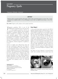
514 Pregnancy Epulis<br />
518 Ludwig’s Angina: A Rare Case Report<br />
522 Management of an Unusual Crown Root Fracture of Mandibular First Primary Molar<br />
526 Follicular Adenomatoid Odontogenic Tumor<br />
529 Sodium Hypochlorite Solution Enhances Healing of Periapical Lesion by Nonsurgical<br />
Method<br />
532 Vital Bleaching with Diode Laser<br />
535 Replantation of Avulsed Tooth after Trauma: A One Year Follow-up Study
Indian Journal of<br />
Multidisciplinary Dentistry<br />
Executive Editor<br />
S Bhuminathan<br />
IJMD’s Editorial Panel<br />
Editor-in-Chief<br />
KMK Masthan<br />
IJMD Advisory Board<br />
<strong>Volume</strong> 2, <strong>Issue</strong> 3<br />
<strong>May</strong>-<strong>Jul</strong>y 2012<br />
Associate Editor<br />
N Aravindha Babu<br />
Prosthodontics<br />
Mahesh Verma<br />
Srinisha J<br />
Raghavendra Jayesh S<br />
Suresh V Nayar (UK)<br />
Sanjna Nayar<br />
Conservative Dentistry/<br />
Endodontics<br />
Sukumaran VG<br />
Subbiya A<br />
Swaminathan S (Singapore)<br />
Implantology<br />
John W Thurmond (USA)<br />
Genetics<br />
Aravind Ramanathan<br />
Oncology<br />
Abraham Kuriakose M<br />
Oral and Maxillofacial<br />
Surgery<br />
Ramakrishna Shenoi<br />
Vijay Ebenezer<br />
Raj Kutta (USA)<br />
Oral Pathology and<br />
Microbiology<br />
Vinay K Hazarey<br />
Ipe Vargese V<br />
Puneet Ahuja<br />
Sangeeta P Wanjari<br />
Gouse Mohiddin<br />
Orthodontics<br />
Krishna Nayak US<br />
Dhandapani G<br />
Murali RV<br />
Deepak C<br />
Pharmacology<br />
Muthiah NS<br />
IJCP’s Editorial Panel<br />
Elumalai M<br />
General Medicine<br />
Rajendran SM<br />
Periodontics<br />
Chandrasekaran SC<br />
Ash Vasanthan (USA)<br />
Oral Medicine and<br />
Radiology<br />
Nalini Aswath<br />
Panjab V Wanjari<br />
Praveen BN<br />
Mubeen<br />
Pedodontics<br />
Krishan Gauba<br />
Ashima Gauba<br />
Biochemistry<br />
<strong>Jul</strong>ius A<br />
Microbiology<br />
Mahalakshmi K<br />
Dr Sanjiv Chopra<br />
Prof. of Medicine & Faculty Dean<br />
Harvard Medical School<br />
Group Consultant Editor<br />
Dr Deepak Chopra<br />
Chief Editorial Advisor<br />
Dr KK Aggarwal<br />
CMD, Publisher and Group<br />
Editor-in-Chief<br />
Dr Veena Aggarwal<br />
Joint MD & Group Executive Editor<br />
Anand Gopal Bhatnagar<br />
Editorial Anchor<br />
IJMD is included in the databases of Genamics Journal Seek, Ulrich International periodical directory,<br />
Index Copernicus International Ltd., HINARI, CINAHL, EBSCO Publishing, Proquest, Chemical Abstracts<br />
Service Source Index (CASSI) and Google Scholar.<br />
482<br />
Advisory Bodies<br />
Heart Care Foundation of India, Non-Resident Indians Chamber of Commerce & Industry,<br />
World Fellowship of Religions<br />
Indian Journal of Multidisciplinary Dentistry, Vol. 2, <strong>Issue</strong> 3, <strong>May</strong>-<strong>Jul</strong>y 2012
From the Editor’s desk<br />
From the Editor-in-chief<br />
xxxxxxxxx<br />
Dr KMK Masthan<br />
Professor and Head,<br />
Department of Oral Pathology and Microbiology<br />
Sree Balaji Dental College and Hospital<br />
Chennai<br />
My editorial in the previous issue on academics and research elicited a mixed response ranging from<br />
strong criticisms to “You are stepping on my toes” to surprising applause. My only response to all of<br />
them is what Somerset Maugham once said “It is very hard to be a gentleman and a writer”. In this<br />
issue I write about palliative care since, I was a witness to one patient’s final moments last month. I felt his last<br />
days would have been better if he had received some form of palliative care instead of the well meaning deceit of<br />
his relatives who kept telling him that he was going to get better. Hence, I share what feel about such care with<br />
the readers.<br />
Palliative care is the care given to the dying, encompassing physical, psychological, social and spiritual<br />
dimensions. It is not the efforts of the medical profession alone, but includes the family members and the<br />
society. This is not some thing new and was practised by King Asoka twenty-four centuries back. He had<br />
installed several hospices to attend to the needs of the dying with special care and attention. All countries<br />
face the rapidly increasing burden of patients nearing their end due to cardiovascular disorders, cancers,<br />
diabetes, respiratory diseases, neurological disabilities and psychiatric ailments. In our country especially, certain<br />
factors like extreme changes in lifestyle during the past four decades have brought about higher incidence of<br />
hypertension, diabetes mellitus, cancers due to tobacco chewing and smoking and coronary artery disease due to<br />
junk/fatty food and hence more number of patients facing premature death.<br />
Whereas, we, as Indians, pride ourselves to be more spiritual and religious, the reality is our dying<br />
elders do not get the dignity due to them and the rightful care they deserve. Busy life, mind set, financial<br />
obligations, poverty, trend towards abolition of joint families all contribute to this insensitivity on the part of the<br />
family members and so the due palliative care is not provided to the dying. Another factor that must be mentioned<br />
is the present medical care system including paid hospitals is more geared to cures and alleviation rather than<br />
support and care. The governmental medical care is totally oblivious and frankly resistant to this palliation concept<br />
at all, the common instruction to the patient’s relative being “Take the patient home’’.<br />
In palliative care, most care givers are faced with situations that have obvious solutions, but unsuitable for the<br />
recipient. For example, whether to advise cardio-pulmonary resuscitation for a patient under palliative care. For<br />
a normal person whose heart has failed due to heart attack or electric shock, it is a life saving procedure. But for<br />
a person who is expected to succumb to his/her disease in a few days, is it justified to subject them to this? My<br />
opinion is a definite ‘No’.<br />
Indian Journal of Multidisciplinary Dentistry, Vol. 2, <strong>Issue</strong> 3, <strong>May</strong>-<strong>Jul</strong>y 2012<br />
483
From the Editor-in-chief<br />
The solution is to train community volunteers and to empower them to avail the help of nurses, doctors and<br />
hospices. They can be trained by medical personnel and they can be given access to any other information through<br />
toll free numbers, free on-line training, open access websites and periodic free training at the cost of the NGOs<br />
and government. Even small things like daily visits, emotional support, spiritual counseling, basic patient care<br />
techniques like how to avoid bedsores, advising a suitable diet within the means of the patient, awareness of the<br />
difference between communicable and noncommunicable diseases can help the patient greatly during the last few<br />
days of their life.<br />
One question the care-giver has to face all the time from the patient is “How long will I live?’’. Let us leave all the<br />
mercy and mental agony issues aside and handle this question in a more pragmatic manner. No medical or nonmedical<br />
person can exactly specify when and at what time the patient is likely to die. But patients may have some<br />
goals like settling their properties in the way they choose or seeing their daughters or sons married before they die<br />
etc. Such expectations are not unreasonable and hence the palliative care-giver can clearly inform the patients to<br />
expedite matters on their wishes. Another issue that always hampers the palliative care giver is the pressure of the<br />
close relatives not to tell the patient that the end is nearing. A fair analogy is if I were to be given some money<br />
and am allowed to spend it, without being told only the last few rupees are remaining, will I consider that as fair?<br />
It is always better if we know when we are nearing the end of our resources. So it is more merciful if the patients<br />
were told that their end is nearing. Probably such information will cause a few upset moments.But everyone<br />
knows that when there is birth, there is death. So they will come to terms with it and handle it better.By not<br />
revealing that, we may actually do injustice to the patient. Because he/she might want to speak to certain friends<br />
and relatives, express his/her opinions, concerns and fears better before the end. Another aspect of this revelation<br />
is that the patients may choose not to waste their meagre resources any further on treatments and medicines. If<br />
a person has worked for 20 years earning money for the marriage of his/her daughter, then it is not logical to<br />
let them spend it when the care giver surely knows the outcome. The trouble with concealment is that one can<br />
quite easily drift into deception. I feel minimal levels of wisdom and massive doses of idealism probably lead the<br />
medical professional to adapt this well meaning deceit and frank injustice to the patient. That logic is as circular<br />
as a Mobius strip where an ant can traverse the entire strip without touching edge anywhere. I would welcome<br />
guidance on this multi-faceted issue from the well informed. It is the province of the knowledge to speak and it<br />
is the privilege of wisdom to listen. Now it is time for the readers to speak their mind.<br />
Best wishes.<br />
484<br />
Indian Journal of Multidisciplinary Dentistry, Vol. 2, <strong>Issue</strong> 3, <strong>May</strong>-<strong>Jul</strong>y 2012
From the Desk of IJCP Group Editor-in-Chief<br />
xxxxxxxxx<br />
American College of Radiology Five Things<br />
Physicians and Patients should Question<br />
Dr KK Aggarwal<br />
Padma Shri and Dr BC Roy National Awardee<br />
Sr. Physician and Cardiologist, Moolchand Medcity<br />
President, Heart Care Foundation of India<br />
Group Editor-in-Chief, IJCP Group<br />
Editor-in-Chief, eMedinewS<br />
Chairman Ethical Committee, Delhi Medical Council<br />
Director, IMA AKN Sinha Institute (08-09)<br />
Hony. Finance Secretary, IMA (07-08)<br />
Chairman, IMA AMS (06-07)<br />
President, Delhi Medical Association (05-06)<br />
emedinews@gmail.com<br />
http://twitter.com/DrKKAggarwal<br />
Krishan Kumar Aggarwal (Facebook)<br />
• Don’t do imaging for uncomplicated headache. Imaging headache patients without specific risk factors for<br />
structural disease is not likely to change management or improve outcome. Those patients with a significant<br />
likelihood of structural disease requiring immediate attention are detected by clinical screens that have been<br />
validated in many settings. Many studies and clinical practice guidelines concur. Also, incidental findings lead<br />
to additional medical procedures and expense that do not improve patient well-being.<br />
• Don’t image for suspected pulmonary embolism (PE) without moderate or high pre-test probability. While<br />
deep vein thrombosis (DVT) and PE are relatively common clinically, they are rare in the absence of elevated<br />
blood d-Dimer levels and certain specific risk factors. Imaging, particularly computed tomography (CT)<br />
pulmonary angiography, is a rapid, accurate and widely available test, but has limited value in patients who<br />
are very unlikely, based on serum and clinical criteria, to have significant value. Imaging is helpful to confirm<br />
or exclude PE only for such patients, not for patients with low pre-test probability of PE.<br />
• Avoid admission or preoperative chest X-rays for ambulatory patients with unremarkable history and physical<br />
exam. Performing routine admission or preoperative chest X-rays is not recommended for ambulatory patients<br />
without specific reasons suggested by the history and/or physical examination findings. Only 2% of such<br />
images lead to a change in management. Obtaining a chest radiograph is reasonable if acute cardiopulmonary<br />
disease is suspected or there is a history of chronic stable cardiopulmonary disease in a patient older than age<br />
70 who has not had chest radiography within six months.<br />
• Don’t do computed tomography (CT) for the evaluation of suspected appendicitis in children until<br />
ultrasound has been considered as an option. Although CT is accurate in the evaluation of suspected<br />
appendicitis in the pediatric population, ultrasound is nearly as good in experienced hands. Since ultrasound<br />
will reduce radiation exposure, ultrasound is the preferred initial consideration for imaging examination in<br />
Indian Journal of Multidisciplinary Dentistry, Vol. 2, <strong>Issue</strong> 3, <strong>May</strong>-<strong>Jul</strong>y 2012<br />
485
From the Desk of IJCP Group Editor-in-Chief<br />
children. If the results of the ultrasound exam are equivocal, it may be followed by CT. This approach is costeffective,<br />
reduces potential radiation risks and has excellent accuracy, with reported sensitivity and specificity<br />
of 94%.<br />
• Don’t recommend follow-up imaging for clinically inconsequential adnexal cysts. Simple cysts and<br />
hemorrhagic cysts in women of reproductive age are almost always physiologic. Small simple cysts in<br />
postmenopausal women are common, and clinically inconsequential. Ovarian cancer, while typically cystic,<br />
does not arise from these benign-appearing cysts. After a good quality ultrasound in women of reproductive<br />
age, don’t recommend follow-up for a classic corpus luteum or simple cyst
ORIGINAL RESEARCH<br />
Interferon IRF6 Gene Variants and the Risk of Isolated Cleft lip or<br />
Palate in South Indian Dravidian Population<br />
S Kishore Kumar*, MR Sukumar*, B Saravanan**, Arvind Ramanathan † , M Boominathan ‡<br />
Abstract<br />
Nonsyndromic clefts of the lip and palate (CL, CP, CL/P) are among the most common congenital defects caused by multifactorial<br />
etiological factors that include both environmental and genetic factors. There is sufficient evidence to hypothesize<br />
that disease locus for this condition can be identified by candidate genes. The purpose of this study is to investigate the<br />
prevalence of mutation in exon 7 of IRF6 gene to determine whether this mutation is implicated in the South Indian Dravidian<br />
population. Material and methods: Blood samples were collected with informed consent from 10 subjects with nonsyndromic<br />
cleft lip/palate and genomic DNA was extracted from the blood samples, polymerase chain reaction was performed and the<br />
products were subjected to direct sequencing. Results: There was a significant positive association between the occurrence of<br />
homozygous valine polymorphic variant and isolated CL, CP and CL or CP (90%, n = 9) relative to heterozygous valine and<br />
isoleucine variant (10%, n = 1) in the present study. Conclusion: The study is clinically significant because it has for the first<br />
time identified the genetic status of exon 7 of IRF6 in Tamil speaking Dravidian population.<br />
Key words: Nonsyndromic cleft lip and palate, IRF6 gene variant, polymerase chain reaction<br />
Development of the head and face comprises<br />
of one of the most complex events during<br />
embryonic development, coordinated by a<br />
network of gene expressions that include transcription<br />
factors and signaling molecules, which confer polarity<br />
of cells. Disturbance of this tightly regulated network<br />
of signaling events may interfere with otherwise normal<br />
cellular function and consequently may result in the<br />
failure of meeting and fusion of the developing facial<br />
primordia, thereby causing orofacial cleft. The extent<br />
of orofacial cleft phenotype varies among the affected<br />
children with some having cleft lip (CL) or cleft palate<br />
(CP) (isolated CL or CP), while the others have cleft lip<br />
with cleft palate (CL/P). Clefts may involve either onehalf<br />
of the oral cavity or both and accordingly they are<br />
classified as unilateral or bilateral clefts. Such orofacial<br />
cleft may either occur as an isolated event (designated as<br />
nonsyndromic) or as a part of complex malformations<br />
(designated as syndromic). Nonsyndromic cleft makes<br />
about 70% of all orofacial clefts, while the remaining<br />
*<br />
Professor<br />
**<br />
Reader, Dept. of Orthodontics<br />
†<br />
Principal, Investigator, Human Genetics Laboratory<br />
‡<br />
Postgraduate Student, Dept. of Orthodontics<br />
Sree Balaji Dental College and Hospital, Chennai<br />
Address for correspondence<br />
Dr S Kishore kumar<br />
E-mail: spkishorekumar@yahoo.co.in<br />
30% are accounted for syndrome associated clefts. 1,26,29<br />
The etiology seems complex 2,,3,9,11,12,16 but genetics<br />
plays a major role. 1,4,6,8,15 Various candidate genes have<br />
been associated with nonsyndromal cleft lip/palate<br />
in different populations, but Interferon regulatory<br />
factor-6 (IRF6) is strongly related in various populations<br />
on a consistent basis. 19,20,23,25,27,28 Identification of<br />
etiologic explanation for clefting has included extensive<br />
evaluation of genes. 22<br />
IRF6 belongs to a family of nine transcription<br />
factors that share a highly-conserved helix-turn-helix<br />
DNA-binding domain. The DNA-binding domain<br />
is essential for IRF6 to bind the promoter region of<br />
the genes it regulates (activates). Mutations in IRF6<br />
were first reported in van der Woude syndrome<br />
(VWS). 13 Investigation of the genetic status of IRF6<br />
in nonsyndromic CL/P patients identified common<br />
polymorphic variant G>A at position 820 in the<br />
coding DNA of IRF6 gene. This causes the conversion<br />
of GTC to ATC and creates a valine→isoleucine<br />
substitution at amino acid 274 in the protein-binding<br />
domain of IRF6 gene. 29 GTC encoding valine amino<br />
acid has been found to be significantly associated with<br />
cleft in several populations.<br />
Material and Methods<br />
Indian Journal of Multidisciplinary Dentistry, Vol. 2, <strong>Issue</strong> 3, <strong>May</strong>-<strong>Jul</strong>y 2012<br />
487
original research<br />
The sample consisted of 10 subjects reporting to the<br />
Dept. of Orthodontics and Dentofacial Orthopedics,<br />
Sree Balaji Dental College and Hospitals, Chennai,<br />
India. The study was carried out after approval from<br />
Institutional Ethical Committee and guidelines from<br />
Helinsiki declaration were followed. Written consents<br />
were obtained from all subjects. Patients with CL or<br />
CP (isolated CL or CP) associated with any history<br />
of developmental disabilities, including learning<br />
disabilities and attention deficits, hearing impairment<br />
and speech deficits or abnormalities were excluded<br />
from the study. Blood samples (1.5 ml) were obtained<br />
from subjects and genomic DNA was purified by<br />
conventional phenol: Chloroform extraction and<br />
ethanol precipitation procedure. A 100 ng DNA was<br />
used as a template to amplify the mutant region in<br />
exon 7 by polymerase chain reaction (PCR) with<br />
the primer sequences mentioned in Table 1. Twenty<br />
microliter l aliquots of amplified PCR products were<br />
subjected to agarose gel electrophoresis in a 1.5%<br />
agarose gel containing ethidium bromide at 100 V<br />
for 30 minutes with 1X TAE (Tris Acetate EDTA)<br />
buffer. The DNA bands were visualized in a long<br />
wavelength UV (364 nm) transilluminator and the<br />
exon 7 specific bands were cut with a clean surgical<br />
blade. Gel blocks containing the exon 7 specific bands<br />
were transferred to a fresh 1.5 ml microcentrifuge tube<br />
and three volumes of solubilization buffer was added.<br />
The tubes were incubated at 55°C for 10 minutes with<br />
intermittent agitation to solubilize the gel blocks. After<br />
the incubation period, 1 gel volume of isopropanol<br />
was added to each tube and mixed by vortex mixer,<br />
following which the contents were transferred to a spin<br />
column. The spin column tubes were centrifuged at<br />
10,000 rpm for one minute at room temperature to<br />
enable binding of the DNA (PCR product of exon<br />
7 of IRF6 gene) to the silica membrane in the spin<br />
columns. The bound DNA was eluted with 40 µl of<br />
elution buffer and 10 ng of the eluted product was<br />
Table 1. IRF6 Exon 7 Mutant Region Primers<br />
Set 1<br />
Sequence Length (T.M*)<br />
Left primer 19 57.35<br />
Aaccttgcagtgactgacc<br />
Right Primer 18 57.47<br />
Atcaggttgggagcaaca<br />
sequenced with sequencing grade primers (A*STAR<br />
facility, Singapore).<br />
Results<br />
DNA size marker<br />
Lane # 1 2<br />
500 bp<br />
400 bp<br />
300 bp<br />
200 bp<br />
100 bp<br />
Figure 1. Initial PCR product of IRF6 gene (353 bp).<br />
A 100 ng aliquot of the total genomic DNA was used<br />
as template to amplify the exon 7 of IRF6 gene, which<br />
is known to carry the genetic mutation in CP patients<br />
in other races. The mutation converts ‘GTC’, which<br />
is the genetic code for ‘valine’ amino acid to ‘ATC’<br />
the genetic code for ‘isoleucine’ amino acid. In order<br />
to analyze for the presence of the above mutation, we<br />
downloaded the sequence of IRF6 coding region from<br />
the public domain database and designed the primers<br />
to specifically amplify exon 7 (Table 1). Amplifications<br />
in all the samples were of the expected size<br />
(353 bp) and a representative of two samples is shown in<br />
Figure 1 (lanes 1 and 2).<br />
Identification of Genetic Polymorphism in<br />
Exon 7 of IRF6 Gene<br />
A 2 µl aliquot of the eluted DNA was sequenced in<br />
a 20 µl reaction volume and the sequenced data was<br />
analyzed with BioEdit software. The genetic code GTC<br />
that encodes for valine amino acid was found in all the<br />
patients, while ATC that encodes for isoleucine was<br />
not found as an isolated event in any of the patients.<br />
However, ATC occurred in heterozygous state along<br />
with GTC in one of 10 samples (10% of samples)<br />
that were analyzed. There was a significant positive<br />
association between the occurrence of homozygous<br />
valine polymorphic variant and isolated CL, CP and<br />
CL/CP (90%, n = 9) relative to heterozygous valine and<br />
isoleucine variant (10%, n = 1). The data was further<br />
analyzed for the distribution pattern of homozygous<br />
488<br />
Indian Journal of Multidisciplinary Dentistry, Vol. 2, <strong>Issue</strong> 3, <strong>May</strong>-<strong>Jul</strong>y 2012
original research<br />
valine or valine and isoleucine in heterzygous state in<br />
each of the conditions - isolated CL or isolated CP<br />
or CL with CP. Valine/Valine homozygous vairant was<br />
found in 20% (n = 2) of isolated CL, 0% (n = 0) of<br />
isolated CP and 70% of CL with CP (n = 7), while<br />
valine/isoleucine heterozygous variant were found to<br />
be 10% (n = 1) in isolated CL, 0% (n = 0) in isolated<br />
CP and 0% (n = 0) in CL with CP.<br />
Taken together valine/valine homozygosity was<br />
significantly associated with clefting relative to valine/<br />
isoleucine heterozygosity. This pattern is consistent with<br />
a recessive effect of the valine allele, which requires to<br />
be in homozygous condition to cause orofacial cleft.<br />
While in the heterozygous state, the valine allele being<br />
recessive may not be able to cause orofacial cleft in the<br />
presence of a normal isoleucine allele.<br />
Discussion<br />
A total of 10 patients of tamil speaking dravidian race,<br />
with isolated nonsyndromic CL, CP or CL/CP were<br />
screened for genetic polymorphism (silent mutation)<br />
in exon 7 of IRF6 gene. The polymorphism converts<br />
GTC that encodes for valine amino acid to ATC,<br />
which encodes for isoleucine amino acid. The screen<br />
identified valine (GTC) in 90% (n = 9) of patients<br />
with CL, CP or CL/CP and both valine (GTC) and<br />
isoleucine (ATC) in heterzygous state in 10% (n = 1) of<br />
them. None of them were found to carry homozygous<br />
isoleucine (ATC) variant.<br />
The IRF6 gene has been shown to be mutated in<br />
patients with VWS and/or Popliteal pterygium<br />
syndrome (PPS) in several populations. 13,17 VWS is<br />
a dominantly inherited disorder characterized by the<br />
presence of pits and/or sinuses on the lower lip in 85%<br />
of cases, CL/P in 50% of the patients and hypodontia<br />
in 20% of them. 7,14,18,21,24 PPS is a less frequent allelic<br />
orofacial clefting disorder. In addition to the signs of<br />
VWS, PPS includes webbing of the knee, syndactyly<br />
(or absence) of the toes and digits, ankyloblepharon,<br />
syngnathia and genital abnormalities. 5 More than 59<br />
different mutations in IRF6 gene have been reported,<br />
which includes silent mutations in protein-binding<br />
domain, frame-shift and nonsense mutations and<br />
deletions all of which either alter or render the protein<br />
functionless. 2,10<br />
IRF6 is expressed at key stages of facial development<br />
in mouse embryos. Specifically, a high level<br />
of IRF6 expression is detected in the ectoderm covering<br />
the facial processes during their fusion to form the<br />
lip and primary palate. Zucchero et al investigated<br />
the prevalence of mutations in IRF6 gene in patients<br />
with nonsyndromic CL, CP or CL/CP, by sequencing<br />
the entire coding region of the IRF6 gene. The study<br />
found strong evidence of overtransmission (67%) of<br />
the valine (GTC) variant at position 274 relative to<br />
isoleucine (ATC) variant in IRF6 protein in Japanese,<br />
Chinese, Vietnamese and Filipino populaiton but not<br />
in Europeans and Indians. 29 In the present study, we<br />
have analyzed a cohort of 10 patients of tamil speaking<br />
dravidian race with CL, CP or CL with CP and found<br />
valine variant to be transmitted in 90% of them<br />
(p ≤ 0.05). When the data was analyzed for stratified<br />
distribution of valine/valine alleles in isolated CL or CP<br />
or CL with CP, the association was found to be significant<br />
relative to valine/isoleucine heterozygous alleles.<br />
Summary and Conclusion<br />
The present study, however, has to be interpreted<br />
carefully since it did not involve analysis of IRF6 gene<br />
from normal individuals. The distribution of valine/<br />
valine and valine/isoleucine alleles in normal individuals<br />
is required to arrive at a more affirmative conclusion.<br />
Nevertheless, the present study has helped us to<br />
understand the genetic status of exon 7 of IRF6 in the<br />
tamil speaking dravidian race. Besides we also made an<br />
interesting observation that 10% of the patients that<br />
we examined carried both valine/isoleucine alleles in<br />
heterozygous state, which is in contrary to Zucchero<br />
et al study 29 who reported this to be rare event in the<br />
Indian population. This may be explained by the fact<br />
that the patients that we investigated were from tamil<br />
speaking dravidian race, while those that were analyzed<br />
by Zucchero et al were from West Bengal.<br />
References<br />
1.<br />
2.<br />
3.<br />
Lidral AC, Moreno LM, Bullard SA. Genetic Factors and<br />
Orofacial Clefting. Semin Orthod 2008;14(2):103-114.<br />
Jugessur A, Murray JC. Orofacial clefting: recent<br />
insights into a complex trait. Curr Opin Genet Dev<br />
2005;15(3):270-8.<br />
Beaty TH, Maestri NE, Hetmanski JB, Wyszynski DF,<br />
Vanderkolk CA, Simpson JC, et al. Testing for interaction<br />
between maternal smoking and TGFA genotype among<br />
oral cleft cases born in Maryland 1992-1996. Cleft Palate<br />
Indian Journal of Multidisciplinary Dentistry, Vol. 2, <strong>Issue</strong> 3, <strong>May</strong>-<strong>Jul</strong>y 2012<br />
489
original research<br />
4.<br />
5.<br />
6.<br />
7.<br />
8.<br />
9.<br />
10.<br />
11.<br />
12.<br />
13.<br />
14.<br />
15.<br />
16.<br />
Craniofac J 1997;34(5):447-54.<br />
Blanton SH, Cortez A, Stal S, Mulliken JB, Finnell<br />
RH, Hecht JT. Variation in IRF6 contributes to<br />
nonsyndromic cleft lip and palate. Am J Med Genet A<br />
2005;137A(3):259-62.<br />
Froster-Iskenius UG. Popliteal pterygium syndrome. J<br />
Med Genet 1990;27(5):320-6.<br />
Ghassibé M, Bayet B, Revencu N, Verellen-Dumoulin<br />
C, Gillerot Y, Vanwijck R, et al. Interferon regulatory<br />
factor-6: a gene predisposing to isolated cleft lip with<br />
or without cleft palate in the Belgian population. Eur J<br />
Hum Genet 2005;13(11):1239-42.<br />
Gorlin R, Cohen MJ, Hennekam R. Syndromes of<br />
the Head and Neck: Orofacial Clefting Syndromes:<br />
Common Syndromes. 4th edition, Oxford University<br />
Press: New York, 2001.<br />
Indian Genome Variation Consortium. Genetic<br />
landscape of the people of India: a canvas for disease gene<br />
exploration. J Genet2008;87(1):3-20.<br />
Murray JC. Face facts: genes, environment, and clefts.<br />
Am J Hum Genet 1995;57(2):227-32.<br />
Murray JC, Schutte BC. Cleft palate: players, pathways,<br />
and pursuits. J Clin Invest 2004;113(12):1676-8.<br />
Shi M, Wehby GL, Murray JC. Review on genetic variants<br />
and maternal smoking in the etiology of oral clefts and<br />
other birth defects. Birth Defects Res C Embryo Today<br />
2008;84(1):16-29.<br />
Rothman KJ, Moore LL, Singer MR, Nguyen US,<br />
Mannino S, Milunsky A. Teratogenicity of high vitamin<br />
A intake. N Engl J Med 1995;333(21):1369-73.<br />
Kondo S, Schutte BC, Richardson RJ, Bjork BC, Knight<br />
AS, Watanabe Y, et al. Mutations in IRF6 cause Van der<br />
Woude and popliteal pterygium syndromes. Nat Genet.<br />
2002;32(2):285-9.<br />
Lacombe D, Pedespan JM, Fontan D, Chateil JF, Verloes<br />
A. Phenotypic variability in van der Woude syndrome.<br />
Genet Couns 1995;6(3):221-6.<br />
Nemana LJ, Marazita ML, Melnick M. Genetic analysis<br />
of cleft lip with or without cleft palate in Madras, India.<br />
Am J Med Genet 1992;42(1):5-9.<br />
Lammer EJ, Shaw GM, Iovannisci DM, Finnell RH.<br />
Maternal smoking, genetic variation of glutathione s-<br />
transferases, and risk for orofacial clefts. Epidemiology<br />
2005;16(5):698-701.<br />
17.<br />
18.<br />
19.<br />
20.<br />
21.<br />
22.<br />
23.<br />
24.<br />
25.<br />
26.<br />
27.<br />
28.<br />
LMR parnalba Lack of association between IRF6<br />
polymorhisms and non syndromic cleft lip or palate in<br />
Brazilian population. 2008.<br />
Mossey PA, Little J, Munger RG, Dixon MJ, Shaw WC.<br />
Cleft lip and palate. Lancet 2009;374(9703):1773-85.<br />
Mossey P, Little J. Addressing the challenges of cleft lip<br />
and palate research in India. Indian J Plast Surg 2009;42<br />
Suppl:S9-S18.<br />
Park JW, McIntosh I, Hetmanski JB, Jabs EW, Vander<br />
Kolk CA, Wu-Chou YH, et al. Association between IRF6<br />
and nonsyndromic cleft lip with or without cleft palate<br />
in four populations. Genet Med 2007;9(4):219-27.<br />
Stottmann RW, Bjork BC, Doyle JB, Beier DR.<br />
Identification of a Van der Woude syndrome mutation<br />
in the cleft palate 1 mutant mouse. Genesis 2010;48(5):<br />
303-8.<br />
Romitti PA, Sun L, Honein MA, Reefhuis J, Correa<br />
A, Rasmussen SA. Maternal periconceptional alcohol<br />
consumption and risk of orofacial clefts. Am J Epidemiol<br />
2007;166(7):775-85.<br />
Scapoli L, Palmieri A, Martinelli M, Pezzetti F, Carinci<br />
P, Tognon M, Carinci F. Strong evidence of linkage<br />
disequilibrium between polymorphisms at the IRF6<br />
locus and nonsyndromic cleft lip with or without cleft<br />
palate, in an Italian population. Am J Hum Genet<br />
2005;76(1):180-3.<br />
Van der woude A. Fistula labii inferioris congenita and<br />
its association with cleft lip and palate. Am J Hum Genet<br />
1954;6(2):244-56.<br />
Vieira AR, Cooper ME, Marazita ML, Orioli IM, Castilla<br />
EE. Interferon regulatory factor 6 (IRF6) is associated<br />
with oral-facial cleft in individuals that originate in South<br />
America. Am J Med Genet A 2007;143A(17):2075-8.<br />
Wyszynski DF, Beaty TH, Maestri NE. Genetics of<br />
nonsyndromic oral clefts revisited. Cleft Palate Craniofac<br />
J 1996;33(5):406-17.<br />
Pan Y, Ma J, Zhang W, Du Y, Niu Y, Wang M, et al.<br />
IRF6 polymorphisms are associated with nonsyndromic<br />
orofacial clefts in a Chinese Han population. Am J Med<br />
Genet A 2010;152A(10):2505-11<br />
Zeiger JS, Beaty TH, Liang KY. Oral clefts, maternal<br />
smoking, and TGFA: a meta-analysis of gene-environment<br />
interaction. Cleft Palate Craniofac J 2005;42(1):58-63.<br />
29. Zucchero TM, Cooper ME, Maher BS, Daack-Hirsch S,<br />
490<br />
Indian Journal of Multidisciplinary Dentistry, Vol. 2, <strong>Issue</strong> 3, <strong>May</strong>-<strong>Jul</strong>y 2012
Role of ‘Live Microorganisms’ (Probiotics) in Prevention of<br />
Caries: Going on the Natural Way Towards Oral Health<br />
Vineet Agrawal*, Sonali Kapoor**, Nimisha Shah †<br />
*Senior Lecturer<br />
**Professor<br />
Dept. of Conservative and Endodontics, MP Dental College and Oral<br />
Research Institute, Vadodara<br />
†<br />
Professor, Dept. of Conservative and Endodontics, KM Shah Dental<br />
College, Vadodara<br />
Address for correspondence<br />
Dr Vineet Agrawal<br />
E-mail: vineetdent@yahoo.co.in<br />
Abstract<br />
Science is providing us the tools to diagnose and treat an infection before it causes damage. For some decades now, bacteria<br />
known as probiotics have been added to various foods because of their beneficial effects for human health. Very encouraging<br />
studies have come up in recent past exploring probiotics in fields of caries, periodontal diseases and few other areas and the<br />
results tend to suggest beneficial effects of probiotics on oral health and on whole body in general. The application of probiotic<br />
strategies may, in near future, provide an end to many infections occurring in oral cavity. This article reviews the probiotic<br />
approaches, such as genetically modified Streptococcus mutans and targeted antimicrobials in the prevention of caries and<br />
discuss its future directions.<br />
Key words: Probiotics, dental caries, prevention, Bifidobacterium, Lactobacillus<br />
W<br />
D Miller first described dental caries as a<br />
bacterially-mediated process more than<br />
100 years ago. 1 Today, we know that dental<br />
caries is a multifaceted disease process. Several models<br />
have been put forward describing mechanism of caries<br />
formation. One of the earlier models that is familiar<br />
to most dentists was put forth by Fitzgerald and<br />
Keyes. 2 They used three overlapping circles describing<br />
the host, bacteria and nutrients required to foment<br />
the production of organic acids and the subsequent<br />
demineralization activity. The beauty of this model is<br />
that all three elements must be present for the disease<br />
to progress. Since all three are required for disease<br />
initiation and progression, removal of any one element<br />
ostensibly leads to the interception of the disease<br />
process.<br />
The surgical approach has been the predominate<br />
mode of caries management for the past 150 years.<br />
Dentistry has, however, in recent years moved<br />
toward an antibiotic/antimicrobial model of disease<br />
management. This approach, however, raises serious<br />
questions: 1) Do the antibiotic/antimicrobial agents<br />
Review article<br />
(chlorhexidine, povidone-iodine, fluoride, etc.) kill<br />
all offending organisms?; 2) if so, do the agents<br />
preclude the re-entry of the same organisms from<br />
external sources? and 3) if the agents do kill all the<br />
offending organisms, do any remaining pathogenic<br />
organisms have selective advantage in repopulating the<br />
tooth surfaces? To overcome the problems inherent<br />
in an antibiotic/antimicrobial approach, probiotic<br />
methods are currently under study as means of caries<br />
management.<br />
What are Probiotics, Prebiotics and<br />
Synbiotics<br />
The term ‘probiotic’ is derived from the Latin preposition<br />
pro (‘for’) and the Greek adjective (biotic), the latter<br />
deriving from the noun (bios, ‘life’). 3 It was first used<br />
by Lilly and Stillwell in 1965 to describe “substances<br />
secreted by one microorganism, which stimulates the<br />
growth of another” and thus was contrasted with the<br />
term antibiotic. 4 Today, two main definitions are used.<br />
According to a WHO/FAO report (2002), probiotics<br />
are “live microorganisms which, when administered in<br />
adequate amounts, confer a health benefit on the host”.<br />
International Life Science Institute (ILSI) Europe<br />
suggests a definition according to which a probiotic is<br />
“a live microbial food ingredient that, when ingested<br />
in sufficient quantities, exerts health benefits on the<br />
consumer”. Probiotics are microorganisms, basically<br />
bacteria, that when ingested would confer health<br />
benefit beyond the basic nutrition. 5<br />
Indian Journal of Multidisciplinary Dentistry, Vol. 2, <strong>Issue</strong> 3, <strong>May</strong>-<strong>Jul</strong>y 2012<br />
491
Review Article<br />
The term prebiotic was introduced by Gibson and<br />
Roberfroid who exchanged ‘pro’ for ‘pre’ which<br />
means ‘before’ or ‘for’. They defined prebiotics as a<br />
“nondigestible food ingredient that beneficially affects<br />
the host by selectively stimulating the growth and/or<br />
activity of one or a limited number of bacteria in the<br />
colon.” 6 More specifically, prebiotics are short-length<br />
carbohydrates, such as fructooligosaccharides, that<br />
resist digestion in upper gastrointestinal tract or are<br />
fermented in the colon to produce short-chain fatty<br />
acids, such as acetate, butyrate and propionate, which<br />
have positive effects on colonic cell growth and stability,<br />
generate many of the same bacteria as provided in<br />
probiotics. 7<br />
The term synbiotic is used when a product contains<br />
both probiotics and prebiotics. According to this<br />
approach, a food or food supplement will include both<br />
the live cells of the beneficial bacteria and the selective<br />
substrate. The idea being that the beneficial bacterial<br />
cells can grow quickly and competitively because of<br />
the presence of selective substrate and establish their<br />
predominance. 6<br />
Table 1. Possible Mechanism of Probiotics in Oral<br />
Health<br />
Production of antimicrobial substances<br />
Organic acids<br />
Hydrogen peroxide<br />
Bacteriocins<br />
Binding in oral cavity<br />
Compete with pathogens for adhesion sites<br />
Involvement in metabolism of substrates (competing with<br />
oral microorganisms for substrates<br />
available)<br />
Immunomodulatory<br />
Stimulate nonspecific immunity<br />
Modulate humoral and cellular immune<br />
response<br />
Modify oral conditions<br />
Modulating pH<br />
Modification of oxidation reduction potential<br />
Mechanism of Probiotics<br />
Probiotics can help prevent and treat disease through<br />
several mechanisms. 8<br />
• Direct interaction: Probiotics interact directly<br />
with the disease-causing microbes, making it<br />
harder for them to cause the disease.<br />
• Competitive exclusion: Beneficial microbes<br />
directly compete with the disease, developing<br />
microbes for nutrition or enterocyte adhesion<br />
sites.<br />
• Modulation of host immune response: Probiotics<br />
interact with and strengthen the immune system<br />
and help prevent disease.<br />
In oral cavity, probiotics tend to create a biofilm,<br />
acting as a protective lining for oral tissues against oral<br />
diseases. Such a biofilm keeps bacterial pathogens off<br />
oral tissues by filling a space, which could have served<br />
as a niche for pathogens in future; and competing with<br />
cariogenic bacteria. Table 1 describes the mechanism<br />
of action of probiotics in oral health. 9<br />
Potential Benefits of Probiotics<br />
Probiotics have traditionally been used for prevention<br />
of colon cancer, 10 lowering cholesterol, 10 lowering blood<br />
pressure, 10 managing lactose intolerance, 11 Helicobacter<br />
pylori, 12 improving immune function and preventing<br />
infections, 13 antibiotic-associated diarrhea, 14 reducing<br />
inflammation, 15 improving mineral absorption, 15<br />
preventing harmful bacterial growth under stress, 16<br />
irritable bowel syndrome and colitis, 16 and managing<br />
urogenital health. 16<br />
Common Strains Used in Oral Probiotics<br />
The most commonly-used probiotic strains belong<br />
to the genera, Lactobacillus, Bifidobacterium, 17 and<br />
Streptococcus. 18 Streptococcus salivarius, Streptococcus<br />
mitis and Streptococcus sanguinis showed a significantly<br />
more pronounced reduction in total anerobic bacteria,<br />
black-pigmented bacteria and Campylobacter rectus.<br />
Probiotic strains of Lactobacillus species include<br />
L. salivarius, L. reuteri, L. acidophilus, L. fermentum,<br />
L. lactis, L. helveticus and L. rhamnosus. Lactobacilli<br />
produce different antimicrobial components, such as<br />
organic acids, hydrogen peroxide, low molecular weight<br />
antimicrobial substances, bacteriocins and adhesion<br />
inhibitors. Similarly, Bifidobacterium strains include<br />
B. bifidum, B. longum and B. infantis. 19<br />
492<br />
Indian Journal of Multidisciplinary Dentistry, Vol. 2, <strong>Issue</strong> 3, <strong>May</strong>-<strong>Jul</strong>y 2012
Review Article<br />
Administration of Probiotic<br />
Different means of probiotic administration for oral<br />
health purpose are: 20<br />
• A culture concentrate added to a beverage or food<br />
(such as a fruit juice)<br />
• Inoculated into prebiotic fibers<br />
• Inoculants into a milk-based food (dairy products<br />
such as milk, milk drink, yoghurt)<br />
• Yogurt drink, cheese, kefir, biodrink<br />
• As concentrated and dried cells packaged as dietary<br />
supplements (nondairy products)<br />
• Such as powder, capsule, gelatin tablets.<br />
Role of Probiotics in Dental Caries<br />
A number of researchers are developing ‘probiotic’<br />
methods to treat the caries causing pathogens. ‘Probiotic’,<br />
as used here, means that mechanisms are employed to<br />
selectively remove only the (odonto) pathogen while<br />
leaving the remainder of the oral ecosystem intact. 21<br />
One of the replacement therapy options entails the<br />
application of a genetically engineered ‘effector strain’<br />
of S. mutans that will replace the cariogenic or ‘wild<br />
strain’ to prevent or arrest caries and to promote optimal<br />
remineralization of tooth surfaces that have been<br />
demineralized but that have not become cavitated.<br />
In caries, there is an increase in acidogenic and acid<br />
tolerating species such as mutans streptococci and<br />
lactobacilli, although other bacteria with similar<br />
properties can also be found like Bifidobacteria,<br />
nonmutans streptococci, Actinomyces spp.,<br />
Propionibacterium spp., Veillonella spp. and Atopobium<br />
spp. Use of probiotics and molecular genetics to replace<br />
and displace cariogenic bacteria with noncariogenic<br />
bacteria has shown promising results. These studies<br />
have employed different approaches: 22<br />
• Early studies concentrated on utilizing bacteria that<br />
expressed bacteriocins or bacteriocin-like inhibitory<br />
substances (BLIS) that specifically prevented the<br />
growth of cariogenic bacteria.<br />
• One approach has been to identify food grade and<br />
probiotic bacteria, which have ability to colonize<br />
teeth and influence the supragingival plaque.<br />
• Also, strains have been screened for suitable<br />
antagonistic activity against relevant oral bacteria.<br />
• Another approach utilized recombinant strain of<br />
S. mutans expressing urease, which was shown to<br />
reduce the cariogenicity of plaque in an animal<br />
model.<br />
• Similarly, genetically modified probiotics with<br />
enhanced properties can be developed (‘designer<br />
probiotics’). For example, a recombinant strain<br />
of Lactobacillus that expressed antibodies<br />
targeting one of the major adhesions of S. mutans<br />
(antigen I/II) was able to reduce both the viable<br />
counts of S. mutans and the caries score in a rat<br />
model.<br />
• A different way of accomplishing the removal of the<br />
pathogens is to develop ‘targeted antimicrobials’.<br />
The basic idea is to develop an inexpensive<br />
targeting molecule that will reliably attach to only<br />
the organism of interest, in this case S. mutans,<br />
S. sobrinus or other chosen pathogen. Once the<br />
targeting molecule is perfected, then a ‘killer’<br />
molecule is optimized and chained to the targeting<br />
molecule. The combined unit then selectively<br />
eliminates the infection of interest. In the case<br />
of the oral cavity and dental caries, this system is<br />
attractive from the perspective of eliminating all<br />
the pathogens thereby precluding the regrowth<br />
of the original infection. There is also compelling<br />
evidence from clinical trials and laboratory efforts<br />
demonstrating that once the bacterial ecosystem<br />
is free of S. mutans, it is difficult to reintroduce<br />
the organisms (another competitive inhibition<br />
situation). 21,22<br />
Various Studies Involving Probiotics for<br />
Decreasing Dental Caries<br />
Considering the growing body of evidence about<br />
the role of probiotics on caries pathogens, however,<br />
it has been suggested that the operative approach in<br />
caries treatment might be challenged by probiotic<br />
implementation with subsequent less invasive<br />
intervention in clinical dentistry and thus, recently,<br />
many studies are been carried on probiotics.<br />
The first randomized, double-blind, placebo-controlled<br />
intervention study, 23 examining the effect of milk<br />
containing L. rhamnosus GG on caries and the risk<br />
of caries in children when compared with normal<br />
milk was completed in 2001; the study included<br />
594 children, 1-6 years old, who consumed milk<br />
for seven months. Probiotic milk was able to reduce<br />
S. mutans counts at the end of the trial and a<br />
Indian Journal of Multidisciplinary Dentistry, Vol. 2, <strong>Issue</strong> 3, <strong>May</strong>-<strong>Jul</strong>y 2012<br />
493
Review Article<br />
significant reduction of caries risk was also observed.<br />
L. rhamnosus is one of the most extensively studied<br />
probiotic and of particular interest in oral biology<br />
since it does not readily ferment sucrose and is safer for<br />
teeth than lactic acid-producing bacteria. Controlled<br />
studies have shown the effectiveness of L. rhamnosus<br />
in reducing caries. L. rhamnosus was found to inhibit<br />
cariogenic S. mutans but colonization of oral cavity by<br />
L. rhamnosus seems improbable. 24 A study aimed at<br />
benefit of cheese containing L. rhamnosus showed that<br />
probiotic intervention helped in reducing the highest<br />
level of Streptococcus mutans. 25<br />
In order to assess whether naturally occurring oral<br />
lactobacilli have probiotic properties, lactobacilli were<br />
isolated from saliva and plaque from children and<br />
adolescents, with or without caries lesions. Twenty-three<br />
Lactobacillus spp. completely inhibited the growth of<br />
all mutans streptococci tested. Species with maximum<br />
interference capacity against mutans streptococci<br />
included Lactobacillus paracasei, Lactobacillus plantarum<br />
and L. rhamnosus. 26<br />
Calgar et al (2006) investigated the effect of probiotic<br />
bacterium L. reuteri on levels of mutans streptococci<br />
and lactobacilli, which was introduced by two different<br />
straw containing L. reuteri and lozenges containing<br />
L. reuteri and concluded that short-term daily ingestion<br />
of lactobacilli-derived probiotics delivered by prepared<br />
straws or lozenges reduced the levels of salivary mutans<br />
streptococci in young adults. 27<br />
Comelli et al (2002) studied 23 dairy bacterial strains<br />
for the prevention of dental caries and reported that<br />
only two strains namely Streptococcus thermophilus and<br />
L. lactis were able to adhere to saliva-coated<br />
hydroxyapatite and were further successfully<br />
incorporated into a biofilm similar to the dental<br />
plaque. Furthermore, they could grow together with<br />
five strains of oral bacterial species commonly found in<br />
supragingival plaque. In this system, L. lactis was able<br />
to modulate the growth of the oral bacteria, and in<br />
particular to diminish the colonization of Streptococcus<br />
oralis, Veillonella dispar, Actinomyces naeslundii and of<br />
the cariogenic S. sobrinus. 28<br />
Few studies have reported reduction in mutans<br />
streptococci levels in saliva following use of probiotic<br />
containing yoghurts but it is not clear whether this<br />
decrease is due to the bactericidal activity of yoghurt<br />
or other mechanisms. Petti et al (2008) investigated<br />
the differences in susceptibility of strains of viridians<br />
streptococci. In vitro, yoghurt with live bacteria showed<br />
selective antimutans activity, suggesting that the overall<br />
decrease in mutans streptococci in vivo could be due to<br />
a bactericidal effect on S. mutans. 29<br />
Calgar et al (2007) evaluated the effect of xylitol and<br />
probiotic chewing gums on salivary mutans streptococci<br />
and lactobacilli and concluded that daily chewing on<br />
gums containing probiotic bacteria or xylitol reduced<br />
the levels of salivary mutans streptococci in a significant<br />
way. However, a combination of probiotic and xylitol<br />
gums did not seem to enhance this effect. 30<br />
Kang et al (2006) did a study in which they found<br />
out that the water-soluble polymers produced from<br />
sucrose by the Weissella cibaria isolates inhibited the<br />
formation of S. mutans biofilm. In the clinical study,<br />
the subjects mouthrinsed with a solution containing<br />
W. cibaria CMS1 and exhibited plaque index reduction<br />
of approximately 20.7%. 31<br />
To study the effect of bifidobacteria a doubleblind,<br />
randomized crossover study was performed.<br />
A statistically significant reduction of salivary mutans<br />
streptococci was recorded after the probiotic yoghurt<br />
consumption containing Bifidobacterium, which<br />
was in contrast to the controls. A similar trend was<br />
seen for lactobacilli, but this decrease failed to reach<br />
statistical significance. Investigators concluded that<br />
probiotic bifidobacteria in yoghurt may reduce the<br />
levels of selected caries-associated microorganisms in<br />
saliva. 18 In a similar study using Bifidobacterium lactis a<br />
statistically significant reduction (p < 0.05) of salivary<br />
mutans streptococci was recorded after consumption<br />
of the probiotic ice-cream in adults. 32<br />
Conclusion<br />
Concept of probiotics is emerging as a fascinating<br />
field and it prompts a new horizon on the relationship<br />
between diet and oral health. Probiotic strategies are<br />
part of the continuing evolution of the treatment of<br />
oral infection that produces the clinical manifestations<br />
of dental caries. As a profession, we are slowly moving<br />
away from the purely surgical approach to treating this<br />
disease. Science is providing us the tools to diagnose<br />
and treat the infection before it causes damage.<br />
The application of probiotic strategies may, in the<br />
494<br />
Indian Journal of Multidisciplinary Dentistry, Vol. 2, <strong>Issue</strong> 3, <strong>May</strong>-<strong>Jul</strong>y 2012
Review Article<br />
not-distant future, provide the end of new cavities in<br />
treated populations.<br />
Future Directions<br />
Probiotics can be used as passive local immunization<br />
against dental caries. High titers of antibodies can<br />
also be directed against human cariogenic bacteria<br />
produced in bovine colostrum over the vehicle of<br />
fermented milk.<br />
Studies have been largely conducted in animals, and<br />
human studies have not been of sufficient duration to<br />
assess the impact on caries. Most studies on the effects of<br />
probiotics on caries prevention are aimed at decreasing<br />
the number of mutans streptococci. Primarily probiotic<br />
Lactobacillus and Bifidobacterium strains have been<br />
used along with few more strains. Unfortunately, in<br />
most cases, the study groups were relatively small, and<br />
the studies were fairly short. Preliminary data obtained<br />
has been encouraging, but numerous randomized<br />
clinical studies will be required to clearly establish the<br />
potential of probiotics in prevention of dental caries.<br />
Also complete understanding of the broad ecological<br />
changes induced in the mouth by probiotics or<br />
prebiotics will be essential to assess their long-term<br />
consequences for oral health and disease.<br />
References<br />
1.<br />
2.<br />
3.<br />
4.<br />
5.<br />
6.<br />
7.<br />
8.<br />
Miller W. Micro-organisms of the Human Mouth. SS<br />
White; Philadelphia, 1890.<br />
Keyes PH. Research in dental caries. J Am Dent Assoc<br />
1968;76(6):1357-73.<br />
Hamilton-Miller JM, Gibson GR, Bruck W. Some<br />
insights into the derivation and early uses of the word<br />
‘probiotic’. Br J Nutr 2003;90(4):845.<br />
Lilly DM, Stillwell RH. Probiotics: growth-promoting<br />
factors produced by microorganisms. Science<br />
1965;147(3659):747-8.<br />
de Vrese M, Schrezenmeir J. Probiotics, prebiotics, and<br />
synbiotics. Adv Biochem Eng Biotechnol 2008;111:<br />
1-66.<br />
Schrezenmeir J, de Vrese M. Probiotics, prebiotics, and<br />
synbiotics - approaching a definition. Am J Clin Nutr<br />
2001;73(2 Suppl):361S-364S.<br />
Isolauri E. Probiotics in human disease. Am J Clin Nutr<br />
2001;73(6):1142S-1146S.<br />
Meurman JH. Probiotics: do they have a role in oral<br />
medicine and dentistry? Eur J Oral Sci 2005;113(3):<br />
188-96.<br />
9.<br />
10.<br />
11.<br />
12.<br />
13.<br />
14.<br />
15.<br />
16.<br />
17.<br />
18.<br />
19.<br />
20.<br />
21.<br />
22.<br />
23.<br />
Haukioja A. Probiotics and oral health. Eur J Dent<br />
2010;4(3):348-55.<br />
Sanders ME. Considerations for use of probiotic<br />
bacteria to modulate human health. J Nutr 2000;130(2S<br />
Suppl):384S-390S.<br />
Brady LJ, Gallaher DD, Busta FF. The role of probiotic<br />
cultures in the prevention of colon cancer. J Nutr<br />
2000;130(2S Suppl):410S-414S.<br />
Reid G, Jass J, Sebulsky MT, McCormick JK. Potential<br />
uses of probiotics in clinical practice. Clin Microbiol Rev<br />
2003;16(4):658-72.<br />
Ouwehand AC, Salminen S, Isolauri E. Probiotics: an<br />
overview of beneficial effects. Antonie Van Leeuwenhoek<br />
2002;82(1-4):279-89.<br />
Szajewska H, Ruszczyński M, Radzikowski A. Probiotics<br />
in the prevention of antibiotic-associated diarrhea in<br />
children: a meta-analysis of randomized controlled trials.<br />
J Pediatr 2006;149(3):367-372.<br />
Famularo G, De Simone C, Pandey V, Sahu AR,<br />
Minisola G. Probiotic lactobacilli: an innovative tool to<br />
correct the malabsorption syndrome of vegetarians? Med<br />
Hypotheses 2005;65(6):1132-5.<br />
Reid G. Probiotic agents to protect the urogenital<br />
tract against infection. Am J Clin Nutr 2001;73(2<br />
Suppl):437S-443S.<br />
Comelli EM, Guggenheim B, Stingele F, Neeser JR.<br />
Selection of dairy bacterial strains as probiotics for oral<br />
health. Eur J Oral Sci 2002;110(3):218-24.<br />
Caglar E, Sandalli N, Twetman S, Kavaloglu S,<br />
Ergeneli S, Selvi S. Effect of yogurt with Bifidobacterium<br />
DN-173 010 on salivary mutans streptococci and<br />
lactobacilli in young adults. Acta Odontol Scand<br />
2005;63(6):317-20.<br />
Meurman JH, Stamatova I. Probiotics: contributions to<br />
oral health. Oral Dis 2007;13(5):443-51.<br />
Caglar E, Kargul B, Tanboga I. Bacteriotherapy and<br />
probiotics’ role on oral health. Oral Dis 2005;11(3):<br />
131-7.<br />
Anderson MH, Shi W. A probiotic approach to caries<br />
management. Pediatr Dent 2006;28(2):151-3; discussion<br />
192-8.<br />
Bhusan J, Chachra S. Probiotics: their role in prevention<br />
of dental caries. J Oral Health Commun Dent<br />
2010;4(3):78-82.<br />
Näse L, Hatakka K, Savilahti E, Saxelin M, Pönkä A,<br />
Poussa T, et al. Effect of long-term consumption of a<br />
probiotic bacterium, Lactobacillus rhamnosus GG, in<br />
milk on dental caries and caries risk in children. Caries<br />
Res 2001;35(6):412-20.<br />
Indian Journal of Multidisciplinary Dentistry, Vol. 2, <strong>Issue</strong> 3, <strong>May</strong>-<strong>Jul</strong>y 2012<br />
495
Review Article<br />
24.<br />
25.<br />
26.<br />
27.<br />
28.<br />
Yli-Knuuttila H, Snäll J, Kari K, Meurman JH.<br />
Colonization of Lactobacillus rhamnosus GG in the oral<br />
cavity. Oral Microbiol Immunol 2006;21(2):129-31.<br />
Ahola AJ, Yli-Knuuttila H, Suomalainen T, Poussa T,<br />
Ahlström A, Meurman JH, et al. Short-term consumption<br />
of probiotic-containing cheese and its effect on dental<br />
caries risk factors. Arch Oral Biol 2002;47(11):799-804.<br />
Simark-Mattsson C, Emilson CG, Håkansson EG,<br />
Jacobsson C, Roos K, Holm S. Lactobacillus-mediated<br />
interference of mutans streptococci in caries-free vs.<br />
caries-active subjects. Eur J Oral Sci 2007;115(4):<br />
308-14.<br />
Caglar E, Cildir SK, Ergeneli S, Sandalli N, Twetman S.<br />
Salivary mutans streptococci and lactobacilli levels after<br />
ingestion of the probiotic bacterium Lactobacillus reuteri<br />
ATCC 55730 by straws or tablets. Acta Odontol Scand<br />
2006;64(5):314-8.<br />
Comelli EM, Guggenheim B, Stingele F, Neeser JR.<br />
29.<br />
30.<br />
31.<br />
32.<br />
Selection of dairy bacterial strains as probiotics for oral<br />
health. Eur J Oral Sci 2002;110(3):218-24.<br />
Petti S, Tarsitani G, Simonetti D’Arca A. Antibacterial<br />
activity of yoghurt against viridans streptococci in vitro.<br />
Arch Oral Biol 2008;53(10):985-90.<br />
Caglar E, Kavaloglu SC, Kuscu OO, Sandalli N,<br />
Holgerson PL, Twetman S. Effect of chewing gums<br />
containing xylitol or probiotic bacteria on salivary<br />
mutans streptococci and lactobacilli. Clin Oral Investig<br />
2007;11(4):425-9.<br />
Kang MS, Chung J, Kim SM, Yang KH, Oh JS. Effect of<br />
Weissella cibaria isolates on the formation of Streptococcus<br />
mutans biofilm. Caries Res 2006;40(5):418-25.<br />
Caglar E, Kuscu OO, Selvi Kuvvetli S, Kavaloglu Cildir<br />
S, Sandalli N, Twetman S. Short-term effect of ice-cream<br />
containing Bifidobacterium lactis Bb-12 on the number<br />
of salivary mutans streptococci and lactobacilli. Acta<br />
Odontol Scand 2008;66(3):154-8.<br />
496<br />
Indian Journal of Multidisciplinary Dentistry, Vol. 2, <strong>Issue</strong> 3, <strong>May</strong>-<strong>Jul</strong>y 2012
Oral Health and Wellness on Wheels!!!<br />
Review article<br />
R Sushma*, D Nagabhushana**<br />
Abstract<br />
Fully equipped mobile dental clinics provide on-the-spot diagnostic, preventive, interceptive and curative services to the<br />
doorsteps of the underprivileged, rural population. It’s an innovative, on-site, dental outreach provider to bring state of<br />
the art, preventive dental care to those in need in the most comfortable and effective way possible.<br />
Key words: Mobile dental service, outreach program, mobile dental unit, portable dentistry<br />
The greatest equity of access is said to exist<br />
when need, rather than structural or individual<br />
factors determine who gains entry to the<br />
healthcare system.<br />
Healthcare is a right, not a privilege but is healthcare<br />
accessible?<br />
Basic oral care facilities should be accessible to every<br />
individual since oral health is an important and crucial<br />
part of one’s overall health and wellness. Over the ages,<br />
oral healthcare has been delivered to the community,<br />
in different ways. The horse back dentistry of olden<br />
days has evolved into the most modern painless dental<br />
procedures.<br />
All over the world, different countries have different<br />
healthcare delivery systems. In our country, different<br />
state governments have established the dental clinics<br />
at different levels from the state capitals to rural areas,<br />
where salaried dentists give dental treatment. In India<br />
70% of the dentists practice in urban areas and we<br />
seldom find dental clinics in rural areas except for a few<br />
government establishments, which lack the required<br />
infrastructure.<br />
Providing universal health insurance coverage and<br />
developing integrated delivery systems may fail to<br />
*Lecturer, Dept. of Public Health Dentistry<br />
**Reader, Dept of Oral Medicine and Radiology<br />
JSS Dental College and Hospital, JSS University, Mysore, Karnataka<br />
Address for correspondence<br />
Dr R Sushma<br />
E-mail: hisushhere@yahoo.co.in<br />
provide universal access. Fully equipped mobile dental<br />
clinics to provide effective dental care to the doorsteps<br />
of the underprivileged, rural population is the need<br />
of the hour. A mobile dental clinic offers dentists<br />
the freedom to offer patients access to care whenever,<br />
wherever. 1<br />
The most persistent problems in healthcare, especially<br />
rural healthcare are:<br />
• Provider shortages<br />
• Fragmented delivery systems<br />
• Cultural and language barriers<br />
• Uninsured populations<br />
• Geographic isolation.<br />
These are just some of the challenges to be<br />
encountered. 2<br />
With the help of dedicated professionals, volunteers<br />
and community support ‘creative solutions’ can provide<br />
vital services to the communities through outreach<br />
programs.<br />
Need for Mobile Dental Service<br />
Areas where services do not exist and people are in real<br />
need of it:<br />
• Rural and frontier residents<br />
• The disabled<br />
• The frail elderly<br />
• At-risk pregnant women and their infants and<br />
children<br />
• The homeless, poor.<br />
Indian Journal of Multidisciplinary Dentistry, Vol. 2, <strong>Issue</strong> 3, <strong>May</strong>-<strong>Jul</strong>y 2012<br />
497
Review Article<br />
How Mobile Services Started with<br />
Dentistry?<br />
In the early 1970s when dentistry was in its infancy,<br />
introduction of public health dentistry initiated the<br />
need for making dental students aware that there<br />
are people who are beyond the reach of available<br />
services. The objective was to expose students to work<br />
in rural setup of country, so that they will be able to<br />
work in rural areas after graduation. This was the act<br />
of reaching out. With this exposure, students enjoyed<br />
working for the needy people, saw more patients, felt<br />
like real dentists and came in contact with other health<br />
professionals.<br />
Mobile Dental Units are Used in many<br />
Ways and Many Places<br />
• School programs (children)<br />
• Retirement homes (elderly)<br />
• Small communities (rural)<br />
• Corporate (employees)<br />
• Community agencies<br />
• Organizations<br />
• Families in need of oral health services.<br />
The general concept is to drive the ‘clinic on wheels’<br />
to residents of outlying communities where limited<br />
resources and travel are obstacles for receiving timely<br />
dental care. 3<br />
Mobile Dental Clinic is Involved in the<br />
Following Activities 4<br />
Community Programs<br />
• Training of dental students in community dental<br />
services<br />
• Community awareness and oral health promotion<br />
Dental Services<br />
• Dental check-up and treatment<br />
Research<br />
• Oral health surveys<br />
• Screening of oral diseases<br />
Mobile Dental Clinic 5,6<br />
Advantages<br />
• Moderate start up costs<br />
• It addresses the problem of transportation to the<br />
clinics<br />
• It decreases missed appointments when run in<br />
conjunction with schools<br />
• Services can be made available at multiple sites<br />
• Services are made available to the needy<br />
population<br />
• Excellent patient attendance<br />
• Treat child without parent<br />
• Transportation issues eliminated<br />
Disadvantages<br />
• High maintenance costs<br />
• Difficult to access and store patient records<br />
• Provides limited services and follow-up may be<br />
difficult<br />
• Requires permission for site use<br />
• High administrative needs<br />
• High productivity difficult<br />
• Location of appropriate parking<br />
• Patient record access/storage<br />
• Computer and phone access difficult<br />
• Multiple weather related problems<br />
Factors to be Considered to Pursue a<br />
Mobile Unit<br />
Purchasing a mobile unit to deliver healthcare services<br />
can be an expensive undertaking for anyone interested<br />
in pursuing this option. Yet, little information is found<br />
in the literature on planning or designing such vehicles.<br />
A set of guidelines could help administrators to make<br />
better decisions regarding this approach for delivering<br />
healthcare. 7<br />
The process of deciding to pursue a van purchase is<br />
complicated, and administrators may best be served by<br />
obtaining experienced consultants to help them fully<br />
comprehend the issues involved. After the decision to<br />
purchase a mobile unit is made, it is necessary to focus<br />
on van requirements and design. 8<br />
The mobile dental clinic should be equipped with two<br />
dental chairs with all attachments and seating space for<br />
15-20 people. 9<br />
• Equipments to be fitted inside the clinic. 10,11<br />
• Dental chair-Hydraulically operated dental chair<br />
with water connection, spittoon and tumbler.<br />
• Air ventury suction with flow control valve, auto<br />
drain and auto flush system.<br />
498<br />
Indian Journal of Multidisciplinary Dentistry, Vol. 2, <strong>Issue</strong> 3, <strong>May</strong>-<strong>Jul</strong>y 2012
Review Article<br />
• Aerotor, micromotor and scaler with three scaling<br />
tips.<br />
• 3-way-syringe.<br />
• Light cure unit with gun, eye protection shield.<br />
• Multifunctional foot control<br />
• Transparent water booster<br />
• Basin<br />
• Stainless steel instrument tray<br />
• X-ray viewer.<br />
• Dental operator’s stool<br />
• Operating light with two intensity, fixed with<br />
hinge on the top of the Van<br />
• Dental X-ray unit 70 KV, 8 mA with digital arm<br />
timer and day light manual developer.<br />
• Autoclave<br />
• High speed automatic instrument autoclave with<br />
digital timer for wet and cycles, which can achieve<br />
135°C, minimum capacity of 20 lt. Screw type<br />
handle for the door locking to prevent sudden<br />
opening of the door.<br />
• Glass bead sterilizer; Portable, easy to handle with<br />
a very low current consumption. Instruments may<br />
be kept only for 10-30 seconds and will be ready<br />
for use.<br />
• Metal cabinets with wash basin<br />
• Portable dental unit<br />
• Compact compressor: Built in 0.25 HP oil-free,<br />
medical grade Monobloc compressor fitted with<br />
auto head air release valve, safety release valve and<br />
over heat thermo cut off.<br />
• Stabilizer: Highly accurate stabilizer of 4 KV.<br />
It should have high correction speed with the<br />
input range of 170-270 V and output range of<br />
220/230 V.<br />
• Generator: It should be a portable generator with<br />
4 KVA capacity with petrol start and run<br />
• Water Tank: 400 lt capacity<br />
• Oxygen cylinder<br />
• Public address system<br />
• TV and DVD player<br />
• Health education models<br />
The mobile clinic requires a garage with proper security.<br />
The driver has to be full time and an integral part of<br />
the care delivery team.<br />
Conclusion<br />
The focus should be on reducing the major disparities<br />
in oral health status and inequities in access to oral<br />
healthcare, while providing the highest caliber of<br />
dentistry for patients in a highly efficient manner. Most<br />
developing countries cannot afford to build adequate<br />
modern healthcare infrastructures to be accessed by<br />
every citizen. The key to a successful dental practice is a<br />
cohesive dental team, which will create an atmosphere<br />
of cooperation resulting in the achievement of the goal<br />
of oral health.<br />
In order to provide dental health curative and<br />
restorative services along with primary prevention of<br />
dental diseases, it is proposed that there should be well<br />
equipped mobile dental clinics so that the services can<br />
be rendered to the rural masses at their doorsteps, more<br />
so in various remote and inaccessible areas. 12<br />
References<br />
1.<br />
2.<br />
3.<br />
4.<br />
5.<br />
6.<br />
7.<br />
8.<br />
9.<br />
10.<br />
11.<br />
12.<br />
Griffith J. Establishing a dental practice in a rural, lowincome<br />
county health department. J Public Health Manag<br />
Pract 2003;9(6):538-41.<br />
Lewis JH, Andersen RM, Gelberg L. Health care for<br />
homeless women. J Gen Intern Med 2003;18(11):921-8.<br />
Krust KS, Schuchman L. Out-of-office dentistry:<br />
an alternative delivery system. Spec Care Dentist<br />
1991;11(5):189-93.<br />
Auceda R. Outreach: big wheel surgery. Perspectives in<br />
health volume 1 – No. 2 1996.<br />
Morreale JP, Dimitry S, Morreale M, Fattore I. Setting up a<br />
mobile dental practice within your present office structure.<br />
J Can Dent Assoc 2005;71(2):91.<br />
Carr BR, Isong U, Weintraub JA. Identification<br />
and description of mobile dental programs - a brief<br />
communication. J Public Health Dent 2008;68(4):234-7.<br />
Lalumandier JA, Molkentin KF. Establishing, funding, and<br />
sustaining a university outreach program in oral health.<br />
Health Aff (Millwood) 2004;23(6):250-4.<br />
Moulavi D, Bushy A, Peterson J, Stullenbarger E. Thinking<br />
about a mobile health unit to deliver services? Things to<br />
consider before buying. Aust J Rural Health 2000;8(1):<br />
6-16.<br />
Lee EE, Thomas CA, Vu T. Mobile and portable dentistry:<br />
alternative treatment services for the elderly. Spec Care<br />
Dentist 2001;21(4):153-5.<br />
Doherty NJ, Crakes G. Estimating the costs of public<br />
dental programmes: mobile clinics. Community Dent<br />
Health 1987;4(2):151-6.<br />
Berkey DB, Ela KM, Berg RG. Advances in portable<br />
and mobile equipment systems. Int Dent J 1993;43(5):<br />
455-65.<br />
Douglass JM. Mobile dental vans: planning considerations<br />
and productivity. J Public Health Dent 2005;65(2):110-3.<br />
Indian Journal of Multidisciplinary Dentistry, Vol. 2, <strong>Issue</strong> 3, <strong>May</strong>-<strong>Jul</strong>y 2012<br />
499
Review article<br />
Programmed Self-cell Suicide (Apoptosis) – Current Review,<br />
Concepts and Future Prospects<br />
JP Rajguru*, KMK Masthan**, TS Thirugnanasambandan † , N Aravindha Babu ‡<br />
Abstract<br />
Homeostasis of tissues depends upon cell division and proliferation. Well-organized or programmed cell death (apoptosis) is<br />
an intrinsic mechanism of our human body playing in various physiological and pathological processes during evolution. This<br />
type of programmed cell death (PCD) is essential for development of highly cellular organisms. Apoptosis plays a major role<br />
in embryogenesis and many diseases like neoplasia, necrosis, acquired-immunodeficiency syndrome (AIDS) and neurogenic<br />
disorders. It releases new essential activated death receptors and mitochondria, which are the beginning of the pathway proposed<br />
for initiating apoptosis. This process is regulated by intra-and extrasomatic signals. Damage of cell results in activation of<br />
a family of caspases (CASP). Caspases are released by inactivated proenzymes activating various organelles in cytosol and<br />
nucleus. This leads to cellular monopoly change and cell death. Uncontrolled mechanisms of signals lead to pathology in the<br />
body. Hence, clinically much pathology is the ultimate result of either increased or decreased apoptosis.<br />
Key words: Apoptosis, necrosis, clinical considerations<br />
Apoptosis (programmed cell death) is a Greek<br />
terminology, meaning “falling of leaves from<br />
tree”. Earlier, it was known as physiological cell<br />
death. Kerr and co-worker (1972), 1 coined the term<br />
Apoptosis. It is a well-organized regulated mechanism<br />
in eukaryotes during the process of embryogenesis.<br />
It is also known as cellular self-destruction; cell self<br />
suicide or programmed cell death (PCD). 2 Apoptosis<br />
is mandatory for normal physiological development<br />
and removal of transformed cells. 3,4 Genetically, it is<br />
a controlled process regulated by complex molecular<br />
signaling systems. In this system, cells undergo change<br />
an organized fashion, an energy-dependant enzymatic<br />
breakdown resulting in cellular fragments. DNA<br />
fragmentation, chromatin condensation, blebbing of<br />
cellular membrane, cell shrinkage and apoptotic bodies<br />
known as Councilman bodies are seen. These fragments<br />
*<br />
Senior Lecturer, Dept. of Oral Pathology<br />
Saraswati Dental College and Hospital, Lucknow<br />
**<br />
Professor and Head, Dept. of Oral Pathology and Microbiology<br />
Sree Balaji Dental College and Hospital, Chennai<br />
†<br />
Professor, Dept. of Oral Pathology<br />
Rajah Muthiah Dental College and Hospital<br />
Annamalai University, Chidambaram<br />
‡<br />
Professor, Dept. of Oral Pathology and Microbiology<br />
Sree Balaji Dental College and Hospital, Chennai<br />
Address for correspondence<br />
Dr JP Rajguru<br />
E-mail: drgurumdsop@gmail.com<br />
are degraded and phagocytosed. 5 Programmed<br />
cell death plays a central role in etiopathogenesis<br />
of human diseases. When apoptotic process is<br />
suppressed, overexpressed or mutated, the imbalanced<br />
or uncontrolled apoptosis leads to pathology of human<br />
diseases. Ischemic cell death can cause nuclear as well as<br />
cytoplasmic swelling and karyolysis. Normal stimulus<br />
are absent in apoptosis but can be seen in necrosis<br />
as shown in Table 1. This mechanism will also cause<br />
disordered apoptosis as shown in Table 2.<br />
Difference between Apoptosis and<br />
Necrosis<br />
Self-suicide is an organized program, through which<br />
unstipulated or destroyed cells undergo destruction with<br />
activated genes. 7 It results in shrinkage of cell, cellular<br />
detachment and fragmentation of bodies preserving<br />
the membrane. Glueksmann distinguished between<br />
apoptosis (physiologically natural cell death) and necrosis<br />
(accidental cell death due to injury or toxins) as shown<br />
in Table 2. 8 Ischemic cell death leads to cytoplasmic<br />
and nuclear swelling. Apoptosis process results in<br />
phagocytosis. 9-12 To overcome noxious stimulus (toxins/<br />
ischemia) cells undergo cell aging and necrosis. 13 On<br />
the other hand, apoptosis refers to cell death occurring<br />
during normal embryogenesis of immature organs and<br />
the maturation of tissues or organs. 14<br />
500<br />
Indian Journal of Multidisciplinary Dentistry, Vol. 2, <strong>Issue</strong> 3, <strong>May</strong>-<strong>Jul</strong>y 2012
Review Article<br />
Table 1. Apoptosis vs Necrosis<br />
Apoptosis<br />
Late loss of membrane<br />
integrity<br />
Asynchronous process in<br />
single cells<br />
Genetically controlled<br />
Physiological and<br />
pathological<br />
No inflammatory reaction<br />
Cell shrinkage<br />
Condensation of nuclear<br />
contents<br />
Table 2. Apoptotic Genes<br />
Pro-apoptotic genes<br />
P 53<br />
Bcl-xl, Bax, Bak, Bad<br />
Ced-3<br />
Pathophysiology<br />
Necrosis<br />
Early loss of membrane integrity<br />
Occurs synchronously in multiple<br />
cells<br />
Caused by overwhelming<br />
noxious stimuli<br />
Always pathological<br />
Inflammatory reaction<br />
Generalized cell and nucleus<br />
swelling<br />
Nuclear chromatin disintegration<br />
Anti-apoptotic genes<br />
Bcl-2<br />
Abl<br />
Caspases family ---<br />
Ced-9<br />
Apoptotic Incentive 17,18<br />
Four group of stimuli are found, which includes group<br />
stimuli: I, II, III, IV.<br />
Group I stimuli includes ionizing and alkylating<br />
agents. They will further induce DNA damage.<br />
Group II stimuli cause apoptosis by stimulation of<br />
death receptors. Group III stimuli include biochemical<br />
agents, which will increase the downstream components<br />
of apoptotic pathway. (e.g.), (phosphatase and kinase<br />
inhibitors including calphotin C). Group IV stimuli<br />
may cause cell boundary damage either by heat, light<br />
and oxidizing agents. If the dose increases, then it may<br />
cause necrosis.<br />
PCD (apoptosis) is a multidirectional process. The<br />
genes activity and mediators influence the cell’s likelihood<br />
of activating it`s self-death programmers. If the<br />
decision phase is properly executed then cell death may<br />
occur.<br />
Mechanism 19-21<br />
Apoptosis is an unexplored highly complicated process.<br />
Decision of cell death is not a light mechanism.<br />
Apoptosis process involves two pathways.<br />
Pathway of Apoptosis<br />
It consists of two mechanisms: (Fig. 1)<br />
Apoptosis includes three phases.<br />
• In first phase, cells get detached from its<br />
substratum and adjacent cells. There will<br />
be absence of microvilli and desmosomes. 15<br />
Fragmentation of DNA by specific endonucleases<br />
gets packed into vesicles. We can observe strand<br />
breakage and nuclear chromatin condensation.<br />
The rough endoplasmic reduction (RER) and<br />
smooth endoplasmic reduction (SER) swells and<br />
cell becomes dense and shrinkage of cytoplasm<br />
is seen.<br />
• In second phase, cell produce cell buds by<br />
breaking into multiple membranes and result in<br />
apoptolic bodies.<br />
• In third phase, the permeability of cell membrane<br />
is increased to stain. Later, the apoptotic bodies are<br />
phagocytosed.<br />
The duration of this mechanism is around 15-25<br />
minutes. 16<br />
Figure 1. Pathway or mechanism of apoptosis.<br />
Indian Journal of Multidisciplinary Dentistry, Vol. 2, <strong>Issue</strong> 3, <strong>May</strong>-<strong>Jul</strong>y 2012<br />
501
Review Article<br />
• Activation of cell surface death receptors - extrinsic<br />
phase.<br />
• Release of cytochrome C from mitochondria -<br />
intrinsic phase.<br />
Activated Cell Surface Death Receptorinduced<br />
Apoptosis: (Extrinsic Phase)<br />
Apoptosis-induced cell surface death receptors are<br />
Fas and tumor necrosis factor (TNF). Fas receptor<br />
is CD95 or APO-1. It is a cytoplasmic protein. It is<br />
activated by binding of Fas legend to cell membrane.<br />
This mechanism is very important in regulating<br />
immune response cytototoxic T lymphocytes and<br />
induces apoptosis. TNF receptor systems show some<br />
differentiation in biochemical pathway. TRIAL (TNFrelated<br />
apoptosis inducing legend) binds to TNF<br />
receptor system and form TRADD (TNF receptor<br />
associated death domain) by following two phases.<br />
Judgment Phase<br />
Important genes, which control apoptosis, are Bcl-2<br />
andp53. Bcl-2 is oncogene 22 and blocks apoptosis. 23<br />
It is also known as cell death suppressor gene.<br />
It directly regulates apoptosis. If the concentration<br />
of Bcl-2 is increased, it prevents apoptosis. Apoptosis<br />
induced death receptors are:<br />
• TNF receptors - TNF receptors system<br />
• FAS receptor<br />
Fas receptor is also known as CD95/APO-1. It is a<br />
transmembrane glycoprotein death receptor. It is<br />
activated by binding Fas legend (Fas-L) to cell molecule.<br />
FADD (Fas-associated death domain) is produced.<br />
These are necessary for controlling immune response<br />
of cytotoxic T lymphocytes and apoptosis.<br />
TNF receptors systems mediate another biochemical<br />
pathway. TNF-related apoptosis inducing legend<br />
(TRAIL) fix to TNF receptor system and create TRADD<br />
(TNF-receptor associated death domain) through two<br />
phases. Two genes are going to regulate the apoptosis<br />
(1) Bcl-2 (2) p53. Bcl-2 is an oncogene or cell death<br />
suppressor gene, because it may suppress apoptosis.<br />
Families of Bcl-2 are - Bcl XL<br />
, Bax, Bak and Bad. They<br />
promote Apoptotic - Proapoptotic proteins. 24 Bcl-2 and<br />
Bcl-XL, present apoptotic - proapoptotic proteins. All<br />
cells depend upon proapoptotic or else antiapoptosis<br />
prevails. P53 gene is a 53 Kda nuclear phosphoprotein,<br />
Table 3. Apoptosis-induced in Various Condition<br />
Decrease apoptosis<br />
Neoplasia<br />
Follicular lymphoma<br />
Carcinoma With P 53 Mutations<br />
Autoimmune disorders<br />
Viral disorders<br />
Herpes viruses<br />
Pox viruses<br />
Adenoviruses<br />
which is seen in chromosome mutation of P 53 . These<br />
genes are predominant in 50% of human cancers and<br />
are associated with resistance to treatment. 25 It is a<br />
proapoptotic mediator. If there is any DNA damage,<br />
then p53 restrict the replication and gives sufficient<br />
time for repairing of the cell. If cell repair can’t be done,<br />
apoptosis will be induced preventing multiplication<br />
of the damaged cell. Cell arrest is quite impossible in<br />
neoplastic cells in which p53 activity is mutated. 26 p53<br />
gene is an important cell growth regulator. Decrease<br />
in p53 in a cell also makes it resistant for radiation<br />
and chemotherapy and inhibiting cancer treatment<br />
(Table 3 Apoptotic supporting genes). 28,29<br />
Implementation Phase<br />
In this phase, proteolysis and mitochondrial inactivation<br />
occurs. Cellular distruption results from activation of<br />
a cystine proteases family known as caspases (CASP). 30<br />
Up to now, CASP 1-10 have been discovered. They are<br />
subclassified in to three subgroups.<br />
• Group - 1: CASP-1, 4 and 5.<br />
They are going to support proinflammatory cytokines.<br />
• Group - 2. CASP-2, 3 and 7.<br />
They are involved in cleavage of apoptotic substrates.<br />
• Group -3. CASP - 6, 8 and 9.<br />
They activate Group-2 caspases. 31<br />
increased Apoptosis<br />
AIDS<br />
Neurogenerative disorders<br />
Alzheimer’s disease<br />
Parkinson’s disease<br />
Amyotrophic lateral<br />
serosis<br />
Retinitis pigmentosa<br />
Few intermediate factors like oncogene C-myc<br />
transcription for E 2<br />
F-1 32 and Ras oncoprotein are<br />
involved in the internal regulation of apoptosis. E 2<br />
F-1<br />
is a positive regulator of C-myc protein.<br />
Release of Cytochrome C from Mitochondria<br />
(Intrinsic Phase) 33-38<br />
502<br />
Indian Journal of Multidisciplinary Dentistry, Vol. 2, <strong>Issue</strong> 3, <strong>May</strong>-<strong>Jul</strong>y 2012
Review Article<br />
Various stimulations induce binding of proapoptotic<br />
Bcl-2 family members to chief cell organelles<br />
Bcl-2 family members. This will cause release of<br />
cytochrome C and binds cytoplasmic protein Apaf-1.<br />
Apaf-1 activates procaspase-9 allosterically, which again<br />
activates procapase-3 and 7. Activation and activity<br />
is tightly regulated. Mitochondrial pathway signal is<br />
controlled as anti-apoptotic Bcl-2 family members<br />
who inhibit release of cytochrome C.<br />
Apoptosis Promoting Factors<br />
This is a flavoprotein, initiating the caspase independent<br />
pathway by causing fragmentation of DNA and<br />
chromatin condensation. This factor also participates<br />
in the regulation of apoptotic mitochondrial membrane<br />
peremeability and exhibits an NADH oxidase activity.<br />
Under normal physiological process, AIF is programmed<br />
behind the outer mitochondrial membrane. In case<br />
of apoptosis, AIF translocates to the cytoplasm and<br />
nucleus. Decrease in this factor results in resistance of<br />
embryonic stem cell to death following the withdrawal<br />
of GF. Caspase-independent effects can be contributed<br />
to AIF. 39 The mammalian AIF precursors contain<br />
an N-terminal mitochondrial localization sequence.<br />
In humans, one AIF sequence has been recently<br />
discovered, which promotes proapoptotic function.<br />
The redox reaction catalyze by AIF in mitochondrial<br />
in the living cell is in questioned. It has been proposed<br />
that AIF might interact with cytochrome Bcl complex<br />
and catalyze the electron transfer to the mitochondrial<br />
repertory chain. AIF can do a caspase-independent<br />
death receptor. When caspase activation occurs early<br />
during apoptosis activated caspases induced the caspaseactivated<br />
protein t-Bid, which can trigger the regulation<br />
of AIF from mitochondrial. So AIF is released from<br />
mitochondria before cell death occurs. It indicates that,<br />
AIF is required for cytochrome C40 dependent caspaseactivation<br />
cascade. MMP can operate apoptosis. AIF is<br />
an important factor in regulation of apoptosis. Bcl-2<br />
family regulates the release of AIF.<br />
Significance of Apoptosis<br />
Unregulated cell death can be a significant component<br />
of diseases such as cancer, Alzheimer’s disease and<br />
Hutingoton’s disease. Few of them are showing under<br />
expression as well as over expression. All diseases of<br />
human are associated with disordered apoptosis.<br />
In normal human physiology, apoptosis places a key<br />
role to maintain homeostasis. It has been estimated that<br />
around 10 billion cells/day are being made of which few<br />
are lost and few survive. 41 They balanced those dying<br />
by apoptosis. It is an essential mechanism, removing<br />
pathogens invaded cells and plays an important role in<br />
wound healing. 42<br />
apoptosis is also important to estimate aggressive<br />
immune cells. It is also mentioned that adaptive stress<br />
plays an important role in pathophysiology. 43,44<br />
Apoptosis in Various Disease and<br />
Conditions (Table 4 and 5)<br />
Immune System<br />
Autoimmunity is an important factor in apoptosis. 45<br />
Dysregulation of apoptosis cause critical autoimmune<br />
disease, immunodeficiency and lymphoid malignancy.<br />
Apoptosis dysregulation can also cause rheumatoid<br />
arthritis, systemic lupus erythematosus (SLE), bowel<br />
diseases and insulin-dependent diabetes mellitus<br />
(IDDM). 46,47 Increased apoptosis cause Aplastic-A,<br />
β thalassemia. 48<br />
Viral Disorders<br />
Virology also shows a major mode of cell death as<br />
in cytotoxic lymphocyte (CTF)-induced cell killing.<br />
Certain viruses show anti-apoptotic proteins that<br />
lead to development of cancer (e.g. HPV and<br />
adenovirus). 49 HIV can be regarded as a pathological<br />
imbalance between CD 4<br />
cell death rate and cell<br />
replacement. HIV shows depletion of CD 4<br />
T<br />
lymphocytes, which is to immunodeficiency 50 and<br />
lymphoma. In HIV infection, CD 4<br />
T cells are gradually<br />
lost due to increase apoptosis and leads to AIDS. 50<br />
Central Nervous System<br />
In embryogenesis, the nervous system produces<br />
a surplus of cells. apoptosis are programmed cell<br />
death that removes those neuron cells, which<br />
fail to reach the target. Cytokines (TNF-α)<br />
and reactive oxygen special (ROS) may induced<br />
PCD. 51 Increased apoptosis plays an important<br />
role in neurodegenerative diseases 52 and aging. It<br />
is commonly seen like Alzheimer’s and parkinson’s<br />
disease and malignancies of neuron. 53,54<br />
Indian Journal of Multidisciplinary Dentistry, Vol. 2, <strong>Issue</strong> 3, <strong>May</strong>-<strong>Jul</strong>y 2012<br />
503
Review Article<br />
Table 4. Human Diseases associated with<br />
Disordered Apoptosis 6<br />
Decreased apoptosis<br />
• Epithelial tissue • Carcinogenesis<br />
• Blood vessels • Intimal hyperplasia<br />
• Lymphocytes • Autoimmune disorders<br />
• Hemopoeitic • Leukemia, lymphoma<br />
systems<br />
Increased apoptosis<br />
• Macrophages Bacillary dysentery, peri-infarct<br />
Border zone lymphocytes Depletion<br />
In HIV injections and sepsis<br />
• Myocardium neurodegenerative diseases like<br />
Alzheimer’s and Parkinson’s<br />
• Lymphocytes<br />
disease<br />
CNS<br />
Table 5. Apoptosis associated with Various Diseases<br />
Decreased<br />
apoptosis<br />
Cancer<br />
• Viral disorders<br />
• Herpesviruses<br />
• Poxviruses<br />
• Adenoviruses<br />
Follicular lymphomas<br />
Carcinomas with<br />
P53 mutations<br />
Cardiovascular System<br />
Myocytic degeneration occurs in apoptosis as well as<br />
necrosis. In case of necrosis it occurs due to hypoxia and<br />
ischemia. apoptosis is seen in myocardial infarction,<br />
reperfusion, increases free radical production and<br />
intercellular calcium, which are main inducer of<br />
apoptosis. In cardiac development, apoptosis plays a<br />
major role. Increased apoptosis leads to bradycradia<br />
and sudden death.<br />
Neoplasia<br />
Increased apoptosis<br />
Aids<br />
• Neurodegenerative disorders<br />
• Alzheimer’s disease<br />
• Parkinson’s disease<br />
• Amyotrophic lateral sclerosis<br />
• Retinitis pigmentosa<br />
• Cerebellar degeneration<br />
Myelodysplastic syndromes/ Aplastic<br />
anemia<br />
Ischemic injury/Myocardial infarction/<br />
Stroke/ Reperfusion injury<br />
It is associated with accumulation of neoplastic cells<br />
due to enhanced cell proliferation, decrease cell turn<br />
over or both. Decrease apoptosis plays an important<br />
role in carcinogenic process. 55-57<br />
Gastrointestinal Disorders<br />
We can appreciate decreased or increased apoptosis in<br />
gastrointestinal diseases. Colorectal cancer is associated<br />
with inhibition of apoptosis; Mutated gene may be<br />
seen in case of colonic and gastric cancer. Hepatitis<br />
shows decreased apoptosis. 58<br />
Renal Disorders<br />
In case of renal malignancy, the apoptotic level is<br />
increased. 59<br />
Reproductive System<br />
Due to presence of trophic hormone, apoptosis is<br />
properly regulated. 60<br />
Future Prospects<br />
Since last few years,’ many of advances have been made<br />
to exploit mutated expression of “inhibitors of apoptotic<br />
proteins (IAPs)” for detection and treatment of human<br />
diseases. Many preclinical studies have provided end<br />
line of evidence, that IAPs can be cross-checked by<br />
antisense oligonucleotides, RNA interference or small<br />
molecule compounds. This leads to a new line of<br />
treatment of cancer. But still the question of using<br />
these strategies as diagnostic or therapeutic tools in<br />
clinical management of cancer, autoimmune disorders<br />
or neurodegenerative diseases is to be answered.<br />
References<br />
1.<br />
2.<br />
3.<br />
4.<br />
5.<br />
6.<br />
Kerr JF, Wyllie AH, Currie AR. Apoptosis: a basic<br />
biological phenomenon with wide-ranging implications<br />
in tissue kinetics. Br J Cancer 1972;26(4):239-57.<br />
Savitz SI, Rosenbaum DM. Apoptosis in neurological<br />
disease. Neurosurgery 1998;42(3):555-72; discussion<br />
573-4.<br />
White E. Life, death, and the pursuit of apoptosis. Genes<br />
Dev 1996;10(1):1-15.<br />
Weil M, Jacobson MD, Coles HS, Davies TJ, Gardner<br />
RL, Raff KD, et al. Constitutive expression of the<br />
machinery for programmed cell death. J Cell Biol<br />
1996;133(5):1053-9.<br />
Wyllie AH, Kerr JF, Currie AR. Cell death: the significance<br />
of apoptosis. Int Rev Cytol 1980;68:251-306.<br />
Pritchard DM, Watson AJ. Apoptosis and gastrointestinal<br />
pharmacology. Pharmacol Ther 1996;72(2):149-69.<br />
504<br />
Indian Journal of Multidisciplinary Dentistry, Vol. 2, <strong>Issue</strong> 3, <strong>May</strong>-<strong>Jul</strong>y 2012
Review Article<br />
7.<br />
8.<br />
9.<br />
10.<br />
11.<br />
12.<br />
13.<br />
14.<br />
15.<br />
16.<br />
17.<br />
18.<br />
19.<br />
20.<br />
21.<br />
22.<br />
23.<br />
24.<br />
25.<br />
Martin SJ. Apoptosis: suicide, execution or murder?<br />
Trends Cell Biol 1993;3(5):141-4.<br />
Kam PC, Ferch NI. Apoptosis: mechanisms and clinical<br />
implications. Anaesthesia 2000;55(11):1081-93.<br />
Majno G, Joris I. Apoptosis, oncosis, and necrosis. An<br />
overview of cell death. Am J Pathol 1995;146(1):3-15.<br />
Farber E. Programmed cell death: necrosis versus<br />
apoptosis. Mod Pathol 1994;7(5):605-9.<br />
Farber E. Chemical carcinogenesis. N Engl J Med<br />
1981;305(23):1379-89.<br />
Hockenbery D. Defining apoptosis. Am J Pathol<br />
1995;146(1):16-9.<br />
Lennon SV, Martin SJ, Cotter TG. Dose-dependent<br />
induction of apoptosis in human tumour cell lines by<br />
widely diverging stimuli. Cell Prolif 1991;24(2):203-14.<br />
Cobb JP, Hotchkiss RS, Karl IE, Buchman TG.<br />
Mechanisms of cell injury and death. Br J Anaesth<br />
1996;77(1):3-10.<br />
Wyllie AH. Apoptosis: an overview. Br Med Bull<br />
1997;53(3):451-65.<br />
Weedon D, Searle J, Kerr JF. Apoptosis. Its nature and<br />
implications for dermatopathology. Am J Dermatopathol<br />
1979;1(2):133-44.<br />
Rudin CM, Thompson CB. Apoptosis and disease:<br />
regulation and clinical relevance of programmed cell<br />
death. Annu Rev Med 1997;48:267-81.<br />
MacLellan WR, Schneider MD. Death by design.<br />
Programmed cell death in cardiovascular biology and<br />
disease. Circ Res 1997;81(2):137-44.<br />
Haimovitz-Friedman A, Kan CC, Ehleiter D, Persaud<br />
RS, McLoughlin M, Fuks Z, et al. Ionizing radiation acts<br />
on cellular membranes to generate ceramide and initiate<br />
apoptosis. J Exp Med 1994;180(2):525-35.<br />
Santana P, Peña LA, Haimovitz-Friedman A, Martin S,<br />
Green D, McLoughlin M, et al. Acid sphingomyelinasedeficient<br />
human lymphoblasts and mice are defective in<br />
radiation-induced apoptosis. Cell 1996;86(2):189-99.<br />
Narula J, Kharbanda S, Khaw BA. Apoptosis and the<br />
heart. Chest 1997;112(5):1358-62.<br />
Hockenbery D, Nuñez G, Milliman C, Schreiber<br />
RD, Korsmeyer SJ. Bcl-2 is an inner mitochondrial<br />
membrane protein that blocks programmed cell death.<br />
Nature 1990;348(6299):334-6.<br />
Korsmeyer SJ. Bcl-2: an antidote to programmed cell<br />
death. Cancer Surv 1992;15:105-18.<br />
Steller H. Mechanisms and genes of cellular suicide.<br />
Science. 1995 Mar 10;267(5203):1445-9.<br />
Donehower LA, Harvey M, Slagle BL, McArthur MJ,<br />
Montgomery CA Jr, Butel JS, et al. Mice deficient for<br />
26.<br />
27.<br />
28.<br />
29.<br />
30.<br />
31.<br />
32.<br />
33.<br />
34.<br />
35.<br />
36.<br />
37.<br />
38.<br />
39.<br />
40.<br />
p53 are developmentally normal but susceptible to<br />
spontaneous tumours. Nature 1992;356(6366):215-21.<br />
Lane DP. Cancer. p53, guardian of the genome. Nature<br />
1992;358(6381):15-6.<br />
Clarke AR, Maandag ER, van Roon M, van der Lugt<br />
NM, van der Valk M, et al. Requirement for a functional<br />
Rb-1 gene in murine development. Nature. 1992 Sep<br />
24;359(6393):328-30.<br />
Yuan J, Shaham S, Ledoux S, Ellis HM, Horvitz HR.<br />
The C. elegans cell death gene ced-3 encodes a protein<br />
similar to mammalian interleukin-1 beta-converting<br />
enzyme. Cell 1993;75(4):641-52.<br />
Nicholson DW, Thornberry NA. Caspases: killer<br />
proteases. Trends Biochem Sci 1997;22(8):299-306.<br />
Oberhammer F, Wilson JW, Dive C, Morris ID,<br />
Hickman JA, Wakeling AE, et al. Apoptotic death in<br />
epithelial cells: cleavage of DNA to 300 and/or 50 kb<br />
fragments prior to or in the absence of internucleosomal<br />
fragmentation. EMBO J 1993;12(9):3679-84.<br />
Green D, Kroemer G. The central executioners of<br />
apoptosis: caspases or mitochondria? Trends Cell Biol<br />
1998;8(7):267-71.<br />
Wu X, Levine AJ. p53 and E2F-1 cooperate to mediate<br />
apoptosis. Proc Natl Acad Sci U S A 1994;91(9):<br />
3602-6.<br />
Gross A, McDonnell JM, Korsmeyer SJ. BCL-2 family<br />
members and the mitochondria in apoptosis. Genes Dev<br />
1999;13(15):1899-911.<br />
Earnshaw WC, Martins LM, Kaufmann SH. Mammalian<br />
caspases: structure, activation, substrates, and functions<br />
during apoptosis. Annu Rev Biochem 1999;68:383-<br />
424.<br />
Rodriguez J, Lazebnik Y. Caspase-9 and APAF-1 form an<br />
active holoenzyme. Genes Dev 1999;13(24):3179-84.<br />
Henkart PA. ICE family proteases: mediators of all<br />
apoptotic cell death? Immunity 1996;4(3):195-201.<br />
Schulze-Osthoff K, Ferrari D, Los M, Wesselborg S,<br />
Peter ME. Apoptosis signaling by death receptors. Eur J<br />
Biochem 1998;254(3):439-59.<br />
Reed JC. Bcl-2 family proteins. Oncogene<br />
1998;17(25):3225-36.<br />
Miramar MD, Costantini P, Ravagnan L, Saraiva LM,<br />
Haouzi D, Brothers G, et al. NADH oxidase activity of<br />
mitochondrial apoptosis-inducing factor. J Biol Chem<br />
2001;276(19):16391-8.<br />
Cregan SP, Fortin A, MacLaurin JG, Callaghan SM,<br />
Cecconi F, Yu SW, et al. Apoptosis-inducing factor<br />
is involved in the regulation of caspase-independent<br />
neuronal cell death. J Cell Biol 2002;158(3):507-17.<br />
Indian Journal of Multidisciplinary Dentistry, Vol. 2, <strong>Issue</strong> 3, <strong>May</strong>-<strong>Jul</strong>y 2012<br />
505
Review Article<br />
41.<br />
42.<br />
43.<br />
44.<br />
45.<br />
46.<br />
47.<br />
48.<br />
49.<br />
50.<br />
51.<br />
Renehan AG, Booth C, Potten CS. What is apoptosis,<br />
and why is it important? BMJ 2001;322(7301):<br />
1536-8.<br />
Greenhalgh DG. The role of apoptosis in wound healing.<br />
Int J Biochem Cell Biol 1998;30(9):1019-30.<br />
Harman D. Role of free radicals in aging and disease.<br />
Ann N Y Acad Sci 1992;673:126-41.<br />
Ozawa T. Mechanism of somatic mitochondrial DNA<br />
mutations associated with age and diseases. Biochim<br />
Biophys Acta 1995;1271(1):177-89.<br />
Shortman K, Egerton M, Spangrude GJ, Scollay R. The<br />
generation and fate of thymocytes. Semin Immunol<br />
1990;2(1):3-12.<br />
Elkon KB. Mechanisms of autoantibody production and<br />
their role in disease. Mt Sinai J Med 1994;61(4):283-<br />
90.<br />
Emlen W, Niebur J, Kadera R. Accelerated in vitro<br />
apoptosis of lymphocytes from patients with systemic<br />
lupus erythematosus. J Immunol 1994;152(7):3685-92.<br />
Solary E, Dubrez L, Eymin B. The role of apoptosis in<br />
the pathogenesis and treatment of diseases. Eur Respir J<br />
1996;9(6):1293-305.<br />
Young LS, Dawson CW, Eliopoulos AG. Viruses and<br />
apoptosis. Br Med Bull 1997;53(3):509-21.<br />
Terai C, Kornbluth RS, Pauza CD, Richman DD,<br />
Carson DA. Apoptosis as a mechanism of cell death in<br />
cultured T lymphoblasts acutely infected with HIV-1. J<br />
Clin Invest 1991;87(5):1710-5.<br />
Louis JC, Magal E, Takayama S, Varon S. CNTF<br />
protection of oligodendrocytes against natural<br />
and tumor necrosis factor-induced death. Science<br />
1993;259(5095):689-92.<br />
52.<br />
53.<br />
54.<br />
55.<br />
56.<br />
57.<br />
58.<br />
59.<br />
60.<br />
61.<br />
Loo DT, Copani A, Pike CJ, Whittemore ER,<br />
Walencewicz AJ, Cotman CW. Apoptosis is induced by<br />
beta-amyloid in cultured central nervous system neurons.<br />
Proc Natl Acad Sci U S A 1993;90(17):7951-5.<br />
Wu JK, Ye Z, Darras BT. Frequency of p53 tumor<br />
suppressor gene mutations in human primary brain<br />
tumors. Neurosurgery 1993;33(5):824-30; discussion<br />
830-1.<br />
Bögler O, Huang HJ, Kleihues P, Cavenee WK. The<br />
p53 gene and its role in human brain tumors. Glia<br />
1995;15(3):308-27.<br />
Evan GI, Wyllie AH, Gilbert CS, Littlewood TD, Land<br />
H, Brooks M, et al. Induction of apoptosis in fibroblasts<br />
by c-myc protein. Cell 1992;69(1):119-28.<br />
Reed JC. Bcl-2 and the regulation of programmed cell<br />
death. J Cell Biol 1994;124(1-2):1-6.<br />
Allsop TE, Wyatt S, Paterson HF, Davies AM. The photooncogene<br />
BCL-2 can selectivity rescue neurotrophic<br />
factor-dependant neurons from apoptosis. Cell 1993;73:<br />
293-307.<br />
Hiramatsu N, Hayashi N, Katayama K, Mochizuki K,<br />
Kawanishi Y, Kasahara A, et al. Immunohistochemical<br />
detection of Fas antigen in liver tissue of patients with<br />
chronic hepatitis C. Hepatology 1994;19(6):1354-9.<br />
Veis DJ, Sorenson CM, Shutter JR, Korsmeyer SJ.<br />
Bcl-2-deficient mice demonstrate fulminant lymphoid<br />
apoptosis, polycystic kidneys, and hypopigmented hair.<br />
Cell 1993;75(2):229-40.<br />
Gosden R, Spears N. Programmed cell death in the<br />
reproductive system. Br Med Bull 1997;53(3):644-61.<br />
Ishizaki Y, Burne JF, Raff MC. Autocrine signals<br />
enable chondrocytes to survive in culture. J Cell Biol<br />
1994;126(4):1069-77.<br />
506<br />
Indian Journal of Multidisciplinary Dentistry, Vol. 2, <strong>Issue</strong> 3, <strong>May</strong>-<strong>Jul</strong>y 2012
Oral Health Aspects of Cannabis Use<br />
Review Article<br />
Ramandeep Singh Gambhir*, Prabhleen Brar**, Sameer Anand † , Amaninder Ranhawa ‡ , Heena Kakar #<br />
Abstract<br />
The use of cannabis, both medicinal and recreational, is growing. There are three main forms of cannabis: Marijuana, hash<br />
and hash oil, all of which contain the main psychoactive constituent THC. Many people are getting addicted to Marijuana,<br />
ignorant of its harmful effects on health. Today, cannabis abuse is a major concern because of its negative effects on general<br />
and oral health. Cannabis users are more prone to develop dental caries, xerostomia, alveolar bone loss, pre-cancerous oral<br />
lesions and other oral infections. The debate over the personal use of marijuana in around the world is extremely contentious<br />
with supporters for decriminalization and legalization, and others who assert the importance of strict prohibition. Public<br />
should have the best information at their disposal about the harms and risks associated with using cannabis in any form.<br />
The present review will throw a spot light on the global prevalence of cannabis use and some of the important oral health<br />
effects of cannabis abuse, which are an important concern to a dental professional.<br />
Key words: Cannabis, oral health, oral cancer, legislation<br />
Cannabis is the generic term used for the<br />
psychoactive substance derived from the three<br />
species of the cannabis plant. The cannabis<br />
plant, cannabis sativa, originated in central Asia and<br />
was introduced into India in the 8th century BC,<br />
where it was used for religious ceremonies and medical<br />
purposes. Subsequently, cannabis was widely used<br />
to treat gastric complaints, headaches, coughing,<br />
hepatitis, gout, ‘hard tumors’, tetanus and rabies. 1<br />
Cannabis contains a unique group of chemicals,<br />
namely cannabinoids, some of which are psychoactive.<br />
Cannabis contains 66 cannabinoids. The most potent<br />
psychoactive substance is delta-9-tetrahydrocannabinol<br />
(THC). However, despite the potential benefits, the<br />
nonmedical use of cannabis can have adverse effects on<br />
the general, mental and oral health of users particularly<br />
when used regularly for an extended period of time. 2<br />
There are three main forms of cannabis: Marijuana,<br />
hash and hash oil. Cannabis has become more closely<br />
*Senior Lecturer, Dept. of Public Health Dentistry<br />
Gian Sagar Dental College and Hospital, Rajpura, Punjab<br />
**Assistant Professor, Dept. of Conservative Dentistry and Endodontics<br />
Punjab University Dental College, Chandigarh<br />
†<br />
Senior Lecturer, Dept. of Periodontics<br />
Rayat and Bahra Dental College, Punjab<br />
‡<br />
Senior Lecturer, Dept. of Public Health Dentistry<br />
Sri Guru Ram Dass Dental College, Amritsar<br />
#<br />
Consultant, Apollo Dental Centre, Chandigarh<br />
Address for correspondence<br />
Dr Ramandeep Singh Gambhir<br />
E-mail: raman1g@yahoo.co.in<br />
linked to youth culture and the age of initiation is<br />
usually lower than for other drugs. Cannabis, in the<br />
form of hash and marijuana is thought to be the most<br />
frequently used drug in the United States. 3,4 There<br />
has been a documented link shown between cannabis<br />
smoking and many intraoral disturbances. 5 The present<br />
paper focuses on some of the major implications on<br />
oral health regarding the use of cannabis by people<br />
worldwide.<br />
Global Prevalence of Cannabis Use<br />
Cannabis is the most widely used illicit drug in Europe,<br />
Australia and throughout the western world. About<br />
147 million people, 2.5% of the world population,<br />
consume cannabis (annual prevalence) compared<br />
with 0.2% consuming cocaine and 0.2% consuming<br />
opiates. Nearly, 40% of Australian population aged 14<br />
and above (over 5 million people) have tried cannabis,<br />
and 18% have used it in the last 12 months. As many<br />
as 45% of 14-19 years old and 64% of 20-29 years old<br />
have used cannabis at least once in their life. Estimates<br />
suggest that by the age of 21, 80% of young people<br />
in New Zealand will have used cannabis on at least<br />
one occasion with 10% developing a pattern of heavy<br />
dependent use. 6 In Europe, one out of 5 adults has used<br />
cannabis at least once in his or her lifetime. Estimates<br />
of the actual use of cannabis (use during the past 12<br />
months) in 15-34 years old in Europe vary from 5%<br />
to 20%. On a global basis, regular use of cannabis is<br />
Indian Journal of Multidisciplinary Dentistry, Vol. 2, <strong>Issue</strong> 3, <strong>May</strong>-<strong>Jul</strong>y 2012<br />
507
Review Article<br />
highest in Canada and Spain (>15%) and Switzerland<br />
(18.3%) (European Monitoring Centre for Drugs and<br />
Drug Addiction [EMCDDA]. 1 During the past decade,<br />
regular cannabis use by young people (15-24 years old)<br />
has increased in Switzerland. Cannabis consumption<br />
has also increased in the developing world during the<br />
past few years. 7 An estimated 38,200,000 African adults<br />
(or 7.7% of the adult population) consume the drug<br />
each year - far higher than the 3.8% of cannabis users<br />
among the world population aged 15-64. 8 According<br />
to a study reports, smoking, drinking and cannabis<br />
use are common and clustered among adolescents in<br />
Seychelles, a rapidly developing country in the Indian<br />
Ocean. 9 Results of a survey conducted among students<br />
in northern Thailand showed that, at some time in<br />
their lives, 30-40% of the male respondents and 3-6%<br />
of the female respondents had used cannabis. 10<br />
Routes of Cannabis Intake<br />
Smoking marijuana is the most common and efficient<br />
way of using cannabis as it is easy to prepare and<br />
its effects are rapid. Marijuana is smoked in a handrolled<br />
cigarette, which may contain varying amounts<br />
of tobacco to assist burning and, on average, 0.5-l g<br />
of leaves, stalks, flowers or seeds. 2,6 A typical joint<br />
contains 0.5-1 g of leaves. A variety of pipes are also<br />
used to smoke marijuana, the most common being a<br />
water pipe (‘a bong’); smoke is sucked through a layer<br />
of water, which cools it and removes some of the tar<br />
and irritants. Smokers inhale deeply and hold their<br />
breath to maximize absorption.<br />
Hashish can be baked and eaten in foods such as cookies<br />
and cakes because it is soluble in fats and alcohol. It<br />
may also be mixed with tobacco and smoked, or heated<br />
and the vapours inhaled. More commonly, hash oil is<br />
spread on the tip or paper wrapping of a cigarette and<br />
smoked. 6<br />
Pharmacology of Cannabis<br />
About 50% of the THC in a joint of herbal cannabis<br />
is inhaled in the mainstream smoke; nearly all of this<br />
absorbed through the lungs, rapidly enters the bloodstream<br />
and reaches the brain within minutes. Effects<br />
are perceptible within seconds and fully apparent in<br />
a few minutes. 2 Peak levels of THC occur within<br />
10 minutes of smoking and decline to 5-10% of initial<br />
levels within an hour. THC is metabolized in the liver<br />
and forms the major metabolite 11-hydroxy-THC,<br />
which is also a psychoactive agent. 2,11 Because THC is<br />
extremely lipid soluble, it accumulates in fatty tissues,<br />
reaching peak concentrations in 4-5 days. It is then<br />
slowly released back into other body compartments,<br />
including the brain. 2 The tissue elimination half-life of<br />
THC is approximately seven days, and total elimination<br />
may take upto 30 days. When ingested, the amount of<br />
cannabis absorbed is 25-30% less than that of smoking<br />
the same amount due to the first-pass metabolism by<br />
the liver. Therefore, the onset of the effects is delayed<br />
by about 30 minutes to two hours, but the duration of<br />
effects is prolonged. 6<br />
Cannabis exerts its effects on the body by interaction<br />
with specific endogenous receptors, CB 1<br />
and CB 2<br />
. 12<br />
These receptors normally modulate neuronal activity by<br />
affecting second messenger and ion transport systems.<br />
CB 1<br />
receptors are located in the central nervous<br />
system (cerebellum, cerebrum and hippocampus). CB 2<br />
receptors are found in cells in the immune system,<br />
predominantly the macrophages. As there are very few<br />
CB 1<br />
receptors in the brainstem, vital functions are not<br />
affected by the use of cannabis. 2<br />
Cannabis Abuse and Oral Health<br />
Cannabis users are prone to oral infections. Generally,<br />
Cannabis abusers have poorer oral health than nonusers,<br />
with higher decayed, missing and filled (DMF)<br />
teeth scores, 1 higher plaque scores and less healthy<br />
teeth gums. 13 An important side effect of cannabis<br />
abuse is xerostomia (dryness of the mouth caused by<br />
malfunctioning salivary glands). According to a study<br />
report, cannabis smoking and chewing causes changes<br />
in the oral epithelium, termed ‘cannabis stomatitis’. Its<br />
symptoms include irritation and superficial anesthesia<br />
of the oral membranous tissue covering internal organs.<br />
With chronic use, this may progress to neoplasia (growth<br />
of a tumor). 14 Dental treatment on intoxicated patients<br />
can result in the patient experiencing acute anxiety,<br />
dysphoria and psychotic-like paranoiac thoughts. The<br />
use of local anesthetic solutions containing epinephrine<br />
may seriously prolong tachycardia already induced by<br />
an acute dose of cannabis.<br />
Cannabis Abuse Causes Oral Cancer<br />
Chronic smokers of cannabis have an increased risk<br />
of developing oral leukoplakia (thick white patches on<br />
508<br />
Indian Journal of Multidisciplinary Dentistry, Vol. 2, <strong>Issue</strong> 3, <strong>May</strong>-<strong>Jul</strong>y 2012
Review Article<br />
mucous membranes of the oral cavity, including the<br />
tongue. It often occurs as a pre-cancerous growth), oral<br />
cancer and other oral infections. Oral cancer related<br />
to cannabis usually occurs on the anterior floor of<br />
the mouth and the tongue. 14,15 Marijuana smoke is<br />
associated with dysplastic changes within the epithelium<br />
of the buccal mucosa (anucleated squamous cells,<br />
immature cell forms, increased nuclear pleomorphism<br />
and increased mitotic activity and abnormalities).<br />
Although smoking marijuana is associated with oral<br />
premalignant lesions, including leukoplakia and<br />
erythroplakia but results of a large population based<br />
study found no association between marijuana use<br />
and development of oral squamous cell carcinoma. 16<br />
The increased incidence of intraoral candidiais in<br />
persons who smoke cannabis may be because of the<br />
hydrocarbons present in marijuana, which act as an<br />
energy source for certain types of Candida species.<br />
Additional factors such as compromised immune<br />
response due to chronic use of marijuana, poor<br />
denture hygiene and nutritional factors should also be<br />
considered. 17<br />
Cannabis Use and Dental Caries<br />
The hypothesis that cannabis increases the risk of<br />
caries was not confirmed according to a study report. 1<br />
However, the difference between groups in the<br />
incidence of decayed surfaces was highly significant.<br />
Cannabis users had considerably more open carious<br />
lesions than those who did not use cannabis. Shortterm<br />
xerostomia and consumption of cariogenic food<br />
and beverages after using cannabis may be responsible<br />
for the high incidence of caries on smooth surfaces.<br />
The cariogenic diet, reduced frequency of oral hygiene<br />
and rare dental control visits indicate that the lifestyle<br />
of cannabis users makes an important contribution to<br />
the incidence of caries. Therefore, the combination of<br />
cannabis use and an unhealthy lifestyle increases the<br />
risk of caries on smooth surfaces. 1,13<br />
Cannabis and Periodontal Disease<br />
Smoking cannabis can affect the nerve endings in the<br />
mouth, masking any sensitivity that may be occurring.<br />
Various effects like fiery-red gingivitis, alveolar<br />
bone loss, gingival inflammation and hyperplastic<br />
gingiva are reported in cannabis smokers. 13,18 Current<br />
knowledge on the effects of cannabis on periodontal<br />
health is inadequate. Controlled epidemiologic studies<br />
are difficult to undertake as the frequency, amount,<br />
duration and mode of administration of cannabis<br />
differ amongst individuals. Personal risk factors<br />
including age, oral hygiene, general health, concurrent<br />
tobacco smoking and poly drug use make it difficult<br />
to identify the specific influence of cannabis abuse on<br />
susceptibility to periodontitis.<br />
The Legal Regulation of Cannabis Use<br />
Cannabis is a controversial drug; debate continues<br />
over its illegal nature and whether or not it should be<br />
legalized. This has become more problematic in recent<br />
years with the emergence of cannabis as a potential<br />
therapeutic agent for some medical problems (such as<br />
multiple sclerosis). Significantly, the issue of the longterm<br />
health effects of cannabis use remains unresolved.<br />
Much of the scientific debate has become entangled<br />
with the wider social question of whether its use should<br />
remain illegal or not. 19 The difficulties with cannabis<br />
prohibition have been noted in a number of reviews,<br />
which have pointed to the difficulties and injustices of<br />
attempting to criminalize the use of a substance, which<br />
is widely used. 20,21<br />
An important legislative issue that requires attention<br />
is the issue of the supply of cannabis to young people<br />
under the age of 18. There is increasing evidence to<br />
suggest that this age group is the most vulnerable to<br />
the effects of cannabis 22,23 and accordingly there are<br />
grounds for suggesting that sentencing in cases of the<br />
supply of cannabis should take into account the ages<br />
of the individuals to whom cannabis is being supplied<br />
with supply to adolescent populations attracting more<br />
severe penalties.<br />
Drug Education in Schools<br />
One approach that has been widely advocated has been<br />
the use of drug education in schools. In particular it<br />
has been argued that by educating young people about<br />
the harms of drugs including cannabis, risks of future<br />
drug use and abuse may be reduced. 24,25 However, the<br />
evidence in support of school-based drug education<br />
is not strong. In general, studies of drug education<br />
programs have found these programs to be most<br />
effective in increasing knowledge about the risks of<br />
drug abuse. 26 Evaluations have found that the program<br />
is effective in increasing student knowledge but that<br />
Indian Journal of Multidisciplinary Dentistry, Vol. 2, <strong>Issue</strong> 3, <strong>May</strong>-<strong>Jul</strong>y 2012<br />
509
Review Article<br />
the effects decrease with time and do not appear to<br />
alter later risks of drug abuse.<br />
Treatment of Cannabis Abuse and<br />
Dependence<br />
There is a need to develop effective clinical services<br />
for the treatment and management of cannabis abuse<br />
and dependence. There are now an increasing number<br />
of studies that have examined the use of a number of<br />
therapeutic approaches to the treatment of cannabis<br />
abuse and dependence. 27,28 These approaches include<br />
cognitive behavioral therapy, motivational enhancement<br />
and contingency management training. While these<br />
treatments have been found in randomized controlled<br />
trials to have some efficacy, 29 their major benefits appear<br />
to be a reduction in levels of cannabis use rather than<br />
ensuring complete abstinence from cannabis. These<br />
results raise issues about the extent to which such<br />
therapy should focus on moderation of cannabis use<br />
rather than complete abstinence.<br />
Conclusion and Recommendations<br />
Cannabis abuse causes a wide array of physical,<br />
psychological, economic and legal issues for the user.<br />
It can lead to serious general as well as oral health<br />
problems. Despite the controversy that surrounds<br />
marijuana use, clinicians will encounter patients who<br />
use it either medicinally or recreationally, and the<br />
oral side effects that accompany its use. The dentist<br />
must use certain precautions while dealing with a<br />
patient who is known to use cannabis in any form in<br />
order to avoid any possible contraindications during<br />
dental treatment and be able to refer such patients, if<br />
so desired by the patient, to the proper professionals<br />
for counseling. Beverages and mouthrinses containing<br />
alcohol should not be prescribed to the patients because<br />
of their drying effects on the oral cavity. Adhering to<br />
a low cariogenic diet and following an effective oral<br />
healthcare regimen that includes fluoride exposure are<br />
also key to inhibiting caries in patients experiencing<br />
xerostomia because of cannabis use. The increasing<br />
prevalence of cannabis use demands awareness of the<br />
diverse adverse effects of cannabis abuse. People should<br />
know about these effects and take timely action in<br />
order to stay away from its negative implications. Laws<br />
should be enforced to regulate cannabis use in different<br />
parts of the world to protect young people from using<br />
cannabis.<br />
References<br />
1.<br />
2.<br />
3.<br />
4.<br />
5.<br />
6.<br />
7.<br />
8.<br />
9.<br />
10.<br />
11.<br />
12.<br />
13.<br />
14.<br />
15.<br />
16.<br />
Schulz-Katterbach M, Imfeld T, Imfeld C. Cannabis and<br />
caries--does regular cannabis use increase the risk of caries<br />
in cigarette smokers? Schweiz Monatsschr Zahnmed<br />
2009;119(6):576-83.<br />
Ashton CH. Pharmacology and effects of cannabis: a<br />
brief review. Br J Psychiatry 2001;178:101-6.<br />
Rees TD. Oral effects of drug abuse. Crit Rev Oral Biol<br />
Med 1992;3(3):163-84.<br />
Grotenhermen F. Pharmacokinetics and<br />
pharmacodynamics of cannabinoids. Clin Pharmacokinet<br />
2003;42(4):327-60.<br />
Darling MR. Cannabis abuse and oral health care: review<br />
and suggestions for management. SADJ 2003;58(5):<br />
189-90.<br />
Cho CM, Hirsch R, Johnstone S. General and oral health<br />
implications of cannabis use. Aust Dent J 2005;50(2):<br />
70-4.<br />
Hall W, Degenhardt L. Prevalence and correlates of<br />
cannabis use in developed and developing countries.<br />
Curr Opin Psychiatry 2007;20(4):393-7.<br />
United Nations Office on Drugs and Crime. Cannabis<br />
in Africa (Monograph on the Internet). United Nations;<br />
2007. (Cited 2012 sept. 1). Available from: www.unodc.<br />
org.<br />
Faeh D, Viswanathan B, Chiolero A, Warren W, Bovet<br />
P. Clustering of smoking, alcohol drinking and cannabis<br />
use in adolescents in a rapidly developing country. BMC<br />
Public Health 2006;6:169.<br />
Suwanwela C, Poshyachinda V. Drug abuse in Asia. Bull<br />
Narc 1986;38(1-2):41-53.<br />
Kumar RN, Chambers WA, Pertwee RG. Pharmacological<br />
actions and therapeutic uses of cannabis and cannabinoids.<br />
Anaesthesia 2001;56(11):1059-68.<br />
Iversen L. Cannabis and the brain. Brain. 2003;126(Pt<br />
6):1252-70.<br />
Darling MR, Arendorf TM. Review of the effects<br />
of cannabis smoking on oral health. Int Dent J<br />
1992;42(1):19-22.<br />
Negative Implications of Cannabis Abuse on General<br />
and Oral Health. Effect on the Oral Health (Monograph<br />
of the Internet). 2012 (Cited Sept. 3, 2012). Available<br />
from: www.worldwidehealth.com.<br />
Firth NA. Marijuana use and oral cancer: a review. Oral<br />
Oncol 1997;33(6):398-401.<br />
Rosenblatt KA, Daling JR, Chen C, Sherman<br />
KJ, Schwartz SM. Marijuana use and risk of oral<br />
squamous cell carcinoma. Cancer Res 2004;64(11):<br />
4049-54.<br />
510<br />
Indian Journal of Multidisciplinary Dentistry, Vol. 2, <strong>Issue</strong> 3, <strong>May</strong>-<strong>Jul</strong>y 2012
Review Article<br />
17.<br />
18.<br />
19.<br />
20.<br />
21.<br />
22.<br />
23.<br />
Darling MR, Arendorf TM, Coldrey NA. Effect of<br />
cannabis use on oral candidal carriage. J Oral Pathol<br />
Med 1990;19(7):319-21.<br />
Versteeg PA, Slot DE, van der Velden U, van der Weijden<br />
GA. Effect of cannabis usage on the oral environment: a<br />
review. Int J Dent Hyg 2008;6(4):315-20.<br />
Australian Transport Safety Bureau. Cannabis and<br />
its Effects on Pilot Performance and Flight Safety<br />
(Monograph on the Internet). Civic Square (ACT):<br />
Austtralian Government; 2004 (Cited Sept. 3, 2012).<br />
Available from: http://www.skybrary.aero/books//1108.<br />
pdf.<br />
Lenton S. Cannabis policy and the burden of proof: is it<br />
now beyond reasonable doubt that cannabis prohibition<br />
is not working? Drug Alcohol Rev2000;19(1): 95-100.<br />
Fergusson DM, Swain-Campbell NR, Horwood LJ.<br />
Arrests and convictions for cannabis related offences<br />
in a New Zealand birth cohort. Drug Alcohol Depend<br />
2003;70(1):53-63.<br />
Hall WD. Cannabis use and the mental health of young<br />
people. Aust N Z J Psychiatry 2006;40(2):105-13.<br />
Kelly E, Darke S, Ross J. A review of drug use and<br />
24.<br />
25.<br />
26.<br />
27.<br />
28.<br />
29.<br />
driving: epidemiology, impairment, risk factors and risk<br />
perceptions. Drug Alcohol Rev 2004;23(3):319-44.<br />
Botvin GJ. Preventing drug abuse in schools: social<br />
and competence enhancement approaches targeting<br />
individual-level etiologic factors. Addict Behav<br />
2000;25(6):887-97.<br />
Botvin GJ, Griffin KW. School-based programmes to<br />
prevent alcohol, tobacco and other drug use. Int Rev<br />
Psychiatry 2007;19(6):607-15.<br />
Faggiano F, Vigna-Taglianti FD, Versino E, Zambon<br />
A, Borraccino A, Lemma P. School-based prevention<br />
for illicit drugs use: a systematic review. Prev Med<br />
2008;46(5):385-96.<br />
Nordstrom BR, Levin FR. Treatment of cannabis<br />
use disorders: a review of the literature. Am J Addict<br />
2007;16(5):331-42.<br />
Denis C, Lavie E, Fatséas M, Auriacombe M.<br />
Psychotherapeutic interventions for cannabis abuse and/<br />
or dependence in outpatient settings. Cochrane Database<br />
Syst Rev 2006;(3):CD005336.<br />
Budney AJ, Roffman R, Stephens RS, Walker D.<br />
Marijuana dependence and its treatment. Addict Sci<br />
Clin Pract 2007;4(1):4-16.<br />
Indian Journal of Multidisciplinary Dentistry, Vol. 2, <strong>Issue</strong> 3, <strong>May</strong>-<strong>Jul</strong>y 2012<br />
511
Review Article<br />
Ayur Health for Dentist’s Wealth<br />
Pramod S Prasad*, R Jonathan**, Arvind Kumar †<br />
Abstract<br />
This review reminds us about nature’s hand in healing and relieving some of the cumulative trauma disorders commonly<br />
associated with dentists in general and endodontists in particular. This depicts the problems we face in our day-to-day practice<br />
like backache, carpal tunnel syndrome, cervical spondylitis, chronic bronchitis, hand arm vibration syndrome and the different<br />
natural therapies available to gain relief from the associated symptoms.<br />
Key words: Ergonomics, cumulative trauma disorders, hand arm vibration syndrome, carpal tunnel syndrome<br />
There is always a quest for fame and money<br />
among humans as a race. Dentists as<br />
professionals are not an exception to this, as a<br />
result most dentists practice beyond their physiologic<br />
and psycological limits. Whereby, there is always a<br />
tendency among most of the dentists to violate the<br />
principles of ergonomics, which is the study of man in<br />
relation to his working environment, the adaptation of<br />
machines and general conditions to fit the individual, so<br />
that he may work with maximum efficiency. Where will<br />
this culminate in? They fall as victims to one of the<br />
many occupational related diseases, to be more precise<br />
cumulative trauma disorders (CTD). The common<br />
cumulative trauma disorders encountered by dental<br />
surgeons are cervical spondylitis, chronic bronchitis,<br />
carpal tunnel syndrome, hand arm vibration syndrome<br />
and backache. In this present world of complementary<br />
medicine where both allopathic and ayurvedic forms<br />
combined are gaining in popularity, herbal treatment<br />
modalities are gaining acceptance as safe and effective<br />
adjuncts.<br />
Considering the availability, safeness 1 and affordability<br />
of herbal medicines, these can be used to heal or gain<br />
relief from many of the cumulative trauma disorders.<br />
*Postgraduate Student<br />
**Professor and Head<br />
†<br />
Reader<br />
Dept. of Conservative Dentistry and Endodontics<br />
Rajas Dental College and Hospital, Tirunelveli, Tamil Nadu<br />
Address for correspondence<br />
Dr Pramod S Prasad<br />
Postgraduate Student<br />
E-mail: drpspkollam@gmail.com<br />
Some of the commonly encountered cumulative<br />
trauma disorders among dental surgeons and their<br />
herbal remedies are reviewed in brief.<br />
Cervical Spondylitis<br />
This is an inflammatory condition affecting the vertebral<br />
and paravertebral structures in the neck and shoulder<br />
region. Two herbs which are very effective for this<br />
condition are ginger and pineapple. Ginger (Zingiber<br />
officinale) 2,3 contains a proteolytic enzyme called<br />
zingibain, which is a powerful anti-inflammatory agent.<br />
Moreover, ginger contains >12 antioxidants. Increase<br />
of ginger content in our side dishes can be of immense<br />
help. Another most important herb is the pineapple.<br />
Pineapple (Ananas comosus) 2 contains a proteolytic<br />
enzyme called bromelain which is a powerful antiinflammatory<br />
agent. A very famous ayurvedic topical<br />
medicine called ‘Kedaki mooladhi’ 4 is prepared from<br />
pineapple. Pineapple is also very effective when taken<br />
as a diet supplement.<br />
Chronic Bronchitis<br />
It is the chronic inflammation of bronchi in the<br />
lungs caused mainly by cross-contamination from<br />
patients and inhalation of aerosols. Most common<br />
herbal remedy for this are eucalyptus and peppermint.<br />
Eucalyptus (Eucalyptus globulus) 2 and peppermint<br />
(Mentha piperita) 2 oil can be used as steam inhalation.<br />
It helps in loosening the phlegm. Tea can be made<br />
with the leaves of these herbs. Roots of Indian<br />
ginseng (Withania somnifera) commonly called as the<br />
‘ashwagandha’ 5 can be used to improve the immunity<br />
by activating the white blood cells.<br />
512<br />
Indian Journal of Multidisciplinary Dentistry, Vol. 2, <strong>Issue</strong> 3, <strong>May</strong>-<strong>Jul</strong>y 2012
Review Article<br />
Hand Arm Vibration Syndrome<br />
Previously, this was known as ‘white finger’- a part of<br />
Raynaud’s phenomenon. This occurs due to decreased<br />
blood flow and is commonly seen in persons who<br />
continuously use high frequency vibrating machines.<br />
Studies reveal that the dentists are more prone for this<br />
disorder than rock drillers. Garlic (Allium sativum) 5<br />
and ginkgo are effective in increasing the circulation.<br />
Systemic garlic consumption can be increased through<br />
our diet. Ginkgo (Ginkgo biloba) 2 leaf extract contains<br />
flavonoids, glycosides, terpenic lactones (ginkgolides),<br />
which increases the elasticity of vessel walls and<br />
rheologic properties of blood.<br />
Backache<br />
This is a very common disorder seen among dentists,<br />
which usually originates from the muscles, nerves,<br />
bones or joints. Red pepper and devils claw are<br />
very effective in alleviating back ache. Red pepper<br />
(Capsicum annuum) 6 contains capsaicin, which is a<br />
powerful anti-inflammatory and analgesic agent. It<br />
can be mashed and applied directly over the affected<br />
region. The secondary storage root of devils claw<br />
(Harpagophytum procumbens) commonly called as ‘puli<br />
nakham’ 3 contains glycosides, phenols and flavinoids<br />
and is found to be an effective anti-inflammatory and<br />
analgesic. The harpagosides inhibits the lipooxygenase<br />
and cyclooxygenase pathways of inflammation.<br />
Carpal Tunnel Syndrome<br />
It is considered as a repetitive stress injury more prone to<br />
endodontists caused by frequent flexion and extension<br />
as in a filing motion. Numbness, tinkling or burning<br />
sensation in the thumb and fingers, particularly the<br />
index and middle fingers are the common symptoms<br />
associated with this syndrome. The tunnel formed<br />
by the carpal bones of the wrist houses the median<br />
nerve which gets compressed by inflammation of the<br />
tendons, which pass through it. Resin from bark of<br />
boswellia (Boswellia serrata) 2 is an anti-inflammatory<br />
(lipooxygenase inhibitor) and analgesic agent. Tea<br />
made from the bark of willow (Salix babylonica) 5<br />
provides anti-inflammatory action by inhibiting the<br />
cyclooxygenase pathway. It is called as ‘natural asprin’<br />
as it contains salicin, which has the chemical structure<br />
similar to aspirin. Another most important herb is<br />
turmeric (Curcuma longa) 6 which contains curcumin.<br />
It is a powerful anti-inflammatory agent. Turmeric<br />
inhibits both cyclooxygenase and lipooxygenase<br />
pathway of inflammation.<br />
Conclusion<br />
Hope this review can go a long way in encountering and<br />
preventing most of the cumulative trauma disorders in<br />
a more acceptable way and in accordance with nature.<br />
Thereby, helping the dental surgeon to maintain a<br />
healthy professional life - the ayur way.<br />
References<br />
1.<br />
2.<br />
3.<br />
4.<br />
5.<br />
6.<br />
Ashtanga Hridayam (Vagbhata Published by Chaukhamba<br />
Sanskrit pratishthana).<br />
Bhaishajya Ratnavali (Govinda Dasa; Published by<br />
Motilal Banarasi Das).<br />
Charaka Samhita (Charaka; Published by Chaukhamba<br />
Sanskrit Pratishthana).<br />
www.herbalremediesworld.com<br />
www.herbalremedypro.com<br />
www.who.int (WHO Geneva 2004; Guidelines on<br />
Herbal Pharmacovigilance).<br />
Indian Journal of Multidisciplinary Dentistry, Vol. 2, <strong>Issue</strong> 3, <strong>May</strong>-<strong>Jul</strong>y 2012<br />
513
case report<br />
Pregnancy Epulis<br />
T Saravanan*, KR Shakila*, K Shanthini*<br />
Abstract<br />
Pregnancy epulis is a pyogenic granuloma of the gingiva, which develops rarely during pregnancy in women. Here, we report<br />
an unusual case of pregnancy epulis in a 20-year-old pregnant woman, which was surgically excised and give a review of the<br />
literature.<br />
Key words: Pregnancy epulis, pregnancy tumor, pyogenic granuloma<br />
Pyogenic granuloma (PG) is one of the<br />
inflammatory hyperplasia seen in the oral cavity<br />
as a tissue response to irritation. The first case<br />
was reported in 1844 by Hullihen 1 and term pyogenic<br />
granuloma or granuloma pyogenicum was coined in<br />
1904 by Hartzell. 2 It is common in skin and oral<br />
cavity especially gingivae, which is keratinized. 3<br />
Currently preferred histologic term is lobular capillary<br />
hemangioma as it represents a benign neoplasm, a<br />
form of capillary hemangioma, rather than a reactive<br />
infectious or traumatic process. Pyogenic granuloma<br />
has a diagnostic, lobular arrangement of capillaries at<br />
its base. 3<br />
Females are slightly more affected than males and<br />
the age at presentation ranges from 18 months to<br />
93 years. The pathogenesis of this benign lesion is<br />
not well-understood. Trauma is felt to be the most<br />
common initiating event but is not always present in<br />
the history. The occasional presence of microorganisms<br />
has led to speculation of an infectious cause. This<br />
has not been proven. There is a higher incidence of<br />
pyogenic granuloma in women during pregnancy. 4<br />
Pyogenic granuloma of the gingiva develops in upto<br />
5% of pregnancies and hence terms like ‘granuloma<br />
gravidaram’ and ‘pregnancy tumor’ are commonly<br />
used. 5<br />
Case Report<br />
A 20-year-old female patient reported to the OPD of<br />
Karpaga Vinayaga Institute of Dental Sciences, with a<br />
chief complaint of painful mass on the gingiva over a<br />
period of four months (Fig. 1). The history revealed<br />
that the growth had gradually increasing in size to<br />
the present size with ulceration and bleeding from<br />
the growth. Clinical examination of the oral cavity<br />
revealed two lobulated hemorrhagic masses one in<br />
palate, of size measuring about 3 × 2 cm and other in<br />
gingiva, 2 × 2 cm in the region of left molars (27, 28)<br />
(Fig. 2 and 3). On examination, the molar teeth (27,<br />
28) were mobile. Radiographic evidence could not be<br />
provided as the patient was in her third trimester of<br />
pregnancy and not cooperative. Routine hemogram<br />
was done. A provisional diagnosis of pregnancy epulis<br />
was given.<br />
The patient was then subjected to excisional biopsy<br />
under local anesthesia and the excised mass was<br />
*Senior Lecturer<br />
Dept. of Oral Medicine and Radiology<br />
Karpaga Vinayaga Institute of Dental Sciences<br />
Chinna Kolampakkam, Kanchipuram, Tamil Nadu<br />
Address for correspondence<br />
Dr T Saravanan<br />
E-mail: sharvy79@gmail.com<br />
Figure 1 and 2. Photograph showing intraoral view of two<br />
lobulated masses.<br />
514<br />
Indian Journal of Multidisciplinary Dentistry, Vol. 2, <strong>Issue</strong> 3, <strong>May</strong>-<strong>Jul</strong>y 2012
Case Report<br />
Figure 3. Photograph of intraoperative excisional biopsy.<br />
Figure 4. Photograph of excisional biopsy with extracted<br />
tooth.<br />
Figure 5. Photomicrograph (40x) showing parakeratinized<br />
stratified squamous epithelium associated with fibrovascular<br />
connective tissue.<br />
Figure 6. Postoperative photograph shows good healing<br />
after one day.<br />
sent for histopathological examination (Fig. 4).<br />
Histopathological examination revealed parakeratinized<br />
stratified squamous epithelium associated with<br />
fibrovascular connective tissue. In most of the areas<br />
epithelium was ulcerated. The underlying connective<br />
tissue exhibited numerous dilated blood vessels,<br />
proliferating endothelial cells and extravasated red blood<br />
cells (RBCs). There was diffuse chronic inflammatory<br />
cell infiltration throughout the tissue (Fig.5). Thus, the<br />
final diagnosis of ‘pyogenic granuloma’ was confirmed.<br />
There was a uneventful healing on next day (Fig. 6).<br />
Discussion<br />
Gingiva is often the site of localized growths that are<br />
considered to be reactive rather than neoplastic in<br />
nature. Most of the lesions in the gingiva are reactive<br />
chronic inflammatory hyperplasia’s with minor trauma<br />
and chronic irritation being the main etiologic factors.<br />
They found an almost equal distribution of lesions<br />
between the maxilla and mandible, with the anterior<br />
maxilla the most prevalent site. 6 It predominantly<br />
occurs in young females in their 2nd and 3rd decades<br />
due to hormonal influences on vasculature.<br />
There is a higher incidence of pyogenic granuloma<br />
in women during pregnancy termed as pregnancy<br />
epulis. Clinically, the pregnancy epulis appears as a<br />
smooth or lobulated and ulcerated mass that is usually<br />
pedunculated or sometimes sessile. Younger tumors are<br />
soft in consistency, progressing to a rubbery texture<br />
on maturation. The color may range from pink to<br />
bright red to purple or brown. 4 Such lesions begin to<br />
develop in first trimester and their incidence increases<br />
Indian Journal of Multidisciplinary Dentistry, Vol. 2, <strong>Issue</strong> 3, <strong>May</strong>-<strong>Jul</strong>y 2012<br />
515
Case Report<br />
upto 7th month of pregnancy. The cause for the<br />
pyogenic granuloma in pregnancy is the raised levels<br />
of progesterone and estrogen and it is seen that the<br />
tumor usually regresses postparturition. 4<br />
The hormonal imbalance coincident with pregnancy<br />
heightens the organism’s response to irritation 7<br />
however, bacterial plaque and gingival inflammation<br />
are necessary for subclinical hormone alterations<br />
leading to gingivitis. 8 The development of this<br />
particular kind of gingivitis, typical in pregnancy, not<br />
different from that appearing in nonpregnant women,<br />
suggests the existence of a relationship between the<br />
gingival lesion and the hormonal condition observed in<br />
pregnancy. Sometimes pregnancy gingivitis can show a<br />
tendency towards localized hyperplasia, which is called<br />
pregnancy granuloma. Generally, it appears in the<br />
2nd - 3rd month of pregnancy, the persistent influence<br />
of plaque induces catarrhal inflammation of the gingiva<br />
that serves as a base for development of hyperplastic<br />
gingivitis during the last months, modulated by the<br />
cumulating hormonal stimuli. In uncontrolled cases,<br />
pyogenic granuloma may arise. This lesion is rarely<br />
observed in women with poor oral hygiene in areas<br />
with local irritating factors such as improperly fitting<br />
restorations or dental calculus. During pregnancy,<br />
pyogenic grenuloma when treated by surgical excision<br />
may reappear due to incomplete excision or inadequate<br />
oral hygiene. 9<br />
The molecular mechanism behind the development and<br />
regression of pyogenic granuloma during pregnancy<br />
is due to changes associated with the functions and<br />
structure of the blood and lymph microvasculature<br />
of the skin and mucosa due to profound endocrine<br />
upheaval. 10 Recent studies have revealed that sex<br />
hormones manifest a variety of biological and<br />
immunological effects. Estrogen accelerates wound<br />
healing by stimulating nerve growth factor (NGF)<br />
production in macrophages, granulocyte-macrophagecolony<br />
stimulating factor (GM-CSF) production in<br />
keratinocytes and basic fibroblast growth factor (bFGF)<br />
and transforming growth factor beta 1 (TGF-β1)<br />
production in fibroblasts, leading to granulation tissue<br />
formation. Estrogen enhances vascular endothelial<br />
growth factor (VEGF) production in macrophages, an<br />
effect that is antagonized by androgens and which may<br />
be related to the development of pyogenic grenuloma<br />
during pregnancy. The molecular mechanism for the<br />
regression of pyogenic granuloma after the pregnancy<br />
is not clear. It is proposed that in the absence of VEGF,<br />
the Angiopoietin (Ang-2) causes the blood vessels to<br />
regress and VEGF, which was found high in pregnancy<br />
was found undetectable after parturition.<br />
There are two histological types of pyogenic<br />
granuloma. One type is characterized by proliferating<br />
blood vessels that are organized in lobular aggregates<br />
although superficially the lesion frequently undergoes<br />
no specific change like edema, capillary dilation or<br />
inflammatory granulation tissue reaction. This is<br />
known as lobular capillary hemangioma type, whereas<br />
the second type nonlobular capillary hemangioma<br />
type consists of highly vascular proliferation that<br />
resembles granulation tissue. In the case presented, the<br />
histological picture was that of chronic inflammatory<br />
cell infiltration, which showed that it was nonlobular<br />
capillary hemangioma.<br />
Differential diagnosis includes pyogenic granuloma,<br />
peripheral giant cell granuloma, peripheral ossifying<br />
fibroma and metastatic cancer. The clinical features<br />
of growth with ulceration and bleeding present<br />
interdentally during the period of pregnancy made us<br />
give a provisional diagnosis of pregnancy epulis.<br />
Possible treatment modalities are excision, curettage,<br />
cryotherapy, chemical and electric cauterization,<br />
and the use of lasers. The lasers commonly used are<br />
argon lasers, continuous wave (CW) Nd:YAG laser,<br />
pulsed dye laser and CW carbon dioxide laser, which<br />
permits rapid, minimally invasive surgical treatment,<br />
but the nonspecific coagulation may lead to scars. 11<br />
The management of pyogenic granuloma depends<br />
on the severity of symptoms. Excisional biopsy is<br />
indicated for treatment of pyogenic granuloma,<br />
except when the procedure would produce marked<br />
deformity. 12 Recurrence rate after excision ranges from<br />
0% to 16%. Pyogenic granuloma of pregnancy often<br />
regresses postparturition, they need not be excised<br />
unless symptomatic. 4 As the patient presented with<br />
huge painful mass, which was ulcerated and bleeding<br />
we decided to excise completely.<br />
Treatment considerations during pregnancy are very<br />
important as it is considered that there is a biological<br />
plausibility that periodontal diseases in pregnancy are<br />
associated with pregnancy complications like preterm<br />
births, preterm low birth weight (LBW) babies or even<br />
516<br />
Indian Journal of Multidisciplinary Dentistry, Vol. 2, <strong>Issue</strong> 3, <strong>May</strong>-<strong>Jul</strong>y 2012
Case Report<br />
pre-eclampsia. 13 Surgical and periodontal treatment<br />
should be completed, when possible.<br />
Precautions to be taken for teeth and gums during<br />
pregnancy are:<br />
• More frequent visits to your dentist are advisable.<br />
• Try to reduce snacking on food high in sugar<br />
content.<br />
References<br />
1.<br />
2.<br />
3.<br />
4.<br />
5.<br />
6.<br />
Hullihen SP. Case of aneurysm by anastomosis of the<br />
superior maxillae. Am J Dent Sc 1844;4:160-2.<br />
Hartzell MB. Granuloma pyogenicum. J Cutan Dis<br />
Symph 1904;22:520-5.<br />
Willies-Jacobo LJ, Isaacs H Jr, Stein MT. Pyogenic<br />
granuloma presenting as a congenital epulis. Arch Pediatr<br />
Adolesc Med 2000;154(6):603-5.<br />
Sheth SN, Gomez C, Josephson GD. Pathological<br />
case of the month: diagnosis and discussion; pyogenic<br />
granuloma of the tongue. Arch Pediatr Adolesc Med<br />
2001;155:1065-6.<br />
Sills ES, Zegarelli DJ, Hoschander MM, Strider WE.<br />
Clinical diagnosis and management of hormonally<br />
responsive oral pregnancy tumor (pyogenic granuloma).<br />
J Reprod Med 1996;41(7):467-70.<br />
Buchner A, Shnaiderman-Shapiro A, Vered M. Relative<br />
frequency of localized reactive hyperplastic lesions of the<br />
7.<br />
8.<br />
9.<br />
10.<br />
11.<br />
12.<br />
13.<br />
gingiva: a retrospective study of 1675 cases from Israel.<br />
J Oral Pathol Med 2010;39(8):631-8.<br />
Eversole LR. Clinical outline of oral pathology: diagnosis<br />
and treatment. 3rd edition, Decker BC (Ed.), Hamilton<br />
2002:p.141-2.<br />
Sooriyamoorthy M, Gower DB. Hormonal influences<br />
on gingival tissue: relationship to periodontal disease.<br />
J Clin Periodontol 1989;16(4):201-8.<br />
Boyarova TV, Dryankova MM, Bobeva AI, Genadiev GI.<br />
Pregnancy and gingival hyperplasia. Folia Med (Plovdiv)<br />
2001;43(1-2):53-6.<br />
Henry F, Quatresooz P, Valverde-Lopez JC, Piérard GE.<br />
Blood vessel changes during pregnancy: a review. Am J<br />
Clin Dermatol 2006;7(1):65-9.<br />
Raulin C, Greve B, Hammes S. The combined continuouswave/pulsed<br />
carbon dioxide laser for treatment of<br />
pyogenic granuloma. Arch Dermatol 2002;138(1):33-7.<br />
Jafarzadeh H, Sanatkhani M, Mohtasham N. Oral<br />
pyogenic granuloma: a review. J Oral Sci 2006;48(4):<br />
167-75.<br />
Bobetsis YA, Barros SP, Offenbacher S. Exploring the<br />
relationship between periodontal disease and pregnancy<br />
complications. J Am Dent Assoc 2006;137 Suppl:<br />
7S-13S.<br />
Indian Journal of Multidisciplinary Dentistry, Vol. 2, <strong>Issue</strong> 3, <strong>May</strong>-<strong>Jul</strong>y 2012<br />
517
case report<br />
Ludwig’s Angina: A Rare Case Report<br />
S Vijay Parthiban*, R Sathish Muthukumar**, M Alagappan † , M Karthi ‡<br />
Abstract<br />
Ludwig’s angina is a rapidly progressing cellulitis characterized by the bilateral involvement of the submandibular, sublingual<br />
and submental spaces. It typically originates from an infected or recently extracted tooth, commonly the lower second and<br />
third molars. We present a case of Ludwig’s angina in a 50-year-old man.<br />
Key words: Induration, airway obstruction, incision and drainage<br />
Ludwig’s angina is a potentially life-threatening<br />
infection of the neck and floor of the mouth.<br />
It is a rapidly progressing cellulitis of the<br />
floor of the mouth characterized by firm induration<br />
and elevation of the tongue leading to severe airway<br />
obstruction. This was described by William Frederick<br />
Von Ludwig in 1836, 1 when he presented a clinical<br />
observation and necropsy finding of a patient with the<br />
same clinical condition. He described a firm connective<br />
tissue tumefaction that extends uniformly about<br />
the periphery of the neck, under the chin region of the<br />
jaw and beyond to involve the tissues between larynx<br />
and floor of the mouth.<br />
Criteria for accurate diagnosis of Ludwig’s angina<br />
have been described by Ludwig and Grodinsky. They<br />
describe Ludwig’s angina as cellulitic infection of<br />
submandibular space, usually involving more than<br />
one neck space, producing firm induration of floor<br />
of mouth and posterior displacement of tongue. It<br />
spreads by continuity along the fascial planes, then<br />
by lymphatics and rarely involves the glandular<br />
structures. The condition is known for its aggressive<br />
course, airway compromise and high mortality when<br />
not treated promptly. 2-6 We report a case of Ludwig’s<br />
*Senior Lecturer, Dept. of Oral and Maxillofacial Surgery<br />
**Professor, Dept. of Oral and Maxillofacial Pathology<br />
†<br />
Reader, Dept. of Oral and Maxillofacial Surgery<br />
‡<br />
Reader, Dept. of Oral and Maxillofacial Pathology<br />
Chettinad Dental College and Research Institute, Chennai<br />
Address for correspondence<br />
Dr S Vijay Parthiban<br />
E-mail: drvijayparthiban79@gmail.com<br />
angina in a 50-year-old and review the presentation<br />
and management of this disease.<br />
Case Presentation<br />
A 50-year-old man weighing 60 kg and 165 cm in<br />
height, presented with complaints of swelling of lowerhalf<br />
of face and neck with difficulty in breathing and<br />
swallowing and inability to open the mouth for the<br />
past three days, and had been spitting out saliva.<br />
He had pain in the right back tooth region one week<br />
before swelling appeared. He was nil by mouth for more<br />
than eight hours. On physical examination, he had no<br />
respiratory distress, but was uncomfortable because of<br />
pain and intraoral drainage of pus. Patient was febrile<br />
(38.8 0 C) with the pulse rate of 106 beats/minute, blood<br />
pressure of 140/90 mmHg and a respiratory rate of<br />
25 breaths/minute. The mouth opening was restricted<br />
with inter-incisal gap of 1 cm. There was a diffuse,<br />
tender and indurated neck swelling, warm on palpation<br />
particularly in submandibular and submental space.<br />
Neck extension was painful and limited. On intraoral<br />
examination, floor of the mouth was erythematous and<br />
indurated. Tongue was elevated from the floor of the<br />
mouth and he was not able to protrude the tongue<br />
beyond the corner of mouth, which is characteristics<br />
of Ludwig’s angina.<br />
A diagnosis of Ludwig’s angina was made and he<br />
was scheduled for emergency drainage of abscess. He<br />
was admitted and observed for 10 days in the ward.<br />
Submental and sublingual incision and drainage was<br />
done and the pus was sent for culture and antibiotic<br />
sensitivity. Corrugated rubber drain was placed through<br />
518<br />
Indian Journal of Multidisciplinary Dentistry, Vol. 2, <strong>Issue</strong> 3, <strong>May</strong>-<strong>Jul</strong>y 2012
Case Report<br />
Figure 1. Photograph showing submandibular, submental<br />
swelling.<br />
Figure 2. Restricted mouth opening, tongue protrusion.<br />
Figure 3 and 4. Photograph showing submental incision and drain in place.<br />
Figure 5 and 6. Photograph showing improved tongue protrusion and mouth opening, respectively.<br />
Indian Journal of Multidisciplinary Dentistry, Vol. 2, <strong>Issue</strong> 3, <strong>May</strong>-<strong>Jul</strong>y 2012<br />
519
Case Report<br />
muscle into to submaxillary space below and<br />
sublingual space above. The infection spreads among<br />
both the spaces via the posterior edge of mylohyoid<br />
muscle. Further progression occurs superiorly from<br />
submaxillary space to the sublingual space producing<br />
firm induration of floor of the mouth, elevation and<br />
posterior displacement of tongue leading to airway<br />
compromise. If untreated, it can spread posteriorly<br />
along the intrinsic tongue muscles to parapharyngeal<br />
and retropharyngeal spaces, which may progress to the<br />
mediastinum.<br />
Figure 7. Photograph of the patient on the day of<br />
discharge.<br />
a submental incision. Periodontally affected 44, 47<br />
and 48 were extracted. Empirical antibiotic regimen<br />
IV cefotaxime 1 g b.i.d., metronidazole 500 mg b.i.d,<br />
IV dexamethasone 8 mg was started immediately.<br />
The culture and antibiotic sensitivity test reported a<br />
predominant growth of Staphylococcus aureus that was<br />
sensitive to amikacin and ofloxacin. Based on the<br />
antibiotic sensitivity test, the drug regimen was altered.<br />
The patient was kept under observation for 10 days<br />
and discharged following complete recovery.<br />
Discussion<br />
While described as far back as the writings of<br />
Hippocrates and Galen, necrotizing fasciitis Ludwig’s<br />
angina was first detailed by Wilhelm Frederick<br />
Von Ludwig in 1836. 7 Ludwig’s angina is a rapidly<br />
progressing cellulitis involving the submandibular,<br />
sublingual and submental space. 8 Ludwig’s angina is<br />
odontogenic in origin in 90% of cases. Various other<br />
causes are oral lacerations, mandible fracture and<br />
infection of oral malignant tumor. Recent infection<br />
or extraction of lower 2 nd or 3 rd molar are the most<br />
common cause for Ludwig’s angina as their roots<br />
extend below the mylohyoid line of the mandible.<br />
To understand the pathophysiology of Ludwig’s angina<br />
requires the knowledge of anatomy of submandibular<br />
space. This space is bounded superiorly by the mucosa<br />
of floor of the mouth and inferiorly by superficial layer<br />
of deep cervical fascia as it extends from hyoid bone<br />
to mandible. This space is subdivided by mylohyoid<br />
Ludwig’s angina originates from infected or recently<br />
extracted tooth, most commonly mandibular second<br />
and third molars. 8 Various other causes reported<br />
are mandible fracture, submandibular sialadenitis,<br />
peritonsillar abscess, epiglottitis and oral malignancy.<br />
It begins as a moderate infection and can progress<br />
rapidly to brawny bilateral swelling of upper neck with<br />
pain, trismus and tongue elevation accompanied with<br />
dysphagia and fever. The most serious complication of<br />
Ludwig’s angina is asphyxia due to expanding edema<br />
of soft tissues of neck. 9 Another common cause of<br />
death is acute loss of airway during intervention to<br />
control the condition. 10 Stridors, anxiety, cyanosis,<br />
sitting posture are late signs of impending airway<br />
obstruction and indicate the need for immediate airway<br />
management. 3 Spread of infection to mediastinum,<br />
carotid sheath, skull base and meninges are other<br />
complications. Ludwig’s angina was formerly fatal, but<br />
now with adequate medical and surgical treatment, has<br />
a reduced rate of mortality. 11 Even after the advent of<br />
newer antibiotics Ludwig’s still remains a potentially<br />
life-threatening infection because of the impending<br />
airway crisis. 5 So, the early recognition, diagnosis<br />
and treatment of Ludwig’s angina is very important.<br />
The cornerstone of medical management is the use of<br />
antibiotics active against streptococci, staphylococci and<br />
anaerobic species. Steroid therapy has been suggested<br />
as an adjunct to halt the progression of edema and<br />
prevent the need for artificial airway.<br />
Conclusion<br />
Ludwig’s angina is a life-threatening infection of floor<br />
of the mouth and neck. Early diagnosis and immediate<br />
treatment is the key for successful management of<br />
Ludwig’s angina. In advanced cases, securing the airway,<br />
surgical drainage and antibiotics following culture and<br />
sensitivity test are important.<br />
520<br />
Indian Journal of Multidisciplinary Dentistry, Vol. 2, <strong>Issue</strong> 3, <strong>May</strong>-<strong>Jul</strong>y 2012
Case Report<br />
References<br />
1.<br />
2.<br />
3.<br />
4.<br />
5.<br />
6.<br />
Murphy SC. The person behind the eponym: Wilhelm<br />
Frederick von Ludwig (1790-1865). J Oral Pathol Med<br />
1996;25(9):513-5.<br />
Kurien M, Mathew J, Job A, Zachariah N. Ludwig’s angina.<br />
Clin Otolaryngol Allied Sci 1997;22(3):263-5.<br />
Marple BF. Ludwig angina: a review of current airway<br />
management. Arch Otolaryngol Head Neck Surg<br />
1999;125(5):596-9.<br />
Neff SP, Merry AF, Anderson B. Airway management<br />
in Ludwig’s angina. Anaesth Intensive Care 1999;27(6):<br />
659-61.<br />
Barakate MS, Jensen MJ, Hemli JM, Graham AR. Ludwig’s<br />
angina: report of a case and review of management issues.<br />
Ann Otol Rhinol Laryngol 2001;110(5 Pt 1):453-6.<br />
Nguyen VD, Potter JL, Hersh-Schick MR. Ludwig angina:<br />
an uncommon and potentially lethal neck infection. AJNR<br />
7.<br />
8.<br />
9.<br />
10.<br />
11.<br />
Am J Neuroradiol 1992;13(1):215-9.<br />
Tshiassny K. Ludwig’s angina: an anatomic study of the<br />
lower molar teeth in its pathogenesis. Arch Otolaryngol<br />
Head Neck Surg 1943;38:485-96.<br />
Durand M, Joseph M. Infections of the upper respiratory<br />
tract. In: Harrison’s Principles of Internal Medicine. <strong>Volume</strong><br />
1. 16th edition, Braunwald E, Fauci AS, Kasper DL, et al<br />
(Eds.), McGraw-Hill: New York 2001:p.191.<br />
Spitalnic SJ, Sucov A. Ludwig’s angina: case report and<br />
review. J Emerg Med 1995;13(4):499-503.<br />
Ovassapian A, Tuncbilek M, Weitzel EK, Joshi CW. Airway<br />
management in adult patients with deep neck infections:<br />
a case series and review of the literature. Anesth Analg<br />
2005;100(2):585-9.<br />
Iwu CO. Ludwig’s angina: report of seven cases and review<br />
of current concepts in management. Br J Oral Maxillofac<br />
Surg 1990;28(3):189-93.<br />
Indian Journal of Multidisciplinary Dentistry, Vol. 2, <strong>Issue</strong> 3, <strong>May</strong>-<strong>Jul</strong>y 2012<br />
521
case report<br />
Management of an Unusual Crown Root Fracture of<br />
Mandibular First Primary Molar<br />
A Vasanthakumari*, R Bharathan**<br />
Abstract<br />
Crown root fractures are seldom observed in the primary molars. The extensive involvement of the pulp dictates the treatment<br />
of such teeth for extraction. Early extraction of primary molars can lead to transient or permanent malocclusion, esthetic,<br />
phonetic and functional problems. The aim of this case report is to describe the diagnosis of an unusual complicated crownroot<br />
fracture involving the primary molars of a 4-year-old girl child as well as to describe its management in order to preserve<br />
them as a functional unit of the dentition.<br />
Key words: Crown-root fracture, primary molar<br />
The frequency of traumatic dental injuries in<br />
children and teenagers varies considerably<br />
because of the influence of factors such as<br />
gender, age and dentition. Their prevalence in early<br />
ages varies from 4.6% to 30.2% and more specifically<br />
it is about 15% in primary dentition. The peak of<br />
incidence of dentoalveolar trauma in primary dentition<br />
occurs between the age of 2-4 and 8-11 years in mixed<br />
dentition. 1,2<br />
Commonly reported causes for dental injuries are<br />
motor vehicle accidents, contact sports and fall. Boys<br />
are more prone to dental trauma than girls. Increased<br />
overjet and incomplete lip closure are predisposing<br />
factors for trauma. 7<br />
The crown and root fracture is defined as fractures<br />
involving enamel, dentin and cementum and also<br />
classified as complicated and uncomplicated according<br />
to the pulpal involvement. 3 About 86.5% of dental<br />
trauma suffered by preschool children cause injury to<br />
primary incisors, whereas only 0.5% of these cause<br />
injury to primary molars. The incidence of crown and<br />
root fracture in primary molars had been reported to<br />
be only 0.8%. The purpose of the present paper is to<br />
describe the management of an unusual crown root<br />
fracture of mandibular first primary molars. 10<br />
Case Report<br />
A 4-year-old girl came accompanied by her parents,<br />
with the chief complaint of pain in the lower left back<br />
tooth for past two days. History as given by mother<br />
revealed patient had a fall one week back while playing<br />
at home. Patient got laceration to the chin region<br />
(Fig. 1) and consulted a nearby private physician and<br />
suture was placed in the chin region. Broken lower<br />
teeth were asymptomatic due to medication and so<br />
no dental treatment was carried out that time. After<br />
two days the patient developed pain while taking food<br />
and water and her sleep was disturbed. On extraoral<br />
* Professor and Head<br />
**Postgraduate Student<br />
Dept. of Pedodontics and Preventive Dentistry<br />
Sri Ramachandra University, Porur, Chennai<br />
Address for correspondence<br />
Dr A Vasanthakumari<br />
Professor and Head, Dept. of Pedodontics and Preventive Dentistry<br />
Faculty Dental Sciences<br />
Sri Ramachandra University, Porur, Chennai - 600 116<br />
E-mail: vkpedo@gmail.com<br />
Figure 1. Skin of chin exposing scar.<br />
522<br />
Indian Journal of Multidisciplinary Dentistry, Vol. 2, <strong>Issue</strong> 3, <strong>May</strong>-<strong>Jul</strong>y 2012
Case Report<br />
Figure 2. Fracture evident in 74.<br />
Figure 6. Access opening in 74.<br />
Figure 3. OPG<br />
Figure 7. Pulp therapy with entrance filling in 74.<br />
Figure 4. Preoperative IOPA.<br />
Figure 5. Reattaching fragment in 74.<br />
Figure 8. Placement of SSC in 74.<br />
Indian Journal of Multidisciplinary Dentistry, Vol. 2, <strong>Issue</strong> 3, <strong>May</strong>-<strong>Jul</strong>y 2012<br />
523
Case Report<br />
the length of time that has passed between the accident<br />
and treatment. 12 Treatment may also depend on the<br />
ability of the child to co-operate with the treatment<br />
and number of teeth involved. 6 The increased incidence<br />
of traumatic injuries to anterior teeth is a consequence<br />
of modern leisure activities and the most common<br />
injuries are crown fractures. 11<br />
The following cases were reported in the literature<br />
wherein the fractured teeth were extracted owing<br />
to either a delay in the treatment instituted or the<br />
inability to provide a secure post-endodontic restoration<br />
and only two cases were reported for preserving<br />
the teeth. 8<br />
Figure 9. Postoperative IOPA.<br />
examination wound dressing in the chin region was<br />
observed, no gross asymmetry; no deviation and<br />
no extraoral swelling were observed. On intraoral<br />
examination revealed a vertical fracture of 74, fracture<br />
line extending mesiodistaly and occluso-gingivally<br />
towards the lingual side in 74 (Fig. 2).<br />
Extraoral examination showed a healing laceration on<br />
the chin. Mouth opening was normal and there was<br />
no pain on examination of temporomandibular joints.<br />
Orthopantomogram (OPG) (Fig. 3) and intraoral<br />
periapical (IOPA) (Fig. 4) confirmed the fracture line<br />
extension to pulpal region in 74. Extraction is usually<br />
the treatment of choice. But, the child being be too<br />
young to lose her teeth, an attempt was made to save<br />
the tooth 74 by attaching fragment with glass ionomer<br />
cement (GIC) type 1X (Fig. 5) followed by pulpectomy<br />
using metapex (Figs. 6 and 7), with placement of<br />
stainless steel crown (Figs. 8 and 9).<br />
Discussion<br />
Crown root fractures of primary molars are extremely<br />
rare and usually occur as a result of trauma to the<br />
chin, as occurred in this case. Although anterior teeth<br />
are more prone to trauma than the posterior teeth, it<br />
is essential that the posterior teeth are also carefully<br />
examined to ensure an accurate diagnosis, especially<br />
when there has been an injury to the chin. 9<br />
Treatment of the fractured tooth or teeth depends on<br />
the severity and position of the fracture line as well as<br />
The literature reports several different treatments<br />
for this kind of problem, ranging from the<br />
maintenance and use of the tooth fragment either as<br />
a temporary or permanent crown, definitive crown<br />
after an orthodontic or surgical extrusion or a crown<br />
lengthening to an extraction of the residual tooth<br />
followed by an immediate or delayed implant surgery,<br />
or fixed partial denture. 13 There have been reports of<br />
fractured primary molars being successfully treated by<br />
pulp therapy and restoration with preformed metal<br />
crowns but in many cases extraction will be the<br />
necessary treatment. 4<br />
The need for a multidisciplinary approach in the<br />
treatment of routine dental problems has been<br />
recognized for some time, especially for dental traumas<br />
that require comprehensive treatment and an accurate<br />
diagnosis and treatment plan, respecting the biological,<br />
functional and esthetic aspects as well as the patient’s<br />
will. 5<br />
In the present case report, we have attempted to save<br />
the teeth by pulpectomy with placement of stainless<br />
steel crown in order to maintain the masticatory<br />
function and thereby prevented the complicated<br />
clinical problems that may arise after extraction of the<br />
primary molars in such a very young patient.<br />
Conclusion<br />
Treatment of the dental trauma is complex and requires<br />
a comprehensive and accurate diagnosis and suitable<br />
treatment plan. It is also important to consider the<br />
biological, functional, esthetic and economic aspects<br />
as well as the patient’s desire.<br />
524<br />
Indian Journal of Multidisciplinary Dentistry, Vol. 2, <strong>Issue</strong> 3, <strong>May</strong>-<strong>Jul</strong>y 2012
Case Report<br />
In aiming to minimize the developmental disturbances<br />
in the permanent dentition, the most effective methods<br />
are firstly to obtain an exact diagnosis to provide correct<br />
first aid treatments and lastly to perform regular followup<br />
until the permanent successor has erupted.<br />
References<br />
1.<br />
2.<br />
3.<br />
4.<br />
5.<br />
6.<br />
Andreasen JO. Etiology and pathogenesis of traumatic<br />
dental injuries. A clinical study of 1,298 cases. Scand J<br />
Dent Res 1970;78(4):329-42.<br />
Andreasen JO, Andreasen FM. Textbook and Colour<br />
Atlas of Traumatic Injuries to the Teeth. 3rd edition,<br />
Copenhagen, Munksgaard: Denmark; 1994.<br />
Kenny DJ, Barrett EJ. Recent developments in dental<br />
traumatology. Pediatr Dent 200;23(6):464-8.<br />
Götze Gda R, Barreira AK, Maia LC. Crown-root fracture<br />
of a lower first primary molar: report of an unusual case.<br />
Dent Traumatol 2008;24(3):e377-80.<br />
Abdelnur JP, da Rosa Götze G, Barreira AK, Maia LC.<br />
Parasymphyseal fracture associated with fracture of a<br />
maxillary primary molar in a child: case report. Dent<br />
Traumatol 2009;25(2):e21-4.<br />
Klein H, Bimstein E. Conservative treatment of multiple<br />
accidental fractures of primary molars and bilateral<br />
7.<br />
8.<br />
9.<br />
10.<br />
11.<br />
12.<br />
13.<br />
fractures of the condyles: report of case. ASDC J Dent<br />
Child 1977;44(3):234-6.<br />
Maréchaux SC. Chin trauma as a cause of primary<br />
molar fracture: report of case. ASDC J Dent Child<br />
1985;52(6):452-4.<br />
Sockalingam SNMP, Mahyuddin A. Complicated crown<br />
root fracture treatment option: a case report. Arch Orofac<br />
Sci 2009;4(1):25-8.<br />
Needleman HL, Wolfman MS. Traumatic posterior<br />
dental fractures: report of a case. ASDC J Dent Child<br />
1976;43(4):262-4.<br />
Sasaki H, Ogawa T, Kawaguchi M, Sobue S, Ooshima T.<br />
Multiple fractures of primary molars caused by injuries<br />
to the chin: report of two cases. Endod Dent Traumatol<br />
2000;16(1):43-6.<br />
Croll TP. Primary molar shattered by a BB: clinical<br />
report. Pediatr Dent 1985;7(2):145-7.<br />
Soviero VM, Guimarães L, Miasato JM, Ramos ME,<br />
Alto LA. Traumatic fractures of primary molars: a case<br />
report. Int J Paediatr Dent 1997;7(4):255-8.<br />
Tejani Z, Johnson A, Mason C, Goodman J. Multiple<br />
crown-root fractures in primary molars and a suspected<br />
subcondylar fracture following trauma: a report of a case.<br />
Dent Traumatol 2008;24(2):253-6.<br />
Indian Journal of Multidisciplinary Dentistry, Vol. 2, <strong>Issue</strong> 3, <strong>May</strong>-<strong>Jul</strong>y 2012<br />
525
case report<br />
Follicular Adenomatoid Odontogenic Tumor<br />
S Loganathan*, H Srinvasan**, R Veerakumar † , M Arul Pari ‡<br />
Abstract<br />
Adenomatoid odontogenic tumor is an uncommon odontogenic lesion, composed of odontogenic epithelium, characterized<br />
histologically by duct like structures with amyloid like deposits, noninvasive lesion with slow but progressive growth. Here<br />
we are reporting a case of adenomatoid odontogenic tumor in a 16-year-old female patient in the maxillary region. This<br />
paper provides the controversies regarding its origin and management in light of recent findings, clinical, radiographic,<br />
histopathologic and therapeutic features of the adenomatoid odontogenic tumor.<br />
Key words: Adenomatoid odontogenic tumor, dentigerous cyst, impacted teeth<br />
Adenomatoid odontogenic tumor, is an<br />
uncommon benign epithelial lesion of<br />
odontogenic origin, accounting for 3-7%<br />
of odontogenic tumors, and was first described<br />
by Drieibaldt in 1907. 1 According to the second<br />
edition of the World Health Organization (WHO)<br />
“Histological typing of odontogenic tumors”, 2<br />
adenomatoid odontogenic tumor is defined as “A tumor<br />
of odontogenic epithelium with duct-like structures<br />
and with varying degrees of inductive change in the<br />
connective tissue. The tumor may be partly cystic, and<br />
in some cases the solid lesion may be present only as<br />
masses in the wall of a large cyst.”<br />
The epithelial lining of the odontogenic cyst may<br />
transform into an odontogenic neoplasm like<br />
ameloblastoma. There are three variants of adenomatoid<br />
odontogenic tumor, follicular variant (73%), which has<br />
a central lesion associated with an embedded tooth, the<br />
extrafollicular variant (24%), which has a central lesion<br />
and no connection with the tooth and the peripheral<br />
variety (3%). 3 The report describes a intraosseous<br />
follicular adenomatoid odontogenic tumor in the<br />
maxilla illustrating the clinical, histopathological and<br />
biological features of the tumor and emphasizes the<br />
importance of the relation between the dental follicle<br />
and the tumor tissue.<br />
Case Report<br />
A 16-year-old female patient reported with a chief<br />
complaint of unerupted tooth and pain in the upper<br />
anterior left maxillary region. The medical history<br />
was insignificant. Intraoral examination disclosed<br />
a nontender, expansible lesion of the left maxilla,<br />
surrounded by normal mucosa and retained deciduous<br />
canine and missing left permanent canine (Fig. 1).<br />
Orthopantomogram (OPG) and maxillary occlusal<br />
view revealed the presence of a significant unilocular<br />
radiolucent area with well-defined sclerotic borders,<br />
*Senior Lecturer, Dept. of Oral and Maxillofacial Surgery, Priyadarshini<br />
Dental College, Pandur, Thiruvallur<br />
**Reader, Dept. of Oral Surgery<br />
†<br />
Reader<br />
‡<br />
Senior Lecturer, Dept. of Pedodontia<br />
Address for correspondence<br />
Dr S Loganathan<br />
Priyadarshini Dental College - Pandur, Thiruvallur, Tamil Nadu<br />
E-mail: drloganathans@gmail.com, srini11@hotmail.com<br />
Figure 1. Intraoral picture showing asymmetry on the left<br />
maxillary region and missing left permanent canine, retained<br />
deciduous canine and malpositioned left lateral incisor.<br />
526<br />
Indian Journal of Multidisciplinary Dentistry, Vol. 2, <strong>Issue</strong> 3, <strong>May</strong>-<strong>Jul</strong>y 2012
Case Report<br />
involving an impacted upper left permanent canine<br />
(Figs. 2 and 3). According to the clinical and radiological<br />
findings, the lesion was diagnosed as an adenomatoid<br />
odontogenic tumor. Under local anesthesia, excisional<br />
biopsy was performed with excavation of upper left<br />
canine (Fig. 4).<br />
The differential diagnosis was dentigerous cyst, calcified<br />
epithelial odontogenic tumor and odontogenic<br />
keratocyst.<br />
Histopathological Features<br />
Figure 2. Panoramic radiograph reveals radiolucency<br />
surrounding the impacted left permanent canine, retained<br />
deciduous canine and displaced lateral incisor.<br />
Figure 3. Occlusal radiograph reveals radiolucency<br />
surrounding the impacted left permanent canine, retained<br />
deciduous canine and displaced lateral incisor.<br />
Figure 4. Picture showing the tumor and the impacted<br />
canine.<br />
Odontogenic epithelium is arranged in the form of<br />
sheets, rods and few odontogenic cells, arranged in<br />
duct like structures with eosinophilic material in the<br />
center. A well-defined firm thick fibrous tissue capsule<br />
is seen at the periphery, which confirms the diagnosis<br />
of adenomatoid odontogenic tumor.<br />
Discussion<br />
Adenomatoid odontogenic tumor is a slow growing<br />
lesion, constituting only 3% of all odontogenic tumors<br />
with a predilection for the anterior maxilla (ratio 2:1) 4<br />
Rick et al have reported adenomatoid odontogenic<br />
tumor to occur with many types of cysts and neoplasm’s<br />
including dentigerous cyst, calcifying odontogenic<br />
cyst, odontoma and ameloblastoma, etc. 5 In relation<br />
with a dentigerous cyst the adenomatoid odontogenic<br />
tumor may demonstrate, grossly and microscopically,<br />
one or more associated cystic cavities. Some of these<br />
cysts are lined by nonkeratinized stratified squamous<br />
epithelium, which is similar to the lining of the<br />
dentigerous cyst or lined by less structured membrane<br />
that may demonstrate bud like extensions into the<br />
connective tissue. In our case, a moderate amount<br />
of the inflammatory component was evident in the<br />
sections, which could cause the cystic epithelium to<br />
lose its characteristic features and hence restrict the<br />
typing to an odontogenic cyst alone.<br />
Odontogenesis is a complex process wherein neoplastic<br />
or hamartomatous lesions can occur at any stage of<br />
odontogenesis. The secondary development of an<br />
ameloblastic proliferation, whether hyperplastic or<br />
neoplastic is well-known, but remains controversial.<br />
In the present case, the multifocal cellular proliferation<br />
had the structure of an AOT although larger lesions<br />
reported in the literature are usually in the dimensions<br />
of 2-3 cm. Radiographically they usually appear as<br />
Indian Journal of Multidisciplinary Dentistry, Vol. 2, <strong>Issue</strong> 3, <strong>May</strong>-<strong>Jul</strong>y 2012<br />
527
Case Report<br />
unilocular lesion, may contain fine calcifications with<br />
or without root resorption. 6,7 This appearance must be<br />
differentiated from various types of disease, such as<br />
calcifying odontogenic tumor or cysts. The differential<br />
diagnosis can also be made with ameloblastoma,<br />
ameloblastic fibroma and ameloblastic fibro-odontoma.<br />
The tumor is well-encapsulated and shows an identical<br />
benign behavior. Therefore, conservative surgical<br />
enucleation produces excellent outcome without<br />
recurrence. 8,9 Our patient has been under follow-up<br />
for eight months.<br />
Conclusion<br />
Our case report supports the general description of<br />
adenomatoid odontogenic tumor in the previous<br />
studies. We conclude that the rarity of adenomatoid<br />
odontogenic tumor may be associated with its slowly<br />
growing pattern and symptomless behavior. Therefore,<br />
it should be distinguished from more common<br />
lesions of odontogenic origin in routine dental<br />
examinations.<br />
References<br />
1.<br />
Neville BW, Damm DD, Allen CM, Bouquot JE. Oral<br />
and maxillofacial pathology. In: Odontogenic Cysts and<br />
Tumors. Warldon CA (Ed.), WB Saunders: Philadelphia,<br />
Pa, USA 2002:p.589-642.<br />
2.<br />
3.<br />
4.<br />
5.<br />
6.<br />
7.<br />
8.<br />
9.<br />
Jing W, Xuan M, Lin Y, Wu L, Liu L, Zheng X, et al.<br />
Odontogenic tumours: a retrospective study of 1642<br />
cases in a Chinese population. Int J Oral Maxillofac Surg<br />
2007;36(1):20-5.<br />
Bravo M, White D, Miles L, Cotton R. Adenomatoid<br />
odontogenic tumor mimicking a dentigerous cyst. Int J<br />
Pediatr Otorhinolaryngol 2005;69(12):1685-8.<br />
Swasdison S, Dhanuthai K, Jainkittivong A, Philipsen<br />
HP. Adenomatoid odontogenic tumors: an analysis of 67<br />
cases in a Thai population. Oral Surg Oral Med Oral<br />
Pathol Oral Radiol Endod 2008;105(2):210-5.<br />
Nigam S, Gupta SK, Chaturvedi KU. Adenomatoid<br />
odontogenic tumor - a rare cause of jaw swelling. Braz<br />
Dent J 2005;16(3):251-3.<br />
Larsson A, Swartz K, Heikinheimo K. A case of multiple<br />
AOT-like jawbone lesions in a young patient - a new<br />
odontogenic entity? J Oral Pathol Med 2003;32(1):<br />
55-62.<br />
Dayi E, Gürbüz G, Bilge OM, Ciftcioğlu MA. Adenomatoid<br />
odontogenic tumour (adenoameloblastoma). Case report<br />
and review of the literature. Aust Dent J 1997;42(5):<br />
315-8.<br />
Philipsen HP, Reichart PA, Nikai H. The adenomatoid<br />
odontogenic tumor (AOT): an update. J Oral Pathol<br />
Med 1997;2:55-60.<br />
Motamedi MH, Shafeie HA, Azizi T. Salvage of an<br />
impacted canine associated with an adenomatoid<br />
odontogenic tumour: a case report. Br Dent J 2005;<br />
199(2):89-90.<br />
528<br />
Indian Journal of Multidisciplinary Dentistry, Vol. 2, <strong>Issue</strong> 3, <strong>May</strong>-<strong>Jul</strong>y 2012
Sodium Hypochlorite Solution Enhances Healing of Periapical<br />
Lesion by Nonsurgical Method<br />
Subrata Sarkar *, Soumyabrata Sarkar**, Badruddin Ahmed Bazmi † , Sarbani Ghosh ‡<br />
case report<br />
Abstract<br />
Sodium hypochlorite (NaOC1) is a broad-spectrum antimicrobial agent effective against bacteria, spores, yeast and viruses.<br />
It provides 100% bacterial reduction as it contains 50 ppm available chlorine at 6.7-10.7 pH at 20 0 C in one minute. 5.25%<br />
NaOCl solution has a pH 11-12 and it provides immediate antibacterial action during root canal irrigation. 2.5-3% solution<br />
has a pH of 11-12, which also gives good results. Grossman (1978) and others observed healing of large periapical lesions<br />
by nonsurgical methods using NaOC1 solution, though the exact mechanism of healing is not clear but it is proved that<br />
NaOC1 has good action against bacteria.<br />
Key words: Sodium hypochlorite, root canal irrigation, nonsurgical method<br />
Periapical infection of tooth/teeth is one of the<br />
common problems in young children. Various<br />
factors are responsible for this, of which caries and<br />
trauma are the prime causes. Neglected trauma causes<br />
apical swelling, pain and swallowing problem, which<br />
are the common signs. 1-5 Radiologic examination shows<br />
large radiolucent areas in relation to affected tooth,<br />
which may be an apical abscess, granuloma or cyst.<br />
Gram-positive anaerobic bacteria are cultured and gramnegetive<br />
anaerobic bacteria cause pathological change<br />
in the apical region. This lesion has a connection with<br />
root canals of the tooth. Various types of treatments<br />
have been advocated to overcome this problem such as<br />
root canal treatment along with surgical curettage in<br />
the apical region. 6<br />
Recently, various investigators 7-13 suggested a<br />
nonsurgical treatment procedure, which will control<br />
apical infection and promote healing of large periapical<br />
lesions. Present paper reflects the management of<br />
a periapical lesion of a young boy by a nonsurgical<br />
method.<br />
*Professor and Head, Dept. of Pedo-Preventive Dentistry<br />
**Senior Lecturer, Dept. of Oral Diagnosis<br />
Oral Medicine and Oral Radiology<br />
†<br />
Senior Lecturer, Dept. of Pedo-Preventive Dentistry<br />
‡<br />
Clinical Tutor, Dept. of Community Dentistry<br />
Guru Nanak Institute of Dental Sciences and Research, Panihati, Kolkata<br />
Address for correspondence<br />
Dr Subrata Sarkar<br />
7, PC Ghosh Road Kolkata - 700 048<br />
E-mail: drssarkar44@yahoo.com<br />
Case Report<br />
A 12-year-young boy came with complaints of pain<br />
and swelling in 41, 42 region for last seven days.<br />
He gave history of trauma in 41, 42 region one year<br />
back. Recently, he developed sudden apical swelling<br />
along with pain, fever, lymphadenitis. After proper<br />
antibiotics, anti-inflammatory and mouth rinse history<br />
of pain and fever subsided.<br />
Investigation: Intraoral periapical X-ray in 41, 42<br />
region was advised. Which showed large radiolucent<br />
area in the region (Fig. 1).<br />
Provisional diagnosis: Chronic periapical abscess in<br />
41, 42 region.<br />
Treatment plan: Nonsurgical endodontic treatment<br />
approach.<br />
Treatment procedure: Thermal and electrical pulp<br />
testing was done in 41, 42 region, which failed to<br />
respond indicating nonvital teeth. The access cavity<br />
was prepared with the help of Round-end Fissure<br />
Bur. Canal was kept open for 24 hours to drain out<br />
pus from the canal. After 24 hours, 5.25% sodium<br />
hypochlorite (NaOC1) irrigation was done drop by<br />
drop slowly (Fig. 2).<br />
After 48 hours, with the help of protaper, enlargement<br />
and removal of root canal debris was done. Then again<br />
irrigation was done with 5.25% NaOCl. Access cavity<br />
was sealed with Cavit cement (3M). Same procedure<br />
Indian Journal of Multidisciplinary Dentistry, Vol. 2, <strong>Issue</strong> 3, <strong>May</strong>-<strong>Jul</strong>y 2012<br />
529
Case Report<br />
Patient was recalled after one month and intraoral<br />
periapical X-ray was taken, which showed absence of<br />
radiolucent zone in apical region of 41, 42. Healing<br />
had taken place. Radio-opaque root canal fillings were<br />
seen in 41, 42 (Fig. 3).<br />
Figure 1. Intraoral periapical X-ray of 41, 42 region showing<br />
large radiolucency in periapical region.<br />
Figure 2. Sodium hypochlorite irrigation with side vented<br />
needle drop by drop slowly.<br />
Figure 3. Healing of the periapical region in relation to<br />
41, 42 with radio-opaque gutta-percha in root canals.<br />
was repeated after 48 hours intervals for five times.<br />
Canals were debrided and dried and obturated with<br />
gutta-percha and zinc oxide eugenol.<br />
Discussion<br />
Treatment of effected pulp restore normal physiological<br />
function of tooth. Dental caries, trauma, attrition,<br />
abrasion, erosion, etc., all cause change of pulpal status<br />
and ultimately cause loss of vitality and house various<br />
bacterial growth. Long-standing pathological change of<br />
pulpal status leads to pathologic changes in periapical<br />
region of tooth like granuloma, apical abscess and<br />
radicular cyst.<br />
First evidence of endodontic treatment was reported in<br />
Israel in 2 nd and 3 rd century (BC). After that throughout<br />
the world endodontic treatment was performed by<br />
various investigators in deciduous and permanent<br />
teeth. Various endodontists believe proper and adequate<br />
biochemical preparation can control pulpal infection<br />
and restore normal physiological functions of tooth.<br />
In the year 1978, Grossman stated only biochemical<br />
instrumentation and cleaning of root canal would<br />
not lead to healing of apical region of nonvital tooth.<br />
Root canal irrigation during endodontic treatment<br />
was first introduced in the year 1859. 14 Various<br />
irrigating solutions like normal saline, hydrogen<br />
peroxide (20%vol), povidone-iodine, calcium<br />
hydroxide, mixture of tetracycline, acid detergent,<br />
[MTAD], ethylenediaminetetraacetic acid (EDTA),<br />
soluble terramycin tablet, neem leaves and other<br />
herbal solution) were used by endodontists for proper<br />
debridement of canals by dissolving organic matter.<br />
Ingle and Beveridge 1976, 7 Nicholls 1977, 8 Grossman<br />
1978 were of this opinions that NaOCl was the<br />
best irrigating solutions because NaOC1 has good<br />
antimicrobial property, property of dissolving pulpal<br />
remnants and debris material and heals large periapical<br />
lesions. Shih et al 1970, 9 Ayhan et al (1999), 10<br />
Ercan et al (2004), 11 Abdullah et al (2005), 12 Berber<br />
(2006), 13 and others are of same opinion that NaOC1<br />
is a broad-spectrum antimicrobial irrigating solution<br />
effective against bacteria, spores, yeast and virus. Ingle<br />
and Beveridge (1976), 7 Nicholls (1997) 8 and others<br />
suggested that NaOC1 helps in healing of apical lesions<br />
and debridement of root canal.<br />
530<br />
Indian Journal of Multidisciplinary Dentistry, Vol. 2, <strong>Issue</strong> 3, <strong>May</strong>-<strong>Jul</strong>y 2012
Case Report<br />
Various endodontists observed NaOC1 has good<br />
bacterial killing efficiency. Thus large apical healing<br />
can be obtained by a nonsurgical method. Present<br />
study supports the above views.<br />
Conclusion<br />
Dental pulpal infection can cause periapical lesion.<br />
Initially endodontists advocated proper root canal<br />
treatment with apical surgical curettage. Other group<br />
of investigators suggested that NaOC1 is broadspectrum<br />
antimicrobial irrigating solution, which can<br />
kill various microorganisms. Proper healing of apical<br />
region by NaOC1 is a nonsurgical method, which can<br />
control pulpal pathology.<br />
References<br />
1.<br />
2.<br />
3.<br />
4.<br />
Lalonde ER. A new rationale for the management of periapical<br />
granulomas and cysts: an evaluation of histopathological<br />
and radiographic findings. J Am Dent Assoc 1970;80<br />
(5):1056-9.<br />
Baskar SN. Periapical lesions - types, incidence and clinical<br />
features. Oral Surg Oral Med Oral Pathol 1966;21:657-71.<br />
Calişkan MK. Prognosis of large cyst-like periapical lesions<br />
following nonsurgical root canal treatment: a clinical review.<br />
Int Endod J 2004;37(6):408-16.<br />
Simon JH. Incidence of periapical cysts in relation to the<br />
root canal. J Endod 1980;6(11):845-8.<br />
5.<br />
6.<br />
7.<br />
8.<br />
9.<br />
10.<br />
11.<br />
12.<br />
13.<br />
14.<br />
Nair PN. New perspectives on radicular cysts: do they heal?<br />
Int Endod J 1998;31(3):155-60.<br />
Grossman LI. Endodontic practice. 9th edition, Lea &<br />
Febiger: Philadelphia 1978:p.191.<br />
Ingle JL, Beveridge EE. Endodontics. 2nd edition, Lea &<br />
Febiger: Philadelphia 1977:p.138.<br />
Nicholls E. Endodontics. 2nd edition, John Wright & Sons<br />
Ltd., Bristol 1977:p.138.<br />
Shih M, Marshall FJ, Rosen S. The bacterial efficacy of<br />
sodium hypochlorite as an endodontic irrigant. Oral Surg<br />
Oral Med Oral Pathol 1970;29:613-9.<br />
Ayhan H, Sultan N, Cirak M, Ruhi MZ, Bodur H.<br />
Antimicrobial effects of various endodontic irrigants on<br />
selected microorganisms. Int Endod J 1999;32(2):99-102.<br />
Ercan E, Ozekinci T, Atakul F, Gül K. Antibacterial<br />
activity of 2% chlorhexidine gluconate and 5.25% sodium<br />
hypochlorite in infected root canal: in vivo study. J Endod<br />
2004;30(2):84-7.<br />
Abdullah M, Ng YL, Gulabivala K, Moles DR, Spratt DA.<br />
Susceptibilties of two Enterococcus faecalis phenotypes to root<br />
canal medications. J Endod 2005;31(1):30-6.<br />
Berber VB, Gomes BP, Sena NT, Vianna ME, Ferraz CC,<br />
Zaia AA, et al. Efficacy of various concentrations of NaOCl<br />
and instrumentation techniques in reducing Enterococcus<br />
faecalis within root canals and dentinal tubules. Int Endod<br />
J 2006;39(1):10-7.<br />
Miller WD. An introduction to the study of the bacteriopathology<br />
of the dental pulp. Dent Cosmos 1894;36:<br />
505-27.<br />
Indian Journal of Multidisciplinary Dentistry, Vol. 2, <strong>Issue</strong> 3, <strong>May</strong>-<strong>Jul</strong>y 2012<br />
531
case report<br />
Vital Bleaching with Diode Laser<br />
Sharath Pare*, SC Loganathan**<br />
Abstract<br />
Every individual with discolored teeth desires to have whiter teeth. Bleaching corrects or improves the color of teeth, and<br />
it is also the least expensive esthetic treatment option. Introduction of Lasers in dentistry has led to a new era of dental<br />
bleaching.<br />
Key words: Discolored teeth, Laser, bleaching<br />
Discolored anterior teeth are often perceived as an<br />
esthetic detraction. Because of the growing need<br />
for beautiful, white teeth and the establishment<br />
of esthetic treatment methods, the bleaching of discolored<br />
teeth has become increasingly important in recent years.<br />
The objective of laser bleaching is to achieve the ultimate<br />
power bleaching process using the most efficient energy<br />
source, while avoiding any adverse effect. 1 Bleaching is<br />
defined as the lightening of the color of a tooth through<br />
the application of a chemical agent to oxidize the organic<br />
pigmentation in the tooth. 2<br />
The release of hydroxyl-radicals from peroxide is<br />
accelerated by a rise in temperature according to the<br />
following equation:<br />
H 2<br />
O 2<br />
+ 211kJ/mol→2HO.<br />
This is in accordance with an increase in speed of<br />
decomposition of a factor of 2.2 for each temperature<br />
rise of 10 0 C. 3<br />
Hydrogen peroxide bleaching proceeds via perhydroxyl<br />
anion and a hydroxyl radical is formed. Use of light source<br />
such as Light Amplification by Stimulated Emission of<br />
Radiation (LASER) increases the formation of hydroxyl<br />
radicals. 4<br />
Classification: 5<br />
• Nonvital bleaching<br />
• In office bleaching<br />
*PG Student<br />
**Professor<br />
Dept. of Conservative Dentistry and Endodontics<br />
Thai Moogambigai Dental College and Hospital, Chennai<br />
Address for correspondence<br />
Dr Sharath Pare<br />
E-mail: sharathpare@gmail.com<br />
• Walking bleach<br />
• Vital bleaching<br />
• In office (power bleaching)<br />
• Night guard bleaching<br />
Etiology of intrinsic discolorations 6<br />
• Pre-eruptive causes<br />
• Medications (tetracycline)<br />
• Metabolism (fluorosis)<br />
• Genetics (hyperbilirubinemia, amelogenesis<br />
imperfecta, cystic fibrosis of the pancreas)<br />
• Dental trauma<br />
• Post-eruptive causes<br />
• Pulpal necrosis<br />
• Intrapulpal hemorrhage<br />
• Residual pulp tissue after endodontic<br />
treatment<br />
• Endodontic materials<br />
• Filling materials<br />
• Root resorption<br />
• Aging process<br />
Redox reaction: 7 The reaction by which bleaching<br />
occurs.<br />
Tooth + Bleaching agent<br />
Hydroxyl radicals react with unsaturated bonds<br />
Simpler molecules are formed<br />
Reflects less light or becomes colorless<br />
Larger stain molecules are converted into smaller ones<br />
532<br />
Indian Journal of Multidisciplinary Dentistry, Vol. 2, <strong>Issue</strong> 3, <strong>May</strong>-<strong>Jul</strong>y 2012
Case Report<br />
Case 1<br />
A 22-year-old female patient reported to the Dept.<br />
of Conservative Dentistry and Endodontics, Thai<br />
Moogambigai Dental College, Chennai with chief<br />
complaint of color change in her left upper front tooth<br />
region. There was relevant medical history and patient<br />
gave no history of trauma. Oral examination showed that<br />
21 was slightly discolored 36, 37, 46 were restored. Soft<br />
tissue around the tooth was normal. RVG and thermal<br />
testing were used as diagnostic aids. RVG revealed no<br />
periapical changes and coronal portion was calcified<br />
in 21. Thermal test revealed tooth was vital.<br />
Oral prophylaxis was done and bleaching was carried<br />
out using 35% hydrogen peroxide gel and diode<br />
laser. Gingival barrier was applied and light cured for<br />
20 seconds. Then hydrogen peroxide gel is applied over<br />
the affected tooth and laser beam is used at 3 watts<br />
Postoperative (Case 1)<br />
for 20 seconds per tooth in pulse mode. Then the gel<br />
was left on the tooth for 20 minutes. The peroxide<br />
gel was wiped with cotton and the gingival barrier was<br />
removed. Immediately after the treatment, the patient<br />
was happy with the degree of color change. The patient<br />
reported after six months and one year. No change in<br />
the color was seen.<br />
Case 2<br />
Preoperative (Case 1)<br />
A 23-year-old female patient reported to the Dept.<br />
of Conservative Dentistry and Endodontics, Thai<br />
Moogambigai Dental College, Chennai, with chief<br />
complaint of color change in her front teeth. Patient<br />
gave no relevant medical history and no history of<br />
trauma. Oral examination revealed all anteriors were<br />
slightly discolored and soft tissue around the teeth<br />
were normal.<br />
Laser activation (Case1)<br />
Preoperative (Case 2)<br />
Indian Journal of Multidisciplinary Dentistry, Vol. 2, <strong>Issue</strong> 3, <strong>May</strong>-<strong>Jul</strong>y 2012<br />
533
Case Report<br />
of ‘photo initiator’ in the gel. Mechanisms of tooth<br />
whitening by peroxide occur by the diffusion of<br />
peroxide through enamel to cause oxidation and hence<br />
lightening the colored species, particularly within the<br />
dentinal regions. The efficacy of light activated systems<br />
of bleaching has better effect on whitening procedure.<br />
In addition, it has been speculated that the light<br />
source can energise the tooth stain to aid the overall<br />
acceleration of the bleaching process. 8<br />
Conclusion<br />
Postoperative (Case 2)<br />
Oral prophylaxis was done and bleaching was carried<br />
out using 35% hydrogen peroxide gel and diode laser.<br />
Gingival barrier was applied and light cured for 20<br />
seconds. Then hydrogen peroxide gel was applied over<br />
the affected teeth and laser beam was used at 3 watts<br />
for 20 seconds per tooth in pulse mode. Then the gel<br />
was left on the tooth for 20 minutes. The peroxide<br />
gel was wiped with cotton and the gingival barrier was<br />
removed. Patient was reviewed after six months with<br />
no change in color.<br />
Discussion<br />
The use of high-intensity light, for raising the<br />
temperature of the hydrogen peroxide and accelerating<br />
the rate of chemical bleaching of teeth was reported in<br />
1918 by Abbot. 4 If heat or light activation is applied, it<br />
is strongly advised to follow manufacturer’s instructions<br />
with limited duration of heat activation to a short period<br />
of time, in order to avoid undesired pulpal responses.<br />
The light source can be laser (argon, CO 2<br />
), halogen,<br />
plasma arc, light emitting diodes (LED). Light activated<br />
tooth whitening systems such as ‘Brite smile’ system<br />
(400-500 nm) ‘Zoom’ system (350-400 nm). These<br />
systems use light, which matches the wavelength<br />
The light source can activate peroxide to accelerate the<br />
chemical redox reactions of the bleaching process. 9 But<br />
if proper regimens are not undertaken pulpal reactions<br />
can occur. Care should be taken during laser bleaching<br />
with a use of pulse mode, which prevents the increase<br />
of intrapulpal temperature. Therefore, application of<br />
activated bleaching procedures should be critically<br />
assessed considering the physical, physiological and<br />
pathophysiological implications.<br />
References<br />
1.<br />
2.<br />
3.<br />
4.<br />
5.<br />
6.<br />
7.<br />
8.<br />
9.<br />
Dostalova T, Jelinkova H, Housova D, Sulc J, Nemec M,<br />
Miyagi M, et al. Diode laser-activated bleaching. Braz<br />
Dent J 2004;15 Spec No:SI3-8.<br />
Sturdevant’s Art and Science of Operative Dentistry. 5th<br />
edition. Edited by Roberson, Heyman and Swift, 2009.<br />
Buchalla W, Attin T. External bleaching therapy with<br />
activation by heat, light or laser - a systematic review.<br />
Dent Mater 2007;23(5):586-96.<br />
Joiner A. The bleaching of teeth: a review of the literature.<br />
J Dent 2006;34(7):412-9.<br />
Ingle’s Endodontics. 6th edition by Ingle, Bakland and<br />
Baumgartner, 2008.<br />
Plotino G, Buono L, Grande NM, Pameijer CH, Somma<br />
F. Nonvital tooth bleaching: a review of the literature and<br />
clinical procedures. J Endod 2008;34(4):394-407.<br />
Text Book of Endodontics. Edited by Anil Kohli, 2010.<br />
Smigel I. Laser tooth whitening. Dent Today<br />
1996;15(8):32-6.<br />
Sun G. The role of lasers in cosmetic dentistry. Dent Clin<br />
North Am 2000;44(4):831-50.<br />
534<br />
Indian Journal of Multidisciplinary Dentistry, Vol. 2, <strong>Issue</strong> 3, <strong>May</strong>-<strong>Jul</strong>y 2012
Replantation of Avulsed Tooth after Trauma: A<br />
One Year Follow-up Study<br />
Swaty Jhamb*, Lalit Bida**<br />
case report<br />
Abstract<br />
Clinical practice has shown that it avulsed teeth are replanted after a delayed extra-alveolar time it compromises the prognosis<br />
of replantation. In case of delayed replantation, the use of adequate media for storage and transportation of avulsed teeth may<br />
improve the prognosis considerably. The case reported in the study is of an accidentally avulsed maxillary right central incisor<br />
that was kept in milk from the moment of trauma until its replantation, 30 minutes later. One year follow-up revealed absence<br />
of root resorption, ankylosis or abnormal mobility, which demonstrates the feasibility of keeping avulsed teeth in milk.<br />
Key words: Replantation, root resorption, ankylosis, prognosis, avulsion<br />
Dentoalveolar traumas are most commonly<br />
observed in children and adolescents,<br />
particularly boys but may affect individuals of<br />
any age. 1,2 Studies have demonstrated that replantation<br />
of avulsed teeth occurs most frequently between one<br />
and 4 hours after avulsion. 1,2 Despite the recognized<br />
therapeutic value of immediate tooth replantation,<br />
clinical practice has shown that most avulsed teeth are<br />
replanted after an extrabuccal time that extrapolates the<br />
adequate conditions for maintenance of the integrity of<br />
periodontal ligament cells. 3 In such cases, wet storage is<br />
considered the best way to store avulsed teeth. 3,4 Some<br />
characteristics of storage medium i.e. pH, osmolarity 5,6<br />
and temperature should be compatible with the survival<br />
of periodontal ligament cells. 4,6 Storage media as Milk,<br />
Hanks balanced salt solution and Viaspan have been<br />
proved to maintain cell viability after long periods. 7<br />
Case Report<br />
A 20-year-old male patient was referred to Dept.<br />
of Conservative Dentistry and Endodontics after<br />
falling from a motorbike and sustaining dental<br />
trauma.<br />
Routine protocol for management of trauma patients<br />
was carried out. On arrival, the patient was examined<br />
for extraoral signs of injury, including swelling and<br />
asymmetry of face and head. Inspection of facial bones<br />
revealed normal mouth opening. No area of ecchymosis,<br />
crepitus or pain on palpation was observed, which<br />
removed the suspicion of underlying fractures.<br />
Intraoral examination revealed avulsion of maxillary<br />
right central incisor (Fig. 1). The patient had difficulty<br />
This article reports, the case of an accidentally avulsed<br />
right permanent maxillary central incisor that was<br />
kept in milk from the moment of trauma until its<br />
replantation, 30 minutes later. The successful clinical<br />
and radiographic findings observed after 1-year followup<br />
are described.<br />
*Senior Lecturer, Dept. of Conservative and Endodontics<br />
**Senior Lecturer, Dept. of Prosthodontics<br />
Dr. HS Institute of Dental Sciences and Hospital, Chandigarh<br />
Address for correspondence<br />
Dr Swaty Jhamb<br />
H.No. 70/1, Sec-38A, Chandigarh<br />
E-mail: drswaty2007@yahoo.co.in<br />
Figure 1. Preoperative photograph.<br />
Indian Journal of Multidisciplinary Dentistry, Vol. 2, <strong>Issue</strong> 3, <strong>May</strong>-<strong>Jul</strong>y 2012<br />
535
Case Report<br />
Figure 2. Tooth stored in milk.<br />
Figure 4. Working length radiograph.<br />
Figure 3a. Replanted tooth in socket.<br />
Figure 5. Post-obturation radiograph.<br />
Figure 3b. Preoperative radiograph with splinted tooth.<br />
Figure 6. Postoperative radiograph after 1-year of<br />
restoration.<br />
in keeping the tooth in the oral cavity so was instructed<br />
to keep the tooth in milk (Fig. 2). The total time elapsed<br />
from the moment of trauma until tooth replantation<br />
was half an hour.<br />
The treatment consisted of replantation of 11 into<br />
socket after meticulous inspection and irrigation of the<br />
avulsed tooth with saline (Fig. 3a). Splinting was carried<br />
from tooth 12 to 11 using resin composite (Fig. 3b).’<br />
536<br />
Indian Journal of Multidisciplinary Dentistry, Vol. 2, <strong>Issue</strong> 3, <strong>May</strong>-<strong>Jul</strong>y 2012
Case Report<br />
Antibiotics were administered for 7 days and 0.12%<br />
chlorhexidine mouth rinses daily were prescribed for<br />
7 days. One week after replantation, the root canal<br />
of 11 was biomechanically prepared using step back<br />
technique (Fig. 4). A Calcium Hydroxide paste was<br />
used as an intracanal dressing and was changed 14 days<br />
later, when splinting was removed. Radiographs were<br />
taken and intracanal medication was changed at 30 and<br />
60 days after replantation. The root canal of the tooth<br />
was obturated at 90 days with Gutta-percha points<br />
and Sealapex that is a Calcium Hydroxide based sealer<br />
(Fig 5). The patient wanted restoration of aesthetics so,<br />
a fixed bridge of metal –ceramic was given. The patient<br />
was kept on continuous recall.<br />
The clinical and radiographic findings after 1-year<br />
follow-up revealed absence of root radiolucency,<br />
absence of root resorption, ankylosis and abnormal<br />
mobility of the replanted tooth (Fig. 6).<br />
Discussion<br />
Milk is mostly used as a storage medium for accidentally<br />
avulsed teeth and therefore, the case reported is<br />
important in the clinical routine or management of<br />
tooth replantation.<br />
Lack of knowledge and possibility of immediate<br />
replantation and unawareness of ideal conditions and<br />
storage media for exarticulated teeth have contributed<br />
to a poor prognosis. Both, the length of extra-alveolar<br />
time and type of storage are significant factors that<br />
can affect the long-term survival of replanted teeth.<br />
Immersion of avulsed teeth in milk at room temperature<br />
preserves the viability of periodontal ligament cells<br />
for upto one hour; whereas, storage in refrigerated<br />
milk is reported to maintain cell viability for additional<br />
45 minutes. 4,8<br />
Irrespective of the type of root surface treatment,<br />
there is consensus in the literature that replanted teeth<br />
should be endodontically treated because the necrotic<br />
pulp and its toxins affect the periodontal ligament<br />
cells through the dentinal tubules and play a decisive<br />
role in the resorption process. 3,9,10 In this case, calcium<br />
hydroxide is the most recommended material for root<br />
canal filling of teeth to be replanted because of its<br />
well-known capacity of controlling the progression of<br />
inflammatory resorption. 11,12<br />
Another aspect of dental replantation is the preparation<br />
of socket, which consists of removal of destructions as<br />
blood clots and bone fragments in order to facilitate<br />
the replantation. 12-15<br />
Contention of replanted teeth is another variable<br />
that might affect the prognosis of tooth replantation.<br />
Basically, it should not interfere with oral hygiene,<br />
allow physiological mobility and remain for a short<br />
time in order to reduce the incidence of ankylosis. 2,16<br />
The goal of antibiotic therapy is to avoid bacterial<br />
proliferation in the area of ongoing process and<br />
contribute to the prevention of inflammatory<br />
resorption. Ideally a broad-spectrum antibiotic should<br />
be administered for seven days. 17<br />
Nevertheless, in the case presented in this paper,<br />
the 1-year clinical and radiographic controls showed<br />
maintainence of root integrity, intact Lamina dura<br />
periradicularly and absence of abnormal mobility,<br />
which are indicative of successful replantation.<br />
Certain precautions were taken while planning the<br />
replantation procedure. The tooth was immersed<br />
in saline prior to replantation to eliminate cell lysis<br />
products resulting from traumatic injury on root<br />
surface, as well as debris and bacteria from saliva. 18-20<br />
Systemic antibiotic therapy was administered and tooth<br />
was endodontically treated to prevent inflammatory<br />
resorption. 21<br />
Root resorption and ankylosis are frequently observed<br />
complications post-replantation. Therefore, despite<br />
the positive results observed after 1-year, clinical and<br />
radiographic follow-up of tooth replanted under the<br />
condition hereby described should be carried for a<br />
longer period.<br />
References<br />
1.<br />
2.<br />
3.<br />
4.<br />
Grossman LI, Ship II. Survival rate of replanted teeth.<br />
Oral Surg Oral Med Oral Pathol 1970;29(6):899-906.<br />
Andreasan JO, Andreason FM. Textbook and Color Atlas<br />
of Traumatic Injuries to Teeth. 3rd edition, Munksgaard:<br />
Copenhagen 1994:p.771.<br />
Andreasen JO, Borum MK, Jacobsen HL, Andreasen<br />
FM. Replantation of 400 avulsed permanent incisors. 4.<br />
Factors related to periodontal ligament healing. Endod<br />
Dent Traumatol 1995;11(2):76-89.<br />
Lekic P, Kenny D, Moe HK, Barretti E, McCulloch CA.<br />
Relationship of clonogenic capacity to plating efficiency<br />
and vital dye staining of human periodontal ligament<br />
cells: implications for tooth replantation. J Periodontal<br />
Res 1996;31(4):294-300.<br />
Indian Journal of Multidisciplinary Dentistry, Vol. 2, <strong>Issue</strong> 3, <strong>May</strong>-<strong>Jul</strong>y 2012<br />
537
Case Report<br />
5.<br />
6.<br />
7.<br />
8.<br />
9.<br />
10.<br />
11.<br />
12.<br />
Blomlöf L, Otteskog P, Hammarström L. Effect of storage<br />
in media with different ion strengths and osmolalities on<br />
human periodontal ligament cells. Scand J Dent Res<br />
1981;89(2):180-7.<br />
Sigalas E, Regan JD, Kramer PR, Witherspoon DE,<br />
Opperman LA. Survival of human periodontal ligament<br />
cells in media proposed for transport of avulsed teeth.<br />
Dent Traumatol 2004;20(1):21-8.<br />
Hiltz J, Trope M. Vitality of human lip fibroblasts in<br />
milk, Hanks balanced salt solution and Viaspan storage<br />
media. Endod Dent Traumatol 1991;7(2):69-72.<br />
Blomlöf L, Lindskog S, Hammarström L. Periodontal<br />
healing of exarticulated monkey teeth stored in milk or<br />
saliva. Scand J Dent Res 1981;89(3):251-9.<br />
Andreasen JO. Relationship between cell damage in the<br />
periodontal ligament after replantation and subsequent<br />
development of root resorption. A time-related study in<br />
monkeys. Acta Odontol Scand 1981;39(1):15-25.<br />
Ehnevid H, Jansson L, Lindskog S, Weintraub A,<br />
Blomlöf L. Endodontic pathogens: propagation of<br />
infection through patent dentinal tubules in traumatized<br />
monkey teeth. Endod Dent Traumatol 1995;11(5):<br />
229-34.<br />
Trope M, Moshonov J, Nissan R, Buxt P, Yesilsoy C.<br />
Short vs. long-term calcium hydroxide treatment of<br />
established inflammatory root resorption in replanted<br />
dog teeth. Endod Dent Traumatol 1995;11(3):124-8.<br />
Flores MT, Andreasen JO, Bakland LK, Feiglin B,<br />
Gutmann JL, Oikarinen K, et al; International<br />
Association of Dental Traumatology. Guidelines for the<br />
evaluation and management of traumatic dental injuries.<br />
Dent Traumatol 2001;17(5):193-8.<br />
13.<br />
14.<br />
15.<br />
16.<br />
17.<br />
18.<br />
19.<br />
20.<br />
21.<br />
Trope M, Hupp JG, Mesaros SV. The role of the socket<br />
in the periodontal healing of replanted dogs’ teeth stored<br />
in ViaSpan for extended periods. Endod Dent Traumatol<br />
1997;13(4):171-5.<br />
Trope M. Clinical management of the avulsed tooth:<br />
present strategies and future directions. Dent Traumatol<br />
2002;18(1):1-11.<br />
Andreasen JO. The effect of removal of the coagulum<br />
in the alveolus before replantation upon periodontal and<br />
pulpal healing of mature permanent incisors in monkeys.<br />
Int J Oral Surg 1980;9(6):458-61.<br />
von Arx T, Filippi A, Buser D. Splinting of traumatized<br />
teeth with a new device: TTS (Titanium Trauma Splint).<br />
Dent Traumatol 2001;17(4):180-4.<br />
Sae-Lim V, Wang CY, Trope M. Effect of systemic<br />
tetracycline and amoxicillin on inflammatory root<br />
resorption of replanted dogs’ teeth. Endod Dent<br />
Traumatol 1998;14(5):216-20.<br />
Andreasen JO. Effect of extra-alveolar period and<br />
storage media upon periodontal and pulpal healing after<br />
replantation of mature permanent incisors in monkeys.<br />
Int J Oral Surg 1981;10(1):43-53.<br />
Loe H, Waerhaug J. Experimental replantation of teeth<br />
in dogs and monkeys. Arch Oral Biol 1961;3:176-84.<br />
Cvek M, Granath LE, Hollender L. Treatment of nonvital<br />
permanent incisors with calcium hydroxide. 3.<br />
Variation of occurrence of ankylosis of reimplanted<br />
teeth with duration of extra-alveolar period and storage<br />
environment. Odontol Revy 1974;25(1):43-56.<br />
Hammarström L, Blomlöf L, Feiglin B, Andersson<br />
L, Lindskog S. Replantation of teeth and antibiotic<br />
treatment. Endod Dent Traumatol 1986;2(2):51-7.<br />
538<br />
Indian Journal of Multidisciplinary Dentistry, Vol. 2, <strong>Issue</strong> 3, <strong>May</strong>-<strong>Jul</strong>y 2012
Indian Journal of<br />
Multidisciplinary Dentistry<br />
Information for Authors<br />
Manuscripts should be prepared in accordance with the ‘Uniform<br />
requirements for manuscripts submitted to biomedical journals’ compiled<br />
by the International Committee of Medical Journal Editors (Ann. Intern.<br />
Med. 1992;96:766-767). The Journal strongly disapproves of the submission<br />
of the same articles simultaneously to different journals for consideration as<br />
well as duplicate publication and will decline to accept fresh manuscripts<br />
submitted by authors who have done so.<br />
The boxed checklist will help authors in preparing their manuscript<br />
according to our requirements. Improperly prepared manuscripts may be<br />
returned to the author without review. The checklists should accompany<br />
each manuscript.<br />
Covering Letter: The covering letter should explain if there is any<br />
deviation from the standard IMRAD format (Introduction, Methods,<br />
Results and Discussion) and should outline the importance of the<br />
paper. Principal/Senior author must sign the covering letter indicating<br />
full responsibility for the paper submitted, preferably with signatures of<br />
all the authors. Articles must be accompanied by a declaration by all<br />
authors stating that the article has not been published in any Journal/<br />
Book. Authors should mention complete designation and departments,<br />
etc., on the manuscript.<br />
Manuscript: Three complete sets of the manuscript should be submitted<br />
and preferably with a CD; typed double spaced throughout (including<br />
references, table and legends to figures). The manuscript should be<br />
arranged as follow: Covering letter, Checklist, Title page, Abstract,<br />
Keywords (for indexing, if required), introduction, Methods, Results,<br />
Discussion, References, Tables, Legends to Figures and Figures. All pages<br />
should be numbered consecutively beginning with the title page.<br />
Types of Submission: Original Research articles, Review articles, Case<br />
reports and Clinical study<br />
Title Page: Should contain the title, short title, names of all the authors<br />
(without degrees of diplomas), names and full location of the departments<br />
and institutions where the work was performed, name of the corresponding<br />
authors, acknowledgement of financial support and abbreviations used. The<br />
title should be of no more than 80 characters and should represent the<br />
major theme of the manuscript. A subtitle can be added if necessary.<br />
A short title of not more than 50 characters (including inter-word spaces)<br />
for use as a running head should be included. The name, telephone<br />
and fax numbers, e-mail and postal addresses of the author to whom<br />
communications are to be sent should be typed in the lower right corner<br />
of the title page.<br />
Abstract: The abstract of not more than 200 words. It must convey the<br />
essential features of the paper. It should not contain abbreviations, footnotes<br />
or references.<br />
Introduction: The introduction should state why the study was carried out<br />
and what were its specific aims/objectives were.<br />
Material and Methods: Theses should be described in sufficient details to<br />
permit evaluation and duplication of the work by others. Ethical guidelines<br />
followed by the investigations should be described.<br />
Results: These should be concise and include only the tables and figures<br />
necessary to enhance the understanding of the text.<br />
Discussion: This should consist of a review of the literature and relate<br />
the major findings of the article to other publications on the subject. The<br />
particular relevance of the results to healthcare in India should be stressed,<br />
e.g., practically and cost.<br />
References: These should conform to the Vancouver style. References<br />
should be numbered in the order in which they appear in the texts and<br />
these numbers should be inserted above the lines on each occasion the<br />
author is cited.<br />
Tables: These should be typed double spaces on a separate sheet and<br />
figure number (in Roman Arabic numerals) and title above the table and<br />
explanatory notes below the table.<br />
Legends: These should be typed double spaces on a separate sheet and<br />
figure numbers (in Arabic numerals) corresponding with the order in which<br />
the figures are presented in the text. The legend must include enough<br />
information to permit interpretation of the figure without reference to the<br />
text.<br />
Figures: Two complete sets of glossy prints of high quality should be<br />
submitted. The labeling must be clear and neat. All photomicrographs<br />
should indicate the magnification of the print. Special features should be<br />
indicated by arrows or letters which contrast with the background. The<br />
back of each illustration should bear the first author’s last name, figure<br />
number and an arrow indicating the top. This should be written lightly<br />
in pencil only. Please do not use a hard pencil, ball point or felt pen.<br />
Color illustrations will be accepted if they make a contribution to the<br />
understanding of the article. Do not use clips/staples on photographs and<br />
artwork. Illustrations must be drawn neatly by an artist and photographs<br />
must be sent on glossy paper. No captions should be written directly on<br />
the photographs or illustration. Legends to all photographs and illustrations<br />
should be typed on a separate sheet of paper. All illustrations and figures<br />
must be referred to in text and abbreviated as ‘Fig’.<br />
Please complete the following checklist and attach to the manuscript:<br />
1. Classification (e.g. original article, review, etc.)_________________<br />
2. Total number of pages____________________________________<br />
3. Number of tables________________________________________<br />
4. Number of figures_______________________________________<br />
5. Special requests_________________________________________<br />
6. Suggestions for reviewers (name and postal address)<br />
Indian 1.______________ Foreign 1. _______________<br />
2._____________________ 2._______________<br />
7. All author’s signatures____________________________________<br />
8. Corresponding author’s name, current postal and e-mail address and<br />
telephone and fax numbers<br />
__________________________________________________________<br />
For Editorial Correspondence<br />
Dr KMK Masthan<br />
Professor and Head<br />
Department of Oral Pathology and Microbiology<br />
Sree Balaji Dental College and Hospital<br />
Velachery Main Road, Narayanapuram, Pallikaranai<br />
Chennai - 600 100, E-mail: masthankmk@yahoo.com,<br />
ijmdent@gmail.com, www.ijmdent.com<br />
Indian Journal of Multidisciplinary Dentistry, Vol. 2, <strong>Issue</strong> 3, <strong>May</strong>-<strong>Jul</strong>y 2012<br />
539
540<br />
Indian Journal of Multidisciplinary Dentistry, Vol. 2, <strong>Issue</strong> 3, <strong>May</strong>-<strong>Jul</strong>y 2012