HT Protein Charge Variant Kit User Guide - PerkinElmer
HT Protein Charge Variant Kit User Guide - PerkinElmer
HT Protein Charge Variant Kit User Guide - PerkinElmer

Create successful ePaper yourself
Turn your PDF publications into a flip-book with our unique Google optimized e-Paper software.
<strong>HT</strong> <strong>Protein</strong> <strong>Charge</strong> <strong>Variant</strong> <strong>Kit</strong> <strong>User</strong> <strong>Guide</strong><br />
Contents<br />
SPECIFICATIONS .................................................................................................................................... 2<br />
KIT CONTENTS ........................................................................................................................ 3<br />
ADDITIONAL ITEMS REQUIRED .................................................................................................. 4<br />
PREPARATION PROCEDURE ............................................................................................................... 5<br />
CHIP PREPARATION ................................................................................................................. 5<br />
SAMPLE PREPARATION ............................................................................................................ 6<br />
RUNNING THE ASSAY ............................................................................................................... 7<br />
CHIP CLEANUP AND STORAGE .................................................................................................. 8<br />
TYPICAL RESULTS................................................................................................................................. 9<br />
PEAKS IN THE ELECTROPHEROGRAM ........................................................................................ 9<br />
ELIMINATION OF EXTRANEOUS PEAKS ...................................................................................... 9<br />
RUNNING BUFFER PH OPTIMIZATION .............................................................................................. 10<br />
TROUBLESHOOTING ........................................................................................................................... 11<br />
FREQUENTLY ASKED QUESTIONS ................................................................................................... 14<br />
LABCHIP KIT ESSENTIAL PRACTICES .............................................................................................. 17<br />
GENERAL .............................................................................................................................. 17<br />
REPRIMING A CHIP ................................................................................................................ 17<br />
CHIP WELL ASPIRATION USING A VACUUM .............................................................................. 17<br />
REORDERING INFORMATION ............................................................................................................. 18<br />
TECHNICAL SUPPORT ........................................................................................................................ 18<br />
Caliper - a <strong>PerkinElmer</strong> Company Page 1 of 18 PN: CLS135474 Rev. 02<br />
68 Elm Street<br />
Hopkinton, MA 01748-1668<br />
1-877-LABCHIP (1-877-522-2447)<br />
_____
<strong>HT</strong> <strong>Protein</strong> <strong>Charge</strong> <strong>Variant</strong> <strong>Kit</strong> <strong>User</strong> <strong>Guide</strong><br />
Specifications<br />
Sample Type<br />
Monoclonal antibody (mAb)<br />
pI Range 7.0 - 9.5<br />
Amount of Sample Required<br />
Resolution<br />
Reproducibility<br />
Carryover<br />
Assay Run Time<br />
Number of Samples per <strong>Kit</strong><br />
25 µL with concentration between 0.5 - 10 mg/mL<br />
(12.5 µg to 250 µg of mAb, total)<br />
Optimal concentration: 2 mg/mL<br />
Comparable to IEX and conventional CZE<br />
CV < 5% for varying concentration from 1 - 3 mg/mL<br />
CV < 3% at constant concentration<br />
< 0.5% of previous sample can be detected in following<br />
sample.<br />
1.8 - 3 hr for a 96-well plate.<br />
Three Assay durations: 68 s, 90 s, and 110 s.<br />
120 samples<br />
Number of Samples per Chip Prep<br />
Chip Life Time 1<br />
96 samples<br />
500 samples per chip<br />
1 Expected chip lifetime is based on use under normal laboratory conditions and adherence to Caliper preparation protocols, sample guidelines and storage<br />
conditions. Individual results may vary.<br />
_________________________________________________________________________________________<br />
Caliper - a <strong>PerkinElmer</strong> Company Page 2 of 18 PN: CLS135474 Rev. 02<br />
68 Elm Street<br />
Hopkinton, MA 01748-1668<br />
1-877-LABCHIP (1-877-522-2447)
<strong>HT</strong> <strong>Protein</strong> <strong>Charge</strong> <strong>Variant</strong> <strong>Kit</strong> <strong>User</strong> <strong>Guide</strong><br />
<strong>Kit</strong> Contents<br />
<strong>HT</strong> <strong>Protein</strong> <strong>Charge</strong> <strong>Variant</strong> <strong>Kit</strong> (P/N: CLS760670)<br />
Items at -20 ºC<br />
(<strong>HT</strong> <strong>Protein</strong> <strong>Charge</strong> <strong>Variant</strong> Labeling <strong>Kit</strong>)<br />
Vial<br />
Quantity<br />
Labeling Buffer Orange 1 vial, 1 mL<br />
Dye Concentrate<br />
Items at 4 ºC<br />
(<strong>HT</strong> <strong>Protein</strong> <strong>Charge</strong> <strong>Variant</strong> Buffer <strong>Kit</strong>)<br />
Blue<br />
Bottle<br />
5 vials, 5 µL<br />
(24 samples / vial)<br />
Quantity<br />
Running Buffer pH 5.6 Red 1 bottle, 25 mL<br />
Running Buffer pH 7.2 Green 1 bottle, 25 mL<br />
Storage Buffer Purple 1 bottle, 25 mL<br />
Consumable Items Supplier Number Quantity<br />
Ladder Tubes Genemate, Cat. # C-3258-1 10, 0.2 mL PCR tubes<br />
Centrifuge Tubes Eppendorf, Cat. # 022363352 5, 2.0 mL centrifuge tubes<br />
Detection Window Cleaning Cloth VWR, Cat. # 21912-046 1 clean room cloth<br />
Swab ITW Texwipe®, Cat. # TX758B 3<br />
Buffer Tubes E&K Scientific, Cat. # 697075-NC 10, 0.75 mL tubes<br />
<strong>HT</strong> DNA5K/RNA/CZE LabChip (P/N: 760435)<br />
<strong>HT</strong> DNA5K/RNA/CZE Chip 1<br />
Safety Warning & Precautions<br />
! WARNING ! For Research Use Only. Not recommended or intended for diagnosis of disease in<br />
humans or animals. Do not use internally or externally in humans or animals.<br />
CAUTION We recommend that this product and components be handled only by those who have been trained<br />
in laboratory techniques and that it is used in accordance with the principles of good laboratory practice. As all<br />
chemicals should be considered as potentially hazardous, it is advisable when handling chemical reagents to<br />
wear suitable protective clothing, such as laboratory overalls, safety glasses, and gloves. Care should be taken<br />
to avoid contact with skin or eyes. In case of contact with skin or eyes, wash immediately with water.<br />
! WARNING ! Dye concentrate contains DMAC. Avoid inhalation and contact with skin and eyes.<br />
Storage: When not in use, store chips and buffers refrigerated at 4 °C; store labeling reagents at -20 °C.<br />
Do not leave chips, buffers and reagents at room temperature overnight.<br />
_________________________________________________________________________________________<br />
Caliper - a <strong>PerkinElmer</strong> Company Page 3 of 18 PN: CLS135474 Rev. 02<br />
68 Elm Street<br />
Hopkinton, MA 01748-1668<br />
1-877-LABCHIP (1-877-522-2447)
<strong>HT</strong> <strong>Protein</strong> <strong>Charge</strong> <strong>Variant</strong> <strong>Kit</strong> <strong>User</strong> <strong>Guide</strong><br />
Additional Items Required<br />
• LabChip GXII instrument with LabChip GX software version 4.0 or greater. May be downloaded from<br />
http://www.caliperls.com/support/software-downloads.htm<br />
• Anhydrous (99.8%) N,N-dimethylformamide (Sigma Aldrich Cat. No. 227056-100mL)<br />
• Commercially available desalting method, for example a Zeba Spin Desalting Plate or Column (Thermo-<br />
Pierce: Cat# 89807 or 89882).<br />
• Plastic, disposable syringe (> 0.2 mL) with needle.<br />
• Centrifuge with rotor that accepts a 96-well PCR plate.<br />
• 96-well PCR plate.<br />
_________________________________________________________________________________________<br />
Caliper - a <strong>PerkinElmer</strong> Company Page 4 of 18 PN: CLS135474 Rev. 02<br />
68 Elm Street<br />
Hopkinton, MA 01748-1668<br />
1-877-LABCHIP (1-877-522-2447)
<strong>HT</strong> <strong>Protein</strong> <strong>Charge</strong> <strong>Variant</strong> <strong>Kit</strong> <strong>User</strong> <strong>Guide</strong><br />
Preparation Procedure<br />
Allow the Chip and Reagents to equilibrate to room temperature for 30 minutes before<br />
use. Protect the Dye Concentrate from exposure to light.<br />
Chip Preparation<br />
Keep the chip in its container during preparation and when carrying from one location to another.<br />
Once a chip has been used for the <strong>HT</strong> <strong>Protein</strong> <strong>Charge</strong> <strong>Variant</strong> assay, it should be designated for this<br />
assay only. Do not run other LabChip GXII assays with this chip.<br />
1. Rinse and aspirate each active well (1, 3, 4, 7, 8, & 10) of the chip once, with molecular biology-grade water.<br />
2. Mix the pH 5.6 and pH 7.2 Running Buffers at the ratio corresponding to the desired pH (see Table 1,<br />
Table 2, and Optimization of Running Buffer pH section).<br />
3. Vortex and microcentrifuge the Running Buffer mixture for about 10 seconds.<br />
4. Add 75 µL of the Running Buffer mixture to wells 3, 4, 7, 8, & 10 of the chip (as shown in Figure 1).<br />
5. Add 750 µL of the Running Buffer mixture to the provided buffer tube; place chip and buffer tube in<br />
instrument. Each chip preparation is sufficient for running 96 samples.<br />
Table 1: Running Buffer pH Adjustment<br />
pH 5.6 (µL) pH 7.2 (µL) pH (± 0.1)<br />
0 1200 7.2<br />
60 1140 6.9<br />
120 1080 6.6<br />
150 1050 6.5<br />
300 900 6.2<br />
420 780 6.1<br />
600 600 5.9<br />
840 360 5.8<br />
1200 0 5.6<br />
Figure 1: Chip Preparation<br />
Buffer<br />
Tube<br />
Thoroughly clean the electrodes of the instrument with molecular biology-grade water before placing the<br />
chip in the instrument if the <strong>HT</strong> <strong>Protein</strong> Express, the <strong>HT</strong> Low MW <strong>Protein</strong>, the <strong>HT</strong> Pico <strong>Protein</strong>, or the<br />
ProfilerPro Glycan Profiling assay was run previously.<br />
_________________________________________________________________________________________<br />
Caliper - a <strong>PerkinElmer</strong> Company Page 5 of 18 PN: CLS135474 Rev. 02<br />
68 Elm Street<br />
Hopkinton, MA 01748-1668<br />
1-877-LABCHIP (1-877-522-2447)
<strong>HT</strong> <strong>Protein</strong> <strong>Charge</strong> <strong>Variant</strong> <strong>Kit</strong> <strong>User</strong> <strong>Guide</strong><br />
Sample Preparation<br />
1. (Recommended) If the mAb sample contains cell culture media, salt (>10mM), surfactant, or excipients, then<br />
desalt the sample before labeling. Use a commercially available desalting method, for example a Zeba Spin<br />
Desalting Plate or Column (Thermo-Pierce: Cat# 89807 or 89882). Note: The <strong>HT</strong> <strong>Protein</strong> <strong>Charge</strong> <strong>Variant</strong> dye<br />
reacts with the ε-amino group of lysine residues via an amide linkage; avoid using amine-containing buffers.<br />
2. To a single well of a 96-well plate, add 5 µL of Labeling Buffer and 25 µL of Sample (2 mg/mL is optimal,<br />
0.5 - 10 mg/mL is allowed).<br />
3. Add 5 µL of Dye Concentrate to 145 µL anhydrous (99.8%) N,N-dimethylformamide in a microcentrifuge<br />
tube and vortex for 10s (use a syringe to extract ~200 µL of the N,N-dimethylformamide from the bottle and<br />
dispense into an intermediate tube). Each 150 µL aliquot of Dye Mixture is sufficient for labeling 24 samples.<br />
Note: Dye Mixture should be used immediately (begin dispensing within 5 minutes of mixing). Prepare the<br />
Dye Mixture after the Labeling Buffer and Samples have been dispensed into wells. If more than 24 samples<br />
are to be labeled, prepare and dispense Dye Mixture in batches of 24 samples when using a single-channel<br />
pipette; for a multi-channel pipette or liquid handler, multiple aliquots of Dye Mixture may be combined.<br />
We recommend that the users read the safety information in the MSDS for N,N-dimethylformamide.<br />
4. To each sample, add 5 µL of Dye Mixture and mix by pipetting up and down.<br />
5. Incubate sample plate at room temperature for 10 minutes, protected from light.<br />
6. To each sample, add 60 µL of molecular biology-grade water and mix by pipetting up and down or with a<br />
plate shaker. Centrifuge sample plate for 1 minute at 1000 rpm.<br />
7. (Optional) Remove excess dye using Zeba Spin Desalting Plate or Column.<br />
8. Place sample plate in instrument.<br />
_________________________________________________________________________________________<br />
Caliper - a <strong>PerkinElmer</strong> Company Page 6 of 18 PN: CLS135474 Rev. 02<br />
68 Elm Street<br />
Hopkinton, MA 01748-1668<br />
1-877-LABCHIP (1-877-522-2447)
<strong>HT</strong> <strong>Protein</strong> <strong>Charge</strong> <strong>Variant</strong> <strong>Kit</strong> <strong>User</strong> <strong>Guide</strong><br />
Running the Assay<br />
1. Click the Run button in the upper left corner of the LabChip GX software.<br />
2. The Start Run dialog box will appear. At the top of the box are tabs labeled Run, Output, and Advanced.<br />
3. In the Run tab, select the appropriate Assay, Plate Name, and Well Pattern. For selection of the<br />
appropriate Assay (<strong>HT</strong> <strong>Protein</strong> <strong>Charge</strong> <strong>Variant</strong> 68s, <strong>HT</strong> <strong>Protein</strong> <strong>Charge</strong> <strong>Variant</strong> 90s, or <strong>HT</strong> <strong>Protein</strong><br />
<strong>Charge</strong> <strong>Variant</strong> 110s), refer to Table 2.<br />
4. In the Output tab, select the destination of the file and the filename convention. We recommend including<br />
Running Buffer pH in the File prefix.<br />
5. In the Advanced tab, select the number of times each well is to be sampled and the sample names.<br />
6. Click Start to begin the run.<br />
Table 2: Recommended Run Conditions<br />
pI of Main <strong>Variant</strong> Running Buffer pH Assay<br />
9.5 - 9.1 7.2 <strong>HT</strong> <strong>Protein</strong> <strong>Charge</strong> <strong>Variant</strong> 68s<br />
9.0 - 8.7 6.2 <strong>HT</strong> <strong>Protein</strong> <strong>Charge</strong> <strong>Variant</strong> 68s<br />
8.6 - 8.0 5.9 <strong>HT</strong> <strong>Protein</strong> <strong>Charge</strong> <strong>Variant</strong> 90s<br />
7.9 - 7.0 5.6 <strong>HT</strong> <strong>Protein</strong> <strong>Charge</strong> <strong>Variant</strong> 110s<br />
The pH values listed in Table 2 are recommendations for achieving high resolution of charge variants<br />
within the time allowed by the indicated assay. If required, the resolution may be increased by increasing<br />
the pH. However, migration speeds decrease with increasing pH, and thus a longer assay may be<br />
required.<br />
_________________________________________________________________________________________<br />
Caliper - a <strong>PerkinElmer</strong> Company Page 7 of 18 PN: CLS135474 Rev. 02<br />
68 Elm Street<br />
Hopkinton, MA 01748-1668<br />
1-877-LABCHIP (1-877-522-2447)
<strong>HT</strong> <strong>Protein</strong> <strong>Charge</strong> <strong>Variant</strong> <strong>Kit</strong> <strong>User</strong> <strong>Guide</strong><br />
Chip Cleanup and Storage<br />
After use, the chip must be cleaned and stored in the chip container.<br />
1. Remove the Running Buffer from each well using vacuum.<br />
2. Rinse and thoroughly aspirate each active well (1, 3, 4, 7, 8, & 10) once with molecular biology-grade water.<br />
3. Add 75 µL of Storage Buffer to each active well, and 120 µL to the waste well, as shown in Figure 2.<br />
4. Place the chip back on the LabChip GXII and ensure that a Buffer Tube containing Running Buffer is in the<br />
Buffer Tube slot. Click the Wash button.<br />
5. After the wash is complete, remove the chip from the LabChip GXII and place in container.<br />
6. Cover all wells with Parafilm, close container, and store at 4 ºC.<br />
Figure 2: Chip Storage<br />
_________________________________________________________________________________________<br />
Caliper - a <strong>PerkinElmer</strong> Company Page 8 of 18 PN: CLS135474 Rev. 02<br />
68 Elm Street<br />
Hopkinton, MA 01748-1668<br />
1-877-LABCHIP (1-877-522-2447)
<strong>HT</strong> <strong>Protein</strong> <strong>Charge</strong> <strong>Variant</strong> <strong>Kit</strong> <strong>User</strong> <strong>Guide</strong><br />
Typical Results<br />
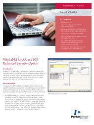
Peaks in the Electropherogram<br />
The profile of charge variants for each<br />
sample is shown in the Electropherogram<br />
tabs of the LabChip GXII software. The<br />
software analyzes these profiles, assigning<br />
a migration time (and other parameters) to<br />
each detected peak. The more basic<br />
variants migrate faster (and appear earlier)<br />
than the more acidic variants. Unless<br />
removed via desalting (see Sample<br />
Preparation section), the electropherogram<br />
will contain a free dye peak that typically<br />
elutes at 30 ± 3 sec, depending on the pH of<br />
the Running Buffer. The free dye peak<br />
should be excluded from the analysis, as<br />
described in the next section.<br />
Figure 3. Electropherogram of a therapeutic mAb. The free dye<br />
peak is labeled with an “X”. The profile of this particular mAb<br />
includes two basic variants (labeled “B1” and “B2”), one main<br />
variant (labeled “Main”), and one acidic variant (labeled “A1”).<br />
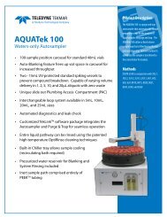
Eliminating Extraneous Peaks<br />
To accurately quantify the % Relative Amount of<br />
each variant, extraneous peaks (such as the free dye<br />
peak and low-signal peaks that appear along the<br />
baseline – see Figure 4) must be excluded from the<br />
analysis. To exclude these peaks, several default<br />
settings of the Peak Find tab of the Analysis Settings<br />
can be adjusted. The default analysis setting of<br />
Minimum Peak Height is 0.2, to allow most peaks to<br />
be detected automatically. To exclude low-signal,<br />
extraneous peaks along the baseline, increase the<br />
Minimum Peak Height. Note that this may also<br />
exclude low-signal peaks associated with the sample<br />
profile. To exclude any remaining peaks in an<br />
electropherogram, right click them individually and<br />
select “Exclude Peak”. Alternatively, if all the sample<br />
profiles in a file span similar time ranges, adjusting<br />
the Start Time (sec) and End Time (sec) to a<br />
narrower range will also eliminate extraneous peaks.<br />
Finally, the Excluded Peaks function under Analysis<br />
Settings may be used to eliminate peaks that appear<br />
at approximately the same migration time in each<br />
profile of a plate, e.g., the free dye peak.<br />
Figure 4. Electropherograms with extraneous peaks<br />
included (top) and excluded (bottom) from analysis.<br />
_________________________________________________________________________________________<br />
Caliper - a <strong>PerkinElmer</strong> Company Page 9 of 18 PN: CLS135474 Rev. 02<br />
68 Elm Street<br />
Hopkinton, MA 01748-1668<br />
1-877-LABCHIP (1-877-522-2447)
<strong>HT</strong> <strong>Protein</strong> <strong>Charge</strong> <strong>Variant</strong> <strong>Kit</strong> <strong>User</strong> <strong>Guide</strong><br />
Optimization of Running Buffer pH<br />
As noted in the Chip Preparation section, the pH of the Running Buffer has a strong effect on the resolution of<br />
charge variants. Typically, as the pH increases, the resolution improves. However, this improvement comes at<br />
the cost of slower migration. The optimum pH for a particular sample balances these two factors. To find this<br />
optimum, we recommend the following procedure:<br />
Initial Test<br />
0) Run samples using the recommendations of Table 2 (see Running the Assay section).<br />
a. If the pIs of samples fall in multiple pI ranges in Table 2, use lowest pH and longest assay type.<br />
pH Optimization<br />
1) Rinse and aspirate each active well of chip (see Chip Preparation section).<br />
2) Prepare Running Buffer at 0.2 or 0.3 units higher than pH used previously; add to wells of chip and to a<br />
new Buffer Tube.<br />
3) Run samples using Running Buffer at higher pH.<br />
a. The samples from step 0 may be re-used throughout the optimization.<br />
b. If the profile of the sample was near the end of the separation time in the previous run, use a<br />
longer assay type.<br />
4) Visually compare the profiles from the run at the higher pH to those from the run at the previous pH to<br />
determine whether the resolution has increased.<br />
a. Indications of increased resolution:<br />
i. Separation between peaks has increased.<br />
ii. Number of peaks has increased.<br />
5) Repeat steps 1 - 4 until the resolution no longer improves. Use the final pH in subsequent runs of<br />
sample.<br />
Additionally, it is possible to determine the<br />
optimal pH by examining the reproducibility of<br />
the % Relative Amounts of the variants, as a<br />
function of pH. Higher pH should provide a<br />
higher reproducibility, as the resolution<br />
increases. For this method of optimization,<br />
we recommend labeling and running multiple<br />
samples of the same mAb (at least three), to<br />
increase the statistical significance of results.<br />
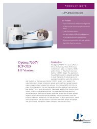
Figure 5 shows the change in the profile of a<br />
mAb when run at various pH. As the pH of<br />
the Running Buffer increases, both the<br />
resolution and the migration time of the<br />
variants increase. Note also, that the peak<br />
height decreases and that the peak width<br />
increases. Finally, the separation between<br />
the free dye peak and the mAb profile<br />
increases at higher pH, so samples with high<br />
pI that may co-elute with the free dye peak at<br />
low pH can be moved away by using a higher<br />
pH.<br />
Figure 5. <strong>Charge</strong> variant profiles of a mAb sample (pI ~8.8) at<br />
various pH.<br />
_________________________________________________________________________________________<br />
Caliper - a <strong>PerkinElmer</strong> Company Page 10 of 18 PN: CLS135474 Rev. 02<br />
68 Elm Street<br />
Hopkinton, MA 01748-1668<br />
1-877-LABCHIP (1-877-522-2447)
<strong>HT</strong> <strong>Protein</strong> <strong>Charge</strong> <strong>Variant</strong> <strong>Kit</strong> <strong>User</strong> <strong>Guide</strong><br />
Troubleshooting<br />
Why don’t I see a profile for my sample?<br />
1. The running buffer pH may be too high or the chosen<br />
assay type may be too short: Try a longer assay<br />
and/or a lower pH.<br />
2. The plate well may contain air bubbles or not enough<br />
sample: The sipper may not be reaching the sample<br />
due to bubbles or low volume. Verify that there is<br />
sufficient sample volume to reach the sipper. To<br />
eliminate bubbles, centrifuge the sample plate for 1<br />
minute at 1000rpm before starting the run.<br />
Alternatively, manually dislodge the bubble using a<br />
pipette tip. Rerun these sample wells.<br />
3. The chip sipper may be clogged: Take the LabChip<br />
out of the instrument; carefully insert the sipper into<br />
the pipette tip attached to the vacuum that was used<br />
to aspirate the wells during chip preparation, until the pipette tip reaches the glue bead. With vacuum<br />
running, hold chip in place until several beads of liquid have emerged from the end of the sipper. Place<br />
chip in instrument and restart run.<br />
Why is the signal of my sample low?<br />
1. The Dye Mixture may have been left at room<br />
temperature for too long: The Dye Mixture should be<br />
used immediately (begin dispensing within 5 minutes<br />
of mixing with N,N-dimethylformamide). Extended<br />
incubation of the mixture at room temperature will<br />
cause degradation of the dye and decrease the<br />
amount of dye that can label the sample.<br />
2. The salt and/or excipient content of the sample may<br />
be too high: Higher conductivity of the sample<br />
typically results in less field amplified stacking of the<br />
sample during on-chip injection. Try desalting the<br />
sample before or after the labeling reaction.<br />
Alternatively, if there are high concentrations of<br />
amine-containing buffer or excipient present, then the<br />
signal of the mAb may decrease through competition for dye molecules during labeling.<br />
3. The mAb sample concentration may be too low. The preferred range of input concentration is 0.5 - 10<br />
mg/mL, and the optimal concentration is 2 mg/mL. Some samples, depending on the number of charge<br />
variants, give lower signals than others; make sure that the sample concentration is ≥ 2 mg/mL.<br />
Why are there extra peaks in the profile of my<br />
sample?<br />
Excipients with amines, such as histidine, can be labeled by the <strong>HT</strong><br />
<strong>Protein</strong> <strong>Charge</strong> <strong>Variant</strong> dye and appear in the profile as extra peaks.<br />
To remove these peaks desalt the sample before or after the labeling<br />
reaction.<br />
_________________________________________________________________________________________<br />
Caliper - a <strong>PerkinElmer</strong> Company Page 11 of 18 PN: CLS135474 Rev. 02<br />
68 Elm Street<br />
Hopkinton, MA 01748-1668<br />
1-877-LABCHIP (1-877-522-2447)
<strong>HT</strong> <strong>Protein</strong> <strong>Charge</strong> <strong>Variant</strong> <strong>Kit</strong> <strong>User</strong> <strong>Guide</strong><br />
Why is the resolution of charge variants in my sample so low?<br />
1. The pH of the Running<br />
Buffer may be too low:<br />
Increase the pH to<br />
improve resolution. Higher<br />
pH running buffer<br />
increases the migration<br />
time and allows better<br />
separation. The higher pH<br />
may require the use of a<br />
longer assay time. See<br />
the Optimization of<br />
Running Buffer pH section<br />
for more detail. If the resolution is not sufficient at the highest pH of the Running Buffer system, then the<br />
pI of the sample is most likely outside the allowed range.<br />
2. Excipients or contaminants may be co-migrating with or affecting the migration of charge variants: Desalt<br />
the sample to remove any excipients that may interfere with or alter the profile. If the <strong>HT</strong> <strong>Protein</strong><br />
Express, <strong>HT</strong> Pico <strong>Protein</strong>, or <strong>HT</strong> Glycan Profiling assay was run before the <strong>HT</strong> <strong>Protein</strong> <strong>Charge</strong> <strong>Variant</strong><br />
assay, then clean the electrodes with molecular biology-grade water.<br />
How can I eliminate carryover, if it is greater than<br />
0.5%?<br />
1. Desalt the sample: Samples that contain cell culture media<br />
or high concentrations of salt / excipients / amine-containing<br />
buffer can potentially increase carryover. Desalting the<br />
sample prior to labeling may reduce the carryover.<br />
2. Use a longer assay type: Longer assays are better at<br />
suppressing carryover.<br />
Why does the migration time of the same sample vary?<br />
The microchannels of the chip have a dynamic coating that suppresses electroosmotic flow during the<br />
separation; variation in the degree of coating causes variation in migration time. Typically, the migration time of a<br />
sample will vary by 0.5 - 2 seconds, with low pI samples having higher variability than high pI samples. Different<br />
chips, GXII instruments, and Running Buffer lots can contribute to the variability. Furthermore, the variability in<br />
migration time is typically lower for chips that have run more samples or have been stored with <strong>HT</strong> <strong>Protein</strong><br />
<strong>Charge</strong> <strong>Variant</strong> Storage Buffer for a longer time. Variation in migration time should not impact resolution or %<br />
relative amount reproducibility. When significant variation in migration time or abnormally slow migration (i. e.,<br />
when the free dye peak elutes after ~33 s) is observed, we recommend that the chip is washed two or three<br />
times with storage buffer and then stored overnight at room temperature, to regenerate the dynamic coating.<br />
_________________________________________________________________________________________<br />
Caliper - a <strong>PerkinElmer</strong> Company Page 12 of 18 PN: CLS135474 Rev. 02<br />
68 Elm Street<br />
Hopkinton, MA 01748-1668<br />
1-877-LABCHIP (1-877-522-2447)
<strong>HT</strong> <strong>Protein</strong> <strong>Charge</strong> <strong>Variant</strong> <strong>Kit</strong> <strong>User</strong> <strong>Guide</strong><br />
Why is the CV of the % Relative Amount greater than the specified value?<br />
Adjust the pH of the Running Buffer to confirm that the resolution is optimal. When running multiple samples of<br />
the same molecule, dilute the samples so the concentration of each differs by ±0.5 mg/mL or less, to eliminate<br />
this source of variability. Desalt samples before or after labeling to (1) remove excipients that may interfere with<br />
the profile, and (2) increase the signal.<br />
In existing data, check the Show Peak Baselines option of the Properties tab to make sure that the software<br />
analyzes each profile of the same sample in the same way (same number of peaks, with peak baselines defined<br />
at the same position within the profile). Adjust the Peak Find parameters in the Analysis Settings interface, such<br />
as the Filter Width (sec) or Slope Threshold (/sec), to improve the reproducibility of the peak find algorithm. Make<br />
sure all extraneous peaks are excluded (see Typical Results section). Also make sure that all relevant peaks are<br />
included, if summing basic and acidic variants.<br />
What are good samples to use as positive controls?<br />
Amino acids such as lysine or histidine can be used as positive control samples to make sure that the<br />
electrophoretic separation and the labeling reaction are working properly. The amino acids are labeled at a<br />
concentration of 1-10 mM. As shown in the figures below, the migration time of lysine decreases slightly with<br />
increasing Running Buffer pH. Conversely, the migration time of histidine increases significantly with increasing<br />
Running Buffer pH.<br />
Why do electropherograms change when wells from different assays are<br />
selected at the same time?<br />
If a well from the longer assay is selected first, then all data will appear in the electropherogram (including the<br />
irrelevant data of the shorter assay, collected during the sip – see Assay Performance section of FAQ). However,<br />
if a well from the shorter assay is selected first, then all data will be truncated at the total assay time of the<br />
shorter assay (68s or 90s). The additional 20 s of irrelevant data in the shorter run may include artifact peaks,<br />
particularly if the profile of the sample is near the end of the separation time (48 s or 70 s); set the end time to 48<br />
s or 70 s, for the 68s and 90s assay, respectively, to exclude these artifact peaks from the analysis. This issue<br />
will be addressed in a future software update.<br />
_________________________________________________________________________________________<br />
Caliper - a <strong>PerkinElmer</strong> Company Page 13 of 18 PN: CLS135474 Rev. 02<br />
68 Elm Street<br />
Hopkinton, MA 01748-1668<br />
1-877-LABCHIP (1-877-522-2447)
<strong>HT</strong> <strong>Protein</strong> <strong>Charge</strong> <strong>Variant</strong> <strong>Kit</strong> <strong>User</strong> <strong>Guide</strong><br />
Why is the baseline noisy, the migration of my sample slow, and the resolution<br />
of the variants poor?<br />
If the <strong>HT</strong> <strong>Protein</strong> Express, the <strong>HT</strong> Low MW <strong>Protein</strong>, the <strong>HT</strong> Pico<br />
<strong>Protein</strong>, or the ProfilerPro Glycan Profiling assay was run on the<br />
instrument, then the electrodes must be cleaned before running<br />
the <strong>HT</strong> <strong>Protein</strong> <strong>Charge</strong> <strong>Variant</strong> assay. If the electrodes are not<br />
cleaned, then residual gel will contaminate the chip and<br />
adversely affect the performance. To fix this problem, remove the<br />
chip from the instrument, aspirate and rinse the wells of the chip<br />
as described in the Chip Preparation section, and add fresh<br />
Running Buffer. Next, replace the buffer tube with a new tube<br />
containing fresh Running Buffer. Then, thoroughly clean the<br />
electrodes of the instrument, using molecular biology-grade<br />
water and a swab. Place the chip back in the instrument and<br />
restart the run.<br />
_________________________________________________________________________________________<br />
Caliper - a <strong>PerkinElmer</strong> Company Page 14 of 18 PN: CLS135474 Rev. 02<br />
68 Elm Street<br />
Hopkinton, MA 01748-1668<br />
1-877-LABCHIP (1-877-522-2447)
<strong>HT</strong> <strong>Protein</strong> <strong>Charge</strong> <strong>Variant</strong> <strong>Kit</strong> <strong>User</strong> <strong>Guide</strong><br />
Frequently Asked Questions<br />
<strong>Kit</strong> Components<br />
1. Is there a lower marker for this assay?<br />
No. The assay has been optimized to minimize variation in migration time (see Troubleshooting section<br />
for more details). Alignment to a lower marker should not be necessary.<br />
2. Is there a charge or pI ladder for this assay?<br />
No. Similar to conventional CZE assays, this assay does not provide pI information.<br />
Sample Preparation<br />
1. May other desalting methods be used, besides Zeba Desalting plates and columns?<br />
Yes, however Zeba Desalting plates and columns were used in the validation of the assay and are<br />
therefore preferred.<br />
2. What happens to the dye when it is not used immediately after dilution with DMF?<br />
When left at room temperature for extended periods of time (hours), the dye mixture will degrade,<br />
causing inefficient labeling and resulting in low sample signal.<br />
3. Can labeled samples be used on another day?<br />
We recommend that samples are analyzed immediately after labeling. However, labeled samples may<br />
be frozen and analyzed later. A loss in signal may result from this storage.<br />
4. Can labeled samples be reconstituted in less or more than 60 µL of molecular-grade water?<br />
The 60 µL dilution gives the optimal signal and profile reproducibility. Other dilutions may be used;<br />
however, signal and reproducibility may be compromised.<br />
5. Can the samples be labeled for more than 10 minutes, or less than 10 minutes?<br />
The recommended 10 minutes is the shortest labeling time that will give the specified reproducibility.<br />
Shorter labeling times will compromise reproducibility; longer labeling times are not likely to affect<br />
reproducibility.<br />
6. What is the lowest sample concentration that I can use?<br />
The recommended range is 0.5 - 10 mg/mL. The reproducibility of % Relative Amount measurements<br />
may decrease for concentrations < 0.5 mg/mL, but detection below this limit is possible.<br />
7. Can I label less than 24 samples?<br />
Yes. To label less than 24 samples, reduce the volume of Dye Concentrate and N,Ndimethylformamide<br />
proportionately. For example, to label 12 samples, add 2.5 µL Dye Concentrate to<br />
72.5 µL N,N-dimethylformamide.<br />
8. Can I analyze a sample with a pI outside of the specified range?<br />
Probably not. Lower pI mAbs may migrate too slowly and not elute before the end of the separation<br />
time, even for the 110s assay. Higher pI mAbs may not be sufficiently resolved, even at pH 7.2.<br />
9. Can proteins other than monoclonal antibodies be tested using this assay?<br />
The <strong>HT</strong> <strong>Protein</strong> <strong>Charge</strong> <strong>Variant</strong> assay was developed using only monoclonal antibodies. However, it<br />
may be possible to resolve the variants of other proteins with pI in the range of 7 - 9.5.<br />
_________________________________________________________________________________________<br />
Caliper - a <strong>PerkinElmer</strong> Company Page 15 of 18 PN: CLS135474 Rev. 02<br />
68 Elm Street<br />
Hopkinton, MA 01748-1668<br />
1-877-LABCHIP (1-877-522-2447)
<strong>HT</strong> <strong>Protein</strong> <strong>Charge</strong> <strong>Variant</strong> <strong>Kit</strong> <strong>User</strong> <strong>Guide</strong><br />
Assay Performance<br />
1. How does this assay compare to conventional IEC, CZE and cIEF?<br />
Typically, the resolution of the <strong>HT</strong> <strong>Protein</strong> <strong>Charge</strong> <strong>Variant</strong> assay is the same or higher than that of IEC.<br />
The resolution of the <strong>HT</strong> <strong>Protein</strong> <strong>Charge</strong> <strong>Variant</strong> assay is comparable to conventional CZE, and in<br />
most cases, the profile is similar to cIEF. The % Relative Amounts of variants reported by the <strong>HT</strong><br />
<strong>Protein</strong> <strong>Charge</strong> <strong>Variant</strong> assay are similar to those given by conventional assays. The analysis time of<br />
the <strong>HT</strong> <strong>Protein</strong> <strong>Charge</strong> <strong>Variant</strong> assay is typically 10- to 50-fold faster than that of conventional assays.<br />
2. Why does the electropherogram end 20 seconds before the time stated in the assay name?<br />
The next sample is sipped after the end of the separation of the current sample, for 20 seconds. During<br />
this time, no relevant data are collected and so these 20 seconds are excluded from the<br />
electropherogram. In the assay names, we have given the sum of the times required for the sip and the<br />
separation steps to provide the total time required to analyze each sample.<br />
3. Can I run multiple samples that have different pI at the same pH?<br />
It is possible to run multiple samples with varying pI at the same pH and achieve adequate resolution<br />
of charge variants for each sample. We recommend limiting the range of pI to no more than the ranges<br />
given in Table 2. However, even with this limit, the resolution for all samples may not be optimal.<br />
4. What should I do if I don’t know the pI of my sample?<br />
We suggest running the longest assay, using the running buffer at pH 5.6. Depending on the<br />
resolution, proceed with the optimization of the pH of the running buffer, as described in the<br />
Optimization of Running Buffer pH section.<br />
5. Can I use my own running buffer or modify the current running buffer?<br />
The Running Buffer provided in the kit has been optimized for resolution and reproducibility. We do not<br />
support the use of customer-developed running buffers.<br />
6. Once a chip is used for the <strong>HT</strong> <strong>Protein</strong> <strong>Charge</strong> <strong>Variant</strong> assay, can it be used for DNA/RNA<br />
assays, or vice versa?<br />
No. Once the chip has been used for the <strong>HT</strong> <strong>Protein</strong> <strong>Charge</strong> <strong>Variant</strong> assay, it should be designated for<br />
this assay only. Running other assays with the same chip will compromise the performance of the chip.<br />
DNA/RNA assays may still be run with the same chip.<br />
7. Can this assay be run on the LabChip 90?<br />
No. The LapChip 90 software does not support this assay.<br />
8. If I am encountering difficulties, how do I confirm that the kit is working properly?<br />
Amino acids such as lysine and histidine can be used as control samples, to check the labeling<br />
reaction and the chip. See Troubleshooting section.<br />
_________________________________________________________________________________________<br />
Caliper - a <strong>PerkinElmer</strong> Company Page 16 of 18 PN: CLS135474 Rev. 02<br />
68 Elm Street<br />
Hopkinton, MA 01748-1668<br />
1-877-LABCHIP (1-877-522-2447)
<strong>HT</strong> <strong>Protein</strong> <strong>Charge</strong> <strong>Variant</strong> <strong>Kit</strong> <strong>User</strong> <strong>Guide</strong><br />
LabChip <strong>Kit</strong> Essential Practices<br />
To ensure proper assay performance please follow the important handling practices described below. Failure to<br />
observe these guidelines may void the LabChip <strong>Kit</strong> product warranty. 1<br />
NOTE: It is important to keep particulates out of the chip wells, channels and capillary. Many of the following<br />
guidelines are designed to keep the chips particulate free.<br />
For assay and instrument troubleshooting, refer to the LabChip GX Software Help file, or call Caliper Technical<br />
Support at 1-877-LABCHIP.<br />
General<br />
1. Avoid use of powdered gloves. Use only non-powdered gloves when handling chips, reagents, sample<br />
plates, and when cleaning the instrument electrodes and electrode block.<br />
2. Calibrate laboratory pipettes regularly to ensure proper reagent dispensing. Only the Caliper-supplied<br />
clean room cloth can be used on the chip to clean the detection window.<br />
3. The entire chip surface must be thoroughly dry before use.<br />
4. The sipper must be kept immersed in liquid at all times and should not be exposed to an open<br />
environment for long periods of time.<br />
5. Use care in handling the chip to prevent sipper damage. Damage to the sipper can result in inconsistent<br />
sampling.<br />
6. Avoid exposing the chip to dust by keeping it in a closed environment such as in the chip container, or in<br />
the instrument before and after chip preparation.<br />
Repriming a Chip<br />
1. Press the CHIP button on the front instrument panel to eject the chip cartridge.<br />
2. Reinsert the cartridge by pushing the cartridge back into the instrument.<br />
3. Press the Run button on the main screen of the LabChip GX software to start another run.<br />
Chip Well Aspiration Using a Vacuum<br />
Aspirating with a pipette can leave used reagents in the chip wells. For this reason, we recommend vacuuming<br />
the wells instead. This can be performed by attaching a permanent pipette tip to a house vacuum line with trap<br />
(see Figures 6a and 6b). To avoid contamination, attach a new pipette tip to the permanent tip for each chip<br />
aspiration (see Figure 6c).<br />
Figure 6a Figure 6b Figure 6c<br />
1 Caliper - a <strong>PerkinElmer</strong> Company warrants that the LabChip <strong>Kit</strong> meets specification at the time of shipment, and is free from defects in material and<br />
workmanship. LabChip <strong>Kit</strong>s are warranted for 60 days from the date of shipment. All claims under this warranty must be made within 30 days of the discovery of<br />
the defect.<br />
_________________________________________________________________________________________<br />
Caliper - a <strong>PerkinElmer</strong> Company Page 17 of 18 PN: CLS135474 Rev. 02<br />
68 Elm Street<br />
Hopkinton, MA 01748-1668<br />
1-877-LABCHIP (1-877-522-2447)
<strong>HT</strong> <strong>Protein</strong> <strong>Charge</strong> <strong>Variant</strong> <strong>Kit</strong> <strong>User</strong> <strong>Guide</strong><br />
Reordering Information<br />
Product<br />
Part Number<br />
<strong>HT</strong> <strong>Protein</strong> <strong>Charge</strong> <strong>Variant</strong> <strong>Kit</strong><br />
CLS760670<br />
<strong>HT</strong> DNA5K/RNA/CZE LabChip 760435<br />
Buffer Tube<br />
E&K Scientific 697075-NC<br />
Technical Support<br />
Caliper - a <strong>PerkinElmer</strong> Company<br />
68 Elm Street<br />
Hopkinton, MA 01748-1668<br />
Phone: 1-877-LABCHIP (522-2447)<br />
Fax: 1-508-435-3439<br />
For additional assay and instrument troubleshooting, refer to the LabChip <strong>HT</strong> Software Help file, or call Caliper<br />
Technical Support at 1-877-LABCHIP.<br />
The chip and reagents supplied with this kit are sold with limited rights of use. The chip may only be used with<br />
the specific quantity of reagents supplied with this kit. The purchaser has no right or license to refurbish, reuse,<br />
remanufacture, or otherwise use the chip with any other reagents than those specifically supplied in this kit. For<br />
more information on the terms and conditions of use of these chips and reagents, please read your LabChip<br />
GX <strong>User</strong> <strong>Guide</strong>. Caliper, the Caliper logo, LabChip, and the LabChip logo are registered trademarks of Calipera<br />
<strong>PerkinElmer</strong> Company.<br />
© Copyright Caliper - a <strong>PerkinElmer</strong> Company 2012<br />
http://www.perkinelmer.com<br />
_________________________________________________________________________________________<br />
Caliper - a <strong>PerkinElmer</strong> Company Page 18 of 18 PN: CLS135474 Rev. 02<br />
68 Elm Street<br />
Hopkinton, MA 01748-1668<br />
1-877-LABCHIP (1-877-522-2447)