Teaching Cardiac Arrhythmias: A Focus on ... - CiteSeerX
Teaching Cardiac Arrhythmias: A Focus on ... - CiteSeerX
Teaching Cardiac Arrhythmias: A Focus on ... - CiteSeerX

Create successful ePaper yourself
Turn your PDF publications into a flip-book with our unique Google optimized e-Paper software.
<str<strong>on</strong>g>Teaching</str<strong>on</strong>g> <str<strong>on</strong>g>Cardiac</str<strong>on</strong>g> <str<strong>on</strong>g>Arrhythmias</str<strong>on</strong>g>: A <str<strong>on</strong>g>Focus</str<strong>on</strong>g> <strong>on</strong> Pathophysiology and<br />
Pharmacology<br />
J<strong>on</strong> E. Sprague 1<br />
The Raabe College of Pharmacy, Ohio Northern University, Ada OH 45810<br />
PROLOGUE<br />
The following manuscript reviews a method of teaching the<br />
pathophysiology and pharmacology of cardiac arrhythmias.<br />
This manuscript is supplemented with a complimentary web<br />
site (http://www.<strong>on</strong>u.edu/user/fs/jsprague/AJPE.html). Those<br />
interested in obtaining further informati<strong>on</strong> can use this site for<br />
lecture notes, animati<strong>on</strong>s of cellular mechanisms, practice case<br />
reports, electrocardiograms (ECG) strips, and Powerpoint®,<br />
lecture presentati<strong>on</strong>s. The author hopes that other find this<br />
informati<strong>on</strong> useful in developing strategies for teaching this<br />
subject area.<br />
INTRODUCTION<br />
The teaching of the pathophysiology and pharmacological<br />
treatment of cardiac arrhythmias can be a challenging task. The<br />
integrated modular system (for descripti<strong>on</strong> see reference 1) utilized<br />
at Ohio Northern University (ONU) allows for the pathophysiology<br />
and pharmacology to be followed by the medicinal<br />
chemistry and therapeutic principles involved in the management<br />
of cardiac arrhythmias. Background material necessary<br />
for the comprehensi<strong>on</strong> of cardiac arrhythmias is provided in<br />
the Biomedical Science Modules. Before entering the<br />
Cardiovascular Module, where cardiac arrhythmias are covered<br />
in detail, students have been instructed in i<strong>on</strong>-channel<br />
functi<strong>on</strong> in muscle c<strong>on</strong>tracti<strong>on</strong>, acti<strong>on</strong> potential physiology and<br />
the basics of ECG. The following text is a summary of a fourhour<br />
lecture sequence presented in the Cardiovascular Module<br />
<strong>on</strong> the pathophysiology and pharmacological treatment of cardiac<br />
arrhythmias. These lectures are supplemented by a web<br />
site that c<strong>on</strong>tains primary literature references in a downloadable<br />
format, animati<strong>on</strong>s of cellular mechanisms and practice<br />
case reports, ECG strips, and Powerpoint®, lecture presentati<strong>on</strong>s.<br />
This manuscript is written in a fashi<strong>on</strong> to complement a<br />
web site created specifically for the readers of the American<br />
Journal of Pharmaceutical Educati<strong>on</strong> that c<strong>on</strong>tains all the<br />
above and can be accessed at (http://www.<strong>on</strong>u.edu/user/<br />
fs/jsprague/AJPE.html). General references utilized in developing<br />
these lectures included references 2-4.<br />
MECHANISMS UNDERLYING CARDIAC<br />
ARRHYTHMIAS<br />
Most cardiac arrhythmias result from disorders of impulse formati<strong>on</strong>,<br />
impulse c<strong>on</strong>ducti<strong>on</strong> or a combinati<strong>on</strong> of both.<br />
Disturbances in impulse formati<strong>on</strong> or automaticity can involve<br />
no pathological change in the pacemaker site generating sinus<br />
bradycardia (< 60 bpm) due to slowed sp<strong>on</strong>taneous sinoatrial<br />
(SA) firing or sinus tachycardia (> 100 bpm) due to rapid firing<br />
of the SA node. The development of an ectopic focus can<br />
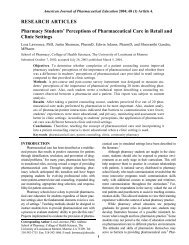
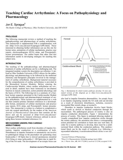
Fig. 1. Mechanisms by which reentry pathways develop. To view animated<br />
versi<strong>on</strong> of this diagram go to (http://www.<strong>on</strong>u.edu/user/fs<br />
/jsprague/AJPE. html).<br />
also lead to impulse formati<strong>on</strong> abnormalities. An ectopic focus<br />
is an impulse originating outside the SA node and can develop<br />
as a result of electrolyte disturbances, ischemia, excessive<br />
myocardial fiber stretch, drugs, or toxins(5).<br />
Disorders in impulse c<strong>on</strong>ducti<strong>on</strong> involve heart blocks,<br />
which result in slowed or blocked c<strong>on</strong>ducti<strong>on</strong> through the<br />
myocardium. The pathological process of reentry is also an<br />
impulse c<strong>on</strong>ducti<strong>on</strong> abnormality. Figure 1 dem<strong>on</strong>strates the<br />
process of reentry. This figure is animated allowing student to<br />
visually comprehend how impulse c<strong>on</strong>ducti<strong>on</strong> circles through<br />
the reentry pathway; the animati<strong>on</strong> also draws a corresp<strong>on</strong>ding<br />
acti<strong>on</strong> potential for correlati<strong>on</strong> to heart rate (see web link). In<br />
order for a reentry pathway to develop, there must be a unidirecti<strong>on</strong>al<br />
block within the c<strong>on</strong>ducti<strong>on</strong> pathway. This unidirecti<strong>on</strong>al<br />
block can be the result of ischemia (e.g. following a<br />
myocardial infarcti<strong>on</strong>). A unidirecti<strong>on</strong>al block al<strong>on</strong>e is not suf<br />
1 Associate Professor of Pharmacology.<br />
2 Pfizer U.S. Pharmaceuticals, Dofetalide package insert (2000).<br />
Am. J. Pharm. Educ., 65, 169-177(2001); received 12/12/00, accepted 3/1/01.<br />
American Journal of Pharmaceutical Educati<strong>on</strong> Vol. 65, Summer 2001 169
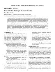
Fig. 2. Premature atrial c<strong>on</strong>tracti<strong>on</strong>s.<br />
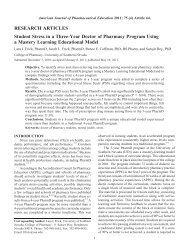
Fig. 4. Multifocal atrial tachycardia.<br />
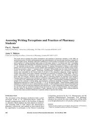
Fig. 3. Sinus tachycardia.<br />
ficient to generate the arrhythmia. At least <strong>on</strong>e of the following<br />
characteristics must be present for the arrhythmia to develop;<br />
l<strong>on</strong>g reentry pathway, short refractory period, or slowed c<strong>on</strong>ducti<strong>on</strong><br />
velocity. All three of these c<strong>on</strong>diti<strong>on</strong>s will allow the<br />
surrounding myocardial tissue to be out of its refractory period<br />
so when the circulating impulse reaches the myocardium a premature<br />
c<strong>on</strong>tracti<strong>on</strong> is generated. Each of these events is<br />
explained in detail. Hand drawings of the reentry pathway<br />
illustrating all three pathological events are given to the class.<br />
Genetic abnormalities in voltage-gated i<strong>on</strong> channel functi<strong>on</strong><br />
have also been linked to arrhythmia generati<strong>on</strong>(6). For example,<br />
the inherited potassium channel disorder that results in the<br />
l<strong>on</strong>g-QT syndrome(6). These examples are discussed in the<br />
previous Biomedical Sciences Module.<br />
TYPES OF CARDIAC ARRHYTHMIAS (5)<br />
After review of a normal ECG and the definiti<strong>on</strong>s of some general<br />
terminology (Appendix A), the students are introduced to<br />
the types of cardiac arrhythmia. The expectati<strong>on</strong> is that they<br />
will be able to identify the arrhythmia type <strong>on</strong> lead II and learn<br />
the standard Advanced <str<strong>on</strong>g>Cardiac</str<strong>on</strong>g> Life Support (ACLS) treatment<br />
guidelines for this form of arrhythmia (7,12). ACLS guidelines<br />
are also utilized in assisting the students in arrhythmia identificati<strong>on</strong><br />
and can be accessed by the students <strong>on</strong> the courses<br />
webpage. After the following types of arrhythmias are discussed,<br />
the students are given a handout of twenty different<br />
forms of arrhythmias to identify. A sample of the handout strips<br />
is in Appendix B and a sample handout can be downloaded<br />
from the web link.<br />
The discussi<strong>on</strong> begins with supraventricular arrhythmias.<br />
The students are informed that when they see these types of<br />
arrhythmias to think “protect the ventricles.” They are asked to<br />
recall some previously covered agents (e.g. p-blockers, verapamil,<br />
diltiazem, digoxin) that slow atrioventricular (AV)<br />
nodal c<strong>on</strong>ducti<strong>on</strong> that may be used to “protect the ventricles.”<br />
“Protect the ventricles” basically becomes the theme of this<br />
secti<strong>on</strong> and is reinforced over the entire week that arrhythmias<br />
are covered. The ECG strip in Figure 2 depicts an atrial premature<br />
beat. This form of arrhythmia is similar to a normal<br />
beat except for timing, and possible distorti<strong>on</strong> of the P wave.<br />
Fig. 5. PSVT or PAT.<br />
The square heart diagram (left side of Figure 2) suggests that<br />
this premature beat may be the result of an ectopic focus. Note<br />
that the P wave often has c<strong>on</strong>tours slightly different from sinus<br />
beats and the PR interval is often l<strong>on</strong>g and the QRS complex is<br />
narrow (< 0.10 sec<strong>on</strong>d). No treatment may be necessary for<br />
this type of arrhythmia.<br />
With atrial tachycardias the heart rate is rapid (approximately<br />
150 beats per minute) with atrial impulse generati<strong>on</strong>.<br />
Ventricular rate is also corresp<strong>on</strong>dingly increased and is driven<br />
by the atrial impulses (“protect the ventricles”). Sinus tachycardia<br />
(Figure 3) has complexes that appear normal and are<br />
evenly spaced. The <strong>on</strong>ly apparent abnormality <strong>on</strong> the ECG is<br />
that the rate is greater than 100 beats per minute (bpm).<br />
Multifocal atrial tachycardia (Figure 4) may be the result<br />
of several ectopic foci firing at different rates. P waves can be<br />
c<strong>on</strong>toured resulting if varying lengths of the PR, PP and RR<br />
intervals. Inverted P waves suggest that the impulse generati<strong>on</strong><br />
occurs in a retrograde fashi<strong>on</strong>.<br />
Paroxysmal Supraventricular Tachcardia (PSVT, Figure 5)<br />
are rapid heart rates that result from a regular successi<strong>on</strong> of<br />
ectopic beats in the atria or from a reentry pathway within the<br />
AV node. A PSVT can last anywhere from a few sec<strong>on</strong>ds to as<br />
l<strong>on</strong>g as several days. Two impulse pathways exist within the<br />
AV node. The a and (3 pathways typically allow for directed<br />
impulse c<strong>on</strong>ducti<strong>on</strong> through the AV node. Figure 5 shows that<br />
if a unidirecti<strong>on</strong>al block develops a recycling of the impulse<br />
can occur. A PSVT may result in atrial rates of 160 to 220 bpm,<br />
with normal or inverted P waves. The QRS complex can be<br />
normal, narrow or widened. The shapes of the QRS complex<br />
assist in making therapeutic drug selecti<strong>on</strong>. Therapeutic drug<br />
selecti<strong>on</strong> is discussed in the subsequent therapeutic lectures.<br />
Atrial flutters and fibrillati<strong>on</strong>s can be differentiated from<br />
each other by looking for a rhythmic pattern <strong>on</strong> the ECG,<br />
which indicates a flutter or a “wavy” n<strong>on</strong>-cyclic pattern to the<br />
baseline between QRS complexes, suggesting a fibrillati<strong>on</strong>.<br />
Atrial flutter (Figure 6) can induce rapid atrial rates in excess<br />
of 300 bpm with <strong>on</strong>ly every sec<strong>on</strong>d or third atrial impulse<br />
being c<strong>on</strong>ducted to the ventricles, giving rise to a ventricular<br />
rate of 100-150 bpm (“protect the ventricles”). Rapid flutter (F)<br />
waves may be seen between each of the QRS complexes. A<br />
170 American Journal of Pharmaceutical Educati<strong>on</strong> Vol. 65, Summer 2001
Fig. 6. Atrial flutter.<br />
Fig. 9. Premature ventricular beats or c<strong>on</strong>tracti<strong>on</strong>s (PVC).<br />
Fig. 7. Atrial fibrillati<strong>on</strong>.<br />
Fig. 10. Ventricular tachycardia.<br />
Fig. 8. Juncti<strong>on</strong>al rhythm.<br />
flutter is defined as a rhythmic cycling of an electrical impulse<br />
and a fibrillati<strong>on</strong> is defined as uncoordinated and “out-of-c<strong>on</strong>trol”<br />
impulse c<strong>on</strong>ducti<strong>on</strong>.<br />
Atrial fibrillati<strong>on</strong> (Figure 7) results from quivering, uncoordinated<br />
atrial activity, which produces an irregular ventricular<br />
rhythm. There are two types of atrial fibrillati<strong>on</strong>: course and<br />
fine fibrillati<strong>on</strong>. A course atrial fibrillati<strong>on</strong> is characterized by<br />
a “saw-tooth” like baseline before each QRS complex.<br />
Whereas a fine atrial fibrillati<strong>on</strong> is a smooth wavy almost flat<br />
line before each QRS complex. P waves may be absent and the<br />
ventricular resp<strong>on</strong>se is irregular and again can be narrow or<br />
wide.<br />
Juncti<strong>on</strong>al rhythm (nodal rhythm, Figure 8) results from<br />
the cells at the juncti<strong>on</strong> of the atrium and the AV node depolarizing<br />
sp<strong>on</strong>taneously and may even become the “pacemaker”<br />
site determining overall cardiac rhythm (“Escape beat”).<br />
Normal anterograde c<strong>on</strong>ducti<strong>on</strong> into the ventricles results in a<br />
typical QRS complex, whereas retrograde c<strong>on</strong>ducti<strong>on</strong> into the<br />
atria produces a P wave that is often inverted and may actually<br />
occur after the QRS complex or not at all.<br />
Ventricular premature beats (Figure 9) are characterized as<br />
ventricular c<strong>on</strong>tracti<strong>on</strong>s not coupled to an atrial impulse and<br />
occur prior to the next expected normal SA-initiated QRS<br />
resp<strong>on</strong>se. A series of premature ventricular c<strong>on</strong>tracti<strong>on</strong>s can be<br />
suggestive of ventricular tachycardia may so<strong>on</strong> follow.<br />
Subsequently, the therapeutic lectures examine the rate of premature<br />
ventricular c<strong>on</strong>tracti<strong>on</strong>s (PVC) and when and how<br />
aggressively they need to be treated. The QRS complex is typically<br />
abnormal and distorted in shape.<br />
Fig. 11. Ventricular fibrillati<strong>on</strong>.<br />
Ventricular tachycardia (Figure 10) result in rapid ventricular<br />
rates not initiated by SA, atrial, or AV sources (recall these<br />
would be termed “supraventricular” tacycardia). This form of<br />
arrhythmia can result in heart rates in excess of 120 bpm and<br />
are comm<strong>on</strong>ly seen in associati<strong>on</strong> with ischemic tissue damage<br />
resulting in circulating or reentry of impulses within the<br />
ischemic z<strong>on</strong>e. Students are told to think of ventricular tachycardia<br />
as the “flutter” of the ventricles. That is they typically<br />
have some form of c<strong>on</strong>sistent pattern associated with their<br />
development.<br />
Ventricular fibrillati<strong>on</strong> (Figure 11) results from chaotic<br />
ventricular activity depicted by bizarre and uncoordinated<br />
ECG traces. Circulatory arrest occurs within sec<strong>on</strong>ds and death<br />
within minutes if not corrected immediately. As with atrial fibrillati<strong>on</strong>,<br />
two forms of ventricular fibrillati<strong>on</strong> are seen: coarse<br />
and fine. The typical progressi<strong>on</strong> of these last three forms of<br />
ventricular arrhythmia is PVC, followed by ventricular tachycardia,<br />
followed by ventricular fibrillati<strong>on</strong>. A discussi<strong>on</strong> of<br />
how fine ventricular fibrillati<strong>on</strong> may be mistaken for asystole<br />
then takes place.<br />
Heart block is a delayed or interrupti<strong>on</strong> in the normal<br />
impulse c<strong>on</strong>ducti<strong>on</strong> between the atria and the ventricles. Firstdegree<br />
heart block (Figure 12) is characterized by all impulses<br />
being c<strong>on</strong>ducted through the AV juncti<strong>on</strong> but the c<strong>on</strong>ducti<strong>on</strong><br />
time (PR interval) is abnormally prol<strong>on</strong>ged (> 0.20 sec<strong>on</strong>ds).<br />
Sec<strong>on</strong>d degree heart block results from partial blockade to<br />
impulse c<strong>on</strong>ducti<strong>on</strong>; some impulses are c<strong>on</strong>ducted to the ventricles<br />
but others are blocked. Mobitz I (Wenckebach, Figure<br />
13) is characterized by repetitive cycles of progressively<br />
American Journal of Pharmaceutical Educati<strong>on</strong> Vol. 65, Summer 2001 171
Fig. 12. First-degree heart block.<br />
Fig. 14. Mobitz II or n<strong>on</strong>-Wenckebach heart block.<br />
Fig. 15. Third-degree heart block.<br />
Fig. 13. Mobitz I or Wenckebach heart block.<br />
lengthening of AV c<strong>on</strong>ducti<strong>on</strong> time, eventually leading to n<strong>on</strong>c<strong>on</strong>ducti<strong>on</strong><br />
of <strong>on</strong>e beat (“dropped beat”). This form of arrhythmia<br />
typically has a pattern in that the PR interval gets l<strong>on</strong>ger,<br />
l<strong>on</strong>ger, l<strong>on</strong>ger and then drops out completely and then the pattern<br />
then repeats. Mobitz II (n<strong>on</strong>-Wenckebach, Figure 14)<br />
involves c<strong>on</strong>ducti<strong>on</strong> of some impulses with a c<strong>on</strong>stant AV c<strong>on</strong>ducti<strong>on</strong><br />
time, and n<strong>on</strong>c<strong>on</strong>ducti<strong>on</strong> of other impulses resulting in<br />
the sudden and unpredictable dropping of QRS complexes.<br />
Third degree heart block (Figure 15) results from the loss<br />
of communicati<strong>on</strong> between the atria and ventricles resulting in<br />
the atria and ventricles c<strong>on</strong>tracting in an unorganized fashi<strong>on</strong>.<br />
The c<strong>on</strong>sequence of this can lead to inadequate ventricular filling<br />
and reduced cardiac output resulting in the patient becoming<br />
hemodynamically unstable. With most forms of bradycardia,<br />
treatment may not be necessary unless the patient is hemodynamically<br />
unstable (decreased blood pressure, shock, pulm<strong>on</strong>ary<br />
c<strong>on</strong>gesti<strong>on</strong> etc.). If the patient is hemodynamically<br />
unstable, start treatment with atropine(7). Students are then<br />
challenged to recall why and how atropine would increase<br />
heart rate.<br />
Escape beats are ectopic beats resulting from sinus node<br />
failure. This serves a protective functi<strong>on</strong> by initiating a cardiac<br />
impulse in the absence of the normal pacemaker activity, and<br />
thereby prevents cardiac standstill. If arrest of the SA node<br />
occurs, a variable period of asystole will usually give way to an<br />
escape beat. Depending <strong>on</strong> the site of origin of the escape beat,<br />
the ECG reflects the resultant c<strong>on</strong>ducti<strong>on</strong> abnormality (Figure<br />
16). Time is spent explaining how the site of origin of the<br />
escape beat results in the various changes in the ECG depicted<br />
in Figure 16.<br />
Some brief comments <strong>on</strong> some other forms of arrhythmia<br />
that the students may encounter are then discussed. Wolf-<br />
Parkins<strong>on</strong> White Syndrome (WPW, Figure 17) is a form of cardiac<br />
arrhythmia that results from the AV node being by-passed<br />
via the Bundle of Kent. This form of arrhythmia is characterized<br />
by the presence of a delta wave prior to the QRS complex.<br />
Fig.16. Different sites of origin of escape beats and their effects <strong>on</strong><br />
the ECG strip.<br />
The students are asked what is the rule for SVTs?...”protect<br />
the ventricles.” Examining the diagram in Figure 17, blocking<br />
AV nodal c<strong>on</strong>ducti<strong>on</strong> would result in increased impulse c<strong>on</strong>ducti<strong>on</strong><br />
through the Bundle of Kent and ventricular rate could<br />
actually increase. Our “rule” doesn't hold for this from of SVT.<br />
Torsades de Pointes, meaning twisting of points, is typically<br />
drug induced. The twisting of points refers to the QRS<br />
complexes twisting al<strong>on</strong>g the isoelectric line. Typically, this is<br />
dem<strong>on</strong>strated by a hand drawing and a sample case report is<br />
then utilized in breakout groups for further discussi<strong>on</strong> of<br />
Torsades. Breakout groups not <strong>on</strong>ly focus <strong>on</strong> Torsades but <strong>on</strong><br />
at least four or five other forms of arrhythmia discussed in<br />
class.<br />
The final form of arrhythmia discussed is Pulseless<br />
Electrical Activity (PEA), which is characterized by the<br />
absence of any detectable pulse in the presence of some electrical<br />
activity. PEA can be caused by hypovolemia, hypoxia,<br />
tensi<strong>on</strong> pneumothorax, hypothermia, and hyperkalemia.<br />
172 American Journal of Pharmaceutical Educati<strong>on</strong> Vol. 65, Summer 2001
Table I. Vaughan Williams classificati<strong>on</strong> of antiarrhythmic<br />
agents<br />
Class Acti<strong>on</strong> Drugs<br />
I<br />
IA<br />
Sodium channel blockade<br />
quinidine<br />
procainamide<br />
disopyramide<br />
IB<br />
lidocaine<br />
tocainide<br />
mexiletine<br />
phenytoin<br />
Fig. 17. Wolf-Parkins<strong>on</strong> White syndrome.<br />
THE PHARMACOLOGY OF ANTIARRHYTHMIC<br />
AGENTS(2,4,8)<br />
After discussing the pathophysiology of cardiac arrhythmias<br />
and how to recognize them, the pharmacology of the agents<br />
used in arrhythmia therapy is discussed. One difficulty that<br />
must be overcome when explaining the methods for treating<br />
cardiac arrhythmias is the fact that drug therapy can result in<br />
the development of another arrhythmia (proarrhythmia) and<br />
other toxicities. Because of the lack of effective resp<strong>on</strong>se and<br />
some studies showing that antiarrhythmic agents can actually<br />
increase mortality (<str<strong>on</strong>g>Cardiac</str<strong>on</strong>g> Arrhythmia Suppressi<strong>on</strong> Trial<br />
[CAST](9), several newer techniques are being developed for<br />
cardiac arrhythmias. The use of ablati<strong>on</strong> therapy for c<strong>on</strong>ducti<strong>on</strong><br />
disorders such as WPW has been very successful(10). This<br />
method of treatment uses radiofrequency to destroy the Bundle<br />
of Kent and return c<strong>on</strong>ducti<strong>on</strong> to normal(10). Ablati<strong>on</strong> therapy<br />
is discussed in the subsequent therapeutic discussi<strong>on</strong>s. In the<br />
future, drug therapy may indeed become sec<strong>on</strong>dary to these<br />
other methods of regulating cardiac arrhythmias. Until this<br />
time arises the pharmacological agents are the main stay in<br />
treating arrhythmias and need to be discussed in detail.<br />
For clarificati<strong>on</strong>, the Vaughan Williams Classificati<strong>on</strong>(11)<br />
of antiarrhythmic agents is still used in the Cardiovascular<br />
Module. This classificati<strong>on</strong> method is beginning to lose favor<br />
for classifying antiarrhythmics because a few agents do not fit<br />
into this classificati<strong>on</strong> system. However, the Vaughan Williams<br />
Classificati<strong>on</strong> allows the anti-arrhythmic agents to be classified<br />
into four classes with just a few excepti<strong>on</strong>s. The Class I agents<br />
(see Table I) are sodium channel blockers that are subdivided<br />
into subgroups based <strong>on</strong> their potency and differential effects<br />
<strong>on</strong> repolarizati<strong>on</strong>. The therapeutic selectivity is provided by the<br />
greater affinity these agents have for active (phase 0) and inactive<br />
(phase 1,2, and 3) sodium channels, but very low affinity<br />
for resting channels. A few minutes are spent reviewing the<br />
three phases of the sodium channel (see web link). The Class<br />
IA agents have moderate potency for sodium channel block<br />
and prol<strong>on</strong>ging repolarizati<strong>on</strong> (potassium efflux block). Class<br />
IB agents have the lowest potency for the sodium channel and<br />
they actually shorten repolarizati<strong>on</strong>. The Class IB agents are<br />
c<strong>on</strong>sidered the safest of the Class I agents and are most comm<strong>on</strong>ly<br />
used first line in the acute treatment of cardiac arrhythmias.<br />
The Class IC agents are the most potent sodium channel<br />
blockers and have limited effects <strong>on</strong> repolarizati<strong>on</strong>. The Class<br />
IC agents are associated with the greatest degree of adverse<br />
reacti<strong>on</strong>s (CAST trial) and are c<strong>on</strong>sidered the least safe(9). The<br />
IC<br />
IA,B,C (mixed)<br />
flecainide<br />
propafen<strong>on</strong>e<br />
encainide<br />
moricizine<br />
II ß-Adrenergic blockers propranolol<br />
acebutolol<br />
esmolol<br />
sotalol<br />
III Agents that prol<strong>on</strong>g Phase 3 bretylium<br />
amiodar<strong>on</strong>e<br />
ibutalide<br />
sotalol<br />
dofetilide<br />
IV Calcium channel blockade verapamil<br />
diltiazem<br />
Digitalis<br />
glycosides<br />
Adenosine<br />
Increase slope of Phase 4<br />
Reduce SA node automaticity<br />
Reduce AV node c<strong>on</strong>ducti<strong>on</strong><br />
digoxin<br />
digitoxin<br />
adenosine<br />
final class is the mixed agent with IA,B,C qualities that has<br />
moderate sodium channel blockade and <strong>on</strong>ly slight effects <strong>on</strong><br />
repolarizati<strong>on</strong>.<br />
Class II agents are the (3-adrenergic blocking agents that<br />
depress phase 4 depolarizati<strong>on</strong> by blocking the ß 1 , receptors.<br />
The depressi<strong>on</strong> of phase 4 depolarizati<strong>on</strong> can be a c<strong>on</strong>fusing<br />
c<strong>on</strong>cept and the students are taught to think of this terminology<br />
in the following way. Depressi<strong>on</strong> of phase 4 depolarizati<strong>on</strong><br />
results in increased time between acti<strong>on</strong> potential generati<strong>on</strong>.<br />
Correlating this resp<strong>on</strong>se to the ECG would transpire to an<br />
increase in the PR interval. This is typically hand drawn with<br />
both an acti<strong>on</strong> potential and ECG trace.<br />
Class III antiarrhythmic agents prol<strong>on</strong>g phase 3 repolarizati<strong>on</strong><br />
predominantly by blocking potassium efflux. Dofetilide<br />
and ibutilide prol<strong>on</strong>g phase 3 repolarizati<strong>on</strong> by mechanisms<br />
other than potassium efflux blockade.<br />
Class IV antiarrhythmic agents are the calcium channel<br />
blockers (CCB) that depress phase 4 depolarizati<strong>on</strong> and prol<strong>on</strong>g<br />
phases 1 and 2. Only two CCBs, verapamil and diltiazem,<br />
are used for the treatment of arrhythmias (Table I). The students<br />
are asked questi<strong>on</strong>s as to why this would be the<br />
case...because of a reflex tachycardia seen with dihydropyridine<br />
CCBs like nifedipine.<br />
Finally, the digitalis gylcosides and adenosine are discussed<br />
separately in the initial presentati<strong>on</strong> of the classificati<strong>on</strong><br />
American Journal of Pharmaceutical Educati<strong>on</strong> Vol. 65, Summer 2001 173
of antiarrhythmic agents. The students are also presented with<br />
acr<strong>on</strong>yms for remembering the different agents in each class.<br />
For example, for the Class IA agents, procainamide, disopyramide,<br />
and quinidine, they are given PDQ (Pretty Darn Quick).<br />
Once a general overview of the different classes of antiarrhythmic<br />
agents is presented, the individual agents are discussed<br />
in detail. Structures of the agents are given at this point<br />
so as to compliment the subsequent medicinal chemistry lectures.<br />
Class IA<br />
Quinidine. Quinidine is the most comm<strong>on</strong>ly used oral antiarrhythmic<br />
agent. Quinidine's therapeutic pharmacological<br />
effects are to depress the pacemaker rate and to reduce c<strong>on</strong>ducti<strong>on</strong><br />
and excitability. <str<strong>on</strong>g>Cardiac</str<strong>on</strong>g> toxicity due to the drug's<br />
antimuscarinic activity may overcome myocardial depressant<br />
effects and lead to an increase in sinus rate and increased AV<br />
c<strong>on</strong>ducti<strong>on</strong>. The c<strong>on</strong>cept of proarrhythmia is introduced here<br />
and explained as an antiarrhythmic drugs ability to cause or<br />
unmask another arrhythmia. Although an older method for<br />
administrati<strong>on</strong>, digoxin may be administered prior to quinidine<br />
in the presence of atrial fibrillati<strong>on</strong> or flutter to prevent ventricular<br />
tachycardia. Digoxin will slow AV nodal c<strong>on</strong>ducti<strong>on</strong><br />
and protect the ventricles. This treatment strategy is <strong>on</strong>ly used<br />
acutely due to quinidine's ability to decrease the renal clearance<br />
of digoxin. An early sign of serious toxicity with any<br />
Class IA agent is an increase in the QRS complex width by ><br />
30 percent. Students are questi<strong>on</strong>ed as to why QRS complex<br />
may widen with toxicity. This allows for a review of the role of<br />
sodium channels in the development of the QRS complex.<br />
Hypotensi<strong>on</strong> may result from a reduced cardiac output as well<br />
as from a vasodilati<strong>on</strong> caused by a-receptor antag<strong>on</strong>ism.<br />
Quinidine is c<strong>on</strong>traindicated in partial or complete AV block,<br />
severe renal disease resulting in azotemia, digitalis-induced<br />
arrhythmias, myasthenia gravis (students are asked<br />
why?...myasthenia gravis is covered in the previous quarter),<br />
and history of Torsades de Pointes. Torsades de Pointes may be<br />
seen with any of the agents that have the ability to inhibit<br />
potassium efflux such as quinidine. This fact is reiterated<br />
throughout the remaining discussi<strong>on</strong> of agents used in the treatment<br />
of cardiac arrhythmias. Quinidine can be used to treat<br />
atrial arrhythmias such as PAC, Atrial Fibrillati<strong>on</strong>, Atrial<br />
Flutter; SVTs such as WPW, AV nodal reciprocating tachycardia<br />
and ventricular arrhythmias such as PVC, ventricular<br />
tachycardia and for the preventi<strong>on</strong> of ventricular fibrillati<strong>on</strong>.<br />
Procainamide. The direct effects of procainamide <strong>on</strong> the heart<br />
are very similar to quinidine, but has some indirect effects that<br />
are quite different from those of quinidine. Procainamide has<br />
much weaker anticholinergic activity <strong>on</strong> the heart and does not<br />
produce a-receptor antag<strong>on</strong>ism. Additi<strong>on</strong>ally, procainamide has<br />
weak gangli<strong>on</strong>ic blocking activity giving it greater negative<br />
inotropic effects than quinidine. Unlike quinidine, procainamide<br />
may produce a syndrome resembling lupus erythematosus,<br />
which is characterized by arthralgia and arthritis. An antinuclear<br />
antibody (ANA) test can be performed here to c<strong>on</strong>firm a diagnosis<br />
similar to what we had already discussed with hydralazine<br />
during the hypertensi<strong>on</strong> lectures. Procainamide is also acetylated<br />
to an active metabolite acecainide or n-acetylprocainamide<br />
(NAPA). Finally, because of the drugs ability to induce proarrhythmias<br />
and b<strong>on</strong>e marrow suppressi<strong>on</strong>, procainamide is typically<br />
reserved for arrhythmias deemed life-threatening.<br />
Disopyramide. Pharmacologically, disopyramide is similar to<br />
quinidine but does not have α or β receptor activity.<br />
Disopyramide is structurally related to the anticholinergic<br />
agent, isopropamide. Therefore, typical anticholinergic side<br />
effects can be seen. Students are asked to predict some examples<br />
of these anticholinergic side effects. Disopyramide can<br />
also reduce cardiac output and reduce left ventricular performance<br />
by a direct depressant effect and cauti<strong>on</strong> is warranted in<br />
heart failure patients. The fact that disopyramide is reserved for<br />
life-threatening arrhythmias is also stressed to the students.<br />
Class IB<br />
Lidocaine. Because of the low incidence of toxicity associated<br />
with class IB agents, lidocaine is the most comm<strong>on</strong>ly used<br />
intravenous (IV) antiarrhythmic agent. Lidocaine has extraordinarily<br />
high degree of efficacy, especially in treating ventricular<br />
arrhythmias occurring after cardiac surgery or acute<br />
myocardial infarcti<strong>on</strong>. The IV route of administrati<strong>on</strong> is rapid,<br />
safe and coupled with a fast decline <strong>on</strong>ce the IV infusi<strong>on</strong> is terminated.<br />
Lidocaine blocks both activated and inactivated sodium<br />
channels. A large fracti<strong>on</strong> of unblocked sodium channels<br />
will become blocked during each acti<strong>on</strong> potential in the<br />
Purkinje fibers and the ventricular myocardial cells, which<br />
have l<strong>on</strong>g plateau phases. Lidocaine suppresses the electrical<br />
activity of the depolarized, arrhythmogenic tissue while minimally<br />
interfering with the electrical activity of normal tissue.<br />
Neurological side effects are the most comm<strong>on</strong> and are associated<br />
with the local anesthetic effects produced by central sodium<br />
channel blockade. The discussi<strong>on</strong> is interrupted at this<br />
point and the students are questi<strong>on</strong>ed, “how many of you have<br />
dispensed lidocaine viscous?” A discussi<strong>on</strong> then ensues <strong>on</strong> how<br />
blocking sodium channels can interfere with neurotransmissi<strong>on</strong><br />
and how this can c<strong>on</strong>tribute to lidocaine's adverse reacti<strong>on</strong><br />
profile. Lidocaine undergoes very extensive first-pass hepatic<br />
metabolism with <strong>on</strong>ly 30 percent of an orally administered<br />
dose appearing in the plasma. Lidocaine is typically c<strong>on</strong>sidered<br />
the drug of choice in suppressing ventricular tachycardia and<br />
preventi<strong>on</strong> of ventricular fibrillati<strong>on</strong> following a MI. Lidocaine<br />
is rarely effective in treating supraventricular arrhythmias but<br />
is effective for those associated with digitalis toxicity.<br />
Tocainde and Mexiletine. These agents are c<strong>on</strong>geners of lidocaine<br />
that are more resistant to gastric acid and relatively resistant<br />
to first-pass metabolism. Electrophysiology and antiarrhythmic<br />
acti<strong>on</strong>s are similar to those of lidocaine. These agents<br />
also have similar indicati<strong>on</strong>s and neurological side effect profiles.<br />
Phenytoin. Phenytoin is currently not approved to treat cardiac<br />
arrhythmias. However, phenytoin is quite effective in treating<br />
life-threatening atrial and ventricular arrhythmias caused by<br />
digitalis overdose, which have failed to resp<strong>on</strong>d to potassium<br />
salts.<br />
Class IC<br />
Flecainide and Encainide. The manufacturer voluntarily<br />
recalled encainide in 1991, but it can be obtained for compassi<strong>on</strong>ate<br />
use by c<strong>on</strong>tacting the manufacturer directly. Both of<br />
these agents have rather selective depressant effects <strong>on</strong> the fast<br />
sodium channels and reduce the velocity and amplitude of<br />
phase 0. They have also been shown to slow c<strong>on</strong>ducti<strong>on</strong> in cardiac<br />
tissue especially the His-Purkinje system. These agents<br />
are <strong>on</strong>ly indicated for life-threatening ventricular arrhythmias.<br />
174 American Journal of Pharmaceutical Educati<strong>on</strong> Vol. 65, Summer 2001
Table II. Side effect table for propafen<strong>on</strong>e<br />
GI (most comm<strong>on</strong>) Cardiovascular (most serious) Neurological<br />
nausea proarrhythmia (5-10%) dizziness<br />
vomiting negative inotropic effects headache<br />
c<strong>on</strong>stipati<strong>on</strong> CHF visual changes<br />
dry mouth<br />
AV and bundle branch block<br />
bitter, metallic or unusual taste<br />
This is mainly due to the results of the CAST study which<br />
showed that flecainide and encainide increases mortality 2.5<br />
times over no treatment(9). Other adverse reacti<strong>on</strong>s that are<br />
discussed are proarrhythmia and negative inotropy.<br />
Propafen<strong>on</strong>e. Propafen<strong>on</strong>e does a little bit of everything needed<br />
for the treatment of arrhythmia. Propafen<strong>on</strong>e is structurally<br />
similar to propranolol and possess about l/40th the ß-blocking<br />
activity of propranolol. Propafen<strong>on</strong>e also has weak Class III<br />
properties, which results in prol<strong>on</strong>gati<strong>on</strong> of repolarizati<strong>on</strong>. To<br />
complete its “little bit of everything” activity propafen<strong>on</strong>e also<br />
has some weak calcium channel blocking activity. The side<br />
effect profile of propafen<strong>on</strong>e is also discussed in detail as to the<br />
cellular mechanisms involved in producing the adverse<br />
resp<strong>on</strong>se (Table II). Finally, the discussi<strong>on</strong> of propafen<strong>on</strong>e ends<br />
with the discussi<strong>on</strong> of some major drug interacti<strong>on</strong>s with<br />
digoxin and warfarin.<br />
Class 1A, B, C (Mixed)<br />
Moricizine. Moricizine is a phenothiazine derivative, without<br />
significant activity <strong>on</strong> the dopaminergic system. Moricizine<br />
reduces the rate of phase 0 depolarizati<strong>on</strong> without affecting<br />
maximum diastolic potential or acti<strong>on</strong> potential amplitude.<br />
C<strong>on</strong>tradictory to what we have discussed to this point is the<br />
fact that the acti<strong>on</strong> potential durati<strong>on</strong> (APD) and effective<br />
refractory period (ERP) are both decreased. This requires a<br />
hand drawing to explain how shortening the APD may actually<br />
“normalize” rate in some sick tissue. This can result in the<br />
normalizati<strong>on</strong> of heart rate in some tissue. Moricizine has minimal<br />
effects <strong>on</strong> the sinus node or atrial tissue. Thus, this agent<br />
is most extensively used for the suppressi<strong>on</strong> of ventricular<br />
arrhythmias. As with most anti-arrhythmic agents, moricizine<br />
may lead to a proarrhythmic resp<strong>on</strong>se, which could result in<br />
sudden cardiac death.<br />
Class II: ß-Adrenergic Blocking Agents<br />
The ß-blocking agents are covered in much detail in the<br />
lectures <strong>on</strong> hypertensi<strong>on</strong> and the introductory aut<strong>on</strong>omic lectures.<br />
Thus, for time utilizati<strong>on</strong> the ß-blocking agents are<br />
reviewed very rapidly. The discussi<strong>on</strong> focuses <strong>on</strong> the following<br />
agents: propranolol, acebutolol, esmolol. Sotalol is menti<strong>on</strong>ed<br />
at this point, but is predominantly a Class III anti-arrhythmic<br />
agent. Most antiarrhythmic effects of these agents are a direct<br />
result of the receptor antag<strong>on</strong>ist activity at the cardiac ß 1 -<br />
receptor. These agents are used to treat supraventricular<br />
arrhythmias. By blocking the ß 1 -receptor influences <strong>on</strong> the AV<br />
node, ERP increases and cardiac impulse c<strong>on</strong>ducti<strong>on</strong> of rapid<br />
atrial depolarizati<strong>on</strong> into the ventricles is reduced (“protect the<br />
ventricles”).<br />
Class III: Prol<strong>on</strong>g Phase 3 Repolarizati<strong>on</strong><br />
Before the discussi<strong>on</strong> <strong>on</strong> the Class III agents begin, the<br />
students are asked, “what form of arrhythmia may be induced<br />
by an agent that prol<strong>on</strong>gs phase 3 repolarizati<strong>on</strong> or blocks<br />
potassium efflux? The answer that the class typically yells out<br />
is “Torsades.”<br />
Bretylium. Bretylium is similar to the post-gangli<strong>on</strong>ic blockers,<br />
quanadrel and quanethidine, that were discussed during the<br />
lectures <strong>on</strong> hypertensi<strong>on</strong>. Bretylium was used as an antihypertensive<br />
in the 1950s. Like the post-gangli<strong>on</strong>ic blockers, bretylium<br />
interferes with catecholamine release from adrenergic neur<strong>on</strong>s.<br />
It is taken up into the nerve terminal, releases norepinephrine<br />
initially (hypertensive resp<strong>on</strong>se) and then prevents its<br />
release later (hypotensive resp<strong>on</strong>se). The use of triclycic antidepressants<br />
can block these resp<strong>on</strong>ses. Students are asked to<br />
recall the mechanism by which this would work. Bretylium's<br />
anti-arrhythmic properties are independent of aut<strong>on</strong>omic<br />
effects. Bretylium lengthens ventricular (but not atrial) APD<br />
and ERP especially in ischemic cells and raises the threshold<br />
for ventricular fibrillati<strong>on</strong>. Up<strong>on</strong> initial administrati<strong>on</strong>, bretylium<br />
causes a release of catecholamines. This may produce an<br />
early positive inotropic effect and may increase the firing rate<br />
of Purkinje fibers. Bretylium is typically reserved for lifethreatening<br />
ventricular arrhythmias and for the prophylaxis and<br />
treatment of ventricular fibrillati<strong>on</strong>.<br />
Amiodar<strong>on</strong>e. Amiodar<strong>on</strong>e has effects that overlap with Class<br />
I and II anti-arrhythmic agents. It shares some properties with<br />
bretylium in that amiodar<strong>on</strong>e slows repolarizati<strong>on</strong> and increases<br />
ventricular fibrillati<strong>on</strong> threshold. Like the Class I agents,<br />
amiodar<strong>on</strong>e is an effective blocker of inactive sodium channels.<br />
It has weak calcium channel blocking activity and is a<br />
n<strong>on</strong>competitive inhibitor of α- and ß-receptors. These effects<br />
result in prol<strong>on</strong>gati<strong>on</strong> of repolarizati<strong>on</strong> and the subsequent<br />
lengthening of the APD and ERP. Amiodar<strong>on</strong>e will also slow<br />
sinus rate and AV nodal c<strong>on</strong>ducti<strong>on</strong>. Extracardic effects include<br />
peripheral vascular dilati<strong>on</strong> as a result of a-blockade and calcium<br />
channel blockade. Pulm<strong>on</strong>ary toxicity includes pulm<strong>on</strong>ary<br />
fibrosis, interstitial pneum<strong>on</strong>itis and alveolitis.<br />
Looking at the structure of amiodar<strong>on</strong>e (Figure 18), the iodine<br />
grouping lend toward the thyroid toxicity seen with amiodar<strong>on</strong>e.<br />
Patients <strong>on</strong> amiodar<strong>on</strong>e should have thyroid functi<strong>on</strong><br />
test performed periodically. Pharmacokinetically, amiodar<strong>on</strong>e<br />
has an extremely l<strong>on</strong>g half-life of greater than a m<strong>on</strong>th in some<br />
patients. Despite all these potential problems, amiodar<strong>on</strong>e has<br />
recently become very popular clinically for the suppressi<strong>on</strong> of<br />
life-threatening ventricular arrhythmias(12). A discussi<strong>on</strong> <strong>on</strong><br />
how clinical applicati<strong>on</strong> of the drug seems to c<strong>on</strong>tradict the<br />
pharmacological and toxicological properties of the compound.<br />
Thus, if dose is adjusted correctly and the patient m<strong>on</strong>itored<br />
closely aminodar<strong>on</strong>e can be used safely.<br />
Sotalol. Because sotalol prol<strong>on</strong>gs repolarizati<strong>on</strong> it is classified<br />
as a class III antiarrhythmic agent. Sotalol is also a n<strong>on</strong>selective<br />
ß-blocking blocker, which possess no intrinsic sympathomimetic<br />
or local anesthetic activity. Its side effect profile is<br />
similar to those typically seen with ß-blockers. Sotalol has<br />
American Journal of Pharmaceutical Educati<strong>on</strong> Vol. 65, Summer 2001 175
Fig. 18. Structure of amiodar<strong>on</strong>e.<br />
some proarrhythmic potential. Such drugs as antihistamines<br />
and tricyclic antidepressants likely exaggerate this proarrhythmic<br />
potential. Sotalol is indicated for the treatment of lifethreatening<br />
arrhythmias.<br />
Ibutalide. Ibutalide delays repolarizati<strong>on</strong> by activati<strong>on</strong> of a<br />
slow, inward current of sodium rather than blocking outward<br />
potassium currents. This makes ibutalide different from all<br />
other Class III agents. In order to explain this effect, an AP is<br />
drawn and the effects of a slow inward flux of sodium <strong>on</strong> prol<strong>on</strong>ging<br />
the APD is explained. Ibutalide is indicated for the<br />
treatment of atrial fibrillati<strong>on</strong>/flutter.<br />
Dofetilide. Dofetilide is a fairly new Class III antiarrhythmic<br />
agent. The students are asked to recall the different forms of<br />
potassium channels. Dofetalide is different than the typical<br />
Class III agents in that it blocks the delayed inward-rectifier<br />
potassium current. Again, an AP is drawn to clarify the effects<br />
of this blockade <strong>on</strong> prol<strong>on</strong>ging APD. Dofetilide is indicated for<br />
“highly symptomatic” atrial fibrillati<strong>on</strong> and flutter. Dofetilide<br />
is reserved for this type of arrhythmia because of the high risk<br />
of developing Torsades de Pointes. In fact, dofetilide is marketed<br />
with a training program to ensure appropriate dosing(12).<br />
Class IV: Calcium-Channel Blocking Agents<br />
The calcium channel blocking (CCB) agents are covered<br />
in great detail in the hypertensi<strong>on</strong> lectures. Thus, for time utilizati<strong>on</strong><br />
the CCB agents are reviewed very rapidly. The discussi<strong>on</strong><br />
here focuses <strong>on</strong> verapamil and diltiazem. These agents<br />
have their most marked effects <strong>on</strong> the SA and AV nodes, which<br />
depend up<strong>on</strong> the calcium current for activati<strong>on</strong>. They result in<br />
depressed SA and AV nodal c<strong>on</strong>ducti<strong>on</strong> and prol<strong>on</strong>gati<strong>on</strong> of the<br />
ERP of the AV node. They slow ventricular rate in the presence<br />
of atrial fibrillati<strong>on</strong> and flutter and are therefore indicated for<br />
reentrant SVT, and PSVT.<br />
Digitalis Glycosides<br />
The cardiac glycosides have indirect vagomimetic acti<strong>on</strong><br />
that decrease ventricular rate and improve ventricular functi<strong>on</strong><br />
by increasing the ERP of the AV node. These agents are indicated<br />
for the treatment of atrial fibrillati<strong>on</strong> and flutter in order<br />
to protect the ventricles. The specific details of the mechanisms<br />
of digoxin are covered prior to this lecture in the discussi<strong>on</strong> of<br />
c<strong>on</strong>gestive heart failure.<br />
Adenosine<br />
Adenosine is an endogenous nucleoside that produces a<br />
bradycardia which is resistant to atropine. Adenosine depresses<br />
SA nodal automaticity and AV nodal c<strong>on</strong>ducti<strong>on</strong>. The electrophysiological<br />
mechanisms involve an increase in potassium<br />
c<strong>on</strong>ductance, reduced calcium mediated slow channel c<strong>on</strong>ducti<strong>on</strong>,<br />
and possible antag<strong>on</strong>ism of catecholamine-mediated<br />
effects. Adenosine will produce a rapid flatline <strong>on</strong> the ECG<br />
m<strong>on</strong>itor, which is shocking but expected. Adenosine could be<br />
c<strong>on</strong>sidered the pharmacological shocking of the heart.<br />
Adenosine is rapidly cleared from the plasma in less than 30<br />
sec<strong>on</strong>ds. Adenosine is the drug of choice in the treatment of<br />
PSVT (12). As with all the antiarrhythmic agents, adenosine is<br />
not devoid of potentially serious adverse reacti<strong>on</strong>s. The students<br />
are informed that as pharmacist they should be aware that<br />
the following adverse reacti<strong>on</strong>s my be seen following adenosine<br />
administrati<strong>on</strong>; dyspnea (12 percent), flushing, retrosternal<br />
chest pain, heart block, asystole, and a transient arrhythmia<br />
at the time of c<strong>on</strong>versi<strong>on</strong>. Potential drug interacti<strong>on</strong>s are also<br />
stressed, dipyridamole inhibits the cellular uptake of adenosine<br />
and will potentiate its effects. Methylxanthines such as caffeine<br />
and theophylline are receptor antag<strong>on</strong>ist of adenosine and may<br />
interfere with its electrophysiologic effects.<br />
PREDICTING DRUG-INDUCED ECG CHANGES<br />
In order to ensure that the students have a thorough understanding<br />
of the pharmacological mechanisms of the antiarrhythmic<br />
agents, they are asked to predict what effects the<br />
agents would have <strong>on</strong> the ECG. Table III list some of the antiarrhythmic<br />
agents discussed in class and the standard ECG<br />
changes induced by the drug (4). For example, with amiodar<strong>on</strong>e,<br />
the PR interval increases as a result of weak calcium<br />
channel blocking and β-blocking activity. The QRS interval<br />
increases due to sodium channel blocking activity and the QT<br />
interval increases by potassium channel blockade induced by<br />
amiodar<strong>on</strong>e. We focus <strong>on</strong> understanding the mechanism and<br />
not memorizing the table. This forces the student to apply their<br />
understanding of drug mechanisms to ECG changes. The class<br />
is then given several drugs to practice predicting ECG changes<br />
<strong>on</strong> and that are then discussed the following day. The exams<br />
list several drugs and ask for predicted ECG changes. This type<br />
of questi<strong>on</strong> test several c<strong>on</strong>cepts emphasized in class from drug<br />
mechanism to understanding an ECG. Furthermore, pharmacy<br />
students then can get a better feel for the importance of understanding<br />
drug mechanism when the pharmacological resp<strong>on</strong>se<br />
can actual be coupled to an ECG change.<br />
DISCUSSION<br />
The teaching of cardiac arrhythmias to pharmacy students can<br />
be a difficult and challenging task. The lectures highlighted<br />
within this manuscript and corresp<strong>on</strong>ding web site<br />
(http://www.<strong>on</strong>u.edu/user/fs/jsprague/AJPE.html) are just <strong>on</strong>e<br />
approach that is used in teaching this subject material in the<br />
Cardiovascular Module at ONU. These lectures are then reinforced<br />
in subsequent lectures <strong>on</strong> medicinal chemistry and therapeutics.<br />
The students also practice analyzing case reports<br />
involving these drugs and arrhythmias in the breakout groups.<br />
Finally, in a capst<strong>on</strong>e course <strong>on</strong>e-year later, the students work<br />
in groups of three and evaluate and give therapeutic recommendati<strong>on</strong>s<br />
for the treatment of randomly generated ECG<br />
strips. During this time, many of the c<strong>on</strong>cepts discussed in this<br />
manuscript are reinforced <strong>on</strong> a more <strong>on</strong>e-<strong>on</strong>-<strong>on</strong>e basis before<br />
the students enter into their clinical rotati<strong>on</strong>s.<br />
Acknowledgements. The author would like to thank Dr.<br />
176 American Journal of Pharmaceutical Educati<strong>on</strong> Vol. 65, Summer 2001
Table III. Antiarrhythmic electrocardiogram effects a<br />
SA nodal AV nodal His-Purkinje<br />
Class Example agent c<strong>on</strong>ducti<strong>on</strong> c<strong>on</strong>ducti<strong>on</strong> c<strong>on</strong>ducti<strong>on</strong> HR PR QRS QT<br />
IA Procainamide +/- +/- _ +/- +/- ↑ ↑<br />
IB Lidocaineb 0 0 0 0 0 0 0-↑<br />
IC Propafen<strong>on</strong>e 0 ↓ ↓ 0 ↑ ↑ ↑<br />
II Esmolol ↓ ↓ 0 ↓ 0-↑ 0 0-↓<br />
III Amiodar<strong>on</strong>e ↓ ↓ ↓ ↓ ↑ ↑ ↑<br />
IV Verapamil ↓ ↓ 0 ↓ ↑ 0 0<br />
Digoxin 0-↓ ↓ 0-↓ ↓ ↑ 0 ↓<br />
Adenosine ↓ ↓ 0 ↑ ↑ 0 0<br />
a for expanded chart see Facts and Comparis<strong>on</strong>'s 1998.<br />
b lidocaine's major effect is <strong>on</strong> an ectopic pacemaker.<br />
Note: each agent in the above table not <strong>on</strong>ly produces changes characteristic of its class (e.g. IA) but also it own unique pharmacology.<br />
D<strong>on</strong>nie Sullivan and Mr. Jeffery Blake for their thorough critique<br />
and evaluati<strong>on</strong> of several drafts of this manuscript.<br />
References<br />
(1) Sprague, J.E. Christoff, J. Allis<strong>on</strong>, J. Kisor, D. and Sullivan, D.,<br />
“Development and Implementati<strong>on</strong> of an Integrated Cardiovasicular<br />
Module in a Pharm.D. Curriculum,” Am. J. Pharm. Edu., 64, 20-<br />
26(2000).<br />
(2) Roden, D.M., “Antiarrhythmic drugs,” in Goodman and Gilman's<br />
Pharmacological Basis of Therapeutics, (edit., Harman, J.E. Limbird,<br />
L.E. Molinoff, P.B. Rudd<strong>on</strong>, R.W. and Gilman, A.B.), 9th editi<strong>on</strong><br />
McGraw-Hill, NY (1996) pp. 839-874.<br />
(3) Advanced <str<strong>on</strong>g>Cardiac</str<strong>on</strong>g> Life Support Committee, Advanced <str<strong>on</strong>g>Cardiac</str<strong>on</strong>g> Life<br />
Support, American Heart Associati<strong>on</strong> (1997).<br />
(4) Drug Facts and Comparis<strong>on</strong>, Drugs Facts and Comparis<strong>on</strong>, St. Louis<br />
MO (2000), pp. 405-437.<br />
(5) Advanced <str<strong>on</strong>g>Cardiac</str<strong>on</strong>g> Life Support Committee “<str<strong>on</strong>g>Arrhythmias</str<strong>on</strong>g>,” in Advanced<br />
<str<strong>on</strong>g>Cardiac</str<strong>on</strong>g> Life Support, American Heart Associati<strong>on</strong>, (1997), pp. 3.1-3.24.<br />
(6) Splawski, I. Shen, J. Timothy, K. Lehmann, M. Priori S. Robins<strong>on</strong>, J.<br />
Moss, A. Schwartz, P., Towbin, J. Vincent, M. and Keatin, M., “Spertrum<br />
of mutati<strong>on</strong>s in l<strong>on</strong>g-QT syndrome genes KVLQT1, HERG, SCN5A,<br />
KCNE1, andKCNE2,” Circulati<strong>on</strong>, 102, 1178-1185(2000).<br />
(7) Internati<strong>on</strong>al Liais<strong>on</strong> Committee <strong>on</strong> Resuscitati<strong>on</strong> (ILCOR), “7C: A<br />
guide to the Agents internati<strong>on</strong>al ACLS algorithms,” ibid., 102(suppl I),<br />
1-142-1157(2000).<br />
(8) Internati<strong>on</strong>al Liais<strong>on</strong> Committee <strong>on</strong> Resuscitati<strong>on</strong> (ILCOR), “Secti<strong>on</strong> 5:<br />
Pharmacology I: For arrhythmias,” ibid., 102(suppl I), 1-112-1-157(2000)<br />
(9) CAST investigators, Preliminary report: Effects of encainide, and flecainide<br />
<strong>on</strong> mortality in a randomized trial of arrhythmia suppressi<strong>on</strong> after<br />
myocardial infarcti<strong>on</strong>. (The <str<strong>on</strong>g>Cardiac</str<strong>on</strong>g> Arrhythmia Suppressi<strong>on</strong> Trail),” TV.<br />
Engl. J. Med., 321, 406-412(1989).<br />
(10) Panescu, D., “Intraventricular electrogram mapping and radiofrequency<br />
cardiac ablati<strong>on</strong> for ventricular tachycardia,” Physiol. Meas., 18, 1-<br />
38(1997).<br />
(11) Vaughan Williams, E.M., “Classifying antiarrhythmic acti<strong>on</strong>s: by facts or<br />
speculati<strong>on</strong>,” J. Clin. Pharmacol., 32, 964-977(1992).<br />
(12) Internati<strong>on</strong>al Liais<strong>on</strong> Committee <strong>on</strong> Resuscitati<strong>on</strong> (ILCOR)., “7D: The<br />
tachycardia algorithms,” Circulati<strong>on</strong>, 2000; 102(suppl I), I-158-I-<br />
165(2000).<br />
Tachycardia - rapid heart rate (> 100 bpm)<br />
Ectopic beat - cardiac impulse originating outside of the SA node<br />
Escape beats - ectopic beats resulting from sinus node failure which<br />
serves a protective functi<strong>on</strong> by initiating a cardiac impulse in the<br />
absence of the normal pacemaker activity, and thereby preventing<br />
cardiac standstill<br />
Premature beats - cardiac impulses arising from cardiac sites other<br />
than the SA node and prior to the normal nodal initiati<strong>on</strong> of the<br />
impulse<br />
APPENDIX B.<br />
Sample ECG strip that students are asked to identify. The first five<br />
questi<strong>on</strong>s are answered following the ECG strip identificati<strong>on</strong> lectures.<br />
Questi<strong>on</strong>s six and seven are answered after the pharmacology<br />
and therapeutic lectures.<br />
1. Is there a normal looking QRS complex?<br />
2. Is there a P wave?<br />
3. What is the relati<strong>on</strong>ship between the P wave and the QRS complex?<br />
4. What type of arrhythmia do you predict this might be?<br />
5. What is the arrhythmic mechanisms behind the development of<br />
this type of arrhythmia?<br />
6. What class and give an example of an agent that might be used<br />
to treat this form of arrhythmia?<br />
7. What is the mechanism of acti<strong>on</strong> of this agent?<br />
APPENDIX A. CARDIAC ARRHYTHMIAS<br />
TERMINOLOGY<br />
Asystole - incomplete or absent systole; cardiac arrest<br />
Extrasystole - premature cardiac beat<br />
Arrhythmia - disturbance of cardiac rhythm resulting from alterati<strong>on</strong>s<br />
in myocardial electrophisology<br />
Bradycardia - slow heart rate (< 60 bpm)<br />
American Journal of Pharmaceutical Educati<strong>on</strong> Vol. 65, Summer 2001 177