Homozygous and compound heterozygous mutations at ... - Unifesp
Homozygous and compound heterozygous mutations at ... - Unifesp
Homozygous and compound heterozygous mutations at ... - Unifesp
You also want an ePaper? Increase the reach of your titles
YUMPU automatically turns print PDFs into web optimized ePapers that Google loves.
© 1993 Oxford University Press Human Molecular Genetics, 1996, Vol. 5, No. 12 1909–1913<br />
<strong>Homozygous</strong> <strong>and</strong> <strong>compound</strong> <strong>heterozygous</strong> <strong>mut<strong>at</strong>ions</strong><br />
<strong>at</strong> the Werner syndrome locus<br />
Junko Oshima*, Chang-En Yu 1 , Charles Piussan 2 , Georg Klein 3 , Jörg Jabkowski 3 ,<br />
Sevim Balci 4 , Tetsuro Miki 5 , Jun Nakura 5 , Toshio Ogihara 5 , James Ells 6 ,<br />
Marilia de A. C. Smith 7 , Maria I. Melaragno 7 , Marco Fraccaro 8 , Susi Scapp<strong>at</strong>icci 8 ,<br />
John M<strong>at</strong>thews 9 , Samir Ouais 10 , Amy Jarzebowicz, Gerard D. Schellenberg 1 <strong>and</strong><br />
George M. Martin<br />
Box 357470, Department of P<strong>at</strong>hology, University of Washington, Se<strong>at</strong>tle, WA 98195, USA, 1 Geri<strong>at</strong>rics Research<br />
Educ<strong>at</strong>ion <strong>and</strong> Clinical Center, Veterans Affairs Puget Sound Health Care System, Se<strong>at</strong>tle Division, Se<strong>at</strong>tle, WA, USA,<br />
2 Pedi<strong>at</strong>ric Genetics, University of Amiens, Amiens, 80054, France, 3 Derm<strong>at</strong>ologische Abteilung, Allgem. offentliche<br />
Krankenhaus der Elisabethinen, Linz, 239 Austria, 4 Department of Clinical Genetics, Hacettepe University Children’s<br />
Hispital, Ankara, Turkey, 5 Department of Geri<strong>at</strong>ric Medicine, Osaka University, Osaka, 565, Japan, 6 Internal Medicine<br />
Clinic, Nellis Air Force Base Hospital, Las Vegas, NV 89101, USA, 7 Division of Genetics, Escola Paulista de Medicina,<br />
São Paulo, 04023, Brazil, 8 Medical Genetics, University of Pavia, Pavia, Italy, 9 Group Health, Fairfax, VA 22039, USA<br />
<strong>and</strong> 10 Section of Endocrinology, Damascus City Hospital, Damascus, Syria<br />
Received June 22, 1996; Revised <strong>and</strong> Accepted September 23, 1996<br />
The Werner syndrome (WS) is a rare autosomal<br />
recessive progeroid disorder. The Werner syndrome<br />
gene (WRN) has recently been identified as a member<br />
of the helicase family. Four distinct <strong>mut<strong>at</strong>ions</strong> were<br />
previously reported in three Japanese <strong>and</strong> one Syrian<br />
WS pedigrees. The l<strong>at</strong>ter mut<strong>at</strong>ion was originally<br />
described as a 4 bp deletion spanning a spliced junction.<br />
It is now shown th<strong>at</strong> this mut<strong>at</strong>ion results in a 4 bp<br />
deletion <strong>at</strong> the beginning of an exon. Nine new WRN<br />
<strong>mut<strong>at</strong>ions</strong> in 10 additional WS p<strong>at</strong>ients, both Japanese<br />
<strong>and</strong> Caucasian, are described. These include three<br />
<strong>compound</strong> heterozygotes (one Japanese <strong>and</strong> two<br />
Caucasian). The new <strong>mut<strong>at</strong>ions</strong> are loc<strong>at</strong>ed all across<br />
the coding region.<br />
INTRODUCTION<br />
Werner syndrome (WS) is a rare autosomal recessive segmental<br />
progeroid syndrome (2). P<strong>at</strong>ients exhibit not only an appearance<br />
of acceler<strong>at</strong>ed aging (prem<strong>at</strong>ure graying, thinning of hair, skin<br />
<strong>at</strong>rophy <strong>and</strong> <strong>at</strong>rophy of subcutaneous f<strong>at</strong>), but also several<br />
disorders commonly associ<strong>at</strong>ed with aging. These include<br />
bil<strong>at</strong>eral c<strong>at</strong>aracts, diabetes mellitus, osteoporosis, several forms<br />
of arteriosclerosis <strong>and</strong> a variety of benign <strong>and</strong> malignant<br />
neoplasms (3,4).<br />
WS fibroblasts have very limited prolifer<strong>at</strong>ive capacities as<br />
compared with age-m<strong>at</strong>ched controls (5–7). A prolong<strong>at</strong>ion of<br />
the S phase has been demonstr<strong>at</strong>ed both in WS fibroblasts <strong>and</strong><br />
lymphoblastoid cell lines (8). Cultured cells exhibit a propensity<br />
for chromosomal <strong>and</strong> intragenic <strong>mut<strong>at</strong>ions</strong> (9–12). The r<strong>at</strong>e of<br />
repair of X-ray- or UV-damaged DNA appears to be normal in<br />
WS fibroblasts (13).<br />
WRN was initially mapped to chromosome 8p (14,15).<br />
Physical <strong>and</strong> genetic maps of the region were constructed<br />
(16–19). WRN has recently been identified (GenBank accession<br />
number L76937) <strong>and</strong> four distinct WRN <strong>mut<strong>at</strong>ions</strong> were described<br />
(1). The WRN gene encodes a 1432 amino acid protein<br />
partially homologous to RecQ helicases (20). The WRN protein<br />
contains seven helicase motifs; two of them have been identified<br />
in all ATP-binding proteins (21).<br />
DNA helicases have been implic<strong>at</strong>ed in a number of molecular<br />
processes. One of the most important functions of DNA helicases<br />
is the unwinding of DNA during DNA replic<strong>at</strong>ion as a component<br />
in a replic<strong>at</strong>ion complex (22–24). Another function of helicase<br />
involves DNA repair. It has been hypothesized th<strong>at</strong> some forms<br />
of nucleotide excision repair are coupled with transcription;<br />
mutant helicases responsible for the DNA instability syndromes<br />
may impair lesion recognition <strong>and</strong>/or lesion removal of the<br />
damaged nucleotides during transcription (25–27). Examples<br />
include: ERCC2 helicase, which complements xeroderma pigmentosum<br />
B <strong>and</strong> its yeast homologue RAD3 (28,29); ERCC3,<br />
which complements xeroderma pigmentosum D <strong>and</strong> its yeast<br />
homologue RAD25 (30–34); ERCC3 <strong>and</strong> ERCC6, which complement<br />
a Cockayne syndrome mut<strong>at</strong>ion (35,36). In Escherichia<br />
coli, the RecQ helicase is involved in the initial step of DNA<br />
repair by recombin<strong>at</strong>ion (37).<br />
Helicases are required for accur<strong>at</strong>e chromosomal segreg<strong>at</strong>ion.<br />
In yeast, precise chromosome segreg<strong>at</strong>ion requires Sgs1, a<br />
eukaryotic homologue of RecQ (38).<br />
*To whom correspondence should be addressed
1910<br />
Human Molecular Genetics, 1996, Vol. 5, No. 9<br />
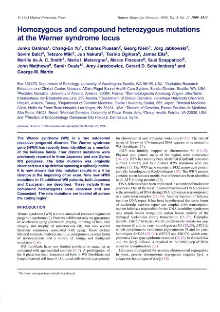
Figure 1. Loc<strong>at</strong>ions of the WRN <strong>mut<strong>at</strong>ions</strong>. The rectangular box indic<strong>at</strong>es the predicted WRN protein. The light shadowed segment indic<strong>at</strong>es the highly acidic repe<strong>at</strong><br />
region; dark shadows indic<strong>at</strong>e the loc<strong>at</strong>ions of the helicase consensus motifs. The loc<strong>at</strong>ions of the WRN <strong>mut<strong>at</strong>ions</strong> are grouped based upon the type of mut<strong>at</strong>ion <strong>and</strong> are<br />
shown underne<strong>at</strong>h the WRN protein along with Registry codes. Parentheses indic<strong>at</strong>e the <strong>heterozygous</strong> <strong>mut<strong>at</strong>ions</strong>. ZM <strong>and</strong> MH <strong>mut<strong>at</strong>ions</strong> were previously described (1).<br />
Given the several potential roles of the WRN protein, a careful<br />
deline<strong>at</strong>ion of spontaneous <strong>mut<strong>at</strong>ions</strong> <strong>at</strong> this locus could facilit<strong>at</strong>e<br />
the characteriz<strong>at</strong>ions of its functions. We report nine WRN gene<br />
<strong>mut<strong>at</strong>ions</strong> from 10 WS p<strong>at</strong>ients, three of which were <strong>compound</strong><br />
heterozygotes. Two other <strong>mut<strong>at</strong>ions</strong> found in three p<strong>at</strong>ients have<br />
been previously reported (1). These various <strong>mut<strong>at</strong>ions</strong> involve<br />
sequences throughout the coding region.<br />
RESULTS<br />
Four Japanese <strong>and</strong> eight non-Japanese WS p<strong>at</strong>ients were selected<br />
from our Intern<strong>at</strong>ional Registry. Six of them (AUS, KO, MIM3,<br />
SEP, TUR, UH) were classified as ‘definite WS’ <strong>and</strong> three (LGS,<br />
OW, SUG) as ‘probable WS’. Clinical <strong>and</strong> labor<strong>at</strong>ory d<strong>at</strong>a for<br />
members of BLS, KUN <strong>and</strong> SYR remain incomplete, but the<br />
affected subjects had been diagnosed as WS by the submitting<br />
physicians.<br />
Three new <strong>mut<strong>at</strong>ions</strong> were found in regions N-terminal with<br />
respect to the helicase consensus motifs. The point mut<strong>at</strong>ion <strong>at</strong> nt<br />
1336, CGA (Arg) to TGA (Stp), was found as a homozygous<br />
mut<strong>at</strong>ion in one Caucasian (LGS) <strong>and</strong> two consanguineous<br />
Japanese (OW, KO) WS subjects <strong>and</strong> as a <strong>heterozygous</strong> mut<strong>at</strong>ion<br />
in one Japanese WS subject (KUN). LGS denied consanguinity;<br />
non-consanguinity was supported by haplotype d<strong>at</strong>a (19). A<br />
single nucleotide deletion <strong>at</strong> 1194–1196, AAA to AA, was seen<br />
as a <strong>heterozygous</strong> mut<strong>at</strong>ion in AUS. This mut<strong>at</strong>ion would cre<strong>at</strong>e<br />
a frameshift which ends <strong>at</strong> 1406–1408 TGA (Stp). A four<br />
nucleotide insertion (ATCT) between 1509 <strong>and</strong> 1520 was<br />
homozygous in MIM3. This frameshift mut<strong>at</strong>ion would termin<strong>at</strong>e<br />
<strong>at</strong> 1535–1537 TGA (Stp).<br />
Three <strong>mut<strong>at</strong>ions</strong> were found within or just 3′ to the helicase<br />
motifs in two Caucasian p<strong>at</strong>ients. One (SEP) mut<strong>at</strong>ion was a 105<br />
bp insertion between 2319 <strong>and</strong> 2320. The insertion results in a<br />
termin<strong>at</strong>ion codon, cre<strong>at</strong>ing a trunc<strong>at</strong>ed protein th<strong>at</strong> excludes<br />
helicase domains III <strong>and</strong> the subsequent C terminus of the WRN<br />
protein. A second mut<strong>at</strong>ion was a deletion of nucleotide<br />
2320–3056 seen in SUG as a <strong>heterozygous</strong> mut<strong>at</strong>ion, termin<strong>at</strong>ing<br />
<strong>at</strong> nt 3081–3083 TGA (Stp). The third mut<strong>at</strong>ion was a <strong>heterozygous</strong><br />
termin<strong>at</strong>ion mut<strong>at</strong>ion found in SUG, loc<strong>at</strong>ed 30 amino acids<br />
after the last helicase motif.<br />
Three new <strong>mut<strong>at</strong>ions</strong> were found in regions C-terminal to the<br />
helicase motifs. A Japanese p<strong>at</strong>ient, IB, was homozygous for an<br />
A deletion <strong>at</strong> nt 3677. The mut<strong>at</strong>ed protein stops <strong>at</strong> nt 3713–3715<br />
TAG (Stp). BLS (French) <strong>and</strong> TUR (Turkish) p<strong>at</strong>ients shared the<br />
same mut<strong>at</strong>ion <strong>at</strong> nt 3724, CGA (Gln) to TGA (Stp), which was<br />
previously found in the Japanese SY family (1). A 74 bp deletion<br />
of nt 3541–3614 was seen as a <strong>heterozygous</strong> mut<strong>at</strong>ion in a<br />
Japanese WS, KUN. This deletion results in a termin<strong>at</strong>ion <strong>at</strong><br />
3720–3722 TAG (Stp). A 113 bp deletion of nt 3691–3803, which<br />
would result in a termin<strong>at</strong>ion <strong>at</strong> nt 3816–3818 TGA (Stp), was<br />
found as a <strong>heterozygous</strong> deletion in the Caucasian WS, AUS.<br />
These <strong>mut<strong>at</strong>ions</strong> were confirmed by sequencing of genomic<br />
PCR products, using the primers from the intron sequences of<br />
WRN (39). A summary of the newly discovered <strong>mut<strong>at</strong>ions</strong> is<br />
given in Figure 1.<br />
The mut<strong>at</strong>ion in the SYR pedigree was previously reported as<br />
a 4 bp deletion <strong>at</strong> the intron–exon boundary, 2 bp from the<br />
put<strong>at</strong>ive intron <strong>and</strong> 2 bp from the contiguous exon (gtagACA-<br />
GACC <strong>at</strong> the DNA level). This was expected to cause an in-frame<br />
deletion of the exon. Our RT–PCR protocol, however, showed a<br />
deletion of 4 bp, ACAG, from the beginning of this exon. The<br />
ACAG deletion would result in a termin<strong>at</strong>ion <strong>at</strong> nt 3971–3973<br />
TAG (Stp).<br />
DISCUSSION<br />
In our original report of the positional cloning of the WRN locus,<br />
four distinct homozygous <strong>mut<strong>at</strong>ions</strong> in the 3′ region of the WRN<br />
gene were described (1). Using the present RT–PCR str<strong>at</strong>egy<br />
<strong>mut<strong>at</strong>ions</strong> were readily found in various loc<strong>at</strong>ions within the gene.<br />
The biochemical consequences of these <strong>mut<strong>at</strong>ions</strong> are not known.<br />
All of the WRN <strong>mut<strong>at</strong>ions</strong> we have found to d<strong>at</strong>e either cre<strong>at</strong>e<br />
a stop codon mut<strong>at</strong>ion or cause frameshifts th<strong>at</strong> lead to prem<strong>at</strong>ure<br />
termin<strong>at</strong>ions. We have not yet found an amino acid substitution<br />
in WRN th<strong>at</strong> seems to be responsible for the p<strong>at</strong>hogenesis of WS.<br />
It is quite possible th<strong>at</strong> the various trunc<strong>at</strong>ed WRN proteins may<br />
be rapidly degraded, resulting in comparable null <strong>mut<strong>at</strong>ions</strong> <strong>and</strong><br />
comparable phenotypes. Such altered mRNAs are thought to be<br />
degraded via a specific p<strong>at</strong>hway (40). In preliminary experiments,<br />
we do observe evidence for reduced levels of WRN mRNA<br />
expression in WS LCLs with four different <strong>mut<strong>at</strong>ions</strong>.<br />
Identical <strong>mut<strong>at</strong>ions</strong> were found across a variety of ethnic<br />
groups, raising the question of potential mut<strong>at</strong>ionally susceptible<br />
sequences. Although the total number of <strong>mut<strong>at</strong>ions</strong> so far found<br />
in the WRN protein is not extensive, c<strong>and</strong>id<strong>at</strong>e sequences for such<br />
susceptibility would include nt 3677–3920, nt 1336–1395 <strong>and</strong> nt<br />
2319–2320.<br />
Three instances of <strong>compound</strong> <strong>heterozygous</strong> <strong>mut<strong>at</strong>ions</strong> were<br />
found: KUN (Japanese), AUS (Caucasian) <strong>and</strong> SUG (Caucasian).<br />
There have been numerous reports of <strong>compound</strong> heterozygotic
1911<br />
Nucleic Human Acids Molecular Research, Genetics, 1994, 1996, Vol. 22, Vol. No. 5, No. 1 9 1911<br />
<strong>mut<strong>at</strong>ions</strong> in ‘disease genes’ (41,42). However, compar<strong>at</strong>ively<br />
few <strong>compound</strong> heterozygotes have been reported in the genomic<br />
instability syndromes. Given the compar<strong>at</strong>ively low prevalence<br />
of consanguinity in the USA, clinicians should therefore be alert<br />
to the diagnosis of WS in the absence of a history of<br />
consanguinity. Our experience suggests th<strong>at</strong> WS is underdiagnosed<br />
in the USA.<br />
MATERIALS AND METHODS<br />
Samples<br />
WS p<strong>at</strong>ients were from an Intern<strong>at</strong>ional Registry of Werner<br />
Syndrome (George M. Martin, MD, Junko Oshima, MD, PhD,<br />
Amy Jarzebowicz, BS). Diagnostic criteria were previously<br />
described (18). This study was approved by the University of<br />
Washington Institutional Review Board.<br />
RT–PCR<br />
Five µg of poly(A) RNA, isol<strong>at</strong>ed from total RNA, using Oligotex<br />
(Qiagen Inc.) was reverse-transcribed with r<strong>and</strong>om hexamers in<br />
100 µl reaction volume with GeneAmp RNA PCR kit (Perkin<br />
Elmer Cetus). Two µl of the RT product were amplified in a 50 µl<br />
PCR reaction buffer containing 5 units Taq DNA polymerase,<br />
10 mM Tris–HCl (pH 8.3), 50 mM KCl, 1.5 mM MgCl 2 , 50 µM<br />
each of dGTP, dATP, dTTP <strong>and</strong> dCTP. The cycle program was<br />
typically: 94C for 5 min, then 94C for 45 s, 55C for 45 s, 72C<br />
for 3.5 min with 2 s increase per cycle for 35 cycles, followed by<br />
72C for 10 min. Five µl aliquots of the first amplific<strong>at</strong>ion products<br />
were subjected to a nested second amplific<strong>at</strong>ion in 100 µl reaction<br />
volumes. The primer sequences for RT–PCR are listed in Table 1.<br />
The secondary PCR products were separ<strong>at</strong>ed on 1% agarose/1×TBE<br />
(100 mM Tris–HCl pH 8.0, 90 mM boric acid <strong>and</strong><br />
1 mM ethylenediaminetetraacetic acid) to estim<strong>at</strong>e the concentr<strong>at</strong>ions<br />
of DNA before sequencing.<br />
Table 1. Primer sequences for the RT–PCR sequencing templ<strong>at</strong>e<br />
Size of<br />
Region of the amplific<strong>at</strong>ion 1st amplific<strong>at</strong>ion primers (5′ to 3′) 2nd amplific<strong>at</strong>ion primers (5′ to 3′) PCR product<br />
5′ end GTGGTGGCGCTCCACAGTCATCC AAGACCTGTTGGACTGGATCTTCTC 838<br />
CTTTATGAAGCCAATTTCTACCC<br />
TACTCCAAAATCTCTAAATTTCGG<br />
Transl<strong>at</strong>ion start site GTGGTGGCGCTCCACAGTCATCC TAGGACTTTCAAAGATGAGTG 1936<br />
to helicase region CTTTATGAAGCCAATTTCTACCC CGTATACAATCCGGTATTTACC<br />
Helicase region GTGGTGGCGCTCCACAGTCATCC AGATGTACTTTGGCCATTCCAG 1218<br />
CTTTATGAAGCCAATTTCTACCC<br />
GCAATGATCCAATCTGGACC<br />
3′ region GCATTAATAAAGCTGACATTCGCC CATTACGGTGCTCCTAAGGACATG 1946<br />
CGGAAGGCTGATTTAAGATGCC<br />
CGGAAGGCTGATTTAAGATGCC<br />
Table 2. WRN <strong>mut<strong>at</strong>ions</strong> in Japanese <strong>and</strong> Caucasian WS p<strong>at</strong>ients<br />
Registry no. Country Ethnicity M/F Loc<strong>at</strong>ion Mut<strong>at</strong>ion Predicted protein<br />
LGS90610 USA Caucasian F 1336 CGA–TGA 368<br />
Arg Stp<br />
OW90650 Japan Japanese M 1336 CGA–TGA 368<br />
Arg Stp<br />
KO90375 Japan Japanese M 1336 CGA–TGA 368<br />
Arg Stp<br />
KUN9001 Japan Japanese M 1336 CGA–TGA 368<br />
Arg Stp<br />
3541–3614 Deletion 1138<br />
AUS40025 Austria Caucasian M 1395 A deletion 391<br />
3691–3803 Deletion 1157<br />
MIM37100 Brazil Caucasian F 1509 ATCT insertion 429<br />
SEP9000 Sardinia Caucasian F 2319–2320 105 bp insertion 708<br />
SUG17802 USA Caucasian M 2320–3056 Deletion 704<br />
2896 CGA–TGA 888<br />
Arg Stp<br />
IB90550 Japan Japanese F 3677 A deletion 1160<br />
BLS60010 France Caucasian M 3724 CAG–TAG 1164<br />
Gln Stp<br />
TUR90010 Turkey Caucasian M 3724 CAG–TAG 1164<br />
Gln Stp<br />
SYR10006 Syria Syrian M 3919–3922 ACAG deletion 1245
1912<br />
Human Molecular Genetics, 1996, Vol. 5, No. 9<br />
Direct sequencing of PCR products<br />
RT–PCR products were sequenced using a T7 sequence PCR<br />
product sequencing kit (UBS, Amersham Life Science, Inc.).<br />
Seven µl of PCR product was pretre<strong>at</strong>ed with 15 U of exonuclease<br />
I <strong>and</strong> 1.5 U of shrimp alkaline phosph<strong>at</strong>ase <strong>at</strong> 37C for 15 min<br />
followed by inactiv<strong>at</strong>ion of the enzymes <strong>at</strong> 80C for 15 min, then<br />
mixed with 100 ng of sequencing primers. The sequencing<br />
reaction followed the manufacturer’s instructions.<br />
The sequencing gel contained 6.6% LongRanger polyacrylamide<br />
(J. T. Baker Inc.), 6 M urea <strong>and</strong> 1.2× TBE. The running buffer<br />
contained 0.6× TBE. The gel was run <strong>at</strong> 55 W, dried <strong>and</strong> exposed<br />
overnight to Biomax MR film (Eastman Kodak Co.).<br />
ACKNOWLEDGEMENTS<br />
We thank Dr Goberdhan P. Dimri for a human cDNA library, an<br />
important contribution leading to the original positional cloning<br />
of WRN. We also thank Annette Smith, Charles E. Ogburn, Thao<br />
Dang, Susan Fredell, Ellen Nemens <strong>and</strong> Deanne Sparlin for their<br />
technical support. This work was supported by N<strong>at</strong>ional Institute<br />
on Aging grants P1 AG08303 (GMM), R37 AG08303 (GMM),<br />
T32 AG00057 (GMM), RO1 AG12019 (GDS) <strong>and</strong> CNPq <strong>and</strong><br />
FAPESP, Brazil (MIM).<br />
ABBREVIATIONS<br />
WS, Werner syndrome; WRN, Werner syndrome gene; UV,<br />
ultraviolet; ERCC, excision repair–cross-complementing; LCL,<br />
lymphoblastoid cell line; RT–PCR, reverse transcription–<br />
polymerase chain reaction; PCR, polymerase chain reaction.<br />
REFERENCES<br />
1. Yu, C.E., Oshima, J., Fu, Y.W., Hisama, F., Wijsman, E.M., Alisch, R.,<br />
M<strong>at</strong>thews, S., Nakura, J., Miki, T., Ouais, S., Martin, G.M., Mulligan, J. <strong>and</strong><br />
Schellenberg, G.D. (1996) Positional cloning of the Werner’s syndrome gene.<br />
Science, 272, 258–262.<br />
2. Martin, G.M. (1978) Genetic syndromes in man with potential relevance to<br />
the p<strong>at</strong>hology of aging. Birth Defects, 14, 5–39.<br />
3. Epstein, C.J., Martin, G.M., Schultz, A.L. <strong>and</strong> Motulsky, A.G. (1966)<br />
Werner’s syndrome: a review of its symptom<strong>at</strong>ology, n<strong>at</strong>ural history,<br />
p<strong>at</strong>hologic fe<strong>at</strong>ures, genetics <strong>and</strong> rel<strong>at</strong>ionships to the n<strong>at</strong>ural aging process.<br />
Medicine, 45, 172–221.<br />
4. Tollefsbol, T.O. <strong>and</strong> Cohen, H.J. (1984) Werner’s syndrome: an underdiagnosed<br />
disorder resembling prem<strong>at</strong>ure aging. Age, 7, 75–88.<br />
5. Martin, G.M., Sprague, C.A. <strong>and</strong> Epstein, C.J. (1970) Replic<strong>at</strong>ive life-span of<br />
cultiv<strong>at</strong>ed human cells. Effects of donor’s age, tissue <strong>and</strong> genotype. Lab.<br />
Invest., 23, 86–92.<br />
6. Salk, D., Bryant, E., Hoehn, H., Johnston, P. <strong>and</strong> Martin, G.M. (1985) Growth<br />
characteristics of Werner syndrome cells in vitro. Adv. Exp. Med. Biol., 190,<br />
305–311.<br />
7. Kill, I.R., Faragher, R.G., Lawrence, K. <strong>and</strong> Shall, S. (1994) The expression<br />
of prolifer<strong>at</strong>ion-dependent antigens during the lifespan of normal <strong>and</strong><br />
progeroid human fibroblasts in culture. J. Cell Sci., 107, 571–579.<br />
8. Poot, M., Hoehn, H., Runger, T.M. <strong>and</strong> Martin, G.M. (1992) Impaired<br />
S-phase transit of Werner syndrome cells expressed in lymphoblastoid cell<br />
lines. Exp. Cell. Res., 202, 267–273.<br />
9. Hoehn, H., Bryant, E.M., Au, K., Norwood, T.H., Boman, H. <strong>and</strong> Martin,<br />
G.M. (1975) Varieg<strong>at</strong>ed transloc<strong>at</strong>ion mosaicism in human skin fibroblast<br />
culture. Cytogenet. Cell. Genet., 15, 282–298.<br />
10. Fukuchi, K., Martin, G.M. <strong>and</strong> Monnet, R.J. (1989) Mut<strong>at</strong>or phenotype of<br />
Werner syndrome is characterized by extensive deletion. Proc. N<strong>at</strong>l Acad.<br />
Sci. USA, 86, 5893–5897.<br />
11. Fukuchi, K., Tayama, K., Kumahara, Y., Murano, K., Pride, A.B., Martin,<br />
G.M., Monnet, R.J. (1990) Increased frequency of 6-thioguanine-resistant<br />
peripheral blood lymphocytes in Werner syndrome p<strong>at</strong>ients. Hum. Genet., 84,<br />
249–252.<br />
12. Runger, T.M., Bauer, C., Dekant, B., Moller, K., Sobotta, P., Czerny, C., Poot,<br />
M. <strong>and</strong> Martin, G.M. (1994) Hypermutable lig<strong>at</strong>ion of plasmid DNA ends in<br />
cells from p<strong>at</strong>ients with Werner syndrome. J. Invest. Derm<strong>at</strong>ol., 102, 45–48.<br />
13. Fujiwara, Y., Higashikawa, T. <strong>and</strong> T<strong>at</strong>sumi, M. (1977) A retarded r<strong>at</strong>e of DNA<br />
replic<strong>at</strong>ion <strong>and</strong> normal level of DNA repair in Werner’s syndrome fibroblasts<br />
in culture. J. Cell. Physiol., 92, 365–374.<br />
14. Goto, M., Rubenstein, M., Weber, J., Woods, K. <strong>and</strong> Drayna, D. (1992)<br />
Genetic linkage of Werner’s syndrome to five markers on chromosome 8.<br />
N<strong>at</strong>ure, 355, 735–758.<br />
15. Schellenberg, G.D., Martin, G.M., Wijsman, E.M., Nakura, J., Miki, T. <strong>and</strong><br />
Ogihara, T. (1992) Homozygosity mapping <strong>and</strong> Werner syndrome. Lancet,<br />
339, 1002.<br />
16. Yu, C.E., Oshima, J., Goddard, K.A.B., Miki, T., Nakura, J., Ogihara, T.,<br />
Fraccaro, M., Puissan, C., Martin, G.M., Schellenberg, G.D. <strong>and</strong> Wijsman,<br />
E.M. (1994) Linkage disequilibrium <strong>and</strong> haplotype analysis of chromosome<br />
8p11.1–21.1 markers <strong>and</strong> Werner syndrome. Am. J. Hum. Genet., 55,<br />
356–364.<br />
17. Oshima, J., Yu, C.E., Boehnke, M., Weber, J.L., Edelhoff, S., Wagner, M.J.,<br />
Wells, D.E., Wood, S., Disteshe, D.M., Martin, G.M. <strong>and</strong> Schellenberg, G.D.<br />
(1994) Integr<strong>at</strong>ed mapping analysis of the Werner syndrome region of<br />
chromosome 8. Genomics, 23, 100–113.<br />
18. Nakura, J., Wijsman, E.J., Miki, T., Kamino, K., Yu, C.E., Oshima, J.,<br />
Fukuchi, K.I., Weber, J.L., Puissan, C., Melaragno, M.I., Epstein, C.J.,<br />
Scapp<strong>at</strong>icci, S., Fraccaro, M., M<strong>at</strong>usmura, T., Murano, S., Yoshida, S.,<br />
Fujiwara, Y., Saida, T., Ogihara, T., Martin, G.M. <strong>and</strong> Schellenberg, G.D.<br />
(1994) Homozygosity mapping of the Werner syndrome locus (WRN).<br />
Genomics, 23, 600–608.<br />
19. Goddard, K.A.B., Yu, C.E., Oshima, J., Martin, G.M., Schellenberg, G.D. <strong>and</strong><br />
Wijsman, E.M. (1996) Towards localiz<strong>at</strong>ion of the Werner syndrome gene by<br />
linkage disequilibrium <strong>and</strong> ancestral haplotyping: lessons learned from<br />
analysis of 35 chromosome 8p11.1–21.1 markers. Am. J. Hum. Genet., 58,<br />
1286–1302.<br />
20. Schmid, S.R. <strong>and</strong> Linder, P. (1992) D-E-A-D protein family of put<strong>at</strong>ive RNA<br />
helicases. Mol. Microbiol., 6, 283–292.<br />
21. Gorbalenya, A.E., Koonin, E.V., Donchenko, A.P. <strong>and</strong> Blinov, V.M. (1989)<br />
Two rel<strong>at</strong>ed superfamilies of put<strong>at</strong>ive helicases involved in replic<strong>at</strong>ion,<br />
recombin<strong>at</strong>ion, repair <strong>and</strong> expression of DNA <strong>and</strong> RNA genome. Nucleic<br />
Acids Res., 17, 4713–4730.<br />
22. Lohman, T.M. (1993) Helicase-c<strong>at</strong>alyzed DNA unwinding. J. Biol. Chem.,<br />
268, 2269–2272.<br />
23. Conaway, R.C. <strong>and</strong> Lehman, L.R. (1982) A DNA primase activity associ<strong>at</strong>ed<br />
with DNA polymerase alpha from Drosophila melanogaster embryos. Proc.<br />
N<strong>at</strong>l Acad. Sci. USA, 79, 2523–2527.<br />
24. Dodson, M.S. <strong>and</strong> Lehman, I.R. (1991) Associ<strong>at</strong>ion of DNA helicase <strong>and</strong><br />
primase activities with subassembly of the herpes simples virus 1 helicaseprimase<br />
composed of the UL5 <strong>and</strong> UL52 gene products. Proc. N<strong>at</strong>l Acad. Sci.<br />
USA, 88, 1105–1109.<br />
25. Hanawalt, P.C. (1994) Transcription-coupled repair <strong>and</strong> human disease.<br />
Science, 266,1957–1958.<br />
26. Drapkin, R. <strong>and</strong> Reinberg, D. (1992) The essential twist. N<strong>at</strong>ure, 369,<br />
523–533.<br />
27. Bootsma, D., Weeda, G., Vermeulen, W., van Vuuren, H., Troelstra, C., van<br />
der Spek, P. <strong>and</strong> Hoeijmakers, J. (1995) Nucleotide excision repair syndrome:<br />
molecular basis <strong>and</strong> clinical symptoms. Phil. Trans. R. Soc. London B. Biol.<br />
Sci., 347, 75–81.<br />
28. Weber, C.A., Salazer, E.P., Stewart, S.A. <strong>and</strong> Thompson, L.H. (1990)<br />
ERCC2: cDNA cloning <strong>and</strong> molecular characteriz<strong>at</strong>ion of a human<br />
nucleotide excision repair gene with high homology to yeast RAD3. EMBO<br />
J., 9, 1437–1447.<br />
29. Guzder, S.N., Qiu, H., Sommers, C.H., Sung, P., Prakash, L. <strong>and</strong> Prakash, S.<br />
(1994a) DNA repair gene RAD3 of S. cerevisiae is essential for transcription<br />
by RNA polymerase II. N<strong>at</strong>ure, 367, 91–94.<br />
30. Weeda, G., van Ham, R.C.A., Masurel, R., Westerveld, A., Odijk, H., de Wit,<br />
J., Bootsma, D., van del Eb, A.J. <strong>and</strong> Hoeijmakers, J.H.J. (1990a) Molecular<br />
cloning <strong>and</strong> biological characteriz<strong>at</strong>ion of the human excision repair gene<br />
ERCC-3. Mol. Cell. Biol., 10, 2570–2581.<br />
31. Flejter, W.L., McDaniel, L.D., Johns, D., Friedberg, E.C. <strong>and</strong> Schults, R.A.<br />
(1992) Correction of xeroderma pigmentosum complement<strong>at</strong>ion group D<br />
mutant cell phenotypes by chromosome <strong>and</strong> gene transfer: involvement of<br />
human ERCC2 DNA repair gene. Proc. N<strong>at</strong>l Acad. Sci. USA, 89, 261–265.
1913<br />
Nucleic Human Acids Molecular Research, Genetics, 1994, 1996, Vol. 22, Vol. No. 5, No. 1 9 1913<br />
32. Sung, P., Bailly, V., Weber, C., Thompson, L.H., Prakash, L. <strong>and</strong> Prakash, S.<br />
(1993) Human xeroderma pigmentosum group D gene encodes a DNA<br />
helicase. N<strong>at</strong>ure, 365, 852–855.<br />
33. Guzder, S.N., Sung, P., Bailly, V., Prakash, L. <strong>and</strong> Prakash, S. (1994) RAD25<br />
is a DNA helicase required for DNA repair <strong>and</strong> RNA polymerase II<br />
transcription. N<strong>at</strong>ure, 369, 578–581.<br />
34. Ma, L., Westbroek, A. Jochemsen, A.G., Weeda, G., Bosch, A., Bootsma, D.,<br />
Hoeijmakers, J.H. <strong>and</strong> van der Eb, A.J. (1994) Mut<strong>at</strong>ional analysis of<br />
ECRR3, which is involved in DNA repair <strong>and</strong> transcription initi<strong>at</strong>ion:<br />
identific<strong>at</strong>ion of domains essential for the DNA repair function. Mol. Cell.<br />
Biol., 14, 4126–4136.<br />
35. Weeda, G., van Ham, R.C.A., Vermeulen, W., Bootsma, D., van der Eb, A.J.<br />
<strong>and</strong> Hoeijmakers, J.H.J. (1990) A presumed DNA helicase encoded by<br />
ERRC-3 is involved in the human repair disorders xeroderma pigmentosum<br />
<strong>and</strong> Cockayne’s syndrome. Cell, 62, 777–791.<br />
36. Troelstra, C., van Gool, A., de Wit, J., Vermeulen, W., Bootsma, D. <strong>and</strong><br />
Hoeijmaker, J.H.J. (1992) ECCR6, a member of a subfamily of put<strong>at</strong>ive<br />
helicases, is involved in Cockayne’s syndrome <strong>and</strong> preferential repair of<br />
active gene. Cell, 71, 939–953.<br />
37. West, S.C. (1994) The processing of recombin<strong>at</strong>ion intermedi<strong>at</strong>es: mechanistic<br />
insights from studies of bacterial protein. Cell, 76, 9–15.<br />
38. W<strong>at</strong>t, P.M., Louis, E.J., Borts, R.H., Hickson, I.D. (1995) Sgs1: A eukaryotic<br />
homolog of E. coli RecQ th<strong>at</strong> interacts with topoisomerase II in vivo <strong>and</strong> is<br />
required for faithful chromosome segreg<strong>at</strong>ion. Cell, 81, 253–260.<br />
39. Yu, C.E., Oshima, J., Wijsman, E. M., Nakura, J., Miki, T., Puissan, C.,<br />
M<strong>at</strong>thews, S., Martin, G.M., Schellenberg, G.D. <strong>and</strong> the Werner’s Syndrome<br />
Collabor<strong>at</strong>ive Group. Mut<strong>at</strong>ions in the consensus helicase domains of the<br />
Werner’s syndrome gene (submitted).<br />
40. Jacobson, A. <strong>and</strong> Peltz, S.W. (1996) Interrel<strong>at</strong>ionships of the p<strong>at</strong>hways of<br />
mRNA decay <strong>and</strong> transl<strong>at</strong>ion in eukaryotic cells. Annu. Rev. Biochem., 65,<br />
693–739.<br />
41. S<strong>at</strong>ok<strong>at</strong>a, I., Tanaka, K. <strong>and</strong> Okada, Y. (1992) Molecular basis of group A<br />
xeroderma pigmentosum: a missense mut<strong>at</strong>ion <strong>and</strong> two deletions loc<strong>at</strong>ed in a<br />
zinc finger consensus sequence of the XPAC gene. Hum. Genet., 88,<br />
603–607.<br />
42. S<strong>at</strong>o, M., Nishigori, C., Yagi, T. <strong>and</strong> Tanabe, H. (1996) Aberrant splicing <strong>and</strong><br />
trunc<strong>at</strong>ed-protein expression due to a newly identified XPA gene mut<strong>at</strong>ion.<br />
Mut<strong>at</strong>. Res., 362, 199–208.


