AEPC Heart Lung Interaction Handout
AEPC Heart Lung Interaction Handout
AEPC Heart Lung Interaction Handout

Create successful ePaper yourself
Turn your PDF publications into a flip-book with our unique Google optimized e-Paper software.
HEART LUNG INTERACTION OF THE INFANT WITH CONGENITAL HEART DISEASE :<br />
EFFECTS OF EXTUBATION, NON INVASIVE VENTILATION AND CHEST PHYSIOTHERAPY<br />
Grollmuss O. (1), Boet A. (2), Fattal S (1), Hervé Ph. (1), Belli E (1)<br />
Centre Chirurgical Marie Lannelongue, Le Plessis Robinson, France (1)<br />
Centre Hospitalier Universitaire Antoine Beclère, Clamart, France (2)<br />
Introduction<br />
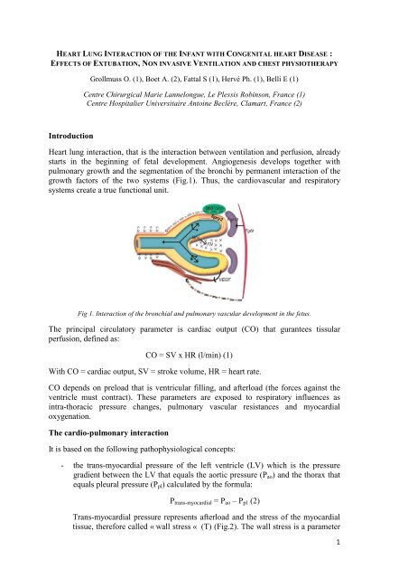
<strong>Heart</strong> lung interaction, that is the interaction between ventilation and perfusion, already<br />
starts in the beginning of fetal development. Angiogenesis develops together with<br />
pulmonary growth and the segmentation of the bronchi by permanent interaction of the<br />
growth factors of the two systems (Fig.1). Thus, the cardiovascular and respiratory<br />
systems create a true functional unit.<br />
Fig 1. <strong>Interaction</strong> of the bronchial and pulmonary vascular development in the fetus.<br />
The principal circulatory parameter is cardiac output (CO) that gurantees tissular<br />
perfusion, defined as:<br />
CO = SV x HR (l/min) (1)<br />
With CO = cardiac output, SV = stroke volume, HR = heart rate.<br />
CO depends on preload that is ventricular filling, and afterload (the forces against the<br />
ventricle must contract). These parameters are exposed to respiratory influences as<br />
intra-thoracic pressure changes, pulmonary vascular resistances and myocardial<br />
oxygenation.<br />
The cardio-pulmonary interaction<br />
It is based on the following pathophysiological concepts:<br />
- the trans-myocardial pressure of the left ventricle (LV) which is the pressure<br />
gradient between the LV that equals the aortic pressure (P ao ) and the thorax that<br />
equals pleural pressure (P pl ) calculated by the formula:<br />
P trans-myocardial = P ao – P pl (2)<br />
Trans-myocardial pressure represents afterload and the stress of the myocardial<br />
tissue, therefore called « wall stress « (T) (Fig.2). The wall stress is a parameter<br />
1
particularly important in a failing ventricle because it depends on the intraventricular<br />
pressure, the thickness of the myocardium and the enddiastolic<br />
diameter of the ventricle.<br />
Fig 2 : Ventricular components of the myocardial « wall stress ».<br />
Therefore afterload of the contracting ventricle will rise in the presence of<br />
negative intra-thoracic pressure (spontaneous inspiration) because of a decrease<br />
of pleural pressure, whereas an augmentation of pleural pressure (ventilation<br />
with positive pressure) will lead to a decrease of LV afterload as a<br />
counterbalance to the wall stress.<br />
- The right ventricular (RV) function is directly exposed to respiration in a way<br />
that intrathoracical pressure influences blood return of the caval veins into the<br />
right atrium (RA) and therefore RV filling. At the same time, it directly<br />
influences the pulmonary vessels modifying their resistance and therefore their<br />
after-load and their ejection volume. Other respiratory parameters like paCO 2 ,<br />
PaO 2 and blood pH also influence the pulmonary vascular tonus and thus right<br />
ventricular after-load.<br />
- The third important concept is that of the intra- and extra-thoracic<br />
compartments. Indeed, the major part of the systemic vessels and the inferior V.<br />
cave are located in an extra-thoracic compartment, the abdomen, and thus<br />
exposed to variations of the atmosphere pressure instead of pleural pressure.<br />
This is the driving force for systemic venous return during spontaneous<br />
inspiration with a decrease of pleural pressure.<br />
- Finally, there is the phenomenon of ventricular interdependence. The two<br />
ventricles share the same non extensible envelope, the pericardium, with a<br />
common contractile element, the inter-ventricular septum. Thus, any<br />
modification of a ventricle’s end-diastolic filling or its contractility modifies the<br />
volume and the contractility of the other.<br />
Mechanical ventilation with positive pressure notably modifies these interactions.<br />
- First, mechanical ventilation positively affects left heart function by optimizing<br />
the conditions of pulmonary filling. Indeed, as mechanical ventilation influences<br />
lung volume it also modifies its blood capacitance in a cyclic manner and<br />
therefore the amount of blood that the lungs may store in their vessels. Thus,<br />
ventilation influences pulmonary venous return.<br />
2
- Also, it modifies LV after-load. Arterial systemic pressure rises, but also the<br />
gradient between the intra- and extra-thoracic vessels, thus creating an effect of<br />
blood expulsion from the intra- to the extra-thoracic aorta.<br />
- And, by changing pleural pressure to positive values it reduces the transmyocardial<br />
gradient, therefore the LV afterload.<br />
- On the other hand, positive pressure ventilation augments the alveolar pressure<br />
thus rising pulmonary vascular resistances and therefore RV afterload.<br />
- Moreover, by augmenting pleural pressures through mechanical ventilation, RA<br />
pressure rises thus impeding systemic venous return, therewith reducing RV<br />
preload.<br />
In conclusion, mechanical ventilation has a beneficial effect on left heart circulation but<br />
rather a negative one on right heart function.<br />
Extubation<br />
Extubation is a critical period in the post-surgical intensive care of patients with<br />
congenital heart disease. Respiratory complications may be possible and, in their<br />
majority, occur rapidly, 22% during the first minutes after extubation. They are<br />
predominantly obstructive due to the anatomy of the infant. Its airways are finer,<br />
especially in the sub-glottis region. Also frequently, when passing the intubation’s tube,<br />
laryngospasm may occur as a reflex that protects the infant from secret aspiration. Other<br />
complications or even a failure of extubation might be due to physical weakness or<br />
fatigue of the infant.<br />
Besides its respiratory complications, extubation may also have hemodynamic<br />
consequences. First, there is a rise of arterial blood pressure and heart rate reflecting the<br />
excretion of catecholamines under stress that may lead to a reduction of stroke volume<br />
through an augmentation of LV after-load in patients with heart failure. To this systolic<br />
dysfunction, a diastolic dysfunction may add, through a reduction of the coronary<br />
perfusion due to a more important trans-myocardial gradient of the LV.<br />
The pathophysiology of heart failure<br />
<strong>Heart</strong> failure or heart insufficiency is defined as the inability of the heart to guarantee an<br />
adequate perfusion of the tissues and organs. It is modified by load changes of the<br />
ventricles and particularly aggravated in the presence of a higher afterload. Afterload<br />
represents all the forces against which the ventricle must contract and exerts an effect of<br />
stress on the myocardial tissue: the “wall stress” (Fig. 2) . This may lead to a decrease<br />
of myocardial contractility with, subsequently, a reduction of stroke volume and thus<br />
cardiac output. The decrease of CO activates the rennin-angiotensin-aldosteron (RAAS)<br />
system and, as a counter-reaction, the secretion of brain natriuretic peptide (BNP).<br />
BNP is a hormone synthesized in the ventricles as a response to the stretch of the<br />
myocardial fibers in the presence of wall stress. To resume, its functions result in a<br />
decrease of the systemic vascular resistances and the plasmatic volume (Fig. 3) thus the<br />
ventricular afterload.<br />
3
Fig 3 : Pathophysiology of the Renin-Angiotensin-Aldosteron System (RAAS) and the role of BNP<br />
The aim of our studies was to observe the modifications of the heart lung interaction in<br />
infants after pediatric cardiac surgery under conditions of extubation after mechanical<br />
ventilation, under non invasive ventilation after extubation and under chest<br />
physiotherapy in an intubated and ventilated patient.<br />
1 st study: respiratory and hemodynamic parameters before and after extubation, is there<br />
a cut-off for BNP as a pre-extubational diagnostic parameter indicating a certain risk for<br />
compromised hemodynamics after extubation?<br />
2 nd study: respiratory and hemodynamic parameters before and after extubation with<br />
and without non invasive ventilation immediately after extubation.<br />
3 rd study: respiratory and hemodynamic effects of respiratory physiotherapy in an<br />
intubated and ventilated patient with congenital heart disease.<br />
Materials and Methods<br />
1) Ventilators<br />
The infants enrolled in the studies were ventilated in a conventional manner (IPPV,<br />
SIMV, CPAP before extubation) by one of the following respirators: Evita XL ®<br />
(Dräger), Servo i ® (Maquet), and Engström ® (General Electrics), the latter equipped with<br />
a module for FRC measurement.<br />
For NIV, we used the Infantflow ® respirator (SEBAC) which limited the maximum<br />
weight of the patients enrolled in the NIV study at 5 kg.<br />
2) Electrical velocimetry (EV)<br />
Cardiac output was measured in the infants non invasively by the AESCULON ® or<br />
ICON ® bioimpédance monitor (Osypka Medical Inc. Berlin, San Diego) that, on the<br />
basis of electrical velocimetry, allow the measurement of SV and CO by measuring<br />
changes in thoracic impedance that are due to the ejection of blood from the left<br />
ventricle into the aorta. They also allow to measure an index of contractility of the left<br />
ventricle (ICON). The principle of electrical velocimetry is based on the concept of<br />
changes in thoracic impedance related to the red blood cells alignment in the ascending<br />
aorta during systole (Fig. 4).<br />
4
Fig 4:The principles of electrical velocimetry.<br />
3) Patients<br />
The patients included in the studies were newborns or infants with stable respiratory and<br />
hemodynamic conditions, without shunt in order not to falsify the CO measurements.<br />
During the measurement period there were no changes of therapy (analgesia, sedation,<br />
vaso-active treatment, ventilation). The infants of every study were distinct cohorts. The<br />
patients in the 1 st and 2 nd study that were extubated received all the same preextubational<br />
weaning: PEEP +4 cm H 2 O, Inspiratory aid (IA) +12 cm H 2 O and FiO 2<br />
40% during 30 minutes.<br />
Results<br />
1st study : Extubation<br />
Patients enrolled in the study : 60 newborns (mean age 10 days at surgery, mean weight<br />
3.3 kg) after uncomplicated switch procedure for transposition of the great vessels<br />
(TGV).<br />
Paramètres Avant extubation 6h après extubation p<br />
pH 7,41 (7,34 – 7,56) 7,38 (7,30 – 7,48) < 0,001<br />
Base excess (BE)<br />
(mval/l)<br />
1,5 (-2,3 – 9,8) -0,05 (-3,6 – 6,3) = 0,005<br />
Volume ejection (VE) 5,157 ± 0,2 4,681 ± 0,25 =0,006<br />
BNP (pg/ml) 554,5 (172 – 6522) 1165 (288 – 10354) < 0,001<br />
Table 1 : Per definition (BNP > 400 pg/ml), the patients suffer already from heart insufficiency before<br />
extubation. Extubation doubles BNP indicating an important augmentation of ventricular after-load that<br />
is associated with a decrease of SV, pH and BE. The metabolic changes are, in the absence of respiratory<br />
interferences (paCO2, respiratory rate) rather due to a compromised cardiac output.<br />
5
In order define a pre-extubational BNP cut-off for the patients that are likely to suffer<br />
from an alteration of their metabolism, we calculated ROCs for the target parameters<br />
BE and SV. (Fig 5) :<br />
Fig 5 : ROC for cut-off calculation between pre-extubational BNP and BE (left) and SV (right).<br />
We then wanted divided the patients into subgroups : first those with a pre-extubational<br />
BNP under the cut-off point of 379 pg/ml (group 1 = “low secretors”). Then the patients<br />
with BNP above the cut-off (group 2). In group 2 we defined a group of “high<br />
secroters” (> 75% level of BNP of all patients = 1137 pg/ml) in the following text called<br />
group 2B (group 2 A would then be the patients with BNP from 379 to 1137 pg =<br />
“medium secretors”). We then compared relative and absolute post-extubational BNP<br />
secretion between the different subgroups. The groups behaved differently : Highest<br />
relative BNP secretion was found in group 1with, highest absolute secretion in group 2B<br />
whereas the most important decrease of BE was found in group 2A. This leads to the<br />
conclusion that the “low” and “high” BNP “secretors” are probably better protected<br />
against metabolic alterations after extubation than the “medium secretors” (Fig 6).<br />
Fig 6 : Behavior of the different pre-extubational BNP “secretor” groups compared to their metabolic<br />
course after extubation (before-after plot). Further explications in the text.<br />
6
Conclusion :<br />
1. Extubation induces an afterload augmentation demonstrated by an increase of<br />
BNP and, at the same time, a decrease of SV.<br />
2. The afterload increase together with SV decrease is responsible for the<br />
metabolism turning to acidosis..<br />
3. BNP can serve as a marker for better detection of those heart-insufficient<br />
patients who might suffer from low cardiac output with metabolic impact after<br />
extubation.<br />
2 nd study : Non invasive ventilation<br />
The questions for this study were : Is there an impact of extubation on a heterogeneous<br />
cohort of infants with heart insufficiency ? Can this impact be modified using NVI<br />
immediately after extubation? Can BNP help to identify the need for and the efficacy of<br />
NVI?<br />
The population of this study was: n = 48 infants (25 with spontaneous breathing after<br />
extubation, 23 under NIV) with different congenital heart diseases, < 5 kg, BNP before<br />
extubation > 400n pg/ml. Design of the study: group comparison : spontaneous<br />
breathing versus NII (CPAP + 4 cmH 2 O, FiO 2 0.4) immediately after extubation. No<br />
significant respiratory, hemodynamic and metabolic differences before extubation.<br />
Results (Fig 7). Only significant results are shown.<br />
Fig 7 : Spontaneous breathing : Significant decrease of BE and SV, at the same time increase of BNP<br />
indicating a more important afterload. Under NIV these effects are not visible any more.<br />
7
Conclusion :<br />
1. Extubation has a hemodynamic and metabolic impact in a heterogeneous group<br />
of patients with heart insufficiency (as documented by an elevated BNP) after<br />
cardiac surgery.<br />
2. NIV installed immediately after extubation is able to ameliorate the<br />
hemodynamic conditions as can be seen by the stability of BNP. BNP thus<br />
serves as a marker of ventricular load or insufficiency and the efficacy of NVI.<br />
3. NVI in the infant is well tolerated (besides a slight increase of respiratory rate).<br />
It should be introduced rapidly after extubation in a patient with imminent risks<br />
for heart failure whereas – from a hemodynamic point of view - it is useless in<br />
patients without compromised cardiac function.<br />
3rd study : Respiratory physiotherapy<br />
Patients enrolled in the study : n = 42, 51 measurements, mean age 73 days, infants with<br />
congenital heart disease after cardiac surgery, no intra- or extra-cardiac shunt,<br />
respiratory and hemodynamic stability under mechanical ventilation. Respiratory<br />
physiotherapy with acceleration of the expiratory flow (FET), aspiration, mask<br />
ventilation.<br />
The study was designed to find answers to the following questions:<br />
- What are the respiratory effects of the respiratory physiotherapy in these<br />
patients?<br />
- What are its hemodynamic effects?<br />
Results (Tab2, p
Tab 2 : The short term respiratory effects of respiratory physiotherapy (immediately/1h after therapy)<br />
are : TV plus 16%/4%, PIP minus 16%/13%, C plus 35%/20%, R minus24%/17%, FRC plus 48%/44%.<br />
These respiratory changes are corresponding to hemodynamic changes as: MVTI plus 13%/17%, SV plus<br />
11%/7%, ICON plus 29%/ 23%, BNP minus 8% (1h) suggesting a decrease of after-load due to a<br />
respiration – related increase of CO and ventricular contractility.<br />
There were good and even strong correlations between the respiratory and<br />
hemoydnamic parameters creating evidence for lung heart interaction (not shown).<br />
Conclusion :<br />
1. Respiratory physiotherapy in patients with congenital heart disease under<br />
conditions of ventilation with positive pressure has positive respiratory effects :<br />
increase of tidal volume, compliance, FRC, decrease of inspiratory pressure and<br />
airway resistance.<br />
2. There are positive circulatory effects, too : better LV filling (MVTI) with<br />
increase of SV and LV contractility (Frank-Starling) conditioning an after-load<br />
reduction.<br />
3. The respiratory positive effects seem to lead to positive hemodynamic effects in<br />
the sense of lung heart interaction. Thus, there is a positive short time effect of<br />
respiratory physiotherapy in these patients.<br />
General conclusion :<br />
1. The lungs, the pulmonary vessels and the heart are an anatomic and<br />
physiological unit.<br />
2. In the patient with heart disease, this functional unit is at stake. It is therefore<br />
necessary to maintain a good pulmonary function (mechanical ventilation, NIV,<br />
respiratory physiotherapy) and a correct oxygenation in order to guarantee a<br />
correct ventricular function.<br />
3. In patients with congenital heart disease, BNP may serve as a marker of cardiac<br />
function as well as of cardiorespiratory interaction and therapy effects on it.<br />
4. Respiratory physiotherapy seems to have beneficial (at least) short time<br />
respiratory and therefore circulatory effects. It stays open whether there are also<br />
long term indications and conditions for this treatment. In any case, respiratory<br />
physiotherapy and NIV must be adapted to the needs and evolutions of the<br />
individual patient.<br />
5. These studies demand further investigations on larger and more different<br />
populations.<br />
9