Inflammatory dendritic cells migrate in and out of transplanted ...
Inflammatory dendritic cells migrate in and out of transplanted ...
Inflammatory dendritic cells migrate in and out of transplanted ...

You also want an ePaper? Increase the reach of your titles
YUMPU automatically turns print PDFs into web optimized ePapers that Google loves.
Research article<br />
<strong>Inflammatory</strong> <strong>dendritic</strong> <strong>cells</strong> <strong>migrate</strong><br />
<strong>in</strong> <strong>and</strong> <strong>out</strong> <strong>of</strong> <strong>transplanted</strong> chronic<br />
mycobacterial granulomas <strong>in</strong> mice<br />
Heidi A. Schreiber, 1,2 Jeffrey S. Hard<strong>in</strong>g, 1,2 Oliver Hunt, 1 Christopher J. Altamirano, 1,3<br />
Paul D. Hulseberg, 1,2 Danielle Stewart, 1,2 Zsuzsanna Fabry, 1,2 <strong>and</strong> Matyas S<strong>and</strong>or 1,2,3<br />
1<br />
Department <strong>of</strong> Pathology <strong>and</strong> Laboratory Medic<strong>in</strong>e, School <strong>of</strong> Medic<strong>in</strong>e <strong>and</strong> Public Health, 2 Cellular <strong>and</strong> Molecular Pathology Tra<strong>in</strong><strong>in</strong>g Program,<br />
<strong>and</strong> 3 Microbiology Doctoral Tra<strong>in</strong><strong>in</strong>g Program, University <strong>of</strong> Wiscons<strong>in</strong>, Madison, Wiscons<strong>in</strong>, USA.<br />
An estimated one-third <strong>of</strong> the world’s population is <strong>in</strong>fected with Mycobacterium tuberculosis, although most<br />
affected <strong>in</strong>dividuals ma<strong>in</strong>ta<strong>in</strong> a latent <strong>in</strong>fection. This control is attributed to the formation <strong>of</strong> granulomas, cell<br />
masses largely compris<strong>in</strong>g <strong>in</strong>fected macrophages with T <strong>cells</strong> aggregated around them. <strong>Inflammatory</strong> DCs,<br />
characterized as CD11c + CD11b + Ly6C + , are also found <strong>in</strong> granulomas <strong>and</strong> are an essential component <strong>of</strong> the<br />
acute immune response to mycobacteria. However, their function dur<strong>in</strong>g chronic <strong>in</strong>fection is less well understood.<br />
Here, we report that CD11c + <strong>cells</strong> dynamically traffic <strong>in</strong> <strong>and</strong> <strong>out</strong> <strong>of</strong> both acute <strong>and</strong> chronic granulomas<br />
<strong>in</strong>duced by Mycobacterium bovis stra<strong>in</strong> bacillus Calmette-Guér<strong>in</strong> (BCG) <strong>in</strong> mice. By transplant<strong>in</strong>g Mycobacterium-<strong>in</strong>duced<br />
granulomas conta<strong>in</strong><strong>in</strong>g fluorescently labeled CD11c + <strong>cells</strong> <strong>and</strong> bacteria <strong>in</strong>to unlabeled mice,<br />
we were able to follow CD11c + cell traffick<strong>in</strong>g <strong>and</strong> T cell activation. We found that half <strong>of</strong> the CD11c + <strong>cells</strong> <strong>in</strong><br />
chronic granulomas were exchanged with<strong>in</strong> 1 week. Compared with tissue-resident DC populations, CD11c +<br />
<strong>cells</strong> migrat<strong>in</strong>g <strong>out</strong> <strong>of</strong> granuloma-conta<strong>in</strong><strong>in</strong>g tissue had an unexpected systemic dissem<strong>in</strong>ation pattern. Despite<br />
low antigen availability, systemic CD4 + T cell prim<strong>in</strong>g still occurred dur<strong>in</strong>g chronic <strong>in</strong>fection. These data demonstrate<br />
that surveillance <strong>of</strong> granulomatous tissue by CD11c + <strong>cells</strong> is cont<strong>in</strong>uous <strong>and</strong> that these <strong>cells</strong> are dist<strong>in</strong>ct<br />
from tissue-resident DC populations <strong>and</strong> support T cell prim<strong>in</strong>g dur<strong>in</strong>g both stages <strong>of</strong> Mycobacterium<br />
<strong>in</strong>fection. This <strong>in</strong>tense DC surveillance may also be a feature <strong>of</strong> Mycobacterium tuberculosis <strong>in</strong>fection <strong>and</strong> other<br />
granuloma-associated diseases.<br />
Introduction<br />
The <strong>in</strong>itiation <strong>and</strong> ma<strong>in</strong>tenance <strong>of</strong> an adequate cellular immune<br />
response have enabled an estimated 2 billion people worldwide<br />
to control, but rarely elim<strong>in</strong>ate, <strong>in</strong>fection with Mycobacterium<br />
tuberculosis (1). This control requires the formation <strong>of</strong> granulomas,<br />
the histopathologic hallmark <strong>of</strong> disease compris<strong>in</strong>g<br />
<strong>in</strong>fected macrophages surrounded by a close aggregation <strong>of</strong><br />
leukocytes. The close <strong>in</strong>teraction <strong>of</strong> antigen-specific T <strong>cells</strong> <strong>and</strong><br />
<strong>in</strong>fected macrophages afforded by the granuloma architecture<br />
enables the host to conta<strong>in</strong> <strong>in</strong>fection <strong>and</strong> prevent dissem<strong>in</strong>ation<br />
(2). The role DCs play dur<strong>in</strong>g early mycobacteria <strong>in</strong>fection has<br />
recently been characterized <strong>and</strong> is now considered an essential<br />
cellular component <strong>in</strong> the <strong>in</strong>itiation <strong>of</strong> adaptive immunity (3).<br />
By transiently deplet<strong>in</strong>g DCs us<strong>in</strong>g pCD11c-diphtheria tox<strong>in</strong><br />
receptor transgenic mice or by elim<strong>in</strong>at<strong>in</strong>g a primary chemok<strong>in</strong>e<br />
network utilized by DCs en r<strong>out</strong>e to lymph nodes us<strong>in</strong>g plt mice,<br />
which lack CCR7 lig<strong>and</strong>s CCL19 <strong>and</strong> CCL21ser, recent studies<br />
have demonstrated the necessity <strong>of</strong> mycobacteria transport <strong>and</strong><br />
subsequent T cell activation by DCs (4, 5).<br />
The presence <strong>of</strong> DCs <strong>in</strong> both human <strong>and</strong> mur<strong>in</strong>e M. tuberculosis<br />
<strong>and</strong> bacillus Calmette-Guér<strong>in</strong> (BCG) chronic granulomas<br />
is appreciated; however, their exact role dur<strong>in</strong>g this time<br />
is unknown (6–9). When address<strong>in</strong>g the role <strong>of</strong> DCs dur<strong>in</strong>g<br />
chronic <strong>in</strong>fection, it is critical to take <strong>in</strong>to account that acute<br />
<strong>and</strong> chronic granulomas are different <strong>in</strong> terms <strong>of</strong> their cellular<br />
composition, bacterial load, <strong>and</strong> cytok<strong>in</strong>e <strong>and</strong> chemok<strong>in</strong>e<br />
Conflict <strong>of</strong> <strong>in</strong>terest: The authors have declared that no conflict <strong>of</strong> <strong>in</strong>terest exists.<br />
Citation for this article: J Cl<strong>in</strong> Invest. 2011;121(10):3902–3913. doi:10.1172/JCI45113.<br />
milieu (10). It is unknown whether these differences allow for<br />
antigenic sampl<strong>in</strong>g, DC traffick<strong>in</strong>g, <strong>and</strong> a susta<strong>in</strong>ed Mycobacterium-specific<br />
T cell response dur<strong>in</strong>g chronic <strong>in</strong>fection.<br />
In the present study, we <strong>in</strong>vestigate DC migration <strong>in</strong>to <strong>and</strong> <strong>out</strong> <strong>of</strong><br />
both acute <strong>and</strong> chronic BCG–<strong>in</strong>duced granulomas. While the BCG<br />
<strong>in</strong>fection model <strong>in</strong> mice has its limitations compared with that <strong>of</strong><br />
M. tuberculosis <strong>in</strong>fection, it also has its advantages. M. tuberculosis burden<br />
<strong>in</strong> mice rema<strong>in</strong>s stable through<strong>out</strong> <strong>in</strong>fection, with mice eventually<br />
succumb<strong>in</strong>g to disease (11, 12). However, the strong majority<br />
<strong>of</strong> humans <strong>in</strong>fected with M. tuberculosis control <strong>in</strong>fection for an<br />
<strong>of</strong>ten long, <strong>in</strong>def<strong>in</strong>ite period <strong>of</strong> time. Unlike M. tuberculosis, mice<br />
<strong>in</strong>fected with BCG also control <strong>in</strong>fection. Currently, there are limited<br />
models to address granuloma traffic <strong>and</strong> antigenic sampl<strong>in</strong>g<br />
<strong>in</strong> the mammalian system. A study by Egen et al. elegantly demonstrated<br />
the cont<strong>in</strong>uous movement <strong>of</strong> T <strong>cells</strong> <strong>and</strong> relative immobility<br />
<strong>of</strong> macrophages with<strong>in</strong> the granuloma (13). Aga<strong>in</strong>, us<strong>in</strong>g <strong>in</strong>travital<br />
2-photon microscopy, this same group more recently demonstrated<br />
that myeloid <strong>and</strong> lymphoid populations <strong>in</strong> M. tuberculosis–<strong>in</strong>duced<br />
hepatic granulomas behaved the same as <strong>in</strong> BCG-<strong>in</strong>duced granulomas<br />
<strong>in</strong> terms <strong>of</strong> motility <strong>and</strong> T cell arrest (14). A study by Davis et al.<br />
tracked macrophage egression from primary granulomas dur<strong>in</strong>g<br />
early <strong>in</strong>fection <strong>of</strong> Mycobacterium mar<strong>in</strong>um <strong>in</strong> zebra fish embryos (15).<br />
However, neither <strong>of</strong> these studies <strong>in</strong>vestigated DC motility. Here,<br />
we present a kidney capsule liver transplant model that allows us<br />
to monitor DC migration <strong>in</strong>to <strong>and</strong> egression from both acute <strong>and</strong><br />
chronic granulomas <strong>and</strong> the result<strong>in</strong>g T cell response. Collectively,<br />
these data demonstrate that CD11c + <strong>cells</strong> enter <strong>and</strong> exit, although<br />
at different rates, both acute <strong>and</strong> chronic Mycobacterium–<strong>in</strong>duced<br />
granulomas. Interest<strong>in</strong>gly, compared with naive tissue, we observed<br />
3902 The Journal <strong>of</strong> Cl<strong>in</strong>ical Investigation http://www.jci.org Volume 121 Number 10 October 2011
esearch article<br />
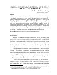
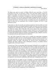
Figure 1<br />
CD11c + <strong>cells</strong> <strong>in</strong> acute <strong>and</strong> chronic granulomas. C57BL/6 mice were<br />
systemically <strong>in</strong>fected i.p. with BCG. (A) H&E sta<strong>in</strong><strong>in</strong>g <strong>of</strong> formal<strong>in</strong>-fixed<br />
liver tissue show<strong>in</strong>g 3- <strong>and</strong> 10-week granulomas. Orig<strong>in</strong>al magnification,<br />
×400. (B) Top panels, CD11c <strong>and</strong> CD11b populations <strong>in</strong> liver<br />
granuloma cell suspensions. Plot obta<strong>in</strong>ed by gat<strong>in</strong>g on the population<br />
display<strong>in</strong>g high side scatter (SSC) <strong>and</strong> forward scatter (FSC), exclud<strong>in</strong>g<br />
lymphocytes. Numbers with<strong>in</strong> gate denote frequency <strong>of</strong> CD11c +<br />
<strong>cells</strong> with<strong>in</strong> high SSC <strong>and</strong> high FSC population. Bottom panels, Ly6C<br />
expression on gated CD11c + population from above plots. Gate set<br />
based on known Ly6C-negative populations <strong>and</strong> numbers denote distribution<br />
<strong>of</strong> Ly6C-positive <strong>and</strong> -negative expression on CD11c + population.<br />
(C) Orig<strong>in</strong>al magnification, ×100. Fluorescent microscopy image<br />
taken <strong>of</strong> liver from CD11c-EYFP mouse <strong>in</strong>fected for 10 weeks with<br />
dsRED BCG. Granulomas <strong>out</strong>l<strong>in</strong>ed with white dashed l<strong>in</strong>es. CD11c-<br />
EYFP <strong>cells</strong> are shown <strong>in</strong> green, <strong>and</strong> DAPI nuclear sta<strong>in</strong> <strong>in</strong> blue. (D)<br />
Digital magnification <strong>of</strong> red box <strong>in</strong> C. Orig<strong>in</strong>al magnification, ×1000.<br />
Red arrows po<strong>in</strong>t to dsRED BCG rods. (E) CD11c-EFYP cell from D<br />
with anti-CD4 sta<strong>in</strong><strong>in</strong>g (red). Red arrow po<strong>in</strong>ts to CD4 + cell, <strong>and</strong> yellow<br />
arrow po<strong>in</strong>ts to merged CD4 + YFP + sta<strong>in</strong><strong>in</strong>g. Orig<strong>in</strong>al magnification,<br />
×1000. (F) Representation <strong>of</strong> observations <strong>in</strong> D <strong>and</strong> E. Representative<br />
plots <strong>and</strong> images from at least 3 or more <strong>in</strong>dependent experiments.<br />
that CD11c + <strong>cells</strong> leav<strong>in</strong>g both 3- <strong>and</strong> 10-week-<strong>in</strong>fected tissue had<br />
a unique systemic dissem<strong>in</strong>ation pattern. We found that the early,<br />
cont<strong>in</strong>uous CD11c + surveillance <strong>and</strong> CCR7-dependent migration<br />
to the lymph node supported T cell activation, but the recipients’<br />
MHCII + <strong>cells</strong> were necessary to prime Mycobacterium-specific T <strong>cells</strong><br />
dur<strong>in</strong>g both phases <strong>of</strong> <strong>in</strong>fection.<br />
Results<br />
Both acute <strong>and</strong> chronic BCG-<strong>in</strong>duced granulomas conta<strong>in</strong> a population <strong>of</strong><br />
CD11c + <strong>cells</strong>, which can be found harbor<strong>in</strong>g mycobacteria <strong>out</strong>side granulomas<br />
<strong>in</strong> chronically <strong>in</strong>fected mice. Acute granulomas (3 weeks after<br />
<strong>in</strong>fection) are generally more numerous <strong>and</strong> less well organized<br />
compared with chronic granulomas (10 weeks after <strong>in</strong>fection)<br />
(Figure 1A <strong>and</strong> Figure 2, B <strong>and</strong> C). It is well known that the acute<br />
lesions have both a high bacterial burden <strong>and</strong> a high frequency <strong>of</strong><br />
IFNγ + CD4 + T <strong>cells</strong>, while the latter chronic granulomas have less <strong>of</strong><br />
both (10). Acute granulomas are associated with a high <strong>in</strong>cidence<br />
<strong>of</strong> bacterial kill<strong>in</strong>g, while chronic lesions are associated with longterm<br />
bacterial survival (16). The 10-week chronic granuloma typically<br />
has 2–3 logs fewer bacilli than 3-week acute lesions. One po<strong>in</strong>t<br />
<strong>of</strong> view is that this difference <strong>in</strong> bacterial burden may be the s<strong>in</strong>gle<br />
most important factor responsible for the chang<strong>in</strong>g immunological<br />
microenvironment with<strong>in</strong> the granuloma. Figure 1B shows that<br />
both acute <strong>and</strong> chronic granulomas conta<strong>in</strong> similar proportions <strong>of</strong><br />
CD11c + CD11b + Ly6C + <strong>cells</strong> (Figure 1B). This subset is <strong>of</strong>ten referred<br />
to as the monocyte-derived “<strong>in</strong>flammatory” DC subset (17, 18).<br />
Support for a population <strong>of</strong> DCs capable <strong>of</strong> migrat<strong>in</strong>g <strong>in</strong>to <strong>and</strong><br />
<strong>out</strong> <strong>of</strong> chronic granulomas came from observ<strong>in</strong>g liver sections <strong>of</strong><br />
10-week dsRED BCG-<strong>in</strong>fected CD11c enhanced yellow fluorescent<br />
prote<strong>in</strong> (CD11c-EYFP) mice with ubiquitously fluoresc<strong>in</strong>g DCs<br />
(19). Albeit rare, CD11c-YFP + <strong>cells</strong> conta<strong>in</strong><strong>in</strong>g dsRED bacilli could<br />
be observed <strong>out</strong>side <strong>of</strong> granulomas <strong>in</strong> chronically <strong>in</strong>fected mice<br />
(Figure 1, C–F). Costa<strong>in</strong><strong>in</strong>g with anti-CD4 <strong>of</strong>ten revealed these <strong>cells</strong><br />
<strong>in</strong> close contact with CD4 + T <strong>cells</strong> (Figure 1, E <strong>and</strong> F). Unbeknownst<br />
to the orig<strong>in</strong> or dest<strong>in</strong>ation <strong>of</strong> this observed BCG-<strong>in</strong>fected CD11c +<br />
cell, this f<strong>in</strong>d<strong>in</strong>g also demonstrates the limitations <strong>of</strong> the current<br />
model for study<strong>in</strong>g DC traffic <strong>in</strong>to <strong>and</strong> <strong>out</strong> <strong>of</strong> granulomas <strong>and</strong> the<br />
subsequent need for a new methodical approach.<br />
Liver tissue conta<strong>in</strong><strong>in</strong>g <strong>in</strong>tact granulomas with mycobacteria <strong>and</strong><br />
CD11c-EYFP + <strong>cells</strong> can be <strong>transplanted</strong> underneath the kidney capsule<br />
<strong>of</strong> a recipient. In order to better study the traffic <strong>of</strong> CD11c + <strong>cells</strong><br />
from granulomatous lesions, we have made use <strong>of</strong> the well-characterized<br />
kidney capsule transplant protocol to develop a model<br />
to track, quantify, <strong>and</strong> measure CD11c + cellular traffic, along<br />
with the immunological <strong>out</strong>come (20). The kidney capsule transplantation<br />
is a well-characterized model that has been used for<br />
decades based on the fact that it is one <strong>of</strong> the highly vascularized<br />
regions <strong>in</strong> the body. Complete revascularization <strong>of</strong> the graft<br />
only takes several days, result<strong>in</strong>g <strong>in</strong> a highly oxygenized graft.<br />
With a skilled h<strong>and</strong>, close to 100% <strong>of</strong> the grafts are accepted <strong>and</strong><br />
the wound heals properly. This model is achieved by systemically<br />
The Journal <strong>of</strong> Cl<strong>in</strong>ical Investigation http://www.jci.org Volume 121 Number 10 October 2011 3903
esearch article<br />
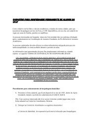
Figure 2<br />
Transplantation <strong>of</strong> liver granulomas under recipient’s kidney capsule. (A) Schematic <strong>of</strong> transplantation model. Liver specimen (~0.025 g ± 10%)<br />
conta<strong>in</strong><strong>in</strong>g granulomas from a 3- or 10-week dsRED BCG–<strong>in</strong>fected CD11c-EYFP donor mouse is <strong>transplanted</strong> underneath the kidney capsule <strong>of</strong><br />
a colorless C57BL/6 recipient. (B <strong>and</strong> C) CD11c-EYFP mice <strong>in</strong>fected 3 weeks (B) <strong>and</strong> 10 weeks (C). Far left images show <strong>in</strong>fected liver; orig<strong>in</strong>al<br />
magnification, ×400. Second column shows white-boxed granuloma; orig<strong>in</strong>al magnification, ×1000. White arrows po<strong>in</strong>t to CD11c-EYFP + <strong>cells</strong><br />
with<strong>in</strong> granuloma, <strong>and</strong> red arrows po<strong>in</strong>t to dsRED BCG. Third column shows CD11c-EYFP liver specimen under kidney capsule <strong>of</strong> colorless<br />
recipient; orig<strong>in</strong>al magnification, ×100. Far right column demonstrates <strong>transplanted</strong> granulomas conta<strong>in</strong><strong>in</strong>g both CD11c-EYFP + <strong>cells</strong> (white arrows)<br />
<strong>and</strong> dsRED BCG (red arrows); orig<strong>in</strong>al magnification, ×1000. 3- <strong>and</strong> 10-week-<strong>in</strong>fected donor images representative <strong>of</strong> 3 <strong>in</strong>dependent experiments<br />
each, <strong>and</strong> <strong>transplanted</strong> kidney capsule images are representative <strong>of</strong> 3–6 mice per time po<strong>in</strong>t from 3 or more <strong>in</strong>dependent experiments.<br />
<strong>in</strong>fect<strong>in</strong>g CD11c-EYFP mice with dsRED BCG <strong>and</strong> wait<strong>in</strong>g until<br />
either acute or chronic lesions are formed with<strong>in</strong> the liver (Figure<br />
2, B <strong>and</strong> C). As depicted <strong>in</strong> Figure 2A, a small piece (0.025 g ±<br />
10%) <strong>of</strong> liver from the acutely or chronically <strong>in</strong>fected CD11c-EYFP<br />
mouse is <strong>transplanted</strong> underneath the kidney capsule <strong>of</strong> a syngeneic<br />
WT recipient (Figure 2A). The images <strong>in</strong> Figure 2, B <strong>and</strong> C,<br />
show the <strong>transplanted</strong> YFP + liver piece underneath the capsule <strong>of</strong><br />
a colorless recipient. Importantly, CD11c-EYFP + <strong>cells</strong> <strong>and</strong> dsRED<br />
BCG bacilli can be found <strong>in</strong> granulomatous lesions <strong>in</strong> the piece<br />
<strong>of</strong> <strong>transplanted</strong> liver (Figure 2, B <strong>and</strong> C). These data demonstrate<br />
both the feasibility <strong>of</strong> the transplant <strong>and</strong> ability to transfer either<br />
<strong>in</strong>tact acute or chronic granulomatous lesions.<br />
CD11c-EYFP + <strong>cells</strong> <strong>migrate</strong> <strong>out</strong> <strong>of</strong> both acute <strong>and</strong> chronic lesions to peripheral<br />
secondary organs. By transplant<strong>in</strong>g granuloma-conta<strong>in</strong><strong>in</strong>g liver<br />
pieces from CD11c-EYFP + donors (Figure 3A), we can track YFP +<br />
cellular egression from granulomas. Sent<strong>in</strong>el CD11c-EYFP + <strong>cells</strong><br />
are present <strong>in</strong> the non<strong>in</strong>fected liver (Figure 3A); however, many<br />
more CD11c-EYFP + <strong>cells</strong> are present <strong>in</strong> <strong>in</strong>fected livers (Figure 3A).<br />
At both acute <strong>and</strong> chronic <strong>in</strong>fection time po<strong>in</strong>ts, there are fewer<br />
CD11c-EYFP + <strong>cells</strong> <strong>in</strong> the <strong>in</strong>terstitial tissue space <strong>out</strong>side granulomas<br />
compared with an un<strong>in</strong>fected liver (Figure 3A). When quantified,<br />
<strong>in</strong> both acute <strong>and</strong> chronic stages, the statistical majority <strong>of</strong><br />
CD11c-EYFP + <strong>cells</strong> are associated with granulomatous lesions<br />
(P = 0.0001 <strong>and</strong> P = 0.0004, respectively) (Figure 4A). This may be<br />
3904 The Journal <strong>of</strong> Cl<strong>in</strong>ical Investigation http://www.jci.org Volume 121 Number 10 October 2011
esearch article<br />
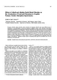
Figure 3<br />
Migration <strong>of</strong> CD11c-EYFP <strong>cells</strong> <strong>out</strong> <strong>of</strong> transplant. (A) CD11c-EYFP distribution <strong>in</strong> donor liver tissue <strong>of</strong> non<strong>in</strong>fected (left), 3-week-<strong>in</strong>fected (middle), <strong>and</strong><br />
10-week-<strong>in</strong>fected (right). Orig<strong>in</strong>al magnification, ×400. (B) In both recipients <strong>of</strong> acute <strong>and</strong> chronic <strong>in</strong>fected donors, CD11c-EYFP <strong>cells</strong> were found <strong>in</strong> the<br />
tRLN 3 days after transplant. Orig<strong>in</strong>al magnification, ×1000 magnification. Images representative from at least 3 or more <strong>in</strong>dependent experiments.<br />
due to the migration <strong>of</strong> stressed liver-resident DCs to the dra<strong>in</strong><strong>in</strong>g<br />
lymph nodes or <strong>in</strong>to the granulomas. The latter is less likely, as<br />
most <strong>of</strong> the CD11c + <strong>cells</strong> <strong>in</strong> the granulomas are Ly6C + , strongly<br />
suggestive <strong>of</strong> their hematogenous arrival (21). Nevertheless, it is<br />
important to note that the vast majority <strong>of</strong> <strong>transplanted</strong> CD11c-<br />
EYFP + <strong>cells</strong> are granuloma associated. Fluorescent microscopy <strong>of</strong><br />
the sectioned tRLN 3 days after transplant revealed the presence<br />
<strong>of</strong> YFP + <strong>cells</strong> (Figure 3B). Although rare, both the <strong>in</strong>herent fluorescence<br />
<strong>and</strong> morphological dendrite protrusions make these <strong>cells</strong><br />
easily dist<strong>in</strong>guishable as transplant-orig<strong>in</strong>ated DCs. Validat<strong>in</strong>g<br />
our previous observation from Figure 1, which suggests a CD11c +<br />
population capable <strong>of</strong> migrat<strong>in</strong>g <strong>out</strong> <strong>of</strong> chronic lesions, we were<br />
able to f<strong>in</strong>d YFP + DCs <strong>in</strong> the tRLN <strong>of</strong> both 3- <strong>and</strong> 10-week-<strong>in</strong>fected<br />
mice (Figure 3B). Interest<strong>in</strong>gly, we have never found a dsRED BCG<br />
rod <strong>in</strong> any <strong>of</strong> the CD11c-EYFP + <strong>cells</strong> that have <strong>migrate</strong>d from the<br />
transplant. This observation was further strengthened by CFU<br />
on lymph nodes, spleen, <strong>and</strong> liver, which were repeatedly negative<br />
(Supplemental Figure 1; supplemental material available onl<strong>in</strong>e<br />
with this article; doi:10.1172/JCI45113DS1). To confirm that viable<br />
BCG rema<strong>in</strong>ed <strong>in</strong> the <strong>transplanted</strong> granulomas over the course<br />
<strong>of</strong> <strong>in</strong>fection, we removed the grafted liver piece 14 days follow<strong>in</strong>g<br />
transplant <strong>and</strong> performed CFU <strong>and</strong> a thorough microscopy search<br />
for fluorescent BCG (Supplemental Figure 1A). Indeed, viable<br />
BCG was still present <strong>and</strong> conta<strong>in</strong>ed with<strong>in</strong> the grafted liver piece<br />
through<strong>out</strong> the duration <strong>of</strong> our experiments. To further confirm<br />
the viability <strong>of</strong> BCG with<strong>in</strong> the graft, we removed the <strong>transplanted</strong><br />
liver piece <strong>and</strong> grafted <strong>in</strong>to TNF-α–deficient recipients. TNF-α is<br />
required to ma<strong>in</strong>ta<strong>in</strong> the granulomas’ cellular composition <strong>and</strong><br />
anti-bacterial dissem<strong>in</strong>ation properties (22). Under TNF-α–deficient<br />
conditions, the BCG was not conta<strong>in</strong>ed <strong>and</strong> dissem<strong>in</strong>ated. In<br />
accordance, transplantation <strong>of</strong> Rag-deficient donors <strong>in</strong>to WT also<br />
does not conta<strong>in</strong> <strong>in</strong>fection <strong>and</strong> dissem<strong>in</strong>ation is observed (Supplemental<br />
Figure 1B). We concluded that over the course <strong>of</strong> our<br />
<strong>in</strong>vestigational period, the BCG localization <strong>and</strong> viability rema<strong>in</strong><br />
constant. Hav<strong>in</strong>g immunocompetent donors <strong>and</strong> recipients is sufficient<br />
to conta<strong>in</strong> the <strong>in</strong>fection with<strong>in</strong> the granuloma <strong>and</strong> at the<br />
same time ma<strong>in</strong>ta<strong>in</strong> Mycobacterium viability.<br />
CD11c-EYFP + <strong>cells</strong> migrat<strong>in</strong>g <strong>out</strong> <strong>of</strong> both acute <strong>and</strong> chronic <strong>in</strong>fected donor<br />
tissue have a systemic dissem<strong>in</strong>ation pattern, but differ <strong>in</strong> the quantity <strong>and</strong><br />
rate <strong>of</strong> egression. To systemically track CD11c-EYFP + cell egression<br />
from the <strong>transplanted</strong> granulomas, we used real-time PCR to measure<br />
YFP transcript (Figure 4, B <strong>and</strong> C). A st<strong>and</strong>ard curve was generated<br />
from known quantities <strong>of</strong> purified CD11c-EYFP + <strong>cells</strong> diluted<br />
<strong>in</strong>to WT YFP – <strong>cells</strong> (Figure 4B). The equation generated from the<br />
The Journal <strong>of</strong> Cl<strong>in</strong>ical Investigation http://www.jci.org Volume 121 Number 10 October 2011 3905
esearch article<br />
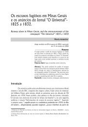
Figure 4<br />
CD11c-EYFP <strong>cells</strong> <strong>migrate</strong> <strong>out</strong> <strong>of</strong> <strong>transplanted</strong> granuloma-conta<strong>in</strong><strong>in</strong>g liver piece to systemic sites. (A) Distribution <strong>of</strong> CD11c-EYFP <strong>cells</strong> <strong>in</strong> donor<br />
liver. All CD11c-EYFP <strong>cells</strong> <strong>in</strong> 10–15 ×400 objective fields from 3 mice per time po<strong>in</strong>t were counted <strong>and</strong> determ<strong>in</strong>ed to be either with<strong>in</strong> or <strong>out</strong>side<br />
<strong>of</strong> granulomas, determ<strong>in</strong>ed by DAPI nuclear sta<strong>in</strong>. (B) Real-time PCR st<strong>and</strong>ard curve generated from known values <strong>of</strong> purified CD11c-EYFP<br />
<strong>cells</strong> diluted <strong>in</strong>to WT <strong>cells</strong>. (C) 3 (top row) <strong>and</strong> 7 (bottom 2 rows) days after transplant, RT-PCR was performed to detect YFP transcript from<br />
<strong>transplanted</strong> kidney (tKid), opposite kidney (oKid), transplant-dra<strong>in</strong><strong>in</strong>g renal lymph node (tRLN), opposite renal lymph node (oRNL), cervical<br />
lymph nodes (CLN), spleen (Sp), <strong>and</strong> liver (Liv). Graphs show percentage <strong>of</strong> total YFP + <strong>cells</strong> detected by PCR. Bottom row shows total percentage<br />
distribution <strong>of</strong> dissem<strong>in</strong>ated YFP + <strong>cells</strong> from dashed box above, <strong>and</strong> n value above box <strong>in</strong>dicates absolute total number <strong>of</strong> YFP + <strong>cells</strong>. Error<br />
bars represent mean ± SEM <strong>and</strong> data representative <strong>of</strong> 3 <strong>in</strong>dependent experiments per time po<strong>in</strong>t with 2–3 mice per group.<br />
st<strong>and</strong>ard curve along with whole <strong>and</strong> partial organ weights were<br />
used henceforth to determ<strong>in</strong>e the absolute number <strong>of</strong> CD11c-<br />
EYFP + <strong>in</strong> various tissues. At 3 <strong>and</strong> 7 days after transplant <strong>of</strong> liver<br />
pieces from non<strong>in</strong>fected or 3- or 10-week BCG <strong>in</strong>fected CD11c-<br />
EYFP mice, the <strong>transplanted</strong> kidney, opposite kidney, transplantdra<strong>in</strong><strong>in</strong>g<br />
renal lymph node (tRLN), opposite renal lymph node, cervical<br />
lymph nodes, spleen, <strong>and</strong> liver were removed <strong>and</strong> homogenized<br />
(Figure 4C). Real-time PCR was performed on total isolated RNA<br />
from tissue homogenate to detect YFP transcript. At 3 days after<br />
transplant, the vast majority <strong>of</strong> CD11c-EYFP + <strong>cells</strong> rema<strong>in</strong>ed <strong>in</strong><br />
both the non<strong>in</strong>fected <strong>and</strong> 3-week-<strong>in</strong>fected donor liver; however, 41%<br />
± 13% <strong>migrate</strong>d <strong>out</strong> <strong>of</strong> the chronic 10-week-<strong>in</strong>fected donor piece to<br />
all peripheral sites sampled, particularly the tRLN. On average, the<br />
10-week tRLN conta<strong>in</strong>ed 9,803 CD11c-EYFP + <strong>cells</strong> compared with<br />
540 <strong>and</strong> 46 <strong>in</strong> the 3-week <strong>and</strong> non<strong>in</strong>fected tRLNs, respectively. At 7<br />
days after transplant, those CD11c-EYFP + <strong>cells</strong> that regressed from<br />
the acute transplant also <strong>migrate</strong>d to all sampled systemic organs,<br />
but those from the non<strong>in</strong>fected liver <strong>migrate</strong>d nearly exclusively<br />
to the tRLN. Interest<strong>in</strong>gly, these data demonstrate that compared<br />
with non<strong>in</strong>fected tissue, CD11c + <strong>cells</strong> orig<strong>in</strong>at<strong>in</strong>g from acutely or<br />
chronically Mycobacterium-<strong>in</strong>fected tissue have the ability to dissem<strong>in</strong>ate<br />
to peripheral sites. Furthermore, for what we believe is<br />
the first time, these data also suggest that DCs presumably associated<br />
with chronic granulomas readily <strong>migrate</strong> to secondary lymphoid<br />
organs, <strong>and</strong> compared with DCs from non<strong>in</strong>fected tissue,<br />
DCs from Mycobacterium-<strong>in</strong>fected tissue have a different migratory<br />
r<strong>out</strong>e given their different end po<strong>in</strong>ts.<br />
Recipient CD11c + <strong>cells</strong> <strong>migrate</strong> <strong>in</strong>to chronic <strong>transplanted</strong> granulomas<br />
more than <strong>in</strong>to acute granulomas. After observ<strong>in</strong>g different patterns <strong>of</strong><br />
DC egression from acute <strong>and</strong> chronic lesions, we next determ<strong>in</strong>ed<br />
whether the same was true for recipient CD11c + <strong>cells</strong>’ access <strong>in</strong>to<br />
<strong>transplanted</strong> granulomas by transplant<strong>in</strong>g un<strong>in</strong>fected or 3- or<br />
10-week-<strong>in</strong>fected colorless C57BL/6 liver pieces under the kidney<br />
capsule <strong>of</strong> CD11c-EYFP mice. At 3 <strong>and</strong> 7 days after transplant, the<br />
<strong>transplanted</strong> liver piece was excised, homogenized, <strong>and</strong> analyzed<br />
by flow cytometry (Figure 5A). To determ<strong>in</strong>e the distribution <strong>of</strong><br />
3906 The Journal <strong>of</strong> Cl<strong>in</strong>ical Investigation http://www.jci.org Volume 121 Number 10 October 2011
esearch article<br />
Figure 5<br />
CD11c-EYFP + migration <strong>in</strong>to <strong>transplanted</strong> granulomas. Non<strong>in</strong>fected or 3- or 10-week-<strong>in</strong>fected liver pieces from colorless donors were <strong>transplanted</strong><br />
<strong>in</strong>to CD11c-EYFP recipients. (A) 3 <strong>and</strong> 7 days post transplant (dpt), donor liver tissue was excised <strong>and</strong> prepared for flow cytometry.<br />
CD11c-EYFP histograms generated from CD11c + surface sta<strong>in</strong> gate. Numbers denote frequency <strong>of</strong> CD11c-EYFP positive <strong>and</strong> negative <strong>cells</strong><br />
among total CD11c + population. (B) Fluorescent microscopy images <strong>of</strong> <strong>transplanted</strong> kidney 3 <strong>and</strong> 7 days after transplant. In first <strong>and</strong> third<br />
columns, white dashed l<strong>in</strong>es <strong>in</strong>dicate borders <strong>of</strong> <strong>transplanted</strong> piece <strong>and</strong> kidney. In second <strong>and</strong> fourth columns, red <strong>in</strong>dicates donor anti-CD11c<br />
surface sta<strong>in</strong> (D), <strong>and</strong> green or yellow/orange <strong>in</strong>dicate recipient CD11c + cell (R). Orig<strong>in</strong>al magnification, ×400 (first <strong>and</strong> third columns); ×1000<br />
(second <strong>and</strong> fourth columns). D, donor; R, recipient. (C) Mean distribution <strong>of</strong> all donor (white bars) <strong>and</strong> recipient (gray bars) CD11c + <strong>cells</strong> per<br />
granuloma 3 <strong>and</strong> 7 days after transplant. CD11c + cellular distribution was determ<strong>in</strong>ed from 10 granulomas per time po<strong>in</strong>t. (D) Histograms show<strong>in</strong>g<br />
surface expression <strong>of</strong> MHCII <strong>and</strong> activat<strong>in</strong>g costimulatory molecules CD40 <strong>and</strong> CD86. Red dashed l<strong>in</strong>e represents background expression.<br />
(E) Mean MFI <strong>of</strong> MHCII <strong>and</strong> costimulatory molecule expression. Data shown representative <strong>of</strong> 2 <strong>in</strong>dependent experiments with 4 kidneys per<br />
group. **P < 0.05; ***P < 0.001. Error bars represent mean ± SEM.<br />
The Journal <strong>of</strong> Cl<strong>in</strong>ical Investigation http://www.jci.org Volume 121 Number 10 October 2011 3907
esearch article<br />
Figure 6<br />
Proliferation <strong>of</strong> Mycobacteria-specific Ag85B CD4 + T <strong>cells</strong> after transplant <strong>of</strong> acutely <strong>and</strong> chronically <strong>in</strong>fected liver tissue. (A) Fluorescent microscopy<br />
<strong>of</strong> transplant tRLN 7 days after transplant. CD11c-EYFP <strong>cells</strong> (green), <strong>and</strong> CD4 + T <strong>cells</strong> (red). Yellow arrows po<strong>in</strong>t to merged green <strong>and</strong> red sta<strong>in</strong><strong>in</strong>g.<br />
Orig<strong>in</strong>al magnification, ×1000. (B) Experimental schematic. Briefly, CD11c-EYFP mice were systemically <strong>in</strong>fected with dsRED BCG for 3 weeks, 10<br />
weeks, or 7 months. 1 day prior to transplant, 5 × 10 5 CFSE-labeled dsRED Ag85B CD4 + T <strong>cells</strong> were adoptively transferred. 7 days after transplant,<br />
the tRLNs were removed <strong>and</strong> analyzed by flow cytometry. (C) Top row: adoptively transferred Tg T <strong>cells</strong> were identified by CD4 + dsRED + gat<strong>in</strong>g,<br />
plots shown were obta<strong>in</strong>ed from lymphocyte gate on SSC versus FSC plot. Bottom 2 rows: CFSE dilution histogram from Tg gate from upper row.<br />
(D) Graph shows average percentage <strong>of</strong> dsRED Ag85B CD4 + T <strong>cells</strong> <strong>in</strong> cycle. Graph representative <strong>of</strong> 2–5 <strong>in</strong>dependent experiments per time po<strong>in</strong>t<br />
with an average <strong>of</strong> 2–6 mice per group. Error bars represent mean ± SEM, <strong>and</strong> statistical significance between groups is shown <strong>in</strong> graph.<br />
donor CD11c + <strong>cells</strong> <strong>and</strong> recipient CD11c + <strong>cells</strong> <strong>in</strong> the transplant,<br />
histograms were generated from the surface-sta<strong>in</strong>ed CD11c +<br />
gate <strong>and</strong> evaluated for YFP + fluorescent expression. Interest<strong>in</strong>gly,<br />
CD11c-EYFP + <strong>cells</strong> readily <strong>migrate</strong>d <strong>in</strong>to non<strong>in</strong>fected <strong>and</strong> 10-week<br />
chronically <strong>in</strong>fected donor tissue by day 3, where a m<strong>in</strong>ority (~5%)<br />
<strong>of</strong> CD11c + <strong>cells</strong> <strong>in</strong> the acute donor were <strong>of</strong> recipient orig<strong>in</strong> (Figure<br />
5A). By 7 days after transplant, approximately 75% <strong>of</strong> CD11c + <strong>cells</strong><br />
<strong>in</strong> the chronically <strong>in</strong>fected donor tissue were newly recruited recipient<br />
CD11c + <strong>cells</strong>. This suggests a high turnover <strong>of</strong> granuloma-associated<br />
CD11c + <strong>cells</strong> <strong>in</strong> these lesions. The visible network <strong>of</strong> sent<strong>in</strong>el<br />
kidney YFP + CD11c + <strong>cells</strong> afforded us the ability to easily detect<br />
the colorless <strong>transplanted</strong> piece by fluorescent microscopy (Figure<br />
5B). At 3 days after transplant, recipient CD11c-EYFP + <strong>cells</strong> (R) had<br />
already <strong>in</strong>filtrated chronic granulomas <strong>and</strong> could be found <strong>in</strong> both<br />
the periphery <strong>and</strong> center <strong>of</strong> the lesions (Figure 5B), whereas few<br />
recipient CD11c-EYFP + <strong>cells</strong> could be found <strong>in</strong> acute 3-week granulomas.<br />
Only donor CD11c + <strong>cells</strong> (D) could be found <strong>in</strong> acute lesions<br />
(Figure 5B). Unlike the chronic donor, by day 7 after transplant,<br />
those CD11c-EYFP + <strong>cells</strong> that had <strong>in</strong>filtrated the acute donor tissue<br />
were found primarily <strong>in</strong> the lymphocytic cuff around the lesions<br />
(Figure 5B). Like the distribution <strong>of</strong> EYFP – <strong>and</strong> EYFP + CD11c + <strong>cells</strong><br />
observed with<strong>in</strong> the entire transplant by flow cytometry (Figure<br />
5A), a similar pattern was observed locally with<strong>in</strong> the granuloma<br />
after quantify<strong>in</strong>g the distribution <strong>of</strong> donor CD11c + <strong>cells</strong> <strong>and</strong> recipient<br />
CD11c-EYFP + <strong>cells</strong> on a s<strong>in</strong>gle lesion basis (Figure 5C). Together<br />
with the previous observation that CD11c + <strong>cells</strong> egress from chronic<br />
granulomas faster than from acute lesions, these data collectively<br />
demonstrate that after transplantation, chronic granulomas are<br />
more quickly <strong>in</strong>filtrated by recipient CD11c + <strong>cells</strong> compared with<br />
acute granulomas.<br />
Once recipient CD11c + <strong>cells</strong> <strong>migrate</strong> <strong>in</strong>to the <strong>transplanted</strong> granulomas,<br />
their maturation status is likely to respond accord<strong>in</strong>gly.<br />
Previously, we found that CD11c + <strong>cells</strong> <strong>in</strong> acute granulomas were<br />
extremely activated with high expression <strong>of</strong> MHCII <strong>and</strong> T cell<br />
3908 The Journal <strong>of</strong> Cl<strong>in</strong>ical Investigation http://www.jci.org Volume 121 Number 10 October 2011
esearch article<br />
Figure 7<br />
Mycobacteria-specific Ag85B CD4 + T cell proliferation is dependent<br />
on MHCII expression on recipients’ <strong>cells</strong>. (A) 5 × 10 5 CFSE-labeled<br />
dsRED P25 CD4 + T <strong>cells</strong> were adoptively transferred <strong>in</strong>to recipient<br />
C57BL/6 mice or MHCII-deficient mice 1 day prior to transplant <strong>of</strong><br />
3- <strong>and</strong> 10-week BCG-<strong>in</strong>fected WT donors (3–5 mice per time po<strong>in</strong>t<br />
representative <strong>of</strong> 1–3 <strong>in</strong>dependent experiments), <strong>and</strong> 3- <strong>and</strong> 10-week<br />
BCG-<strong>in</strong>fected Ccr7 –/– donors (6 mice per time po<strong>in</strong>t). CFSE dilution<br />
histogram <strong>of</strong> Tg P25 CD4 + T <strong>cells</strong> <strong>in</strong> the tRLN 7 days after transplant.<br />
Numbers denote frequency <strong>of</strong> proliferat<strong>in</strong>g <strong>cells</strong> determ<strong>in</strong>ed by<br />
set gate. Cells shown from same Tg gat<strong>in</strong>g strategy used <strong>in</strong> Figure<br />
6C. (B) Mean percentage <strong>of</strong> P25 Tg CD4 + T <strong>cells</strong> 7 days after transplant<br />
<strong>in</strong> tRLN <strong>in</strong> cycle. Values obta<strong>in</strong>ed from gat<strong>in</strong>g strategy shown<br />
<strong>in</strong> A. Statistical significance from WT is as follows: ****P = 0.00001;<br />
***P = 0.0001; **P = 0.001. Error bars represent mean ± SEM.<br />
costimulatory molecules CD40 <strong>and</strong> CD86 (21). However, CD11c +<br />
<strong>cells</strong> <strong>in</strong> chronic lesions <strong>of</strong> both BCG-<strong>in</strong>fected mice <strong>and</strong> the lungs<br />
<strong>of</strong> M. tuberculosis–<strong>in</strong>fected mice had much lower expression <strong>of</strong> these<br />
markers (21, 23). Here, we found that when the CD11c-EYFP + <strong>cells</strong><br />
<strong>migrate</strong>d <strong>in</strong>to either un<strong>in</strong>fected or 3- <strong>and</strong> 10-week-<strong>in</strong>fected transplants,<br />
they achieved a maturation status similar to that <strong>of</strong> the<br />
resident CD11c + <strong>cells</strong>. Compared with CD11c-EYFP + <strong>cells</strong> that<br />
entered either non<strong>in</strong>fected or 10-week chronically <strong>in</strong>fected tissue,<br />
those found <strong>in</strong> 3-week-<strong>in</strong>fected donor tissue had significantly<br />
higher expression <strong>of</strong> MHCII <strong>and</strong> T cell costimulatory molecules<br />
CD40 <strong>and</strong> CD86 (Figure 5, D <strong>and</strong> E), thus demonstrat<strong>in</strong>g, that<br />
newly arrived CD11c + <strong>cells</strong> <strong>in</strong> the granuloma take on the phenotype<br />
<strong>of</strong> the resident CD11c + <strong>cells</strong>.<br />
Antigenic sampl<strong>in</strong>g results <strong>in</strong> proliferation <strong>of</strong> mycobacteria-specific Ag85B<br />
CD4 + T <strong>cells</strong> after transplant <strong>of</strong> acutely <strong>and</strong> chronically <strong>in</strong>fected BCG <strong>and</strong><br />
M. tuberculosis liver granulomas. One <strong>of</strong> the questions that rema<strong>in</strong><br />
regard<strong>in</strong>g <strong>in</strong>fection with pathogenic mycobacteria is whether there<br />
is cont<strong>in</strong>uous antigenic sampl<strong>in</strong>g from chronic granulomas <strong>and</strong><br />
subsequent T cell prim<strong>in</strong>g. However, <strong>in</strong> order to ask this question,<br />
one has to rule <strong>out</strong> the presence <strong>of</strong> mycobacteria <strong>in</strong> the lymph<br />
nodes where prim<strong>in</strong>g occurs <strong>and</strong> rule <strong>out</strong> the possibility <strong>of</strong> T cell<br />
prim<strong>in</strong>g as a result <strong>of</strong> newly formed lesions dur<strong>in</strong>g chronic time<br />
po<strong>in</strong>ts. The transplant model presented here allows us to address<br />
the question <strong>of</strong> antigenic sampl<strong>in</strong>g from chronic lesions. We have<br />
observed migration <strong>of</strong> CD11c-EYFP + <strong>cells</strong> <strong>in</strong> <strong>and</strong> <strong>out</strong> <strong>of</strong> both acute<br />
<strong>and</strong> chronic granuloma–laden transplants (Figures 3–5). Collectively,<br />
our data demonstrate that a portion <strong>of</strong> CD11c + <strong>cells</strong> with<strong>in</strong><br />
the chronic granuloma are able to reach the dra<strong>in</strong><strong>in</strong>g lymph<br />
nodes, the primary site <strong>of</strong> T cell prim<strong>in</strong>g. To see whether newly<br />
e<strong>migrate</strong>d CD11c-EYFP + <strong>cells</strong> engage with T <strong>cells</strong> <strong>in</strong> the tRLN,<br />
we used fluorescent microscopy (Figure 6A). CD11c-EYFP + <strong>cells</strong><br />
from both 3- <strong>and</strong> 10-week-<strong>in</strong>fected donors were found <strong>in</strong> close<br />
contact with CD4 + T <strong>cells</strong> (Figure 6A). To test whether T cell prim<strong>in</strong>g<br />
occurred, donor CD11c-EYFP mice were <strong>in</strong>fected for 3 weeks,<br />
10 weeks, or 7 months (Figure 6B). Un<strong>in</strong>fected CD11c-EYFP mice<br />
were used as negative controls. One day prior to transplant, all<br />
mice received an adoptive transfer <strong>of</strong> 5 × 10 5 CFSE-labeled dsRED<br />
P25 CD4 + T <strong>cells</strong>, which have TCR specificity for Mycobacterium<br />
Ag85B <strong>of</strong> M. tuberculosis <strong>and</strong> BCG. On the day <strong>of</strong> the transplant,<br />
C57BL/6 recipients received a piece <strong>of</strong> either un<strong>in</strong>fected or 3-week,<br />
10-week, or 7-month <strong>in</strong>fected liver. Seven days after transplant,<br />
P25 CD4 + T <strong>cells</strong> <strong>in</strong> the <strong>transplanted</strong> tRLN were analyzed by flow<br />
cytometry (Figure 6C). P25 CD4 + T <strong>cells</strong> were traced by co-CD4<br />
<strong>and</strong> -dsRED expression. CFSE dilution <strong>of</strong> these <strong>cells</strong> was observed<br />
<strong>in</strong> all <strong>in</strong>fected donor recipients (Figure 6C). Compared with the<br />
non<strong>in</strong>fected antigen-free donor, statistically more P25 CD4 +<br />
T <strong>cells</strong> were <strong>in</strong> cycle <strong>in</strong> both the acute <strong>and</strong> chronic <strong>in</strong>fected donor<br />
groups <strong>and</strong> detectable <strong>in</strong> the 7-month donor recipient as well<br />
(Figure 6D). In addition to BCG, we exam<strong>in</strong>ed P25 CD4 + T cell<br />
prim<strong>in</strong>g after transplant <strong>of</strong> both 3- <strong>and</strong> 10-week M. tuberculosis<br />
stra<strong>in</strong> mc 2 6020 (ΔlysA ΔpanCD mutant), auxotrophic for lys<strong>in</strong>e<br />
<strong>and</strong> pantothenate, <strong>in</strong>fected liver (Supplemental Figure 2). Infection<br />
with this M. tuberculosis mutant results <strong>in</strong> 2 features similar to<br />
latency: low CFU <strong>and</strong> a nonproliferative state (24). We found that<br />
T cell prim<strong>in</strong>g occurred after transplant <strong>of</strong> both acute <strong>and</strong> chronic<br />
M. tuberculosis-<strong>in</strong>duced granulomas. However, due to the low bacterial<br />
load observed with this stra<strong>in</strong>, both time po<strong>in</strong>ts resulted<br />
<strong>in</strong> similar levels <strong>of</strong> T cell prim<strong>in</strong>g. These data beg<strong>in</strong> to address<br />
the long-st<strong>and</strong><strong>in</strong>g question <strong>of</strong> cont<strong>in</strong>uous T cell prim<strong>in</strong>g dur<strong>in</strong>g<br />
chronic Mycobacterium <strong>in</strong>fection. Although not as robust as<br />
<strong>in</strong> response to acute lesions, which have a much higher bacterial<br />
burden, here we show that systemic Mycobacterium-specific CD4 +<br />
T <strong>cells</strong> divide after transplant <strong>of</strong> both 10-week <strong>and</strong> 7-month chronic<br />
donors, despite the low bacterial burden.<br />
Recipient MHCII molecule expression is required for prim<strong>in</strong>g <strong>of</strong> P25<br />
CD4 + T <strong>cells</strong> follow<strong>in</strong>g transplant <strong>of</strong> both acutely <strong>and</strong> chronically <strong>in</strong>fected<br />
liver granulomas. As previously mentioned, the DCs associated<br />
with Mycobacterium-<strong>in</strong>duced granulomas are <strong>of</strong> the myeloid<br />
monocyte–derived “<strong>in</strong>flammatory” DC subset, characterized by<br />
CD11c <strong>in</strong>t–hi CD11b hi Ly6C hi . However, previous studies <strong>in</strong>vestigat<strong>in</strong>g<br />
the P25 CD4 + T cell activation capacity <strong>of</strong> this subset dur<strong>in</strong>g<br />
early M. tuberculosis <strong>in</strong>fection found them to be poor T cell stimulators<br />
that elicit much less IFN-γ production compared with other<br />
DC subsets (5). There is a paradigm that newly arrived migratory<br />
DCs <strong>in</strong> the lymph node tend to “h<strong>and</strong> <strong>of</strong>f” antigen to lymph node–<br />
The Journal <strong>of</strong> Cl<strong>in</strong>ical Investigation http://www.jci.org Volume 121 Number 10 October 2011 3909
esearch article<br />
resident DCs (25, 26). To determ<strong>in</strong>e whether the granuloma-orig<strong>in</strong>ated<br />
DCs were responsible for T cell activation or whether the<br />
recipients’ DCs were acquir<strong>in</strong>g antigen <strong>and</strong> prim<strong>in</strong>g, we adoptively<br />
transferred 5 × 10 5 CFSE-labeled dsRED P25 CD4 + T <strong>cells</strong> <strong>in</strong>to<br />
MHCII-deficient recipients <strong>and</strong> <strong>transplanted</strong> 3- or 10-week-<strong>in</strong>fected<br />
liver pieces from MHCII-express<strong>in</strong>g donors. Seven days after<br />
transplant, P25 CD4 + T <strong>cells</strong> <strong>in</strong> the <strong>transplanted</strong> kidney-dra<strong>in</strong><strong>in</strong>g<br />
renal lymph node were analyzed by flow cytometry (Figure 7A).<br />
In both 3- <strong>and</strong> 10-week-<strong>in</strong>fected donor MHCII-deficient recipients,<br />
the frequency <strong>of</strong> P25 CD4 + T <strong>cells</strong> <strong>in</strong> cycle was significantly<br />
decreased (Figure 7, A <strong>and</strong> B). These data <strong>in</strong>dicate that granuloma<br />
MHCII + <strong>cells</strong> are not sufficient for prim<strong>in</strong>g Mycobacterium-specific<br />
P25 CD4 + T <strong>cells</strong>. This would suggest that recipient MHCII +<br />
<strong>cells</strong> obta<strong>in</strong> antigen. The acquisition <strong>of</strong> antigen by recipient DCs<br />
may occur with<strong>in</strong> the lymph node after the migration <strong>of</strong> granuloma-derived<br />
APCs, or with<strong>in</strong> the transplant by obta<strong>in</strong><strong>in</strong>g antigen<br />
from resident macrophages or DCs, or engulf<strong>in</strong>g any extracellular<br />
bacteria. In recipients <strong>of</strong> both 3- <strong>and</strong> 10-week-<strong>in</strong>fected donors, we<br />
found CD11c-EYFP + recipient <strong>cells</strong> <strong>in</strong> close proximity with donor<br />
CD11c + <strong>cells</strong> with<strong>in</strong> the transplant (Supplemental Figure 3A) <strong>and</strong><br />
recipient CD11c-EYFP + <strong>cells</strong> with viable dsRED BCG at 7 days<br />
after transplant <strong>of</strong> a 3-week-<strong>in</strong>fected donor (Supplemental Figure<br />
3B). Furthermore, 3- <strong>and</strong> 10-week donor CD11c-EYFP + <strong>cells</strong><br />
could be found <strong>in</strong> direct contact with recipient CD11c + <strong>cells</strong> <strong>in</strong><br />
the dra<strong>in</strong><strong>in</strong>g renal lymph nodes (Supplemental Figure 3C). Based<br />
on the previous observation that CD11c + <strong>cells</strong> are able to readily<br />
<strong>in</strong>filtrate chronic, but not acute, granulomas by day 3, we hypothesized<br />
that if antigenic transfer required the shuttl<strong>in</strong>g <strong>of</strong> mycobacterial<br />
antigen to the lymph nodes by granuloma CD11c + <strong>cells</strong>, this<br />
could be facilitated by either donor or recipient CD11c + <strong>cells</strong> dur<strong>in</strong>g<br />
chronic <strong>in</strong>fection, but only donor CD11c + <strong>cells</strong> dur<strong>in</strong>g acute<br />
<strong>in</strong>fection. To test this, we <strong>transplanted</strong> liver pieces from 3- <strong>and</strong><br />
10-week-<strong>in</strong>fected CCR7-deficient mice. CCR7, chemok<strong>in</strong>e receptor<br />
7, is expressed by DCs <strong>and</strong> T lymphocytes <strong>and</strong> is a receptor responsible<br />
for DC migration to the lymph nodes <strong>in</strong> response to lig<strong>and</strong>s<br />
CCL19 <strong>and</strong> CCL21 (27). The Ccr7 –/– mice had a similar proportion<br />
<strong>and</strong> localization <strong>of</strong> CD11c + <strong>cells</strong> <strong>in</strong> the granuloma at both 3- <strong>and</strong><br />
10-week <strong>in</strong>fection time po<strong>in</strong>ts (data not shown). Transplant <strong>of</strong><br />
3-week-<strong>in</strong>fected Ccr7 –/– mice resulted <strong>in</strong> a significant decrease <strong>in</strong><br />
P25 activation (Figure 7, A <strong>and</strong> B). However, after transplantation<br />
<strong>of</strong> 10-week-<strong>in</strong>fected Ccr7 –/– donor liver, the level <strong>of</strong> P25 CD4 + T cell<br />
activation surpassed that observed <strong>in</strong> 10-week WT donors (Figure<br />
7, A <strong>and</strong> B). The bacterial burden <strong>in</strong> a 10-week Ccr7 –/– animal is<br />
slightly higher as compared with 10-week WT (data not shown),<br />
which could result <strong>in</strong> higher antigen availability <strong>and</strong>, therefore,<br />
<strong>in</strong>creased prim<strong>in</strong>g.<br />
Discussion<br />
DC function is <strong>in</strong>herently associated with migration from the site<br />
<strong>of</strong> immune surveillance to the dra<strong>in</strong><strong>in</strong>g lymph node. The use <strong>of</strong><br />
DC, T cell, <strong>and</strong> bacteria-specific fluorescent cod<strong>in</strong>g, <strong>in</strong> comb<strong>in</strong>ation<br />
with the well-characterized kidney capsule transplant model<br />
presented here, allows us to ask questions regard<strong>in</strong>g DC migration<br />
to <strong>and</strong> from granulomas <strong>and</strong> the potential consequence(s) it may<br />
have on the immune response. This model addresses granulomaantigen<br />
prim<strong>in</strong>g by exclud<strong>in</strong>g the possibility <strong>of</strong> extra-granulomatous<br />
bacteria or the possibility <strong>of</strong> T cell prim<strong>in</strong>g as a result <strong>of</strong><br />
newly formed lesions. Additionally, the transplantation itself also<br />
bears medical relevance, as tuberculosis rema<strong>in</strong>s a serious risk to<br />
liver transplant recipients Although most <strong>in</strong>stances are the result<br />
<strong>of</strong> reactivation <strong>of</strong> latent M. tuberculosis <strong>in</strong> the recipient follow<strong>in</strong>g<br />
transplantation, there are cases <strong>of</strong> transmission through the <strong>transplanted</strong><br />
liver, particularly <strong>in</strong> develop<strong>in</strong>g countries (28, 29). Along<br />
with its advantages, the transplant model also has several restrictions.<br />
Like any surgery, the physical stress <strong>of</strong> the transplant itself is<br />
likely to <strong>in</strong>duce nonspecific migration; however, the environment<br />
surround<strong>in</strong>g the granulomatous <strong>in</strong>flammation may be similarly<br />
stressed. We <strong>in</strong>clude non<strong>in</strong>fected transplants to establish a basel<strong>in</strong>e<br />
as an important control. Importantly, tissue <strong>in</strong>tegrity <strong>of</strong> the<br />
transplant for the duration <strong>of</strong> these studies (≤ 7 days) is excellent<br />
<strong>and</strong> granulomas with<strong>in</strong> the transplant can be easily detected.<br />
On a cellular level, our data support a chang<strong>in</strong>g view <strong>of</strong> chronic<br />
granulomas by show<strong>in</strong>g that one-third <strong>of</strong> DCs are exchanged by<br />
traffic <strong>in</strong> <strong>and</strong> <strong>out</strong> <strong>of</strong> the granuloma with<strong>in</strong> 1 week. Interest<strong>in</strong>gly,<br />
we found that CD11c + <strong>cells</strong> had significantly better access to enter<br />
<strong>and</strong> exit chronic granulomas compared with acute. This f<strong>in</strong>d<strong>in</strong>g<br />
was unexpected, consider<strong>in</strong>g the robust <strong>in</strong>flammation associated<br />
with acute lesions. One would expect considerable CD11c + recruitment<br />
dur<strong>in</strong>g this time due to a stronger chemok<strong>in</strong>e gradient (30).<br />
Additionally, the extracellular matrix that surrounds both acute,<br />
<strong>and</strong> to a somewhat higher extent, chronic granulomas, does not<br />
seem to prevent the entry <strong>of</strong> CD11c + <strong>cells</strong> <strong>in</strong>to chronic lesions.<br />
Additional studies are needed to underst<strong>and</strong> why chronic granulomas,<br />
the predom<strong>in</strong>ate site <strong>of</strong> long-term bacterial persistence, are<br />
so thoroughly exposed to immune-survey<strong>in</strong>g <strong>cells</strong>.<br />
We were also able to track the migration <strong>of</strong> CD11c + <strong>cells</strong> <strong>out</strong><br />
<strong>of</strong> acute <strong>and</strong> chronic granulomas. Interest<strong>in</strong>gly, compared with<br />
CD11c-EYFP + <strong>cells</strong> associated with un<strong>in</strong>fected tissue, which solely<br />
<strong>migrate</strong>d to the transplant-dra<strong>in</strong><strong>in</strong>g renal lymph node, CD11c-<br />
EYFP + <strong>cells</strong> orig<strong>in</strong>at<strong>in</strong>g from granulomas dissem<strong>in</strong>ated to many<br />
systemic sites (Figure 4). This may be attributed to the subset<br />
<strong>of</strong> DCs associated with un<strong>in</strong>fected <strong>and</strong> <strong>in</strong>fected tissue. The former<br />
subset <strong>in</strong>cludes <strong>in</strong>terstitial liver DCs, predom<strong>in</strong>antly compris<strong>in</strong>g<br />
CD11c + CD11b + Ly6c – DCs (31), whereas the DC subset<br />
almost exclusively associated with Mycobacterium <strong>in</strong>flammation<br />
<strong>in</strong> the acute <strong>and</strong> chronic granuloma are the monocyte-derived<br />
<strong>in</strong>flammatory (CD11b + CD11c + Ly6C + ) DCs (21). The stress <strong>of</strong><br />
the surgery associated with the transplant may be sufficient to<br />
<strong>in</strong>duce the migration <strong>of</strong> tissue-resident DCs <strong>in</strong> the un<strong>in</strong>fected<br />
transplant to dra<strong>in</strong> <strong>in</strong>to the renal lymph node. However, the activated<br />
CD11b + CD11c + Ly6C + DCs <strong>in</strong> the <strong>in</strong>fected tissue are more<br />
equipped to <strong>migrate</strong> to many systemic sites follow<strong>in</strong>g transplantation.<br />
It will be important to determ<strong>in</strong>e whether <strong>in</strong>flammatory<br />
DCs <strong>in</strong> other <strong>in</strong>fectious models have the same systemic migration<br />
pattern or whether this is unique to the mycobacterial granuloma<br />
system. DC migration is <strong>in</strong>dispensable for immune surveillance,<br />
mak<strong>in</strong>g future studies <strong>in</strong>vestigat<strong>in</strong>g the underly<strong>in</strong>g mechanism<br />
for this migration pattern necessary <strong>in</strong> order to better underst<strong>and</strong><br />
chronic Mycobacterium <strong>in</strong>fection.<br />
We demonstrate here that there is antigenic sampl<strong>in</strong>g from the<br />
chronic granulomas, therefore support<strong>in</strong>g the hypothesis that<br />
despite low antigenic levels <strong>in</strong> chronic lesions, localized bacteria<br />
susta<strong>in</strong> a systemic immune response. By 7 days after transplant<br />
<strong>of</strong> acute <strong>and</strong> chronic BCG- <strong>and</strong> M. tuberculosis mc 2 6020–<strong>in</strong>duced<br />
granulomas, adoptively transferred Mycobacteterium-specific P25<br />
CD4 + T <strong>cells</strong> proliferated (Figure 6 <strong>and</strong> Supplemental Figure 2).<br />
Future studies will require <strong>in</strong>vestigation <strong>of</strong> chronic pulmonary<br />
granulomas <strong>in</strong>duced by virulent M. tuberculosis <strong>and</strong> dur<strong>in</strong>g human<br />
disease. The presence <strong>of</strong> mycobacterial antigen–conta<strong>in</strong><strong>in</strong>g DCs <strong>in</strong><br />
the lymph nodes <strong>of</strong> M. tuberculosis–<strong>in</strong>fected <strong>in</strong>dividuals already sug-<br />
3910 The Journal <strong>of</strong> Cl<strong>in</strong>ical Investigation http://www.jci.org Volume 121 Number 10 October 2011
esearch article<br />
gests a level <strong>of</strong> cont<strong>in</strong>uous antigenic prim<strong>in</strong>g; however, the source<br />
<strong>and</strong> arrival time <strong>of</strong> that antigen is unknown (8). Antigenic prim<strong>in</strong>g<br />
dur<strong>in</strong>g chronic stages was further supported by a recent study that<br />
identified latent antigen-specific T <strong>cells</strong> <strong>in</strong> the peripheral blood <strong>of</strong><br />
latently <strong>in</strong>fected <strong>in</strong>dividuals (32).<br />
To better underst<strong>and</strong> the relative role <strong>of</strong> donor granuloma<br />
APCs <strong>and</strong> recipient APCs <strong>in</strong> acute <strong>and</strong> chronic prim<strong>in</strong>g <strong>of</strong> granuloma<br />
antigen, we used both chemok<strong>in</strong>e receptor– <strong>and</strong> MHCIIdeficient<br />
mice (Figure 7). The lack <strong>of</strong> P25 CD4 + T cell prim<strong>in</strong>g<br />
observed when WT tissue was <strong>transplanted</strong> <strong>in</strong>to MHCII-deficient<br />
recipients demonstrates an absolute requirement for<br />
recipient APCs. This was not surpris<strong>in</strong>g, as the same subset <strong>of</strong><br />
DCs <strong>in</strong>volved with M. tuberculosis <strong>in</strong>fection have been shown to<br />
be poor <strong>in</strong>ducers <strong>of</strong> Th1 immunity (5). Accord<strong>in</strong>gly, a recent<br />
study by McCurley <strong>and</strong> Mellman found that this monocytederived<br />
subset <strong>in</strong> humans quickly degrades <strong>in</strong>tracellular antigen,<br />
which is likely to affect antigen presentation <strong>and</strong> T cell prim<strong>in</strong>g<br />
(33). We observed both donor CD11c + –recipient CD11c +<br />
contact locally with<strong>in</strong> the transplant <strong>and</strong> with<strong>in</strong> the dra<strong>in</strong><strong>in</strong>g<br />
lymph node, an accepted site for DC-DC antigen exchange. We<br />
also noticed recipient CD11c + <strong>cells</strong> with BCG <strong>in</strong> the transplant<br />
(Supplemental Figure 3). While these data only provide <strong>in</strong>direct<br />
evidence for DC-DC antigen exchange, they do support the feasibility<br />
<strong>of</strong> recipient MHCII + <strong>cells</strong> obta<strong>in</strong><strong>in</strong>g antigen for prim<strong>in</strong>g.<br />
T cell prim<strong>in</strong>g did not occur after transplant <strong>of</strong> a CCR7 KO<br />
acute 3-week donor, suggest<strong>in</strong>g that DC traffic to the dra<strong>in</strong><strong>in</strong>g<br />
lymph nodes is required for T cell prim<strong>in</strong>g. However, transplant<br />
<strong>of</strong> a CCR7 KO chronic 10-week donor resulted <strong>in</strong> T cell prim<strong>in</strong>g.<br />
Consider<strong>in</strong>g the higher traffic <strong>of</strong> DCs <strong>in</strong>to chronic granulomas,<br />
it is likely that CCR7-sufficient recipient DCs are recruited <strong>in</strong>to<br />
the lesion, obta<strong>in</strong> antigen, <strong>and</strong> carry it to the lymph node. A<br />
recent study by Celli <strong>and</strong> colleagues demonstrat<strong>in</strong>g the early<br />
<strong>in</strong>filtration <strong>of</strong> <strong>in</strong>flammatory monocytes <strong>and</strong> DCs <strong>in</strong>to allografted<br />
ear tissue <strong>and</strong> their ability to ferry antigen to the dra<strong>in</strong><strong>in</strong>g<br />
lymph node for T cell activation is <strong>in</strong> accordance with our<br />
observations (34). Collectively, these data suggest that prim<strong>in</strong>g<br />
<strong>in</strong> response to acute <strong>in</strong>fection requires that antigen be shuttled<br />
to the lymph node by granuloma-orig<strong>in</strong>at<strong>in</strong>g CCR7 + APCs <strong>and</strong><br />
this process is abrogated <strong>in</strong> either MHCII-deficient recipients or<br />
CCR7-deficient donors. Even with sufficient antigen pick-up by<br />
<strong>in</strong>com<strong>in</strong>g CD11c + <strong>cells</strong>, prim<strong>in</strong>g dur<strong>in</strong>g chronic stage was also<br />
entirely dependent on MHCII expression on recipient APCs.<br />
Data presented <strong>in</strong> this study demonstrate that CD11c + <strong>cells</strong> have<br />
access to both acute <strong>and</strong> chronic granulomas, but once <strong>in</strong>side, they<br />
acquire a different phenotype. CD11c + <strong>cells</strong> <strong>in</strong> acute granulomas<br />
express molecules that support T cell reactivation, while CD11c +<br />
<strong>cells</strong> <strong>in</strong> chronic lesions display a more tolerogenic phenotype (Figure<br />
5, D <strong>and</strong> E). Recently, we reported that the change <strong>in</strong> CD11c +<br />
phenotype may affect IFN-γ availability with<strong>in</strong> the granuloma <strong>and</strong><br />
alter the bactericidal capacity <strong>of</strong> the granulomatous environment<br />
(21). A more recent <strong>in</strong>travital imag<strong>in</strong>g study by Egen <strong>and</strong> colleagues<br />
demonstrated that newly recruited, primed P25 CD4 + T <strong>cells</strong> did<br />
not form long-last<strong>in</strong>g engagements with granuloma APCs due to<br />
<strong>in</strong>sufficient antigen presentation (14). Not only does this support<br />
our f<strong>in</strong>d<strong>in</strong>g that granuloma CD11c + <strong>cells</strong> are poor <strong>in</strong>ducers <strong>of</strong> T cell<br />
prim<strong>in</strong>g, but it further highlights the fact that cellular recruitment<br />
<strong>and</strong> <strong>in</strong>teractions with<strong>in</strong> the granuloma microenvironment<br />
dramatically <strong>in</strong>fluence the immune response <strong>and</strong> disease course.<br />
Imag<strong>in</strong>g Mycobacterium-<strong>in</strong>duced granulomas has significantly<br />
<strong>in</strong>creased our underst<strong>and</strong><strong>in</strong>g <strong>of</strong> mycobacterial pathophysiology<br />
(13, 35, 36). Here, we present a new approach for study<strong>in</strong>g CD11c +<br />
cellular traffic <strong>and</strong> T cell prim<strong>in</strong>g from granulomas that comb<strong>in</strong>es<br />
both cell- <strong>and</strong> bacteria-specific fluorescence <strong>and</strong> transplantation.<br />
The chronic granuloma is thought to be the site where low levels<br />
<strong>of</strong> bacteria can survive <strong>in</strong>def<strong>in</strong>itely. A better underst<strong>and</strong><strong>in</strong>g <strong>of</strong><br />
specific immunity dur<strong>in</strong>g the late phase <strong>of</strong> <strong>in</strong>fection will help us<br />
better underst<strong>and</strong> <strong>and</strong> treat latent mycobacterial <strong>in</strong>fections. As a<br />
blood-derived cell, the <strong>in</strong>flammatory DC subset may also play an<br />
important role <strong>in</strong> other <strong>in</strong>fectious granuloma models. In addition<br />
to Mycobacterium-<strong>in</strong>duced granulomas, <strong>cells</strong> mak<strong>in</strong>g up leishmania-<br />
<strong>and</strong> schistosome-<strong>in</strong>duced granulomas <strong>and</strong> autoimmune-associated<br />
granulomas (i.e., Crohn <strong>and</strong> sarcoidosis) are also largely<br />
recruited from the blood. Future studies <strong>in</strong>vestigat<strong>in</strong>g the traffic<br />
<strong>of</strong> this DC subset to <strong>and</strong> from these lesions <strong>and</strong> the effect they<br />
have on local <strong>and</strong> systemic immunity, will also be significant.<br />
Methods<br />
Mice. C57BL/6 (H2 b ), CCR7-deficient (B6.129P2(C)-Ccr7 tm1Rfor /J), <strong>and</strong> Actb-<br />
DsRED.T3 Tg mice were purchased from Jackson Laboratory. CD11c-EYFP<br />
Tg mice on the C57BL/6 background were a gift from Michel C. Nussenzweig<br />
(Rockefeller University, New York, New York, USA) (19). P25 transgenic<br />
mice were a gift from Antonio G. Rothfuchs <strong>and</strong> Alan Sher (NIH,<br />
Bethesda, Maryl<strong>and</strong>, USA). MHCII-deficient mice were a gift from Chella<br />
David (Mayo Cl<strong>in</strong>ic, Rochester, M<strong>in</strong>nesota, USA). P25 mice were bred with<br />
Actb-DsRED.T3 transgenic mice to obta<strong>in</strong> dsRED P25 mice. Mice were<br />
housed <strong>and</strong> bred <strong>in</strong> a pathogen-free facility at the University <strong>of</strong> Wiscons<strong>in</strong><br />
Animal Care Unit (Madison, Wiscons<strong>in</strong>, USA) accord<strong>in</strong>g to the guidel<strong>in</strong>es<br />
<strong>of</strong> the Institutional Animal Care <strong>and</strong> Use Committee.<br />
Infection. Kanamyc<strong>in</strong>-resistant dsRED-express<strong>in</strong>g BCG, a gift from<br />
Lalita Ramakrishnan (University <strong>of</strong> Wash<strong>in</strong>gton, Seattle, Wash<strong>in</strong>gton,<br />
USA), was grown <strong>in</strong> Middlebrook 7H9 supplemented with 0.05% Tween<br />
80 <strong>and</strong> 10% oleic acid–dextrose–catalase supplement (Difco) <strong>in</strong> the presence<br />
<strong>of</strong> kanamyc<strong>in</strong> (50 μg/ml) <strong>and</strong> stored at –80°C. M. tuberculosis stra<strong>in</strong><br />
mc 2 6020 (ΔlysA ΔpanCD mutant) was a gift from William R. Jacobs Jr.<br />
(Albert E<strong>in</strong>ste<strong>in</strong> College <strong>of</strong> Medic<strong>in</strong>e, New York, New York, USA) <strong>and</strong><br />
grown as previously described (24). For <strong>in</strong>fections, ampoules were<br />
thawed, diluted <strong>in</strong> PBS, <strong>and</strong> briefly sonicated to obta<strong>in</strong> s<strong>in</strong>gle-cell suspensions.<br />
For systemic <strong>in</strong>fection, a nonlethal dose <strong>of</strong> 1 × 10 7 CFU <strong>in</strong> 100 μl<br />
was i.p. <strong>in</strong>jected. CFU on liver, spleen, <strong>and</strong> lymph node homogenates was<br />
performed as previously described (37).<br />
Transplant. Mice were anesthetized by i.p. <strong>in</strong>jection <strong>of</strong> a ketam<strong>in</strong>e<br />
(90 mg/kg)/xylaz<strong>in</strong>e (10 mg/kg) mixture <strong>and</strong> s.c. <strong>in</strong>jected with meloxicam<br />
for pa<strong>in</strong> management. An area on the dorsal side <strong>of</strong> the mouse<br />
toward the posterior end was shaved <strong>and</strong> swabbed with iod<strong>in</strong>e. A 1-cm<br />
longitud<strong>in</strong>al <strong>in</strong>cision through the sk<strong>in</strong> <strong>and</strong> peritoneum was made above<br />
the last rib <strong>and</strong> hip jo<strong>in</strong>t, <strong>and</strong> the kidney then withdrawn. A 1-mm <strong>in</strong>cision<br />
along the kidney capsule was made <strong>and</strong> the capsule then drawn<br />
away from the kidney, creat<strong>in</strong>g an open pocket between the organ <strong>and</strong><br />
the capsule. Two pieces <strong>of</strong> donor liver <strong>of</strong> approximately 0.025 g ± 10%<br />
<strong>in</strong> mass were <strong>in</strong>serted under the capsule. The peritoneum was then<br />
sutured <strong>and</strong> the sk<strong>in</strong> <strong>in</strong>cision closed with surgical staples. Mice received<br />
kanamyc<strong>in</strong> (5 mg/kg) <strong>in</strong> their water 1 day prior to surgery <strong>and</strong> for the<br />
duration <strong>of</strong> their recovery.<br />
Mononuclear cell isolation <strong>and</strong> flow cytometry. Isolation <strong>of</strong> splenocytes,<br />
lymph nodes, <strong>and</strong> granuloma-<strong>in</strong>filtrat<strong>in</strong>g <strong>cells</strong> was performed as previously<br />
described (38). Transplanted liver tissue was processed between 2 glass<br />
slides <strong>and</strong> treated with 5 mg/ml type I collagenase (Sigma-Aldrich) at 37°C<br />
for 40 m<strong>in</strong>utes with shak<strong>in</strong>g. The s<strong>of</strong>tened granulomas were disrupted by<br />
repeated expulsion through a syr<strong>in</strong>ge for 1 m<strong>in</strong>ute <strong>and</strong> washed. Isolated<br />
<strong>cells</strong> were processed for flow cytometry. A total <strong>of</strong> 10 6 <strong>cells</strong> was <strong>in</strong>cubated<br />
The Journal <strong>of</strong> Cl<strong>in</strong>ical Investigation http://www.jci.org Volume 121 Number 10 October 2011 3911
esearch article<br />
for 30 m<strong>in</strong>utes on ice with saturat<strong>in</strong>g concentrations <strong>of</strong> labeled Abs with<br />
40 μg/ml unlabeled 2.4G2 mAb to block b<strong>in</strong>d<strong>in</strong>g to Fc receptors <strong>and</strong><br />
washed 3 times with sta<strong>in</strong><strong>in</strong>g buffer (PBS plus 1% BSA). Fluorochromelabeled<br />
Abs aga<strong>in</strong>st CD11c (HL3), CD11b (Mac-1), MHCII I-A b (AF6-120.1),<br />
CD40 (3/23), <strong>and</strong> CD86 (GL1) were purchased from BD Biosciences. Anti-<br />
CD16/CD32 (2.4G2) was produced from hybridomas. Cell surface sta<strong>in</strong><strong>in</strong>g<br />
was acquired on a FACSCalibur or LSRII (BD Biosciences) <strong>and</strong> analyzed<br />
with FlowJo (Tree Star) s<strong>of</strong>tware, version 5.4.5.<br />
Fluorescent microscopy. Organs fixed overnight <strong>in</strong> 3% formal<strong>in</strong>/25%<br />
sucrose <strong>in</strong> PBS were frozen <strong>in</strong> O.C.T. Compound (Tissue-Tek Sakura). 5- to<br />
10-μm–thick cryosections were cut from O.C.T.-embedded tissue samples<br />
<strong>and</strong> fixed for 10 m<strong>in</strong>utes <strong>in</strong> ice-cold acetone, then washed 3 times with<br />
PBS <strong>and</strong> <strong>out</strong>l<strong>in</strong>ed with a Pap pen. Sections were then surface sta<strong>in</strong>ed, with<br />
40 μg/ml <strong>of</strong> 2.4G2 block<strong>in</strong>g Ab, for 2 hours at room temperature <strong>in</strong> PBS<br />
<strong>and</strong> washed with PBS for 30 m<strong>in</strong>utes. Sections were mounted us<strong>in</strong>g Pro-<br />
Long Gold Antifade Reagent with DAPI (Invitrogen). All images were<br />
acquired with a camera (Optronics Inc.) mounted on a fluorescence microscope<br />
(Olympus BX41; Leeds Precision Instruments). PictureFrame s<strong>of</strong>tware<br />
(Optronics Inc.) was used to obta<strong>in</strong> JPEG images.<br />
Real-time PCR. For st<strong>and</strong>ard curve, CD11c + <strong>cells</strong> were purified from<br />
CD11c-EYFP transgenic mice us<strong>in</strong>g a CD11c MicroBeads Cell Separation<br />
Kit from Miltenyi Biotec. Greater than 90% purity was confirmed<br />
by flow cytometry. Known quantities <strong>of</strong> purified CD11c-EYFP <strong>cells</strong> were<br />
diluted <strong>in</strong>to WT <strong>cells</strong> <strong>and</strong> lysed <strong>in</strong> TRIzol Reagent (Invitrogen). Total RNA<br />
was extracted <strong>and</strong> reverse transcribed us<strong>in</strong>g MMLV reverse transcriptase<br />
(Invitrogen) us<strong>in</strong>g oligo(dT) as primer accord<strong>in</strong>g to the manufacturer’s<br />
protocol. Real-time PCRs were performed us<strong>in</strong>g the follow<strong>in</strong>g YFP primers:<br />
forward 5′-CCACATGAAGCAGCAGGACTT-3′ <strong>and</strong> reverse 5′-GGT-<br />
GCGCTCCTGGACGTA-3′ (Integrated DNA Technologies), β2-microglobul<strong>in</strong><br />
primers (sense) 5′-TGACCGGCTTGTATGCTATC-3′ <strong>and</strong> (antisense)<br />
5′-CAGTGTGAGCCAGGATATAG-3′, <strong>and</strong> 10,000-fold diluted SYBR Green<br />
stock solution (Molecular Probes) <strong>in</strong> a Smart Cycler version 1.2f (Cepheid).<br />
For real-time PCR on tissue samples, both whole organs <strong>and</strong> fractions <strong>of</strong><br />
organs were weighed prior to homogenization. To both determ<strong>in</strong>e st<strong>and</strong>ard<br />
curve <strong>and</strong> quantify absolute number <strong>of</strong> YFP + <strong>cells</strong>, PCR curves were plotted<br />
on a logarithmic scale to reveal exponential growth region. Cycle number<br />
at the log fluorescence <strong>of</strong> 10 was determ<strong>in</strong>ed <strong>and</strong> plotted aga<strong>in</strong>st orig<strong>in</strong>al<br />
YFP + dilution. Threshold values <strong>of</strong> log fluorescence 10 were determ<strong>in</strong>ed<br />
for the various tissues, <strong>and</strong> correspond<strong>in</strong>g cycle number was plugged <strong>in</strong>to<br />
equation generated from the st<strong>and</strong>ard curve.<br />
Adoptive transfer. Pooled splenocytes <strong>and</strong> lymphocytes from dsRED P25<br />
transgenic mice were CFSE labeled as previously described (39) (Molecular<br />
Probes). 5 × 10 5 Vβ11 + CD4 + transgenic <strong>cells</strong> were adoptively transferred i.v.<br />
via retroorbital <strong>in</strong>jection <strong>in</strong>to recipient mice 1 day prior to transplant.<br />
Statistics. Results are given as means ± SEM or means ± SD. Comparisons<br />
between groups was done us<strong>in</strong>g Student’s 2-tailed t test analysis. P < 0.05<br />
was considered significant.<br />
Study approval. All animal experiments were approved by the Animal Care<br />
<strong>and</strong> Use Committee <strong>of</strong> the University <strong>of</strong> Wiscons<strong>in</strong>.<br />
Acknowledgments<br />
We would like to thank Toshi K<strong>in</strong>oshita for expert histopathology<br />
services <strong>and</strong> members <strong>of</strong> our laboratory for helpful discussions<br />
<strong>and</strong> constructive criticisms <strong>of</strong> this work. We express special thanks<br />
to Michel C. Nussenzweig (Rockefeller University, New York, New<br />
York, USA), Antonio G. Rothfuchs <strong>and</strong> Alan Sher (NIH, Bethesda,<br />
Maryl<strong>and</strong>, USA), <strong>and</strong> Chella David (Mayo Cl<strong>in</strong>ic, Rochester,<br />
M<strong>in</strong>nesota) for their gift <strong>of</strong> the CD11c-EYFP, P25, <strong>and</strong> MHCII<br />
KO mice, respectively. Also, thank you to William Jacobs (Albert<br />
E<strong>in</strong>ste<strong>in</strong> College <strong>of</strong> Medic<strong>in</strong>e) for his gift <strong>of</strong> M. tuberculosis stra<strong>in</strong><br />
mc 2 6020, <strong>and</strong> Lalita Ramakrishnan (University <strong>of</strong> Wash<strong>in</strong>gton)<br />
for her gift <strong>of</strong> the dsRED BCG plasmid. Work was supported by<br />
the Bill <strong>and</strong> Mel<strong>in</strong>da Gates Foundation, <strong>and</strong> NIH fund<strong>in</strong>g R01-<br />
A1048087 <strong>and</strong> R21-A1072638 (to M. S<strong>and</strong>or).<br />
Received for publication September 14, 2010, <strong>and</strong> accepted <strong>in</strong><br />
revised form July 20, 2011.<br />
Address correspondence to: Matyas S<strong>and</strong>or, 5468 MSC, 1300 University<br />
Ave., Madison, Wiscons<strong>in</strong> 53705, USA. Phone: 608.265.8715;<br />
Fax: 608.262.0846; E-mail: ms<strong>and</strong>or@wisc.edu.<br />
Heidi A. Schreiber’s present address is: The Rockefeller University,<br />
New York, New York, USA.<br />
1. Ulrichs T, Kaufmann SH. New <strong>in</strong>sights <strong>in</strong>to the<br />
function <strong>of</strong> granulomas <strong>in</strong> human tuberculosis.<br />
J Pathol. 2006;208(2):261–269.<br />
2. Saunders BM, Britton WJ. Life <strong>and</strong> death <strong>in</strong> the<br />
granuloma: immunopathology <strong>of</strong> tuberculosis.<br />
Immunol Cell Biol. 2007;85(2):103–111.<br />
3. Cooper AM. Cell-mediated immune responses <strong>in</strong><br />
tuberculosis. Annu Rev Immunol. 2009;27:393–422.<br />
4. Tian T, Woodworth J, Skold M, Behar SM. In vivo<br />
depletion <strong>of</strong> CD11c+ <strong>cells</strong> delays the CD4+ T cell<br />
response to Mycobacterium tuberculosis <strong>and</strong><br />
exacerbates the <strong>out</strong>come <strong>of</strong> <strong>in</strong>fection. J Immunol.<br />
2005;175(5):3268–3272.<br />
5. Wolf AJ, et al. Mycobacterium tuberculosis <strong>in</strong>fects<br />
<strong>dendritic</strong> <strong>cells</strong> with high frequency <strong>and</strong> impairs their<br />
function <strong>in</strong> vivo. J Immunol. 2007;179(4):2509–2519.<br />
6. Schreiber HA, S<strong>and</strong>or M. The role <strong>of</strong> <strong>dendritic</strong> <strong>cells</strong><br />
<strong>in</strong> mycobacterium-<strong>in</strong>duced granulomas. Immunol<br />
Lett. 2010;130(1–2):26–31.<br />
7. Uehira K, et al. Dendritic <strong>cells</strong> are decreased <strong>in</strong><br />
blood <strong>and</strong> accumulated <strong>in</strong> granuloma <strong>in</strong> tuberculosis.<br />
Cl<strong>in</strong> Immunol. 2002;105(3):296–303.<br />
8. Tailleux L, et al. DC-SIGN is the major Mycobacterium<br />
tuberculosis receptor on human <strong>dendritic</strong><br />
<strong>cells</strong>. J Exp Med. 2003;197(1):121–127.<br />
9. Tsai MC, et al. Characterization <strong>of</strong> the tuberculous<br />
granuloma <strong>in</strong> mur<strong>in</strong>e <strong>and</strong> human lungs: cellular<br />
composition <strong>and</strong> relative tissue oxygen tension.<br />
Cell Microbiol. 2006;8(2):218–232.<br />
10. Co DO, Hogan LH, Kim SI, S<strong>and</strong>or M. Mycobacterial<br />
granulomas: keys to a long-last<strong>in</strong>g host-pathogen<br />
relationship. Cl<strong>in</strong> Immunol. 2004;113(2):130–136.<br />
11. H<strong>in</strong>gley-Wilson SM, Samb<strong>and</strong>amurthy VK, Jacobs<br />
WR Jr. Survival perspectives from the world’s most<br />
successful pathogen, Mycobacterium tuberculosis.<br />
Nat Immunol. 2003;4(10):949–955.<br />
12. Flynn JL. Lessons from experimental Mycobacterium<br />
tuberculosis <strong>in</strong>fections. Microbes Infect.<br />
2006;8(4):1179–1188.<br />
13. Egen JG, Rothfuchs AG, Feng CG, W<strong>in</strong>ter N,<br />
Sher A, Germa<strong>in</strong> RN. Macrophage <strong>and</strong> T cell<br />
dynamics dur<strong>in</strong>g the development <strong>and</strong> dis<strong>in</strong>tegration<br />
<strong>of</strong> mycobacterial granulomas. Immunity.<br />
2008;28(2):271–284.<br />
14. Egen JG, Rothfuchs AG, Feng CG, Horwitz MA,<br />
Sher A, Germa<strong>in</strong> RN. Intravital imag<strong>in</strong>g reveals<br />
limited antigen presentation <strong>and</strong> T cell effector<br />
function <strong>in</strong> mycobacterial granulomas. Immunity.<br />
2011;34(5):807–819.<br />
15. Davis JM, Ramakrishnan L. The role <strong>of</strong> the granuloma<br />
<strong>in</strong> expansion <strong>and</strong> dissem<strong>in</strong>ation <strong>of</strong> early<br />
tuberculous <strong>in</strong>fection. Cell. 2009;136(1):37–49.<br />
16. Thompson-Snipes L, Skamene E, Radzioch D.<br />
Acquired resistance but not <strong>in</strong>nate resistance to<br />
Mycobacterium bovis bacillus Calmette-Guer<strong>in</strong><br />
is compromised by <strong>in</strong>terleuk<strong>in</strong>-12 ablation. Infect<br />
Immun. 1998;66(11):5268–5274.<br />
17. Leon B, Lopez-Bravo M, Ardav<strong>in</strong> C. Monocyte-derived<br />
<strong>dendritic</strong> <strong>cells</strong>. Sem<strong>in</strong> Immunol.<br />
2005;17(4):313–318.<br />
18. Geissmann F, Manz MG, Jung S, Sieweke MH,<br />
Merad M, Ley K. Development <strong>of</strong> monocytes,<br />
macrophages, <strong>and</strong> <strong>dendritic</strong> <strong>cells</strong>. Science. 2010;<br />
327(5966):656–661.<br />
19. L<strong>in</strong>dquist RL, et al. Visualiz<strong>in</strong>g <strong>dendritic</strong> cell networks<br />
<strong>in</strong> vivo. Nat Immunol. 2004;5(12):1243–1250.<br />
20. Robertson NJ, Fairchild PJ, Waldmann H. Ectopic<br />
transplantation <strong>of</strong> tissues under the kidney capsule.<br />
Methods Mol Biol. 2007;380:347–353.<br />
21. Schreiber HA, et al. Dendritic <strong>cells</strong> <strong>in</strong> chronic<br />
mycobacterial granulomas restrict local anti-bacterial<br />
T cell response <strong>in</strong> a mur<strong>in</strong>e model. PLoS One.<br />
2010;5(7):e11453.<br />
22. K<strong>in</strong>dler V, Sapp<strong>in</strong>o AP, Grau GE, Piguet PF, Vassalli<br />
P. The <strong>in</strong>duc<strong>in</strong>g role <strong>of</strong> tumor necrosis factor <strong>in</strong> the<br />
development <strong>of</strong> bactericidal granulomas dur<strong>in</strong>g<br />
BCG <strong>in</strong>fection. Cell. 1989;56(5):731–740.<br />
23. Gonzalez-Juarrero M, Orme IM. Characterization<br />
<strong>of</strong> mur<strong>in</strong>e lung <strong>dendritic</strong> <strong>cells</strong> <strong>in</strong>fected<br />
with Mycobacterium tuberculosis. Infect Immun.<br />
2001;69(2):1127–1133.<br />
24. Samb<strong>and</strong>amurthy VK, et al. Long-term protection<br />
aga<strong>in</strong>st tuberculosis follow<strong>in</strong>g vacc<strong>in</strong>ation with a<br />
severely attenuated double lys<strong>in</strong>e <strong>and</strong> pantothenate<br />
auxotroph <strong>of</strong> Mycobacterium tuberculosis. Infect<br />
Immun. 2005;73(2):1196–1203.<br />
25. Belz GT, et al. Dist<strong>in</strong>ct migrat<strong>in</strong>g <strong>and</strong> nonmi-<br />
3912 The Journal <strong>of</strong> Cl<strong>in</strong>ical Investigation http://www.jci.org Volume 121 Number 10 October 2011
esearch article<br />
grat<strong>in</strong>g <strong>dendritic</strong> cell populations are <strong>in</strong>volved <strong>in</strong><br />
MHC class I-restricted antigen presentation after<br />
lung <strong>in</strong>fection with virus. Proc Natl Acad Sci U S A.<br />
2004;101(23):8670–8675.<br />
26. Allan RS, et al. Migratory <strong>dendritic</strong> <strong>cells</strong> transfer<br />
antigen to a lymph node-resident <strong>dendritic</strong> cell<br />
population for efficient CTL prim<strong>in</strong>g. Immunity.<br />
2006;25(1):153–162.<br />
27. Lukacs-Kornek V, Engel D, Tacke F, Kurts C. The<br />
role <strong>of</strong> chemok<strong>in</strong>es <strong>and</strong> their receptors <strong>in</strong> <strong>dendritic</strong><br />
cell biology. Front Biosci. 2008;13:2238–2252.<br />
28. S<strong>in</strong>gh N, Paterson DL. Mycobacterium tuberculosis<br />
<strong>in</strong>fection <strong>in</strong> solid-organ transplant recipients:<br />
impact <strong>and</strong> implications for management. Cl<strong>in</strong><br />
Infect Dis. 1998;27(5):1266–1277.<br />
29. Yehia BR, Blumberg EA. Mycobacterium tuberculosis<br />
<strong>in</strong>fection <strong>in</strong> liver transplantation. Liver Transpl.<br />
2010;16(10):1129–1135.<br />
30. Peters W, Ernst JD. Mechanisms <strong>of</strong> cell recruitment<br />
<strong>in</strong> the immune response to Mycobacterium tuberculosis.<br />
Microbes Infect. 2003;5(2):151–158.<br />
31. Sumpter TL, Abe M, Tokita D, Thomson AW. Dendritic<br />
<strong>cells</strong>, the liver, <strong>and</strong> transplantation. Hepatology.<br />
2007;46(6):2021–2031.<br />
32. Schuck SD, et al. Identification <strong>of</strong> T-cell antigens<br />
specific for latent mycobacterium tuberculosis<br />
<strong>in</strong>fection. PLoS One. 2009;4(5):e5590.<br />
33. McCurley N, Mellman I. Monocyte-derived <strong>dendritic</strong><br />
<strong>cells</strong> exhibit <strong>in</strong>creased levels <strong>of</strong> lysosomal<br />
proteolysis as compared with other human <strong>dendritic</strong><br />
cell populations. PLoS One. 2010;5(8):e11949.<br />
34. Celli S, Albert ML, Bousso P. Visualiz<strong>in</strong>g the<br />
<strong>in</strong>nate <strong>and</strong> adaptive immune responses underly<strong>in</strong>g<br />
allograft rejection by two-photon microscopy. Nat<br />
Med. 2011;17(6):744–749.<br />
35. Volkman HE, Pozos TC, Zheng J, Davis JM,<br />
Rawls JF, Ramakrishnan L. Tuberculous granuloma<br />
<strong>in</strong>duction via <strong>in</strong>teraction <strong>of</strong> a bacterial<br />
secreted prote<strong>in</strong> with host epithelium. Science.<br />
2010;327(5964):466–469.<br />
36. Ulrichs T, et al. Modified immunohistological<br />
sta<strong>in</strong><strong>in</strong>g allows detection <strong>of</strong> Ziehl-Neelsen-negative<br />
Mycobacterium tuberculosis organisms <strong>and</strong><br />
their precise localization <strong>in</strong> human tissue. J Pathol.<br />
2005;205(5):633–640.<br />
37. Co DO, et al. Interactions between T <strong>cells</strong> respond<strong>in</strong>g<br />
to concurrent mycobacterial <strong>and</strong> <strong>in</strong>fluenza<br />
<strong>in</strong>fections. J Immunol. 2006;177(12):8456–8465.<br />
38. Hogan LH, et al. Mycobacterium bovis stra<strong>in</strong> bacillus<br />
Calmette-Guer<strong>in</strong>-<strong>in</strong>duced liver granulomas conta<strong>in</strong><br />
a diverse TCR repertoire, but a monoclonal T cell<br />
population is sufficient for protective granuloma<br />
formation. J Immunol. 2001;166(10):6367–6375.<br />
39. L<strong>in</strong>g C, S<strong>and</strong>or M, Suresh M, Fabry Z. Traumatic<br />
<strong>in</strong>jury <strong>and</strong> the presence <strong>of</strong> antigen differentially<br />
contribute to T-cell recruitment <strong>in</strong> the CNS. J Neurosci.<br />
2006;26(3):731–741.<br />
The Journal <strong>of</strong> Cl<strong>in</strong>ical Investigation http://www.jci.org Volume 121 Number 10 October 2011 3913