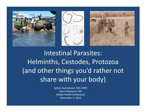
Intestinal Parasites: Helminths, Cestodes, Protozoa (and other ...
Intestinal Parasites: Helminths, Cestodes, Protozoa (and other ...
Intestinal Parasites: Helminths, Cestodes, Protozoa (and other ...

Create successful ePaper yourself
Turn your PDF publications into a flip-book with our unique Google optimized e-Paper software.
<strong>Intestinal</strong> <strong>Parasites</strong>:<br />
<strong>Helminths</strong>, <strong>Cestodes</strong>, <strong>Protozoa</strong><br />
(<strong>and</strong> <strong>other</strong> things you’d rather not<br />
share with your body)<br />
James Gaensbauer, MD, MPH<br />
Kevin Messacar, MD<br />
Global Health Conference<br />
November 5, 2013
Learning Objectives 1:<br />
Public Health Issues<br />
• Underst<strong>and</strong> the contribution of poverty,<br />
sanitation, <strong>and</strong> clean water on the worldwide<br />
prevalence of intestinal parasites<br />
• Recognize the effects of intestinal parasites on<br />
child health, nutrition, <strong>and</strong> development<br />
• Underst<strong>and</strong> the preventative public health<br />
measures recommended for intestinal parasite<br />
control, including school‐based deworming<br />
<strong>and</strong> screening
Learning Objectives 2:<br />
Presentation of Specific <strong>Parasites</strong><br />
• Underst<strong>and</strong> the basics of transmission, life<br />
cycle, clinical manifestations, diagnosis <strong>and</strong><br />
treatment of the most common intestinal<br />
parasites worldwide.<br />
– Focus on the unique aspects of each pathogen<br />
– Underst<strong>and</strong> the way in which intestinal parasites<br />
in the developing setting can mimic more<br />
common clinical diseases in the developed world<br />
• When you hear footsteps, think horses, not zebras
Know your local epidemiology!<br />
Until you are surrounded by zebras… then think zebras, not horses
Part 2: Overview<br />
• <strong>Helminths</strong><br />
– Roundworm: Ascaris lumbricoides<br />
– Whipworm: Trichuris trichuria<br />
– Hookworms: Necator americanus, Acylostoma duodenale<br />
– Strongyloides stercoralis<br />
• <strong>Cestodes</strong><br />
– Taenia solium<br />
– Echinococcus granulosus<br />
• <strong>Protozoa</strong><br />
– Entamoeba histolytica<br />
– Giardia lamblia<br />
• Trematodes<br />
– Schistosomiasis
Case 1<br />
A 4 year old girl with hx of<br />
asthma presents to your<br />
local rural hospital in<br />
Paraguay with abdominal<br />
pain <strong>and</strong> distension. She<br />
has vomited everything<br />
she has eaten <strong>and</strong> has not<br />
passed stool in 4 days.<br />
Today her belly feels firm,<br />
<strong>and</strong> she is acting ill.
Ascaris lumbricoides<br />
• Ingest eggs<br />
• Larvae invade<br />
intestines<br />
• Lung<br />
• GI tract<br />
• Excrete eggs
Ascariasis<br />
• Clinical Manifestions<br />
– Lung: Loeffler’s syndrome‐ mimics asthma<br />
– GI tract:<br />
• Malabsorption/malnutrition: Vitamin A, Fe<br />
• Obstruction<br />
– Children‐ ileal, appendiceal‐ mimics obstruction,<br />
intussusception, volvulus, appendicitis<br />
– Adults: hepatobiliary, pancreatic obstruction‐ mimics<br />
cholecysitis, pancreatitis<br />
– Worms migrate with high fever or anesthesia<br />
• Screen before elective surgery in endemic area
• Diagnosis:<br />
Ascariasis: Diagnosis<br />
<strong>and</strong> Treatment<br />
– Stool O+P<br />
– Imaging: Ultrasound, Endoscopy<br />
• Treatment:<br />
– Medical: Albendazole X 1<br />
• Mebendazole X 1 (only for Ascaris)<br />
• If obstruction: piperzine citrate relaxes worms<br />
– Surgery/ERCP: removal of obstruction
Case 1 (continued)<br />
You get an X‐ray <strong>and</strong> note<br />
dilated loops of<br />
intestine with air fluid<br />
levels. Ultrasound<br />
notes a mass in her<br />
ileum. Surgery removes<br />
complete obstruction<br />
by a bolus of ascaris<br />
worms. You tx with<br />
albendazole <strong>and</strong> she<br />
shows full recovery.
Case 2<br />
A 12 year old F presents to<br />
your rural clinic in<br />
Cambodia. She<br />
complains of diarrhea<br />
mixed with mucous <strong>and</strong><br />
blood, <strong>and</strong> straining<br />
after stooling.<br />
• On her growth chart<br />
you note she has lost<br />
3kg in the past 6<br />
months<br />
• On exam she has digital<br />
clubbing, appears pale<br />
<strong>and</strong> fatigued<br />
• Blood spot Hct 29
Whipworm: Trichuris trichuria<br />
• Ingest eggs<br />
• Stays in GI tract<br />
• Adult worms in cecum<br />
• Excrete eggs
Tricuriasis<br />
• Clinical Manifestations:<br />
– Light infection: usually asymptomatic<br />
• Malabsorption, malnutrition<br />
– Heavy InfectionTrichuris dysentery syndrome: colitis<br />
can mimic IBD with bloody, mucousy diarrhea,<br />
tenesmus, impaired growth, abdominal pain, anemia,<br />
finger clubbing<br />
• Diagnosis: stool O+P<br />
• Treatment:<br />
– Albendazole X 1<br />
– Mebendazole X 3 days
Case 2 (continued)<br />
• Knowing the local<br />
epidemiology, you do a<br />
stool O+P which<br />
demonstrates barrel shaped<br />
eggs of Trichuriasis<br />
• You treat with Mebendazole<br />
100mg twice daily for three<br />
days<br />
• You start Iron supplements<br />
<strong>and</strong> give Vitamin A<br />
supplementation<br />
• On follow‐up 2 months<br />
later, her anemia has<br />
resolved <strong>and</strong> she has<br />
regained 3kg
You conduct a school visit at an<br />
elementary school on an<br />
isl<strong>and</strong> in Lake Victoria,<br />
Kenya. You note that the<br />
children are barefoot <strong>and</strong><br />
play in the shallow water on<br />
the s<strong>and</strong>y coastline. There<br />
are no public latrines <strong>and</strong><br />
the children run to the lake<br />
to defecate. The children in<br />
the school appear pale,<br />
malnourished, some are<br />
chewing on rocks, soil.<br />
Case 3
Hookworms: Necator americanus,<br />
Ancylostoma duodenale<br />
• Penetrate skin<br />
• Lungs<br />
• GI tract<br />
– Attach<br />
• Excrete Eggs
Hookworm: Clinical Manifestations<br />
• Skin penetration: “Ground itch”<br />
• Lung: eosinophilic pneumonitis<br />
• GI tract:<br />
– <strong>Intestinal</strong> attachmentblood loss<br />
• Fe deficiency anemia, Pica<br />
• Hypoproteinemia <strong>and</strong> anasarca
Hookworm: GI blood loss<br />
• Adult worms use cutting<br />
apparatus to attach to<br />
intestinal mucosa<br />
• Contract muscular esophagi<br />
to create negative pressure<br />
<strong>and</strong> suck tissue plug<br />
• Hydrolytic enzymes,<br />
mechanical disruption of<br />
blood vessels causes bleeding<br />
Necator americanus
• Diagnosis:<br />
– Stool O+P<br />
• Treatment:<br />
Hookworm:<br />
Diagnosis <strong>and</strong> Treatment<br />
– Albendazole X 1<br />
– Mebendazole X 3 days<br />
– Fe supplementation
Case 3 (continued)<br />
• You take stool samples <strong>and</strong> discover the<br />
majority of children are carrying Ancylostoma<br />
Duodenale<br />
• You conduct school deworming with<br />
albendazole<br />
• You work with local government to construct<br />
latrines for the school <strong>and</strong> water sanitation<br />
education<br />
• Deworming program is established every 6<br />
months<br />
• You follow Hct, weight <strong>and</strong> height for students<br />
over time <strong>and</strong> note a substantial improvement<br />
over the next 2 years
Case 4<br />
• You are evaluating a 6 year old F with recent<br />
onset asthma at a referral hospital in Ghana.<br />
She began having dyspnea <strong>and</strong> wheezing 1<br />
month ago <strong>and</strong> has been treated with a<br />
prolonged prednisone course X 3 weeks for<br />
refractory symptoms. She is also complaining<br />
of abdominal pain, <strong>and</strong> is now ill appearing<br />
<strong>and</strong> complaining of headaches.
Strongyloides stercoralis<br />
• Penetrate skin<br />
• Lungs<br />
• GI tract<br />
– Adults in Duodenum<br />
– Eggs hatch in intestine<br />
– Autoinfective Cycle<br />
• Visceral Migration<br />
• Larvae (not eggs)<br />
excreted in stool
Autoinfection <strong>and</strong> Hyperinfection<br />
Autoinfection<br />
• Eggs hatch in intestine,<br />
larvae in intestine can<br />
penetrate to increase<br />
infection without<br />
reinfection from outside<br />
world<br />
• Persistence of infection<br />
for decades in<br />
untreated host<br />
Hyperinfection<br />
• Immunosuppression<br />
(steroids,<br />
chem<strong>other</strong>apy) leads to<br />
multiple rounds of<br />
autoinfection<br />
• Visceral migration <strong>and</strong><br />
dissemination to<br />
multiple organs,<br />
including brain
Strongyloides: Clinical Manifestations<br />
• Skin: pruritis at site, perianal irritation<br />
• Lung: wheezing, cough, hemoptysis<br />
• GI tract: Abdominal pain, diarrhea<br />
• Visceral migration: mortality 87%
Strongyloides:<br />
Diagnosis <strong>and</strong> Treatment<br />
• Diagnosis: low sensitivity, underestimated burden<br />
of disease<br />
– Stool O+P is difficult due to larvae in stools, not eggs<br />
– String test: examines duodenal contents, misses lower<br />
– Serum antibody testing<br />
• Treatment<br />
– Ivermectin X 2 days (80% cure rate)<br />
– Albendazole X 7 days<br />
– Treat empirically in pts who are going to receive<br />
immunosuppression in endemic areas
Case 4 (continued)<br />
Knowing the local epidemiology of her area, you<br />
suspect Strongyloidiasis now exacerbated by<br />
prolonged steroid course. You send a serum<br />
ELISA for Strongyloides which is positive. You<br />
stop her steroids <strong>and</strong> start her on Ivermectin.<br />
Despite your best efforts, she passes away<br />
after 2 days in the intensive care unit from<br />
Stongyloides hyperinfection.
Case 5<br />
An 18 year old previously healthy<br />
F presents to your local<br />
hospital in Guatemala with a<br />
1 st seizure this afternoon. She<br />
had been suffering L sided<br />
headaches for the past month.<br />
Today she began with right<br />
sided with clonic activity then<br />
generalized to tonic clonic<br />
seizure for 2 minutes. On<br />
exam you note a right‐sided<br />
hemiplegia with hyperreflexia.
Taenia solium: Taeniasis, Cysticercosis<br />
• Taeniasis:<br />
– Ingestion of infected<br />
pork<br />
– GI tract<br />
– Excrete eggs in stool<br />
• Cysticercosis:<br />
– Ingestion of humanexcreted<br />
eggs<br />
– lodge in subcu, muscles,<br />
eye, brain
Taeniasis<br />
• Life cycle: ingest cysticerci, larva<br />
hatch in intestine <strong>and</strong> forms<br />
segments, detach <strong>and</strong> excreted in<br />
stool with eggs<br />
• Clinical Manifestations:<br />
– Usually asymptomatic, do not seek<br />
care <strong>and</strong> continue to shed<br />
– GI tract: abdominal pain, distension,<br />
diarrhea, nausea
Cysticercosis<br />
• Life Cycle: eggs liberate embryo when in<br />
gastric acid bloodtissues (brain) encyst<br />
as cysticerci<br />
• Clinical manifestations<br />
– Subcutaneous: small painless nodules<br />
– Muscle: incidental finding on imaging<br />
– Eye: cysts floating in vitreous cause visual<br />
disturbance<br />
– Brain…
Neurocysticercosis<br />
• Cysticerci elicit few inflammatory changes initially<br />
• Parasite degenerates over time immunemediated<br />
inflammation<br />
– Local Inflammation: Seizures, headaches<br />
– Mass effect/CSF blockage: Hydrocephalus, increased<br />
ICP<br />
• Eventually forms calcified scars
Taenia Solium: Diagnosis <strong>and</strong><br />
Treatment<br />
Taeniasis:<br />
• stool O+P poor, stool<br />
ELISA better<br />
• Niclosamide X 1 (not<br />
absorbed, stays in GI<br />
tract), or praziquantel X 1<br />
Neurocysticercosis:<br />
• Antibody testing of CSF or<br />
serum<br />
• Imaging: CT or MRI<br />
– Cystic lesion with mural<br />
nodule (scolex)<br />
• Anti‐epileptics<br />
• Treatment of<br />
symptomatic patients<br />
with 1 or more lesions<br />
– Albendazole with steroids<br />
• Surgery
Case 5 (continued)<br />
You obtain a STAT head CT in the ED <strong>and</strong> note 5<br />
moderate sized cysts with surrounding<br />
inflammation <strong>and</strong> edema. You treat with<br />
albendazole <strong>and</strong> steroids for 8 days. She<br />
initially seizes with the start of therapy, but<br />
afterwards improves <strong>and</strong> demonstrates partial<br />
recovery.
Case 6<br />
A 58 year old man<br />
presents to your clinic<br />
in Bangalore, India<br />
with intermittent RUQ<br />
pain for 2 months,<br />
fullness of his<br />
abdomen <strong>and</strong> jaundice<br />
of his skin
Echinococcus granulosus<br />
• Ingest eggs from canine<br />
feces<br />
• GI tract portal<br />
circulation<br />
• Liver<br />
• Lung<br />
Intermediate host: sheep, <strong>other</strong>s<br />
Definitive host: Canines
Echinococcus: Clinical Manifestations<br />
• Liver:<br />
– Asymptomatic for years, grow 1cm per year<br />
– Mass effect: Biliary obstruction resembling<br />
cholecystitis<br />
– Cyst rupture: Anaphylactic reaction, cholangitis<br />
• Lung: dyspnea, coughing up grape‐skin, salty<br />
fluid
Echinococcus: Diagnosis <strong>and</strong><br />
Treatment<br />
Diagnosis<br />
• Serum antibody testing<br />
• Imaging<br />
Medical Treatment<br />
• Albendazole X 3<br />
months, can add<br />
praziquantel<br />
Surgical Treatment:<br />
• Risk of peritonitis,<br />
anaphylaxis from spill<br />
– Pre‐operative<br />
albendazole<br />
• PAIR:<br />
– Puncture under<br />
ultrasound guidance<br />
– Aspirate fluid<br />
– Inject protoscolicide<br />
– Re‐aspirate after 15‐20m
Case 6 (continued)<br />
You conduct an ultrasound of his<br />
abdomen <strong>and</strong> note a giant<br />
hepatic cyst obstructing his<br />
common bile duct. You treat him<br />
with 2 days of albendazole preoperatively<br />
<strong>and</strong> your surgeon<br />
takes him to the OR for PAIR<br />
drainage, which demonstrates<br />
protoscolices confirming his<br />
diagnosis of echinococcal hydatid<br />
cyst.
Case 7<br />
You are running a new‐immigrant clinic <strong>and</strong><br />
conducting health screenings. You note on<br />
stool O+P that many of your Ethiopian<br />
immigrant patients have amebic cysts. The<br />
children appear to be growing well <strong>and</strong> do not<br />
complain of GI symptoms.
Entamoeba Histolytica<br />
• <strong>Protozoa</strong> with cyst <strong>and</strong> trophozoite forms<br />
• Life cycle:<br />
– Transmission: Ingest cyst from fecally<br />
contaminated food or water<br />
– GI tract: Cyst releases trophozoite in intestine, can<br />
invade intestinal mucosa<br />
– Liver: Can enter portal circulation <strong>and</strong> lodge in<br />
liver<br />
– Brain, lung or <strong>other</strong> tissues<br />
– Cysts <strong>and</strong> trophozoites shed in stool
Amebiasis: Clinical Manifestations<br />
• Noninvasive infection:<br />
asymptomatic carrier<br />
• GI tract: amebic colitis<br />
– Ulcerates through mucosa,<br />
submucosa<br />
– Cramping abdominal pain,<br />
weight loss, diarrhea with<br />
mucous <strong>and</strong> blood<br />
• Liver: amebic liver abscess<br />
– Fever, hepatomegaly, dull<br />
RUQ pain, distension,<br />
tachypnea
Amebiasis:<br />
Diagnosis <strong>and</strong> Treatment<br />
• Diagnosis:<br />
– Stool O+P unable to differentiate from E. dispar<br />
(nonpathogenic)<br />
– Stool antigen detection + serum antibody testing<br />
– Imaging<br />
• Single lesion in R lobe, nonspecific<br />
• Treatment<br />
– Asymptomatic colonization: paromomycin X 7 days<br />
– Colitis: metronidazole X 7‐10 days followed by luminal<br />
agent (paromomycin)<br />
– Liver Abscess: medications as above<br />
• If >5cm, not responding to medication in 5‐7d drain
Case 7 (continued)<br />
As you recognize that stool O+P cannot often<br />
differentiate E. dispar from E. histolytica, you<br />
send stool antigen testing for E. histolytica. All<br />
of your patients are negative. You astutely<br />
decide not to treat them for the carriage of<br />
these non‐pathogenic amebas <strong>and</strong> they<br />
continue to do well.
You have returned from your 8<br />
month project in rural<br />
Kenya. Though you were<br />
careful with filtering <strong>and</strong><br />
treating your water at the<br />
start of your trip, your<br />
vigilance waned over time.<br />
At your return visit to your<br />
PCP, she asks if you have<br />
had any GI issues. You state<br />
that, besides the 7 months<br />
of diarrhea <strong>and</strong> cramping<br />
abdominal pain, <strong>and</strong> 15 lbs<br />
of weight loss, you haven’t<br />
had any <strong>other</strong> problems<br />
Case 8
Giardia Lamblia<br />
• Flagellated protozoa: cyst <strong>and</strong> trophozoite<br />
form<br />
• Transmission: ingestion of >10‐25 cysts from<br />
fecally contaminated water (human or animal)<br />
– Resistant to chlorination<br />
• GI tract: Excystation in proximal small<br />
bowelattaches to duodenum or jejunum,<br />
does not invade<br />
• Cysts excreted in stool
Giardiasis: Clinical Manifestations<br />
• Asymptomatic shedding<br />
• GI tract:<br />
– sudden onset watery diarrhea progressing to<br />
explosive, foul smelling, greasy stools, abdominal<br />
cramps, bloating, flatulence<br />
• Most clear spontaneously, some have chronic<br />
intermittent sx for months<br />
– Malabsorption <strong>and</strong> weight loss<br />
– Acquired lactose intolerance
Giardia: Diagnosis <strong>and</strong><br />
Treatment<br />
• Diagnosis:<br />
– Stool O&P looking for trophozoites (loose) or cysts<br />
(formed)<br />
– Stool antigen immunoassays (ELISA, DFA)<br />
• Treatment:<br />
– Metronidazole X 5 days<br />
– Tinidazole X 1 dose<br />
– Nitazoxanide X 3 days<br />
– Albendazole/Mebendazole
Case 8 (continued)<br />
You leave a stool sample which is sent for O+P,<br />
antigen testing. Cysts are seen under the<br />
microscope <strong>and</strong> antigen testing returns<br />
positive for Giardia Lamblia. You take 5 days<br />
of metronidazole <strong>and</strong> gain 15 lbs back on<br />
some home cooking. Your next trip you<br />
decide to filter <strong>and</strong> treat all of your drinking<br />
water….
A 10 year old female presents<br />
for checkup <strong>and</strong><br />
vaccinations in your mobile<br />
clinic on the isl<strong>and</strong>s of Lake<br />
Victoria. She is previously<br />
healthy, Tanner II, <strong>and</strong><br />
excitedly tells you that she<br />
is becoming a woman, as<br />
she recently noted some<br />
menstrual bleeding, as her<br />
urine turned red this past<br />
week. She does not attend<br />
school because she helps<br />
her m<strong>other</strong> fish‐mongering<br />
on the beach.<br />
Case 9
Schistosomiasis (Bilharzia)<br />
• Host: freshwater snails<br />
• Penetrates skin<br />
• Blood‐dwelling fluke<br />
• Matures in portal vein<br />
• Migrates to preferred<br />
body part (based on<br />
species) <strong>and</strong> releases eggs<br />
– Bladder/ GU tract<br />
– GI tract<br />
• Eggs excreted in urine or<br />
stool
Schistosomiasis: Clinical<br />
Manifestations<br />
• Skin: swimmer’s itch<br />
• Acute: Katayama Fever (systemic)<br />
hypersensitivity rxn against production of eggs<br />
4‐8 weeks after exp<br />
– Fever, headache, myalgias, bloody diarrhea,<br />
tender hepatomegaly<br />
• Chronic: eggs trapped in tissues secrete<br />
enzymes causing eosinophilic inflammation,<br />
granulomas
Schistosomiasis: Clinical<br />
Manifestations<br />
• Bladder/GU tract: S. haematobium<br />
– Hematuria of terminal urine, dysuria, proteinuria<br />
– Fibrosis, calcification‐> hydronephrosis, RF<br />
– Squamous bladder cancer<br />
• GI tract: S. mansoni, S. japonicum<br />
• Chronic colicky abdominal pain, diarrhea, bloody stools<br />
• Liver: S. mansoni, S. japonicum<br />
• Pipestem fibrosis, cirrhosis, liver failure
Schistosomiasis: Diagnosis <strong>and</strong><br />
• Diagnosis<br />
Treatment<br />
– Stool O+P<br />
– Filtered urine microscopy<br />
– Urine strips for hematuria in highly endemic area<br />
• Treatment<br />
– Praziquantel<br />
– Add steroids in Katayama fever, <strong>and</strong> repeat dose<br />
of praziquantel 4‐6 weeks afterwards
Case 9 (continued)<br />
• You use a urine dipstick<br />
to detect hematuria <strong>and</strong><br />
obtain a filtered urine<br />
for microscopy which<br />
detects Schistosoma<br />
haematobium<br />
• You treat her with<br />
praziquantel <strong>and</strong> her<br />
symptoms resolve
References<br />
• Bethony J, Brooker S, Albonico M, et al. Soil‐transmitted helminth<br />
infections: ascariasis, trichuriasis, <strong>and</strong> hookworm. The Lancet.<br />
2006;367:1521‐1532.<br />
• Hotez PJ, Brooker S, Bethony JM, Bottazzi ME, Loukas A, Xiao S.<br />
Hookworm Infection. N Engl J Med. 2004;351:799‐807.<br />
• Olsen A, van Lieshout L, Marti H, et al. Strongyloidiasis‐‐the most<br />
neglected of the neglected tropical diseases? Trans R Soc Trop Med<br />
Hyg. 2009;103:967‐972.<br />
• García HH, Gonzalez AE, Evans CAW, Gilman RH. Taenia solium<br />
cysticercosis. The Lancet. 2003;362:547‐556.<br />
• McManus DP, Zhang W, Li J, Bartley PB. Echinococcosis. Lancet.<br />
2003;362:1295‐1304.<br />
• Stanley SL. Amoebiasis. Lancet. 2003;361:1025‐1034.<br />
• Ross AG, Bartley PB, Sleigh AC, et al. Schistosomiasis. N Engl J Med.<br />
2002;346:1212‐1220.