
You also want an ePaper? Increase the reach of your titles
YUMPU automatically turns print PDFs into web optimized ePapers that Google loves.
UNIVERSITATEA DE ŞTIIN<br />
TIINŢE E AGRICOLE ŞI I MEDICINĂ VETERINARĂ<br />
„ION IONESCU <strong>de</strong> <strong>la</strong> BRAD"<br />
IAŞI<br />
FACULTATEA DE MEDICINĂ VETERINARĂ<br />
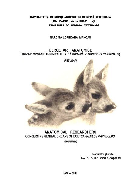
NARCISA-LOREDANA MANCAŞ<br />
CERCETĂRI ANATOMICE<br />
PRIVIND ORGANELE GENITALE LA CĂPRIOARĂ (CAPREOLUS CAPREOLUS)<br />
(REZUMAT)<br />
ANATOMICAL RESEARCHERS<br />
CONCERNING GENITAL ORGANS OF DOE (CAPREOLUS CAPREOLUS)<br />
(SUMMARY)<br />
Conducător ştiinţific,<br />
Prof. Dr. Dr. H.C. VASILE COŢOFAN<br />
IAŞI – 2006
Narcisa Loredana Mancaş<br />
Teză <strong>de</strong> doctorat<br />
Cuvinte cheie: cervi<strong>de</strong>e, organe genitale, diapauză embrionară, ovar, corpi luteali, coarne uterine, caruncul uterin,<br />
p<strong>la</strong>centom, cervix, oglinda.<br />
Teza <strong>de</strong> doctorat, ilustrată prin 46 figuri şi 2 tabele, cuprin<strong>de</strong> 157 pagini şi<br />
este structurată în două părţi: partea I-a, ce inclu<strong>de</strong> datele bibliografice referitoare<br />
<strong>la</strong> tema tezei şi partea a II-a care cuprin<strong>de</strong> rezultatele cercetărilor personale<br />
referitoare <strong>la</strong> morfologia aparatului genital <strong>la</strong> căprioară.<br />
Studiile efectuate până în prezent în domeniul reproducţiei rumegătoarelor<br />
sălbatice fac referire <strong>la</strong> durata sezonului <strong>de</strong> reproducţie, <strong>la</strong> diferiţi parametri ai<br />
ciclului sexual, <strong>la</strong> endocrinologia ciclului sexual şi a gestaţiei.<br />
Strategiile reproductive ale mamiferelor sunt puternic influenţate <strong>de</strong><br />
condiţiile <strong>de</strong> mediu (May şi Rubenstein, 1985). Sincronizarea şi durata perioa<strong>de</strong>lor<br />
<strong>de</strong> rut, rata fertilităţii, numărul <strong>de</strong> <strong>de</strong>scen<strong>de</strong>nţi şi raportul între sexe <strong>la</strong> naştere<br />
(Clutton-Brock şi Iason, 1986) sunt doar câţiva dintre parametrii reproductivi<br />
influenţati <strong>de</strong> mediul ambiental.<br />
În ultimii ani s-a încercat aplicarea <strong>la</strong> animalele sălbatice a unor tehnologii<br />
utilizate în reproducţia animalelor <strong>de</strong> fermă (vacă, oaie, capră – inducere a<br />
superovu<strong>la</strong>ţiei şi transferul <strong>de</strong> embrioni), urmărindu-se în acest fel creşterea<br />
numărului <strong>de</strong> produşi obţinuţi. Se întreve<strong>de</strong>a astfel o posibilitate <strong>de</strong> a înlătura<br />
pericolul <strong>de</strong> dispariţie a unor specii <strong>de</strong> interes cinegetic şi rezolvarea problemelor<br />
legate <strong>de</strong> repopu<strong>la</strong>re.<br />
Toate aceste încercări sunt sortite eşecului dacă nu se acordă atenţia cuvenită<br />
suportului anatomic reprezentat <strong>de</strong> organele aparatului genital.<br />
Prezenta lucrare – “Cercetări anatomice privind organele genitale <strong>la</strong><br />
căprioară ( Capreolus capreolus)” vine să completeze datele existente în literatura<br />
<strong>de</strong> specialitate cu unele caracteristici importante ale reproducţiei speciei Capreolus<br />
şi anume anatomia aparatului reproducător femel şi să pună în evi<strong>de</strong>nţă <strong>de</strong>osebirile<br />
existente între această specie şi rumegătoarele domestice.<br />
Căprioarele sunt cervi<strong>de</strong>e <strong>de</strong> talie mijlocie (22 – 30 kg), fără g<strong>la</strong>ndă<br />
periorbitală, cu coadă rudimentară (2 – 3 cm lungime) şi o durată <strong>de</strong> viaţă <strong>de</strong> până<br />
1
Narcisa Loredana Mancaş<br />
Teză <strong>de</strong> doctorat<br />
<strong>la</strong> 20 <strong>de</strong> ani. Căprioarele sunt rumegătoare monoestrice, durata ciclului estral fiind<br />
obişnuit <strong>de</strong> 36 ore; ele sunt apte <strong>de</strong> împerechere după 14 luni <strong>de</strong> viaţă. Studiile<br />
efectuate <strong>de</strong> Sempere (1992, 1993) sugerează faptul că fotoperioada are efect opus<br />
<strong>la</strong> cele două sexe ale speciei Capreolus capreolus, creşterea ei are efect inhibitor<br />
asupra activităţii sexuale <strong>la</strong> căprioară.<br />
Căprioara este singura dintre speciile <strong>de</strong> ungu<strong>la</strong>te care prezintă o perioadă <strong>de</strong><br />
<strong>la</strong>tenţă pe durata gestaţiei – diapauza embrionară şi în consecinţă are un ciclu<br />
reproductiv diferit <strong>de</strong> cel al speciilor înrudite. Diapauza embrionară constituie o<br />
strategie importantă în supravieţuirea acestei specii. Pe parcursul sezonului cald<br />
(vară-toamnă) femelele acumulează rezerve ce vor fi folosite pe parcursul iernii, iar<br />
prin întârzierea <strong>de</strong>zvoltării embrionului căprioarele s-au adaptat astfel încât<br />
naşterea iezilor are loc atunci când resursele sunt din nou disponibile.<br />
Durata gestaţiei <strong>la</strong> această specie variază fiind în medie <strong>de</strong> 300 <strong>de</strong> zile, din<br />
care gestaţia propriu-zisă durează în jur <strong>de</strong> 150 zile. După fecundaţie, zigotul şi<br />
stadiul <strong>de</strong> morulă sunt găzduite pe o durată <strong>de</strong> 4 – 5 luni în uter fără a se produce<br />
nidaţia. În acest interval activitatea miotică <strong>la</strong> nivelul embrionului este minimă,<br />
întârzierea nidaţiei nu este influenţată <strong>de</strong> durata fotoperioa<strong>de</strong>i ca <strong>la</strong> nevăstuică, ci<br />
<strong>de</strong> gradul <strong>de</strong> <strong>de</strong>zvoltare al b<strong>la</strong>stocistului care comunică organismului mamei<br />
momentul optim pentru imp<strong>la</strong>ntare prin eliberarea unei proteine prezente doar <strong>la</strong><br />
această specie (PAG – Pregnancy Associated Glycoprotein). După imp<strong>la</strong>ntare<br />
(luna ianuarie), embrionul se <strong>de</strong>zvoltă rapid, naşterea producându-se în general în<br />
lunile aprilie-mai.<br />
La fătare, iezii au 1,25 – 1,60 kg, cresc rapid şi îşi dublează greutatea după<br />
primele două săptămâni <strong>de</strong> viaţă (Danilkin, 1996).<br />
Tractusul genital <strong>la</strong> căprioară, ca <strong>de</strong> altfel al tuturor cervi<strong>de</strong>elor, respectă<br />
mo<strong>de</strong>lul general <strong>de</strong>scris <strong>la</strong> ungu<strong>la</strong>te, fiind format din două ovare cu oviductele<br />
corespon<strong>de</strong>nte, un uter <strong>de</strong> tip bicorn subseptat, separat <strong>de</strong> vagin printr-un un gât<br />
uterin lung. Vaginul, <strong>de</strong> asemenea lung, se prelungeşte cu vestibulul vaginal şi<br />
vulva.<br />
2
Narcisa Loredana Mancaş<br />
Teză <strong>de</strong> doctorat<br />
O bună parte dintre organele genitale ale căprioarei sunt adăpostite <strong>de</strong><br />
cavitatea pelvină care se prezintă ca un conduct cilindric, aproape orizontal, cu<br />
<strong>de</strong>schi<strong>de</strong>rea anterioară ova<strong>la</strong>ră şi foarte înclinată în p<strong>la</strong>n ventro-caudal. Traseul<br />
scurt, apertura cranială <strong>la</strong>rgă, absenţa componentelor osoase <strong>la</strong> nivelul peretelui<br />
dorsal al porţiunii posterioare a cavităţii pelvine şi posibilitatea basculării sacrumului<br />
în jurul unui ax ce trece prin centrul feţelor auricu<strong>la</strong>re ale primei vertebre<br />
sacrale constituie tot atâtea adaptări care facilitează pasajul fătului în momentul<br />
parturiţiei.<br />
Ovarele, p<strong>la</strong>sate în p<strong>la</strong>nul bifurcaţiei coarnelor uterine, sunt si<strong>de</strong>fii, uşor<br />
ap<strong>la</strong>tizate şi cu suprafaţa netedă <strong>la</strong> femelele impubere şi ovoi<strong>de</strong>, uşor inegale (cel<br />
drept în general mai mare) şi cu suprafaţa <strong>de</strong>nive<strong>la</strong>tă <strong>de</strong> prezenţa foliculilor<br />
ovarieni în diferite stadii <strong>de</strong> evoluţie sau datorită corpilor luteali, foarte voluminoşi<br />
<strong>la</strong> femelele adulte ale speciei Capreolus capreolus. Bursa ovariană este <strong>de</strong>osebit <strong>de</strong><br />
<strong>la</strong>rgă.<br />
Histostructura ovarului este simi<strong>la</strong>ră cu cea a rumegătoarelor domestice,<br />
predominând zona corticală <strong>la</strong> nivelul căreia se i<strong>de</strong>ntifică 1 – 2 sau chiar 3 foliculi<br />
terţiari pe fiecare ovar, concomitent. Spre <strong>de</strong>osebire <strong>de</strong> alte specii <strong>de</strong> rumegătoare,<br />
corpul luteal nu regresează după ovu<strong>la</strong>ţie, ei persistând peste 5 luni, perioadă care<br />
coinci<strong>de</strong> cu diapauza embrionară. Dacă <strong>la</strong> alte specii <strong>de</strong> cervi<strong>de</strong>e corpii luteali sunt<br />
indicatori <strong>de</strong> gestaţie, <strong>la</strong> specia Capreolus c. ei pot fi întâlniţi şi <strong>la</strong> femelele<br />
negestante până <strong>la</strong> începutul lunii ianuarie. De altfel, s-a observat că nu există o<br />
corespon<strong>de</strong>nţă între numărul corpilor luteali <strong>de</strong> <strong>la</strong> nivelul ovarelor şi numărul <strong>de</strong><br />
embrioni din uter. Sunt consi<strong>de</strong>rate gestante doar căprioarele <strong>la</strong> care corpii luteali<br />
persistă după această dată, <strong>la</strong> care se reia multiplicarea celu<strong>la</strong>ră <strong>la</strong> nivelul<br />
b<strong>la</strong>stocistului şi se produce nidaţia.<br />
Artera ovariană asigură irigarea ovarului, salpinxului şi extremităţii<br />
anterioare a coarnelor uterine şi se <strong>de</strong>sprin<strong>de</strong> din aorta abdominală, aproape <strong>de</strong><br />
terminarea ei. Din ea se <strong>de</strong>sprin<strong>de</strong> o ramură utero-tubară pentru vârful cornului<br />
uterin şi capătul uterin al salpinxului, iar <strong>la</strong> 5 – 7 cm <strong>de</strong> ovar se împarte într-o<br />
3
Narcisa Loredana Mancaş<br />
Teză <strong>de</strong> doctorat<br />
ramură sinuoasă, r. tubară anterioară, pentru pavilionul trompei uterine, o ramură<br />
tubară posterioară, care formează până <strong>la</strong> 10 – 16 flexuozităţi şi câteva (2 – 4)<br />
ramuri ovariene. Ramurile arterei ovariene nu înconjoară vena ovariană ci<br />
formează anse strânse p<strong>la</strong>sate <strong>la</strong> distanţe variabine <strong>de</strong> aceasta.<br />
Conformaţia cavităţii pelvine face ca o bună parte a segmentelor căilor<br />
genitale <strong>la</strong> căprioara adultă să fie p<strong>la</strong>sate în afara acesteia, suspendate <strong>de</strong> pereţii<br />
posteriori ai cavităţii abdominale prin plica peritoneală urogenitală.<br />
Trompele uterine <strong>la</strong> căprioară sunt lungi, fine şi flexuoase. Segmentul<br />
ovarian se prezintă sub forma unei di<strong>la</strong>taţii conice <strong>la</strong>rgi, cu marginea liberă mai<br />
franjurată <strong>de</strong>cât <strong>la</strong> rumegătoarele domestice. Una din fimbriile pavilionului –<br />
fimbria ovarică se inseră pe marginea liberă a ovarului, iar o alta, mai <strong>de</strong>zvoltată,<br />
se dirijează spre vârful cornului uterin şi <strong>de</strong>limitează împreună cu ligamentul uteroovarian<br />
şi mezosalpinx, un şanţ care adăposteşte ovarul. Această dispunere<br />
constituie o structură suplimentară <strong>de</strong> siguranţă pentru prevenirea a că<strong>de</strong>rii<br />
ovulului în cavitatea abdominală, având în ve<strong>de</strong>re modul <strong>de</strong> viaţă al animalului<br />
(obligat, în mod frecvent, să sară sau să alerge).<br />
Corpul oviductului, cu un traseu lung (9 – 10 cm) şi un diametru re<strong>la</strong>tiv<br />
constant, este foarte sinuos în apropierea infundibulului. Se continuă cu vârful<br />
cornului uterin corespunzător, <strong>la</strong> nivelul unei joncţiunii utero-tubare bine<br />
evi<strong>de</strong>nţiate <strong>la</strong> femelele adulte. La căprioară, mucoasa salpingiană formează<br />
22 – 30 <strong>de</strong> cute primare longitudinale, din care se <strong>de</strong>sprind cute mucoase secundare<br />
şi terţiare rezultând un aspect ramificat pe secţiune transversală. În apropierea<br />
joncţiunii tubo-uterine, structura cutelor mucoase <strong>de</strong>vine mai simplă, cutele<br />
primare se reduc <strong>la</strong> 4 – 6, iar cutele secundare şi terţiare lipsesc în majoritatea<br />
cazurilor.<br />
Coarnele uterine au <strong>la</strong> femelele impubere formă <strong>de</strong> con uşor răsucit ventral,<br />
cu vârful bont, iar <strong>la</strong> căprioarele adulte sunt lungi (32 – 40 cm), flexuoase şi uşor<br />
ap<strong>la</strong>tizate, asemănătoare anselor intestinale <strong>de</strong> care se <strong>de</strong>osebesc prin consistenţă şi<br />
prezenţa <strong>la</strong> nivelul suprafeţei lor a unor striaţii longitudinale datorită tunicii<br />
4
Narcisa Loredana Mancaş<br />
Teză <strong>de</strong> doctorat<br />
muscu<strong>la</strong>re <strong>de</strong>zvoltate. Extremităţile caudale ale coarnelor uterine se unesc pe linia<br />
mediană, limita separatoare fiind marcată <strong>la</strong> interior <strong>de</strong> un sept care avansează<br />
2 – 3 mm în interiorul cavităţii uterine şi <strong>la</strong> exterior, pe faţa dorsală, <strong>de</strong> un şanţ<br />
medial mai lung <strong>de</strong>cât septul intercornual. Cele două cavităţi cornuale sunt<br />
cilindrice şi au un diametru interior mediu <strong>de</strong> 0,9 – 1,8 cm.<br />
Cranial se continuă cu lumenul oviductelor, iar caudal se <strong>de</strong>schid <strong>la</strong>rg în<br />
cavitatea uterină.<br />
Mucoasa coarnelor uterine prezintă carunculi uterini reniformi şi ap<strong>la</strong>tizaţi,<br />
în număr <strong>de</strong> 2 – 5 pentru fiecare corn uterin în parte.<br />
La specia Capreolus gestaţia este geme<strong>la</strong>ră (peste 80% din cazuri), dar fără<br />
anastomoze vascu<strong>la</strong>re, iar p<strong>la</strong>centa este <strong>de</strong> tip sinepiteliocorial, oligocotiledonară.<br />
Cordonul ombilical, lung <strong>de</strong> 14 – 18 cm, conţine două artere şi două vene şi<br />
nu s-au remarcat flexuozităţi spira<strong>la</strong>te.<br />
Corpul uterin, foarte scurt (0,5 – 1 cm), apare ca un cilindru ap<strong>la</strong>tizat având<br />
raporturi <strong>de</strong> contiguitate cu rectul şi vezica urinară.<br />
Gâtul uterin are 6,2 – 7 cm lungime, pereţii <strong>de</strong>osebit <strong>de</strong> groşi (0,4 – 0,9 cm)<br />
care <strong>de</strong>limitează un canal cervical foarte îngust şi o mucoasă nepigmentată ce<br />
formează 6 – 7 cute groase, dispuse circu<strong>la</strong>r şi cu marginea liberă înclinată spre<br />
cavitatea vaginală.<br />
Prin răsfrângerea mucoasei cervicale pe peretele vaginal, în jurul <strong>de</strong>schi<strong>de</strong>rii<br />
vaginale a cavităţii uterine se formează Fornix vaginae, iar intrarea în canalul<br />
cervical este p<strong>la</strong>sată <strong>la</strong> nivelul unei „flori involte” cu structură simplă. Ventral,<br />
această cută mucoasă lipseşte, iar capetele ei şi cutele longitudinale ale mucoasei<br />
vaginale creează un canal superficial dirijat spre orificiul uterin extern, p<strong>la</strong>sat în<br />
apropierea peretelui ventral.<br />
Coarnele şi corpul uterin primesc sângele prin ramuri <strong>de</strong>rivate din a. uteroovariană<br />
(r. utero-tubară prin a. anterioară a cornului uterin) şi a. uterină medie. A.<br />
anterioară a cornului uterin trece <strong>la</strong> partea medială a venei uterine anterioare,<br />
dirijându-se spre vârful cornului uterin. A. uterină medie provine din a. iliacă<br />
5
Narcisa Loredana Mancaş<br />
Teză <strong>de</strong> doctorat<br />
internă şi după un traseu iniţial caudal, se curbează în jos şi înainte şi <strong>de</strong>scrie<br />
flexuri <strong>la</strong>rgi. Emite o ramură fină pentru vârful coarnelor uterine, 3 – 5 ramuri<br />
puternice pentru segmentul mijlociu (coarne, corp uterin şi cervix) cu precă<strong>de</strong>re<br />
spre zonele un<strong>de</strong> sunt p<strong>la</strong>saţi carunculii/p<strong>la</strong>centoamele, în pachete <strong>de</strong> 2 – 5 ramuri<br />
pentru fiecare şi o ramură retrogradă din care se <strong>de</strong>taşează terminale pentru<br />
segmentele posterioare ale aparatului genital. Limfa este colectată <strong>de</strong> limfocentrul<br />
lombar format din limfonoduri reduse (până <strong>la</strong> 5 mm diametru) dispuse în ţesutul<br />
gras dintre foiţele seroasei pelvine.<br />
Cavitatea vaginală are calibru uniform (6,5 – 8 cm circumferinţă şi o<br />
lungime <strong>de</strong> 11,5 – 18 cm) şi este complet închisă în perioada <strong>de</strong> repaus sexual<br />
datorită prezenţei mucusului.<br />
Pe faţa dorsală, seroasa vaginală continuă caudal pe cea uterină până în<br />
p<strong>la</strong>nul <strong>de</strong>schi<strong>de</strong>rii uretrei un<strong>de</strong> se răsfrânge şi se întoarce pe faţa ventrală a rectului<br />
şi pe pereţii pelvini formând fundul <strong>de</strong> sac recto-vaginal (p<strong>la</strong>sat <strong>la</strong> 4 – 5 cm anterior<br />
<strong>de</strong>schi<strong>de</strong>rii vulvare). Foiţa peritoneală <strong>de</strong> pe peretele ventral coboară pe gâtul<br />
vezicii urinare şi apoi în sens anterior pe vezica urinară şi ligamentele suspensoare<br />
ale acesteia formând fundul <strong>de</strong> sac vezico-vaginal (genito-urinar), p<strong>la</strong>sat 2 – 3 cm<br />
cranial faţă <strong>de</strong> cel prezentat anterior. Mucoasa vaginală, <strong>de</strong> culoare roz, apare<br />
cutată longitudinal şi transversal, iar arterele şi venele vaginului formează un întins<br />
plex în structurile perivaginale.<br />
Vestibulul vaginal apare ca un conduct turtit transversal, <strong>la</strong>rg şi având<br />
jumătate din lungimea canalului vaginal (6 – 8 cm). P<strong>la</strong>sat în întregime în<br />
interiorul cavităţii pelvine, între tablele ischiatice, este inclus în masa <strong>de</strong> ţesut<br />
conjunctiv ce se găseşte în loja retroperitoneală.<br />
Pe p<strong>la</strong>nşeul cavităţii vestibu<strong>la</strong>re, <strong>la</strong> ~ 5 cm cranial <strong>de</strong> <strong>de</strong>schi<strong>de</strong>rea vulvară, se<br />
găseşte orificiul uretral extern, <strong>la</strong> nivelul unei mici <strong>de</strong>presiuni triunghiu<strong>la</strong>re, iar <strong>la</strong><br />
1,5 – 2 cm caudal acestuia se <strong>de</strong>schid g<strong>la</strong>n<strong>de</strong>le vestibu<strong>la</strong>re mari.<br />
La căprioară, vulva este reprezentată doar <strong>de</strong> <strong>la</strong>biile mari, două pliuri groase,<br />
puţin proeminente faţă <strong>de</strong> p<strong>la</strong>nul regional care mărginesc un orificiu îngust p<strong>la</strong>sat<br />
6
Narcisa Loredana Mancaş<br />
Teză <strong>de</strong> doctorat<br />
oblic <strong>la</strong> 2 – 2,5 cm sub anus, în p<strong>la</strong>nul tuberozităţii ischiatice.<br />
Comisura dorsală mai puţin rotunjită comparativ cu rumegătoarele<br />
domestice, iar cea ventrală ascuţită şi îngustă, adăposteşte clitorisul şi se continuă<br />
cu un smoc <strong>de</strong> peri lungi simi<strong>la</strong>r „pensulei” <strong>de</strong> <strong>la</strong> ţap.<br />
Clitorisul are dimensiuni reduse, fiind p<strong>la</strong>sat <strong>la</strong> aproximativ 1,5 cm <strong>de</strong><br />
marginea comisurii ventrale. Segmentul vizibil al clitorisului (2 – 3 mm lungime şi<br />
1 – 1,5 mm diametru) este orientat caudo-dorsal. Pielea din zona perivulvară este<br />
acoperită cu b<strong>la</strong>nă <strong>de</strong> culoare albă (oglinda), fiecare folicul pilos având muşchi<br />
pilo-erectori puternici, cu rol important în apelul sexual. Contracţia muscu<strong>la</strong>turii<br />
pilo-erectoare modifică orientarea firelor <strong>de</strong> păr, realizând a<strong>de</strong>vărate „semnale<br />
luminoase”, vizibile <strong>de</strong> <strong>la</strong> distanţă. Local, g<strong>la</strong>n<strong>de</strong>le sebacee modificate secretă un<br />
produs cu miros caracteristic; sunt mai active în perioada <strong>de</strong> rut.<br />
G<strong>la</strong>nda mamară <strong>la</strong> căprioară este constituită din patru sferturi perfect<br />
funcţionale, p<strong>la</strong>sate în spaţiul inghinal, fuzionate pe linia mediană care formează<br />
un uger <strong>de</strong> tip „sălbatic”. Mameloanele <strong>de</strong> formă conică, în număr <strong>de</strong> 4, prezentând<br />
în zona apicală un singur orificiu papi<strong>la</strong>r, sunt p<strong>la</strong>sate simetric faţă <strong>de</strong> şanţul<br />
median, în colţurile unui pătrat cu <strong>la</strong>tura <strong>de</strong> 3,5 – 5,5 cm. Ţesutul g<strong>la</strong>ndu<strong>la</strong>r<br />
(parenchimul) are culoare roz-gălbuie şi este organizat în lobi, fiecare format din<br />
mai mulţi lobuli (~ 3 – 5 mm diametru). Alveo<strong>la</strong> mamară constituie unitatea<br />
morfo-funcţională a lobulilor g<strong>la</strong>ndu<strong>la</strong>ri, putându-se i<strong>de</strong>ntifica între 80 – 120 astfel<br />
<strong>de</strong> unităţi/lobul.<br />
Activitatea secretorie a parenchimului mamar este maximă în primele 2 – 3<br />
luni după fătare, se reduce începând cu luna august şi încetează <strong>la</strong> începutul<br />
toamnei; în cazuri rare se pot întâlni femele care au g<strong>la</strong>nda mamară activă şi în<br />
luna <strong>de</strong>cembrie.<br />
7
Narcisa Loredana Mancaş<br />
Teză <strong>de</strong> doctorat<br />
Key words: cervi<strong>de</strong>, genitalia, embryonic diapause, ovary, corpus luteus, uterine horns, uterine caruncu<strong>la</strong>e,<br />
p<strong>la</strong>centome, cervix, the miror.<br />
This thesis, illustrated by 46 figures and 2 tables, contains 157 pages and it<br />
is structured in two parts: first part, which contain bibliographical data concerning<br />
to the theme of the thesis and second part which contain the results of the personal<br />
researches concerning to the morphology of the roe doe genitalia.<br />
The effected studies up to the present in the domain of the wilds ruminants’<br />
reproductions refer to the time of the reproduction season, to the sexual cycle’s<br />
endocrinology and to the pregnancy.<br />
Mammal’s reproductive strategies are strongly influenced upon the<br />
environment’s conditions (May &Rubenstein, 1985).<br />
The synchronization and the duration of the mating period, the rate of<br />
fertility, the number of offspring and the sexration at birth (Clutton-Brock & Jason,<br />
1986) are only some of the reproductive parameters represented by the ambient<br />
environment.<br />
In the <strong>la</strong>st years they tried for the wild animals, some technologies used at<br />
the reproduction of the farm animals (cow, sheep, goat – the induction of the<br />
superovu<strong>la</strong>tion and the embryo transfer), keeping watch over in this way a number<br />
of the gotten new born increase.<br />
It’s appeared a possibility for some species of hunting concern to be out of<br />
extinction’s danger and solution for the territory repopu<strong>la</strong>tion problems.<br />
All these studies will be <strong>de</strong>stined for a failure if you don’t pay attention to<br />
the anatomic support represented by the genital organs.<br />
This study – “Anatomical researches concerning genital organs of doe<br />
(Capreolus capreolus)” comes to complete the offered informations in the<br />
specialty remarks with some important features of Capreolus species reproduction,<br />
namely the anatomy of the doe’s genital organs and to emphasize the differences<br />
between this species and domestic ruminants.<br />
The does are cervi<strong>de</strong>s of middle size (22 – 30 kg), without periorbital g<strong>la</strong>nd,<br />
8
Narcisa Loredana Mancaş<br />
Teză <strong>de</strong> doctorat<br />
with rudimentary tail (2 – 3 cm length) and they live until 20 years.<br />
They are monoestric ruminants, with a 36h usual estrus length; they are able<br />
to matte after 14 months of life. The performed studies by Sempere (1992, 1993)<br />
suggest that the photoperiod has an opposite effect at two sexes of Capreolus<br />
capreolus species; her increase has an inhibiting effect over the doe’s sexual<br />
activity.<br />
The roe <strong>de</strong>er female is only of the ungu<strong>la</strong>te species which presents a <strong>la</strong>tency<br />
period during pregnancy – the embryonic diapause (<strong>de</strong><strong>la</strong>yed imp<strong>la</strong>ntation) and,<br />
therefore it has a different reproductive cycle than any re<strong>la</strong>ted species.<br />
Embryonic diapause is a very important strategy for this species life. During<br />
the warm season (summer – autumn) the female accumu<strong>la</strong>te reserves which will be<br />
used in the winter time and with the <strong>de</strong><strong>la</strong>yed imp<strong>la</strong>ntation, the roe <strong>de</strong>er female are<br />
adapted so that the birth of kids is happening when the food is avai<strong>la</strong>ble again.<br />
The duration of pregnancy at this species is varied, being on the average by<br />
300 days, from which proper pregnancy is during about 150 days.<br />
After fecundation, the zygote and moru<strong>la</strong> stage are hosted 4 – 5 months in<br />
uterus without nidation. In this time, the mitotic activity to the embryo’s level is<br />
minimal, the <strong>de</strong><strong>la</strong>y of nidation is not influenced by the photoperiod time than to the<br />
weasel, but the progress level of b<strong>la</strong>stocyst which communicates the mother’s<br />
organism the best time for imp<strong>la</strong>ntation for a protein release, presented only at this<br />
species (PAG – Pregnancy Associated Glycoprotein). After imp<strong>la</strong>ntation (in<br />
January), the embryo grows up fast, the birth happens usually in April or May. On<br />
birth, the kids have, 1,25 – 1,60 kg, grows up quickly and their weight is doubled<br />
after first two weeks of life (Danilkin, 1996).<br />
The doe’s genital tractus, besi<strong>de</strong>s all simi<strong>la</strong>r cervi<strong>de</strong>e, follows the general<br />
pattern <strong>de</strong>scribed at the ungu<strong>la</strong>tes, being formed by two ovaries with the<br />
correspon<strong>de</strong>nt’s oviducts, a subseptat uterus bicornis, separated fro vagina by a<br />
long cervix. Also, the long vagina, keeps on vaginal vestibule and vulva.<br />
A great part of the doe’s genital organs are p<strong>la</strong>ced in the pelvic cavity, which<br />
looks like a cylindrical duct, almost horizontally, with oval opening and very<br />
9
Narcisa Loredana Mancaş<br />
Teză <strong>de</strong> doctorat<br />
downward in ventro-caudal p<strong>la</strong>ne. The brief route, the <strong>la</strong>rge cranial opening, the<br />
absence of bony constituent at the dorsal wall of the cavity’s posterior pelvic<br />
segment and the possibility of the dumping sacrum around an ax which comes<br />
through the center’s auricu<strong>la</strong>r faces of the first sacral vertebra, these constitute so<br />
many adaptations which facilitate the passage of the fetus in the parturition time.<br />
The ovaries, situated in the uteri’s horn bifurcation p<strong>la</strong>n, are pearly, easily<br />
f<strong>la</strong>tted with a smooth area to young female and ovoid, easily unequal (the right one<br />
is generally bigger), with a ruffled area because of ovarian follicles in different<br />
stages of evolution or because of Corpora lutea, very voluminously at Capreolus<br />
capreolus adult female.<br />
The ovary bursa is very <strong>la</strong>rge.<br />
Ovary histological structure is the same with the domestic ruminants’one,<br />
the cortical zone is prevalent, at this level it <strong>de</strong>tectes 1 – 2 or 3 tertiary follicles on<br />
every ovary in the same time. Comparing of another species ruminants, the Corpus<br />
luteum doesn’t let down after ovu<strong>la</strong>tion, they persistent over 5 months, this period<br />
means with the <strong>de</strong><strong>la</strong>yed imp<strong>la</strong>ntation. If the Corpora lutea are like pregnancy<br />
gui<strong>de</strong>s to the other species of cervi<strong>de</strong>e, to the Capreolus c. species they can be also<br />
meet at non-pregnant female, too, till the beginning of January. Besi<strong>de</strong>s it is<br />
observed that there isn’t a correspon<strong>de</strong>nce between the number of Corpora lutea<br />
and the number of embryo insi<strong>de</strong> the uterus. They consi<strong>de</strong>r them pregnant only the<br />
does with Corpora lutea persistent after this time, when it rep<strong>la</strong>ies the cellu<strong>la</strong>r<br />
multiplications at the b<strong>la</strong>stocyst level and the nidation is bringing.<br />
The ovarian artery assures the irrigation of the ovary, the salpinx and the<br />
anterior extremity of uterine horns and takes out from abdominal aorta, nearly his<br />
finish. From this it takes out an uterotubar branch for the apex of the uterine horn<br />
and the uterine segment of the salpinx and at 5 – 7 cm by ovary it divi<strong>de</strong>s in a<br />
sinuous branch, the tubar anterior branch, for the Infundibulum, a posterior tubar<br />
branch and some ovarian branches (2 – 4) which form until 10 – 16 loops. The<br />
branches of the ovarian artery don’t around the ovarian vein but form tight loops<br />
situated at variable distance of this.<br />
10
Narcisa Loredana Mancaş<br />
Teză <strong>de</strong> doctorat<br />
The conformation of the pelvic cavity <strong>de</strong>termines as a great part of genital<br />
segments to be situated outsi<strong>de</strong> of this, suspen<strong>de</strong>d by posterior walls of abdominal<br />
cavity by urogenital peritoneal fold.<br />
The roe doe has a long, thin and flexuous uterine tubes. The ovarian segment<br />
looks like a <strong>la</strong>rge and conical di<strong>la</strong>tion, with a free edge more fringed than at the<br />
domestic ruminants. One of pavilion’s fringes – Fimbria ovarica - inserted on the<br />
free bor<strong>de</strong>r of ovary and the other, more <strong>de</strong>veloped, is conduced to the extremity of<br />
the uterine horn and <strong>de</strong>limits with the utero-ovarian ligament and mesosalpinx, a<br />
groove which lodges the ovary.<br />
This disposition constitutes a safe supplementary structure for the prevention<br />
of the ovule col<strong>la</strong>pse in the abdominal cavity knowing the doe’s way of life<br />
(frequently forced to run and jump).<br />
The salpinx body, with a long tract (9 – 10 cm) and a re<strong>la</strong>tive constant<br />
diameter is very sinuous nearly to the Infundibulum. He is continued with the top<br />
of uterine horn, at the level of a junction utero-tubar, evi<strong>de</strong>ntly at adult female.<br />
At the doe, the mucous membrane of the salpinx forms 22 – 30 longitudinal<br />
primary folds from which the secondary and tertiary mucous fold is <strong>de</strong>tached and<br />
is appeared a branched aspect by cross section.<br />
In proximity of the tubo-uterine junction, the structure of the mucous folds<br />
becomes simpler, the number of primary folds is reduced at 4 – 6 and the<br />
secondary and tertiary folds are missing in most of the cases.<br />
The uterine horns, at the young female, have a shape cone easily twisted<br />
ventrally with the blunt top, bat at the adult roe does, the uterine horns are flexuous<br />
and easily f<strong>la</strong>ttened, simi<strong>la</strong>r to the intestinal loops of which is distincted by<br />
consistence and by presence on their surface of some longitudinal furrows because<br />
of <strong>de</strong>velopment of muscu<strong>la</strong>r <strong>la</strong>yer.<br />
The caudal extremity of the uterine horns are united on the median line, the<br />
separation bound being marked insi<strong>de</strong> of one septum which advances 2 – 3 mm in<br />
to uterine cavity and in exterior, on the dorsal si<strong>de</strong>, is marked by a medial groove<br />
more longer than intercornual septum. Both uterine horns are cylindrically and<br />
11
Narcisa Loredana Mancaş<br />
Teză <strong>de</strong> doctorat<br />
have a medium interior diameter by 0.9 – 1.8 cm. Cranially it is continued with the<br />
lumen of the oviducts and caudally they are <strong>la</strong>rge opened in the uterine cavity.<br />
The mucous membrane of the uterine horns presents the oval and f<strong>la</strong>ttened<br />
uterine caruncles, 2 – 5 for each uterine horn. At the Capreolus species the<br />
pregnancy is double (above 80%), but without vascu<strong>la</strong>r anastomosys and the<br />
p<strong>la</strong>centa is synepiteliocorial and olygocotiledonal type.<br />
The umbilical cordon, with 14 – 18 cm length, contains two arteries and two<br />
veins, without coiled flexuous.<br />
The uterine body, very short (0.5 – 1 cm), looks like a f<strong>la</strong>ttened cylin<strong>de</strong>r,<br />
having a contiguity touching with the rectum and the urinary b<strong>la</strong>d<strong>de</strong>r.<br />
The cervix has 6.2 – 7 cm length, with very thick walls (0.4 – 0.9 cm), which<br />
makes a very narrow duct. The unpigmented cervical mucous membrane forms<br />
6 – 7 thick folds with a circu<strong>la</strong>r disposition and with the free edge inclined to the<br />
pelvic cavity.<br />
To the cervical mucous membrane turned down on vaginal wall, around to<br />
the vaginal opening of the uterine cavity is formed Fornix vaginae and the entry in<br />
the cervical canal is located at the “involt flower” level, which has a simple<br />
structure.<br />
Ventrally, this mucous fold is missing and her extremities together with the<br />
longitudinal folds of vaginal mucous creates a superficial groove fallowed to the<br />
external uterine orifice, p<strong>la</strong>ced nearly in the ventral wall.<br />
The horns and uterine body receive the blood through the branches <strong>de</strong>tached<br />
from the utero-ovarian artery (a strong utero-tubar branch) and the medium uterine<br />
artery.<br />
The anterior artery of the uterine horn passes to the medial si<strong>de</strong> of anterior<br />
uterine vein, conducting specially to the top of uterine horn.<br />
The medium uterine artery comes from the internal iliac artery and after an<br />
initial caudal route is curving forward and down and <strong>de</strong>scribes 4 – 5 <strong>la</strong>rge loops. It<br />
gives out a thin branch for the top of uterine horns, 3 – 5 strong branches for the<br />
middle segment (uterine horns, uterine body and cervix, especially to the areas<br />
12
Narcisa Loredana Mancaş<br />
Teză <strong>de</strong> doctorat<br />
where are p<strong>la</strong>ced the uterine caruncles/p<strong>la</strong>centomes, in bunches of 2 – 5 branches<br />
for each) and a caudal branch from which are <strong>de</strong>tached the terminals for posterior<br />
segment of the genital tract..<br />
The lymph is collected by the lumbar lymphocenter represented by small<br />
lymph no<strong>de</strong>s (until 5 mm), disposed in the fat tissue between the folds of pelvic<br />
serous membrane.<br />
Vaginal cavity has a uniform caliber (6.5 – 8 cm perimeter and 11.5 – 18 cm<br />
length) and is completely closed in the sexual repose time because of the mucus<br />
present.<br />
On the dorsal si<strong>de</strong>, the serous vaginal membrane continues caudally the<br />
uterine one until the p<strong>la</strong>ne of the urethral opening where is turned down and is<br />
returned on the ventral si<strong>de</strong> of the rectum and on the pelvic walls forming the<br />
recto-vaginal sack bottom (located at 4 – 5 cm before vulva’s opening).<br />
The peritoneal serous membrane from the ventral wall <strong>de</strong>scends to the neck<br />
of the urinary b<strong>la</strong>d<strong>de</strong>r and then <strong>de</strong>scends in the cranial way on the urinary b<strong>la</strong>d<strong>de</strong>r<br />
and on the suspensor ligaments of this, forming the genitor-urinary sack bottom,<br />
located at 2 – 3 cm in front of the previous sack.<br />
The vaginal mucosa, with a pink color, appears fol<strong>de</strong>d longitudinally and<br />
transversally and the arteries and veins of vagina form a <strong>la</strong>rge plexus in the<br />
vagina’s neighborhood <strong>la</strong>yers.<br />
The vaginal vestibule looks like a cross f<strong>la</strong>ttened duct, broad and having a<br />
half of vaginal length (6 – 8 cm). It is located completely insi<strong>de</strong> of pelvic cavity,<br />
between ischiatic bones boards, is inclu<strong>de</strong>d in the conjunctive tissue from the<br />
retroperitoneal lodge. On the vestibu<strong>la</strong>r cavity’s floor, at approximate 5 cm cranial<br />
by the vulva’s opening, there is the external urethral aperture, at the level of a<br />
small, triangu<strong>la</strong>r flute, and 1.5 – 2 cm caudally of this is opened the major<br />
vestibu<strong>la</strong>r g<strong>la</strong>nds.<br />
At roe doe, the vulva is represented only by major lips, two thick folds, little<br />
prominent by the regional p<strong>la</strong>ne which are bor<strong>de</strong>ring a narrow hole p<strong>la</strong>ced oblique<br />
at 2 – 2.5 cm un<strong>de</strong>r the anus, in the ischiatic tuber p<strong>la</strong>ne. The dorsal <strong>la</strong>bial<br />
13
Narcisa Loredana Mancaş<br />
Teză <strong>de</strong> doctorat<br />
commissure is less roun<strong>de</strong>d.comparativelly with the domestic ruminants, and the<br />
ventral one is narrow and sharp, it lodges the clitoris and is continuing with a long<br />
hairs tuft which is simi<strong>la</strong>rly to the roe buck’s “brush”.<br />
The clitoris is small, p<strong>la</strong>ced about 1.5 cm by the ventral commissure’s edge.<br />
The visible part of the clitoris (2 – 3 mm length and 1 – 1.5 mm diameter) is<br />
orientated caudo-dorsally.<br />
The skin from the environs area is covered by white fur (the mirror), each of<br />
the hair follicle has strong erector muscles, with a very important role in the sexappeal<br />
in the matting time. The contraction of the erector muscles modifies the<br />
hair’s orientation and it realizes a real “bright signals”, visible from the distances.<br />
In this area, the changed sebaceous g<strong>la</strong>nds excrete a substance with a<br />
specific odour; they are more active in the mating time.<br />
Located in the inguinal region, the mammary g<strong>la</strong>nd of roe doe is constituted<br />
by four perfect functionally quarters, joined on the median line which form an<br />
ud<strong>de</strong>r of “wild type”.<br />
The conical teats, four in number, presenting in the apical extremity a single<br />
papil<strong>la</strong>r orifice, are p<strong>la</strong>ced symmetrically to the median p<strong>la</strong>ne, in the corners of a<br />
square with a si<strong>de</strong> of 3.5 – 5.5 cm. has one teat and each teat has one opening. The<br />
g<strong>la</strong>ndu<strong>la</strong>r tissue (Parenchyma) has an yellow-pink color and is organized into<br />
lobes, each one is ma<strong>de</strong> up of many lobules (3 – 5 mm in diameter). The mammary<br />
alveoli are the ud<strong>de</strong>r’s morphofunctionaly unit (milk producing unit), existing<br />
80 – 120 alveoli/lobule.<br />
The excretory activity of mammary g<strong>la</strong>nd’s tissue is maximal in the first<br />
2 – 3 months of <strong>la</strong>ctation, he is reduced in August and is stopped at the beginning<br />
of the autumn; rarely can be meet female which have an active mammary g<strong>la</strong>nds in<br />
December, too.<br />
14

![rezumat teză [RO]](https://img.yumpu.com/19764796/1/190x245/rezumat-teza-ro.jpg?quality=85)

![rezumat teză [RO] - Ion Ionescu de la Brad](https://img.yumpu.com/14613555/1/184x260/rezumat-teza-ro-ion-ionescu-de-la-brad.jpg?quality=85)