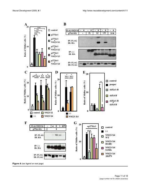
<strong>Neural</strong> <strong>Development</strong> 2009, 4:1 http://www.neuraldevelopment.com/content/4/1/1 Figure 6 (see legend on next page) Page 11 of 18 (page number not for citation purposes)
<strong>Neural</strong> <strong>Development</strong> 2009, 4:1 http://www.neuraldevelopment.com/content/4/1/1 NM23 inhibits p27Xic1-mediated gliogenesis through their interaction Figure 6 (see previous page) NM23 inhibits p27Xic1-mediated gliogenesis through their interaction. (A) Co-expression of a NM23 member with p27Xic1 inhibits p27Xic1-mediated gliogenesis. (B) Interaction of NM23-X4 with deleted versions of p27Xic1. Full length, amino-terminal NT(1–96), and 1–91 portions of p27Xic1 interact with NM23-X4, but the 31–96 portion does not. (C) The interaction between p27Xic1 and NM23 is responsible for the inhibitory function of NM23 on Müller glial cell phenotype. NM23-X4 blocked glial induction by interacting with the amino-terminal and 1–91 portions of p27Xic1 but not with the 31–96 portion. (D) NM23-X4 cannot inhibit gliogenesis mediated by p16Xic2. Müller glial cell percentage in the retina after co-introduction of NM23-X4 and Xic1 or Xic2. (E) Effect of co-introduction of shX4-B and -B constructs in the retina. Activation of gliogenesis by shX4-B requires p27Xic1. (F) Interaction of p27Xic1 with mutants of NM23-X4. Wild type (wt), H148C (H), S150G (S) and KPN were tested for their interaction with p27Xic1. (G) Wild type and the KPN blocked Müller cell induction by p27Xic1, but H148C and S150G did not. Double and triple asterisks correspond to P 0.01, and 0.001, respectively; error bars indicate standard error of the mean. rogenesis versus gliogenesis depending on the presence of neurogenic stimuli in the progenitors [17]. In the Xenopus retina, glial cells are formed after neurogenesis has taken place. The expression of p27Xic1 in the CMZ as retinogenesis progresses is consistent with its role in cell fate determination [13]. As shown in Figure 4D–F, at stage 39, NM23-X4 is expressed at the peripheral side of the CMZ. The expression domain of NM23-X4 overlaps with that of p27Xic1 in the central region of the CMZ (Figure 4F,G). From the expression patterns of NM23-X4 and p27Xic1, their interaction and functions, we propose a model, schematically represented in Figure 8D. According to this, p27Xic1 acts at the central part of the CMZ to induce Müller glial cells. NM23-X4 is responsive to suppress this gliogenic activity of p27Xic1 at the peripheral part of the CMZ, where their two expression domains coincide. This suppression results in inhibition of Müller glial cell production and maintenance of the neurogenic potential of the early progenitors in the retinal cell lineage. Although it was previously reported that NM23-H4 is largely located in mitochondria [39], our analysis using a deletion construct of NM23-X4 lacking its mitochondriasorting signal showed that mitochondrial localization is not required for its gliogenic activity (data not shown). This is supported by the observation that all other NM23 members tested showed similar gliogenic activities (Figure 7Q) and p27Xic1 binds to both wild-type NM23-X4 and its amino-terminal processed form (data not shown). It is more likely that the NM23 family regulates the activity of CDKIs in the cytosol because Cip/Kip CDKIs localize in the cytosol and shuttle to the nucleus depending on the cellular context. This notion is also supported by the cytosolic localization observed when we are staining against tagged forms of exogenous NM23-X4 in embryos in addition to the mitochondrial staining (data not shown). How does NM23-X4 inhibit the gliogenic activity of CDKIs? We showed that direct interaction with the amino-terminal half of p27Xic1 and a specific NM23-X4 activity, probably as a NDPK or protein kinase, are required for the inhibition of p27Xic1. Although several residues of CDKIs are phosphorylated, a majority of the phosphorylation sites are located at the carboxy-terminal half. In the amino-terminal half, only threonine-57 of p21Cip1, serine-10 and tyrosine-88 of p27Kip1 have been reported as phosphorylation sites [40-42]. However, the threonine-57 and serine-10 sites are not conserved in p27Xic1 and, moreover, NM23 family members are not known to possess tyrosine kinase activity, arguing against these sites on p27Xic1 being the ones phosphorylated by NM23-X4. Previously, NM23-H1 was reported to phosphorylate the kinase suppressor of Ras in a histidine dependent manner [29]. Also, the aspartic acid at 319 of aldolase C is phosphorylated by NM23-H1 [43]. In bacteria and plants, histidine kinase activity has very important roles through consequent phosphorylation of aspartic acid and glutamic acid [44,45]. Interestingly, vertebrate CDKIs have three conserved aspartic acids and glutamic acid in the CDK/cyclin binding domain at the amino termini. Our preliminary work has shown that mutation of these residues abrogates the NM23-X4 effect (data not shown). Ongoing work will verify if CDKIs are direct targets of NM23-X4 action. In addition to the inhibitory role of NM23-X4 on gliogenesis, we have shown that a large gain of NM23-X4 activates gliogenesis. We have provided evidence that, at the endogenous level, NM23-X4 works as a negative regulator of p27Xic1-mediated gliogenesis through direct protein interaction as shown by the knock-down analysis. Data from overexpression assays suggest that NM23-X4 works as an activator of gliogenesis in a mechanism largely independent of p27Xic1. Further work will be required to determine how NM23-X4 may act as an activator of gliogenesis. It is evident that it affects cell fate determination. Recent work has shown that purine-mediated signaling has a major role in eye development [46]. It is of great interest that molecules involved in ATP/ADP enzymatic steps are also part of a regulatory network, along with transcription factors and other partners, to affect eye Page 12 of 18 (page number not for citation purposes)