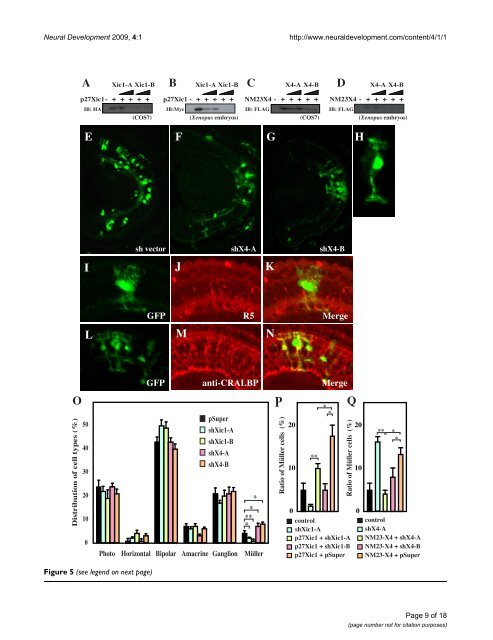
<strong>Neural</strong> <strong>Development</strong> 2009, 4:1 http://www.neuraldevelopment.com/content/4/1/1 Figure 5 (see legend on next page) Page 9 of 18 (page number not for citation purposes)
<strong>Neural</strong> <strong>Development</strong> 2009, 4:1 http://www.neuraldevelopment.com/content/4/1/1 Reduction of NM23-X4 increases the Müller glial cell population Figure 5 (see previous page) Reduction of NM23-X4 increases the Müller glial cell population. (A-D) shRNAs against p27Xic1 and NM23-X4 are efficient in knocking down their respective protein expression in cell culture and Xenopus embryos. The indicated short hairpin RNA (shRNA) constructs and the corresponding tagged expression constructs were co-transfected in COS7 cells and coinjected in two-cell stage Xenopus embryos. The effect was analyzed by immunoprecipitation of total lysate of cells or embryos (see Materials and methods). (E-G) Stage 41 retinal section after transfecting with pSuper vector and GFP (E), with shX4-A and GFP (F), or with shX4-B and GFP (G). (H) Enlarged view of a green fluorescent protein (GFP)-positive Müller glial cell transfected with shX4-B. (I-K) Staining against the Müller glial marker R5 at stage 41 retina transfected with shX4-B: (I) GFP; (J) R5 staining; (K) merged view. (L-N) Anti-CRALBP staining of Müller glial cells at stage 41 retina transfected with shX4-B: (L) GFP, (M) anti-CRALBP, (N) merged view. (O) Cell type distribution in the stage 41 retina transfected with the indicated construct plus GFP expressed in percentages. (P-Q) Rescue of the knock-down effect in the cell type distribution in the retina by cointroduction of shRNA construct with p27Xic1 (P) and NM23X4 (Q) expression constructs. The Müller glial cell percentages for each condition are shown. Single and double asterisks correspond to P 0.05 and 0.01, respectively; error bars indicate standard error of the mean. genic phenotype (Figure 7Q and data not shown). Since loss of function causes an increase in the Müller cell population, one would expect that overexpression of NM23 would have the opposite effect. Possibly, these observations show that NM23s function in a context-dependent manner to regulate neurogenesis versus gliogenesis. Furthermore, it has been observed that the glial-promoting activity of NM23-X4 is not affected by loss of function of p27Xic1 (data not shown), suggesting that the activation of gliogenesis by NM23 does not require p27Xic1. This implies that NM23-X4 might have a second mechanism of action, independent of the suppression of p27Xic1-mediated gliogenesis. Ongoing experiments will further elucidate this. Effect of NM23-X4 on cell cycle regulation It has been evident that p27Xic1 regulates the cell cycle acting as a cell cycle inhibitor. We therefore examined if the NM23-X4 inhibition of p27Xic1-mediated gliogenesis has any effect on the cell cycle. To address this, we performed a bromodeoxyuridine (BrdU) incorporation assay in the retina and a proliferation assay using early Xenopus embryos [13]. Consistent with previous reports [13], p27Xic1 reduces the proliferative potential of cells in both neural retina and early embryos as demonstrated by the decrease of BrdU positive cells (Figure 8A) and the enlargement of blastomeres (Figure 8B,C), respectively. Interestingly, NM23-X4 co-overexpression with p27Xic1 inhibited the p27Xic1-mediated cell cycle inhibition (Figure 8A–C). Furthermore, loss of NM23-X4 function, using shX4-A and -B constructs, inhibited proliferation in the retina as assayed in cells of the inner nuclear and ganglion cell layer (Figure 8A). These observations suggest that endogenous NM23-X4 functions as an inhibitor of the p27Xic1 cell cycle regulatory action. Overexpression of NM23-X4 or NM23-X1 by itself did not alter proliferation in either retinogenesis or early embryogenesis. This observation may suggest that endogenous NM23-X4 levels are already sufficient to suppress the p27Xic1 activity and overexpression above those levels does not affect the outcome. Further, when we analyzed clone sizes of lipofected cells in Xenopus retina, we did not observe any significant changes in the clone size after modulation of NM23-X4 activity (data not shown). This enforces the idea that NM23-X4 primarily acts at the last step of the sequential determination of retinal cell lineage, that is, the production of Müller glia cells without affecting the overall clone proliferation (Figures 5O and 7Q). Last, there was no effect of NM23-X4 on apoptosis in the retina (data not shown). Using a TUNEL assay, we did not observe any change in the ratio of apoptotic cells compared to the control (approximately 3.5% apoptotic cells at stage 33/34). Discussion <strong>Development</strong>al roles of the NM23 family members in retinogenesis NM23 family members possess NDPK activity, which is required for production of ATP or GTP. In addition, several unique activities of NM23 family members have been reported, such as protein kinase, exonuclease and DNA repair activities [24,25]. Although NM23 family members are expressed in the central nervous system, very little was known about their roles in neural development. We report that all Xenopus homologs are expressed in the retina, that NM23-X3 and -X4 are specifically expressed in retinal precursor cells in the CMZ and that NM23-X4 has an essential role in the distribution of retinal cell types through interaction with p27Xic1. The observation that inhibition of NM23-X4 function by shRNA constructs increased the proportion of Müller cells (Figure 5) indicates that endogenous NM23-X4 functions as a regulator of gliogenesis in the retina. This function is associated with its ability to inhibit p27Xic1-mediated gliogenesis (Figure 6A). Our previous work showed that p27Xic1 plays an important role in both neurogenesis and gliogenesis in a context-dependent manner [8,13,14]. This means that CDKIs regulate the decision between neu- Page 10 of 18 (page number not for citation purposes)