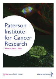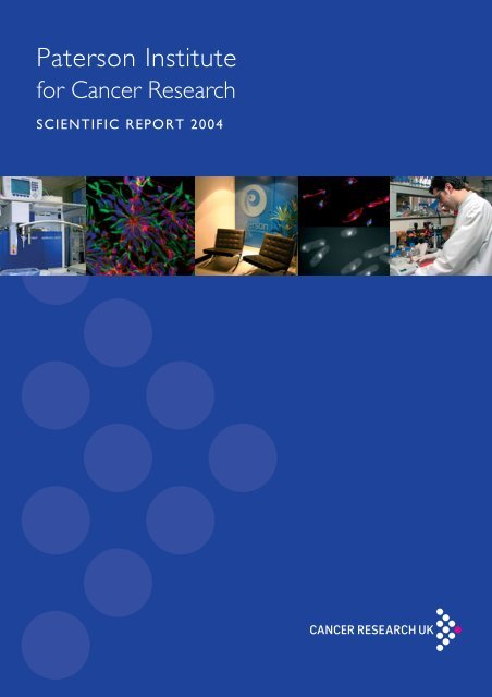
Paterson Institute for Cancer Research SCIENTIFIC REPORT 2004
Paterson Institute for Cancer Research SCIENTIFIC REPORT 2004
Paterson Institute for Cancer Research SCIENTIFIC REPORT 2004

You also want an ePaper? Increase the reach of your titles
YUMPU automatically turns print PDFs into web optimized ePapers that Google loves.
<strong>Paterson</strong> <strong>Institute</strong><br />
<strong>for</strong> <strong>Cancer</strong> <strong>Research</strong><br />
<strong>SCIENTIFIC</strong> <strong>REPORT</strong> <strong>2004</strong>
Immunocytochemistry <strong>for</strong> neural cell markers in E14 ES cells at day<br />
7 of neural differentiation in N2B27. Glial marker GFAP (red), neural<br />
marker nestin (green) and nuclei are marked using DAPI (blue).<br />
Image supplied by Rebecca Baldwin, Medical Oncology<br />
Illustration Credits<br />
Many illustrations in this report were taken by Anne Murtagh of the Christie Hospital<br />
Medical Illustrations Department and by Jenny Varley.
<strong>Cancer</strong> <strong>Research</strong> UK<br />
<strong>Paterson</strong> <strong>Institute</strong><br />
<strong>for</strong> <strong>Cancer</strong> <strong>Research</strong><br />
<strong>SCIENTIFIC</strong> <strong>REPORT</strong> <strong>2004</strong>
<strong>Cancer</strong> <strong>Research</strong> UK <strong>Paterson</strong> <strong>Institute</strong> <strong>for</strong> <strong>Cancer</strong> <strong>Research</strong> Scientific Report <strong>2004</strong><br />
Edited by Professor Jenny Varley, Assistant Director (<strong>Research</strong>)<br />
<strong>Paterson</strong> <strong>Institute</strong> <strong>for</strong> <strong>Cancer</strong> <strong>Research</strong><br />
Wilmslow Road, Manchester M20 4BX<br />
http://www.paterson.man.ac.uk<br />
ISSN 1740-4525<br />
Copyright © 2005 <strong>Cancer</strong> <strong>Research</strong> UK<br />
Printed by Cambrian Printers<br />
<strong>Cancer</strong> <strong>Research</strong> UK<br />
Registered Charity No. 1089464<br />
Registered as a company limited by guarantee in England and Wales No. 4325234<br />
Registered address 61 Lincoln’s Inn Fields, London WC2A 3PX<br />
Tel +44(0) 20 7242 0200<br />
http://www.cancerresearchuk.org
Contents<br />
Director’s Introduction . . . . . . . . . . . . . . . . . . . . . . . . . . . . . . . . . . . . . . . . . . . . . . 2<br />
<strong>Research</strong> Laboratories:<br />
Bioin<strong>for</strong>matics Group . . . . . . . . . . . . . . . . . . . . . . . . . . . Crispin Miller . . . . . . . . . . . . . . . . . . . . 4<br />
Carcinogenesis Group . . . . . . . . . . . . . . . . . . . . . . . . . . Geoff Margison . . . . . . . . . . . . . . . . . . 6<br />
Cell Division Group . . . . . . . . . . . . . . . . . . . . . . . . . . . . Iain Hagan . . . . . . . . . . . . . . . . . . . . . . 8<br />
Cell Regulation Group . . . . . . . . . . . . . . . . . . . . . . . . . . Nic Jones . . . . . . . . . . . . . . . . . . . . . . 10<br />
Cell Signalling Group . . . . . . . . . . . . . . . . . . . . . . . . . . . . Angeliki Malliri . . . . . . . . . . . . . . . . . . 12<br />
Cellular and Molecular Pharmacology Group . . . . . . . Caroline Dive . . . . . . . . . . . . . . . . . . 14<br />
Clinical and Experimental Pharmacology Group . . . . . Caroline Dive and Malcolm Ranson 16<br />
Functional Genomics and Cell Cycle Regulation Group Karim Labib . . . . . . . . . . . . . . . . . . . . 18<br />
Gene Therapy Group . . . . . . . . . . . . . . . . . . . . . . . . . . . Lez Fairbairn . . . . . . . . . . . . . . . . . . . 20<br />
Immunology Group . . . . . . . . . . . . . . . . . . . . . . . . . . . . Peter L Stern . . . . . . . . . . . . . . . . . . 22<br />
Mitotic Spindle Function and Cell Cycle Control Group Elmar Schiebel . . . . . . . . . . . . . . . . . . 24<br />
Radiochemical Targeting and Imaging Group . . . . . . . Jamal Zweit . . . . . . . . . . . . . . . . . . . . 26<br />
Stem Cell Biology Group . . . . . . . . . . . . . . . . . . . . . . . . Georges Lacaud . . . . . . . . . . . . . . . . 28<br />
Stem Cell and Haematopoiesis Group . . . . . . . . . . . . . Valerie Kouskoff . . . . . . . . . . . . . . . . 30<br />
Structural Cell Biology Group . . . . . . . . . . . . . . . . . . . . Terence D Allen . . . . . . . . . . . . . . . . 32<br />
Medical Oncology: Gene-Immunotherapy Group . . . Robert Hawkins . . . . . . . . . . . . . . . . 34<br />
Medical Oncology: Glycoangiogenesis Group . . . . . . . Gordon Jayson . . . . . . . . . . . . . . . . . 36<br />
Medical Oncology: Proteoglycan Group . . . . . . . . . . . John T Gallagher . . . . . . . . . . . . . . . . 38<br />
<strong>Research</strong> Services . . . . . . . . . . . . . . . . . . . . . . . . . . . . . . . . . . . . . . . . . . . . . . . . . . 40<br />
Publications . . . . . . . . . . . . . . . . . . . . . . . . . . . . . . . . . . . . . . . . . . . . . . . . . . . . . . . 46<br />
Seminar Speakers <strong>2004</strong> . . . . . . . . . . . . . . . . . . . . . . . . . . . . . . . . . . . . . . . . . . . . . . 54<br />
Postgraduate Education . . . . . . . . . . . . . . . . . . . . . . . . . . . . . . . . . . . . . . . . . . . . . 56<br />
Administrative Services . . . . . . . . . . . . . . . . . . . . . . . . . . . . . . . . . . . . . . . . . . . . . 58<br />
Acknowledgement <strong>for</strong> Funding of the <strong>Paterson</strong> <strong>Institute</strong> . . . . . . . . . . . . . . . . . . . 60<br />
Career Opportunities . . . . . . . . . . . . . . . . . . . . . . . . . . . . . . . . . . . . . . . . . . . . . . . 62<br />
How to Find Us. . . . . . . . . . . . . . . . . . . . . . . . . . . . . . . . . . . . . . . . . inside back cover<br />
P A T E R S O N I N S T I T U T E S C I E N T I F I C R E P O R T 2 0 0 4<br />
1
Director’s Introduction<br />
The big event of <strong>2004</strong> was the <strong>Paterson</strong> Review in<br />
July. The investment that <strong>Cancer</strong> <strong>Research</strong> UK<br />
makes to the <strong>Institute</strong> each year is very significant<br />
and clearly it is essential that mechanisms are in place<br />
to ensure that the research supported is of the<br />
highest quality. The main vehicle to ensure such<br />
quality is through the quinquennial review system<br />
where the research programme of individual groups<br />
is assessed by experts in the particular research field.<br />
The result of such assessment provides essential<br />
advice to the Director and to <strong>Cancer</strong> <strong>Research</strong><br />
UK and influences the level of continuing<br />
support to the group. In addition to supporting<br />
world-class research groups, core-funded <strong>Institute</strong>s<br />
are also expected to provide added value<br />
in their research ef<strong>for</strong>ts through the development<br />
of a research strategy that maximises<br />
the stability and flexibility of core-research<br />
support. This added value could come from the<br />
development of focused research themes that<br />
promote interaction and synergy between<br />
different research programmes, from support<br />
to long-term and high-risk research as well as<br />
shorter to medium-term projects, from the provision of high quality research support that can<br />
benefit the research programmes in the <strong>Institute</strong>, but also in the wider research community, and<br />
from the provision of an optimal environment <strong>for</strong> the career development of young scientists. In<br />
addition, of particular relevance to the <strong>Paterson</strong> <strong>Institute</strong>, is the potential <strong>for</strong> fostering synergy and<br />
interactions between areas of basic and clinical research and developing the infrastructure and<br />
environment that promotes translational research. In order to assess the overall quality of the<br />
<strong>Institute</strong>, its research strategy, its future plans and the degree of added value that it provides,<br />
<strong>Cancer</strong> <strong>Research</strong> UK carries out an <strong>Institute</strong> review every five years through an international panel<br />
of scientific leaders who have experience in directing significant research centres. It is just such a<br />
review that took place in July.<br />
The results of the review were very satisfying. The review party concluded that the <strong>Paterson</strong> was<br />
an internationally competitive <strong>Institute</strong> and fully endorsed the research strategy that had underpinned<br />
developments over the last five years and the strategic proposals <strong>for</strong> continuing<br />
development over the next five years. Such a resounding endorsement was crucially<br />
important to us since over the last few years we had progressed through a period of very<br />
significant change which was, at times, quite painful. However, we are now in a very<br />
strong position and will, over the coming years, continue to establish our strengths in<br />
key research areas and further develop meaningful and high quality translational research<br />
programmes. To help us reach our goals the review party recommended, and <strong>Cancer</strong><br />
<strong>Research</strong> UK agreed, to increase our core support so that we can establish additional<br />
research groups and significantly increase the number of non-clinical and clinical<br />
research trainees.<br />
Nic Jones, Director of<br />
the <strong>Paterson</strong> <strong>Institute</strong><br />
The review party also highlighted the enormous potential <strong>for</strong> integrated cancer research<br />
programmes in Manchester, particularly now that the new University of Manchester has been<br />
<strong>for</strong>med through the merger of the previous Victoria University of Manchester and UMIST. Closer<br />
interaction between the <strong>Paterson</strong>, the Christie Hospital and the University could lead to the development<br />
of common research goals and strategies. This would be of considerable benefit to further<br />
development of cancer research in Manchester and we look <strong>for</strong>ward to strengthening these interactions<br />
over the coming year.<br />
Angeliki Malliri joined the <strong>Institute</strong> as a new Junior Group Leader at the beginning of the year and<br />
has been busy building a research team to investigate the role of Rho-like GTPases in tumour initiation<br />
and progression. In particular, Angeliki’s group is investigating how the Tiam1 protein, a<br />
2 P A T E R S O N I N S T I T U T E S C I E N T I F I C R E P O R T 2 0 0 4
egulator of the Rho-like protein Rac1, promotes tumorigenesis. Angeliki’s previous studies have<br />
shown that loss of Tiam1 reduces tumour <strong>for</strong>mation in both a skin and intestinal tumorigenesis<br />
model. Elsewhere in the <strong>Institute</strong>, we were happy to welcome Colin Gleeson as our new Health<br />
and Safety Officer. Congratulations to Karim Labib who was an EMBO Young Investigator<br />
awardee and Elmar Schiebel who was awarded the British Society <strong>for</strong> Cell Biology Hooke Medal.<br />
Work has now started on a major refurbishment of the north end of the <strong>Institute</strong>’s laboratories.<br />
It is expected that the work will continue <strong>for</strong> all of 2005 and inevitably will be disruptive to the<br />
activities of the <strong>Institute</strong>. However, when complete it will provide much needed additional<br />
laboratory space that will house new <strong>Institute</strong> research groups, the relocation of the Clinical and<br />
Experimental Pharmacology group, and a new development in Molecular<br />
Pathology. In addition it will provide an upgrade to our seminar room and<br />
common room which are important facilities in ensuring that the <strong>Institute</strong> has<br />
an interactive and stimulating environment. We have continued to invest in<br />
our research services and during <strong>2004</strong> a mass spectrometry facility was established<br />
in the <strong>Institute</strong> that has state-of-the-art equipment to facilitate protein<br />
identification and the analysis of protein modifications. This facility is a joint<br />
venture with the Leukaemia <strong>Research</strong> Fund (LRF) and is embedded within the<br />
research activity of Tony Whetton, an LRF programme grant holder. It is<br />
running extremely smoothly and has already made a significant impact to<br />
research activities within the <strong>Institute</strong>. For example, in the Cell Division group the facility has been<br />
used to identify novel phosphorylation sites on a number of key mitotic regulators which has<br />
opened up new and exciting lines of research and new insights into the control of cell division.<br />
This success has rein<strong>for</strong>ced our belief that the <strong>Institute</strong> needs first class services such as this, on<br />
site, to facilitate the work that we do. Our research services are available not only to the corefunded<br />
groups of the <strong>Institute</strong>, but also to other groups that occupy the <strong>Institute</strong>’s laboratories.<br />
There<strong>for</strong>e they benefit all research on site, a good example of the added value that a core-funded<br />
<strong>Institute</strong> such as the <strong>Paterson</strong> can provide.<br />
In terms of research, significant advances in a number of areas were made during the year. Iain<br />
Hagan and Elmar Schiebel have provided new insights into the role of the spindle pole body in<br />
regulating mitosis and of microtubule binding proteins in spindle orientation. Caroline Dive<br />
continues to investigate factors that affect drug-induced apoptosis, a key event in drug therapy of<br />
cancer. They have identified the nature of the interplay between hypoxia and apoptosis which<br />
involves the down regulation of a key positive regulator of the apoptosis machinery. Lez Fairbairn<br />
and his group have shown that the inducible expression of the homeobox transcription factor<br />
HOXB4 can provide a major boost to the transduction of bone marrow cells and thus may significantly<br />
facilitate future gene therapy approaches. Peter Stern and colleagues have continued to<br />
investigate the potential <strong>for</strong> immunotherapy of 5T4, an oncofoetal antigen they discovered.<br />
During <strong>2004</strong> a 5T4-based vaccine clinical trial was initiated.<br />
In summary, it has been a very significant year in the <strong>Paterson</strong> and the success of the <strong>Institute</strong><br />
review means that we can look <strong>for</strong>ward to a period of further development built on the progress<br />
we have made over the last five years. Manchester is an exciting and thriving city with a rapidly<br />
expanding academic and biotechnology research base. In addition, it has in the Christie NHS Trust<br />
one of the biggest specialist cancer hospitals in Europe and is at the heart of the biggest cancer<br />
research network in the country. All of these factors combined make the city ideally suited to the<br />
support of cancer research that encompasses basic through to clinical research. We look <strong>for</strong>ward<br />
to contributing fully and being at the heart of further expansion of cancer research in the city.<br />
P A T E R S O N I N S T I T U T E S C I E N T I F I C R E P O R T 2 0 0 4<br />
3
GROUP LEADER<br />
Crispin Miller<br />
POSTDOCTORAL<br />
FELLOW<br />
Claire Wilson<br />
RESEARCH<br />
APPLICATIONS<br />
PROGRAMMER<br />
Tim Yates<br />
SYSTEM<br />
ADMINISTRATOR/<br />
<strong>SCIENTIFIC</strong><br />
PROGRAMMER<br />
Zhi Cheng Wang<br />
GRADUATE STUDENTS<br />
Laura Hollins<br />
(with Lez Fairbairn)<br />
Graeme Smethurst<br />
(with Peter Stern)<br />
MSC STUDENT<br />
Hui Sun Leong<br />
Bioin<strong>for</strong>matics Group<br />
http://www.paterson.man.ac.uk/groups/bioin<strong>for</strong>matics.jsp<br />
Bioin<strong>for</strong>matics is the application of Computer Science to the<br />
analysis of biological data. We are a research group that<br />
provides access to bioin<strong>for</strong>matics both within the <strong>Paterson</strong><br />
<strong>Institute</strong> and beyond. Our own research focuses on developing<br />
novel techniques and software tools <strong>for</strong> microarray<br />
analysis, centred on the CR-UK Affymetrix system, housed at<br />
the <strong>Paterson</strong> <strong>Institute</strong>. We collaborate with bench scientists<br />
in the design and analysis of microarray experiments, and this<br />
work is providing the basis <strong>for</strong> much of our own research.<br />
Microarrays measure the expression levels <strong>for</strong> many<br />
thousands of genes at once by determining, <strong>for</strong> each<br />
gene, how much mRNA is present in a sample. At<br />
one level, microarray experiments are easy – one<br />
simply extracts RNA, hybridises it to some chips<br />
and looks <strong>for</strong> the genes that change. In reality, the<br />
process of getting from a set of samples to a set of<br />
differentially expressed genes is a complex one that<br />
brings together biology, biochemistry, physics and<br />
statistics. Successful microarray analysis relies on<br />
understanding how each of these contributes to the<br />
data produced by an experiment. In addition, microarrays<br />
generate large amounts of data. Recent<br />
Affymetrix chips, <strong>for</strong> example, use ~500,000<br />
features to probe <strong>for</strong> ~47,000 different transcripts;<br />
the simplest experiment comparing between two<br />
samples in triplicate generates data <strong>for</strong> about<br />
3,000,000 features at once. A clinical study might<br />
involve hundreds of samples, and generate many<br />
millions of data points <strong>for</strong> further analysis.<br />
Data management and analysis<br />
It is standard practice in microarray analysis to apply<br />
statistical tests to assess the significance of each<br />
change in gene expression; experiments are<br />
repeated a number of times to generate replicates,<br />
and the replicate data used to evaluate the<br />
consistency of the observed differences. These tests<br />
are often accompanied by calculations of foldchange,<br />
produced from the mean values <strong>for</strong> each set<br />
of samples. Knowledge of the replicate structure of<br />
a microarray experiment is fundamental to its<br />
correct interpretation.<br />
Searching or mining gene expression databases<br />
must also have access to this in<strong>for</strong>mation, because<br />
replication introduces redundancy into the data that<br />
must be recognised and dealt with appropriately.<br />
We have been developing a large MIAMEcompliant<br />
database that provides access to<br />
expression data via a Web interface. In order to<br />
allow the database to be searched <strong>for</strong> experiments in<br />
which specified genes are differentially expressed,<br />
the database must have access to in<strong>for</strong>mation<br />
describing the replicate structure of each experiment,<br />
and use this to guide the statistical tests that<br />
underpin the search. We have developed an<br />
annotation system that uses a ‘drag-and-drop’<br />
interface that allows users to build a pictorial representation<br />
of their experiment using a set of icons<br />
that represent the different stages of the experimental<br />
process (see Figure). The system makes use<br />
of this apparently in<strong>for</strong>mal interaction to build a<br />
structured and machine readable representation of<br />
experimental design. This is subsequently used by<br />
the database to group samples together to support a<br />
variety of tasks including data visualisation and<br />
gene-centred searches.<br />
4 P A T E R S O N I N S T I T U T E S C I E N T I F I C R E P O R T 2 0 0 4
BIOINFORMATICS<br />
The majority of data analysis per<strong>for</strong>med by our<br />
group uses BioConductor (www.bioconductor.org),<br />
a package of functions built with the statistical<br />
programming language R. We contribute code to<br />
BioConductor and have also been developing our<br />
own package, ‘simpleaffy’, which implements a<br />
variety of analysis algorithms <strong>for</strong> Affymetrix data,<br />
including Quality Control, signal detection,<br />
expression level generation, a set of graph plotting<br />
and visualisation functions, tools <strong>for</strong> fold-change<br />
comparisons and statistical tests, some of which<br />
have been re-implemented in C <strong>for</strong> speed.<br />
The complex relationship between genes, probes<br />
and transcripts<br />
Affymetrix microarrays record the presence of a<br />
transcript in solution by measuring the level of<br />
hybridisation between the transcript and a set of<br />
short (typically 25mer) oligonucleotide probes<br />
anchored to the array surface. Each ‘probe-set’<br />
consists of a series of ‘perfect match’ (PM) probes,<br />
designed to match exactly to the transcript, and a<br />
series of ‘mismatch probes’ (MM), identical to the<br />
PM probes except that the middle residue has been<br />
changed. Hybridisation conditions are controlled<br />
with the aim of maximising the binding between a<br />
transcript and its PM probes, whilst minimising the<br />
binding to its MM probes (see www.affymetrix.com<br />
<strong>for</strong> more details). The intention is that the PM<br />
probes record the presence of the transcript, whilst<br />
MM probes measure background and non-specific<br />
hybridisation. One advantage of this approach is<br />
that the combination of short oligos and strict<br />
hybridisation conditions makes it possible to use in<br />
silico searches to predict which probes are likely to<br />
bind to which transcripts; in<strong>for</strong>mation that is<br />
important because many transcripts have similar<br />
sequences and certain probes are capable of binding<br />
to more than one mRNA molecule. (e.g. because<br />
alternate splicing can lead to a set of transcripts<br />
being encoded by a single gene, due to homology, or<br />
due to repetitive or low complexity regions). Not<br />
only do some probesets target multiple transcripts,<br />
the reverse is also true – there are multiple probesets<br />
that target a single transcript. This can occur, <strong>for</strong><br />
example, with probe-sets designed to identify<br />
different splice-variants of the same gene, or where<br />
one probeset is designed to identify a gene family,<br />
whilst another targets a particular family member.<br />
Identifying these situations is useful when considering<br />
experimental data in which evidence from a<br />
particular probeset is weak. If all the other probesets<br />
targeting the same transcript behave similarly, this<br />
can provide supporting evidence; if they behave<br />
differently it may be possible to discount the<br />
probeset from further analysis. We have developed<br />
an online database, ADAPT, that allows these<br />
complex relationships to be investigated. Much of<br />
this work was done by Hui Sun Leong, an MSc<br />
project student who worked with us over the<br />
summer. At the time of writing, ADAPT stores data<br />
<strong>for</strong> ~250,000 probesets, mapping to ~178,000<br />
different sequences.<br />
The effects of experimental process on microarray<br />
data<br />
A significant issue associated with microarray<br />
databases is a desire to combine the results from<br />
different experiments into larger in silico studies.<br />
Fundamental to this is the need to develop an<br />
understanding of how changes in experimental<br />
protocol can affect the data, and when these differences<br />
are large enough to thwart this kind of<br />
analysis. It also has a significant impact on what<br />
in<strong>for</strong>mation it is necessary to capture within a<br />
database in order to allow such decisions to be<br />
taken. We collaborate closely with the Molecular<br />
Biology Core Facility (page 44) through an ongoing<br />
series of experiments designed to evaluate new<br />
protocols, to gain a better understanding of how<br />
they are likely to influence experimental data, and to<br />
use this in<strong>for</strong>mation to develop analysis techniques<br />
that work effectively across heterogeneous data sets.<br />
Screenshot of some of the software developed by the <strong>Paterson</strong><br />
<strong>Institute</strong> Bioin<strong>for</strong>matics group. For more details see our website<br />
at http://bioin<strong>for</strong>matics.picr.man.ac.uk.<br />
Publications listed<br />
on page 46<br />
P A T E R S O N I N S T I T U T E S C I E N T I F I C R E P O R T 2 0 0 4<br />
5
GROUP LEADER<br />
Geoff Margison<br />
POSTDOCTORAL<br />
FELLOWS<br />
Rhoderick Elder<br />
Kathryn Harrison<br />
Steven Pearson<br />
Stephen Wharton<br />
CLINICAL FELLOW<br />
Satish Maddineni<br />
<strong>SCIENTIFIC</strong> OFFICERS<br />
Gail McGown<br />
Nicole Pelton<br />
Mary Thorncroft<br />
Mandy Watson<br />
GRADUATE STUDENTS<br />
Vincent Barvaux<br />
Joanna Libby<br />
Yan Zhou<br />
UNDERGRADUATE<br />
STUDENTS<br />
Jennifer Ferguson<br />
Danielle Foulston<br />
David Knowles<br />
Carcinogenesis Group<br />
http://www.paterson.man.ac.uk/groups/carcino.jsp<br />
The group’s objectives are to establish the role of DNA<br />
damage and repair in the biological effects of specific<br />
genotoxic agents and to exploit these in the treatment and<br />
prevention of cancer. Our focus is on certain types of<br />
alkylating agents and ionising radiation which are used extensively<br />
in cancer therapy because of their cytotoxic effects.<br />
Current projects within the group are in the areas of therapeutic<br />
response modification and gene targeting. The <strong>for</strong>mer<br />
has resulted in the development the O 6 -alkylguanine-DNA<br />
alkyltransferase (ATase) inactivating agent, PaTrin-2 and we<br />
previously reported that this drug has entered clinical trials.<br />
Therapeutic response modification<br />
Alkylating agents of various types are used in the<br />
treatment of many cancer patients. Their effectiveness<br />
is limited by adverse toxic side effects in<br />
normal tissues, which defines the maximum<br />
tolerated doses, and by inherent or treatmentinduced<br />
tumour resistance. Until improved therapeutic<br />
agents that circumvent these problems are<br />
developed, the only option <strong>for</strong> better patient<br />
management is to establish the mechanisms of<br />
tumour resistance and normal tissue sensitivity of<br />
currently used agents and then to devise strategies<br />
to improve in their effectiveness. Understanding the<br />
mechanisms of action of these agents has come<br />
from studies of their DNA damaging, carcinogenic<br />
and toxic properties.<br />
Chemotherapeutic alkylating agents generate a large<br />
number of different types of lesions in DNA and<br />
there is increasing understanding of the mechanism<br />
by which some of these lesions result in cell killing.<br />
Thus methylating agents such as DTIC (Dacarbazine)<br />
and the CR-UK drug, Temozolomide<br />
produce O 6 -methylguanine in DNA and this kills<br />
cells via the action of the post replication mismatch<br />
repair (MMR) system.<br />
Repair pathways, which may have evolved to deal<br />
with low levels of damage, reduce the efficacy of<br />
such agents and there is increasing interest in developing<br />
strategies to attenuate the expression of such<br />
pathways in order to improve their efficacy. These,<br />
and especially that repairing damage at the O 6 -<br />
position of guanine, i.e. O 6 -alkylguanine-DNA alkyltransferase<br />
(ATase; also known as MGMT), but also<br />
the base excision repair (BER) system that<br />
processes 3- and 7-alkylpurines in DNA, have<br />
become targets <strong>for</strong> modulation in order both to<br />
enhance tumour response and to increase normal<br />
tissue resistance. ATase removes alkyl groups from<br />
the O 6 -position of guanine by stoichiometric<br />
transfer to a cysteine residue in its active site, a<br />
process that results in its irreversible inactivation.<br />
Alkylpurine-DNA-N-glycosylase (APNG) The<br />
removes 3- and 7-alkylpurines generating apurinic<br />
sites in DNA that activate and are processed by<br />
poly-ADP-ribose polymerase.<br />
One approach to increasing the efficacy of O 6 -<br />
alkyalting agents is the use of pseudosubstrates that<br />
ablate ATase activity but are themselves non-toxic.<br />
In collaboration Prof Brian McMurry and Dr<br />
Stanley McElhinney (Chemistry Department,<br />
Trinity College, Dublin), we have developed<br />
PaTrin-2. This highly potent inactivator of ATase,<br />
which has been licensed by <strong>Cancer</strong> <strong>Research</strong><br />
Technology to KuDOS Pharmaceuticals, has<br />
completed Phase I clinical trials that were carried<br />
out here at Christie Hospital and at University<br />
College, London. Under the auspices of KuDOS<br />
PaTrin-2 is now in Phase II trials and the results are<br />
awaited with interest.<br />
One of the trials is addressing the effectiveness of<br />
PaTrin-2 in inactivating ATase in a number of<br />
tumour types in order to establish if the doses<br />
required <strong>for</strong> complete inactivation of ATase are<br />
different. This will lead on to Phase II studies in<br />
such patients. This has required us to develop and<br />
6 P A T E R S O N I N S T I T U T E S C I E N T I F I C R E P O R T 2 0 0 4
CARCINOGENESIS<br />
validate to Good Clinical Laboratory Practice<br />
standards, a quantitative assay <strong>for</strong> total ATase<br />
protein.<br />
We previously reported, in collaboration with Jim<br />
Heighway (Roy Castle International Centre <strong>for</strong><br />
Lung <strong>Cancer</strong> <strong>Research</strong>) and Mauro Santibanez-<br />
Koref (<strong>Institute</strong> of Human Genetics, Newcastle),<br />
that the two alleles of ATase can be expressed at<br />
highly significantly different levels in the normal<br />
lung tissue of different lung cancer patients. This<br />
has been confirmed in a separate study and again<br />
suggests that there is indeed a genetic component of<br />
interindividual ATase expression levels and that at<br />
least some of this variation maps close to or within<br />
the ATase locus.<br />
The possibility that there are polymorphisms in the<br />
human gene that can be used to predict the basal<br />
levels of expression of ATase is currently being<br />
examined.<br />
Preclinical studies<br />
As mentioned above, MMR is essential <strong>for</strong> the toxic<br />
effects of O 6 -methylguanine to be manifested and it<br />
is know that some colorectal, and less frequently,<br />
other types of cancers do not express MMR<br />
proteins because the gene is silenced by promoter<br />
methylation. Decitabine is an agent that reverses<br />
promoter methylation and in an EU-funded project,<br />
together with Prof Bob Brown of the CR-UK<br />
Beatson <strong>Institute</strong>, we have already shown in MMR<br />
down-regulated human ovarian cancer cells that<br />
combinations of Decitabine and PaTrin-2 can<br />
reverse both MMR and ATase-mediated resistance<br />
to Temozolomide. Depending on the outcome of<br />
Phase I clinical trials of Decitabine being carried out<br />
elsewhere, combinations of this with PaTrin-2<br />
might be proposed <strong>for</strong> clinical studies. More<br />
recently we have shown that the toxic effects of<br />
Temozolomide are increased by inhibiting the BER<br />
pathway by means of an inhibitor of PARP (3-<br />
aminobenzamide, 3-AB) or by using an agent that<br />
prevents the processing of AP sites (methoxyamine.<br />
MX). Combinations of 3-AB and MX have no<br />
additive effects, but either or both agents combined<br />
with PaTrin-2 cause a substantial increase in cell<br />
killing. Such combinations are thus feasible propositions<br />
<strong>for</strong> clinical trials.<br />
The expression of the anti-apoptosis protein Bcl-2<br />
is frequently upregulated in tumours. In collaboration<br />
with Genta, we have now shown that, using<br />
an antisense RNA oligonucleotide (oblimersen)<br />
downregulation of Bcl-2 in a human ovarian tumour<br />
cell line increases its sensitivity to Temozolomide.<br />
In addition, oblimersen in combination with<br />
PaTrin-2 substantially increases the overall toxic<br />
effect of Temozolomide. Again, clinical trials of<br />
such combinations are warranted.<br />
Gene targeting<br />
Work has continued on the characterisation of<br />
murine models lacking specific DNA glycosylases,<br />
base excision repair (BER) enzymes involved<br />
principally in the release of chemically modified<br />
bases from DNA. Initial studies showed that BER<br />
of oxidised pyrimidines still occurs in the absence of<br />
mNTH1 and subsequent biochemical studies<br />
indicated that this residual activity is primarily due<br />
to NEIL1. In collaboration with Dr Will Bohr’s<br />
group (NIH, Baltimore, USA) we have shown that<br />
mouse liver mitochondrial extracts lacking both<br />
NTH1 and OGG1 are also competent <strong>for</strong> the<br />
incision of oligonucleotides containing certain<br />
oxidatively damaged bases. In addition, work in<br />
collaboration with Dr Murat Saparbaev (Villejuif,<br />
France) has indicated that the major AP-endonuclease<br />
in mammalian cells (Ape1) can nick DNA 5´<br />
to certain oxidised bases, providing an alternative<br />
repair pathway, termed nucleotide incision repair.<br />
The role of the compensatory DNA repair activities<br />
in protecting the genome from genotoxins is<br />
continuing using RNA interference in combination<br />
with insertional gene knockouts.<br />
A<br />
In collaboration with Dr Alain Barbin, (IARC,<br />
Lyon, France), we have measured the levels of 1,N 6 -<br />
ethenoadenine (εA) and 3,N 4 -ethenocytosine<br />
following treatment with vinyl carbamate (Vcar). As<br />
expected, there were higher levels and increased<br />
persistence of εA in hepatic DNA from mice<br />
lacking alkylpurine-DNA-N-glycosylase (APNG)<br />
than from wild-type animals. However, no<br />
increased susceptibility of APNG-/- mice to<br />
hepatocarcinogenesis was observed one year after<br />
Vcar treatment. Likewise, rates of cell proliferation<br />
and apoptosis were similar in both wild-type and<br />
APNG-/- strains immediately after Vcar treatment.<br />
Thus, although previous reports suggest a critical<br />
role <strong>for</strong> εA in Vcar induced carcinogenesis, under<br />
the conditions of extensive liver damage observed<br />
in this study, the differences in εA levels were not<br />
directly associated with a higher susceptibility of<br />
APNG-/- mice to hepatocarcinogenesis.<br />
B<br />
Downregulation of<br />
Bcl-2 using antisense<br />
oligoribonucleotides.<br />
Photomicrographs of<br />
anti-Bcl-2 antibody<br />
stained human ovarian<br />
cancer cells (A2780)<br />
be<strong>for</strong>e oblimersen (A)<br />
and after 5 days<br />
exposure to 33 nM<br />
oblimersen (B).<br />
Publications listed<br />
on page 46<br />
P A T E R S O N I N S T I T U T E S C I E N T I F I C R E P O R T 2 0 0 4<br />
7
GROUP LEADER<br />
Iain Hagan<br />
POSTDOCTORAL<br />
FELLOWS<br />
Isabel Alvarez<br />
Agnes Grallert<br />
Michael Leverentz<br />
Alasdair Robertson<br />
Victor Tallada<br />
Tiina Tamm<br />
GRADUATE STUDENTS<br />
Jon Chung<br />
Daphne Garcin<br />
<strong>SCIENTIFIC</strong> OFFICER<br />
Deepti Wilks<br />
Cell Division Group<br />
http://www.paterson.man.ac.uk/groups/celldiv.jsp<br />
Errors in chromosome transmission alter the balance of<br />
tumour suppressor and tumour promoter genes. This<br />
imbalance favours changes in genome composition in the<br />
ensuing cell divisions that can lead to cancer. Chromosome<br />
segregation during mitosis is initiated by the attachment of<br />
the microtubules of the mitotic spindle to the chromosomes.<br />
Once all chromosomes have become attached to both<br />
spindle poles, the chromosomes split into two identical<br />
chromatids that then move to the poles. Because the<br />
regulatory networks that regulate mitotic progression are<br />
highly conserved, studying the complexities of cell division in<br />
the relatively simple unicellular yeasts greatly accelerates the<br />
analysis of the more complex issue of the control of cell<br />
division in man.<br />
We study cell division in the fission yeast Schizosaccharomyces<br />
pombe because it is a simple, unicellular<br />
organism with excellent genetics that is cheap to<br />
grow and divides rapidly. Commitment to mitosis in<br />
S. pombe is regulated by the activity of a protein<br />
kinase called MPF. MPF is composed of a catalytic<br />
sub-unit encoded by the cdc2 + gene and a regulatory<br />
sub-unit called cyclin B. Prior to mitosis MPF is<br />
inhibited via phosphorylation by the protein kinase<br />
Wee1 on a residue (tyrosine 15) that lies in the ATP<br />
binding pocket of p34 cdc2 . This phosphate can be<br />
removed by a protein phosphatase encoded by the<br />
cdc25 + gene. The balance of activity between Cdc25<br />
and Wee1 is the critical factor in determining when<br />
MPF will be activated to drive mitotic commitment.<br />
Once a critical threshold level of MPF is activated a<br />
positive feedback loop is promoted to boost Cdc25<br />
activity and suppress Wee1 activity, thereby driving<br />
full-scale commitment to mitosis. Fully activated<br />
MPF then activates a number of highly conserved<br />
kinases that are named after the founder members<br />
of each group Polo, aurora and NIMA.<br />
Events on the spindle pole regulate MPF<br />
activation during commitment to mitosis<br />
Previous work in our lab has identified a critical role<br />
<strong>for</strong> events on the spindle pole in mitotic control as<br />
they suggest that the MPF positive feedback loop is<br />
primed from the spindle pole body (SPB).<br />
Mutations in the SPB component Cut12 allow cells<br />
to divide without Cdc25 protein. They appear to do<br />
this by promoting Polo kinase activity in interphase<br />
Figure 1<br />
naïve - no recruitment of Fin1<br />
1 cycle old - recruits Fin1 but does not tell its daughter to recruit it<br />
2 cycles old - recruits Fin1 and tells its daughter to recruit it<br />
Product of an SPB that was at least 1 cell cycle old - old parent promotes the association<br />
8 P A T E R S O N I N S T I T U T E S C I E N T I F I C R E P O R T 2 0 0 4
CELL DIVISION<br />
so triggering the feedback loop independently of the<br />
normal requirement <strong>for</strong> a priming impetus from a<br />
small amount of active MPF. The observations in<br />
human cells by Jon Pines group (Gurdon <strong>Institute</strong><br />
Cambridge) that active MPF first appears on the<br />
centrosomes strongly suggest that the networks we<br />
are studying in yeast occur in human cells. In other<br />
words key decisions about whether to divide or not<br />
do not arise from the gradual accumulation of a<br />
“pro mitosis” state. Rather, they are taken at a<br />
discrete location, the spindle pole.<br />
Recruitment of Fin1 kinase shows that the SPB<br />
takes two cell cycles to mature<br />
Because we have previously shown that the fission<br />
yeast NIMA-related kinase Fin1 is involved in the<br />
MPF regulating Cut12/Polo feedback loop we were<br />
keen to learn more about this important cell cycle<br />
regulator. We were not surprised to find that Fin1<br />
associated with the SPB, but were very surprised to<br />
see that in late anaphase cells (the stage in which the<br />
two genomes are pulled to opposite ends of the cell)<br />
Fin1 was found on both SPBs in one half of the<br />
population and on only one in the other half. Yeast<br />
spindle pole bodies divide by a conservative<br />
mechanism – a new one <strong>for</strong>ms next to the old. As<br />
this seemed like a simple mechanism to differentiate<br />
between two SPBs, we asked whether the affinity of<br />
Fin1 <strong>for</strong> an SPB was related to its age. We exploited<br />
a trick developed by our colleagues in the Schiebel<br />
lab in which the slow folding properties of red<br />
fluorescent protein can be harnessed to specifically<br />
label the old SPBs. This showed that whenever Fin1<br />
was asymmetric it was always on the old SPB and<br />
that an SPB could only specify that its daughter<br />
should recruit Fin1 (Fin1 on both late anaphase<br />
SPBs) after it had passed through at least two cell<br />
cycles. This inheritance pattern is strikingly<br />
reminiscent of the maturation of the centrioles in<br />
human centrosomes. Centrioles pass through one<br />
and a half cell cycles be<strong>for</strong>e they acquire the<br />
appendages that are characteristic of a mature<br />
centriole (see Figure 2). As centrosome amplification<br />
occurs in every cancer and cell cycle<br />
progression is controlled from the centrosome we<br />
are hopeful that understanding SPB maturation in<br />
yeast may lead to ways to restore centriole<br />
maturation to the tumours that have suppressed it<br />
to de-regulate growth. Such restoration of<br />
centrosome maturation would be expected to<br />
restore normal growth control.<br />
Fin1 controls mitotic exit as well as entry<br />
We found that the association of Fin1 with the SPB<br />
depended upon the Septum Initiation Network<br />
(SIN). This conserved regulatory network promotes<br />
cytokinesis. If SIN activity is absent cytokinesis is<br />
blocked, if it is hyperactive, multiple rounds of<br />
cytokinesis are induced. The G protein that is<br />
central to SIN function, Spg1, is anchored to the<br />
SPB via the scaffold proteins Cdc11 and Sid4.<br />
Active Spg1 recruits a protein kinase called Cdc7<br />
that then recruits two more protein kinases to the<br />
SPB and septation is triggered. Spg1 activity is<br />
controlled by the Cdc16/Byr4 bipartite GAP<br />
protein complex that represses SIN activity on the<br />
old SPB of late anaphase cells. Fin1 associated with<br />
Byr4 and was required to repress the SIN on the old<br />
SPB in half of the cells in the culture. Fin1 is part of<br />
a SIN negative feedback loop because the<br />
recruitment of Fin1 to the SPB is dependent upon<br />
the positive effectors of the SIN as well as the<br />
repressors and the scaffold molecules. Activation of<br />
the SIN promotes Fin1 recruitment which then<br />
keeps the SIN shut down on the old SPB. The<br />
challenge now is to work out why this only happens<br />
on the old SPB and how the SIN is shut off in the<br />
half of the population in which the SIN remains<br />
repressed on the old SPB of fin1 null cells.<br />
Maturation of<br />
human centrioles<br />
metaphase<br />
G1<br />
anaphase<br />
Fully mature centriole<br />
Daughter centriole<br />
New centriole<br />
Lessons from yeast<br />
The ability to manipulate genes at will in a simple<br />
organism whose primary purpose is to divide is<br />
enabling us to explore the finer points of the<br />
pathways that co-ordinate a successful cell division.<br />
This in<strong>for</strong>mation in<strong>for</strong>ms studies in higher systems<br />
that, in turn, raise models that can be most readily<br />
tested in yeast. This re-iterative cycle of comparative<br />
studies ensures that great strides are being made in<br />
understanding the molecular basis of cell division.<br />
S<br />
Peri-centriolar material<br />
Figure 2<br />
pro-metaphase<br />
G2<br />
Appendages<br />
Publications listed<br />
on page 47<br />
P A T E R S O N I N S T I T U T E S C I E N T I F I C R E P O R T 2 0 0 4<br />
9
GROUP LEADER<br />
Nic Jones<br />
POSTDOCTORAL<br />
FELLOWS<br />
Amna Butt<br />
Wolfgang Breitwieser<br />
Dominic James<br />
Caroline Wilkinson<br />
GRADUATE STUDENT<br />
Gemma Thornton<br />
<strong>SCIENTIFIC</strong> OFFICERS<br />
Keren Dawson<br />
Steve Lyons<br />
Cell Regulation Group<br />
http://www.paterson.man.ac.uk/groups/cellreg.jsp<br />
Cells commonly respond to extracellular signals by<br />
modulating the activity of specific transcription factors and<br />
subsequently the expression of many target genes. We are<br />
particularly interested in the response to cytotoxic and<br />
genotoxic stress which results in the mobilisation of a battery<br />
of protective and repair mechanisms or the induction of<br />
apoptosis. Failure to respond appropriately can result in<br />
cellular damage and thereby drive tumourigenesis.<br />
Figure 1:<br />
ATF2/ATFa double<br />
knockout embryos<br />
arrest development<br />
with severe defects in<br />
foetal heart and liver<br />
The AP-1 transcription factor plays a key role in the<br />
response of cells to extracellular signals. In<br />
mammalian cells it is regulated by a plethora of<br />
physiological and pathological stimuli including<br />
mitogens, hormones, genotoxic agents, stress<br />
signals, viral infections and cytokines. Not surprisingly<br />
there<strong>for</strong>e, it has been linked to many cellular<br />
events including cell proliferation and differentiation<br />
as well as apoptosis. On the organismal level<br />
AP-1 plays important roles in tissue stress responses<br />
such as inflammation and ischaemia, and is implicated<br />
in the onset and progression of tumours. The<br />
factor and its regulation are complex since it is not<br />
a single entity but rather a mixture of dimeric<br />
complexes composed of members of the Jun, Fos,<br />
ATF and MAF protein families. Different dimeric<br />
combinations can recognise slightly different<br />
sequence elements and be regulated by distinct<br />
signalling pathways. A well characterised signalling<br />
cascade involves the activation of the stress-induced<br />
mitogen-activated protein (MAP) kinases JNK and<br />
p38, which directly phosphorylate and modulate the<br />
embryos day 11.5:<br />
ATF2-wt/ATFa-ko<br />
ATF2-ko/ATFa-ko<br />
activity of various members of the AP-1 complex.<br />
Over the last few years considerable progress has<br />
been made in elucidating the function of individual<br />
AP-1 proteins through the characterisation of<br />
genetically modified mice and cells that derive from<br />
them.<br />
Homologues of AP-1 family proteins are found in<br />
all eukaryotic organisms and their involvement in<br />
stress responses is highly conserved. In fission yeast<br />
the major transcriptional responses to stress conditions<br />
are coordinated by the transcription factors<br />
Atf1 and Pap1, which are related to mammalian<br />
ATF and Jun proteins respectively. In addition the<br />
activity of Atf1 is regulated by the Sty1 kinase, a<br />
homologue of the mammalian p38 kinase. Thus<br />
fission yeast serves as a useful model <strong>for</strong> understanding<br />
the role and regulation of AP-1 proteins in<br />
mediating stress responses.<br />
Functional characterisation of ATF-2<br />
ATF-2 is a member of the AP-1 family and can bind<br />
to DNA either as a homodimer or as a heterodimer<br />
with c-Jun. ATF-2 is activated by the p38 or JNK<br />
kinases through phosphorylation of two N-terminal<br />
threonine residues T69 and T71. ATF-2 has been<br />
implicated in stress-induced cellular processes such<br />
as the activation of DNA repair genes in response to<br />
DNA damage, the activation of immune response<br />
genes after induction by inflammatory cytokines as<br />
well as the induction of pro-apoptotic mechanisms<br />
in response to cytotoxic drugs.<br />
To address the biological importance of ATF-2, its<br />
interaction with other AP-1 family members and its<br />
regulation by stress-activated MAP kinases we have<br />
generated a series of genetically modified mice<br />
expressing mutant ATF-2 proteins. Genetic inactivation<br />
of ATF-2 results in death shortly after birth<br />
due to a respiratory defect. A similar phenotype was<br />
observed in mice containing an ATF-2 allele where<br />
the phosphor-acceptor threonine residues (T69 and<br />
T71) are altered to alanines. An additional role <strong>for</strong><br />
ATF-2 in development was revealed by the genetic<br />
10 P A T E R S O N I N S T I T U T E S C I E N T I F I C R E P O R T 2 0 0 4
CELL REGULATION<br />
inactivation of ATF-2 together with ATFa, another<br />
member of the ATF family that is highly conserved.<br />
These double mutant mice die during mid-gestation<br />
with severe anaemia and significant disorganisation<br />
and hypotrophy of embryonic liver and heart. In the<br />
<strong>for</strong>mer case, massive apoptosis of foetal liver cells is<br />
observed. Cells derived from these double mutant<br />
mice are currently being characterised in more<br />
detail. Interestingly, while wild type and double<br />
knockout cells behave similarly during exponential<br />
growth, the knockouts have the ability to grow to<br />
significantly higher cell densities compared to wild<br />
types. Thus ATF-2 may have a role in cell-contactdependent<br />
growth arrest and may explain the observation<br />
that oncogenic trans<strong>for</strong>mation of knock-out<br />
cells results in significantly increased tumour sizes in<br />
xenotransplant experiments compared to wild type<br />
cells.<br />
conditions. This analysis provides a comprehensive<br />
overview of cellular responses to environmental<br />
stress and mechanistic insights into how the cell<br />
integrates in<strong>for</strong>mation concerning the state of the<br />
environment and as a result orchestrates the<br />
expression of the appropriate set of genes. Current<br />
studies focus on the regulation of the Atf1, Pap1<br />
and other factors involved in such orchestration,<br />
their interaction with other proteins and protein<br />
complexes and the nature of the complexes that<br />
bind to promoter elements be<strong>for</strong>e and after stress<br />
conditions.<br />
wt rav1∆ Figure 2:<br />
Doxorubicin uptake in<br />
wild-type and drug<br />
sensitive fission yeast<br />
cells (rav1∆)<br />
To circumvent the early lethality that occurs in the<br />
ATF-2 knockouts we have also generated mice<br />
containing a floxed ATF-2 allele to allow tissuespecific<br />
Cre/loxP mediated inactivation of the gene.<br />
ATF-2-floxed mice have been crossed to a number<br />
of lines expressing Cre recombinase in a tissuespecific<br />
manner. The phenotypes of these mutant<br />
mice are currently under investigation. For example,<br />
inactivation of ATF-2 in endothelial cells using a tie-<br />
1-Cre line results in mice that are born at the<br />
expected rate but soon after develop a number of<br />
phenotypic abnormalities and as a result the<br />
majority of these animals die within two weeks of<br />
birth. The most striking defect is in the gut where<br />
extensive damage is evident resembling the ‘GI<br />
syndrome’ associated with radiation treatment.<br />
Increased apoptosis of endothelial and epithelial<br />
cells is observed possibly as a result of sensitisation<br />
to inflammatory signals that arise during colonisation<br />
of the gastrointestinal tract following birth.<br />
Mice containing specific inactivation of ATF-2 in a<br />
variety of other cell types are currently being investigated.<br />
Stress response in fission yeast<br />
We are using fission yeast as a model system <strong>for</strong><br />
studying stress. The factors Atf1 and Pap1<br />
coordinate most of the changes in gene expression<br />
following stress and we are currently trying to<br />
understand how these factors are regulated, what<br />
target genes they control and how they are differentially<br />
mobilised depending upon the nature and<br />
magnitude of the stress signal.<br />
In collaboration with Jurg Bahler at the Sanger<br />
Centre in Cambridge, we have carried out comprehensive<br />
global microarray analysis of the transcriptional<br />
responses to a variety of different stress<br />
Characterisation of drug resistant mechanisms<br />
Multidrug resistance is the main mechanism by<br />
which many cancers demonstrate resistance to<br />
chemotherapy drugs and is a major factor in failure<br />
of chemotherapy treatment. Significant progress<br />
has been made in understanding the molecular basis<br />
of some <strong>for</strong>ms of drug resistance; many different<br />
mechanisms exist with most being poorly understood<br />
or not yet identified. In mammalian cells<br />
stress-activated protein kinase pathways are<br />
activated by treatment with a number of cancer<br />
chemotherapeutic drugs including cisplatin,<br />
adriamycin and etoposide. Moreover, numerous<br />
multidrug resistant cell lines have been reported to<br />
show increased JNK activation. We are using fission<br />
yeast as a model system to examine the role of<br />
stress-activated pathways in drug resistance and to<br />
identify new resistance mechanisms.<br />
Using a genetic screen <strong>for</strong> multidrug sensitivity,<br />
novel genes have been identified that play a role in<br />
the innate resistance of fission yeast to a range of<br />
chemotherapeutic drugs. Characterisation of these<br />
genes has identified novel drug-resistance pathways.<br />
One gene encodes a ser/thr kinase that regulates the<br />
activity of a membrane-associated potassium transporter<br />
that plays a key role in ion homeostasis and<br />
membrane potential. A second gene encodes a<br />
regulator of vacuolar ATPase, a highly conserved<br />
enzyme complex found in all eukaryotes and<br />
associated with a number of important biological<br />
functions including vacuolar transport. How these<br />
activities give rise to drug resistance and the<br />
potential importance of similar pathways in human<br />
cells is under investigation.<br />
Publications listed<br />
on page 47<br />
P A T E R S O N I N S T I T U T E S C I E N T I F I C R E P O R T 2 0 0 4<br />
11
GROUP LEADER<br />
Angeliki Malliri<br />
POSTDOCTORAL<br />
FELLOWS<br />
Eduardo Castaneda Saucedo<br />
Simon Woodcock<br />
<strong>SCIENTIFIC</strong> OFFICER<br />
Gavin White<br />
Cell Signalling Group<br />
http://www.paterson.man.ac.uk/groups/cellsig.jsp<br />
Tumour initiation and progression result from inappropriate<br />
activation of intracellular signalling cascades. Rho-like<br />
GTPases are molecular switches in signalling pathways that<br />
regulate actin cytoskeleton organisation and gene<br />
transcription. In this way, Rho proteins influence cell<br />
morphology, adhesion, motility, as well as cell cycle<br />
progression and cell survival. Rho proteins are essential <strong>for</strong><br />
Ras-mediated in vitro trans<strong>for</strong>mation. Recently, data has<br />
emerged to directly implicate Rho proteins in tumour initiation<br />
and progression in vivo. Our group focuses on identifying<br />
signalling events downstream of Rho proteins that<br />
modulate tumour susceptibility and disease progression.<br />
Similarly to Ras, Rho proteins such as Rac1, RhoA<br />
and Cdc42 are guanine nucleotide binding proteins<br />
that cycle between an inactive GDP-bound state<br />
and an active GTP-bound state. The activity of Rho<br />
proteins is controlled by guanine nucleotide<br />
exchange factors (GEFs) and GTPase-activating<br />
proteins (GAPs). GEFs activate small GTPases by<br />
promoting the exchange of GDP <strong>for</strong> GTP, whereas<br />
GAPs enhance the intrinsic rate of hydrolysis of<br />
bound GTP <strong>for</strong> GDP, leading to inactivation.<br />
Activation of Rho GTPases results in binding to<br />
various effector molecules that elicit downstream<br />
responses.<br />
Tiam1/Rac signalling and Ras-induced skin tumorigenesis<br />
Tiam1 (<strong>for</strong> T-lymphoma invasion and metastasis<br />
protein) belongs to the GEF family of proteins and<br />
selectively activates Rac in response to growth<br />
factors and cell-substrate interactions. Tiam1-<br />
deficient mice are resistant to the <strong>for</strong>mation of skin<br />
tumours induced by application of a two-stage<br />
chemical carcinogenesis protocol (Malliri et al.,<br />
Nature 2002; 417: 867). This protocol entails<br />
tumour initiation in epidermal keratinocytes by<br />
treatment with the carcinogen 7,12-dimethylbenzanthracene,<br />
which induces oncogenic activation<br />
of the c-Ha-Ras gene. Subsequent repeated<br />
treatments with the tumour promoter 12-O-tetradecanoylphorbol<br />
13-acetate (TPA) result in the<br />
outgrowth and progression of initiated cells. Tiam1-<br />
deficient tumours were not only fewer but also<br />
smaller than wild-type tumours and this correlated<br />
with increased apoptosis and reduced proliferation<br />
in carcinogen-exposed skin of Tiam1-deficient<br />
mice. Ras has been shown to recruit Tiam1 and<br />
subsequently activate Rac in two ways: through a<br />
phosphatidylinositol 3-kinase-dependent mechanism;<br />
and through direct binding of active GTPbound<br />
Ras to a Ras-binding domain located within<br />
the Tiam1 protein (Lambert et al., Nature Cell Biol<br />
2002; 4: 621).<br />
Tiam1/Rac and canonical Wnt-signalling<br />
Tiam1 is also a potent modifier of intestinal tumorigenesis.<br />
Min (multiple intestinal neoplasia) mice,<br />
which are predisposed to the <strong>for</strong>mation of intestinal<br />
tumours due to a mutation in the apc tumour<br />
suppressor gene, develop far fewer tumours in the<br />
absence of Tiam1 (Malliri et al., submitted). Tumour<br />
growth is also reduced. The Apc protein is a core<br />
component of the canonical Wnt signalling<br />
pathway. Tiam1 appears to be, itself, a target of this<br />
pathway. It is expressed in the proliferative<br />
compartments (crypts) of the mouse adult intestine<br />
where the Wnt pathway is normally active. Further,<br />
Tiam1 is over-expressed in adenomatous polyps in<br />
Min mice where the Wnt pathway is hyperactivated.<br />
Significantly, Tiam1 is also up-regulated in<br />
adenomas from patients with either sporadic colorectal<br />
polyps or familial adenomatous polyposis<br />
(FAP). Colon cancer cells over-expressing an<br />
inhibitor of Wnt signalling rapidly lose Tiam1<br />
expression. Conversely, over-expression of<br />
activators of the pathway in non-trans<strong>for</strong>med intestinal<br />
epithelial cells induces Tiam1 expression.<br />
12 P A T E R S O N I N S T I T U T E S C I E N T I F I C R E P O R T 2 0 0 4
CELL SIGNALLING<br />
Thus, these two studies on tumorigenesis in vivo<br />
demonstrate that two independent oncogenic<br />
signalling pathways of major clinical significance<br />
(Ras and Wnt) recruit the Tiam1-Rac signalling<br />
pathway by specific, albeit distinct mechanisms. In<br />
the context of oncogenesis, activation of this<br />
signalling module promotes tumour initiation and<br />
growth.<br />
Tiam1/Rac and malignant progression<br />
The skin carcinogenesis model revealed an<br />
additional role <strong>for</strong> Tiam1 in tumorigenesis. The few<br />
skin tumours arising in Tiam1-deficient mice<br />
progressed more frequently to malignancy than<br />
those in wild-type mice, suggesting that Tiam1<br />
deficiency promotes malignant conversion (Malliri<br />
et al., Nature 2002; 417: 867). Analysis of Tiam1<br />
expression in skin tumours of wild-type mice<br />
revealed that benign papillomas maintained high<br />
levels of Tiam1 expression, whereas expression was<br />
reduced in squamous cell carcinomas and was<br />
completely lost in highly invasive spindle cell carcinomas.<br />
The increased Ras signalling associated with<br />
advanced skin malignancies (resulting from amplification<br />
of the mutated Ras allele) seems to be<br />
responsible <strong>for</strong> the reduction or loss of Tiam1<br />
expression in the later stages of tumour progression,<br />
as demonstrated in vitro <strong>for</strong> Ras-trans<strong>for</strong>med<br />
MDCK cells (Zondag et al., J Cell Biol 2000; 149:<br />
775). Thus, while Tiam1/Rac co-operate with Ras<br />
in establishing tumours, they antagonise Ras during<br />
tumour invasion (Figure).<br />
One probable mechanism by which Tiam1/Rac<br />
antagonises malignant progression is through their<br />
positive effect on cell-cell adhesion. In vitro studies<br />
have shown that overexpression of activated Rac or<br />
Tiam1 can promote the <strong>for</strong>mation of adherens<br />
junctions and the accompanying induction of an<br />
epithelioid phenotype in a number of cell lines<br />
(Malliri & Collard, Curr Opin Cell Biol 2003; 15:<br />
583). Moreover, using both RNA interference and<br />
cells derived from Tiam1-deficient mice, it was<br />
recently shown that endogenous Tiam1 is required<br />
<strong>for</strong> both the <strong>for</strong>mation as well as the maintenance of<br />
cadherin-based adhesions (Malliri et al., J Biol Chem<br />
<strong>2004</strong>; 279: 30092).<br />
Tiam1 appears to make a unique contribution to the<br />
development of the tumour types studied so far,<br />
since its loss cannot be compensated <strong>for</strong> by other<br />
Rac GEFs. Clearly, Rho GEFs do more than simply<br />
activate Rho molecules, and several studies now<br />
point to their role in influencing the choice of<br />
biological response elicited by a given Rho protein.<br />
Our lab is using a combination of in vitro and in vivo<br />
approaches to identify signalling events<br />
downstream of the Tiam1/Rac module that can<br />
influence tumour susceptibility.<br />
Ras<br />
Ras<br />
Tiam1 RAC<br />
Tiam1 RAC<br />
survival proliferation cell-cell adhesion<br />
migration<br />
Interactions of Ras and Rac pathways in tumorigenesis based on<br />
chemical skin carcinogenesis of Tiam1 mutant mice. Tiam1-<br />
deficient mice develop fewer and smaller Ras-induced skin<br />
tumours than wild-type mice. This is related to the positive<br />
effects of Tiam1/Rac signalling on cell survival and proliferation.<br />
However, Tiam1-deficient tumours progressed more frequently<br />
to malignancy. This can be explained by in vitro and in vivo results<br />
showing that high levels of Ras signalling (Ras↑ ↑), as seen in<br />
advanced skin malignancies, down-regulates Tiam1 expression<br />
and thereby Rac activity which is required <strong>for</strong> cell-cell adhesion.<br />
Publications listed<br />
on page 47<br />
P A T E R S O N I N S T I T U T E S C I E N T I F I C R E P O R T 2 0 0 4<br />
13
GROUP LEADER<br />
Caroline Dive<br />
SENIOR LECTURER<br />
Guy Makin<br />
POSTDOCTORAL<br />
FELLOWS<br />
Clare Dempsey<br />
Deema Hussein<br />
Christopher Morrow<br />
Judith Henry Mowatt<br />
Darren Roberts<br />
Arkadiusz Welman<br />
CLINICAL FELLOWS<br />
Marlon Seenath<br />
Gavin Wilson<br />
<strong>SCIENTIFIC</strong> OFFICERS<br />
Karen Brookes<br />
Christopher Cawthorne<br />
Martin Greaves<br />
Lourdes Ponce Perez<br />
Nigel Smith (50%)<br />
GRADUATE STUDENTS<br />
Candida Banks<br />
Jane Barraclough<br />
Isabel Pires<br />
Dan Tennant<br />
Cellular and Molecular<br />
Pharmacology Group<br />
http://www.paterson.man.ac.uk/groups/cmp.jsp<br />
With a focus on colorectal cancer (CRC), CMP study the<br />
impact of tumour hypoxia and the activity of non-receptor<br />
tyrosine kinase c-Src on tumour growth and drug-induced<br />
apoptosis. Our findings in <strong>2004</strong> include the discovery that<br />
Hypoxia Inducible factor 1 represses the transcription of the<br />
potent pro-apoptotic protein Bid in vitro and in vivo; that<br />
kinase dead mutants of c-Src sensitise CRC cells to oxaliplatin-induced<br />
apoptosis via a caspase-8 dependent pathway;<br />
and, using a unique single step system <strong>for</strong> inducible gene<br />
expression, that activated c-Src promotes delayed transit<br />
through G 2 /M phases of the cell cycle.<br />
Colon cancer and c-Src<br />
Almost 50% of the 35,000 patients diagnosed with<br />
colorectal cancer (CRC) each year in the UK will<br />
develop metastases and die of their disease. Without<br />
treatment, the median survival of patients with<br />
advanced CRC is 6 months, with few surviving<br />
beyond one year. For patients with advanced<br />
disease, 5-Fluorouracil (5FU) has been the mainstay<br />
of treatment <strong>for</strong> 40 years and its use increases<br />
median survival to 12 months. The development of<br />
new agents active in the treatment of advanced CRC<br />
has recently altered the management of this disease.<br />
Combinations of irinotecan and oxaliplatin with<br />
5FU prolong survival and improve quality of life,<br />
whilst new oral fluoropyrimidines such as capecitabine,<br />
offer convenient alternatives to intravenous<br />
treatments. However, even after such treatments<br />
with newer agents such as Irinotecan and oxaliplatin,<br />
the median survival is in the order of 18<br />
months and there is still a pressing need <strong>for</strong><br />
improved therapies. The levels and/or activity of c-<br />
Src are 5-40 fold elevated in more than 70% human<br />
colon cancers relative to normal tissues. We are<br />
investigating the role of activated c-Src in CRC with<br />
respect to its impact on tumour growth and drug<br />
responsiveness by manipulating c-Src activity in a<br />
panel of human colon cancer cell lines grown as<br />
monolayers and as tumour xenografts.<br />
To avoid the problems resulting from unspecific<br />
compensatory mechanisms associated with constitutive<br />
over-expression of signalling molecules we<br />
concentrated on the studies utilising doxycycline<br />
regulated gene expression. Based on the recently<br />
described two-step approach (Welman et al., J Cell<br />
Biochem, epub Jan 24 2005), we have further<br />
developed a powerful single-step methodology (the<br />
super module vector system; SMV) designed <strong>for</strong><br />
rapid generation of human cancer cell lines<br />
inducibly expressing a gene of interest. We applied<br />
this system to construct multiple human colon<br />
cancer cell lines inducibly over-expressing c-Src and<br />
diverse c-Src mutants. Following optimisation of<br />
doxycycline delivery to nude mice, c-Src mutants<br />
can now be robustly induced in CRC xenografts<br />
(Figure).<br />
In vitro, induced expression of activated c-Src<br />
(Y527F) in HCT116 cells resulted in the phosphorylation<br />
of MAPK, PKB and STAT3 with minimal<br />
apoptosis observed. In addition, and counter to<br />
reports that activated c-Src promotes cell proliferation,<br />
there was pronounced delay in the G 2 /M<br />
phase of the cell cycle. These events were prevented<br />
by treatment of induced cells with the Src inhibitor<br />
PP2. The acute cell cycle delay was overcome during<br />
prolonged c-Src activation but at no stage did<br />
increased c-Src activity hasten cell proliferation. The<br />
acute negative effects of activation of c-Src on CRC<br />
growth may go some way to explain lack of<br />
14 P A T E R S O N I N S T I T U T E S C I E N T I F I C R E P O R T 2 0 0 4
CELLULAR & MOLECULAR PHARMACOLOGY<br />
activating mutations in c-Src in the vast majority of<br />
clinical CRC. Studies on c-Src activation in<br />
xenografts are under way together with its impact<br />
on drug responses in vivo. The molecular events<br />
that delay G 2 /M transit are under investigation and<br />
may have ramifications regarding the combination<br />
of small molecule inhibitors of c-Src with conventional<br />
DNA damaging anticancer drugs.<br />
With the arrival of two new post-doctoral fellows in<br />
<strong>2004</strong>, the SMV system is now being exploited to<br />
investigate in vitro and in vivo the signalling crosstalk<br />
between c-Src and PI-3K, c-Src dependent STAT3<br />
mediated regulation of anti-apoptotic Bcl-X L , and<br />
the similarities and differences between c-Src and c-<br />
Yes signalling in CRC. Our overall objective is to<br />
dissect out pro- and anti-apoptotic functions of c-<br />
Src in CRC in order to optimise the use of c-Src<br />
inhibitors in the clinic.<br />
- + - + - + doxycycline<br />
pY418c-Src<br />
actin<br />
Tumour hypoxia and apoptosis<br />
Solid tumours contain sub-populations of hypoxic<br />
cells that are refractory to radiotherapy and some<br />
<strong>for</strong>ms of chemotherapy. A key question <strong>for</strong> us is<br />
how does hypoxia affect drug-induced apoptosis in<br />
CRC? Hypoxia down-regulates several proapoptotic<br />
members of the Bcl-2 family (Bax, Bad,<br />
and Bid) in a panel of CRC cell lines grown as<br />
monolayers in vitro or as human tumour xenografts.<br />
The hypoxia mediated down regulation of Bid<br />
occurs via a Hypoxia Inducible Factor-1 (HIF-1)<br />
dependent process in these cell lines (Erler et al.,<br />
Mol Cell Biol <strong>2004</strong>; 24: 2875) and contributes to<br />
drug resistance under hypoxic conditions. In<br />
addition, in a subset of clinical CRC, nuclear HIF-1<br />
expression was inversely correlated with Bid<br />
expression suggesting that this pathway, predicted<br />
to lower the threshold <strong>for</strong> apoptosis, is likely to be<br />
operational in patients’ tumours. Further studies of<br />
the role HIF-1 plays in modulating drug responses<br />
is now under investigation via induction of HIF-1<br />
RNAi or dominant negative HIF-1 in human CRC<br />
xenografts.<br />
Collaborators: Professor Ian J Strat<strong>for</strong>d, Dr Kaye<br />
Williams (School of Pharmacy, University of<br />
Manchester), Miss Sarah T O’Dwyer, Mr Andrew<br />
Renehan and Dr Mark Saunders (Depts Surgery and<br />
Clinical Oncology, Christie Hospital NHS Trust).<br />
Drug resistance in neuroblastoma<br />
Neuroblastoma is the commonest extracranial solid<br />
tumour of childhood. These tumours often display<br />
adverse biological features and are highly resistant<br />
to current chemotherapy. Guy Makin and members<br />
of CMP are studying two elements of drugresistance<br />
in these tumours; the contribution of<br />
hypoxia, and the mechanism of full activation of the<br />
pro-apoptotic protein Bax.<br />
In colon carcinoma cells 16 hours of oxygen deprivation<br />
reduced etoposide-induced apoptosis and<br />
HIF-1-dependent down-regulation of Bid<br />
contributed to this drug resistance (Erler et al., Mol<br />
Cell Biol <strong>2004</strong>; 24: 2875 and see above). Areas of<br />
hypoxia are detectable in neuroblastomas. Neuroblastoma<br />
cell lines rapidly stabilise transcriptionally<br />
active HIF-1 after exposure to hypoxia. However,<br />
unlike colon cells, only prolonged hypoxia was able<br />
to protect against apoptosis induced by vincristine<br />
and etoposide. Prolonged hypoxia had no<br />
protective effect in p53 null cell lines. In neuroblastoma<br />
cells the mechanism of hypoxia-induced<br />
drug resistance does not seem to involve Bcl-2<br />
family proteins, and may instead involve a novel p53<br />
function.<br />
We have previously shown that sequential steps in<br />
the activation of Bax occur regardless of cell fate.<br />
Since either Bak or Bax is essential <strong>for</strong> the release of<br />
apoptogenic factors from mitochondria, we have<br />
been investigating the roles of other Bcl-2 family<br />
proteins in this process. Bcl-X L was unchanged<br />
both in level and in localisation and although Bcl-2<br />
was up-regulated it did not change location or its<br />
phosphorylation status. Bim was up-regulated and<br />
translocated to mitochondria and Bid was also upregulated,<br />
but did not relocalise. Bak levels were<br />
unchanged, but unusually, it relocated from cytosol<br />
to mitochondria. However, none of these changes<br />
predicted eventual cell fate. Recombinant Bid or<br />
Bim were able to release cytochrome c, Smac, and<br />
Omi from mitochondria from neuroblastoma cells,<br />
which have Bax but not Bak at their outer<br />
mitochondrial membrane, suggesting that given<br />
appropriate upstream signals, Bax can be fully<br />
functional. We are currently attempting to define<br />
these upstream signals.<br />
Collaborators: Dr Eddy Estlin, Royal Manchester<br />
Children’s Hospital. Funding from BBSRC and The<br />
Friends of Rosie.<br />
Induction of activated<br />
c-Src in human colorectal<br />
cancer xenograft<br />
tumours. Lysates from<br />
HCT116 xenografts<br />
harvested without or<br />
after doxycycline<br />
treatment were<br />
blotted <strong>for</strong> activated c-<br />
Src and actin, and a<br />
clear doxycycline<br />
induction of activate c-<br />
Src is shown.<br />
Publications listed<br />
on page 47<br />
P A T E R S O N I N S T I T U T E S C I E N T I F I C R E P O R T 2 0 0 4<br />
15
GROUP LEADERS<br />
Caroline Dive<br />
Malcolm Ranson<br />
QUALITY ASSURANCE<br />
SENIOR SCIENTIST<br />
Jeff Cummings<br />
CLINICAL FELLOW<br />
Sarah Danson<br />
<strong>SCIENTIFIC</strong> OFFICERS<br />
Martin Dawson<br />
Olive Denenny<br />
Tim Ward<br />
Clinical and Experimental<br />
Pharmacology Group<br />
http://www.paterson.man.ac.uk/groups/cep.jsp<br />
CEP develops, validates and implements pharmacokinetic<br />
(PK) and pharmacodynamic (PD) assays <strong>for</strong> Phase I clinical<br />
trials. The group is directly linked with the clinical research<br />
facilities of the Derek Crowther Trials Unit (DCU) at the<br />
Christie Hospital headed by Dr Malcolm Ranson. This year<br />
we conducted PK and PD analyses during ongoing, <strong>Cancer</strong><br />
<strong>Research</strong> UK sponsored Phase I trials of the bioreductively<br />
activated alkylating agent RH-1 and of an antisense oligonucleotide<br />
to the inhibitor of apoptosis protein XIAP. PD assay<br />
development <strong>for</strong> anti-angiogenic and anti-vascular drugs<br />
became a focus with trials anticipated in 2005. DCU/CEP<br />
received the Christie Hospital Chief Executive’s Award <strong>for</strong><br />
<strong>2004</strong>.<br />
Clinical trial facilities at the Christie Hospital’s<br />
Derek Crowther Unit (DCU)<br />
The translational research of CEP is directly<br />
associated with the DCU that provides world class<br />
facilities <strong>for</strong> Phase I/II trials. During 2003/<strong>2004</strong>,<br />
the DCU had 80 trials on its database involving 684<br />
patients and almost 5,500 patient visits. The Christie<br />
Hospital NHS trust is a CR-UK Key Centre <strong>for</strong><br />
Phase I trials and also a Key Centre <strong>for</strong> the National<br />
Translational <strong>Cancer</strong> <strong>Research</strong> network (NTRAC).<br />
Development of a good clinical laboratory practise<br />
(GCLP) quality system in CEP<br />
In order to comply with the new European<br />
Directive (2001/20/EC) on Clinical Trials which<br />
entered the UK statute book on the 1st of May <strong>2004</strong><br />
(Statutory Instrument, 1031, HMSO) and facilitate<br />
greater collaboration with the <strong>Cancer</strong> <strong>Research</strong> UK<br />
Drug Development Office and the Pharmaceutical<br />
Industry, CEP progressed the development of its<br />
quality assurance (QA) system based on the<br />
Principle of the Elisa<br />
Basis <strong>for</strong> the Plasma Assay<br />
Figure 1:<br />
The M30-ApoptosenseTM<br />
Plasma Elisa<br />
Assay – a putative<br />
surrogate marker of<br />
tumour cell apoptosis<br />
based on the cleavage<br />
and shedding of cytokeratin<br />
18 from<br />
apoptotic tumour<br />
cells, used by CEP in<br />
several ongoing clinical<br />
trials<br />
(Figure reproduced<br />
with permission from<br />
PEVIVA AB, Bromma,<br />
Sweden).<br />
16 P A T E R S O N I N S T I T U T E S C I E N T I F I C R E P O R T 2 0 0 4
CLINICAL & EXPERIMENTAL PHARMACOLOGY<br />
principles of good laboratory practise (GLP) and<br />
good clinical laboratory practise (GCLP). The CEP<br />
QA web pages plot the progress of the groups QA<br />
system.<br />
During the last year, CEP has put 11 projects<br />
through the QA system, based on Phase I clinical<br />
trials of RH-1 and XIAP antisense (see below) with<br />
an emphasis on the validation of PK and PD<br />
methods (Cummings et al., Br J <strong>Cancer</strong> 2005; 92:<br />
532). Eight new PK/PD methods were successfully<br />
validated to CR-UK and international quality<br />
standards (details on these methods are also<br />
available at the CEP QA web pages). All patient<br />
sample tracking and analysis are per<strong>for</strong>med under<br />
full document and studies are now subject to QA<br />
auditing. With the move to a new suite of custom<br />
designed GLP compliant laboratories in the Translational<br />
<strong>Research</strong> Facility in 2005, this will further<br />
enable the group to achieve full compliance.<br />
staurosporine and collection of media. Measurements<br />
on assay precision and kit-to-kit QC were<br />
always less than 10%. The M30 antigen (CK18-<br />
Asp396) was stable <strong>for</strong> 3 months at -80°C while at<br />
37°C it had a half life of 80–100 hr in healthy<br />
volunteer plasma. To-date these assays have been<br />
applied to the analysis of plasma and PBMCs<br />
collected from 10 subjects, including 7 accrued in<br />
Manchester.<br />
I<br />
III<br />
IV<br />
II<br />
A<br />
Pharmacodynamic evaluation of an antisense<br />
therapeutic AEG 35156 targeting the inhibitor of<br />
apoptosis protein (XIAP) during a Phase I clinical<br />
trial<br />
The inhibitor of apoptosis protein, XIAP is<br />
frequently over expressed in chemo-resistant<br />
tumours. Over the past year, CEP together with<br />
DCU has commenced evaluation of the PD effects<br />
of AEG 35156 during an ongoing Phase I trial.<br />
These studies were conducted in conjunction with<br />
the Canadian biotechnology company Aegera<br />
Therapeutics Inc. Method validation was completed<br />
on three PD assays (Cummings et al., Br J <strong>Cancer</strong><br />
2005; 92: 532). Quantitative RT-PCR (Taqman) was<br />
confirmed to be specific <strong>for</strong> XIAP and assay<br />
linearity extended over four orders of magnitude.<br />
MDA-MB-231/U6-E1 cells and clone X-G4 stably<br />
expressing an RNAi vector against XIAP served as<br />
high and low XIAP expression quality controls<br />
(QCs). Within-day and between-day coefficients of<br />
variation (CVs) in precision <strong>for</strong> cycle threshold (CT)<br />
and delta CT values were always less than 10%.<br />
Western blotting was validated using a GST-XIAP<br />
fusion protein standard and HeLa cells and SF268<br />
cells as high and low XIAP expression QCs. Specificity<br />
was demonstrated with a panel of cell lines<br />
including clone X-G4. The assay was linear over a<br />
29-fold range of protein concentration and yielded<br />
acceptable precision. XIAP protein was also shown<br />
to be stable at -80°C <strong>for</strong> more than 60 days. M30-<br />
ApoptosenseTM plasma Elisa (Figure 1) detects a<br />
caspase-cleaved fragment of cytokeratin 18 in<br />
peripheral blood, believed to be a surrogate marker<br />
<strong>for</strong> tumour cell apoptosis. Generation of an<br />
independent QC was achieved through the<br />
treatment of cells with the apoptosis inducing agent<br />
% DNA cross-linked<br />
% DNA cross-linked<br />
% DNA cross-linked<br />
100<br />
80<br />
60<br />
40<br />
20<br />
0<br />
-2 0<br />
100<br />
90<br />
80<br />
70<br />
60<br />
50<br />
40<br />
30<br />
20<br />
10<br />
0<br />
100<br />
80<br />
60<br />
40<br />
20<br />
0<br />
-2 0<br />
D A Y 1 D A Y 5<br />
4 0 -1 3 5 u g /m 2<br />
5 -10 min 40-120 min 4hr-24hr<br />
200-350 ug/m 2<br />
5-10 min 40-120 min 4hr-24hr<br />
470-810 ug/m 2<br />
5-10 min 40-120 min 4hr-24hr<br />
% DNA cross-linked<br />
% DNA cross-linked<br />
% DNA cross-linked<br />
100<br />
80<br />
60<br />
40<br />
20<br />
0<br />
-20<br />
100<br />
90<br />
80<br />
70<br />
60<br />
50<br />
40<br />
30<br />
20<br />
10<br />
0<br />
100<br />
90<br />
80<br />
70<br />
60<br />
50<br />
40<br />
30<br />
20<br />
10<br />
0<br />
4 0 -1 3 5 u g /m 2<br />
5-10 min 40-120 min 4hr-24hr 24hr day5<br />
200-350 ug/m 2<br />
5-10 min 40-120 min 4hr-24hr 24hr da y5<br />
470-810 ug/m 2<br />
5-10 min 40-120 min 4hr-24hr 24hr da y5<br />
Figure 2:<br />
A. Representative images of PBMC QC standards subjected to the<br />
Comet-X assay. (i) Control, (ii) Irradiated no drug, (iii) Irradiated+ low<br />
dose RH-1, (iv). Irradiated +high dose RH1. Cross-linking by RH1 reduces<br />
the extent of the radiation induced comet “tail”.<br />
B. DNA cross-linking observed in patients 1-9 following RH-1 treatment.<br />
B<br />
Publications listed<br />
on page 48<br />
P A T E R S O N I N S T I T U T E S C I E N T I F I C R E P O R T 2 0 0 4<br />
17
GROUP LEADER<br />
Karim Labib<br />
POSTDOCTORAL<br />
FELLOWS<br />
Ben Hodgson<br />
Masato Kanemaki<br />
Alberto Sanchez-Diaz<br />
<strong>SCIENTIFIC</strong> OFFICER<br />
Frederick van Deursen<br />
GRADUATE STUDENTS<br />
Agnieszka Gambus<br />
Vanessa Marchesi<br />
Hiroko Nishikawa<br />
Functional Genomics and Cell Cycle<br />
Regulation Group<br />
http://www.paterson.man.ac.uk/groups/fungen.jsp<br />
Our group has used a novel <strong>for</strong>m of “functional proteomics”<br />
to identify a number of new cell-cycle proteins. Four of these<br />
proteins are required <strong>for</strong> chromosome replication and,<br />
remarkably, they interact with each other to <strong>for</strong>m a complex<br />
called “GINS”. We have purified GINS from budding yeast<br />
cell extracts and found that it interacts both with the MCM<br />
helicase that is responsible <strong>for</strong> unwinding the DNA duplex at<br />
<strong>for</strong>ks, and also with a number of proteins that regulate the<br />
function and progression of DNA replication <strong>for</strong>ks. Understanding<br />
the structure and regulation of DNA replication<br />
<strong>for</strong>ks has now become the major interest of our group. We<br />
are also studying the function of another protein found in our<br />
proteomics screen that is essential <strong>for</strong> cytokinesis.<br />
The role of the GINS complex at DNA replication<br />
<strong>for</strong>ks<br />
Most eukaryotic cells make a single, near-perfect<br />
copy of their chromosomes during each cell-cycle.<br />
Chromosome replication is initiated from many<br />
origins on each chromosome, leading to the establishment<br />
of many DNA replication <strong>for</strong>ks that move<br />
through the chromosome until they meet a <strong>for</strong>k<br />
from a neighbouring origin. Many other processes<br />
are co-ordinated with DNA synthesis at DNA replication<br />
<strong>for</strong>ks, such as chromatin assembly and the<br />
establishment of cohesion between the newly<br />
<strong>for</strong>med sister-chromatids. In addition, cells monitor<br />
carefully the progression of DNA replication <strong>for</strong>ks,<br />
and activate “checkpoint” mechanisms in response<br />
to problems in DNA synthesis, in order to<br />
somehow maintain the integrity of stalled DNA<br />
replication <strong>for</strong>ks, and thereby preserve genomic<br />
stability. Recent work has suggested that individual<br />
<strong>for</strong>ks carry with them proteins that can inhibit the<br />
progression of DNA replication <strong>for</strong>ks in response<br />
to problems in DNA synthesis. The mode of action<br />
of such proteins is presently unclear, but they<br />
appear to interact with the MCM helicase that is<br />
responsible <strong>for</strong> unwinding the DNA duplex at the<br />
<strong>for</strong>k. Inhibiting the progression of the <strong>for</strong>k in<br />
response to problems in DNA synthesis may help<br />
to reduce the likelihood of generating mutations,<br />
and may also be important to prevent breakage of<br />
the <strong>for</strong>k and the generation of chromosomal translocations.<br />
We have used chromatin immunoprecipitation<br />
(ChIP) experiments to show that the GINS<br />
complex is a novel component of DNA replication<br />
<strong>for</strong>ks. By purifying GINS from yeast cell extracts,<br />
and then identifying associated proteins by mass<br />
spectrometry, we have found that GINS interacts<br />
with the MCM helicase during the process of<br />
chromosome replication. GINS also interacts with<br />
the above mentioned regulators of <strong>for</strong>k progression,<br />
and is essential <strong>for</strong> these proteins to associate with<br />
DNA replication <strong>for</strong>ks. We are now using a variety<br />
of biochemical and genetic approaches to study the<br />
role of GINS in the establishment and regulation of<br />
eukaryotic DNA replication <strong>for</strong>ks.<br />
Studying stalled DNA replication <strong>for</strong>ks at specific<br />
chromosomal loci<br />
DNA replication <strong>for</strong>ks move away from origins at<br />
an average speed of 3kb/minute. One challenge in<br />
studying <strong>for</strong>ks, there<strong>for</strong>e, is knowing where to find<br />
them! The progression of <strong>for</strong>ks can be arrested<br />
temporarily during chromosome replication either<br />
at naturally occurring “pause sites” in the<br />
chromosome, or else by the presence of certain<br />
kinds of DNA damage. The regulation of such<br />
stalled <strong>for</strong>ks is poorly understood, though it’s clear<br />
that some stalled <strong>for</strong>ks activate checkpoint mechanisms<br />
that help stabilise the <strong>for</strong>k until the reason <strong>for</strong><br />
the block has been dealt with, as well as preventing<br />
18 P A T E R S O N I N S T I T U T E S C I E N T I F I C R E P O R T 2 0 0 4
FUNCTIONAL GENOMICS<br />
unwanted events such as premature entry into<br />
mitosis. We have developed yeast strains in which<br />
we can stall individual DNA replication <strong>for</strong>ks at<br />
specific loci, allowing us to study the regulation and<br />
structure of stalled <strong>for</strong>ks in greater detail. We have<br />
exploited DNA sequences from the ribosomal<br />
RNA genes on chromosome 12, which act as<br />
“Replication Fork Barriers” (RFBs). These RFBs<br />
are bound by a protein, Fob1, that is essential <strong>for</strong><br />
the RFB to stall the progression of replication <strong>for</strong>ks,<br />
but is, however, not essential <strong>for</strong> cell viability. We<br />
have made yeast strains in which RFB sequences<br />
have been inserted between two active origins of<br />
DNA replication on chromosome 3 of budding<br />
yeast. By regulating expression of Fob1, we can<br />
synchronise cells in the G1-phase of the cell-cycle,<br />
activate the RFB by switching-on Fob1, and then<br />
allow cells to undergo chromosome replication. We<br />
have used ChIP to ask which replication proteins<br />
remain associated with the stalled <strong>for</strong>k, and we have<br />
also deleted genes encoding potential regulators of<br />
<strong>for</strong>k progression to ask which may be important <strong>for</strong><br />
stalling of <strong>for</strong>ks at the RFB. In this way we have<br />
shown that the stalling of <strong>for</strong>ks at the RFB is an<br />
active process that is probably mediated by the <strong>for</strong>ks<br />
themselves, using proteins that they carry with<br />
them.<br />
Studying the role of Cdc104 in cytokinesis<br />
We identified Cdc104 by “functional proteomics”<br />
as a protein that is essential <strong>for</strong> cytokinesis. We have<br />
found, in collaboration with Gislene Pereira in<br />
Elmar Schiebel’s group, that Cdc104 is localised at<br />
the very end of mitosis at the site where cytokinesis<br />
will subsequently occur. We are currently investigating<br />
how the function of Cdc104 relates to that of<br />
other proteins that have previously been shown to<br />
be important <strong>for</strong> cytokinesis.<br />
GINS allows a large<br />
complex of proteins to<br />
<strong>for</strong>m around the MCM<br />
helicase at eukaryotic<br />
DNA replication <strong>for</strong>ks.<br />
Cdc45 is probably an<br />
essential component<br />
of the helicase, and the<br />
other proteins may<br />
regulate the<br />
progression of DNA<br />
replication <strong>for</strong>ks, or<br />
link the helicase to<br />
other components of<br />
the replisome.<br />
Publications listed<br />
on page 48<br />
P A T E R S O N I N S T I T U T E S C I E N T I F I C R E P O R T 2 0 0 4<br />
19
GROUP LEADER<br />
Lez Fairbairn<br />
POSTDOCTORAL<br />
FELLOWS<br />
Mick Milsom<br />
Tom Southgate<br />
GRADUATE STUDENT<br />
Laura Hollins<br />
(with Crispin Miller)<br />
<strong>SCIENTIFIC</strong> OFFICERS<br />
Dorothy Gagen (50%)<br />
Lorna Wool<strong>for</strong>d<br />
Gene Therapy Group<br />
http://www.paterson.man.ac.uk/groups/gt.jsp<br />
The group has continued with and expanded upon its<br />
chemoprotective/chemoselective studies this year. This has<br />
included development and characterisation of a tamoxifeninducible<br />
<strong>for</strong>m of HOXB4 which provides a pharmacologically<br />
controllable engraftment advantage to repopulating<br />
haemopoietic stem and progenitor cells. This inducible<br />
HOXB4 is also the focus <strong>for</strong> transcriptional profiling experiments<br />
aimed at elucidating the molecular consequences of<br />
HOXB4 expression in haemopoietic cells. Further work with<br />
Mn-dependent superoxide dismutase and XRCC1 has led to<br />
the development of strategies to provide protection to<br />
haemopoietic cells from radiation and topoisomerase I inhibitors.<br />
O 6 -methylguanine-DNA-methyltransferase<br />
(MGMT) and PaTrin2<br />
Lorna Wool<strong>for</strong>d has been investigating the P 140 K<br />
mutant version of MGMT, which we and others<br />
have previously shown to provide O 6 -benzylguanine-insensitive<br />
protection of haemopoietic cells<br />
against the cytotoxic effects of a number of clinically<br />
relevant drugs including temozolomide and<br />
BCNU. She has shown that MGMT P140K is also<br />
resistant to inactivation by the alternative MGMT<br />
inhibitor, O 6 -bromothenylguanine (PaTrin2). Moreover,<br />
she has shown that transduction of haemopoietic<br />
cells with the P 140 K mutant results in<br />
resistance to the cytotoxic effects of temozolomide<br />
in combination with PaTrin2. More importantly,<br />
she has also shown that haemopoietic stem and<br />
progenitor cells expressing MGMT P140K have an in<br />
vivo survival and selective advantage following<br />
challenge of recipient animals with temozolomide/<br />
PaTrin2. These data, along with previous data, <strong>for</strong>m<br />
the basis of a clinical trial proposal which we submit<br />
<strong>for</strong> approval in April 2005.<br />
Tamoxifen-inducible HOXB4<br />
Last year we reported the results of our experiments<br />
exploring the potential of the homeobox<br />
transcription factor, HOXB4, to augment the<br />
selective potential of the DNA repair protein<br />
MGMT. In these experiments, a clear in vivo<br />
selective advantage was seen <strong>for</strong> cells expressing<br />
both HOXB4 and MGMT, over those expressing<br />
either selective marker alone. However, we also<br />
reported that overexpression of HOXB4 conferred<br />
a differentiation delay to myeloid progenitors. This<br />
latter effect may prove to be an unwanted long-term<br />
side effect of HOXB4 and we reasoned that it may<br />
be important to control the function of this protein<br />
in transduced cells. To this end Mick Milsom has<br />
developed a tamoxifen-inducible <strong>for</strong>m of HOXB4,<br />
by fusion of a mutant (tamoxifen-responsive)<br />
version of the oestrogen receptor to the C-terminus<br />
of the homeobox protein (HOXB4-TxR). This has<br />
been incorporated into a retroviral vector and used<br />
to transduce primary murine bone marrow and the<br />
murine myeloid progenitor cell line, FDCP-mix. In<br />
the absence of tamoxifen, we saw no phenotypic<br />
effect of expression of this fusion protein.<br />
However, when tamoxifen was added, we saw clear<br />
evidence of a differentiation delay both in primary<br />
bone marrow and in the cell line. This was manifest<br />
as maintenance of colony-<strong>for</strong>ming cells in the face<br />
of a differentiation pressure (GM-CSF).<br />
We have since investigated the effects of our<br />
inducible HOXB4 in vivo. Following transduction,<br />
murine bone marrow cells were re-transplanted into<br />
recipient animals in competition with untransduced<br />
cells and the extent of reconstitution with transduced<br />
cells determined by FACS analysis of<br />
peripheral blood. In the absence of tamoxifen<br />
treatment, animals were reconstituted with<br />
HOXB4-TxR-transduced cells at a low level (5-9%<br />
GFP positive). In contrast, when mice received<br />
tamoxifen-citrate in their drinking water, the levels<br />
of transduced cells detected in peripheral blood<br />
were significantly higher (14-24% GFP positive).<br />
This is the first report of in vivo, pharmacologically<br />
regulated HOXB4 activity<br />
Transcriptional profiling of HOXB4-expressing<br />
FDCP-mix<br />
Whilst the biological effects of HOXB4-overexpression<br />
on the in vitro expansion and in vivo<br />
engraftment of haemopoietic stem and progenitor<br />
cells are well documented, the molecular events<br />
important in these effects are unknown. It is largely<br />
20 P A T E R S O N I N S T I T U T E S C I E N T I F I C R E P O R T 2 0 0 4
GENE THERAPY<br />
assumed that HOXB4 overexpression alters the<br />
expression profile of cells, leading to the observed<br />
phenotypic changes. To determine the transcriptional<br />
consequences of HOXB4 activation in<br />
haemopoietic progenitors, we have expressed<br />
HOXB4-TxR in FDCP-mix cells. Laura Hollins, a<br />
PhD student, has begun investigating these cells.<br />
She has prepared mRNA from cells cultured in the<br />
absence of tamoxifen or at various times following<br />
addition of this activator and has used these to interrogate<br />
Affymetrix GeneChips. Data analysis is at an<br />
early stage, and we expect to begin identifying lead<br />
targets soon.<br />
Chemoprotection/radioprotection studies<br />
Myelotoxicity is a complication associated with the<br />
administration of both chemotherapeutic agents<br />
and radiopharmaceuticals. We reported last year the<br />
instigation of studies to investigate the utility of the<br />
manganese-dependent superoxide dismutase<br />
(MnSOD) to provide protection against the<br />
cytotoxic and genotoxic effects of ionising<br />
radiation. Retroviral vectors co-expressing MnSOD<br />
with eGFP have been derived and used to transduce<br />
the human erythroleukaemic cell line, K562.<br />
Following isolation of GFP + cells lines, western<br />
blot analysis showed effective expression of<br />
MnSOD in transduced cells. Moreover, K562-<br />
MnSOD cells were shown to have an approx twofold<br />
increase in superoxide dismutase activity.<br />
Increased MnSOD activity in K562 led to a<br />
reduction in the extent of DNA damage following<br />
irradiation, as measured in a Comet assay (Figure a).<br />
In the absence of irradiation the comet tail<br />
contained 3.9±0.5% of total DNA. Following 10Gy<br />
irradiation percentage tail DNA content in the<br />
parental line was 44.9±2.1%, compared to<br />
49.3±2.5% in cells transduced with SFß91-eGFP,<br />
(no significant difference). In cells transduced with<br />
SFß91-MnSOD-IRES-eGFP, however, tail DNA<br />
content was 37.3±1.9% which was significantly<br />
(p
GROUP LEADER<br />
Peter L Stern<br />
POSTDOCTORAL<br />
FELLOWS<br />
Sumia Ali<br />
Rufzan Bibi<br />
Hui-Rong Jiang<br />
David Shaw<br />
Lucy Smyth<br />
Zhou Su Fang<br />
Chris Ward<br />
CLINICAL FELLOWS<br />
Adam Dangoor<br />
Nagarajan Pranesh<br />
Ursula Winters<br />
<strong>SCIENTIFIC</strong> OFFICERS<br />
Debbie Burt<br />
Kate Mulryan<br />
Angela Eastham<br />
GRADUATE STUDENTS<br />
Katie Barrow<br />
Graeme Smethurst<br />
(with Crispin Miller)<br />
Helen Spencer<br />
Immunology Group<br />
http://www.paterson.man.ac.uk/groups/immuno.jsp<br />
We aim to translate knowledge of human papillomavirus<br />
(HPV) driven carcinogenesis or the expression and function<br />
of oncofoetal molecules into new cancer immunotherapies.<br />
In the past year, we have initiated new early phase clinical<br />
trials of different immunotherapies and are developing<br />
further optimisations in preclinical models. Our basic studies<br />
of the 5T4 oncotrophoblast antigen have focused on establishing<br />
the pattern of expression throughout development<br />
and we have extended the practical application in defining<br />
pluripotency of embryonic stem (ES) cells to human ES cells.<br />
HPV associated neoplasia<br />
Different types of therapeutic HPV vaccines<br />
including peptide, protein, DNA or viral vector<br />
based have proven safe and immunogenic in<br />
patients although there is often no simple correlation<br />
with clinical outcome (Stern, Expert Opin<br />
Investig Drugs <strong>2004</strong>; 13: 959). Understanding the<br />
equilibrium between viral and immunological<br />
factors will be important in providing the appropriate<br />
tools to evoke therapeutic immunity.<br />
Drawing on our previous vaccination studies, we<br />
have now designed clinical trials to study local and<br />
systemic immune factors in high grade VIN (vulva<br />
intraepithelial neoplasia) HPV 16 positive patients<br />
who will receive treatment by either imiquimod<br />
followed by photodynamic therapy (PDT) or<br />
imiquimod plus vaccination. Since imiquimod acts<br />
at least partly through inducing a local inflammatory<br />
response, and PDT or vaccination may facilitate T<br />
cell mediated long term effects it raises the possibility<br />
that combinations may facilitate adaptive<br />
immunity overcoming HPV directed immunosuppression<br />
thereby leading to longer-term resolution<br />
of persistent VIN.<br />
Cervix carcinogenesis is initiated with infection with<br />
high-risk HPV types. The subsequent progression<br />
from premalignant cervical intraepithelial neoplasia<br />
(CIN) to invasive cancer is driven by both genetic<br />
and epigenetic processes. In collaboration with Free<br />
University of Amsterdam, we assessed the role of<br />
the gene encoding the adhesion molecule tumour<br />
suppressor in lung cancer 1 (TSLC1) in this<br />
progression. It was shown that TSLC1 gene<br />
silencing via promoter hypermethylation is a<br />
frequent event in the progression from high-risk<br />
HPV-containing, high grade CIN lesions to invasive<br />
cervical cancer. (Steenbergen et al., J Natl <strong>Cancer</strong><br />
Inst <strong>2004</strong>; 96: 294).<br />
5T4 oncofoetal antigen and development<br />
Over-expression of 5T4 oncofoetal antigen, an early<br />
marker of mouse ES cell differentiation, in vitro<br />
increases cellular motility and decreases adhesion,<br />
properties relevant to development and cancer.<br />
Embryonic expression of m5T4 antigen is first<br />
detected on trophectoderm at implantation and is<br />
restricted to extra-embryonic tissues to E11.5. In<br />
the embryo, significant m5T4 expression is detected<br />
at E12.5 in hindbrain roofplate and in various<br />
epithelia derived from all germ layers. In keratin 14-<br />
expressing epithelia, there is a congruent 5T4<br />
expression pattern with many of these cells being<br />
Ki-67 positive. In brain, expression is observed in<br />
roofplate, ependymal layers, choroid plexus and<br />
subventricular zones of lateral ventricles at E14.5.<br />
By E17.5, expression is decreased in the subventricular<br />
zone with further restriction to choroid<br />
plexus in adult brain. Our data demonstrate a<br />
limited 5T4 expression profile during embryogenesis<br />
associated with actively cycling, undifferentiated<br />
epithelial progenitor cells which may<br />
contribute to their migration (Barrow et al.,<br />
submitted).<br />
22 P A T E R S O N I N S T I T U T E S C I E N T I F I C R E P O R T 2 0 0 4
IMMUNOLOGY<br />
A key requirement of human ES cell applications is<br />
to identify and manipulate live pluripotent cells. We<br />
have now shown that surface expression of human<br />
5T4 oncofoetal antigen is associated with loss of the<br />
pluripotent marker Oct-4 in two different human<br />
ES lines. 5T4 induction is correlated with a<br />
membraneous E- to N-cadherin switch, changes<br />
which are likely to facilitate increased motility.<br />
Importantly, human 5T4 specific antibody can be<br />
used to identify and sort ES cells to propagate<br />
homogeneous pluripotent cells or generate efficient<br />
differentiation of particular lineages. For example,<br />
low density 5T4-negative ES colony transfers<br />
cultured in serum generate homogeneous glial<br />
populations, while tyrosine hydroxylase-positive<br />
neurons are obtained with high yields in neurobasal<br />
medium. Thus 5T4 is a cross-species cell surface<br />
marker of ES cell differentiation, allowing isolation<br />
and maintenance of pluripotent populations in vitro<br />
(Ward et al., submitted).<br />
Preclinical studies of cancer vaccines<br />
The restricted expression of 5T4 on tumour tissues<br />
as well as its implication in tumour progression and<br />
bad prognosis makes it a promising candidate <strong>for</strong><br />
immunotherapy. We have shown human monocytederived<br />
dendritic cells infected with a replication<br />
defective adenovirus expressing 5T4 (5T4-Ad) can<br />
efficiently generate 5T4 specific CD8 T cell<br />
responses. In mice, we have used 5T4 retrovirally<br />
transduced DC lines in combination with the 5T4-<br />
Ad in heterologous prime boost regimens where<br />
they are highly efficient in inducing 5T4-specific<br />
CTL activity. However, the different heterologous<br />
prime-boost combinations provide different<br />
survival advantages to B16h5T4 tumour growth in<br />
mice which are h5T4 naïve (protection) or not<br />
(active therapy). The role of T regulatory cells in<br />
these outcomes is now being investigated.<br />
Some B-cell lymphomas lack important costimulatory<br />
properties that could prevent them from<br />
being used as cell-based vaccines. We have shown<br />
that infection of A20 B lymphoma cells with a replication-defective<br />
adenovirus encoding murine (m)<br />
CD40L, but not mIL-2, produces an antigen<br />
presentation phenotype with up-regulation of MHC<br />
class I/II, induction of B7-1/2 molecules and<br />
production of murine IL-12 and MIP1-α. Direct<br />
therapy of pre-established tumours was achieved<br />
with the combination of Ad-mCD40L and AdmIL-2<br />
given at days 4 and 8 at the tumour site with<br />
a significant long-term survival of 85% of tumourbearing<br />
mice (p=0.0001). This work strongly<br />
supports the use of Ad-CD40L and Ad-IL-2 combination<br />
therapy <strong>for</strong> the treatment of patients with B<br />
cell lymphoma (Meziane et al., Int J <strong>Cancer</strong> <strong>2004</strong>;<br />
111: 910). An idiotype-specific T cell line recognising<br />
a naturally processed idiotype derived epitope<br />
of A20 cells has been used to successfully treat mice<br />
with disseminated lymphoma supporting the clinical<br />
use of idiotype specific T cells (Armstrong et al., J<br />
Immunother <strong>2004</strong>; 27: 227).<br />
Clinical trials of 5T4 immunotherapy<br />
A CR-UK sponsored trial in collaboration with<br />
Ox<strong>for</strong>d BioMedica evaluating 5T4-MVA vaccine in<br />
patients undergoing surgical resection of colorectal<br />
liver metastases has recruited ten patients. The<br />
unique aspect of this trial is the potential to investigate<br />
activity in the site of the tumour post vaccination.<br />
Our baseline studies of tumour infiltrating<br />
lymphocytes have shown a mixed representation of<br />
CD4 and CD8 cells which are recoverable and<br />
expandible.<br />
In collaboration with the Radiochemical Targeting<br />
and Imaging Group, we have optimised, and<br />
validated to GMP standard, the methodology <strong>for</strong><br />
indirect labelling with the positron emitting nuclide<br />
124 I of 5T4 antibody targeted superantigen <strong>for</strong> use<br />
in a clinical positron emission imaging study. To<br />
accomplish this, a series of full-scale preparations<br />
were undertaken, during which the reproducible<br />
production of sterile, pyrogen-free, fully functional<br />
labelled antibody of high specific activity was<br />
demonstrated. Preclinical studies show that this<br />
labelled antibody specifically localises to 5T4<br />
positive tumours and is retained <strong>for</strong> several days<br />
after administration.<br />
Immunofluorescence<br />
staining of human ES<br />
cells <strong>for</strong> expression of<br />
5T4 and loss of<br />
pluripotency.<br />
Detection of nuclei<br />
stained by DAPI<br />
(blue), 5T4 expression<br />
(green) and<br />
pluripotent marker<br />
Oct 4 (orange). Areas<br />
which express 5T4 are<br />
shown to be absent in<br />
areas which express<br />
Oct 4 demonstrating<br />
the loss of pluripotency<br />
in 5T4<br />
positive cells.<br />
Publications listed<br />
on page 49<br />
P A T E R S O N I N S T I T U T E S C I E N T I F I C R E P O R T 2 0 0 4<br />
23
GROUP LEADER<br />
Elmar Schiebel<br />
POSTDOCTORAL<br />
FELLOWS<br />
Irina Alexandrova<br />
Yasuhiro Araki<br />
Claudia Florindo<br />
Clare Lawrence<br />
Hiromi Maekawa<br />
Flavia Moreira-Leite<br />
Thomas Höfken<br />
Kang Zeng<br />
<strong>SCIENTIFIC</strong> OFFICERS<br />
Emma Mellors<br />
Graham Walsh<br />
(Placement student)<br />
Verity Nancollis<br />
Mitotic Spindle Function and Cell<br />
Cycle Control Group<br />
http://www.paterson.man.ac.uk/groups/msf.jsp<br />
The overall aim of our research is to understand the<br />
molecular mechanisms that achieve sister chromatid segregation<br />
and regulate cell cycle progression in mitosis. This is a<br />
very active and rapidly evolving research area that is driven<br />
by the parallel analysis of mitosis in organisms such as yeast,<br />
Drosophila, C. elegans and human cells, advances in cell<br />
imaging and proteomics, and genome wide screens <strong>for</strong> genes<br />
that function in mitosis.<br />
Our initial studies on the spindle checkpoint and the<br />
function of kinetochore, spindle and centrosome<br />
components were per<strong>for</strong>med in budding yeast as a<br />
model system. Now, we have reached the point that<br />
our knowledge and expertise allows us to switch to<br />
higher eukaryotes. For the past year we have been<br />
studying the function of the phosphatase Cdc14 in<br />
chicken and mammalian cells. Recently, we have<br />
started to analyse the human family of Mob<br />
proteins.<br />
Spindle alignment<br />
The orientation of the mitotic spindle is in many<br />
cells essential <strong>for</strong> the position of the division plane,<br />
the development of cell-type diversity, cell cycle<br />
progression and accurate chromosome segregation.<br />
As in many polarised animal cells, the yeast spindle<br />
becomes aligned according to a pre-defined cell<br />
polarity axis. The cytoplasmic microtubules<br />
organised by the spindle pole body (SPB), the<br />
functional equivalent of the mammalian<br />
centrosome, interact with the cell cortex and<br />
thereby align the mitotic spindle. Spindle alignment<br />
is an essential and cell cycle regulated process that<br />
immediately starts with the duplication of the SPB<br />
in G1/S of the cell cycle. The cortex of the daughter<br />
cell, the bud, captures the cytoplasmic microtubules<br />
organised by the pre-existing old SPB (SPB duplication<br />
is conservative resulting a pre-existing old<br />
and a newly <strong>for</strong>med SPB). Binding of cytoplasmic<br />
microtubules to the daughter cortex requires the<br />
conserved microtubule binding protein Bim1 and<br />
Kar9, which has limited homology to the tumour<br />
suppressor adenomatous polyposis coli (APC). We<br />
have recently shown that the Kar9-Bim1 complex<br />
first binds to the old SPB and then becomes transported<br />
along microtubules to microtubule ends.<br />
Kar9 then interacts with the myosin Myo2 that<br />
moves along actin cables into the growing bud. This<br />
mechanism positions the developing spindle early in<br />
the cell cycle along the mother-bud axis. In addition,<br />
the cyclin-dependent kinase Cdc28 in complex with<br />
the B-type cyclin Clb4 and the essential subunit<br />
Cks1 become transported to cytoplasmic microtubule<br />
ends through the Kar9-Bim1 complex where<br />
Cdc28-Clb4-Cks1 regulate the duration of microtubule<br />
interactions with the bud cortex.<br />
Kin4 and the spindle position checkpoint<br />
Yeast Cdc14 is a conserved phosphatase, which is<br />
entrapped in the nucleolus during most of the cell<br />
cycle through complex <strong>for</strong>mation with the nucleolar<br />
protein Net1. This entrapment effectively inactivates<br />
the phosphatase. At the beginning of<br />
anaphase, Cdc14 becomes activated through release<br />
from the nucleolus in a stepwise manner. First, in<br />
early anaphase, the FEAR network (cdc Fourteen<br />
Early Anaphase Release), which includes separase<br />
Esp1, promotes a partial release of Cdc14 from the<br />
nucleolus. In a second phase the mitotic exit<br />
network (MEN), a GTPase driven signalling<br />
cascade, triggers the release of the remaining Cdc14.<br />
The Ras-like GTPase Tem1 is one of the most<br />
upstream MEN components and is regulated by the<br />
putative GEF Lte1 (an activator of the MEN) and<br />
the GAP complex Bfa1-Bub2 (MEN inhibitor). The<br />
full activation of Cdc14 by the MEN is essential to<br />
reduce Cdk1 activity, which is a prerequisite <strong>for</strong> cells<br />
to exit mitosis, not only in yeast but also in<br />
mammalian cells. How the MEN controls Cdc14 is<br />
an important and outstanding question. It is only<br />
known that the Dbf2-Mob1 kinase complex is the<br />
most downstream component in the MEN<br />
signalling cascade and as such should regulate<br />
Cdc14.<br />
24 P A T E R S O N I N S T I T U T E S C I E N T I F I C R E P O R T 2 0 0 4
MITOTIC SPINDLE FUNCTION<br />
The association of the MEN activator Lte1 with the<br />
bud cortex led to the proposal that the SPBassociated<br />
Bfa1-Bub2 GAP inactivates Tem1 until<br />
the SPB enters the bud in anaphase where the MEN<br />
becomes activated through Lte1. This coupling of<br />
mitotic exit with nuclear migration prevents<br />
premature mitotic exit in mutants with defects in<br />
spindle orientation and was termed “the spindle<br />
position checkpoint” (SPC). The distinct polar<br />
cellular distribution of MEN activators and inhibitors<br />
is only one element of the SPC. An additional<br />
component is the inhibition of the phosphorylation<br />
of Bfa1 by Cdc5 in cells with a misaligned anaphase<br />
spindle. This keeps the MEN inactive and prevents<br />
mitotic exit. We, in collaboration with Dr G Pereira<br />
(University of Manchester), have shown that the<br />
Kin4 kinase is an essential component of the SPC.<br />
In interphase and early mitosis Kin4 is associated<br />
with the cell cortex and binds in mid-anaphase<br />
predominantly to the SPB in the mother cell<br />
(Figure). In response to a misaligned anaphase<br />
spindle Kin4 localises to both SPBs and inhibits<br />
mitotic exit. This inhibition is important <strong>for</strong> the<br />
survival of cells with spindle alignment defects.<br />
Thus, in cells with misaligned spindle Kin4 inhibits<br />
the MEN in mid-anaphase to prevent premature<br />
mitotic exit.<br />
A<br />
∆kar9 KIN4-GFP KAR9 KIN4-GFP<br />
B<br />
Kin4-GFP Spc42-eqFP merged<br />
*<br />
Merged<br />
phase<br />
Human<br />
In human cells two ORFs named hCdc14A and<br />
hCdc14B that have high homology to CDC14 have<br />
been identified. Notably, the cell cycle arrest of<br />
conditional lethal cdc14-1 cells was complemented<br />
by hCdc14A and hCdc14B indicating that the<br />
hCdc14 proteins fulfil at least some functions of<br />
Cdc14. Despite this complementation, the<br />
molecular roles of hCdc14A and hCdc14B in<br />
human cells are poorly understood. Cdc14A seems<br />
to have a role in centrosome duplication and cytokinesis.<br />
hCdc14B is a nucleolar protein of unknown<br />
function. Whether Cdc14A/B are essential in<br />
metazoae as it is in budding yeast is a matter of<br />
debate due to conflicting reports from C. elegans. It<br />
is also unclear whether a MEN-like pathway<br />
regulates Cdc14A and Cdc14B in mammalian cells.<br />
However, at least some components of the MEN<br />
are conserved in mammalian cells. The MEN<br />
proteins Mob1 and Dbf2 have homologues in<br />
mammalian cells. Dbf2 belongs to a family of<br />
conserved kinases, including the mammalian<br />
tumour suppressor gene LATS, that are important<br />
regulators of cell morphogenesis, cell proliferation,<br />
and cell cycle regulation. Mob4A and Mob4B with<br />
high homology to yeast Mob1 are associated with<br />
centrosomes and the midbody and have an essential<br />
role in cytokinesis.<br />
Cell polarity proteins and mitotic exit<br />
Cell polarity proteins such as the PAK kinases Ste20<br />
and Cla4 are associated with the cortex of the bud<br />
and promote mitotic exit in yeast. In fact, the GEF<br />
Cdc24, the GTPase Cdc42 and Ste20 <strong>for</strong>m a signal<br />
transduction cascade that promotes mitotic exit. We<br />
per<strong>for</strong>med a genetic screen to identify components<br />
of this pathway. Two related bud cortex associated<br />
Cdc42 effectors, Gic1 and Gic2, were obtained as<br />
factors that promoted mitotic exit independently of<br />
Ste20. The mitotic exit function of Gic1 was<br />
dependent upon its activation by Cdc42 and upon<br />
the release of Gic1 from the bud cortex. Moreover,<br />
Gic1 bound directly to Bub2 and prevented binding<br />
of the GTPase Tem1 to Bub2. We propose that in<br />
anaphase the Cdc42-regulated Gic proteins trigger<br />
mitotic exit by interfering with Bfa1-Bub2 GAP<br />
function.<br />
A role of the SPB component Cdc31 in regulating<br />
nuclear export<br />
Centrins are calmodulin-like proteins that function<br />
in the duplication of microtubule-organising<br />
centres. In collaboration with the group of Dr Hurt<br />
at the University of Heidelberg we have described a<br />
new function of the yeast centrin Cdc31. We have<br />
shown that the nuclear export factor Sac3 recruits<br />
Cdc31 and Sus1 (a subunit of the SAGA<br />
transcription complex) to the Sac3-Thp1 complex,<br />
which functions in mRNA export together with<br />
specific nucleoporins at the nuclear basket. A previously<br />
reported cdc31 temperature-sensitive allele,<br />
which is not defective in SPB duplication, induces<br />
mRNA export defects. Thus, Cdc31 has an<br />
unexpected link to the mRNA export machinery.<br />
Kin4 becomes<br />
targeted to both SPBs<br />
in response to spindle<br />
misalignment.<br />
(A) Kin4 (green) is<br />
associated with the<br />
cortex of the mother<br />
cell and binds in early<br />
anaphase to the SPB<br />
(Spc42-eqFP, red) in<br />
the mother cell body<br />
(marked by arrows)<br />
but not the SPB in the<br />
bud. Kin4 is also<br />
associated with the<br />
site of cytokinesis.<br />
(B) When the<br />
anaphase spindle<br />
becomes misaligned in<br />
the mother cell body<br />
(∆kar9 cells) Kin4<br />
associated in 95% of<br />
the cells with both<br />
SPBs (top panel,<br />
arrow). However,<br />
when the spindle of<br />
∆kar9 cells was<br />
correctly aligned along<br />
the mother-bud axis,<br />
Kin4 bound in a polar<br />
manner with only the<br />
SPB in the mother cell<br />
(bottom panel, arrow).<br />
Publications listed<br />
on page 50<br />
P A T E R S O N I N S T I T U T E S C I E N T I F I C R E P O R T 2 0 0 4<br />
25
GROUP LEADER<br />
Jamal Zweit<br />
POSTDOCTORAL<br />
FELLOWS<br />
Nevil Chimon<br />
Bronwen Dekker<br />
James Gillies<br />
Heather Keen<br />
Christian Prenant<br />
CLINICAL FELLOW<br />
Saifee Mullamitha<br />
<strong>SCIENTIFIC</strong> OFFICERS<br />
John Bailey<br />
Lynn Disley<br />
Nigel Smith (50%)<br />
GRADUATE STUDENT<br />
Heather Williams<br />
UNDERGRADUATE<br />
STUDENT<br />
Tracy Gandu<br />
Radiochemical Targeting and<br />
Imaging Group<br />
http://www.paterson.man.ac.uk/groups/rti.jsp<br />
Targeting and imaging of cellular and molecular pathways,<br />
with positron-emitting probes, continues to be the research<br />
focus of the Radiochemical Targeting and Imaging Group.<br />
Over the last year we continued the development of<br />
[ 124 I]4IB-annexin V as a cell death probe, and<br />
compared the directly labelled 124 I-annexin V, with<br />
the indirectly labelled, [ 124 I]4IB-annexin V and<br />
found that the latter has a higher rate of binding to<br />
phosphatidylserine in vitro, lower uptake in<br />
apoptotic liver and preferential renal clearance<br />
compared to the directly labelled. Towards the<br />
development of a cell proliferation probe, we<br />
compared the thymidine phosphorylase cleavage<br />
activity and thymidine kinase activity in an in vitro<br />
and in vivo lung cancer model. The translational<br />
research has concentrated on the laboratory development<br />
of radiolabelled antibodies against the<br />
tumour-associated 5T4 antigen (immunotherapeutic),<br />
and against the α V β 3/5 endothelial integrins<br />
(antiangiogenic). The <strong>for</strong>mer has been developed<br />
into a clinical grade preparation in order to translate<br />
the laboratory development into a clinical pharmacokinetic<br />
study along side a Phase I trial in renal<br />
cancer at the Christie. We continue to underpin our<br />
laboratory and clinical research by technologydriven<br />
development in radiochemistry with a major<br />
focus on the development of nanotechnology<br />
plat<strong>for</strong>ms <strong>for</strong> radiochemical synthesis and analysis.<br />
Targeting and imaging cell death pathways<br />
We are interested in imaging cell death in vivo using<br />
annexin V radiolabelled with iodine-124. We made<br />
[ 124 I]4IB-annexin V and [ 124 I]4IB-ovalbumin using<br />
[ 124 I]N-succinimidyl-4-iodobenzoate and found<br />
that [ 124 I]4IB-annexin V binds to phosphatidylserine-coated<br />
microtitre plates and apoptotic Jurkat<br />
cells, and accumulates in hepatic apoptotic lesions<br />
in mice treated with anti-Fas antibody, while<br />
[ 124 I]4IB-ovalbumin does not. In comparison to<br />
directly labelled 124 I-annexin V, we found that<br />
[ 124 I]4IB-annexin V has a higher rate of binding to<br />
phosphatidylserine in vitro, lower uptake in<br />
apoptotic liver, higher kidney and urine uptake,<br />
lower thyroid and stomach content uptake, greater<br />
plasma stability, and a lower rate of plasma<br />
clearance.<br />
Another feature of programmed cell death is the<br />
activation of caspases – cysteine proteases that<br />
cleave their substrates after aspartate residues. A<br />
successful MSc project showed that BOC-D-fmk<br />
inhibits DEVD-AMC cleavage in vitro. We have<br />
subsequently found that it inhibits apoptosis in<br />
camptothecin treated Jurkat cells and hepatic<br />
apoptosis in mice treated with anti-Fas antibody.<br />
We have made a radiolabelled derivative of this<br />
molecule, [ 125 I]4IB-D-fmk, and experiments to see<br />
whether this molecule could be used to detect<br />
programmed cell death in vivo are in progress.<br />
Cell proliferation PET probes<br />
We have been working on the development of PET<br />
nucleoside analogues as potential probes of cell<br />
proliferation. In non-small cell lung cancer<br />
(NSCLC) models (H460 and H596), we have been<br />
investigating the thymidine analogues [ 124 I]-IUdR,<br />
[ 124 I]-FIAU and [ 18 F]-FLT. In terms of thymidine<br />
phosphorylase activity [ 124 I]-IUdR was found to<br />
have the highest number of iudouracil cleaved<br />
molecules with [ 124 I]-FIAU the least. In both<br />
normal and tumour bearing animals, thymidine<br />
kinase activity was reflected in proliferative tissues<br />
such as spleen, bone marrow and tumours significantly<br />
more than in other tissues. We are currently<br />
investigating the relationship between regional<br />
tissue enzyme activity with the uptake of the radiolabelled<br />
nucleoside.<br />
26 P A T E R S O N I N S T I T U T E S C I E N T I F I C R E P O R T 2 0 0 4
RADIOCHEMICAL TARGETING<br />
Drug targeting and pharmacokinetics<br />
ABR-217620, an immunotherapeutic drug based on<br />
an antibody that recognises the tumour associated<br />
antigen 5T4, is a candidate <strong>for</strong> immunotherapy in<br />
solid tumours in man. In the <strong>for</strong>thcoming year we<br />
plan to conduct a clinical imaging study, in which<br />
positron emission tomography (PET) will be used<br />
to demonstrate proof of concept <strong>for</strong> the mechanism<br />
of action of the drug. To accomplish this, we have<br />
optimised an indirect labelling strategy, in which a<br />
precursor, N-succinimidyl-4-tributylstannylbenzoate,<br />
is oxidatively labelled with the positron<br />
emitting nuclide 124 I, purified then conjugated to<br />
the drug. The labelled drug retains the full native<br />
functionality of the unlabelled compound. In<br />
preclinical models the labelled drug exhibits specific<br />
localisation and retention in 5T4-positive tumours<br />
indicting an imaging window of 24-48 hours after<br />
administration is optimal.<br />
In collaboration with Immunology, we have<br />
developed a PET analogue of the monoclonal<br />
antibody (MAb) specifically targeting the α V β 3/5<br />
integrins. We have demonstrated that the PET<br />
analogue showed specific binding to A375.S2<br />
human melanoma cells expressing the α V β 3/5<br />
integrins. In a rat-bearing melanoma xenograft<br />
model, the radiolabelled protein localised predominantly<br />
to the tumour with minimum uptake in the<br />
rest of the tissues indicating specific binding to<br />
those cells over-expressing the integrin receptors.<br />
Work planned <strong>for</strong> 2005 include imaging of 124 I-<br />
labelled MAb in the xenograft model will allow<br />
detailed pharmacokinetics of the drug.<br />
Pharmacodynamic probes <strong>for</strong> antiangiogenic<br />
therapy<br />
We are developing, in collaboration with Drs JM<br />
Gardiner (Chemistry) and Gordon Jayson (Medical<br />
Oncology) at the University of Manchester, an<br />
oligosaccharide which inhibits bFGF binding to its<br />
receptor. The anti-angiogenic effect will be<br />
quantified indirectly by assessing the expression of a<br />
biomarker (v integrin receptors, uPAR or KDR)<br />
using ligands labelled with 124 I and 18 F (antibodies<br />
anti-uPAR, anti-KDR and RGD-peptide). These<br />
ligands will be investigated in a in a rat tumour<br />
xenograft model using radiotracer kinetic<br />
techniques and PET imaging. The three selected<br />
biomarkers are being assayed by measuring KDR,<br />
uPAR and integrin density on cell cultures<br />
(HUVEC) while varying the amount of bFGF<br />
(initially 5 to 15 nanomol/mL) in the cell culture<br />
medium. Measurements are made by ELISA and<br />
flow cytometry using the antibodies anti-KDR, antiuPAR<br />
and anti-integrin family.<br />
Microfluidics<br />
Our current research involves a new application of<br />
microfluidic technology by combining miniaturisation<br />
and radiotracer techniques in biomedical<br />
research. The inherent characteristics of radiopharmaceutical<br />
research in terms of tracer quantities,<br />
short half-lives of biomedical isotopes and sensitivity<br />
of detection makes it compatible with the<br />
micro-scale nature of miniaturisation. In particular<br />
this work demonstrates, <strong>for</strong> the first time, the feasibility<br />
of applying this technology to PET radiochemistry.<br />
In the long term, the concept of an onchip<br />
integrated system incorporating pharmaceutical<br />
manufacturing and analysis could be realised.<br />
In this work, miniaturisation and PET radiochemistry<br />
has been married to produce a number of PET<br />
radiopharmaceuticals <strong>for</strong> molecular imaging<br />
research. The most important radiotracer used in<br />
PET, 2-[ 18 F]FDG, has been radiosynthesised using<br />
multiple micro-reactors functioning in sequence.<br />
This produced comparable radiochemical yields to<br />
current radiolabelling methods, with an overall<br />
reaction time of 4 seconds at ambient temperature<br />
as opposed to 15 minutes at 90 o C.<br />
Imaging of 124 I-labelled<br />
anti-5T4 antibody in<br />
antigen positive<br />
tumours (B16F10 in<br />
C57BL6 mouse).<br />
Image capture using a<br />
dual HiDac camera<br />
and reconstructed in<br />
1mm slices<br />
Publications listed<br />
on page 50<br />
P A T E R S O N I N S T I T U T E S C I E N T I F I C R E P O R T 2 0 0 4<br />
27
GROUP LEADER<br />
Georges Lacaud<br />
POSTDOCTORAL<br />
FELLOWS<br />
Christophe Lancrin<br />
Flor Perez-Campo<br />
<strong>SCIENTIFIC</strong> OFFICER<br />
Catherine Gavin<br />
GRADUATE STUDENT<br />
Kelly Chiang<br />
UNDERGRADUATE<br />
STUDENT<br />
Suzanne Pilkington<br />
Stem Cell Biology Group<br />
http://www.paterson.man.ac.uk/groups/scbio.jsp<br />
The Runx1 transcription factor is a frequent target of gene<br />
rearrangements and mutations in human acute myelogenous<br />
leukaemia (AML) and acute lymphoblastic leukaemia (ALL).<br />
Consistent with its initial implication in leukaemias, Runx1 has<br />
been shown to be critical <strong>for</strong> normal haematopoietic development.<br />
Moz, a transcriptional coactivator, was initially<br />
identified in AML translocated to the CREB binding protein<br />
loci. Moz function remains largely unknown. Using the in vitro<br />
differentiation of mouse embryonic stem (ES) cells as a<br />
model system, our goals are to further define the role of<br />
Runx1 and MOZ in early haematopoietic development and<br />
investigate how alterations of their function may lead to<br />
leukemogenesis.<br />
Early haematopoietic development<br />
The earliest site of haematopoiesis in the mouse<br />
embryo is the yolk sac where blood islands, derived<br />
from mesodermal cells, develop at approximately<br />
day 7.5 of gestation. The yolk sac blood islands<br />
consist of two lineages, a population of primitive<br />
erythroid cells surrounded by a layer of angioblasts<br />
that eventually <strong>for</strong>m the developing vasculature.<br />
The parallel development of these lineages in close<br />
association provided the basis <strong>for</strong> the hypothesis<br />
that they arise from a common precursor, a cell<br />
called the haemangioblast. Definitive haematopoiesis<br />
is defined by the generation of all lineages<br />
other than primitive erythroid cells and includes the<br />
production of definitive erythroid, myeloid and<br />
lymphoid cells.<br />
Although the blood islands were identified as the<br />
earliest site of haematopoietic and endothelial<br />
development almost 100 years ago, attempts to<br />
identify, isolate and characterise the precursors<br />
representing these initial stages of lineage development,<br />
including the elusive haemangioblast, have<br />
been largely hampered by the inaccessibility of the<br />
early mammalian embryo. The differentiation of<br />
embryonic stem (ES) cells in culture offers a<br />
powerful alternative approach to study the development<br />
of such lineages that are established very<br />
early in embryonic life. When removed from conditions<br />
that maintain them in vitro in an undifferentiated<br />
state, ES cells will spontaneously differentiate<br />
and <strong>for</strong>m colonies or embryoid bodies (EBs) that<br />
contain precursors <strong>for</strong> multiple lineages including<br />
those of the haematopoietic and endothelial<br />
systems. Several studies have established that the<br />
early events of haematopoietic and endothelial<br />
commitment in the ES/EB model are comparable,<br />
if not identical, to that of the early embryo. Using<br />
this model system, a precursor was recently<br />
identified that generates blast colonies containing<br />
precursors of endothelial and haematopoietic<br />
lineages. The blast colony-<strong>for</strong>ming cells (BL-CFC)<br />
that generate these colonies represent a transient<br />
population that appears in the EBs prior to the<br />
emergence of any other haematopoietic lineage<br />
precursors. The characteristics of the BL-CFC<br />
suggest that it represents the in vitro equivalent of the<br />
haemangioblast and as such the earliest stage of<br />
haematopoietic development described to date.<br />
A critical function of Runx1 in haemangioblast<br />
development<br />
To investigate the role of Runx1 at the earliest stage<br />
of haematopoietic commitment, we have analysed<br />
its expression pattern and function during ES/EB<br />
differentiation and in early yolk sac development.<br />
Expression analyses indicated that Runx1 is<br />
expressed in yolk sac mesodermal cells prior to the<br />
establishment of the blood islands and within the<br />
BL-CFC in EBs. Embryoid bodies generated from<br />
the Runx1-/- ES cells did not contain definitive<br />
haematopoietic precursors, reflecting the defect<br />
observed in the mutant mice. Analysis of early EBs<br />
revealed a profound defect in the potential of the<br />
Runx1-/- ES cells to generate blast colonies. Fewer<br />
28 P A T E R S O N I N S T I T U T E S C I E N T I F I C R E P O R T 2 0 0 4
STEM CELL BIOLOGY<br />
colonies were generated by the mutant ES cells and<br />
their potential was restricted to endothelial and<br />
primitive haematopoietic development. In contrast,<br />
Runx1+/+ blast colonies displayed endothelial as<br />
well as both primitive and definitive developmental<br />
potential. Altogether these results provide evidence<br />
that Runx1 does function at the haemangioblast<br />
stage of development and suggest that its functional<br />
requirement may define subpopulations of BL-CFC<br />
precursors.<br />
Downstream targets of Runx1<br />
Runx1 has been shown to participate in the<br />
regulation of expression of a number of different<br />
genes including IL-3, myeloperoxidase, neutrophil<br />
elastase, M-CSF, GM-CSF and T cell receptors that<br />
are involved in later stages of haematopoietic development.<br />
Our results have indicated that Runx1 is<br />
required <strong>for</strong> definitive haematopoietic development<br />
at the level of the BL-CFC and is there<strong>for</strong>e likely to<br />
regulate a different set of genes at this time of development.<br />
To identify these genes, we have compared<br />
the patterns of gene expression from haemangioblast-enriched-cell-populations<br />
derived from<br />
both Runx1 deficient and Runx1 competent ES<br />
cells. The possibility to isolate large number of cells<br />
at the precise stage of the haematopoietic defect<br />
represents a unique advantage of the ES/EB<br />
system. We are now validating the differential<br />
expression of these candidates on samples<br />
generated from the ES/EB system.<br />
An early role of Runx1 in mesoderm development<br />
Our studies have also indicated that although<br />
Runx1+/- EBs generated normal number of blast<br />
colonies, the kinetics of development of the BL-<br />
CFC precursors was accelerated relative to the<br />
pattern observed in wild type EBs. Gene expression<br />
analyses have indicated that the acceleration was<br />
already initiated at the level of mesoderm<br />
commitment. These results suggest that Runx1<br />
could play an important function during the development<br />
of mesoderm in addition to its role in<br />
commitment to the definitive haematopoietic<br />
program.<br />
Runx1 iso<strong>for</strong>ms<br />
Previous studies have shown that Runx1 is<br />
expressed as multiple naturally occurring spliced<br />
iso<strong>for</strong>ms that generate proteins with distinct activities<br />
on target promoters. Recent experiments have<br />
indicated that expressions of some of these<br />
iso<strong>for</strong>ms are differentially regulated during development.<br />
One intriguing hypothesis is that the<br />
different iso<strong>for</strong>ms of this transcription factor could<br />
fulfil distinct functions at the different stages of the<br />
establishment of the haematopoietic system. We are<br />
currently testing this hypothesis by further defining<br />
the pattern of expression of the different Runx1<br />
iso<strong>for</strong>ms and evaluating their respective functions.<br />
ES cell<br />
Primitive<br />
Haemangioblast<br />
Endo<br />
Runx1<br />
Ery P<br />
Mesoderm<br />
Definitive Precursor<br />
Moz and haematopoietic development<br />
Moz was identified in AML at the breakpoint of<br />
translocation with CREB binding protein (CBP),<br />
the nuclear receptor TIF2 and the P300<br />
transcriptional co-activator. All these genes encode<br />
enzymes containing a histone acetyl transferase<br />
domain (HAT) suggesting that aberrant modification<br />
of histones or other factors could provide the<br />
first step in the route to oncogenesis. We have<br />
created ES cells in which one or both allele of Moz<br />
are mutated <strong>for</strong> the HAT activity. We are currently<br />
evaluating the consequences of such alteration upon<br />
in vitro ES cell differentiation and in vivo in mice. In<br />
addition ES cells targeted <strong>for</strong> a deletion of the whole<br />
Moz gene or the closely related Moz2 (MORF) gene<br />
are being generated.<br />
-<br />
Runx1b<br />
Runx1c<br />
Epiblast<br />
Multipotential Haemangioblast<br />
+<br />
Definitive<br />
Haemangioblast<br />
Endo<br />
Multiple functions of<br />
Runx1 during<br />
haematopoietic<br />
development<br />
Publications listed<br />
on page 50<br />
P A T E R S O N I N S T I T U T E S C I E N T I F I C R E P O R T 2 0 0 4<br />
29
GROUP LEADER<br />
Valerie Kouskoff<br />
POSTDOCTORAL<br />
FELLOWS<br />
Arnaud Gandillet<br />
Andrew Williamson<br />
Marc Holland<br />
GRADUATE STUDENT<br />
Kelly Chiang<br />
<strong>SCIENTIFIC</strong> OFFICER<br />
Stella Pearson<br />
Stem Cell and Haematopoiesis<br />
Group<br />
http://www.paterson.man.ac.uk/groups/sch.jsp<br />
During development of the mouse embryo, blood cells are<br />
generated from mesodermal precursors at specific times and<br />
locations. Using various in vivo and in vitro systems, we are<br />
now starting to understand the cascade of molecular events<br />
leading to the commitment of mesoderm and the <strong>for</strong>mation<br />
of the first blood precursors, the haemangioblast. To track<br />
the <strong>for</strong>mation of mesodermal subpopulations and to study<br />
their specification toward blood lineages, we are using the in<br />
vitro differentiation of mouse Embryonic Stem (mES) cells as<br />
a model system.<br />
Mesoderm <strong>for</strong>mation and specification to the<br />
blood lineages<br />
In the mouse embryo, mesoderm is generated from<br />
the epiblast or embryonic ectoderm through the<br />
process of gastrulation that is initiated at approximately<br />
day 6.5 of gestation. At the onset of gastrulation,<br />
the epiblast cells in the region that defines the<br />
posterior part of the embryo undergo an epithelial<br />
to mesenchymal transition and <strong>for</strong>m a transient<br />
structure known as the primitive streak from which<br />
the mesoderm emerges. The newly <strong>for</strong>med<br />
mesoderm migrates away from the primitive streak<br />
and is patterned into various populations with<br />
distinct developmental fates. Brachyury is expressed<br />
in all nascent mesoderm and is down-regulated as<br />
these cells undergo patterning and specification into<br />
derivative tissues including skeletal muscle, cardiac<br />
muscle, kidney, blood and endothelium. The first<br />
mesodermal cells to develop within the embryo<br />
contribute predominantly to the extraembryonic<br />
tissues, giving rise to the haematopoietic and<br />
vascular cells of the yolk sac. Haematopoietic<br />
progenitors are found in the developing yolk sac as<br />
early as day 7.0 of gestation, approximately 12 hours<br />
following the beginning of gastrulation. Flk1, the<br />
receptor 2 <strong>for</strong> VEGF, is expressed in the yolk sac at<br />
this stage and is essential <strong>for</strong> the establishment of<br />
the blood cell and vascular lineages.<br />
The first haematopoietic and endothelial precursors<br />
arise from extra-embryonic mesoderm and differentiate<br />
to <strong>for</strong>m the blood islands in the yolk sac of the<br />
early embryo. The close spatial and temporal development<br />
of these lineages within the blood islands<br />
provided the basis <strong>for</strong> the hypothesis that they arise<br />
from a common progenitor, the haemangioblast.<br />
Indirect evidence supporting the haemangioblast<br />
concept came from studies demonstrating that a<br />
large number of genes are expressed by both<br />
haematopoietic and endothelial lineages and that<br />
some of these genes are essential <strong>for</strong> both blood cell<br />
and vascular development. Direct demonstration<br />
<strong>for</strong> the existence of a progenitor with haemangioblast<br />
properties has been provided by experiments<br />
using a model system based on the in vitro<br />
differentiation potential of ES cells. Recent studies<br />
have now demonstrated the existence of haemangioblast<br />
precursors in gastrulating embryos.<br />
In vitro differentiation of ES cells as a model<br />
system to study lineage specification<br />
Following the initiation of differentiation in culture,<br />
ES cells will <strong>for</strong>m colonies known as embryoid<br />
bodies (EBs) that generate haematopoietic and<br />
endothelial progeny in a temporal pattern recapitulating<br />
the development of these populations in the<br />
yolk sac. Analysis of early EBs, prior to the haematopoietic<br />
and endothelial commitment stages,<br />
revealed the presence of a progenitor with haemangioblast<br />
potential. In response to VEGF, these<br />
progenitors which express Flk1 generate blast<br />
colonies that display both haematopoietic and<br />
endothelial potential. The characteristics of the BL-<br />
30 P A T E R S O N I N S T I T U T E S C I E N T I F I C R E P O R T 2 0 0 4
STEM CELL & HAEMATOPOIESIS<br />
CFC (Blast-colonies <strong>for</strong>ming cell), namely its early<br />
development and its potential to generate haematopoietic<br />
as well as endothelial progeny, suggests that<br />
it represents the in vitro equivalent of the yolk sac<br />
haemangioblast.<br />
To track mesoderm <strong>for</strong>mation and enable us to<br />
isolate this population from developing EBs, we<br />
targeted the green fluorescence protein (GFP) gene<br />
to the brachyury locus in ES cells (referred as GFP-<br />
Bry). EBs generated from this ES cell line expressed<br />
readily detectable levels of GFP when observed<br />
under a fluorescence microscope. The haematopoietic<br />
potential of the heterozygous GFP-Bry ES<br />
cells was normal, indicating that this aspect of<br />
mesoderm development was intact. Importantly,<br />
GFP expression faithfully recapitulated brachyury<br />
expression in differentiating EBs and as such,<br />
provides a unique marker <strong>for</strong> the identification and<br />
isolation of cells expressing this gene. Coexpression<br />
of GFP with Flk1 revealed the<br />
emergence of three distinct cell populations, GFP -<br />
Flk1 - , GFP + Flk1 - and GFP + Flk1 + cells, which<br />
represent a developmental progression ranging<br />
from pre-mesoderm to pre-haemangioblast<br />
mesoderm to the haemangioblast.<br />
Dissecting the molecular cascades leading to<br />
haematopoiesis specification<br />
Using both DNA microarray chip and proteomic<br />
approaches, we have initiated a research program<br />
allowing us to dissect the molecular events culminating<br />
in the <strong>for</strong>mation of the first blood precursor,<br />
the haemangioblast. In a first set of experiments,<br />
four populations are being analysed along the path<br />
of ES cell differentiation: pluripotent ES cells,<br />
epiblast-like cells (GFP - Flk1 - ), mesodermal cells<br />
(GFP + Flk1 - ) and haemangioblast committed cells<br />
(GFP + Flk1 + ). Preliminary results from these experiments<br />
have lead to the identification of several<br />
genes which are distinctively up-regulated in the<br />
specific subpopulations. These genes are now the<br />
focus of intense studies in the lab.<br />
ES cells<br />
Epiblast-Like<br />
GFP - Flk1 -<br />
Mesoderm<br />
GFP + Flk1 -<br />
Haemangioblast<br />
GFP + Flk1 +<br />
Primitive<br />
Mac<br />
Haematopoiesis<br />
E d Mast<br />
Definitive Haematopoiesis<br />
Publications listed<br />
on page 51<br />
P A T E R S O N I N S T I T U T E S C I E N T I F I C R E P O R T 2 0 0 4<br />
31
GROUP LEADER<br />
Terence D Allen<br />
POSTDOCTORAL<br />
FELLOWS<br />
Sheona Drummond<br />
Carole Mead<br />
VISITING FELLOW<br />
Elena Kiseleva<br />
<strong>SCIENTIFIC</strong> OFFICERS<br />
Sandra A Ruther<strong>for</strong>d<br />
Stephen Murray<br />
GRADUATE STUDENTS<br />
Stephanie Carson<br />
Helen Sanderson<br />
Martin Brandenburg<br />
(rotation student)<br />
Structural Cell Biology Group<br />
http://www.paterson.man.ac.uk/groups/scb.jsp<br />
The nuclear envelope and associated structures provide the<br />
defining element of the eukaryotic cell. It is apparent that the<br />
‘nuclear periphery’ has profound influence not only over<br />
transport between nucleus and cytoplasm, but in both<br />
nuclear/cytoplasmic interactions and the overall spatial organisation<br />
of the nuclear interior. Many of the elements of the<br />
nuclear periphery – including the nuclear lamina – have been<br />
implicated in a wide variety of disease states, as well as the<br />
established roles of nucleoporins in several oncogenic fusion<br />
proteins in human leukaemia, and a general overexpression<br />
of Nup88 in a high proportion of all human cancers. As cell<br />
division is fundamental in oncogenic trans<strong>for</strong>mation, a full<br />
understanding of the dynamics of breakdown and reassembly<br />
of the nuclear/cytoplasmic boundary is basic to the understanding<br />
of the disease.<br />
Direct visualisation of the stages of Nuclear Pore<br />
Complex (NPC) re<strong>for</strong>mation in human cells – a<br />
second route to NPC <strong>for</strong>mation<br />
We have previously identified the stages of NPC<br />
<strong>for</strong>mation using a cell-free system of nuclear<br />
<strong>for</strong>mation in vitro. We established a well-characterised<br />
series of intermediate stages, and were able<br />
to modulate these stages of <strong>for</strong>mation with a variety<br />
of inhibitors. Briefly, NPC <strong>for</strong>mation is initiated at<br />
sites of fusion between the inner and outer nuclear<br />
envelope membranes, followed by insertion of core<br />
structures and the progressive accumulation of<br />
structure from centre to periphery on both sides of<br />
the nuclear envelope. As NPCs are accumulated<br />
throughout interphase, the presence of a nuclear<br />
membrane as the starting point <strong>for</strong> NPC <strong>for</strong>mation<br />
appears entirely reasonable. Furthermore, we found<br />
a virtually identical NPC <strong>for</strong>mation in vivo in<br />
Drosophila embryos.<br />
However, a recent collaboration indicated that this<br />
system might not be the case in dividing mammalian<br />
cells. Here we found that post mitotic NPC<br />
assembly initiates on chromatin via early<br />
recruitment of the Nup107-160 complex (Walther et<br />
al., Cell 2003; 113: 195). To ascertain whether the<br />
sites of NPC initiation required the presence of new<br />
nuclear envelope membranes, we visualised stages<br />
of chromosome condensation, separation and<br />
decondensation through mitosis in HeLa cells using<br />
FEISEM with immunolabelling. This required<br />
accessing chromosomal surfaces <strong>for</strong> surface<br />
imaging (see next section). Chromosomes were<br />
imaged without making metaphase spread preparations,<br />
being exposed in situ in dividing cells through<br />
prophase, metaphase and early anaphase, and within<br />
paired daughter cells <strong>for</strong> late anaphase and<br />
telophase, often still connected by the midbody.<br />
Monoclonal Ab 414, which labels several nucleoporins,<br />
was compared with antibodies to Nup107 and<br />
Nup133, both of which are members of the 107-160<br />
Nucleoporin complex. Although the extraction<br />
involved brief exposure to 0.1% Triton-X100, this<br />
was after a fixation step in 2% para<strong>for</strong>maldehyde,<br />
identical to the prefixation standard <strong>for</strong> EM<br />
immunocytochemistry in general. Nuclear envelope<br />
membranes are retained in this protocol in interphase<br />
cells, but because of the possibility that<br />
re<strong>for</strong>ming NE in mitotic cells might be more labile,<br />
we used a ‘hypotonic only’ pre-treatment of the<br />
cells, and obtained similar results.<br />
The most striking finding was the clear appearance<br />
of several stages of NPC <strong>for</strong>mation on the surface<br />
of anaphase chromosomes in the absence of any<br />
NE membranes. NPCs showing well developed<br />
cytoplasmic rings, with some development of<br />
cytoplasmic filaments were clearly imaged at the<br />
surface of the chromatin (see Figure 1). This<br />
indicates that NPCs can assemble directly on<br />
chromatin, without the need <strong>for</strong> pre-existing<br />
membranes (although this must be the case <strong>for</strong><br />
32 P A T E R S O N I N S T I T U T E S C I E N T I F I C R E P O R T 2 0 0 4
STRUCTURAL CELL BIOLOGY<br />
NPC increase during interphase), and appears to<br />
confirm the existence of ‘prepores’ as suggested<br />
from early experiments in the Xenopus cell free<br />
system (Sheehan et al., J Cell Biol 1988; 106: 1). This<br />
is also indicative of two pathways to NPC<br />
<strong>for</strong>mation, firstly in the presence of membrane<br />
(interphase nuclear envelope and annulate lamellae),<br />
and secondly directly on the surface of chromatin in<br />
the absence of NE membranes.<br />
Accessing the surface of mitotic chromosomes in<br />
situ <strong>for</strong> FEISEM and immunocytochemistry<br />
Mitotic cells were either released in situ from a<br />
nocodazole block, or shaken off and harvested by<br />
spinning onto poly-l-lysine coated silicon chips. The<br />
cells were then taken through the FEFF (Fix-<br />
Extract-Fix–Fracture) protocol (Allen et al., Meth<br />
Cell Biol 1998; 53: 125). Mitotic cells fractured in a<br />
satisfactory manner, allowing direct secondary<br />
electron surface imaging of chromosomes and<br />
chromatin at most stages of division. Backscattered<br />
electron (BSE) imaging of the osmium fixed signal<br />
from the chromosomes gave an overall image of the<br />
chromosome complement which was similar to<br />
fluorescence staining in LM, allowing precise<br />
staging of division. At the end of the fix-extract<br />
stage, cells were stained with a variety of antibodies,<br />
reproducing the fixed and permeabilisation stage as<br />
used <strong>for</strong> conventional TEM immunocytochemistry.<br />
Antibody location was visualised with 10nm gold<br />
secondary antibodies by BSE imaging. In spite of<br />
the strong BSE image produced by the underlying<br />
chromatin, and the relatively small difference in Z<br />
number between osmium and gold, the 10nm<br />
colloidal particles were clearly apparent. SE and<br />
BSE images were collected simultaneously to<br />
maintain absolute register, and superimposed/<br />
transferred via a clear layer in Photoshop to display the<br />
combined in<strong>for</strong>mation. In this way we were able to<br />
show Cenp-F labelling at the kinetochore by SEM<br />
<strong>for</strong> the first time, (in collaboration with S Taylor,<br />
University of Manchester)<br />
Is gp210 an essential, functional protein of the<br />
mammalian NPC?<br />
Gp210 is one of only two nucleoporins of the<br />
mammalian NPC that contains a transmembrane<br />
domain. Previous work involved investigation of<br />
the role of gp210 in NPC assembly in the amphibian<br />
Xenopus cell-free system. This work suggested that<br />
the cytoplasmic “tail” of gp210 (a short 60 amino<br />
acid domain extending beyond the pore membrane<br />
towards the centre of the NPC) is involved in the<br />
dilation of nascent nuclear pores.<br />
Recently our work has extended to looking at the<br />
function of gp210 in mammalian species. A panel of<br />
novel pSUPER vectors has been generated (in<br />
collaboration with Dr Helen Pickersgill, Netherlands<br />
<strong>Cancer</strong> <strong>Institute</strong>, Amsterdam) in order to<br />
“knock-down” and potentially “knock-out” gp210<br />
protein expression within HeLa cells. We have<br />
developed the ability to significantly reduce the<br />
amount of gp210 protein in human cells (see Figure<br />
2) and this down-regulation appears to reduce the<br />
number of viable cells in culture indicative of an<br />
essential function <strong>for</strong> this protein. Co-transfection<br />
with vectors that generate fluorescent protein<br />
markers <strong>for</strong> nuclear import (GFP-NLS) or both<br />
nuclear import and export (NES-GFP-NLS) has<br />
revealed no bulk inhibition of nuclear import<br />
capability, however preliminary work does suggest<br />
that nuclear export (of NES-GFP-NLS) may be<br />
reduced or inhibited. Cells with reduced amounts of<br />
gp210 will be visualised at higher resolution with the<br />
aim of determining the effects on NPC number,<br />
distribution, composition and association at defined<br />
time-points throughout the cell-cycle. To this end,<br />
cells will be transfected with a functional gp210-<br />
specific pSUPER vector (gp210-1pSUPER or<br />
gp210-3pSUPER) and, prior to lethality, cells will be<br />
visualised by FEISEM.<br />
gp210-1pSUPER 72hr-post-transfection<br />
DNA<br />
Figure 1:<br />
FEISEM image of<br />
anaphase<br />
chromosome surface,<br />
showing well defined<br />
stages of NPC<br />
<strong>for</strong>mation in the<br />
absence of nuclear<br />
envelope membrane.<br />
Figure 2:<br />
“Knock-down” of<br />
gp210 protein<br />
expression using a<br />
gp210-specific<br />
pSUPER vector<br />
(gp210-1pSUPER).<br />
GFP-CMV used as a<br />
fluorescent marker <strong>for</strong><br />
positive transfectants.<br />
GFP-CMV<br />
(transfection control) gp210 DNA + gp210<br />
Publications listed<br />
on page 51<br />
P A T E R S O N I N S T I T U T E S C I E N T I F I C R E P O R T 2 0 0 4<br />
33
GROUP LEADER<br />
Robert Hawkins<br />
POSTDOCTORAL<br />
FELLOWS<br />
Eleanor Cheadle<br />
David Gilham<br />
Dominic Rothwell<br />
CLINICAL FELLOWS<br />
Richard Griffiths<br />
Riyad Kallingal<br />
Wasat Mansoor<br />
Daren Subar<br />
Fiona Thistlethwaite<br />
<strong>SCIENTIFIC</strong> OFFICERS<br />
Hayley Batha<br />
Natalia Kirillova<br />
Allison O’Neill<br />
Amanda Russell<br />
GRADUATE STUDENTS<br />
John Bridgeman<br />
Christine Cowsill<br />
Rachel Duxbury<br />
RESEARCH / SPECIALIST<br />
NURSES<br />
Jackie O’Dwyer<br />
Carmel Garner<br />
Jackie Fenemore<br />
Caroline Hamer<br />
Andrea Spencer-Shaw<br />
RESEARCH / TRIALS<br />
ADMINISTRATORS<br />
Isabelle Wartelle<br />
Pauline Scott<br />
Medical Oncology:<br />
Gene-Immunotherapy Group<br />
This is an integrated clinical-laboratory group undertaking<br />
early phase clinical trials and translational research. We focus<br />
on the development of novel biological therapies, particularly<br />
immunotherapy. Clinically we are undertaking several trials of<br />
genetic vaccines, trials of antibody and peptide based therapy.<br />
The laboratory research uses gene delivery to develop novel<br />
therapies utilising understanding of the basic immunobiology<br />
of cancer. The major focus is on developing retroviral delivery<br />
of chimeric receptors to T cells to allow optimal recognition<br />
and destruction of cancer cells. This approach is close to<br />
clinical trial. Together with the immunology group and<br />
external collaborators we are also developing improved<br />
cancer vaccines. We are also part of the UK Centre <strong>for</strong><br />
Tissue Engineering investigating gene therapy approaches to<br />
cartilage repair, wound healing and vascular disease.<br />
Clinical trials<br />
We are developing a number of novel biological<br />
therapies. These include a novel peptide (PCK3145)<br />
which induces apoptosis of prostate cancer.<br />
Pharmacodynamic monitoring of the trial also<br />
revealed that the peptide appears to decrease matrix<br />
metalloprotease-IX. Further investigation of the<br />
optimal dosing schedule in relation to pharmacodynamic<br />
effects continues. The major focus of clinical<br />
trials remain 5T4 targeting (see also Immunology<br />
Group, page 22). Phase II trials of Trovax<br />
(Modified Vaccinia virus Ankara expressing 5T4)<br />
vaccination in combination with chemotherapy<br />
indicates that immune responses are still produced<br />
and thus combined therapy is possible. In a <strong>Cancer</strong><br />
<strong>Research</strong> UK trial we are (jointly with the<br />
Immunology Group and the Hepatobiliary Surgery<br />
Unit in the North Manchester General Hospital)<br />
investigating the intra-tumoural, as well as the<br />
systemic immune responses in patients undergoing<br />
vaccination prior to resection of liver metastases.<br />
We are also the lead centre in a number of major<br />
multi-centre trials of renal call cancer.<br />
A major focus of future work will be the evaluation<br />
of Engineered T cells in phase I / II clinical trials.<br />
These will require some inpatient care and both<br />
planning and fundraising <strong>for</strong> this innovative and<br />
important endeavour are under way. A highlight of<br />
the year was an international meeting hosted by our<br />
group and held in Manchester entitled “Cellular<br />
Therapy of <strong>Cancer</strong>”. Leaders in the field from<br />
Europe and the UK together with the leading<br />
American group met to discuss the state of the art<br />
in this important area of cancer research. There was<br />
also a regulatory workshop (combining regulators,<br />
funders and industry representatives) to discuss<br />
ways of facilitating the development of this<br />
relatively complex <strong>for</strong>m of therapy. Locally, we plan<br />
a joint development with the National Blood<br />
Service of larger GMP cell therapy facilities on the<br />
Christie site as part of the developments <strong>for</strong> the Cell<br />
and Gene Therapy Centre – this will facilitate a<br />
range of novel cell based therapies and in particular<br />
will focus on the use of Engineered T cells (see<br />
below). The development of these is facilitated by<br />
the appointment of a specific research fellow to<br />
manage these trials (Fiona Thistlethwaite).<br />
34 P A T E R S O N I N S T I T U T E S C I E N T I F I C R E P O R T 2 0 0 4
MOLECULAR ONCOLOGY<br />
Engineered T cells<br />
We had previously demonstrated effective vaccination<br />
of against B-cell idiotypes in lymphoma<br />
models and identified epitopes recognised by T<br />
cells. Utilising these true tumour specific T cells we<br />
have shown that we can cure lymphoma in animal<br />
models. However, this is not a feasible approach to<br />
the treatment of patients and we are developing<br />
engineered T cells to target tumour associated<br />
antigens. Tumour associated surface antigens have<br />
been shown to be a good target <strong>for</strong> antibody recognition<br />
and chimeric T cell receptors, which link a<br />
single chain antibody to a signal transducing<br />
domain, can potentially bypass defects in antigen<br />
presentation. This chimeric T cell receptor<br />
approach, combined with the ability to efficiently<br />
transduce primary T cells with retroviral vectors,<br />
provides a potentially powerful new method of<br />
cancer treatment. Utilising a single chain antibody to<br />
carcinoembryonic antigen (made from a phage<br />
library) we are testing this system to target gastrointestinal<br />
and many other common cancers. The<br />
chimeric receptor construct has been optimised and<br />
transduces human T cells efficiently. These<br />
engineered T cells effectively (and selectively) kill a<br />
variety of CEA positive tumour cell lines in vitro. In<br />
therapeutic models transduced murine T cells can<br />
be used to treat tumours, and in local therapy<br />
models human cells are also effective. In preparation<br />
<strong>for</strong> clinical trials, important questions have<br />
been addressed such as the potential <strong>for</strong> soluble<br />
antigen to inhibit T cell mediated killing or T cell<br />
migration in response to chemokines. Encouragingly,<br />
there is no detrimental effect of soluble CEA<br />
in either case. This work has also led us to examine<br />
the chemokines produced by natural tumours.<br />
Working with Mr Sherlock in the North Manchester<br />
Hepatobiliary Unit we are exploring the expression<br />
of chemokines from resected liver metastases. We<br />
have also undertaken similar work on targeting<br />
CD19 (a target on B cell malignancies). We plan<br />
trials to target both CEA and CD19 positive malignancies<br />
as soon as GMP virus and validated assays<br />
are available. The CEA targeted trial is planned in to<br />
start in 2005 and the CD19 targeted trial in 2006.<br />
Assay validation and process development is ongoing<br />
at our GMP unit at the Manchester Blood<br />
Centre. An outline of the trial to target CEA is<br />
shown in the figure. We are also investigating 5T4 as<br />
a target <strong>for</strong> engineered T cells either alone or in<br />
combination with Trovax (see above).<br />
Other aspects of our work involve the investigation<br />
of relevant patient immunobiology and target<br />
identification/validation. Patients with advanced<br />
cancer are often relatively immunosuppressed and<br />
we are investigating aspects of this – understanding<br />
the biological basis of this immunosuppression and<br />
determining techniques which may reverse it may be<br />
critical to successful immunotherapy. A current<br />
focus is the examination of regulatory T cells in<br />
patients with renal and gastrointestinal malignancies.<br />
We are also investigating targets in renal<br />
cell cancer and have demonstrated strong<br />
expression of 5T4 which is thus a potentially<br />
attractive target.<br />
1<br />
Chimeric T Cell<br />
Receptor<br />
Collect T cells<br />
from Patient<br />
Patient with cancer<br />
Gene modify with<br />
viral vector<br />
Conditioning<br />
chemotherapy<br />
Expand cells<br />
Return<br />
Engineered<br />
T cells<br />
Tissue engineering<br />
The major focus of this programme remains the<br />
genetic modification of chondrocytes and dermal<br />
fibroblasts. Cultured chondrocytes lose their<br />
chondrocytic phenotype and fail to produce extracellular<br />
matrix and collagen type II. We have shown<br />
that this de-differentiation can be reversed by transduction<br />
with the Sox9 gene using retroviruses.<br />
Culturing transduced cells, particularly as pellet<br />
culture, results in restored production of extracellular<br />
matrix. This can be achieved even with cultures<br />
from diseased joints. Biopsy material is expanded in<br />
a cocktail of growth factors to facilitate transduction<br />
and to expand the numbers of cells. The expanded,<br />
transduced chondrocytes are potentially suitable <strong>for</strong><br />
clinical use – appropriate pre-clinical models are<br />
being developed to test this. Other targets include<br />
wound repair. Based on experiments that show<br />
reduced scarring when recombinant TGFβ3 is<br />
applied to the wound we are investigating gene<br />
therapy based approaches. Cultured dermal fibroblasts<br />
transduced with TGFβ3 can produce appropriate<br />
levels of growth factor – evaluation of ways to<br />
control expression and limit survival of these cells<br />
are being explored to ensure safety of the approach.<br />
2<br />
Analyse Safety and<br />
Efficacy in vitro<br />
3 4<br />
IL-2 T cell<br />
‘support’<br />
therapy<br />
Outline of Trial of<br />
Engineered T cell<br />
therapy. Particular<br />
areas where there are<br />
ongoing refinements<br />
under development<br />
are numbered:<br />
1) Receptor design<br />
2) Ex vivo expansion<br />
conditions<br />
3) Chemotherapy<br />
conditioning regimes<br />
4) IL-2 ‘support’<br />
therapy.<br />
Publications listed<br />
on page 51<br />
P A T E R S O N I N S T I T U T E S C I E N T I F I C R E P O R T 2 0 0 4<br />
35
GROUP LEADER<br />
Gordon Jayson<br />
POSTDOCTORAL<br />
FELLOWS<br />
Alison Backen<br />
Steen Hansen<br />
Xiao-Tong He<br />
Susan McGinty<br />
Bader Salameh<br />
CLINICAL FELLOWS<br />
Andrew Clamp<br />
Sarah Duff<br />
Jurjees Hasan<br />
Saifee Mullamitha<br />
Chan Ton<br />
Melissa Whitworth<br />
Medical Oncology:<br />
Glycoangiogenesis Group<br />
Angiogenesis, the process of new blood vessel <strong>for</strong>mation, has<br />
been identified as a target as it is required by tissues to grow<br />
beyond 2 mm diameter. Inhibition of angiogenesis is<br />
associated with tumour growth restraint in the laboratory<br />
and recently a randomised trial has shown a survival benefit<br />
<strong>for</strong> patients with metastatic cancer treated with chemotherapy<br />
and anti-angiogenic therapy. We are interested in the<br />
angiogenic cytokine, Fibroblast Growth Factor (FGF), which<br />
is dependent on the linear polysaccharide, heparan sulphate<br />
(HS), <strong>for</strong> its biological activity. Our programme focuses on<br />
the development of oligosaccharides which competitively<br />
inhibit heparan sulphate function and thereby inhibit growth<br />
factor activity. We plan to take a lead octasaccharide into<br />
Phase I clinical trial evaluation within two years in our Phase I<br />
anti-angiogenic programme.<br />
Heparan sulphate proteoglycans (HSPG) in<br />
ovarian cancer<br />
HS is found on the surface of all tissue <strong>for</strong>ming<br />
cells, where it is covalently bound to a core protein<br />
in the extracellular matrix or the cell membrane,<br />
<strong>for</strong>ming heparan sulphate proteoglycans (HSPG).<br />
We have completed a comprehensive analysis of<br />
HSPG in ovarian cancer, demonstrating that the<br />
expression of stromal syndecan 1 or endothelial<br />
syndecan 3 were poor prognostic factors.<br />
FGF extracellular signalling apparatus<br />
By combining a number of unique probes and<br />
different in situ techniques we have now shown that<br />
the ovarian cancer endothelium expresses FGF<br />
receptors, biologically active HS and is surrounded<br />
by FGF2. These data highlight FGF2 as a relevant<br />
target cytokine <strong>for</strong> anti-angiogenic agents and<br />
suggest that ovarian cancer might be a suitable<br />
disease to treat with FGF2 inhibitors and corroborate<br />
the findings seen in our analysis of HS<br />
synthetic enzymes.<br />
HS synthetic enzymes<br />
We have completed a semi-quantitative analysis of<br />
HS synthetic enzymes in human cancer and have<br />
shown that the most significant difference between<br />
normal and cancer tissue was seen with the enzyme<br />
6-O-sulfotransferase in ovarian cancer. This is a key<br />
enzyme involved in the synthesis of biologically<br />
active HS and its presence in ovarian cancer<br />
endothelium suggests that the endothelial HS will<br />
be biologically active, that is, have the capacity to<br />
activate FGF2.<br />
Heparanase<br />
We have generated stable anti-sense cell lines that<br />
express different amounts of heparanase. Exposure<br />
of these lines to hypoxic or reducing conditions<br />
increased heparanase activity highlighting a novel<br />
and previously undescribed mechanism of<br />
heparanase activation. As the enzyme is involved in<br />
matrix and heparan sulphate degradation, invasion<br />
and metastasis, the potential to inhibit this enzyme<br />
with oligosaccharides is increasingly important.<br />
Oligosaccharides in vitro and in vivo<br />
We have tested size-fractionated oligosaccharides in<br />
vitro and in vivo. The data show that octa- and<br />
decasaccharides inhibit FGF2-induced mitogenesis<br />
and motogenesis in vitro. In vivo we have shown,<br />
using a double blind protocol, that the same size<br />
species (octa- and decasaccharides) inhibit FGF2-<br />
induced angiogenesis, H460 lung carcinoma<br />
induced angiogenesis and H460 lung carcinoma<br />
growth. Extensive testing of the anti-coagulant<br />
potential of these molecules suggested that the<br />
oligosaccharides do not affect factors X or II and<br />
36 P A T E R S O N I N S T I T U T E S C I E N T I F I C R E P O R T 2 0 0 4
MEDICAL ONCOLOGY<br />
neither do they prolong the APTT; thus it should be<br />
possible to escalate doses without incurring significant<br />
toxicity.<br />
Oligosaccharide organic synthesis<br />
Our data suggest that octa- and decasaccharides<br />
have the capacity to inhibit angiogenesis in vivo.<br />
However, a clinical trial will require a kilogram of<br />
material. We have initiated a chemical organic<br />
synthesis programme to generate this material. One<br />
of the greatest synthetic hurdles is the generation of<br />
L-iduronate, the stereochemistry of which contrasts<br />
with most naturally occurring saccharides. We have<br />
now established a method to generate iduronate<br />
that can be readily achieved and scaled up using<br />
relatively cheap reagents. The rest of the chemistry<br />
is established in precedent.<br />
Phase I programme<br />
We plan to take the oligosaccharides into the clinic<br />
in the next two years. This will be per<strong>for</strong>med<br />
through our Phase I anti-angiogenesis programme<br />
and we now have one of the largest phase I antiangiogenic<br />
programmes in the world. Our portfolio<br />
currently includes drugs that inhibit VEGF, KDR,<br />
PDGF and the α v β 3/5 integrins, using antibodies<br />
and receptor tyrosine kinase inhibitors. We have<br />
established a number of pharmacodynamic<br />
endpoints that are included in our Phase I trials<br />
including dynamic contrast enhanced Magnetic<br />
Resonance Imaging, PET scanning and<br />
measurement of circulating endothelial cell<br />
precursors.<br />
Resistance to anti-angiogenic agents<br />
In the evaluation of the pure PDGF antagonist we<br />
detected unique changes in the vasculature of<br />
patients that were associated with the rapid onset of<br />
ascites. These findings explain the oedema observed<br />
in patients treated with mixed kinase inhibitors (e.g.<br />
imatinib) and caution against developing broad<br />
spectrum anti-angiogenic agents without a thorough<br />
understanding of the underlying biology. In a<br />
further study we have found considerable redundancy<br />
in the VEGF system in colon cancer,<br />
suggesting that although we have seen some benefit<br />
associated with VEGF inhibitors in colon cancer, a<br />
more comprehensive approach could improve the<br />
efficacy of the compounds.<br />
FGF-2/FR1c-AP<br />
binding is limited to<br />
stromal and<br />
endothelial cells in<br />
ovarian serous adenocarcinoma.<br />
A<br />
B<br />
C<br />
D<br />
Publications listed<br />
on page 52<br />
P A T E R S O N I N S T I T U T E S C I E N T I F I C R E P O R T 2 0 0 4<br />
37
GROUP LEADER<br />
John T Gallagher<br />
SENIOR FELLOW<br />
Malcolm Lyon<br />
RESEARCH FELLOW<br />
Cathy Merry<br />
POSTDOCTORAL<br />
FELLOW<br />
Hanane Gouzi<br />
Christopher Robinson<br />
Zheng Wei<br />
CLINICAL FELLOW<br />
Andrew Clamp<br />
<strong>SCIENTIFIC</strong> OFFICERS<br />
Nijole Gasiunas<br />
Graham Rushton<br />
Jonathan Deakin<br />
GRADUATE STUDENTS<br />
Claire Johnson<br />
Rebecca Baldwin<br />
Annie Wat<br />
Medical Oncology:<br />
Proteoglycan Group<br />
Cells of complex organisms contain heparan sulphate proteoglycans<br />
(HSPGs) on their surface; HSPGs play key roles in<br />
sensing and transmitting signals from the microenvironment<br />
to the cell interior. We are studying the mode of action of<br />
HS in the control of cell proliferation and differentiation.<br />
Figure 1:<br />
Disaccharides in<br />
heparan sulphate.<br />
Heparan sulphate is a<br />
linear, negativelycharged<br />
polymer<br />
made up of repeating<br />
disaccharide units in<br />
which the amino sugar<br />
(glucosamine) is N-<br />
acetylated (A) or N-<br />
sulphated (B) and the<br />
uronic acid is in the<br />
glucuronate (GlcA) or<br />
iduronate (IdoA) configuration.<br />
The major<br />
functional groups<br />
involved in growth<br />
factor recognition are<br />
shown in red. The B<br />
units occur in clusters<br />
called S-domains and<br />
<strong>for</strong>m the active sites<br />
<strong>for</strong> binding growth<br />
factors. Variable<br />
sulphation of the B<br />
units confers specificity<br />
to these binding<br />
events.<br />
Heparan sulphate proteoglycans (HSPGs)<br />
Many growth factors and morphogens deliver<br />
signals to cells via a dual receptor mechanism<br />
composed of a high affinity transmembrane signal<br />
transducer and a lower affinity but more abundant<br />
HSPG. We are interested in the molecular recognition<br />
between HS and various classes of growth<br />
factor and in the mode of association, molecular<br />
architecture and stability of multiprotein signalling<br />
assemblies on the plasma membrane. We have an<br />
expanding programme of work on HS and its<br />
dependent growth factors in the regulation of selfrenewal<br />
and commitment of mouse embryonic stem<br />
cells.<br />
A new model <strong>for</strong> heparan sulphate<br />
The HS chain is made up of two related disaccharide<br />
units in which the amino sugar is either N-<br />
acetylated (A) or N-sulphated (B) (Figure 1). These<br />
(A) N-Acetylated<br />
(B) N-Sulphated<br />
CH 2 OH<br />
O<br />
OH<br />
CH 3<br />
C<br />
NH<br />
CH 2 OSO -<br />
3<br />
O<br />
OH<br />
O<br />
O<br />
COO -<br />
OH<br />
GlcNAc α 1-4 GlcA<br />
NH<br />
SO 3<br />
-<br />
O<br />
COO -<br />
OH<br />
O<br />
OH<br />
O<br />
O<br />
SO 3<br />
-<br />
GlcNSO 3 (6S) α 1-4 IdoA(2S)<br />
units are arranged in a non-random, mainly segregated<br />
manner creating a unique molecular design in<br />
which sequences of B units (called S-domains) are<br />
interspersed with extended regions of low<br />
sulphation in which A units predominate. Using a<br />
new enzyme called K5 lyase, which only attacks HS<br />
in non-sulphated regions, we have located a further<br />
type of structural motif, less sulphated than the S-<br />
domains, that <strong>for</strong>ms an interface between lowsulphated<br />
and S-domain regions. These motifs of<br />
intermediate sulphation, termed transition (T)<br />
zones, are composed of alternating A and B units<br />
with variations in uronate isomers and C-6<br />
sulphation (Figure 2). The fusion of T-zones and S-<br />
domains significantly extends and amplifies the<br />
sequence diversity in HS underpinning its nearuniversal<br />
role as a growth factor co-receptor.<br />
Regulation of HGF/SF by glycosaminoglycans<br />
(GAG)<br />
A full molecular description of the GAG coreceptor<br />
sequence required by HGF/SF is<br />
important <strong>for</strong> the design of inhibitors to regulate<br />
aberrant HGF/SF activity in tumours. Gel mobility<br />
shift assay (GMSA) is a new method <strong>for</strong> studying<br />
GAG-protein interactions in which binding events<br />
are detected by shifts in protein or GAG electrophoretic<br />
mobility. Using this method we have<br />
identified tetra- and hexa-saccharides from HS and<br />
dermatan sulphate (DS) respectively as the minimal<br />
sizes of binding sequences <strong>for</strong> HGF/SF. Selectivity<br />
of binding displayed within an unfractionated HS<br />
tetrasaccharide population is indicative of the structural<br />
specificity of this interaction. A purified, fully<br />
sulphated HS tetrasaccharide can be specifically<br />
desulphated under partial reaction conditions to<br />
allow the generation of all positional variants <strong>for</strong> N,<br />
2-O- and 6-O-sulphates, independently or in<br />
combination. Similar, partial de-4-O-sulphation can<br />
be applied to a purified, HGF/SF-binding DS<br />
hexasaccharide, which contains sulphates only at C-<br />
4 of each GalNAc residue. Mixtures of partially<br />
sulphated HS or DS can be fractionated by SAX-<br />
HPLC, and each purified species structurally<br />
38 P A T E R S O N I N S T I T U T E S C I E N T I F I C R E P O R T 2 0 0 4
MEDICAL ONCOLOGY<br />
identified by disaccharide analyses be<strong>for</strong>e being<br />
tested <strong>for</strong> retention, or loss, of HGF/SF–binding<br />
ability by GMSA. Results so far consolidate<br />
previous indications that neither N-sulphation nor<br />
iduronate 2-O-sulphation are necessary <strong>for</strong> HGF/<br />
SF binding to HS.<br />
HS sulphation patterns in early development<br />
Embryonic stem (ES) cells are pluripotent cells<br />
from the inner cell mass/epiblast of pre-implantation<br />
embryos. Differentiation and retention of<br />
pluripotency require ES cells to respond to growth<br />
factors/morphogens, many of which are known to<br />
be regulated by HS. During differentiation along the<br />
neural pathway, which is dependent on FGF4, the<br />
sulphate content of the cellular HS increases, as<br />
does the total amount of HS expressed. We are in<br />
the process of correlating the changes in HS<br />
structure with expression patterns of the enzymes<br />
involved in HS biosynthesis. This approach should<br />
help us to understand how the appearance of<br />
inductive sequence motifs in HS is regulated. We are<br />
also extending this analysis to the mesodermal<br />
lineage (with Georges Lacaud and Valerie<br />
Kouskoff) in which other HS-dependent factors<br />
(e.g. VEGF) play a critical role. Mice that harbour<br />
mutations in HS biosynthetic enzymes present<br />
developmental defects ranging from a failure to<br />
gastrulate in HS null animal to limited and tissue<br />
specific effects such as renal agenesis in mice<br />
deficient in HS-2-O-sulphotransferase. We have<br />
established ES cells from these mutant embryos and<br />
are in the process of studying their in vitro differentiation<br />
properties with a view to identifying which<br />
signalling pathways are disrupted.<br />
RNA interference of HS biosynthetic enzymes<br />
RNA interference is the silencing of gene<br />
expression by double stranded RNA, which elicits<br />
degradation of target mRNA in a homologydependent<br />
manner. Due to the persistence of HS<br />
biosynthetic enzymes, any silencing effect needs to<br />
be long term (10-20 days). To achieve this we have<br />
utilised a plasmid-based expression of short hairpin<br />
RNA (shRNA) constructs. These were designed<br />
against the HS-polymerase gene Ext1 and used to<br />
silence its expression and decrease HS production<br />
in murine ES cells, CHO cells and chondrocyte<br />
precursors. Silencing was evident up to 21 days after<br />
transfection, although only 50-60% of the cells were<br />
affected compared to levels of 85-90% seen in the<br />
first 10 days. We are now expressing tetracyclineinducible<br />
shRNA to Ext1, and also to Oct4, a<br />
transcription factor associated with ES cell pluripotency.<br />
FGF1-FGFR2-heparin binding studies<br />
HS, or its chemical analogue heparin, are coreceptors<br />
<strong>for</strong> the fibroblast growth factors (FGFs).<br />
We have investigated the interaction of heparin<br />
saccharides with FGF1 and its cognate receptor<br />
FGFR2. In the absence of FGF1, FGFR2 interacts<br />
with heparin oligosaccharides in a 1:1 ratio. We have<br />
previously shown that although FGF1 binds<br />
heparin hexasaccharides (dp6) as a monomer, it is<br />
dimerised by longer saccharides (e.g. dp12). It is<br />
likely that these two modes of interaction differ in<br />
their stability. Our findings indicate that dimerisation<br />
of FGF1 is essential <strong>for</strong> the <strong>for</strong>mation of a<br />
stable signalling complex with FGFR2. It is possible<br />
that a cooperative mode of binding between FGF1<br />
and heparin saccharides favours the <strong>for</strong>mation of<br />
FGF1 dimers. This could explain how FGF1 drives<br />
FGFR dimerisation in the presence of large<br />
amounts of HS at the cell surface.<br />
K5 Lyase K5 Lyase K5 Lyase<br />
Heparinase I<br />
Heparinase I<br />
NAc domain [GlcA-GlcNAc] n–<br />
A units<br />
S-domain [GlcNS(+/-6S)-IdoA(2S)] n –<br />
B units<br />
Transition (T) Zones [A-B] n<br />
N-sulphate group<br />
+<br />
Identification and function of GlcNH 3 residues<br />
in HS<br />
The rare N-unsubstituted glucosamine (GlcNH + 3 )<br />
residues in HS have important cell-biological and<br />
pathophysiological roles. However, we lack<br />
methods <strong>for</strong> isolation and identification of<br />
GlcNH + 3 . We have used the bacterial heparinases<br />
commonly employed <strong>for</strong> analysis of HS. Digestion<br />
of HS (chemically enriched in GlcNH + 3 ), using a<br />
mixture of heparinases I, II and III, yielded four<br />
novel GlcNH + 3 -containing disaccharides that were<br />
subsequently identified as HexA-GlcNH + 3 , HexA-<br />
GlcNH + 3 (6S), HexA(2S)-GlcNH + 3 and HexA(2S)-<br />
GlcNH + 3 (6S). Testing of individual heparinases<br />
revealed that heparinase I did not cleave at<br />
GlcNH + 3 residues, whereas heparinase II and III<br />
were active. The action of heparinase II on<br />
+<br />
GlcNH 3 requires additional O-sulphation,<br />
whereas heparinase III acts only in the absence of<br />
sulphation. This study has revealed striking<br />
distinctions in the substrate specificities of bacterial<br />
heparinases that can be exploited to analyse and<br />
quantify GlcNH + 3 in HS.<br />
Figure 2:<br />
Domain structure of a<br />
section of the heparan<br />
sulphate chain. K5<br />
lyase attacks the HS<br />
chain in non-sulphated<br />
regions of the polymer<br />
chain; its site specificity<br />
has enabled us to<br />
propose the new<br />
model of HS shown<br />
above in which the<br />
highly sulphated S-<br />
domains are flanked by<br />
regions of intermediate<br />
sulphation (T<br />
zones) <strong>for</strong>ming areas<br />
of hypervariable<br />
composition with<br />
distinct growth factor<br />
binding properties. S-<br />
domain sequences can<br />
be disrupted by<br />
Heparinase I.<br />
Publications listed<br />
on page 52<br />
P A T E R S O N I N S T I T U T E S C I E N T I F I C R E P O R T 2 0 0 4<br />
39
HEAD OF RESEARCH SERVICES<br />
Jenny Varley<br />
DEPUTY<br />
Caroline Chadwick<br />
<strong>Research</strong> Services<br />
http://www.paterson.man.ac.uk/facilities/scifacs.jsp<br />
<strong>2004</strong> has been a very exciting year <strong>for</strong> the <strong>Research</strong> Services<br />
at the <strong>Paterson</strong> <strong>Institute</strong>. We have again invested heavily in<br />
state-of-the-art equipment which we consider to be essential<br />
to maintain the high level of competitive science. We have<br />
purchased and installed a Deltavision System <strong>for</strong> multidimensional<br />
restorative microscopy, several items <strong>for</strong> the<br />
Histology Unit and a Becton Dickinson FACSAria cell sorter.<br />
In addition we have now greatly expanded our ability to carry<br />
out protein analysis with the joint purchase of an ABI Q Star<br />
XL Quadrupole Time of Flight (Q-ToF) tandem MS and an<br />
ABI 4000 Q Trap tandem MS, together with associated<br />
chromatography equipment.<br />
Advanced Imaging Facility<br />
Head: Steve Bagley<br />
http://www.paterson.man.ac.uk/facilities/advimg.jsp<br />
Owing to the expansion of the Advanced Imaging<br />
Facility, the acquisition of new imaging tools and an<br />
escalation in the breadth of microscope users’ applications,<br />
a large proportion of this year has been<br />
concerned with commissioning of equipment, the<br />
development of new techniques, training and<br />
consolidation. The facility is undergoing a period of<br />
upheaval as the microscopes have been relocated to<br />
temporary accommodation whilst new laboratories<br />
are being developed. The purpose of the new<br />
laboratories is <strong>for</strong> enhanced temperature control<br />
and environmental stability around each of the<br />
microscopes. The equipment will be located in the<br />
new laboratories by June 2005.<br />
A multi-dimensional (x, y, z, t) fluorescent timelapse<br />
system has been acquired <strong>for</strong> primarily<br />
mammalian investigations. The microscope permits<br />
high-resolution UV imaging along with a variety of<br />
lasers <strong>for</strong> photo-activation investigations. The<br />
microscope has the ability of multiple lateral and<br />
axial positions; thus the process of data acquisition<br />
is more meaningful and statistically relevant. The<br />
optics are well characterised which enables image<br />
reconstruction via deconvolution; consequently<br />
noise can be removed from the images which are<br />
generally of low signal and defects in the microscope<br />
light path can be corrected. This equipment<br />
has been in the laboratory <strong>for</strong> four months and is<br />
already over subscribed with four research groups<br />
within the <strong>Paterson</strong> utilising the equipment <strong>for</strong><br />
multi-position volume imaging over time of both<br />
mammalian and yeast imaging.<br />
The procurement of an Argon Ion 200mW laser<br />
and the design of a microscope coupling allows <strong>for</strong><br />
the rapid illumination of samples with a high output<br />
laser light <strong>for</strong> photo-bleaching and ablation studies.<br />
The system runs under Metamorph which<br />
integrates laser exposure with multidimensional<br />
time lapse. Development of the equipment and<br />
technique is continuing, a laser spot size of 300nm<br />
is presented to a sample currently and will prove to<br />
be invaluable in dynamic studies.<br />
Three papers of note have utilised the facility and<br />
extensively employed techniques available within<br />
the visualisation laboratory over the last twelve<br />
months.<br />
Grallert et al. (Genes Dev <strong>2004</strong>; 18: 1007) showed<br />
that maturation of the S. pombe spindle-pole body<br />
occurs over consecutive cell cycles and reveals a role<br />
<strong>for</strong> NIMA-related kinase, Fin1, in modulating<br />
septum initiation network (SIN) activity. This study<br />
was dependent upon the time-lapse system built<br />
within the facility.<br />
In a second study, Maekawa et al. (Genes Dev <strong>2004</strong>;<br />
18: 1709) showed that Cdk1-Clb4 controls the interaction<br />
of astral microtubule plus ends with<br />
subdomains of the daughter cell cortex. This study<br />
utilised spinning disk microscopy to generate 4D<br />
images at a high frequency.<br />
The third report by Hart et al. (Br J <strong>Cancer</strong> 2005; 92:<br />
503) examines the invasive characteristics of human<br />
prostatic epithelial cells utilising laser scanning<br />
40 P A T E R S O N I N S T I T U T E S C I E N T I F I C R E P O R T 2 0 0 4
RESEARCH SERVICES<br />
confocal time lapse microscopy. A series of coculture<br />
models of invasion were defined which<br />
simulated the blood/bone marrow stroma<br />
boundary and allowed the elucidation of the<br />
signalling and mechanics of trans-endothelial<br />
migration within the complex bone marrow<br />
environment. This study highlighted the<br />
requirement to examine the potential roles of<br />
signalling molecules and/or inhibitors, in co-culture<br />
models which mimic the complex environment of<br />
the bone marrow.<br />
Over the coming year the development of new<br />
techniques with respect to presentation to the<br />
microscope and environmental control will be a<br />
necessity. The examination of some of the emerging<br />
novel fluorescent proteins will be vital <strong>for</strong> the field<br />
of co-localisation volume imaging.<br />
Biological Resources<br />
http://www.paterson.man.ac.uk/facilities/bru.jsp<br />
It is now three years since the total refurbishment in<br />
2001 of the Biological Resources Unit and we can<br />
hardly remember what the old unit looked like.<br />
Today we are all used to working in a modern<br />
rodent facility, which runs smoothly and efficiently,<br />
providing research support to many different<br />
groups<br />
The use of animals in medical research remains a<br />
controversial issue and although alternative<br />
methods such as tissue culture are used wherever<br />
possible, there is still a need <strong>for</strong> some research<br />
involving animals in the understanding of cancer<br />
and the development of better treatments <strong>for</strong><br />
patients. All the work at the <strong>Paterson</strong> <strong>Institute</strong> that<br />
involves the use of rodents is covered by licences<br />
issued by the Home Office and reviewed by a local<br />
ethical committee.<br />
Within the BRU all the mice are housed in individually<br />
ventilated cages (IVCs); these cages prevent<br />
the spread of potential disease from one cage to<br />
another, with each cage having an individual Hepa<br />
filtered air supply and exhaust. In addition to<br />
protecting the mice from disease, these cages also<br />
help to protect the staff from exposure to allergens<br />
from the mice and bedding. Exposure of staff to<br />
Laboratory Animal Allergens (LAA) is a current hot<br />
topic with the Health and Safety Executive (HSE).<br />
Long term exposure to LAA can induce asthma and<br />
skin or eye allergies in staff handling the animals.<br />
The use of IVC caging dramatically reduces this<br />
risk. All the cages are provided with environmental<br />
enrichment, in the <strong>for</strong>m of nesting material and<br />
wooden chew blocks or play tunnels. The addition<br />
of these items to the cage provides environmental<br />
stimulation <strong>for</strong> the mice and reduces aggression<br />
amongst males of some inbred strains.<br />
We undertake routine health screening from our<br />
colonies to ensure that the mice are free from a list<br />
of specific pathogens (SPF) and any new strains<br />
brought into the unit are health screened be<strong>for</strong>e<br />
introduction into the facility.<br />
Two thirds of the space in the unit has been given<br />
over to the development and breeding of genetically<br />
altered mice, which are important in providing<br />
mouse models of human disease or understanding<br />
the function of a particular gene in the living<br />
organism.<br />
The transgenic service has provision <strong>for</strong> both<br />
pronuclear and ES cell injection, and over the last<br />
year we have successfully developed several novel<br />
strains. In addition we offer cryo-preservation and<br />
re-derivation of embryos and in conjunction with<br />
the Molecular Biology Core Facility, a full<br />
genotyping service is provided <strong>for</strong> all genetically<br />
altered mice.<br />
The remaining third of the unit is dedicated to the<br />
care and housing of experimental mice and a<br />
dedicated staff of highly skilled technicians can<br />
undertake a range of procedural studies. There is<br />
close liaison between the BRU staff and the scientific<br />
groups within the <strong>Institute</strong>.<br />
Bovine pulmonary<br />
artery endothelial cells<br />
(BPAEC) stained with<br />
BODIPY FL phallacidin<br />
<strong>for</strong> labelling F-actin<br />
visualised on the laser<br />
ablation/photobleaching<br />
system. A<br />
300nm focus laser<br />
spot has been manipulated<br />
to bleach the<br />
fluorescent Actin near<br />
the cell/cell contact<br />
point. The upper<br />
image shows a low<br />
power laser spot and<br />
its position over the<br />
object to be irradiated<br />
by the laser. After an<br />
exposure time of<br />
600msec the area of<br />
interest has been<br />
photobleached (lower<br />
image). These images<br />
demonstrate the utility<br />
of the laser ablation/<br />
photobleaching<br />
system and the precise<br />
definition of the laser<br />
spot.<br />
P A T E R S O N I N S T I T U T E S C I E N T I F I C R E P O R T 2 0 0 4<br />
41
Caroline Chadwick<br />
CR-UK GeneChip Microarray Service<br />
Head: Stuart Pepper<br />
http://bioinf.picr.man.ac.uk/mbcf/index.jsp<br />
At the end of 2003 the GeneChip service had gained<br />
an extra member of staff so that we could provide a<br />
labelling service <strong>for</strong> our users. This extra service has<br />
proved very popular and we are now labelling the<br />
samples <strong>for</strong> all new projects. Given that at any one<br />
time we have 20-25 ongoing projects, this provides<br />
plenty of work in the lab. This year we passed two<br />
milestones – our first 1000 arrays hybridised (now<br />
over 1400) and over 100 projects approved by our<br />
review panel. We have now reached 110 approved<br />
projects distributed between 67 different CR-UK<br />
research groups.<br />
In 2003 we were given funds to upgrade our scanner<br />
to a new high resolution system with an autoloader,<br />
and add a second fluidics station <strong>for</strong> per<strong>for</strong>ming<br />
staining and washing of arrays. With this improved<br />
set up, plus an extra member of staff, we expected<br />
to be able to process around 800 samples a year,<br />
even allowing <strong>for</strong> time to per<strong>for</strong>m labelling<br />
reactions. In fact, by the end of October this year<br />
over 800 arrays had already been processed, and we<br />
are confident that over the coming year we will be<br />
able to keep up with demand <strong>for</strong> the service.<br />
The most commonly used expression arrays are the<br />
Human Genome U133 plus 2.0 array <strong>for</strong> human<br />
work (with 54,000 probe sets) and the Mouse<br />
Expression 430 2.0 array <strong>for</strong> murine samples (with<br />
45,000 probe sets). Use of these new larger arrays<br />
has lead to an increase in the size of data files; we<br />
now routinely supply results to our users on DVD<br />
rather than CD, as even small projects were<br />
spanning multiple discs. Alongside the human and<br />
mouse chips, Affymetrix have been expanding their<br />
range of genomic arrays and now offer 20 different<br />
species, including both S. pombe and S. cerevisiae.<br />
There has been some development in labelling<br />
protocols this year, with Affymetrix releasing two<br />
new validated protocols. For the standard labelling<br />
protocol we now ask users to supply 5µg, rather<br />
than 25µg as <strong>for</strong> the previous protocol. Where<br />
projects utilise laser capture techniques, or use small<br />
biopsies, it is not always possible to obtain<br />
microgram quantities of RNA, <strong>for</strong> these projects we<br />
have a 2 cycle labelling protocol which works with<br />
less than 0.5µg total RNA.<br />
During the year we have run a small project using<br />
SNP arrays <strong>for</strong> mapping chromosomal changes in<br />
tumours. The 10K SNP array has 11,500 probe sets<br />
each interrogating an individual SNP; <strong>for</strong> each SNP<br />
there are actually 10 probe pairs (perfect match and<br />
mismatch control) to confirm the SNP base on each<br />
strand. These arrays were initially designed <strong>for</strong><br />
linkage studies, but are rapidly becoming accepted<br />
as an excellent means <strong>for</strong> high resolution mapping<br />
of loss of heterozygosity (LOH) in tumours. In<br />
collaboration with Maggie Knowles (CR-UK<br />
Clinical Centre, St James’s University Hospital,<br />
Leeds) we have compared the results obtained using<br />
the SNP arrays to previous results from CGH<br />
experiments and found excellent concordance.<br />
With further analysis we have also used the SNP<br />
array data in a quantitative manner to show regions<br />
of amplification in cancer cell lines, again with<br />
excellent correlation to CGH data.<br />
Central Services<br />
Head: Caroline Chadwick<br />
http://www.paterson.man.ac.uk/facilities/censerv.jsp<br />
A modern and efficient central services facility<br />
provides a vital role in supporting the research<br />
carried out in the <strong>Institute</strong>. The roles carried out by<br />
the group include media preparation and sterile<br />
services, central stores and porters. The services<br />
provided can often go un-noticed by staff as the<br />
facility runs smoothly and efficiently, which is a sign<br />
that the group works well. During the year there<br />
have been a few changes with new members of staff<br />
arriving and some leaving. The new staff have fitted<br />
in well and have shown the potential to reach the<br />
eventual goal of staff being able to interchange<br />
between the roles provided by the group without<br />
problem or disruption to the end users.<br />
The service has been going through some modernisation<br />
recently, with email being used to order<br />
goods from central stores which will lead to a new<br />
more efficient ordering system in the New Year<br />
linked to the development of the new intranet.<br />
More modernisation will include the new bar code<br />
system to be installed in lab services and central<br />
stores which aims to help with the increased growth<br />
in use of the stores which now includes well over<br />
one hundred different items from twenty different<br />
suppliers. There is also a continued expansion of the<br />
institute freezers <strong>for</strong> the supply of enzymes,<br />
reagents and media which in late <strong>2004</strong> will number<br />
five.<br />
The media and plate pouring service is proving<br />
more popular than ever with three members of staff<br />
having to work hard to keep up with increasing<br />
demand, and the service continues to grow each<br />
month. All the lab aides are assigned labs in which<br />
42 P A T E R S O N I N S T I T U T E S C I E N T I F I C R E P O R T 2 0 0 4
RESEARCH SERVICES<br />
they tend to spend a few hours a week in helping<br />
with basic technical duties together with the<br />
laboratory staff. Alongside the media services, the<br />
sterile services provide an important role ensuring<br />
the labs are fully stocked with sterile glassware,<br />
simple buffers and boxes of tips. All lab aides are<br />
responsible <strong>for</strong> ensuring the smooth running of the<br />
labs to which they are assigned.<br />
The porters have been running smoothly and still<br />
provide an essential service and remove all rubbish<br />
from the institute as well as moving some<br />
equipment and delivering all goods delivered to the<br />
institute. They also deliver goods ordered from the<br />
central stores to the labs in the afternoons.<br />
Flow Cytometry Unit<br />
Head: Mike Hughes<br />
http://www.paterson.man.ac.uk/facilities/facs.jsp<br />
The Flow Cytometry unit has four Becton<br />
Dickinson instruments. The FACSVantage SE<br />
Turbo sorter, the recently acquired FACSAria and<br />
the FACSCalibur analyser are located in the Flow<br />
Cytometry laboratory and the FACScan analyser is<br />
located in the Kay Kendal Laboratory. The unit has<br />
two full time staff who run the cell sorters. All users<br />
of the analysers undergo a training course followed<br />
by supervision be<strong>for</strong>e they can run their own<br />
samples. At present there are about one hundred<br />
users of the service from all groups within the<br />
institute.<br />
The FACSVantage cell sorter has three lasers and<br />
can sort cells at speeds up to 15,000 per second<br />
whilst collecting data from two scatter parameters<br />
and up to five fluorescent parameters. The new<br />
FACSAria sorter is a bench top machine with three<br />
air cooled lasers. Its new fully digital electronics<br />
enables sorting at rates up to about 25,000 cells per<br />
second. It is also equipped with a new optics system<br />
comprising of an octagon and two trigons in order<br />
to minimise loss of light and there<strong>for</strong>e improve<br />
sensitivity. At present there are two scatter and nine<br />
fluorescence detectors on the Aria but there are also<br />
four spare detectors that can be used as new fluorochromes<br />
become available. The new FACSAria also<br />
has the ability to sort four populations of cells at the<br />
same time and multiple parameters can be used as<br />
thresholds making it easier to sort rare populations<br />
of cells.<br />
The single cell sorting option is available on both<br />
the FACSVantage and the FACSAria, and sample<br />
temperature control and sterile sorts are also<br />
possible on both sorters. Analysis of the stem cell<br />
“side population” is possible on both sorters but the<br />
FACSVantage give a significantly better profile<br />
because of the 357nm laser excitation.<br />
This year, one of our major projects has been the<br />
sorting of mesodermal subpopulations of<br />
embryonic stem (ES) cells <strong>for</strong> the Stem Cell Biology<br />
and Stem Cell Haematopoiesis groups using green<br />
fluorescent protein (GFP) and Flk1 expression. The<br />
haematopoietic and endothelial lineages derive from<br />
mesoderm and develop through the maturation of a<br />
common progenitor, the haemangioblast, which<br />
expresses the tyrosine receptor Flk1. To investigate<br />
the developmental processes regulating mesoderm<br />
induction and specification to the haemangioblast,<br />
the in vitro differentiation of ES cells is used as a<br />
system model. An ES cell line that expresses GFP<br />
exclusively in the mesoderm germ layer, under the<br />
control of Brachyury regulatory sequences has been<br />
engineered to help track mesodermal subpopulations<br />
and study their specification toward blood<br />
lineages.<br />
Following the in vitro differentiation of these ES<br />
cells, developing mesodermal progenitors can be<br />
isolated based on GFP expression. Co-expression<br />
of GFP with Flk1 reveals the emergence of three<br />
distinct cell populations, GFP – Flk1 – , GFP + Flk –<br />
and GFP + Flk1 + cells, which represent a developmental<br />
progression ranging from pre-mesoderm to<br />
pre-haemangioblast to the haemangioblast. We<br />
have sorted these mesoderm subpopulations<br />
allowing the stem cell groups to study their<br />
molecular and biological characteristics.<br />
Histology<br />
Head: Garry Ashton<br />
http://www.paterson.man.ac.uk/facilities/<br />
histology.jsp<br />
Over the last twelve months the unit has been<br />
<strong>for</strong>tunate enough to be able to replace three major<br />
pieces of equipment. A cryostat, which allows <strong>for</strong><br />
the use of disposable blades, whilst also greatly<br />
reducing the problems associated with static, has<br />
seen heavy demand. A new Shandon Excelsior<br />
tissue processor, that relies on specific gravity to<br />
assess the quality of reagents, and a multi-programmable<br />
automated stainer have both brought major<br />
improvements to the service. Linked to the new<br />
equipment, the unit participates in an external<br />
quality assessment program to ensure the highest<br />
standards in both tissue processing and staining are<br />
achieved.<br />
The chamber in the<br />
FACSAria showing the<br />
flow cell illuminated by<br />
lasers.<br />
P A T E R S O N I N S T I T U T E S C I E N T I F I C R E P O R T 2 0 0 4<br />
43
The Arcturus Laser<br />
Capture Microdissection<br />
equipment in<br />
the Histology Unit.<br />
The Molecular Biology<br />
team receiving the<br />
Christies <strong>2004</strong><br />
"<strong>Research</strong> Team of the<br />
Year" staff award from<br />
Sir Alex Ferguson.<br />
From left to right,<br />
Stuart Pepper, Yvonne<br />
Hey, Gillian Newton,<br />
Sir Alex, Yvonne<br />
Connolly and Emma<br />
Saunders.<br />
Generally all areas of the service have seen an<br />
increase in workload, particularly immunocytochemistry<br />
(ICC) and in situ hybridisation (ISH). 5T4<br />
and cytokeratin expression in tumour cell lines was<br />
identified using the alkaline phosphatase antialkaline<br />
phosphatase (APAAP) technique. Other<br />
examples of the ICC per<strong>for</strong>med include (a) the<br />
optimisation and validation of anti-XIAP antibodies<br />
<strong>for</strong> use as a pharmacodynamic endpoint assay, to<br />
determine the biological efficacy of antisense<br />
treatment in target tissue, (b) validation at protein<br />
level of genes that have been identified on a cDNA<br />
array based approach as up-regulated in premalignant<br />
and normal cervix, ultimately leading to<br />
the identification of patients who may not respond<br />
well to treatment.<br />
Additionally, double immunostaining was used to<br />
demonstrate that apoptotic events in ATF2 knock<br />
out mice were derived from the cells of the<br />
endothelial compartment.<br />
ISH has been used to show the expression pattern<br />
of genes expressed specifically in endothelial cells<br />
after induction by stress / inflammatory signals, and<br />
also to show that factors involved in the oxidative<br />
stress response, including members of the GPX and<br />
GST families, show a highly enriched expression<br />
pattern specific to vascular endothelial. An ISH<br />
protocol is also currently being developed to<br />
confirm the differential expression of the<br />
chemokine Eotaxin 2 in colorectal hepatic metastasis.<br />
In conjunction with the Molecular Biology Core<br />
Facility, we have now validated protocols that will<br />
allow expression analysis on the Affymetrix<br />
plat<strong>for</strong>m of samples obtained by laser capture<br />
microdissection (LCM). Initially this was carried out<br />
on Haematoxylin & Eosin stained sections, with<br />
target cells identified by morphology. We are now<br />
evaluating techniques <strong>for</strong> identifying cells by<br />
immunophenotype prior to microdissection and<br />
analysis using Affymetrix GeneChips.<br />
Finally, over the coming months the unit will<br />
purchase a tissue microarray instrument, which<br />
allows <strong>for</strong> the generation of multiple specimen<br />
slides that contain potentially hundreds of<br />
individual tissues. This technology enables<br />
researchers to per<strong>for</strong>m high throughput microscopic<br />
studies using standard histological<br />
techniques. As well as the obvious advantages of<br />
speed and throughput, tissue microarrays conserve<br />
valuable material whilst also making standardisation<br />
of the methodology much simpler.<br />
Molecular Biology Core Facility<br />
Head: Stuart Pepper<br />
http://www.paterson.man.ac.uk/facilities/mbcf.jsp<br />
The last year has seen numerous changes in the core<br />
facility, from minor upgrades to major acquisitions.<br />
Undoubtedly the most exciting new initiative has<br />
been the purchase, in collaboration with the<br />
University of Manchester (Professor Tony<br />
Whetton), of two state-of-the-art mass spectrometers<br />
<strong>for</strong> protein based applications: an ABI Q Star<br />
XL Quadrupole Time of Flight (Q-ToF) tandem<br />
MS and an ABI 4000 Q Trap tandem MS.<br />
The Q Star offers excellent mass accuracy and<br />
resolution, and is well suited to ‘routine’ identification<br />
of sequencing of peptides, whether derived<br />
from gel or non-gel based separation approaches.<br />
The Q Trap is a unique hybrid instrument in that it<br />
is a triple quadrupole instrument with ion trapping<br />
capabilities. This makes the instrument amenable to<br />
studies on post-translational modifications such as<br />
phosphorylation and has allowed development of<br />
novel strategies <strong>for</strong> identification of phosphorylation<br />
sites on known, enriched proteins. To<br />
provide a suitable range of delivery options to<br />
match the capabilities of the two mass spec systems<br />
we have three LC Packings Ultimate Nano LC<br />
systems, two of which are able to per<strong>for</strong>m twodimensional<br />
chromatography. The LC systems are<br />
fitted with Famos autosamplers to allow fully<br />
automated sample injection with maximum<br />
precision and zero sample loss. These machines<br />
have been used in a number of pilot studies and are<br />
already demonstrating very impressive per<strong>for</strong>mance<br />
<strong>for</strong> routine protein identification, either from Silver<br />
stained gel spots or from complex LC fractionation,<br />
as well tremendous sensitivity <strong>for</strong> detecting post<br />
translational modifications such as phosphorylation.<br />
Recently a non-gel based relative quantification<br />
method has become available. The method allows<br />
parallel analysis of up to four samples, and is capable<br />
of generating relative quantitation results on<br />
hundreds of proteins. This Isotope Tag Relative<br />
Absolute Quantification (iTRAQ) technique has<br />
44 P A T E R S O N I N S T I T U T E S C I E N T I F I C R E P O R T 2 0 0 4
RESEARCH SERVICES<br />
huge benefits and has been trialled at the <strong>Paterson</strong><br />
<strong>Institute</strong> in collaboration with ABI. We are now<br />
undertaking a large scale comparison looking at the<br />
correlation of protein and transcript levels in<br />
normal versus tumour cell lines.<br />
Whilst these developments have been happening,<br />
other areas of the core facility have had some<br />
improvements too. We now have a new digital<br />
imaging system, primarily <strong>for</strong> chemiluminescent<br />
imaging, plus image analysis software available<br />
across the PICR network. A new ABI7900 has been<br />
installed <strong>for</strong> real time PCR analysis; this system has<br />
a 384-well card plat<strong>for</strong>m that allows high<br />
throughput PCR without the need <strong>for</strong> complex<br />
robotics to facilitate 384-well pipetting.<br />
Alongside these developments the core services of<br />
plasmid preparation and DNA sequencing have also<br />
been maintained, with both services having a significant<br />
increase in demand. The combination of an<br />
automated fluidics system <strong>for</strong> DNA purification,<br />
coupled with a 16 capillary sequencer, has allowed<br />
us to manage a throughput of up to 150 samples a<br />
day <strong>for</strong> these services. As <strong>2004</strong> comes to a close we<br />
are looking at new reagents <strong>for</strong> DNA preparation<br />
that will allow faster processing times and even<br />
higher daily throughputs.<br />
Kostoris Medical Library<br />
Head: Steve Glover<br />
http://www.christie.nhs.uk/profinfo/departments/library/<br />
In <strong>2004</strong> the medical library has continued to move<br />
towards a predominantly online service and now<br />
provides the staff of the <strong>Paterson</strong> <strong>Institute</strong> with<br />
access to over 2.2 million full text articles available<br />
from the desktop. In collaboration with the<br />
University of Manchester John Rylands Library, this<br />
figure is closer to 3.5 million articles. Access to this<br />
service is centred on the Library’s Intranet pages<br />
and <strong>2004</strong> has seen a high volume of page requests<br />
by <strong>Paterson</strong> staff. Some of this year’s web-log<br />
figures are as follows:<br />
Electronic journals 7461<br />
Electronic databases 2802<br />
Impact factor queries 975<br />
In addition to the high-volume of internal web page<br />
requests, the medical library delivered electronic<br />
tables of contents from more than 120 journal titles<br />
over the course of the year. This equated to approximately<br />
14500 alerts delivered on or ahead of publication<br />
to individual mailboxes.<br />
As a result of these services e-journal usage has<br />
climbed throughout <strong>2004</strong>. Most publishers now<br />
supply usage data to a common standard known as<br />
COUNTER. This allows the library to assess how<br />
well our paid resources are being utilised by PICR<br />
staff. Some of the top utilised titles (number of full<br />
text downloads) from our various licences are:<br />
Nature 5488<br />
New England Journal of Medicine 4580<br />
<strong>Cancer</strong> <strong>Research</strong> 4206<br />
Journal of Clinical Oncology 3936<br />
Proc Natl Acad Sci USA 3538<br />
The Library provides a series of pro-active Current<br />
Awareness Services (CAS) with monthly searches of<br />
databases such as Medline, Embase, BIOSIS, and<br />
ISI’s Web of Science. In <strong>2004</strong> the library will have<br />
conducted over 370 database searches <strong>for</strong> institute<br />
staff, mainly on Web of Science and PubMed and<br />
delivered via email.<br />
In September <strong>2004</strong> Anne Webb and Riz Zafar were<br />
invited to present to the European Association <strong>for</strong><br />
Health In<strong>for</strong>mation and Libraries (EAHIL) biennial<br />
conference in Santander, Spain. The presentation<br />
discussed how the medical library has integrated<br />
local, regional, and national electronic resources<br />
into a hybrid library setting.<br />
In 2005 the library hopes to further develop the<br />
in<strong>for</strong>mation intelligence side of the operation to<br />
help support the needs of the research groups and<br />
support services.<br />
Staff make use of the<br />
library facilities<br />
P A T E R S O N I N S T I T U T E S C I E N T I F I C R E P O R T 2 0 0 4<br />
45
Publications<br />
BIOINFORMATICS GROUP (page 4)<br />
Crispin J Miller<br />
Refereed <strong>Research</strong> Papers<br />
Erler, J.T., Cawthorne, C.J., Williams, K.J., Koritzinsky,<br />
M., Wouters, B.G., Wilson, C., Miller, C., Demonacos, C.,<br />
Strat<strong>for</strong>d, I.J. and Dive, C. (<strong>2004</strong>) Hypoxia-mediated<br />
down-regulation of Bid and Bax in tumors occurs via<br />
hypoxia-inducible factor 1-dependent and -independent<br />
mechanisms and contributes to drug resistance. Mol Cell<br />
Biol, 24, 2875-2904.<br />
Mabey, J.E., Anderson, M.J., Giles, P.F., Miller, C.J.,<br />
Attwood, T.K., Paton, N.W., Bornberg-Bauer, E.,<br />
Robson, G.D., Oliver, S.G. and Denning, D.W. (<strong>2004</strong>)<br />
CADRE: the Central Aspergillus Data REpository. Nucleic<br />
Acids Res, 32 Database issue, D401-405.<br />
Wilson, C.L., Boardman, P.E., Doig, A.J. and Hubbard,<br />
S.J. (<strong>2004</strong>) Improved prediction <strong>for</strong> N-termini of alphahelices<br />
using empirical in<strong>for</strong>mation. Proteins, 57, 322-330.<br />
Wilson, C.L., Pepper, S.D., Hey, Y. and Miller, C.J. (<strong>2004</strong>)<br />
Amplification protocols introduce systematic but reproducible<br />
errors in gene expression studies. Biotechniques, 36,<br />
498-506.<br />
Barvaux, V.A., Ranson, M., Brown, R., McElhinney, R.S.,<br />
McMurry, T.B. and Margison, G.P. (<strong>2004</strong>) Dual repair<br />
modulation reverses temozolomide resistance in vitro. Mol<br />
<strong>Cancer</strong> Ther, 3, 123-127.<br />
Chinnasamy, D., Fairbairn, L.J., Neuenfeldt, J., Treisman,<br />
J.S., Hanson, J.P.J., Margison, G.P. and Chinnasamy, N.<br />
(<strong>2004</strong>) Lentivirus-mediated expression of mutant<br />
MGMTP140K protects human CD34+ cells against<br />
combined toxicity of O 6 -benzylguanine and 1,3-bis(2-<br />
chlorethyl)-nitrosourea or temozolomide. Hum Gene Ther,<br />
15, 758-769.<br />
Gros, L., Ishchenko, A.A., Ide, H., Elder, R.H. and<br />
Saparbaev, M.K. (<strong>2004</strong>) The major human AP endonuclease<br />
(Ape1) is involved in the nucleotide excision repair<br />
pathway. Nucl Acids Res, 32, 73-81.<br />
Lees, N.P., Harrison, K.L., Hall, C.N., Margison, G.P. and<br />
Povey, A.C. (<strong>2004</strong>) Reduced MGMT activity in human<br />
colorectal adenomas is associated with K-ras GC->AT<br />
transition mutations in a population exposed to methylating<br />
agents. Carcinogenesis, 25, 1243-1247.<br />
Maddineni, S.B., Sangar, V.K., Hendry, J.H., Margison,<br />
G.P. and Clarke, N.W. (<strong>2004</strong>) Differential radiosensitisation<br />
by ZD1839 (Iressa), a highly selective epidermal<br />
growth factor receptor tyrosine kinase inhibitor in two<br />
related bladder cancer cell lines. Br J <strong>Cancer</strong>, epub Dec 21<br />
<strong>2004</strong>, printed 2005, 92, 125-130.<br />
Maksimenko, A., Ishchenko, A.A., Sanz, G., Laval, J.,<br />
Elder, R.H. and Saparbaev, M.K. (<strong>2004</strong>) A molecular<br />
beacon assay <strong>for</strong> measuring base excision repair activities.<br />
Biochem Biophys Res Commun, 319, 240-246.<br />
Milsom, M.D., Wool<strong>for</strong>d, L.B., Margison, G.P., Humphries,<br />
R.K. and Fairbairn, L.J. (<strong>2004</strong>) Enhanced in vivo<br />
selection of bone marrow cells by retroviral-mediated<br />
coexpression of mutant O 6 -methylguanine-DNA-methyltransferase<br />
and HOXB4. Mol Ther, 10, 862-873.<br />
Sangar, V.K., Cowan, R., Margison, G.P., Hendry, J.H.<br />
and Clarke, N.W. (<strong>2004</strong>) An evaluation of gemcitabines<br />
differential radiosensitising effect in related bladder cancer<br />
cell lines. Br J <strong>Cancer</strong>, 90, 542-548.<br />
CARCINOGENESIS GROUP (page 6)<br />
Geoff Margison<br />
Refereed <strong>Research</strong> Papers<br />
Barvaux, V.A., Lorigan, P., Ranson, M., Gillum, A.M.,<br />
McElhinney, R.S., McMurry, T.B. and Margison, G.P.<br />
(<strong>2004</strong>) Sensitization of a human ovarian cancer cell line to<br />
temozolomide by simultaneous attenuation of the Bcl-2<br />
antiapoptotic protein and DNA repair by O 6 -alkylguanine-DNA<br />
alkyltransferase. Mol <strong>Cancer</strong> Ther, 3, 1215-1220.<br />
Patents<br />
O 6 -Substituted guanine derivatives, a process <strong>for</strong> their<br />
preparation and their use in treating tumour cells.<br />
McMurry, T.B.H., McElhinney, R.S., McCormick, J.E.,<br />
Elder, R.H., Kelly, J., Margison, G.P., Rafferty, J.A.,<br />
Watson, A.J. and Willington, M.A. International Patent<br />
Application PCT/IE94/0031, published (WO 94/29312,<br />
70 pages) through CRC Technology Ltd. (Chem. Abs.<br />
1995, 122, 239458e).<br />
Pyrimidine derivatives and guanine derivatives, and their<br />
use in treating tumour cells. McMurry, T.B.H., McElhinney,<br />
R.S., McCormick, J.E., Donnelly, D.J., Murray, P.,<br />
Carola, C., Elder, R.H., Kelly, J., Margison, G.P., Watson,<br />
46 P A T E R S O N I N S T I T U T E S C I E N T I F I C R E P O R T 2 0 0 4
A.J., Rafferty, J.A., Willington, M.A. and Willington, M.R.<br />
International Patent Application PCT/IE96/00084, published<br />
(WO97/20843, 129 pages) through CRC Technology<br />
Ltd. (Chem. Abs., 1997, 127, 108802t).<br />
Potentiation of temozolomide in human tumour cells.<br />
Baer, J., Freeman, A.A., Newlands, E.S., Watson, A.J.,<br />
Rafferty, J.A., and Margison, G.P. (USP: 5731304)<br />
through CRC Technology Ltd.<br />
Ionizing radiation or diathermy-switched gene therapy<br />
vectors and their use in antitumour therapy. Margison,<br />
G.P. Marples B., Scott, S.D. and Hendry, J.H. through<br />
CRC Technology Ltd.<br />
CELL DIVISION GROUP (page 8)<br />
Iain Hagan<br />
Refereed <strong>Research</strong> Publications<br />
Grallert, A., Krapp, A., Bagley, S., Simanis, V. and Hagan,<br />
I.M. (<strong>2004</strong>) Recruitment of NIMA kinase shows that<br />
maturation of the S. pombe spindle-pole body occurs over<br />
consecutive cell cycles and reveals a role <strong>for</strong> NIMA in<br />
modulating SIN activity. Genes Dev, 18, 1007-1021.<br />
Hayward, D.G., Clarke, R.B., Faragher, A.J., Pillai, M.R.,<br />
Hagan, I.M. and Fry, A.M. (<strong>2004</strong>) The centrosomal kinase<br />
Nek2 displays elevated levels of protein expression in<br />
human breast cancer. <strong>Cancer</strong> Res, 64, 7370-7376.<br />
CELL SIGNALLING GROUP (page 12)<br />
Angeliki Malliri<br />
Refereed <strong>Research</strong> Paper<br />
Malliri, A., Van Es, S., Huveneers, S. and Collard, J.G.<br />
(<strong>2004</strong>) The Rac exchange factor Tiam1 is required <strong>for</strong><br />
establishment and maintenance of cadherin-based adhesions.<br />
J Biol Chem, 279, 30092-30098.<br />
CELL REGULATION GROUP (page 10)<br />
Nic Jones<br />
Refereed <strong>Research</strong> Papers<br />
Verschuren, E.W., Hodgson, J.G., Gray, J.W., Kogan, S.,<br />
Jones, N. and Evan, G.I. (<strong>2004</strong>) The role of p53 in<br />
suppression of KSHV cyclin-induced lymphomagenesis.<br />
<strong>Cancer</strong> Res, 64, 581-589.<br />
Vivancos, A.P., Castillo, E.A., Jones, N., Ayte, J. and<br />
Hidalgo, E. (<strong>2004</strong>) Activation of the redox sensor Pap1 by<br />
hydrogen peroxide requires modulation of the intracellular<br />
oxidant concentration. Mol Microbiol, 52, 1427-1435.<br />
Wilkinson, C.R., Dittmar, G.A., Ohi, M.D., Uetz, P.,<br />
Jones, N. and Finley, D. (<strong>2004</strong>) Ubiquitin-like protein<br />
Hub1 is required <strong>for</strong> pre-mRNA splicing and localization<br />
of an essential splicing factor in fission yeast. Curr Biol, 14,<br />
2283-2288.<br />
CELLULAR AND MOLECULAR<br />
PHARMACOLOGY GROUP (page 14)<br />
Caroline Dive<br />
Refereed <strong>Research</strong> Papers<br />
Erler, J.T., Cawthorne, C.J., Williams, K.J., Koritzinsky,<br />
M., Wouters, B.G., Wilson, C., Miller, C., Demonacos, C.,<br />
Strat<strong>for</strong>d, I.J. and Dive, C. (<strong>2004</strong>) Hypoxia-mediated<br />
down-regulation of Bid and Bax in tumors occurs via<br />
hypoxia-inducible factor 1-dependent and -independent<br />
mechanisms and contributes to drug resistance. Mol Cell<br />
Biol, 24, 2875-2904.<br />
Griffiths, G.J., Koh, M.Y., Brunton, V.G., Cawthorne, C.,<br />
Reeves, N.A., Greaves, M., Tilby, M.J., Pearson, D.G.,<br />
Ottely, C.J., Workman, P., Frame, M.C. and Dive, C.<br />
(<strong>2004</strong>) Expression of kinase defective mutants of c-Src in<br />
human metastatic colon cancer cells decreases Bcl-xL and<br />
increases oxaliplatin- and Fas-induced apoptosis. J Biol<br />
Chem, 279, 46113-46121.<br />
Henry-Mowatt, J., Dive, C., Martinou, J.C. and James, D.<br />
(<strong>2004</strong>) Role of mitochondrial membrane permeabilization<br />
in apoptosis and cancer. Oncogene, 23, 2850-2860.<br />
P A T E R S O N I N S T I T U T E S C I E N T I F I C R E P O R T 2 0 0 4<br />
47
Kim, J.Y., West, C.M., Valentine, H., Ward, T.H.,<br />
Patterson, A.V., Strat<strong>for</strong>d, I.J., Roberts, S.A. and Hendry,<br />
J.H. (<strong>2004</strong>) Cytotoxicity of the bioreductive agent RH1<br />
and its lack of interaction with radiation. Radiother Oncol,<br />
70, 311-317.<br />
Other Publications<br />
Clamp, A., Danson, S., Nguyen, H., Cole, D. and<br />
Clemons, M. (<strong>2004</strong>) Assessment of therapeutic response<br />
in patients with metastatic bone disease. Lancet Oncol, 5,<br />
607-616.<br />
Cummings, J., Ward, T.H., Ranson, M. and Dive, C.<br />
(<strong>2004</strong>) Apoptosis pathway-targeted drugs-from the bench<br />
to the clinic. Biochim Biophys Acta Rev <strong>Cancer</strong>, 1705, 53-66.<br />
Danson, S., Ward, T.H., Butler, J. and Ranson, M. (<strong>2004</strong>)<br />
DT-diaphorase: a target <strong>for</strong> new anticancer drugs. <strong>Cancer</strong><br />
Treat Rev, 30, 437-449.<br />
Di Francesco, A.M., Ward, T.H. and Butler, J. (<strong>2004</strong>)<br />
Diaziridinylbenzoquinones. Methods Enzymol, 382, 174-<br />
193.<br />
Ranson, M. (<strong>2004</strong>) Epidermal growth factor receptor<br />
tyrosine kinase inhibitors. Br J <strong>Cancer</strong>, 90, 2250-2255.<br />
CLINICAL AND EXPERIMENTAL<br />
PHARMACOLOGY GROUP (page 16)<br />
Caroline Dive & Malcolm Ranson<br />
Refereed <strong>Research</strong> Papers<br />
Barvaux, V.A., Lorigan, P., Ranson, M., Gillum, A.M.,<br />
McElhinney, R.S., McMurry, T.B. and Margison, G.P.<br />
(<strong>2004</strong>) Sensitization of a human ovarian cancer cell line to<br />
temozolomide by simultaneous attenuation of the Bcl-2<br />
antiapoptotic protein and DNA repair by O 6 -alkylguanine-DNA<br />
alkyltransferase. Mol <strong>Cancer</strong> Ther, 3, 1215-1220.<br />
Barvaux, V.A., Ranson, M., Brown, R., McElhinney, R.S.,<br />
McMurry, T.B. and Margison, G.P. (<strong>2004</strong>) Dual repair<br />
modulation reverses temozolomide resistance in vitro. Mol<br />
<strong>Cancer</strong> Ther, 3, 123-127.<br />
Clayton, A.J., Danson, S., Jolly, S., Ryder, W.D., Burt,<br />
P.A., Stewart, A.L., Wilkinson, P.M., Welch, R.S., Magee,<br />
B., Wilson, G., Howell, A. and Wardley, A.M. (<strong>2004</strong>)<br />
Incidence of cerebral metastases in patients treated with<br />
trastuzumab <strong>for</strong> metastatic breast cancer. Br J <strong>Cancer</strong>, 91,<br />
639-643.<br />
Danson, S., Ferry, D., Alakhov, V., Margison, J., Kerr, D.,<br />
Jowle, D., Brampton, M., Halbert, G. and Ranson, M.<br />
(<strong>2004</strong>) Phase I dose escalation and pharmacokinetic study<br />
of pluronic polymer-bound doxorubicin (SP1049C) in<br />
patients with advanced cancer. Br J <strong>Cancer</strong>, 90, 2085-2091.<br />
Ranson, M. and Wardell, S. (<strong>2004</strong>) Gefitinib, a novel, orally<br />
administered agent <strong>for</strong> the treatment of cancer. J Clin<br />
Pharm Ther, 29, 95-103.<br />
FUNCTIONAL GENOMICS AND CELL<br />
CYCLE REGULATION GROUP (page 18)<br />
Karim Labib<br />
Other Publication<br />
Sanchez-Diaz, A., Kanemaki, M., Marchesi, V. and Labib,<br />
K. (<strong>2004</strong>) Rapid depletion of budding yeast proteins by<br />
fusion to a heat-inducible degron. Sci STKE, <strong>2004</strong>, PL8.<br />
GENE THERAPY GROUP (page 20)<br />
Lez Fairbairn<br />
Refereed <strong>Research</strong> Papers<br />
Baxter, M.A., Wynn, R.F., Jowitt, S.N., Wraith, J.E.,<br />
Fairbairn, L.J. and Bellantuono, I. (<strong>2004</strong>) Study of<br />
telomere length reveals rapid aging of human marrow<br />
stromal cells following in vitro expansion. Stem Cells, 22,<br />
675-682.<br />
Chinnasamy, D., Fairbairn, L.J., Neuenfeldt, J., Treisman,<br />
J.S., Hanson, J.P.J., Margison, G.P. and Chinnasamy, N.<br />
(<strong>2004</strong>) Lentivirus-mediated expression of mutant<br />
48 P A T E R S O N I N S T I T U T E S C I E N T I F I C R E P O R T 2 0 0 4
MGMTP140K protects human CD34+ cells against<br />
combined toxicity of O 6 -benzylguanine and 1,3-bis(2-<br />
chlorethyl)-nitrosourea or temozolomide. Hum Gene Ther,<br />
15, 758-769.<br />
Milsom, M.D., Duxbury, R., Gagen, D., Humphries, R.K.,<br />
Schmidt, M., von-Kalle, C. and Fairbairn, L.J. (<strong>2004</strong>)<br />
Overexpression of HOXB4 confers a myelo-erythroid<br />
differentiation delay in vitro. Leukemia, epub Oct 21,<br />
printed 2005, 19, 148-153.<br />
Milsom, M.D., Wool<strong>for</strong>d, L.B., Margison, G.P., Humphries,<br />
R.K. and Fairbairn, L.J. (<strong>2004</strong>) Enhanced in vivo<br />
selection of bone marrow cells by retroviral-mediated<br />
coexpression of mutant O 6 -methylguanine-DNA-methyltransferase<br />
and HOXB4. Mol Ther, 10, 862-873.<br />
Wynn, R.F., Hart, C.A., Corradi-Perini, C., O'Neill, L.,<br />
Evans, C.A., Wraith, J.E., Fairbairn, L.J. and Bellantuono,<br />
I. (<strong>2004</strong>) A small proportion of mesenchymal stem cells<br />
strongly express functionally active CXCR4 receptor<br />
capable of promoting migration to bone marrow. Blood,<br />
104, 2643-2645.<br />
Other Publications<br />
de Grey, A.D., Campbell, F.C., Dokal, I., Fairbairn, L.J.,<br />
Graham, G.J., Jahoda, C.A. and Porterg, A.C. (<strong>2004</strong>) Total<br />
deletion of in vivo telomere elongation capacity: an<br />
ambitious but possibly ultimate cure <strong>for</strong> all age-related<br />
human cancers. Ann N Y Acad Sci, 1019, 147-170.<br />
Milsom, M.D. and Fairbairn, L.J. (<strong>2004</strong>) Protection and<br />
selection <strong>for</strong> gene therapy in the hematopoietic system. J<br />
Gene Med, 6, 133-146.<br />
Southgate, T. and Fairbairn, L.J. (<strong>2004</strong>) Genetic manipulation<br />
of drug sensitivity in haematopoietic cells. Expert<br />
Rev Mol Med, 6, 1-24.<br />
IMMUNOLOGY GROUP (page 22)<br />
Peter L Stern<br />
Refereed <strong>Research</strong> Papers<br />
Ali, S., Curtin, J.F., Zirger, J.M., Xiong, W., King, G.D.,<br />
Barcia, C., Liu, C., Puntel, M., Goverdhana, S., Lowenstein,<br />
P.R. and Castro, M.G. (<strong>2004</strong>) Inflammatory and<br />
anti-glioma effects of an adenovirus expressing human<br />
soluble Fms-like tyrosine kinase 3 ligand (hsFlt3L):<br />
treatment with hsFlt3L inhibits intracranial glioma progression.<br />
Mol Ther, 10, 1071-1084.<br />
Armstrong, A.C., Dermime, S., Mulryan, K., Stern, P.L.,<br />
Bhattacharyya, T. and Hawkins, R.E. (<strong>2004</strong>) Adoptive<br />
transfer of anti-idiotypic T cells cure mice of disseminated<br />
B cell lymphoma. J Immunother, 27, 227-231.<br />
Davidson, E.J., Faulkner, R.L., Sehr, P., Pawlita, M.,<br />
Smyth, L.J., Burt, D.J., Tomlinson, A.E., Hickling, J.,<br />
Kitchener, H.C. and Stern, P.L. (<strong>2004</strong>) Effect of TA-CIN<br />
(HPV 16 L2E6E7) booster immunisation in vulval<br />
intraepithelial neoplasia patients previously vaccinated<br />
with TA-HPV (vaccinia virus encoding HPV 16/18<br />
E6E7). Vaccine, 22, 2722-2729.<br />
Meziane el, K., Bhattacharyya, T., Armstrong, A.C., Qian,<br />
C., Hawkins, R.E., Stern, P.L. and Dermime, S. (<strong>2004</strong>) Use<br />
of adenoviruses encoding CD40L or IL-2 against B cell<br />
lymphoma. Int J <strong>Cancer</strong>, 111, 910-920.<br />
Roeder, G.E., Parish, J.L., Stern, P.L. and Gaston, K.<br />
(<strong>2004</strong>) Herpes simplex virus VP22-human papillomavirus<br />
E2 fusion proteins produced in mammalian or bacterial<br />
cells enter mammalian cells and induce apoptotic cell<br />
death. Biotechnol Appl Biochem, 40, 157-165.<br />
Smyth, L.J., Van Poelgeest, M.I., Davidson, E.J., Kwappenberg,<br />
K.M., Burt, D., Sehr, P., Pawlita, M., Man, S.,<br />
Hickling, J.K., Fiander, A.N., Tristram, A., Kitchener,<br />
H.C., Offringa, R., Stern, P.L. and Van Der Burg, S.H.<br />
(<strong>2004</strong>) Immunological responses in women with human<br />
papillomavirus type 16 (HPV-16)-associated anogenital<br />
intraepithelial neoplasia induced by heterologous primeboost<br />
HPV-16 oncogene vaccination. Clin <strong>Cancer</strong> Res, 10,<br />
2954-2961.<br />
Steenbergen, R.D., Kramer, D., Braakhuis, B.J., Stern,<br />
P.L., Verheijen, R.H., Meijer, C.J. and Snijders, P.J. (<strong>2004</strong>)<br />
TSLC1 gene silencing in cervical cancer cell lines and<br />
cervical neoplasia. J Natl <strong>Cancer</strong> Inst, 96, 294-305.<br />
Ward, C.M., Barrow, K.M. and Stern, P.L. (<strong>2004</strong>)<br />
Significant variations in differentiation potential properties<br />
between independent mouse ES cell lines cultured<br />
under defined conditions. Exp Cell Res, 293, 229-238.<br />
P A T E R S O N I N S T I T U T E S C I E N T I F I C R E P O R T 2 0 0 4<br />
49
Other Publications<br />
Dermime, S., Gilham, D.E., Shaw, D.M., Davidson, E.J.,<br />
Meziane el, K., Armstrong, A., Hawkins, R.E. and Stern,<br />
P.L. (<strong>2004</strong>) Vaccine and antibody-directed T cell tumour<br />
immunotherapy. Biochim Biophys Acta Rev <strong>Cancer</strong>, 1704, 11-<br />
35.<br />
Stern, P.L. (<strong>2004</strong>) Recent developments in human<br />
papillomavirus vaccines. Expert Opin Investig Drugs, 13, 959-<br />
971.<br />
Active Patents<br />
Stern, P.L. and Hole, N. (1989) Improvement relating to<br />
antigens. International Application PCT/GB89/00207,<br />
published under the patent co-operation treaty. Patent<br />
filed 3/3/89. Publication No. WO89/07947. 8th<br />
September, 1989.<br />
Stern P.L. and Ward C.M. (<strong>2004</strong>) 5T4 expression as a<br />
marker of stem cell differentiation. Patent filed 2/7/2002.<br />
Publication number WO<strong>2004</strong>005926 15 th January <strong>2004</strong><br />
toolbox <strong>for</strong> PCR-based tagging of yeast genes: new<br />
fluorescent proteins, more markers and promoter substitution<br />
cassettes. Yeast, 21, 947-962.<br />
Maekawa, H. and Schiebel, E. (<strong>2004</strong>) Cdk1-Clb4 controls<br />
the interaction of astral microtubule plus ends with<br />
subdomains of the daughter cell cortex. Genes Dev, 18,<br />
1709-1724.<br />
Other Publications<br />
Maekawa, H. and Schiebel, E. (<strong>2004</strong>) CLIP-170 family<br />
members: a motor-driven ride to microtubule plus ends.<br />
Dev Cell, 6, 746-748.<br />
Pereira, G. and Schiebel, E. (<strong>2004</strong>) Cdc14 phosphatase<br />
resolves the rDNA segregation delay. Nat Cell Biol, 6, 473-<br />
735.<br />
RADIOCHEMICAL TARGETING AND<br />
IMAGING GROUP (page 26)<br />
Jamal Zweit<br />
Refereed <strong>Research</strong> Papers<br />
Ledingham, K.W.D., McKenna, P., McCanny, T.,<br />
Shimizu, S., Yang, J.M., Robson, L., Zweit, J., Gillies, J.M.,<br />
Bailey, J., Chimon, G.N., Clarke, R.J., Neeley, D., Norreys,<br />
P.A., Collier, J.L., Singhal, R.P., Wei, M.S., Mangles,<br />
S.P.D., Nilson, P., Kruschelnick, K. and Zepf, M. (<strong>2004</strong>)<br />
High powered laser production of short-lived isotopes <strong>for</strong><br />
positron emission tomography. J Phys D Appl Phys, 37,<br />
2341-2345.<br />
Robinson, S., Julyan, P.J., Hastings, D.L. and Zweit, J.<br />
(<strong>2004</strong>) Per<strong>for</strong>mance of a block detector PET scanner in<br />
imaging non-pure positron emitters - modelling and<br />
experimental validation with 124 I. Phys Med Biol, 49, 5505-<br />
5528.<br />
MITOTIC SPINDLE FUNCTION AND<br />
CELL CYCLE GROUP (page 24)<br />
Elmar Schiebel<br />
Refereed <strong>Research</strong> Papers<br />
Fischer, T., Rodriguez-Navarro, S., Pereira, G., Racz, A.,<br />
Schiebel, E. and Hurt, E. (<strong>2004</strong>) Yeast centrin Cdc31 is<br />
linked to the nuclear mRNA export machinery. Nat Cell<br />
Biol, 6, 840-848.<br />
Hoefken, T. and Schiebel, E. (<strong>2004</strong>) Novel regulation of<br />
mitotic exit by the Cdc42 effectors Gic1 and Gic2. J Cell<br />
Biol, 164, 219-231.<br />
Janke, C., Magiera, M.M., Rathfelder, N., Taxis, C., Reber,<br />
S., Maekawa, H., Moreno-Borchart, A., Doenges, G.,<br />
Schwob, E., Schiebel, E. and Knop, M. (<strong>2004</strong>) A versatile<br />
STEM CELL BIOLOGY GROUP (page 28)<br />
Georges Lacaud<br />
Refereed <strong>Research</strong> Papers<br />
Lacaud, G., Keller, G. and Kouskoff, V. (<strong>2004</strong>) Tracking<br />
mesoderm <strong>for</strong>mation and specification to the hemangioblast<br />
in vitro. Trends Cardiovasc Med, 14, 314-317.<br />
Lacaud, G., Kouskoff, V., Trumble, A., Schwantz, S. and<br />
Keller, G. (<strong>2004</strong>) Haploinsufficiency of Runx1 results in<br />
acceleration of mesodermal development and hemangioblast<br />
specification upon in vitro differentiation of ES cells.<br />
Blood, 103, 886-889.<br />
50 P A T E R S O N I N S T I T U T E S C I E N T I F I C R E P O R T 2 0 0 4
STEM CELL AND HAEMATOPOIESIS<br />
GROUP (page 30)<br />
Valerie Kouskoff<br />
Refereed <strong>Research</strong> Papers<br />
Ait-Azzouzene, D., Skog, P., Retter, M., Kouskoff, V.,<br />
Hertz, M., Lang, J., Kench, J., Chumley, M., Melamed, D.,<br />
Sudaria, J., Gavin, A., Martensson, A., Verkoczy, L.,<br />
Duong, B., Vela, J. and Nemazee, D. (<strong>2004</strong>) Toleranceinduced<br />
receptor selection: scope, sensitivity, locus<br />
specificity, and relationship to lymphocyte-positive selection.<br />
Immunol Rev, 197, 219-230.<br />
Huber, T.L., Kouskoff, V., Fehling, H.J., Palis, J. and<br />
Keller, G. (<strong>2004</strong>) Haemangioblast commitment is initiated<br />
in the primitive streak of the mouse embryo. Nature, 432,<br />
625-630.<br />
Kubo, A., Shinozaki, K., Shannon, J.M., Kouskoff, V.,<br />
Kennedy, M., Woo, S., Fehling, H.J. and Keller, G. (<strong>2004</strong>)<br />
Development of definitive endoderm from embryonic<br />
stem cells in culture. Development, 131, 1651-1662.<br />
Lacaud, G., Keller, G. and Kouskoff, V. (<strong>2004</strong>) Tracking<br />
mesoderm <strong>for</strong>mation and specification to the hemangioblast<br />
in vitro. Trends Cardiovasc Med, 14, 314-317.<br />
Lacaud, G., Kouskoff, V., Trumble, A., Schwantz, S. and<br />
Keller, G. (<strong>2004</strong>) Haploinsufficiency of Runx1 results in<br />
acceleration of mesodermal development and hemangioblast<br />
specification upon in vitro differentiation of ES cells.<br />
Blood, 103, 886-889.<br />
STRUCTURAL CELL BIOLOGY<br />
GROUP (page 32)<br />
Terence D Allen<br />
Refereed <strong>Research</strong> Papers<br />
Barber, K.E., Martineau, M., Harewood, L., Stewart, M.,<br />
Cameron, E., Stref<strong>for</strong>d, J.C., Ruther<strong>for</strong>d, S., Allen, T.D.,<br />
Broadfield, Z.J., Cheung, K.L., Harris, R.L., Jalali, G.R.,<br />
Moorman, A.V., Robinson, H.M. and Harrison, C.J.<br />
(<strong>2004</strong>) Amplification of the ABL gene in T-cell acute<br />
lymphoblastic leukemia. Leukemia, 18, 1153-1156.<br />
Kiseleva, E., Allen, T.D., Ruther<strong>for</strong>d, S., Bucci, M., Wente,<br />
S.R. and Goldberg, M.W. (<strong>2004</strong>) Yeast nuclear pore<br />
complexes have a cytoplasmic ring and internal filaments.<br />
J Struct Biol, 145, 272-288.<br />
Other Publications<br />
Drummond, S. and Allen, T.D. (<strong>2004</strong>) Structure, function<br />
and assembly of the nuclear pore complex. In: The Nuclear<br />
Envelope, Editors: Evans, D.E., Hutchison, C.J. and<br />
Bryant, J.A. SEB Symposium Series Vol 16 pp 89-106<br />
Garland Science/ Bios Scientific, Ox<strong>for</strong>d UK.<br />
Allen T.D., Hutchison, E., Murray, S. and Ruther<strong>for</strong>d,<br />
S.A. (<strong>2004</strong>) Surface visualisation and immunological<br />
characterisation of the nuclear cytoplasmic interface in situ.<br />
Microscopy and Microanalysis 10, (Supp 2) 1218-1221.<br />
Kiseleva, E. (<strong>2004</strong>) Nuclear pore complexes in a<br />
Saccharomyces cerevisiae nucleus [Cell of the Month] Nature<br />
Rev Mol Cell Biol, 5, 427.<br />
MEDICAL ONCOLOGY:<br />
GENE-IMMUNOTHERAPY GROUP<br />
(page 34)<br />
Robert Hawkins<br />
Refereed <strong>Research</strong> Papers<br />
Armstrong, A.C., Dermime, S., Mulryan, K., Stern, P.L.,<br />
Bhattacharyya, T. and Hawkins, R.E. (<strong>2004</strong>) Adoptive<br />
transfer of anti-idiotypic T cells cure mice of disseminated<br />
B cell lymphoma. J Immunother, 27, 227-231.<br />
Li, Y., Tew, S.R., Russell, A.M., Gonzalez, K.R.,<br />
Hardingham, T.E. and Hawkins, R.E. (<strong>2004</strong>) Transduction<br />
of passaged human articular chondrocytes with<br />
adenoviral, retroviral, and lentiviral vectors and the effects<br />
of enhanced expression of SOX9. Tissue Eng, 10, 575-584.<br />
Meziane el, K., Bhattacharyya, T., Armstrong, A.C., Qian,<br />
C., Hawkins, R.E., Stern, P.L. and Dermime, S. (<strong>2004</strong>) Use<br />
of adenoviruses encoding CD40L or IL-2 against B cell<br />
lymphoma. Int J <strong>Cancer</strong>, 111, 910-920.<br />
Other Publications<br />
Dermime, S., Gilham, D.E., Shaw, D.M., Davidson, E.J.,<br />
Meziane el, K., Armstrong, A., Hawkins, R.E. and Stern,<br />
P.L. (<strong>2004</strong>) Vaccine and antibody-directed T cell tumour<br />
immunotherapy. Biochim Biophys Acta Rev <strong>Cancer</strong>, 1704, 11-<br />
35.<br />
Pedley, R.B., Sharma, S.K., Hawkins, R.E. and Chester,<br />
K.A. (<strong>2004</strong>) Antibody-directed enzyme-prodrug therapy.<br />
Methods Mol Med, 90, 491-514.<br />
Kiseleva, E., Drummond, S.P., Goldberg, M.W., Ruther<strong>for</strong>d,<br />
S.A., Allen, T.D. and Wilson, K.L. (<strong>2004</strong>) Actin- and<br />
protein-4.1-containing filaments link nuclear pore complexes<br />
to subnuclear organelles in Xenopus oocyte nuclei. J<br />
Cell Sci, 117, 2481-2490.<br />
P A T E R S O N I N S T I T U T E S C I E N T I F I C R E P O R T 2 0 0 4<br />
51
MEDICAL ONCOLOGY:<br />
GLYCOANGIOGENESIS GROUP<br />
(page 36)<br />
Gordon Jayson<br />
Refereed <strong>Research</strong> Papers<br />
Hasan, J., Shnyder, S.D., Bibby, M., Double, J.A., Bicknel,<br />
R. and Jayson, G.C. (<strong>2004</strong>) Quantitative angiogenesis<br />
assays in vivo - a review. Angiogenesis, 7, 1-16.<br />
Ton, N.C. and Jayson, G.C. (<strong>2004</strong>) Resistance to anti-<br />
VEGF agents. Curr Pharm Des, 10, 51-64.<br />
Clamp, A., Adams, M., Atkinson, R., Boven, E., Calvert,<br />
A.H., Cervantes, A., Ganesan, T., Lotz, J., Vasey, P.,<br />
Cheverton, P. and Jayson, G.C. (<strong>2004</strong>) A phase IIA study<br />
of the topoisomerase I inhibitor, exatecan mesylate (DX-<br />
8951f), administered at two different dose schedules in<br />
patients with platinum- and taxane-resistant/refractory<br />
ovarian cancer. Gynecol Oncol, 95, 114-119.<br />
Davies, E.J., Blackhall, F.H., Shanks, J.H., David, G.,<br />
McGown, A.T., Swindell, R., Slade, R.J., Martin-Hirsch,<br />
P., Gallagher, J.T. and Jayson, G.C. (<strong>2004</strong>) Distribution<br />
and clinical significance of heparan sulfate proteoglycans<br />
in ovarian cancer. Clin <strong>Cancer</strong> Res, 10, 5178-5186.<br />
He, X., Brenchley, P.E., Jayson, G.C., Hampson, L.,<br />
Davies, J. and Hampson, I.N. (<strong>2004</strong>) Hypoxia increases<br />
heparanase-dependent tumor cell invasion, which can be<br />
inhibited by antiheparanase antibodies. <strong>Cancer</strong> Res, 64,<br />
3928-3933.<br />
Hipkins, J., Whitworth, M., Tarrier, N. and Jayson, G.<br />
(<strong>2004</strong>) Social support, anxiety and depression after<br />
chemotherapy <strong>for</strong> ovarian cancer: a prospective study. Br<br />
J Health Psychol, 9, 569-581.<br />
Jayson, G.C., Parker, G.J., Mullamitha, S., Valle, J.W.,<br />
Saunders, M., Broughton, L., Lawrance, J., Carrington, B.,<br />
Roberts, C., Issa, B., Buckley, D.L., Cheung, S., Davies,<br />
K., Watson, Y., Zinkewich-Peotti, K., Rolfe, L. and<br />
Jackson, A. (<strong>2004</strong>) Blockade of platelet-derived growth<br />
factor receptor-beta by CDP860, a humanized,<br />
PEGylated di-Fab', leads to fluid accumulation and is<br />
associated with increased tumor vascularized volume. J<br />
Clin Oncol, epub Oct 4, printed 2005, 23, 973-981.<br />
Vasey, P.A., Jayson, G.C., Gordon, A., Gabra, H.,<br />
Coleman, R., Atkinson, R., Parkin, D., Paul, J., Hay, A. and<br />
Kaye, S.B. (<strong>2004</strong>) Phase III randomized trial of docetaxelcarboplatin<br />
versus paclitaxel-carboplatin as first-line<br />
chemotherapy <strong>for</strong> ovarian carcinoma. J Natl <strong>Cancer</strong> Inst, 96,<br />
1682-1691.<br />
Whitworth, M.K., Whitfield, A., Holm, S., Shaffer, J.,<br />
Makin, W. and Jayson, G.C. (<strong>2004</strong>) Doctor, does this mean<br />
I'm going to starve to death? J Clin Oncol, 22, 199-201.<br />
Other Publications<br />
Duff, S.E. and Jayson, G.C. (<strong>2004</strong>) Vascular endothelial<br />
growth factor D: biology, functions and role in cancer. In<br />
VEGF and <strong>Cancer</strong>. Editor: Harmey, J.H. Publisher:<br />
Kluwer Academic/Plenum Publishers/Landes Bioscience.<br />
MEDICAL ONCOLOGY:<br />
PROTEOGLYCAN GROUP<br />
(page 38)<br />
John T Gallagher<br />
Refereed <strong>Research</strong> Papers<br />
Davies, E.J., Blackhall, F.H., Shanks, J.H., David, G.,<br />
McGown, A.T., Swindell, R., Slade, R.J., Martin-Hirsch,<br />
P., Gallagher, J.T. and Jayson, G.C. (<strong>2004</strong>) Distribution<br />
and clinical significance of heparan sulfate proteoglycans<br />
in ovarian cancer. Clin <strong>Cancer</strong> Res, 10, 5178-5186.<br />
Lyon, M., Deakin, J.A., Lietha, D., Gherardi, E. and<br />
Gallagher, J.T. (<strong>2004</strong>) The interactions of hepatocyte<br />
growth factor/scatter factor and its NK1 and NK2<br />
variants with glycosaminoglycans using a modified gel<br />
mobility shift assay. Elucidation of the minimal size of<br />
binding and activatory oligosaccharides. J Biol Chem, 279,<br />
43560-43567.<br />
Murphy, K.J., Merry, C.L., Lyon, M., Thompson, J.E.,<br />
Roberts, I.S. and Gallagher, J.T. (<strong>2004</strong>) A new model <strong>for</strong><br />
the domain structure of heparan sulfate based on the novel<br />
specificity of K5 lyase. J Biol Chem, 279, 27239-27245.<br />
O'Keeffe, D., Olson, S.T., Gasiunas, N., Gallagher, J.,<br />
Baglin, T.P. and Huntington, J.A. (<strong>2004</strong>) The heparin<br />
binding properties of heparin cofactor II suggest an<br />
antithrombin-like activation mechanism. J Biol Chem, 279,<br />
50267-50273.<br />
52 P A T E R S O N I N S T I T U T E S C I E N T I F I C R E P O R T 2 0 0 4
Pankonin, M.S., Gallagher, J.T. and Loeb, J.A. (<strong>2004</strong>)<br />
Specific structural features of heparan sulfate proteoglycans<br />
potentiate neuregulin-1 signaling. J Biol Chem, epub<br />
Nov 04, printed 2005, 280, 383-388.<br />
ten Dam, G.B., van de Westerlo, E.M., Smetsers, T.F.,<br />
Willemse, M., van Muijen, G.N., Merry, C.L., Gallagher,<br />
J.T., Kim, Y.S. and van Kuppevelt, T.H. (<strong>2004</strong>) Detection<br />
of 2-O-sulfated iduronate and N-acetylglucosamine units<br />
in heparan sulfate by an antibody selected against acharan<br />
sulfate (IdoA2S-GlcNAc)n. J Biol Chem, 279, 38346-<br />
38352.<br />
Viviano, B.L., Paine-Saunders, S., Gasiunas, N., Gallagher,<br />
J. and Saunders, S. (<strong>2004</strong>) Domain-specific<br />
modification of heparan sulfate by Qsulf1 modulates the<br />
binding of the bone morphogenetic protein antagonist<br />
Noggin. J Biol Chem, 279, 5604-5611.<br />
Roberts, S.A., Hendry, J.H., Swindell, R., Wilkinson, J.M.<br />
and Hunter, R.D. (<strong>2004</strong>) Compensation <strong>for</strong> changes in<br />
dose-rate in radical low-dose-rate brachytherapy: a radiobiological<br />
analysis of a randomised clinical trial. Radiother<br />
Oncol, 70, 63-74.<br />
Scott, D. (<strong>2004</strong>) Chromosomal radiosensitivity and low<br />
penetrance predisposition to cancer. Cytogenet Genome Res,<br />
104, 365-370.<br />
Zoubiane, G.S., Valentijn, A., Lowe, E.T., Akhtar, N.,<br />
Bagley, S., Gilmore, A.P. and Streuli, C.H. (<strong>2004</strong>) A role<br />
<strong>for</strong> the cytoskeleton in prolactin-dependent mammary<br />
epithelial cell differentiation. J Cell Sci, 117, 271-280.<br />
ADDITIONAL PUBLICATIONS<br />
Refereed <strong>Research</strong> Papers<br />
Barlow, J.W., Mous, M., Wiley, J.C., Varley, J.M., Lozano,<br />
G., Strong, L.C. and Malkin, D. (<strong>2004</strong>) Germ line BAX<br />
alterations are infrequent in Li-Fraumeni syndrome. <strong>Cancer</strong><br />
Epidemiol Biomarkers Prev, 13, 1403-1406.<br />
Booth, C., Booth, D., Williamson, S., Demchyshyn, L.L.<br />
and Potten, C.S. (<strong>2004</strong>) Teduglutide ([Gly2]GLP-2)<br />
protects small intestinal stem cells from radiation damage.<br />
Cell Prolif, 37, 385-400.<br />
Kim, J.Y., Kim, C.H., Strat<strong>for</strong>d, I.J., Patterson, A.V. and<br />
Hendry, J.H. (<strong>2004</strong>) The bioreductive agent RH1 and<br />
gamma-irradiation both cause G2/M cell cycle phase<br />
arrest and polyploidy in a p53-mutated human breast<br />
cancer cell line. Int J Radiat Oncol Biol Phys, 58, 376-385.<br />
Kim, J.Y., Patterson, A.V., Strat<strong>for</strong>d, I.J. and Hendry, J.H.<br />
(<strong>2004</strong>) The importance of DT-diaphorase and hypoxia in<br />
the cytotoxicity of RH1 in human breast and non-small<br />
cell lung cancer cell lines. Anticancer Drugs, 15, 71-77.<br />
P A T E R S O N I N S T I T U T E S C I E N T I F I C R E P O R T 2 0 0 4<br />
53
Seminar Speakers <strong>2004</strong><br />
Our external seminar series was curtailed in late <strong>2004</strong> by<br />
the onset of building works associated with refurbishment<br />
of areas in the North end of the building<br />
adjacent to our lecture theatre. Un<strong>for</strong>tunately this<br />
disruption will continue into 2005, but the refurbishment<br />
will include a new ventilation system in the lecture<br />
theatre, which will be welcomed by all. In addition to our<br />
regular <strong>Paterson</strong> seminars, there are also seminars<br />
organised by the gene and immunotherapy groups and<br />
the Christie Oncology groups, as well as postgraduate<br />
seminars. Last but not least we held our annual <strong>Institute</strong><br />
Colloquium in September, this year in Chester. We<br />
were <strong>for</strong>tunate to have tempted Gerard Evan away from<br />
the Cali<strong>for</strong>nian sun to be our guest speaker, and he not<br />
only gave a spectacular plenary lecture, but also<br />
provided excellent feedback throughout the meeting.<br />
We always greatly appreciate the time that our external<br />
guests give us in their extremely busy schedules and<br />
every Colloquium benefits immensely from their<br />
presence.<br />
January <strong>2004</strong><br />
19 Jan Sergio Moreno (<strong>Cancer</strong> <strong>Research</strong><br />
<strong>Institute</strong>, Salamanca, Spain)<br />
22 Jan Alan Clarke (University of Cardiff)<br />
29 Jan Bruno Amati (Department of<br />
Experimental Oncology,<br />
Milan, Italy)<br />
February <strong>2004</strong><br />
12 Feb Austin Smith (<strong>Institute</strong> <strong>for</strong> Stem Cell<br />
<strong>Research</strong>, Edinburgh)<br />
18 Feb Thomas Goehler (University of<br />
Hamburg, Germany)<br />
19 Feb Tony Kouzarides (Wellcome Trust/CR-<br />
UK <strong>Institute</strong>, Cambridge)<br />
March <strong>2004</strong><br />
4 Mar Brad Ozanne (Beatson <strong>Institute</strong>)<br />
11 Mar Margaret Frame (Beatson <strong>Institute</strong>)<br />
18 Mar Clemens Schmitt (Humboldt University,<br />
Berlin, Germany)<br />
25 Mar Francis Barr (Max Planck <strong>Institute</strong>,<br />
Martinsried, Germany)<br />
April <strong>2004</strong><br />
1 Apr Manfred Schmidt (University of<br />
Freiburg, Germany)<br />
15 Apr Herbie Newell (Northern <strong>Institute</strong> of<br />
<strong>Cancer</strong> <strong>Research</strong>, Newcastle)<br />
22 Apr Simon Boulton (Clare Hall Laboratories,<br />
CR-UK London <strong>Research</strong><br />
<strong>Institute</strong>)<br />
22 Apr Brian Huntly (Harvard University, USA)<br />
29 Apr Keith Caldecott (Genome Centre,<br />
University of Sussex)<br />
May <strong>2004</strong><br />
6 May Francisco Vega (<strong>Cancer</strong> <strong>Research</strong><br />
<strong>Institute</strong>, Salamanca, Spain)<br />
6 May Jan-Michael Peters (IMP, Vienna,<br />
Austria)<br />
13 May Michael Bornens (Institut Curie, Paris,<br />
France)<br />
27 May Dennis Headon (University of<br />
Manchester)<br />
54 P A T E R S O N I N S T I T U T E S C I E N T I F I C R E P O R T 2 0 0 4
June <strong>2004</strong><br />
10 Jun David Ish-Horowitz (CR-UK London<br />
<strong>Research</strong> <strong>Institute</strong>)<br />
16 Jun Adrian Thrasher (<strong>Institute</strong> <strong>for</strong> Child<br />
Health, London)<br />
21 Jun Nagesh Kalakonda (Memorial Sloan<br />
Kettering, New York, USA)<br />
28 Jun Andrew McAinsh (MIT Department Of<br />
Biology, Cambridge, USA)<br />
November <strong>2004</strong><br />
16 Nov Nimesh Joseph (Bangalore)<br />
December <strong>2004</strong><br />
2 Dec Clive Stanway (<strong>Cancer</strong> <strong>Research</strong><br />
Technology)<br />
21 Dec Rene Bernards (Netherlands <strong>Cancer</strong><br />
<strong>Institute</strong>, Amsterdam)<br />
July <strong>2004</strong><br />
12 Jul Jan Paluh (Boston College, USA)<br />
15 Jul Ludger Hengst (Max Planck <strong>Institute</strong>,<br />
Martinsried, Germany)<br />
September <strong>2004</strong><br />
16 Sep Steve Jackson (Wellcome Trust/CR-UK<br />
<strong>Institute</strong>, Cambridge)<br />
30 Sep Norman Maitland (University of York)<br />
October <strong>2004</strong><br />
19 Oct Caroline Alexander (McCardle <strong>Cancer</strong><br />
Centre, Wisconsin, USA)<br />
28 Oct Constanze Bonifer (University of Leeds)<br />
P A T E R S O N I N S T I T U T E S C I E N T I F I C R E P O R T 2 0 0 4<br />
55
POSTGRADUATE TUTOR<br />
Graham J Cowling<br />
EDUCATION<br />
COORDINATOR<br />
Julie Edwards<br />
EDUCATION COMMITTEE <strong>2004</strong><br />
Terry Allen (Chairman, until September)<br />
Iain Hagan (Chairman, from September)<br />
Graham Cowling<br />
Caroline Dive<br />
Dave Gilham<br />
Valerie Kouskoff<br />
Catherine Merry<br />
Crispin Miller<br />
Jenny Varley<br />
Postgraduate Education<br />
http://www.paterson.man.ac.uk/education/<br />
The <strong>Paterson</strong> <strong>Institute</strong> and Christie Hospital is the major The <strong>Paterson</strong> PhD Programme<br />
centre <strong>for</strong> research and teaching in the Division of <strong>Cancer</strong> We welcomed five new research students and<br />
Studies, School of Medicine, University of Manchester. We four clinical research fellows to the <strong>Institute</strong> to<br />
join over 50 postgraduates who are at various<br />
continue to provide an excellent cancer research training<br />
stages of their degrees within the Division of<br />
programme <strong>for</strong> postgraduate students and clinical research <strong>Cancer</strong> Studies. Their contributions to the<br />
fellows studying <strong>for</strong> MPhil, MD and PhD degrees by research. scientific work of the <strong>Institute</strong> are described<br />
This active research environment serves all our students elsewhere in this report. This year the <strong>Institute</strong><br />
equally. They draw on the wide expertise of our scientists<br />
has four CR-UK funded <strong>Paterson</strong> students,<br />
from the UK, Ireland, Germany and Japan.<br />
and clinicians and gain benefit from our modern laboratories<br />
They were selected from over 250 applicants to<br />
and first-rate service units. Our training programme aims to begin our PhD training programme. The CRimprove<br />
effectiveness in ‘cutting edge’ research, provide the UK Department of Medical Oncology also<br />
latest professional and management skills and enhance welcomed postgraduates from the around the<br />
world. Our new student from Hong Kong won<br />
chosen career development.<br />
a prestigious Manchester University Dorothy<br />
Hodgkin’s International Studentship to allow her to study in Manchester. The PhD student who<br />
pioneered our studentships which offer three 10-week rotation periods in different labs mastered<br />
a remarkable range of skills in his first year. The remaining students based in single labs <strong>for</strong> the<br />
entirety of their studies have achieved a strong basis <strong>for</strong> their final thesis research and several have<br />
already published parts of their work. Our programme is still evolving and, in 2005, we are offering<br />
an even more flexible approach to PhD training with many more CR-UK studentships on offer.<br />
Successful UK, European and International candidates will be offered single four-year projects in<br />
one lab, but we will be also able to support a first year rotation option in our research groups if<br />
appropriate. The <strong>for</strong>mer scheme aims to appeal to students who have completed a masters degree<br />
or equivalent or who may have spent a year working in a research laboratory and have a focused<br />
view of the research area they wish to pursue.<br />
<strong>Research</strong> Degree Structure<br />
Postgraduate students entering the <strong>Institute</strong> to study <strong>for</strong> the degree of PhD are under the direct<br />
guidance of an appointed supervisor(s) and are also allocated an advisor, with whom they meet<br />
regularly to review and record progress, set new targets and identify any assistance required. To<br />
further enhance our training programme, in <strong>2004</strong> a monthly series of cancer biology master-classes<br />
has been introduced; this allows group heads in the <strong>Paterson</strong> and Christie Hospital to explain the<br />
“what”, the “how” and the “why” of their area of research. These monthly evening sessions are<br />
proving a success with all of our postgraduate students and many of the postdoctoral fellows<br />
taking part in scientific discussions, often outside of their area. Students also participate in a structured<br />
faculty graduate training programme of lectures and short courses. This includes sessions on<br />
statistics and data handling (project dependent), bioin<strong>for</strong>matics (optional), pharmacology<br />
56 P A T E R S O N I N S T I T U T E S C I E N T I F I C R E P O R T 2 0 0 4
POSTGRADUATE EDUCATION<br />
(optional), safety (compulsory), innovation (compulsory), written and oral presentations<br />
(compulsory), careers, animal usage (compulsory if using animal models) and ethics (compulsory<br />
if using human tissue). Postgraduates are also required to participate in regular research meetings<br />
within their research group and to attend an organised series of seminars by national and international<br />
speakers, which runs throughout the year in the <strong>Paterson</strong> <strong>Institute</strong>, Christie Hospital and<br />
Manchester Universities. Individual students, with advice from their supervisor and advisor, take<br />
combinations of these courses that enhance their field of study over the 3-4 years of study.<br />
In <strong>2004</strong>, the Division of <strong>Cancer</strong> Studies introduced a more <strong>for</strong>malised system <strong>for</strong> assessing the<br />
quality of all MPhil, MD and PhD projects. Based on research council principles, the Education<br />
Committee, with help from internal assessors, ensure that the work will <strong>for</strong>m the basis of a stimulating<br />
and intellectually challenging postgraduate programme. This process takes place well be<strong>for</strong>e<br />
the candidate is chosen. The Education Committee, a body made up of senior scientists, postdoctoral<br />
fellows and student representatives, continue to assess student progress throughout their 3-<br />
4 years of study and ensure, along with their supervisors and advisors, that the student achieves<br />
their degree goal. Assessment is through written reports at 3 months (literature review), 1 year and<br />
2 years and short talks to the <strong>Institute</strong> at similar times (6, 12, 24 months and as part of the final<br />
viva voce). Students self-assess their own per<strong>for</strong>mances with input from their supervisors and<br />
advisors at regular intervals throughout their programme. All students have the opportunity to<br />
give written feedback on any aspect of their degree programme using an annual questionnaire, the<br />
results of which are published. Students can ask advice of any member of the Education<br />
Committee or use their student representatives. All students are automatically members of the<br />
<strong>Cancer</strong> Studies Postgraduate Association, an independent student body at the <strong>Institute</strong> and<br />
Christie Hospital.<br />
Students are offered advice and training in poster and oral communication. All postgraduates are<br />
expected to present their research at group, Education Committee, national or international scientific<br />
meetings including the annual <strong>Paterson</strong> Colloquium. Course units taken, seminars attended<br />
feedback from the Education Committee on various student assessment talks and meetings with<br />
supervisors and advisors are recorded in a Personal Development Record called the Postgraduate<br />
Handbook issued by the University to each student. This is an audited account of progress and<br />
achievement throughout their years of study. Workshops are held at the University in the first<br />
week of study and at the end of each year covering different skills including communication and<br />
exploring future career pathways.<br />
Chained to the bench <strong>for</strong> 4 years?<br />
In the few hours when our students are not working,<br />
our student representatives Kelly Chiang, Laura<br />
Hollins, Becky Baldwin have been busy organising a<br />
programme of activities outside of the laboratory.<br />
This begins in September when all our students join<br />
the <strong>Institute</strong> <strong>for</strong> the annual Colloquium. These three<br />
days of intensive science also includes two nights of<br />
relaxation that allow new students to find their feet<br />
<strong>for</strong> hard work and play. All students are invited to<br />
the annual student social to meet other new<br />
postgraduates and, in <strong>2004</strong>, this took the <strong>for</strong>m of a<br />
“Games” night at a local bar (see picture).<br />
<strong>Paterson</strong> students<br />
enjoying pizza be<strong>for</strong>e<br />
“the games” began.<br />
P A T E R S O N I N S T I T U T E S C I E N T I F I C R E P O R T 2 0 0 4<br />
57
ASSOCIATE DIRECTOR -<br />
ADMINISTRATION<br />
Pippa McNichol<br />
DEPUTY<br />
Margaret Lowe<br />
Administrative Services<br />
http://www.paterson.man.ac.uk/facilities/adminfacs.jsp<br />
Administrative Services provide the foundation upon which<br />
the <strong>Paterson</strong> <strong>Institute</strong> runs. They comprise Admin Services<br />
(including Human Resources & Reception), Director’s Office,<br />
Estates & Facilities, Finance and Purchasing, Health & Safety<br />
and IT. It has been a very busy year <strong>for</strong> the team; firstly all the<br />
administrative services were reviewed to ensure that they<br />
were working in the most efficient and effective way and<br />
secondly (and more importantly) the team undertook a lot<br />
of the preparatory work to support the Director <strong>for</strong> the CR-<br />
UK <strong>Institute</strong> Review in July.<br />
In addition to this, the department commissioned a new logo<br />
and ‘advertising brand’ <strong>for</strong> the <strong>Institute</strong>, redesigned the<br />
<strong>Institute</strong>’s Reception area and oversaw the development of<br />
the <strong>Paterson</strong>’s new website.<br />
Admin Services<br />
(including Human Resources & Reception)<br />
Manager: Julie Hallett<br />
Ben Bridgeman, Trevor Haughton, Shirley<br />
Leonard, Anna Pearson<br />
It has been another demanding and fulfilling year<br />
<strong>for</strong> the Admin team. The department has continued<br />
to develop its services to the groups and has, once<br />
again, undertaken some very unusual, challenging<br />
and rewarding tasks.<br />
Anna, the <strong>Institute</strong>’s HR Advisor, has continued to<br />
develop her role and knowledge, under the<br />
Associate Director’s guidance, and has helped and<br />
advised a number of existing staff and new recruits<br />
on a diverse number of subjects including pensions,<br />
paternity leave and work permits. She should be<br />
congratulated <strong>for</strong> successfully completing another<br />
two electives towards her CIPD (Employment Law<br />
and Employee Reward). Shirley, the <strong>Institute</strong>’s<br />
Recruitment Officer, played a key role in the specification<br />
and development process <strong>for</strong> the new<br />
recruitment software which was installed in<br />
September.<br />
It was also “all change” on Reception in the summer<br />
with the unveiling of the stunning new reception<br />
area. To accompany this change a security company<br />
has been employed to staff reception and Trevor,<br />
the new security/receptionist, has proved to be an<br />
invaluable member of the team.<br />
Director’s Office<br />
Director’s PA: Elaine Mercer<br />
<strong>2004</strong> was a busy year <strong>for</strong> the Director’s Office;<br />
comprehensive support was provided <strong>for</strong> the<br />
<strong>Institute</strong> Review and <strong>for</strong> the Director. Assistance<br />
was also given to various special projects and the<br />
editing and the production of the <strong>Paterson</strong><br />
Newsletter was undertaken by this office.<br />
Estates & Facilities<br />
Manager: Steve Alcock<br />
John Lord, Dennis O’Shea<br />
<strong>2004</strong> was another busy year <strong>for</strong> the Estates & Facilities<br />
team; the department gained two new members<br />
of staff: John Lord, who was appointed as the<br />
<strong>Institute</strong>’s Maintenance Technician, and Dennis<br />
O’Shea who is on secondment from the Christie<br />
Hospital to act as the liaison officer <strong>for</strong> the duration<br />
of the Translational <strong>Research</strong> Facility (TRF) project.<br />
Major areas of the <strong>Institute</strong> have undergone an<br />
upgrade of the ventilation system, and the benefit<br />
will certainly be felt by <strong>Institute</strong> staff in summer<br />
2005. The TRF contract is now under way, which<br />
has resulted in parts of the <strong>Institute</strong> undergoing<br />
some upheaval whilst the enabling works are<br />
completed and staff are relocated to their temporary<br />
accommodation.<br />
58 P A T E R S O N I N S T I T U T E S C I E N T I F I C R E P O R T 2 0 0 4
ADMINISTRATIVE SERVICES<br />
Finance & Purchasing<br />
Manager: Margaret Lowe<br />
Catherine Bentley, Liz Fletcher, Kate McCoy,<br />
Denise Owen, Debbie Suthern<br />
In the light of new groups joining the <strong>Institute</strong> and<br />
the corresponding increase in demands on the<br />
department, the opportunity was taken in <strong>2004</strong> to<br />
review the finance and purchasing services to<br />
ensure that they were tailored to the future needs of<br />
the <strong>Institute</strong>, whilst still complying with the legal<br />
requirements of the Christie Hospital NHS Trust.<br />
Consequently, the team is confident that it can<br />
provide the high levels of service required <strong>for</strong> the<br />
continuing growth of the <strong>Institute</strong>. Each member<br />
of staff is familiar with all aspects of the services<br />
provided by the department to ensure adequate<br />
cover at all times.<br />
Software <strong>for</strong> a new ordering system has now been<br />
identified and is currently being adapted to the<br />
<strong>Institute</strong>’s specific requirements. It is hoped that<br />
this will be in place <strong>for</strong> the new financial year in<br />
April 2005.<br />
Health & Safety<br />
Manager: Colin Gleeson<br />
A series of planned safety inspections of the<br />
<strong>Institute</strong> has been developed and is being implemented.<br />
Following inspection, each department and<br />
/or facility will receive a report detailing any appropriate<br />
remedial actions to improve and safeguard<br />
the health, safety and welfare of their staff. The<br />
inspection programme includes participation by<br />
staff, including union members, to encourage active<br />
participation in health and safety and compliance<br />
with legislative requirements.<br />
A number of guidance documents and <strong>for</strong>ms have<br />
been produced relating to work with biological<br />
agents, genetically modified organisms, compressed<br />
gases, liquid nitrogen, fire, lone working and area<br />
and equipment decontamination. It is envisaged<br />
these documents, and others still in development,<br />
will be rolled out to staff via an updated <strong>Institute</strong><br />
intranet. They will <strong>for</strong>m a valuable source of in<strong>for</strong>mation<br />
concerning the key work-related hazards in<br />
the <strong>Institute</strong>.<br />
The Translational <strong>Research</strong> Facility project has now<br />
begun in earnest. The <strong>Institute</strong> is working hard to<br />
develop a constructive working relationship with<br />
the contractors and their work<strong>for</strong>ce, in order to<br />
minimise the disruption and impact of the building<br />
work on <strong>Institute</strong> activities and maintain a safe<br />
working environment <strong>for</strong> <strong>Institute</strong> staff and<br />
construction workers alike.<br />
The Environmental Agency visited and inspected<br />
the <strong>Institute</strong> to review its arrangements <strong>for</strong> the use,<br />
storage and disposal of radio-chemicals. The report<br />
received was favourable with only one minor<br />
recommendation which has subsequently been<br />
implemented.<br />
The in situ organisational arrangements of the<br />
<strong>Institute</strong> Joint Safety Committee and the Christie<br />
Hospital Trust Health and Safety, Genetic Modification<br />
and Radiation Protection committees<br />
continue to play an important role in overseeing and<br />
monitoring health and safety per<strong>for</strong>mance within<br />
the <strong>Institute</strong>.<br />
IT<br />
Manager: Malik Pervez<br />
Zhi Cheng Wang, Brian Poole, Steve Royle, Mark<br />
Wadsworth, Matt Young<br />
It has been another busy year <strong>for</strong> IT developments.<br />
The team is building upon a robust infrastructure to<br />
provide a faster, more secure working environment.<br />
New ways of providing enhanced productivity and<br />
access to systems have been identified and will be<br />
put into practice in early 2005. One such project will<br />
deal with the provision of full independence from<br />
the desktop by enabling ‘Wire’ free access within the<br />
<strong>Institute</strong>. A number of IT challenges are ahead<br />
including the provision of Disaster Recovery<br />
Services, the expansion of network services and the<br />
development of systems to encompass the new<br />
Translation <strong>Research</strong> Facility building.<br />
Margaret Lowe<br />
The Admin team<br />
P A T E R S O N I N S T I T U T E S C I E N T I F I C R E P O R T 2 0 0 4<br />
59
Acknowledgement <strong>for</strong> Funding of the <strong>Paterson</strong> <strong>Institute</strong><br />
The major source of funding (78%) of the <strong>Paterson</strong> <strong>Institute</strong> is through a core grant from <strong>Cancer</strong> <strong>Research</strong><br />
UK (CR-UK). This is divided between the various scientific groups and service units within the <strong>Institute</strong><br />
to enable them to carry out their research. In addition to this a further 5% of funding has been received<br />
from the CR-UK <strong>for</strong> Project Grant Work and Studentships.<br />
PATERSON INSTITUTE FUNDING<br />
<strong>2004</strong><br />
11%<br />
1%<br />
4%<br />
0% 6%<br />
78%<br />
CR-UK Core Grant<br />
CR-UK Project Grants<br />
CR-UK Student Grants<br />
Christie Hospital Endowments Main Grant<br />
Christie Hospital Endowments Project Grants<br />
Other<br />
The infrastructure of the <strong>Institute</strong> is funded by the Christie Hospital Endowment Fund and together with<br />
specific project grants accounts <strong>for</strong> just over 11% of the total income.<br />
The final 6% of the <strong>Institute</strong>’s funding is received from a number of additional sources. The research carried<br />
out through these additional projects enhances and supports the research undertaken by the core funding.<br />
These sources are as follows:<br />
• National Translational <strong>Cancer</strong> <strong>Research</strong> Network (NTRAC)<br />
• European Commission<br />
• Centocor<br />
• Medical <strong>Research</strong> Council<br />
• Association of International <strong>Cancer</strong> <strong>Research</strong><br />
• Astra Zeneca<br />
• Wellcome Trust<br />
• Active Biotech<br />
• Action <strong>Research</strong><br />
• Friends of Rosie<br />
We are immensely grateful to all our sponsors.<br />
60 P A T E R S O N I N S T I T U T E S C I E N T I F I C R E P O R T 2 0 0 4
Donations to the <strong>Institute</strong> in <strong>2004</strong><br />
Legacies<br />
• In memory of Gladys Maud Emerson<br />
• In memory of Florence Butler<br />
• In memory of Mrs CM Ord<br />
• In memory of Elsie Pierpoint<br />
Donations<br />
• Dr TW Brindle & Mrs D Brindle<br />
• Oldham SNU Church<br />
• Mrs E Longden<br />
• Mrs P Johnson<br />
• Mr Neil Raghib<br />
• Miss Carol Davidson<br />
• Mr R Longden<br />
• Mr & Mrs G Mellor in memory of Lucinda Shallcross and Lucinda Henry<br />
• Mr N Drysdale, Streetbridge Inn<br />
• Mrs M Collier in memory of Bill Collier<br />
• Rotary Club of Colne Noyna<br />
• Mrs P Watkinson in memory of Irene Brown<br />
• The Ladies Committee of Prosperity Lodge No 5206, Accrington<br />
• Women’s Trust Fund<br />
• Holt’s Brewery<br />
• East Lance Christie Crusaders in memory of Simon Brown<br />
• SL Yearsley<br />
Representatives from<br />
Holt’s brewery and Sir<br />
Bobby Charlton<br />
present a cheque to<br />
the <strong>Paterson</strong> <strong>Institute</strong><br />
to enable us to<br />
purchase the new<br />
Shandon Excelsior<br />
tissue processor <strong>for</strong><br />
the Histology Unit<br />
(Photograph courtesy of<br />
the Christie Hospital)<br />
P A T E R S O N I N S T I T U T E S C I E N T I F I C R E P O R T 2 0 0 4<br />
61
Career Opportunities<br />
The <strong>Paterson</strong> <strong>Institute</strong> is located alongside the Christie Opportunities exist at a number of levels in the<br />
<strong>Institute</strong>. We have a well-established<br />
Hospital, and has a strong programme of basic and translational<br />
research. In addition, there are very close links described in the section on Postgraduate<br />
programme of degrees by research which is<br />
with the University of Manchester. The <strong>Institute</strong> offers Education (page 56). We encourage applications<br />
from suitable qualified graduates to apply<br />
excellent laboratory facilities and outstanding core facilities,<br />
to join either the PhD or MD programmes.<br />
including molecular services, a microarray plat<strong>for</strong>m,<br />
proteomics, the production of knock-in/knock-out<br />
Graduates with a first or 2.1 honours degree in<br />
a biological science can apply each year to train<br />
<strong>for</strong> a four-year PhD in one of our research<br />
animal models, real-time PCR and advanced imaging. laboratories. First year students will<br />
compliment their laboratory skills by attending<br />
Details of all groups and facilities are given throughout<br />
a small number of specialised postgraduate<br />
this report, and can guide interested parties to the taught and training courses allowing them to<br />
appropriate contacts.<br />
gain a sound knowledge base of the latest developments<br />
in cancer treatment and research. The<br />
<strong>Institute</strong> also has a well developed process <strong>for</strong> ensuring suitable pastoral care and mentoring <strong>for</strong><br />
all students.<br />
Postdoctoral applicants of high calibre are regularly sought. Although post docs will be<br />
encouraged to apply <strong>for</strong> their own fellowships, funded positions are always available <strong>for</strong><br />
outstanding candidates. Interested applicants should contact the Group Leaders directly, with<br />
details of their area of interest and recent experience. Links to sources of potential funding <strong>for</strong><br />
fellowships are provided on http://www.paterson.man.ac.uk/djs?jid=222.<br />
Fletcher Moss<br />
Botanical Gardens,<br />
Didsbury<br />
(Photograph courtesy of<br />
Steve Royle)<br />
In addition to postgraduate and<br />
postdoctoral opportunities, the<br />
<strong>Institute</strong> is still seeking to recruit<br />
outstanding candidates to the<br />
positions of Junior and Senior Group<br />
Leaders. The packages provided are<br />
extremely attractive and commensurate<br />
with the experience of the<br />
applicant, with significant funding <strong>for</strong><br />
personnel, recurrent expenditure and<br />
equipment. Junior Group Leaders are<br />
appointed <strong>for</strong> an initial six-year<br />
period, with Senior Group Leaders<br />
appointed to non-time limited<br />
positions.<br />
Specific vacancies can be found on our web pages (http://www.paterson.man.ac.uk/vacancies/),<br />
but suitably qualified and enthusiastic individuals should contact the <strong>Institute</strong> at any time to<br />
enquire about career possibilities.<br />
62 P A T E R S O N I N S T I T U T E S C I E N T I F I C R E P O R T 2 0 0 4
How to Find Us<br />
The <strong>Paterson</strong> is well<br />
placed <strong>for</strong> both national<br />
and international travel,<br />
with Manchester Airport<br />
only around a 30-minute<br />
drive away. The region is<br />
very well-served by the<br />
motorway network and by<br />
the West Coast main line<br />
rail service.