PGT INSTRUmENTATIoN - Small Bone Innovations
PGT INSTRUmENTATIoN - Small Bone Innovations
PGT INSTRUmENTATIoN - Small Bone Innovations

Create successful ePaper yourself
Turn your PDF publications into a flip-book with our unique Google optimized e-Paper software.
S.T.A.R. ® Surgical Technique<br />
Su r g i c a l Pr o c e d u r e<br />
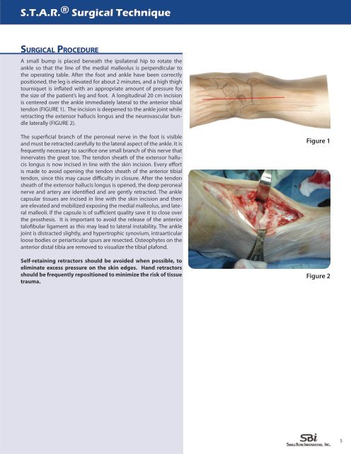
A small bump is placed beneath the ipsilateral hip to rotate the<br />
ankle so that the line of the medial malleolus is perpendicular to<br />
the operating table. After the foot and ankle have been correctly<br />
positioned, the leg is elevated for about 2 minutes, and a high thigh<br />
tourniquet is inflated with an appropriate amount of pressure for<br />
the size of the patient’s leg and foot. A longitudinal 20 cm incision<br />
is centered over the ankle immediately lateral to the anterior tibial<br />
tendon (FIGURE 1). The incision is deepened to the ankle joint while<br />
retracting the extensor hallucis longus and the neurovascular bundle<br />
laterally (FIGURE 2).<br />
The superficial branch of the peroneal nerve in the foot is visible<br />
and must be retracted carefully to the lateral aspect of the ankle. It is<br />
frequently necessary to sacrifice one small branch of this nerve that<br />
innervates the great toe. The tendon sheath of the extensor hallucis<br />
longus is now incised in line with the skin incision. Every effort<br />
is made to avoid opening the tendon sheath of the anterior tibial<br />
tendon, since this may cause difficulty in closure. After the tendon<br />
sheath of the extensor hallucis longus is opened, the deep peroneal<br />
nerve and artery are identified and are gently retracted. The ankle<br />
capsular tissues are incised in line with the skin incision and then<br />
are elevated and mobilized exposing the medial malleolus, and lateral<br />
malleoli. If the capsule is of sufficient quality save it to close over<br />
the prosthesis. It is important to avoid the release of the anterior<br />
talofibular ligament as this may lead to lateral instability. The ankle<br />
joint is distracted slightly, and hypertrophic synovium, intraarticular<br />
loose bodies or periarticular spurs are resected. Osteophytes on the<br />
anterior distal tibia are removed to visualize the tibial plafond.<br />
Self-retaining retractors should be avoided when possible, to<br />
eliminate excess pressure on the skin edges. Hand retractors<br />
should be frequently repositioned to minimize the risk of tissue<br />
trauma.<br />
Figure 1<br />
Figure 2<br />
5