SR⢠MCP Implant System - Small Bone Innovations
SR⢠MCP Implant System - Small Bone Innovations
SR⢠MCP Implant System - Small Bone Innovations

Create successful ePaper yourself
Turn your PDF publications into a flip-book with our unique Google optimized e-Paper software.
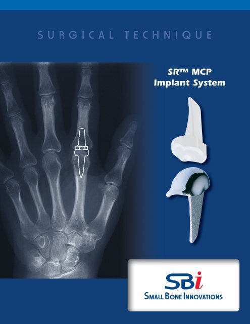
SURGICAL<br />
TECHNIQUE<br />
SR <strong>MCP</strong><br />
<strong>Implant</strong> <strong>System</strong>
SR <strong>MCP</strong> <strong>Implant</strong> <strong>System</strong><br />
SURGICAL TECHNIQUE<br />
CONTENTS<br />
Introduction<br />
Preoperative Assessment & Anatomy 1<br />
Surgical Procedure<br />
1. Incision Options 2<br />
2. Capsular Exposure 2<br />
3. Dorsal Synovectomy 2<br />
4. Metacarpal Articular Head Resection 3<br />
5. Proximal Phalanx Articular Surface Resection 3<br />
6. Trial Preparation 4<br />
7. Trial Placement 5<br />
8. Trial Reduction / <strong>Implant</strong> Placement 5<br />
9. <strong>Implant</strong>ing the Components 6<br />
10. Closure and Centralization 6<br />
11. Initial Post-Operative Treatment 7<br />
12. Rehabilitation 7<br />
Revision or Removal of <strong>MCP</strong> Prostheses 8<br />
Crossed Intrinsic Transfer Procedure 8<br />
Indications and Contraindications 9<br />
Warnings and Precautions 9
Preoperative Assessment & Anatomy<br />
Record the range of motion (ROM) for all the joints of the<br />
hand and wrist and other significant joints. Static deformities,<br />
grip strength and pinch strength should also be<br />
assessed and recorded.<br />
X-ray examination includes AP, lateral and such special<br />
views as may be indicated initially at surgery and at followup<br />
intervals. Sizing templates with a 3% parallax enlargement<br />
are available. Follow-up examinations will include<br />
repetition of the above measurement as well as evaluation<br />
of positioning, alignment, cement mantle adequacy and<br />
evidence of loosening or stem erosion into bone.<br />
Assessment of <strong>MCP</strong> palmar subluxation, ulnar drift, ulnar<br />
intrinsic contracture, extrinsic extensor tendon displacement,<br />
swan neck or boutonniere deformity, and flexor tendon<br />
displacement should be recorded.<br />
TYPICAL RHEUMATOID<br />
DEFORMITY<br />
NORMAL<br />
NORMAL<br />
TYPICAL RHEUMATOID<br />
DEFORMITY<br />
1
SR <strong>MCP</strong> <strong>Implant</strong> <strong>System</strong><br />
SURGICAL PROCEDURE<br />
FIGURE 1<br />
1<br />
Incision Options<br />
Under tourniquet control and adequate anesthesia, incision<br />
is made. Two options are recommended (FIGURE 1):<br />
A: a curved logitudional incision is made over the dorsum<br />
of the metacarpophalangeal (<strong>MCP</strong> joint for single joint<br />
involvement),<br />
B: or, through a transverse incsion across the dorsum of<br />
the <strong>MCP</strong> with multiple joint involvement.<br />
Dissection is carried down through the subcutaneous<br />
tissue to expose the extensor tendons. Retract skin<br />
proximally and distally from the incision, with care<br />
taken to avoid the dorsal veins passing between the<br />
metacarpal heads.<br />
FIGURE 2<br />
Distended doral<br />
capsule<br />
2<br />
Capular Exposure<br />
If the extensor tendon is well centralized, it is split down<br />
the central sulcus of the central tendon and both sides are<br />
reflected from the underlying capsule (FIGURE 2). In<br />
rheumatoid arthritis, the synovial protrusion may have<br />
caused the extensor tendon to ulnarly translate and obliterate<br />
the radial sagittal band. It is usually possible to dissect<br />
the sagittal bands from the capsule and preserve<br />
them so that the extensor can be relocated and the sagittal<br />
bands imbricated to maintain extensor position.<br />
Extensor tendon and<br />
ulnar sagittal band<br />
Attenuated<br />
collateral<br />
ligaments<br />
Radial sagital<br />
band<br />
3<br />
Dorsal Synovectomy<br />
The capsule is then incised longitudinally to expose the<br />
joint (FIGURE 3). A dorsal synovectomy is performed. As<br />
necessary, collateral ligaments are preserved if the joint<br />
can be fully reduced. If not, it may be necessary to release<br />
one or both collateral ligaments partially or wholly from<br />
their tuberosital origins. The ends of the collateral ligaments<br />
should be tagged for later repair to their tuberosital<br />
origins.<br />
FIGURE 3<br />
Joint excisional<br />
lines<br />
2 SR <strong>MCP</strong> <strong>Implant</strong> <strong>System</strong>
4<br />
Metacarpal Articular Head Resection<br />
The metacarpal head is removed first by a vertical saw cut<br />
distal to the collateral ligaments. A second cut at 45° proximovolarly<br />
removes the remainder of the metacarpal<br />
head (FIGURES 4A and 4B). Ligamentous integrity is<br />
retained in so far as possible.<br />
FIGURE 4A<br />
FIGURE 4B<br />
5<br />
Proximal Phalanx Articular Surface Resection<br />
Remove the base of the proximal phalanx with a minimal<br />
slice (FIGURE 5).This is to preserve the collateral ligamentous<br />
attachments in so far as possible. Marked contracture<br />
of the ulnar capsule may require detachment of the ulnar<br />
collateral ligament to assure alignment of the finger to<br />
the metacarpal.<br />
FIGURE 5<br />
3
6<br />
Trial Preparation<br />
An awl is then inserted in the dorsal aspect of the<br />
intramedullary canal of the metacarpal (FIGURE 6A). This<br />
is followed by sequential broaching until proper fit has<br />
been attained. For the index and long finger the<br />
intramedullary preparation is ulnarly displaced slightly<br />
(FIGURE 6B). This provides a better moment arm for the<br />
radial intrinsic and extrinsic tendons to compensate for<br />
the ulnar drift tendency. The same awl and broach procedure<br />
is then followed for the proximal phalanx<br />
(FIGURE 6C).<br />
FIGURE 6A<br />
Awl<br />
Joint removed<br />
FIGURE 6B<br />
Broach<br />
FIGURE 6C<br />
4 SR <strong>MCP</strong> <strong>Implant</strong> <strong>System</strong>
7<br />
Trial Placement<br />
An impactor with a concave surface aids insertion and<br />
optimal proximal displacement of the proximal trial component<br />
(FIGURE 7A).The tip of the prostheses should<br />
pass the midpoint of the metacarpal. Avoid impaction<br />
with metallic instruments which may damage the articular<br />
surfaces of the trial components.<br />
A convex impactor is also provided to aid insertion and<br />
seating of the distal component (FIGURE 7B). Once the<br />
trial components are inserted, check for fit and position,<br />
as well as position under the image intensifier.<br />
FIGURE 7A<br />
FIGURE 7B<br />
8<br />
Trial Reduction/<strong>Implant</strong> Preparation<br />
The joint is reduced with both the metacarpal and phalangeal<br />
trials in place. Static position, ease of passive<br />
motion, plane of motion to full extension, and stability is<br />
checked.With multiple joints, comparison between finger<br />
alignment is checked with the trials in place. Revision<br />
trimming may be necessary to correct muscle or soft tissue<br />
tightness, and to insure a full range of motion.<br />
If release of the collateral ligaments was previously<br />
required, two holes are drilled through the tuberosity at<br />
the dorsal lateral and dorsal medial aspect of the remaining<br />
metacarpal head for reattachment or imbrication of<br />
the ligaments (FIGURE 8). Sutures for repair of the collateral<br />
ligament (4-0 Ticron/mersilene) are inserted.<br />
FIGURE 8<br />
When satisfied with preliminary appearances, the trials<br />
are removed using the trial extractor provided in the<br />
instrument tray.<br />
5
9<br />
<strong>Implant</strong>ing the Components<br />
The intramedullary canal is then irrigated first with saline,<br />
then with 0.5% neomycin solution.The medullary cavities<br />
are dried. Both the metacarpal and phalangeal prostheses<br />
are removed from their sterile packaging and inspected.<br />
Polymethylmethacrylate adhesive (PMMA) is injected in a<br />
liquid state using a size #14 plastic intracath attached to a<br />
10cc syringe (FIGURE 9).<br />
FIGURE 9<br />
The distal component is inserted first. Care is taken to<br />
position the prosthetic components in proper alignment<br />
and rotation. Convex and concave plastic impactors are<br />
provided to assist implant insertion. Avoid impaction with<br />
metallic instruments which, by compromising the surface<br />
finish, may accelerate prosthetic wear.<br />
FIGURE 10A<br />
Radial collateral<br />
ligament tightened<br />
The joint is then extended and checked under the image<br />
intensifier before the cement hardens. Cement fixation of<br />
one finger at a time is advisable if the positioning is difficult.<br />
If multiple joints are done, it may be easier to do the<br />
distal components as a group followed by the proximal<br />
components. After the cement has cured, passive range of<br />
motion is checked to ensure a full range of motion without<br />
impingement of prosthetic binding. If satisfactory, the<br />
collateral ligaments are tightened or reattached to the<br />
tuberosity of the metacarpal head.<br />
FIGURE 10B<br />
10<br />
Closure and Centralization<br />
Capsular closure is undertaken after prosthetic fixation.<br />
Centralization of the extensor tendon and imbrication of<br />
the radial sagittal bands is usually necessary in rheumatoid<br />
hands by appropriate closure of the extensor tendon<br />
retinacular system. A “pants-over-vest”centralization may<br />
be required in cases of moderate-severe ulnar drift along<br />
with crossed-intrinsic transfer. Snug repair of the radial<br />
collateral ligament helps to also realign the flexor assemblage<br />
(See FIGURE A,Page 8).<br />
Reattach radial collateral ligament to the metacarpal<br />
bone (FIGURE 10A). Realign the finger before tightening<br />
sutures. With finger held in slight over correction, imbricate<br />
radial sagittal band over extensor tendon (FIGURE<br />
10B). Complete joint closure by securing radial sagittal<br />
band to the extensor apparatus (FIGURE 10C).<br />
FIGURE 10C<br />
6 SR <strong>MCP</strong> <strong>Implant</strong> <strong>System</strong>
11<br />
Initial Post-Operative Treatment<br />
Postoperative positioning is important and should place<br />
the <strong>MCP</strong> joints in slight flexion and the PIP joints in<br />
approximately 45º flexion. If there has been preoperative<br />
ulnar deviation, or this appears following closure the fingers<br />
should be placed in 5 to 10º radial deviation. These<br />
positions must be held while applying a compressive<br />
dressing (FIGURE 11).<br />
FIGURE 11<br />
Support pad for<br />
flexion PIP<br />
Pad to provide<br />
counter support<br />
Pad to provide<br />
counter support<br />
If there has been a tendency for the fourth and fifth<br />
metacarpals to “droop”, the excessive metacarpal arch curvature<br />
should also be supported.The dressing is removed<br />
at 2 to 4 days and a dynamic splint applied for daytime<br />
exercises. A static rest/nocturnal splint capable of holding<br />
the fingers in the corrected positions is used at other periods<br />
for 4 to 6 weeks.<br />
Pad for dorsal displacement<br />
of proximal phalanges<br />
Palmar support<br />
for wrist<br />
12<br />
Rehabilitation<br />
The initial rehabilitation program benefits greatly from<br />
close supervision of the physiatrist and hand therapist.<br />
The fabrication of the dynamic and static splints requires<br />
expert knowledge for correct fit as well as instruction in<br />
proper use (FIGURE 12). The first week or two are best<br />
carried out with daily or bi-daily supervision followed<br />
with repeat reassessment at the scheduled visits.<br />
FIGURE 12<br />
Fish line<br />
Rubber band<br />
tensioners<br />
Velcro straps<br />
Eyelets<br />
Thermoplastic<br />
cockup splint<br />
Follow-up exams should include range of motion (ROM)<br />
assessment for all the joints of the hand and wrist and<br />
other significant joints. Static deformities, grip strength<br />
and pinch strength should also be assessed and recorded.<br />
X-ray examination includes AP, lateral and such special<br />
views as may also be indicated at follow-up intervals.<br />
Assessment of <strong>MCP</strong> palmar subluxation, ulnar drift, ulnar<br />
intrinsic contracture, extrinsic extensor tendon displacement,<br />
swan neck or boutonniere deformity, and flexor<br />
tendon displacement should also be recorded.<br />
Coat hanger<br />
support for<br />
pulleys<br />
Polythylene pulleys<br />
Set washer to<br />
adjust radial<br />
pull<br />
LOW PROFILE<br />
DYNAMIC SLING<br />
Counteracts palmer and ulnar<br />
subluxations tendencies<br />
Leatherette slings<br />
7
Revision or Removal of<br />
<strong>MCP</strong> Prostheses<br />
Should the SR <strong>MCP</strong> implants loosen, dislocate, suffer<br />
infection, or other complication requiring removal, the<br />
procedure involves retrieval of the implant under sterile<br />
conditions, tourniquet control and adequate anesthesia.<br />
The previous dorsal incision (straight or curvilinear) is<br />
incised. The soft tissue flaps are reflected down to the<br />
extensor tendon system. The extensor tendon is incised<br />
longitudinally on the radial side from the proximal phalanx<br />
base across the metacarpal head and neck.The sagittal<br />
bands are reflected from the capsule (if possible). The<br />
capsule of the joint is incised longitudinally and the<br />
implants are exposed. Release of collateral ligaments<br />
might be necessary to retrieve the implants. The<br />
metacarpal prosthesis is removed first. <strong>Small</strong> 1-2mm<br />
osteotomes are used to free the prosthesis and bone<br />
cement from within the metacarpal canal. A linear<br />
osteotomy of the metacarpal canal may be needed to<br />
remove the implant. A fine burr on a high-speed drill is<br />
used to remove the cement over the dorsal surface of the<br />
proximal component. The distal component may also be<br />
drilled out with the high speed burr.The phalangeal components<br />
can be tapped from the intramedullary canals or<br />
removed with an osteotome.<br />
Salvaging of the joint is by soft tissue volar plate interposition<br />
or alternatively silicone interposition joint (no sepsis).<br />
Collateral ligament reconstruction and extensor tendon<br />
centralization should be performed in revision procedures<br />
similar to the index prosthetic insertion procedure.<br />
Postoperative physiotherapy is unchanged from other<br />
procedures of <strong>MCP</strong> arthroplasty.<br />
FIGURE A<br />
Radial collateral<br />
ligament tightened<br />
FIGURE B<br />
Release ulnar<br />
lateral band<br />
Ulnar deviation<br />
with contracture<br />
ulnar<br />
soft tissues<br />
Realignment<br />
of extensor<br />
and flexor<br />
tendons<br />
Crossed Intrinsic Transfer Procedure<br />
Crossed intrinsic transfers are often helpful in correcting<br />
ulnar deviation. In rheumatoid hands with ulnar capsular<br />
contracture it may be necessary to release the ulnar<br />
intrinsic and all or part of the ulnar collateral ligament,<br />
especially if there is swan neck deformity (FIGURE A).<br />
Release of the ulnar intrinsic lateral band insertion midway<br />
onto the proximal phalanx allows transfer onto the<br />
radial aspect of the adjacent digit at the close of the soft<br />
tissue rebalancing (FIGURE B).<br />
First palmar<br />
interosseous to<br />
radial collateral<br />
ligament 3rd finger<br />
8 SR <strong>MCP</strong> <strong>Implant</strong> <strong>System</strong>
INDICATIONS<br />
> Patient is in need of a revision of failed <strong>MCP</strong> prosthesis(es)<br />
or<br />
> Patient expects to place his/her hands under loading<br />
situations, which preclude the use of an alternative<br />
implant in the painful osteo-arthritic and post<br />
traumatic arthritic <strong>MCP</strong> joint.<br />
CONTRAINDICATIONS<br />
> <strong>Bone</strong>, musculature, tendons, or adjacent soft tissue<br />
compromised by disease, infection, or prior implantation,<br />
which cannot provide adequate support or fixation<br />
for the prosthesis.<br />
> Infection.<br />
> Skeletal Immaturity.<br />
WARNINGS AND PRECAUTIONS<br />
Patient Counseling Information (see also Warnings)<br />
In addition to the patient related information contained in the Warnings and Adverse Events<br />
sections, the following information should be conveyed to the patient:<br />
> While the expected life of total joint replacements is difficult to estimate, it is finite.<br />
These components are made of foreign materials, which are placed within the body for<br />
the potential restoration of mobility or reduction of pain. However, due to the many<br />
biological, mechanical and physiochemical factors which affect these devices, the components<br />
cannot be expected to withstand the activity level and loads of healthy bone for an<br />
unlimited period of time.<br />
> Adverse events of this device may necessitate reoperation, revision, or fusion of the<br />
involved joint.<br />
Precautions<br />
> Do not resterilize. The implant is provided sterile in an undamaged package. If either the<br />
implant or the package appears damaged, expiration date has been exceeded, or if sterility<br />
is questioned for any reason, the implant should not be used.<br />
> Meticulous preparation of the implant and selection of the proper size implant increase the<br />
potential for a successful outcome.<br />
> The implant should be removed from its sterile packaging only after the implant site has<br />
been prepared and properly sized.<br />
> <strong>Implant</strong>s should be handled without blunt instruments to avoid scratching, cutting, or nicking<br />
the device.<br />
Warnings (see Patient Counseling Information Section)<br />
> Patients should be made aware of the increased potential for device failure when excessive<br />
demands are made upon it. Strenuous loading, excessive mobility, and articular instability<br />
all may lead to accelerated wear and eventual failure by loosening, fracture, or dislocation of<br />
the device.<br />
Caution<br />
Federal (United States) law restricts this device to sale, distribution and use by or on the order of a physician.<br />
Humanitarian Device<br />
The <strong>Small</strong> <strong>Bone</strong> <strong>Innovations</strong> SR <strong>MCP</strong> Finger Prosthesis is authorized by Federal Law for use in arthroplasty when<br />
either the:<br />
> patient is in need of a revision of failed <strong>MCP</strong> prosthesis(es); or<br />
> patient expects to place his/her hands under loading situations, which preclude the use of an alternative<br />
implant in the painful osteo-arthritic and post-traumatic arthritic <strong>MCP</strong> joint.<br />
The effectiveness of this device for this use has not been demonstrated.<br />
Intracath is a trademark of Becton Dickinson.<br />
MicroAire® is a registered trademark of MicroAire® Surgical Instruments.<br />
9
<strong>Small</strong> <strong>Bone</strong> <strong>Innovations</strong>, Inc.<br />
1711 South Pennsylvania Ave.<br />
Morrisville, PA 19067<br />
Customer Service (800) 778-8837<br />
Technical Support (866) SBi-TIPS<br />
Fax (215) 428-1795<br />
www.totalsmallbone.com<br />
SBi International, SAS<br />
ZA les Bruyeres<br />
BP 28<br />
01960 Peronnas<br />
France<br />
Tel +33 474 21 58 19<br />
Fax +33 474 21 43 12<br />
info@sbi-intl.com<br />
MKT-10710 Rev. a<br />
Copyright 2006, <strong>Small</strong> <strong>Bone</strong> <strong>Innovations</strong>, Inc.