Teliospore character Size (range) µm Size (mean) µm Colour Exospore Ornamentation in median view ♣ Exospore Ornamentation in surface view <strong>Tilletia</strong> <strong>indica</strong> 1 22–47–(61) (26–55(–64)) † 35–41 (40–44) † Pale orange to mainly dark, reddish brown to opaque-black Sharply pointed to truncate spines (occasionally curved), 1.5–5.0 µm high, covered with a hyaline sheath Spines densely arranged, either individually (densely echinulate) or in closely spaced, narrow ridges (finely cerebriform), <strong>Tilletia</strong> walkeri 2 (23–45) † 30–31 * (34–36) † Pale yellow to mainly dark reddish brown (never opaque) Conical to truncate spines (occasionally curved), 3–6 µm high, covered with a hyaline to yellowish-brown sheath Spines coarsely arranged, forming wide, incompletely cerebriform (to coralloid) ridges or thick clumps T. horrida 3 17–36 (20–38(–41)) † 24–28 ‡ (28) † Pale yellow to mainly light or dark chestnut brown (semi-opaque) Sharply pointed or curved spines, 1.5–4.0 µm high, becoming truncated scales with maturity, covered with a hyaline to tinted sheath Spines appearing as polygonal scales (occasionally spines forming cerebriform ridges or small clumps) ‡ 1 Based on: Bansal et al., (1984); Castlebury & Carris (1999); CMI Description No. 748 (1983); Durán (1987); Durán & Fischer (1961); Khanna & Payak (1968); Mathur & Cunfer (1993); Milbrath et al. (1998); Mundkur (1940); Peterson et al., (1984). 2 Based on: Castlebury & Carris (1999); Cunfer & Castlebury, 1999; Milbrath et al. (1998); Castlebury, 3 1998. As T. barclayana: Castlebury & Carris (1999); CMI Description No. 75 (1965); Durán (1987); Durán & Fischer (1961). Or as T. horrida: Aggarwal et al. (1990); Khanna & Payak (1968); Castlebury (1998). † Castlebury & Carris (1999) report larger spore sizes (in brackets) for teliospores warmed overnight at 45°C in Shear’s solution; Castlebury (1998) also reports larger teliospore sizes. * Milbrath et al., (1998), supported by Author’s data from teliospores ex. Lolium (two isolates ex. ‡ Oregon, USA) in water. Author’s data from teliospores ex. Oryzae (California, USA; Arkanasas, USA) in water; though not reported in the literature, some spores may have ridges in addition to individual spines (see Figure 2.4). ♣ Hawksworth et al., (1995): Ainsworth <strong>and</strong> Bisby’s Dictionary of the Fungi. Table 2.2. Morphological characteristics of <strong>Tilletia</strong> <strong>indica</strong> (Karnal bunt of wheat), T. walkeri (ryegrass bunt) <strong>and</strong> T. horrida (rice smut). <strong>The</strong> literature on spore sizes is often variable. Spore size is affected by the mounting medium <strong>and</strong> by heating treatments. For rice smut (T. horrida, synonym T. barclayana), data from rice is potentially more reliable than data based on T. barclayana sensu lato from various Poaceae as the latter is considered a species complex. <strong>The</strong> rice pathogen is considered distinct from those on Paspalum <strong>and</strong> Pannicum, but it is not known whether it is distinct from T. barclayana s.s. on Pennisetum (Pimentel, 1998; Castlebury, 1998). 12
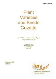
Figure 2.2. Teliospores of <strong>Tilletia</strong> <strong>indica</strong> (Karnal bunt of wheat) showing surface ornamentation patterns: spines densely arranged, either individually (densely echinulate) or in closely spaced, narrow ridges (finely cerebriform). (Figures 2.2–2.4 are at the same scale; 10 mm = 17 µm.) 13