Tilletia indica - The Food and Environment Research Agency - Defra
Tilletia indica - The Food and Environment Research Agency - Defra
Tilletia indica - The Food and Environment Research Agency - Defra

You also want an ePaper? Increase the reach of your titles
YUMPU automatically turns print PDFs into web optimized ePapers that Google loves.
EU RECOMMENDED PROTOCOL FOR THE DIAGNOSIS OF A QUARANTINE<br />
ORGANISM<br />
Identity<br />
<strong>Tilletia</strong> <strong>indica</strong><br />
Name: <strong>Tilletia</strong> <strong>indica</strong> Mitra<br />
Synonyms: Neovossia <strong>indica</strong> (Mitra) Mundkur<br />
Further information on this organism can be obtained from:<br />
Taxonomic position: Fungi: Basidiomycetes: Ustilaginales<br />
Mycology team PL2,<br />
Common Central names: Science Laboratory, Karnal S<strong>and</strong> bunt Hutton, or Partial York bunt YO41 of wheat 1LZ (English)<br />
Carie de Karnal (French)<br />
Indischer Weizenbr<strong>and</strong> (German)<br />
Quarantine status: EPPO A1 list: No 23<br />
EU Annex designation I/AI<br />
Further information on this organism can be obtained from:<br />
Mycology diagnosis, Team PLH2, Central Science Laboratory, S<strong>and</strong> Hutton,<br />
York YO41 1LZ, UK<br />
Diagnostic protocol prepared by A. J. Inman, K. J. D. Hughes <strong>and</strong> R.J. Bowyer<br />
Central Science Laboratory, S<strong>and</strong> Hutton, York YO41 1LZ<br />
Please send suggestions/comments to: a.inman@csl.gov.uk<br />
This protocol has been produced under EU funding <strong>and</strong> ring tested by expert<br />
laboratories within the EU<br />
Version date: 6th January 2003<br />
1
TILLETIA INDICA PROTOCOL<br />
DETECTION AND IDENTIFICATION OF TILLETIA INDICA (KARNAL BUNT)<br />
CONTENTS<br />
CONTENTS 2<br />
LIST OF TABLES 3<br />
LIST OF FIGURES 4<br />
SECTION I: INTRODUCTION 5<br />
Principle hosts 5<br />
Symptoms 5<br />
SECTION II: DIAGNOSTIC SCHEME 6<br />
Scope of the diagnostic scheme<br />
6<br />
Flow diagram for diagnosis 7<br />
Sampling 8<br />
Detection 8<br />
Isolation 9<br />
Identification<br />
9<br />
Comparison with other similar organisms 9<br />
Confirmation 17<br />
Requirements for a positive diagnosis 17<br />
Report on the diagnosis 18<br />
Contact point for protocol 18<br />
List of participants in the ring test 19<br />
SECTION III: PROTOCOLS<br />
A. Protocol for extracting teliospores from seed or grain by size-selective sieving 20<br />
B. Protocol for morphological identification 25<br />
C. Protocol for isolation & germination of teliospores for molecular confirmation 30<br />
D. Protocol for confirmation by traditional PCR using species specific primers **<br />
E. Protocol for confirmation by PCR using TaqMan® **<br />
F. Protocol for confirmation using restriction enzyme analysis **<br />
2
SECTION IV: REFERENCES **<br />
3
LIST OF TABLES<br />
Table 2.1. <strong>The</strong> number of replicate 50 g sub-samples needed to detect differing levels of<br />
contamination with specified confidences, assuming an equal distribution of teliospores.<br />
Table 2.2. Morphological characteristics of <strong>Tilletia</strong> <strong>indica</strong> (Karnal bunt of wheat), T.<br />
walkeri (ryegrass bunt) <strong>and</strong> T. horrida (rice smut).<br />
Table 3.1. Record sheet for teliospores detected in wash tests.<br />
Table 3.2. Scheme for morphologically distinguishing teliospores of <strong>Tilletia</strong> <strong>indica</strong>, T.<br />
walkeri <strong>and</strong> T. horrida detected in size-selective sieving wash tests that use 20 µm <strong>and</strong><br />
53 µm sieves.<br />
8<br />
11<br />
26<br />
28
LIST OF FIGURES<br />
Figure 1.1. Grain infected with <strong>Tilletia</strong> <strong>indica</strong> (Karnal bunt).<br />
Figure 2.1. Flow diagram of diagnostic scheme<br />
Figure 2.2. Teliospores of <strong>Tilletia</strong> <strong>indica</strong> (Karnal bunt of wheat) showing surface<br />
ornamentation patterns.<br />
Figure 2.3. Teliospores of <strong>Tilletia</strong> walkeri (ryegrass bunt) showing surface<br />
ornamentation patterns.<br />
Figure 2.4. Teliospores of <strong>Tilletia</strong> horrida (rice smut) showing surface ornamentation<br />
patterns.<br />
Figure 2.5. Teliospores of <strong>Tilletia</strong> <strong>indica</strong> <strong>and</strong> T. walkeri showing teliospore profiles in<br />
median view after bleaching <strong>and</strong> then staining with lactoglycerol-trypan blue.<br />
Figure 2.6. Colonies of <strong>Tilletia</strong> <strong>indica</strong>, T. walkeri <strong>and</strong> T. horrida after 7, 10 <strong>and</strong> 14 days<br />
on PDA at 19°C <strong>and</strong> a 12 hour dark/light cycle.<br />
Figure 3.1. Size-selective sieving wash test: 20 µm mesh nylon sieve (mounted between<br />
4 cm diameter cylinders), 53 µm sieve (mounted between 11 cm diameter cylinders) <strong>and</strong><br />
a 50 g grain sample in a 250 ml Erlenmeyer flask with 100 ml of 0.01% Tween-20.<br />
Figure 3.2. Size-selective sieving wash test: Arrangement of sieves (20 µm, sieve, left;<br />
53 µm sieve, right) mounted in funnels over 500 ml Erlenmeyer flasks in preparation for<br />
size-selective sieving of wash water from a 50 g grain sample.<br />
Figure 3.3. Size-selective sieving wash test: Grain <strong>and</strong> washings poured onto a 53 µm<br />
sieve over a 500 ml Erlenmeyer flask, together with an aspirator bottle for subsequent<br />
rinsing of the grain on the sieve.<br />
Figure 3.4. Size-selective sieving wash test: <strong>The</strong> final sieve fraction being washed to one<br />
side of the 20 µm sieve with water from a disposable Pasteur pipette.<br />
Figure 3.5. Size-selective sieving wash test: <strong>The</strong> final sieve fraction being recovered<br />
from the 20 µm sieve with water from a disposable Pasteur pipette for subsequent<br />
centrifugation in a conical centrifuge tube.<br />
Figure 3.6. Teliospores of <strong>Tilletia</strong> <strong>indica</strong> in median view.<br />
Figure 3.7. Pictorial key to teliospore ornamentation.<br />
Figure 3.8. Decision tree for morphological identification of teliospores detected in size<br />
selective sieving wash tests<br />
Figure 3.9. <strong>Tilletia</strong> <strong>indica</strong> teliospores germinating on water agar after 10–14 days.<br />
Figure 3.10. Agar plug with germinated teliospores placed on the inside of a Petri dish<br />
lid suspended over potato dextrose broth<br />
5<br />
5<br />
7<br />
12<br />
13<br />
14<br />
15<br />
16<br />
22<br />
22<br />
22<br />
23<br />
23<br />
23<br />
27<br />
29<br />
31<br />
31
SECTION 1: INTRODUCTION<br />
Principle Hosts<br />
<strong>Tilletia</strong> <strong>indica</strong> causes the disease Karnal bunt, or partial bunt, of wheat (Triticum spp.).<br />
Triticale (X Triticosecale) is also naturally infected <strong>and</strong> rye (Secale) is a potential host. T.<br />
<strong>indica</strong> was added to the EC Plant Health Directive 77/93/EEC (now 2000/29/EC) as a I/AI<br />
pest in 1996 <strong>and</strong> quarantine requirements applied to seed <strong>and</strong> grain of Triticum, Secale <strong>and</strong> X<br />
Triticosecale from countries where T. <strong>indica</strong> is known to occur.<br />
Symptoms<br />
T. <strong>indica</strong> is a floret-infecting fungal smut pathogen. Unlike systemic smuts, not all the seeds<br />
on an ear are usually infected <strong>and</strong> the seeds are typically only partially bunted. Seeds are<br />
infected through the germinal end of the grain <strong>and</strong> the fungus develops within the pericarp<br />
where it produces a powdery, brownish-black mass of teliospores. When fresh, the spore<br />
masses produce a foetid, decaying fish-like smell (trimethylamine). Seeds are usually only<br />
partially colonised, showing various degrees of infection. Point infections are most common,<br />
but infection may also spread down the adaxial groove <strong>and</strong>, in severe cases, the whole grain<br />
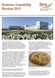
may appear bunted (Figure 1.1).<br />
Figure 1.1. Grain infected with <strong>Tilletia</strong> <strong>indica</strong> (Karnal bunt). Symptoms range from partial<br />
bunting (point infections <strong>and</strong> infections spreading down the adaxial groove) to almost<br />
complete bunting. Photograph courtesy of G. L. Peterson, USDA.
SECTION II: DIAGNOSTIC SCHEME<br />
Scope of the Diagnostic Scheme<br />
<strong>The</strong> scheme for <strong>Tilletia</strong> <strong>indica</strong> describes procedures for:<br />
(i) detection of teliospores in imported seed or grain of wheat by a size-selective sieving<br />
wash test;<br />
(ii) morphological identification of teliospores detected in wash tests;<br />
(iii) isolation <strong>and</strong> germination of teliospores for molecular confirmation;<br />
(iv) molecular confirmation of cultures.<br />
7
Sample<br />
declared<br />
healthy<br />
IDENTIFICATION<br />
based on teliospore<br />
morphology<br />
(Protocol B)<br />
Wash test for tuberculate <strong>Tilletia</strong> teliospores<br />
(Size selective sieving on 50 g sub-samples)<br />
(Protocol A)<br />
negative positive<br />
Many teliospores<br />
present<br />
(c. ≥10)<br />
MORPHOLOGICAL<br />
DIAGNOSIS of<br />
teliospores<br />
(Protocol B)<br />
MOLECULAR CONFIRMATION TESTS<br />
Isolate & germinate suspect teliospores to<br />
produce cultures for molecular confirmation tests<br />
(Protocol C)<br />
MOLECULAR CONFIRMATION:<br />
Restriction enzyme analysis (Protocol D)<br />
PCR with species specific primers (Protocol E)<br />
TaqMan with species specific primers & probe (Protocol F)<br />
IDENTIFICATION OF SPECIES<br />
based on morphological <strong>and</strong> molecular analyses<br />
CONFIRMATION:<br />
microscopic examination of<br />
teliospores from bunted<br />
seeds compared to those in<br />
wash test (Protocol B)<br />
Bunted seeds found<br />
Examine sample for bunted<br />
cereal seeds or bunted seeds<br />
of other Poaceae<br />
No bunted cereal grains<br />
found<br />
Few teliospores present<br />
(c.
Sampling<br />
Seed lots should be sampled according to current ISTA rules. Grain, e.g. for feed or<br />
processing, is typically more difficult to sample because consignments are usually very large<br />
<strong>and</strong> transported or stored as large, loose bulks. However, for monitoring purposes, grain<br />
should be sampled in an appropriate fashion to produce a 1–2 kg thoroughly mixed sample<br />
representative of the consignment.<br />
Detection<br />
For quarantine purposes, detection of T. <strong>indica</strong> is best achieved by a wash test (CABI/EPPO,<br />
1997); infected parts of the grain typically disintegrate so that the teliospores contaminate<br />
other grains in the lot. <strong>The</strong> most efficient <strong>and</strong> rapid wash test method for detecting teliospores<br />
in a sample is a size-selective sieving <strong>and</strong> centrifugation technique (Section III, Protocol A;<br />
Peterson et al., 2000). This method has, on average, an 82% efficiency of recovery <strong>and</strong><br />
microscopic examinations typically require only a few slides per 50 g sub-sample. <strong>The</strong><br />
number of replicate 50 g sub-samples needed to detect differing levels of contamination is<br />
given in Table 2.1.<br />
Contamination level<br />
(No. of spores / 50 g sample)<br />
No. of replicate samples required for detection<br />
according to level of confidence (%)<br />
99 % 99.9 % 99.99 %<br />
1<br />
3<br />
5<br />
6<br />
2 2 3 4<br />
5 1 1 1<br />
Table 2.1. <strong>The</strong> number of replicate 50 g sub-samples needed to detect differing levels of<br />
contamination with specified confidences, assuming an equal distribution of teliospores<br />
(Peterson et al., 2000; Inman & Bowyer, EU SMT4-CT98-2252 evaluation, 2000).<br />
Direct visual examinations for bunted kernals or teliospores contaminating seed surfaces are<br />
not considered reliable methods for quarantine purposes. However, Karnal bunt may be<br />
detected by visual examination with the naked eye <strong>and</strong> low power microscopy (x10–x70<br />
magnification). To help visualise symptoms, seed can be soaked in 0.2% sodium hydroxide<br />
for 24 hours at 20°C; this is especially useful for chemically treated seed lots where coloured<br />
dyes may obscure symptoms (Mathur & Cunfer, 1993; Agarwal & Mathur, 1992). With<br />
severe contamination, teliospores may be seen on the surface of seeds (Mathur & Cunfer,<br />
1993).<br />
9
Isolation<br />
<strong>Tilletia</strong> <strong>indica</strong> is a facultative biotroph. To produce cultures, teliospores are soaked in water,<br />
quickly surface sterilised <strong>and</strong> then germinated on water agar plates (Section III, Protocol C).<br />
After 7-14 days, non-dormant teliospores produce a promycelium bearing 32–128 or more<br />
basidiospores (primary sporidia) at its tip. <strong>The</strong>se basidiospores can then be cultured directly<br />
on solid or liquid nutrient media.<br />
Identification<br />
<strong>The</strong> morphology of <strong>Tilletia</strong> <strong>indica</strong> is as follows (see also: Table 2.2; Figure 2.2; <strong>and</strong> Section<br />
III, Protocol B):<br />
Teliospore shape <strong>and</strong> size * : globose to subglobose, sometimes with a small hyphal fragment<br />
(more common on immature teliospores, but occasionally on mature teliospores), mostly 22–<br />
47 µm in diameter, occasionally larger (mean 35–41 * µm).<br />
Teliospore colour: pale orange to brown to dark, reddish brown; some teliospores black <strong>and</strong><br />
opaque (Figure 2.2).<br />
Teliospore ornamentation * : densely ornamented with sharply pointed to truncate spines,<br />
occasionally with curved tips, 1.5–5.0 µm high, which in surface view appear as either<br />
individual spines (densely echinulate) or as closely spaced, narrow ridges (finely<br />
cerebriform) (Figure 2.2).; the spines are covered by a thin hyaline membrane.<br />
Sterile cells: globose, subglobose to lacrymiform (tear-shaped), yellowish brown, 10–28 x 48<br />
µm, with or without an apiculus (short stalk), with smooth walls up to 7 µm thick <strong>and</strong><br />
laminated. Sterile cells are likely to be uncommon in sieved washings.<br />
* <strong>The</strong> mounting medium <strong>and</strong> heating or warming treatments can affect teliospore size (Aggarwal et al.,<br />
1990; Khanna & Payak, 1968; Castlebury & Carris, 1999). This protocol assumes that spores are<br />
mounted in water <strong>and</strong> not warmed or heated; suspect spores can then be germinated for any<br />
subsequent PCR confirmation. However, surface ornamentation can sometimes not be seen clearly in<br />
water. In such cases, mounting teliospores in lactoglycerol or Shears’s solution (Mathur & Cunfer,<br />
1993) <strong>and</strong> gently heating the slides may improve clarity.<br />
Comparison with Other Similar Organisms<br />
Morphological comparisons<br />
Other tuberculate-spored <strong>Tilletia</strong> species may be confused with T. <strong>indica</strong> (Durán & Fischer,<br />
1961; Durán, 1987). In particularly, the morphologically <strong>and</strong> genetically similar fungus<br />
<strong>Tilletia</strong> walkeri (ryegrass bunt), <strong>and</strong> also T. horrida (rice smut), are known contaminants of<br />
wheat seed or grain (Cunfer & Castlebury, 1999; Castlebury & Carris, 1999; Smith et al.,<br />
1996). <strong>The</strong> most important morphological characters that discriminate T. <strong>indica</strong>, T. walkeri<br />
<strong>and</strong> T. horrida are teliospore size (range <strong>and</strong> mean), exospore ornamentation <strong>and</strong> colour<br />
(Table 2.2; Figures 2.2–2.4; Section III, Protocol B). If sufficient numbers of teliospores are<br />
present, T. horrida teliospores are principally distinguished from T. <strong>indica</strong> by their smaller<br />
size, chestnut-brown colour <strong>and</strong> spines that are frequently curved <strong>and</strong> that appear as<br />
polygonal scales in surface view. T. walkeri <strong>and</strong> T. <strong>indica</strong> have a larger degree of overlap in<br />
morphological characters. However, T. walkeri teliospores are on average smaller, paler in<br />
colour (never black/opaque) <strong>and</strong> have coarser exospore ornamentation which in surface view<br />
gives the appearance of wide, incompletely cerebriform ridges or thick clumps (Castlebury &<br />
Carris, 1999). In median view, the exospore spine profiles may also aid identification. <strong>The</strong><br />
10
median profiles can be enhanced by bleaching the teliospores in 10% domestic bleach<br />
(sodium hypochlorite) for 15–20 minutes; if necessary, spores can then be rinsed twice in<br />
water <strong>and</strong> stained (e.g. trypan blue or cotton blue in lactoglycerol), (Figure 2.5). In general, T.<br />
<strong>indica</strong> teliospores have a smoother, more complete median outline due to their spines being<br />
more densely arranged; profiles of T. walkeri are more irregular with gaps between the spines<br />
due their spines being more coarsely arranged (Figure 2.5).<br />
In culture, T. walkeri <strong>and</strong> T. <strong>indica</strong> produce very similar colonies. On potato dextrose agar<br />
(PDA) after 14 days at 19°C with a 12 hour light cycle, both species typically produce white<br />
to cream-coloured, slow growing, irregular, crustose colonies, about 4–6 mm in diameter<br />
(Figure 2.6). In comparison, comparable cultures of T. horrida grow significantly more<br />
slowly (colonies only 2–3 mm in diameter) due to their higher temperature optima. T. horrida<br />
isolates may also produce a reddish-purple pigment (Figure 2.6), both on PDA <strong>and</strong> potato<br />
dextrose broth.<br />
Other tuberculate-spored <strong>Tilletia</strong> species have teliospores that can appear morphologically<br />
similar to those of T. <strong>indica</strong> (Pimentel, et al., 1998; Durán & Fischer, 1961). <strong>The</strong>se species<br />
are less likely to be found as contaminants of wheat, but they include: T. barclayana (smut of<br />
various gramineae, e.g. Panicum <strong>and</strong> Paspalum), T. eragrostidis (on Eragrostis), T. inolens<br />
(on Deyeuxia forsteri), T. rugispora (on Paspalum), T. boutelouae (on Bouteloua gracilis).<br />
None of these morphologically similar species, or T. walkeri or T. horrida, has been found to<br />
naturally infect wheat.<br />
Molecular comparisons<br />
Diagnostically significant differences exist between T. <strong>indica</strong>, T. walkeri <strong>and</strong> T. horrida in<br />
their nuclear <strong>and</strong> mitochondrial DNA. Interspecific polymorphisms have been identified<br />
using various polymerase chain reaction (PCR) methods, including RAPDs, RFLPs <strong>and</strong><br />
AFLPs (Pimentel et al., 1998; Laroche et al., 1998). In the nuclear ribosomal (rDNA) ITS1<br />
<strong>and</strong> ITS2 regions, there is a >98% similarity between T. walkeri <strong>and</strong> T. <strong>indica</strong> sequences<br />
(Levy et al., 1998). However, within the ITS1 region, T. walkeri has a diagnostically<br />
important restriction enzyme site (ScaI) that is not present with T. <strong>indica</strong>, T. horrida or other<br />
closely related species (Pimentel et al., 1998; Levy et al., 1998). With mtDNA, sequence<br />
differences have enabled species specific primers to be designed to T. <strong>indica</strong> <strong>and</strong> T. walkeri<br />
(Frederick et al., 2000). <strong>The</strong>se primers can be used in conventional PCR assays or in a<br />
TaqMan system in conjunction with a probe (Frederick et al., 2000). <strong>The</strong>re are currently no<br />
species specific primers for T. horrida, but RFLPs can be used to identify cultures (Pimental<br />
et al., 1998). If species specific primers for T. walkeri <strong>and</strong> T. <strong>indica</strong> do not give positive<br />
results on test cultures, RFLPs, RAPDs or AFLPs may be useful tools in identification<br />
(Pimental et al., 1998). See Section III, Protocols C–F for molecular tests.<br />
11
Teliospore<br />
character<br />
Size (range) µm<br />
Size (mean) µm<br />
Colour<br />
Exospore<br />
Ornamentation<br />
in median view<br />
♣<br />
Exospore<br />
Ornamentation<br />
in surface view<br />
<strong>Tilletia</strong> <strong>indica</strong> 1<br />
22–47–(61)<br />
(26–55(–64)) †<br />
35–41<br />
(40–44) †<br />
Pale orange to mainly dark,<br />
reddish brown<br />
to opaque-black<br />
Sharply pointed to truncate<br />
spines (occasionally<br />
curved), 1.5–5.0 µm high,<br />
covered with a hyaline<br />
sheath<br />
Spines densely arranged,<br />
either individually<br />
(densely echinulate) or in<br />
closely spaced, narrow<br />
ridges (finely cerebriform),<br />
<strong>Tilletia</strong> walkeri 2<br />
(23–45) †<br />
30–31 *<br />
(34–36) †<br />
Pale yellow to mainly dark<br />
reddish brown (never<br />
opaque)<br />
Conical to truncate spines<br />
(occasionally curved), 3–6<br />
µm high, covered with a<br />
hyaline to yellowish-brown<br />
sheath<br />
Spines coarsely arranged,<br />
forming wide,<br />
incompletely cerebriform<br />
(to coralloid) ridges or<br />
thick clumps<br />
T. horrida 3<br />
17–36<br />
(20–38(–41)) †<br />
24–28 ‡<br />
(28) †<br />
Pale yellow to mainly light<br />
or dark chestnut brown<br />
(semi-opaque)<br />
Sharply pointed or curved<br />
spines, 1.5–4.0 µm<br />
high, becoming truncated<br />
scales with maturity,<br />
covered with a hyaline to<br />
tinted sheath<br />
Spines appearing as<br />
polygonal scales<br />
(occasionally spines<br />
forming cerebriform ridges<br />
or small clumps) ‡<br />
1<br />
Based on: Bansal et al., (1984); Castlebury & Carris (1999); CMI Description No. 748 (1983); Durán<br />
(1987); Durán & Fischer (1961); Khanna & Payak (1968); Mathur & Cunfer (1993); Milbrath et al.<br />
(1998); Mundkur (1940); Peterson et al., (1984).<br />
2<br />
Based on: Castlebury & Carris (1999); Cunfer & Castlebury, 1999; Milbrath et al. (1998); Castlebury,<br />
3<br />
1998.<br />
As T. barclayana: Castlebury & Carris (1999); CMI Description No. 75 (1965); Durán (1987); Durán<br />
& Fischer (1961). Or as T. horrida: Aggarwal et al. (1990); Khanna & Payak (1968); Castlebury<br />
(1998).<br />
† Castlebury & Carris (1999) report larger spore sizes (in brackets) for teliospores warmed overnight at<br />
45°C in Shear’s solution; Castlebury (1998) also reports larger teliospore sizes.<br />
* Milbrath et al., (1998), supported by Author’s data from teliospores ex. Lolium (two isolates ex.<br />
‡<br />
Oregon, USA) in water.<br />
Author’s data from teliospores ex. Oryzae (California, USA; Arkanasas, USA) in water; though not<br />
reported in the literature, some spores may have ridges in addition to individual spines (see Figure 2.4).<br />
♣ Hawksworth et al., (1995): Ainsworth <strong>and</strong> Bisby’s Dictionary of the Fungi.<br />
Table 2.2. Morphological characteristics of <strong>Tilletia</strong> <strong>indica</strong> (Karnal bunt of wheat), T. walkeri<br />
(ryegrass bunt) <strong>and</strong> T. horrida (rice smut). <strong>The</strong> literature on spore sizes is often variable.<br />
Spore size is affected by the mounting medium <strong>and</strong> by heating treatments. For rice smut (T.<br />
horrida, synonym T. barclayana), data from rice is potentially more reliable than data based<br />
on T. barclayana sensu lato from various Poaceae as the latter is considered a species<br />
complex. <strong>The</strong> rice pathogen is considered distinct from those on Paspalum <strong>and</strong> Pannicum,<br />
but it is not known whether it is distinct from T. barclayana s.s. on Pennisetum (Pimentel,<br />
1998; Castlebury, 1998).<br />
12
Figure 2.2. Teliospores of <strong>Tilletia</strong> <strong>indica</strong> (Karnal bunt of wheat) showing surface<br />
ornamentation patterns: spines densely arranged, either individually (densely echinulate) or<br />
in closely spaced, narrow ridges (finely cerebriform).<br />
(Figures 2.2–2.4 are at the same scale; 10 mm = 17 µm.)<br />
13
Figure 2.3. Teliospores of <strong>Tilletia</strong> walkeri (ryegrass bunt) showing surface ornamentation<br />
patterns: spines coarsely arranged <strong>and</strong> forming wide, incompletely cerebriform to coralloid<br />
ridges or thick clumps.<br />
(Figures 2.2–2.4 are at the same scale; 10 mm = 17 µm.)<br />
14
Figure 2.4. Teliospores of <strong>Tilletia</strong> horrida (rice smut) showing surface ornamentation<br />
patterns: polygonal scales or, occasionally, with cerebriform ridges.<br />
(Figures 2.2–2.4 are at the same scale; 10 mm = 17 µm.)<br />
15
<strong>Tilletia</strong> <strong>indica</strong><br />
<strong>Tilletia</strong> walkeri<br />
Figure 2.5. Teliospores of <strong>Tilletia</strong> <strong>indica</strong> (top) <strong>and</strong> T. walkeri (bottom) showing teliospore<br />
profiles in median view after bleaching <strong>and</strong> then staining with lactoglycerol-trypan blue.<br />
Note the smoother outline on T. <strong>indica</strong> teliospores compared to the more irregular outline of<br />
T. walkeri teliospores with more obvious gaps between spines.<br />
16
7 days<br />
10 days<br />
14 days<br />
T. horrida T. walkeri T. <strong>indica</strong><br />
Figure 2.6. Colonies of <strong>Tilletia</strong> <strong>indica</strong> (right), T. walkeri (centre) <strong>and</strong> T. horrida (left) after 7<br />
days (top), 10 days (centre) <strong>and</strong> 14 days (bottom) on PDA at 19°C <strong>and</strong> a 12 hour dark/light<br />
cycle. Note slower growth, <strong>and</strong> purple pigmentation after 14 days, for T. horrida colonies.<br />
17
Confirmation<br />
Morphological confirmation<br />
If only a few teliospores are present (c.
conform to one specific species (size range, size mean, colour <strong>and</strong> exospore ornamentation<br />
patterns: see Section III, Protocol B). Molecular confirmation is still recommended.<br />
Bunted grains not present; few teliospores detected in wash test<br />
If only a very few spores are present (e.g.
LIST OF PARTICIPANTS IN THE RING TEST<br />
(confirmed participation, as of 6 th December 2001)<br />
Anna Radova<br />
State Phytosanitary Administration<br />
Division of Diagnostics<br />
Slechtitelu 11<br />
78371 Olomouc<br />
CZECH REPUBLIC<br />
Irene Vloutoglou<br />
Benaki Phytopathological Institute<br />
Plant Pathology Department<br />
8, S. Delta Street<br />
14561 Kifissia, Athens<br />
GREECE<br />
Angelo Porta-Puglia<br />
Istituto Sperimentale per la Patologia Vegetale,<br />
Via C.G. Bertero, 20<br />
I-00156 Rome<br />
ITALY<br />
Carla Montuschi<br />
Servizio Fitosanitario Regionale<br />
Via Corticella, 133<br />
40129, Bologna,<br />
ITALY<br />
Ilse van Browershaven<br />
Plantenziektenkundige Dienst<br />
Postbus G102<br />
6700 HC Wageningen<br />
NETHERLANDS<br />
Maria de Jesus Gomes; E. Diogo <strong>and</strong> Mª R. Malheiros<br />
Direcção-Geral de Protecção das Culturas (DGPC)<br />
Edificio 1 - Tapada da Ajuda<br />
1349-018 Lisboa<br />
PORTUGAL<br />
Valerie Cockerell<br />
Scottish Agricultural Science <strong>Agency</strong><br />
East Craigs<br />
Edinburgh<br />
EH12 8NJ<br />
SCOTLAND<br />
Ann Barnes<br />
Central Science Laboratory,<br />
S<strong>and</strong> Hutton,<br />
York YO41 1LZ<br />
UNITED KINGDOM<br />
20
SECTION III. PROTOCOLS<br />
A1. Protocol for extracting teliospores from untreated seed or grain by size-selective<br />
sieving (based on Peterson et al., 2000)<br />
Method<br />
1. Bleach the sieves, funnels <strong>and</strong> flasks by immersion for 15 minutes in 30% bleach. 1<br />
2. Rinse the bleach thoroughly from the equipment with tap water.<br />
3. Weigh 50 g of grain into a new, disposable, large weigh boat. (See Section II, Table 2.1,<br />
for the number of 50 g sub-samples required to detect different levels of contamination; 3<br />
replicates detects a level of 1 spore per 50 g sample with a 99% confidence).<br />
4. Pour the 50 g sub-sample of grains into a 250 ml Erlenmeyer flask (Figure 3.1)<br />
5. Add 100 ml of 0.01% Tween-20 aqueous solution to the flask. Seal the top of the flask<br />
(e.g. with Parafilm or clingfilm).<br />
6. Place the flask on a flask shaker set at an appropriate speed (e.g. 350 oscillations/minute,<br />
or 200 r.p.m for an orbital shaker) to ensure good agitation for 3 minutes to release any<br />
teliospores from the grain. Alternatively the flask can be shaken or swirled by h<strong>and</strong>.<br />
7. Place a 53 µm mesh nylon sieve (11 cm diameter) in a funnel over a clean 500 ml<br />
Erlenmeyer flask (Figure 3.2) then pour the whole contents of the flask (the grain <strong>and</strong> the<br />
wash water) into the sieve (Figure 3.3).<br />
8. Rinse the 250 ml flask with 20–50 ml of distilled water from an aspirator bottle, then pour<br />
evenly over the grain on the 53 µm sieve.<br />
9. Repeat ‘Step 8’ twice more.<br />
10. Thoroughly rinse the grain on the 53 µm sieve by washing with further distilled water<br />
from an aspirator bottle (Figure 3.3) to give a final collected volume of c. 300–400 ml.<br />
11. Remove the 53 µm sieve from the funnel <strong>and</strong> rinse the funnel with two aliquots of 10–20<br />
ml of distilled water, collecting the water in the same 500 ml flask. (NB. Keep the washed<br />
grain sample(s) <strong>and</strong> also the remainder of the submitted sample that has not been tested,<br />
in case there is a need to examine grain directly for disease symptoms – see Protocol B).<br />
12. Place a 20 µm mesh nylon sieve (4 cm diameter; Figure 3.2) in a funnel over a second<br />
500 ml Erlenmeyer flask.<br />
13. Pour the collected washings from Steps 7–11 through the 20 µm nylon sieve. NB. Wet the<br />
sieve membrane prior to use <strong>and</strong> gently tap the outside of the PVC sieve-holder<br />
repeatedly to facilitate a good rate of sieving; otherwise the membrane can quickly<br />
become blocked.<br />
1 Bleach eliminates the risk of false positives by cross contamination from previous samples; bleach<br />
kills teliospores <strong>and</strong> makes them appear hyaline compared with the normally dark, pigmented spores.<br />
<strong>The</strong> bleach solution should be changed regularly, as appropriate.<br />
21
14. Rinse the first 500 ml flask twice with 20 ml of water <strong>and</strong> pour through the 20 µm sieve.<br />
15. Tilt the 20µm sieve to an angle of 30−45° (Figure 3.4). Gently wash the deposit on the<br />
membrane <strong>and</strong> on the sieve walls on to one side of the membrane using distilled water or<br />
0.01% Tween 20 detergent from an aspirator bottle or a disposable Pasteur pipette.<br />
16. Recover the suspension that collects at the edge of the 20 µm sieve using a clean,<br />
disposable Pasteur pipette (Figure 3.5) <strong>and</strong> place the suspension in a new 15 ml<br />
disposable conical centrifuge tube.<br />
17. Repeat ‘Steps 15–16’ until the 20 µm sieve appears clean (this may require 5–10 repeats,<br />
<strong>and</strong> typically results in a final collected volume of about 3–5 ml in the centrifuge tube).<br />
If necessary, the 20 µm sieve can be examined under a low power microscope to check<br />
for any residual teliospores.<br />
18. Centrifuge 2 the collected suspension at 1000 x g for 3 mins. NB. Conical-bottomed tubes<br />
are recommended, as are centrifuges with swing-out arms rather than fixed arms, as these<br />
give better pellets. If debris is seen to adhere to the inside walls of the centrifuge tubes,<br />
re-suspend in 0.01% Tween 20 <strong>and</strong> repeat the centrifugation).<br />
19. Carefully remove the supernatant using a 1 ml pipettor with a plugged, disposable pipette<br />
tip, or a new disposal Pasteur pipette. Take care not to disturb the pellet (discard the<br />
removed supernatant into a disposable waste vessel for quarantine disposal).<br />
20. Re-suspend the pellet using distilled water to give a final volume of 50−100 µl, or more if<br />
the pellet volume requires. NB. If warm laboratory conditions cause water preparations to<br />
dry out quickly, then Shear’s solution, or just a glycerol solution, can be used as an<br />
alternative to water. However, teliospores start be killed after a few minutes exposure in<br />
Shear’s <strong>and</strong> little germination can be expected after 1 hour’s exposure; slides should be<br />
assessed immediately (within 10–20 mins) <strong>and</strong> any spores recovered immediately from<br />
the slide (see Protocol C) <strong>and</strong> washed in water to allow germination <strong>and</strong> the<br />
recommended molecular confirmations.<br />
21. Pipette a 20 µl aliquot of the suspension onto a microscope slide <strong>and</strong> place a cover slip<br />
(18 x 18 mm) on top.<br />
22. Examine the whole slide immediately (the slide can quickly dry out) for teliospores of T.<br />
<strong>indica</strong> (Figure 3.6) using a compound microscope at x100–x400 magnification. Assess<br />
the characteristics of any teliospores at x100 magnification. Teliospores of T. <strong>indica</strong> are<br />
mainly 25–45 µm in diameter, pale orange but mostly reddish-brown to opaque-black,<br />
<strong>and</strong> densely echinulate. (For reference, see: Section II of the Diagnostic Protocol, Table<br />
2.2. & Figure 2.2; Section III, Protocol B).<br />
23. Repeat ‘Step 22’ with further 20 µl aliquots until the whole suspension is examined.<br />
24. If suspect teliospores are found, refer to, <strong>and</strong> follow, the morphological diagnostic<br />
protocol (Section III, Protocol B) <strong>and</strong> the general Diagnostic Scheme (Section II), i.e:<br />
record morphological characters (e.g. size, colour, ornamentation); examine the sample<br />
for bunted seeds; isolate & germinate teliospores for molecular confirmation, if required.<br />
2 Equation for calculating RCF (x g) from RPM: RCF = 1.12 rmax (RPM/1000) 2 , where rmax is the radius (in<br />
mm) from the center of rotation to the bottom of the centrifuge tube.<br />
22
25. Finally, bleach all equipment used <strong>and</strong> rinse with water before re-using (see Steps 1 & 2).<br />
Figure 3.1. Size-selective sieving wash test: 20 µm mesh nylon sieve (mounted between 4<br />
cm diameter cylinders: left), 53 µm sieve (mounted between 11 cm diameter cylinders; right)<br />
<strong>and</strong> a 50 g grain sample in a 250 ml Erlenmeyer flask with 100 ml of 0.01% Tween-20<br />
aqueous solution.<br />
Figure 3.2. Size-selective sieving wash test: arrangement of sieves (20 µm, sieve, left; 53 µm<br />
sieve, right) mounted in funnels over 500 ml Erlenmeyer flasks in preparation for sizeselective<br />
sieving of wash water from a 50 g grain sample.<br />
23
Figure 3.3. Size-selective sieving wash test: grain <strong>and</strong> washings poured onto a 53 µm sieve<br />
over a 500 ml Erlenmeyer flask, together with an aspirator bottle for subsequent rinsing of<br />
the grain on the sieve.<br />
Figure 3.4. Size-selective sieving wash test: the final sieve fraction being washed to one side<br />
of the 20 µm sieve with water from a disposable Pasteur pipette in preparation for recovery.<br />
Figure 3.5. Size-selective sieving wash test: the final sieve fraction being recovered from the<br />
20 µm sieve with water from a disposable Pasteur pipette for subsequent centrifugation in a<br />
conical centrifuge tube.<br />
24
Figure 3.6. <strong>Tilletia</strong> <strong>indica</strong> teliospores in median view (20–50 µm diam.; mean 35–41 µm).<br />
25
Materials<br />
30% Bleach solution (3 parts household bleach: 7 parts water; c. 1.6% active NaOCl)<br />
Wash water = 0.01% aqueous Tween-20 (detergent)<br />
Large weigh boats (8 x 8 cm)<br />
Weighing balance<br />
250 ml Erlenmeyer glass flask<br />
100 ml measuring cylinder<br />
Parafilm‘M’ or clingfilm<br />
Laboratory flask shaker (alternatively, shaking flasks by h<strong>and</strong> is acceptable)<br />
500 ml Erlenmeyer glass flask x 2<br />
Funnel (approx. 13 cm diameter)<br />
53 µm mesh nylon sieve (11 cm external diameter): mesh from Spectrum Laboratories *<br />
20 µm mesh nylon sieve (4 cm external diameter): mesh from BDH or Spectrum Laboratories<br />
Aspirator bottle with distilled water<br />
Sterile disposable 3 ml Pasteur pipettes<br />
Pipettor (100 µl capacity) plus disposable pipettor tips (plugged)<br />
Pipettor (1000 µl capacity) plus disposable pipettor tips (plugged)<br />
15 ml sterile, disposable conical bottom centrifuge tubes<br />
Centrifuge (to take 15 ml centrifuge tubes above)<br />
Autoclavable, disposable waste bottle<br />
Autoclave bags<br />
Glass microscope slides (76 x 21 mm)<br />
Microscope cover slips (18 x 18 mm)<br />
Compound microscope (x 100–400 magnification)<br />
Dissecting microscope (x 10–70 magnification)<br />
Shear’s solution † (as an alternative mounting medium to water if slides are prone to drying;<br />
however, Shear’s starts to kill teliospores after a few minutes exposure <strong>and</strong> little<br />
germination can be expected after 1 hours exposure)<br />
* If 53 µm nylon mesh cannot be sourced, then an alternative mesh size could be<br />
used, e.g. 50 or 70 µm mesh<br />
† Shear’s solution: 300 ml Mc Ilvaine’s buffer ††<br />
6 ml Potassium acetate<br />
120 ml Glycerine<br />
180 ml Ethyl alcohol (95%)<br />
†† Prepare Mc Ilvaine’s buffer (Mathur & Cunfer, 1993) as follows:<br />
Dissolve 19.212 g citric acid (C6H8O7) in 1000 ml distilled water <strong>and</strong><br />
mix thoroughly.<br />
Dissolve 28.392 g of disodium phosphate (Na2HPO4) in 1000 ml of<br />
distilled water <strong>and</strong> mix thoroughly.<br />
Mix 8.25 ml of citric acid solution with 291.75 ml of disodium<br />
phosphate solution <strong>and</strong> mix thoroughly.<br />
26
A2. Protocol for extracting teliospores from fungicide treated seed by size-selective<br />
sieving (adapted from Agarwal & Mathur, 1992 <strong>and</strong> based on Peterson et al., 2000)<br />
Method (added after the ring test, therefore not ring tested)<br />
1. Follow Steps 1–4 from Protocol A1.<br />
2. To the 50 g fungicide treated sample add 100 ml of 0.2% (or 1%) sodium hydroxide 1<br />
(NaOH) <strong>and</strong> incubate for 24 hours.<br />
3. Add 100 ml of 0.01% Tween-20 aqueous solution to the flask (as in Step 5 of Protocol<br />
A1). Seal the top of the flask (e.g. with Parafilm or clingfilm).<br />
4. Continue with Steps 6–25 from Protocol A1.<br />
1 NaOH can help to remove most of the fungicide treatment allowing subsequent sieving.<br />
Without the NaOH treatment, the 20 µm sieve may become blocked by the fungicide<br />
treatment. <strong>The</strong> NaOH treatment does not affect teliospore size <strong>and</strong> colour characteristics,<br />
but does kill the teliospores (Bowyer & Inman, unpublished). An alternative to using<br />
NaOH is to use just the 53µm sieve <strong>and</strong> not the 20µm sieve, if this becomes easily<br />
blocked.<br />
27
B. Protocol for morphological identification<br />
1. If tuberculate teliospores are found in a wash test, record the morphological<br />
characteristics of the teliospores using Table 3.1 (refer also to Figures 2.2–2.4, 3.6 <strong>and</strong><br />
Table 2.2). NB. Tuberculate teliospores detected in wash tests of wheat grain are assumed<br />
to be either <strong>Tilletia</strong> <strong>indica</strong>, T. walkeri or T. horrida. Other tuberculate-spored <strong>Tilletia</strong><br />
species that infect various grasses cannot be excluded as contaminants, but have not<br />
previously been found contaminating wheat (see Section II).<br />
2. Follow the Decision Scheme (Figure 3.8) <strong>and</strong> examine the submitted grain sample<br />
(including the 50 g washed sub-samples) for bunted wheat seeds or bunted seeds of other<br />
Poaceae, e.g. ryegrass seed (see Section II, Figure 2.1).<br />
3. If wheat seeds with Karnal bunt symptoms are found, confirm <strong>Tilletia</strong> <strong>indica</strong> by<br />
microscopic examination of the teliospores in the seed (Section II; Table 2.2; Table 3.2).<br />
4. If bunted ryegrass seeds are found, but no bunted wheat seeds, confirm T. walkeri by<br />
microscopic examination of the teliospores in the seed (Table 3.2). If confirmed, compare<br />
teliospores from the seed with those found in the wash test. If the teliospores are identical<br />
make a diagnosis. Molecular confirmation of the teliospores from the wash test is still<br />
recommended.<br />
5. If wheat seeds infected with T. <strong>indica</strong> or ryegrass seeds infected with T. walkeri are not<br />
found, make a presumptive identification of teliospores found in the wash test: use Table<br />
3.2, in conjunction with the following ‘guiding diagnostic principles’ (adapted from<br />
NAPPO, 1999):<br />
• Samples with teliospores all less than 36 µm, with curved spines, are most likely<br />
to be T. horrida.<br />
• Samples with teliospores >36 µm are most likely to be T. <strong>indica</strong>.<br />
• Samples with teliospores mostly (28–35 µm), translucent brown, never<br />
black/opaque, very spherical, with blunt spines with distinct gaps between (made<br />
more obvious in profile after bleaching) are most likely to be T. walkeri,<br />
especially if grain is from areas where ryegrass is grown alongside wheat or where<br />
ryegrass seeds are present in the sample.<br />
• Samples with mature, dark teliospores less than 25 µm are most likely to be T.<br />
horrida not T. walkeri or T. <strong>indica</strong>.<br />
• Samples with some black, opaque teliospores are most probably T. <strong>indica</strong> (T.<br />
walkeri teliospores are never opaque, black; T. horrida teliospores can be dark,<br />
semi-opaque).<br />
6. If relatively large numbers of teliospores are present (e.g. ≥10 spores), it may be possible<br />
to identify the teliospores morphologically if all morphological criteria (size range; mean<br />
size; colour; ornamentation) clearly conform to any one species (see Table 3.2).<br />
However, molecular confirmation is still recommended if bunted wheat seeds were not<br />
found in the grain sample.<br />
7. If only a few teliospores are detected (e.g.
Teliospore Size (µm) Colour Ornamentation Notes<br />
Number (diam.) (see codes below) (see codes below)<br />
1<br />
2<br />
3<br />
4<br />
5<br />
6<br />
7<br />
8<br />
9<br />
10<br />
11<br />
12<br />
13<br />
14<br />
15<br />
16<br />
17<br />
18<br />
19<br />
20<br />
21<br />
22<br />
23<br />
24<br />
25<br />
26<br />
27<br />
28<br />
29<br />
30<br />
31<br />
32<br />
33<br />
34<br />
35<br />
36<br />
37<br />
38<br />
39<br />
40<br />
41<br />
42<br />
43<br />
44<br />
45<br />
46<br />
47<br />
48<br />
49<br />
50<br />
etc<br />
Size range Mean ± s.d<br />
Provisional<br />
identification<br />
Colour code examples Ornamentation code examples<br />
BO Black/opaque DE Densely echinulate (spines densely <strong>and</strong> individually arranged)<br />
RB Reddish-brown FC Finely cerebriforn (spines forming closely spaced narrow ridges)<br />
CB Chestnut-brown CC Coralloid (ridges much branched)<br />
P (PY, PO, PB) Pale (yellow/orange/brown) CO Coarsely cerebriform (spines coarsely arranged forming wide,<br />
incompletely cerebriform ridges)<br />
TC Thick clumps (spines forming thick clumps)<br />
PS Polygonal scales (curved in profile)<br />
Table 3.1. Example of a record sheet that could be used for teliospores detected in wash tests.<br />
29
<strong>Tilletia</strong> <strong>indica</strong><br />
<strong>Tilletia</strong> walkeri<br />
<strong>Tilletia</strong> horrida<br />
Densely echinulate (DE)<br />
Densely echinulate (DE)<br />
to finely cerebriform<br />
(FC: closely spaced,<br />
narrow ridges)<br />
Coarsely cerebriform<br />
(CO): Coarsely arranged<br />
spines forming wide<br />
incompletely<br />
cerebriform ridges<br />
Incompletely<br />
cerebriform to coralloid<br />
ridges (CC)<br />
Thick clumps (TC)<br />
Figure 3.7. Pictorial key to teliospore ornamentation (see also Figs 2.2–2.4).<br />
Spines as polygonal<br />
scales (PS), sometimes<br />
forming ridges or<br />
clumps (left <strong>and</strong> centre);<br />
Sharply pointed, often<br />
curved spines (right)<br />
30
Sample d<br />
(✔ boxes)<br />
T. horrida<br />
T. walkeri<br />
T. <strong>indica</strong><br />
Max Size (diam., µm) a Mean size (diam., µm) a<br />
36–45–50+ 24–28 30–31 35–41 Pale yellow to<br />
mostly light<br />
or dark<br />
chestnut-<br />
brown (to<br />
semi-opaque)<br />
Colour b Spines (ornamentation) in surface view b <strong>and</strong> median profile b,c<br />
Pale yellow to<br />
mostly<br />
reddishbrown<br />
(never<br />
opaque)<br />
Pale orange,<br />
but mostly<br />
dark reddish-<br />
brown to<br />
opaque black<br />
Echinulate; polygonal<br />
scales in surface view;<br />
occasionally<br />
cerebriform ridges or<br />
rarely clumps.<br />
Sharpely pointed,<br />
becoming truncate,<br />
occasionally curved<br />
Coarse; broad,<br />
incompletely cerebriform<br />
ridges (to coralloid), or<br />
thick clumps.<br />
Conical to truncate (gaps<br />
between spines obvious<br />
in profile after<br />
bleaching) c<br />
Dense; echinulate or<br />
closely spaced narrow<br />
ridges (finely cerebriform)<br />
Sharpely pointed to<br />
truncate, occasionally<br />
curved (few or no gaps<br />
between spines after<br />
bleaching) c<br />
Table 3.2. Scheme for morphologically distinguishing teliospores of <strong>Tilletia</strong> <strong>indica</strong>, T. walkeri <strong>and</strong> T. horrida detected in size-selective sieving wash<br />
tests that use 20 µm <strong>and</strong> 53 µm sieves. a Section II, Table 2.2; b Section II, Table 2.2 <strong>and</strong> Figures 2.2–2.4; c Section II, Figure 2.5;<br />
d Refer to Diagnostic Scheme in Section II (Figure 2.1) <strong>and</strong> Decision scheme (Figure 3.8).<br />
, character conforms to species
Bunted wheat seeds found Bunted wheat NOT<br />
found; Bunted ryegrass<br />
found<br />
CONFIRMATION<br />
Karnal bunt (T. <strong>indica</strong>)<br />
confirmed by microscopic<br />
examination of teliospores<br />
from grains<br />
(Table 2.2 & 3.2)<br />
Molecular confirmation of<br />
teliospores in wash tests<br />
recommended in case of<br />
mixed species populations<br />
ALL morphological<br />
characters satisfy one<br />
specific <strong>Tilletia</strong> species<br />
(see Table 3.2)<br />
MORPHOLOGICAL<br />
IDENTIFICATION<br />
Tuberculate teliospores detected in wash test:<br />
record morphological characters (Table 3.1)<br />
Examine grain sample* <strong>and</strong> the washed sub-samples directly for<br />
bunted seeds of wheat or other Poaceae (e.g. ryegrass)<br />
CONFIRMATION<br />
Ryegrass bunt (T. walkeri)<br />
confirmed if teliospores<br />
from bunted seeds <strong>and</strong><br />
wash test are identical <strong>and</strong><br />
conform totally to T.<br />
walkeri (Table 2.2 & 3.2)<br />
Many teliospores present<br />
(≥10)<br />
NOT all morphological<br />
characters satisfy one<br />
<strong>Tilletia</strong> species<br />
(see Table 3.2)<br />
PRESUMPTIVE<br />
IDENTIFICATION<br />
Bunted seeds<br />
NOT found<br />
PRESUMPTIVE<br />
IDENTIFICATION<br />
Assess morphological<br />
characters of teliospores<br />
i n wash test (Table 3.1)<br />
Few teliospores present<br />
(
C. Protocol for isolation <strong>and</strong> germination of teliospores for molecular confirmation<br />
Method<br />
1. Recover suspect teliospores from both the microscope slide <strong>and</strong> the cover slip by washing<br />
them with distilled water over a clean 20 µm sieve. Recover the teliospores from the sieve<br />
(see Protocol A, step 15–17), pipetting the suspension into a new 15 ml conical centrifuge<br />
tube. Make up the final volume to 3–5 ml with distilled water.<br />
2. Incubate the teliospore suspension overnight at 21°C to hydrate the teliospores <strong>and</strong> make<br />
fungal <strong>and</strong> bacterial contaminants more susceptible to subsequent surface sterilisation.<br />
3. After incubating overnight, pellet the teliospores by centrifuging at 1200 x g for 3 mins.<br />
4. Remove the supernatant with a pipettor with a plugged tip, or a disposable Pasteur<br />
pipette, taking care not to disturb the pellet; pipette the supernatant into a suitable<br />
disposable waste bottle for subsequent quarantine disposal.<br />
5. Resuspend the pellet in 5 ml of 10% domestic bleach 1 (c. 0.3–0.5% active NaOCl);<br />
replace the centrifuge tube cap <strong>and</strong> immediately invert the tube three time to ensure that<br />
the bleach contacts all inner surfaces.<br />
6. Immediately centrifuge at 1200 x g for 1 minute, then quickly <strong>and</strong> aseptically remove the<br />
supernatant <strong>and</strong> resuspend the pellet in 1 ml sterile distilled water (SDW) to wash the<br />
pellet. NB. Some teliospores can be killed if the total time in the bleach exceeds 2<br />
minutes 1 .<br />
7. Centrifuge at 1200 x g for 5 minutes, aseptically remove the supernatant <strong>and</strong> then add<br />
another 1 ml SDW to wash the pellet again.<br />
8. Centrifuge at 1200 x g for 5 minutes, aseptically remove the supernatant <strong>and</strong> resuspend<br />
the final pellet in 1 ml SDW.<br />
9. Transfer 200 µl of the teliospore suspension aseptically onto individual 2% water agar<br />
plates with antibiotics 2 (AWA) <strong>and</strong> spread with a sterile spreader. (NB. 3-day-old plates<br />
are recommended as these quickly absorb the suspension; excessive surface water can<br />
inhibit teliospore germination. Alternatively, prepare the agar plates on the day of use, but<br />
pour the liquid agar when cool <strong>and</strong> do not replace the lids fully until the agar has set.)<br />
10. Incubate the AWA plates at 21°C with a 12 hour light cycle (e.g. TLD 18W/83 Philips<br />
white light tubes). Leave for about 5 days before sealing plates with parafilm or electrical<br />
tape, or placing the plates inside clear polyethylene bags.<br />
11. After 7–14 days, examine the plates for germinated teliospores bearing a tuft of filiform<br />
basidiospores (primary sporidia), or small colonies forming around germinated<br />
teliospores (Figure 3.9). <strong>The</strong>se colonies produce secondary sporidia typically of two<br />
types: filiform <strong>and</strong> allantoid. Cut out small blocks of agar (c. 5x5 to 10x10 mm square)<br />
bearing germinated teliospores or colonies; invert the agar blocks <strong>and</strong> stick them onto the<br />
30
inside surface of a Petri dish lid. Place the lids over Petri dish bases containing<br />
approximately 5 ml potato dextrose broth so that sporidia can then be released onto the<br />
broth surface. Incubate at 21°C with 12 hour light cycle.<br />
12. After 2–3 days, basidiospores deposited onto the broth surface produce small mats of<br />
mycelia approximately 0.5–1.0 cm diameter. With a sterile dissecting needle, remove<br />
each mycelial mat, touching them onto sterile filter paper to remove excessive broth.<br />
Place the mycelia in suitable vials (e.g. 1.5– 2.0 ml microcentrifuge tubes) for immediate<br />
DNA extraction, or store at -80°C for subsequent DNA extraction.<br />
1 Instead of bleach, teliospores can be surface sterilised for 30 minutes in 5–10 ml of acidic<br />
electrolysed water (AEW). AEW effectively surface sterilises teliospores but, compared to a 1–2<br />
minute bleach treatment, AEW stimulates rather than reduces teliospore germination (Bonde et al.,<br />
1999). AEW properties: pH 2.3–2.8; redox potential (ORP) c. 1100 mv; free chlorine 10–15 ppm.<br />
2 AWA prepared using Agar Technical No. 3 (2%) from Oxoid; antibiotics penicillin <strong>and</strong> streptomycin<br />
(60 mg penicillin-G (Na salt) & 200 mg streptomycin sulfate per litre of agar). After autoclaving, add<br />
the antibiotic suspension to the cooled agar prior to pouring plates.<br />
Figure 3.9. <strong>Tilletia</strong> <strong>indica</strong> teliospores germinating on water agar after 10–14 days, producing<br />
a tuft of primary sporidia (basidiospores) at the apex of the promycelium. Primary sporidia<br />
germinate in situ to produce small colonies which produce secondary sporidia of two types:<br />
further filiform sporidia; allantoid sporidia which are forcibly discharged.<br />
Agar plug with germinated teliospore(s)<br />
PD broth<br />
Figure 3.10. Agar plug with germinated teliospores placed on the inside of a Petri dish lid<br />
suspended over potato dextrose broth<br />
31
Materials<br />
Aspirator bottle with distilled water<br />
20 µm mesh nylon sieve (4 cm diameter)<br />
Sterile disposable Pasteur pipettes<br />
Pipettor (1000 µl capacity) <strong>and</strong> sterile, disposable pipettor tips (plugged)<br />
Pipettor (200 µl capacity) <strong>and</strong> sterile, disposable pipettor tips (plugged)<br />
Sterile, disposable 3 ml Pasteur pipettes<br />
15 ml disposable conical bottom centrifuge tubes<br />
Centrifuge (to take 15 ml centrifuge tubes)<br />
Autoclavable disposable waste bottle<br />
Autoclave bags<br />
10% Bleach solution 1 (1 part domestic bleach: 9 parts water; c. 0.3–0.5% active NaOCl)<br />
Sterile distilled water<br />
Sterile disposable spreader<br />
Antibiotic water agar (AWA) plates (2% Technical agar No.3, Oxoid; 60 mg penicillin-G (Na<br />
salt) <strong>and</strong> 200 mg streptomycin sulfate per litre of agar).<br />
Parafilm‘M’ or electrical tape<br />
Scalpel<br />
Potato dextrose broth (Difco)<br />
Dissecting needle<br />
Sterile filter paper<br />
1.5–2.0 ml microcentrifuge tubes<br />
1 Acidic electrolysed water (AEW) with the following properties can be used instead<br />
of 10% bleach (Bonde et al., 1999): pH 2.3–2.8; redox potential (ORP) c. 1100 mv;<br />
free chlorine (5)-10-15 ppm). Equipment: Super Oxseed Labo, Advanced H2O Inc.,<br />
Alameda, CA, USA.<br />
32
D. Protocol for confirmation by traditional PCR using species specific primers<br />
<strong>The</strong> draft protocol is currently being evaluated by the ring testing participants. <strong>The</strong> protocol<br />
will be finalised when comments have been received.<br />
E. Protocol for confirmation by PCR using TaqMan®<br />
<strong>The</strong> draft protocol is currently being evaluated by the ring testing participants. <strong>The</strong> protocol<br />
will be finalised when comments have been received.<br />
F. Protocol for confirmation using restriction enzyme analysis<br />
<strong>The</strong> draft protocol is currently being evaluated by the ring testing participants. <strong>The</strong> protocol<br />
will be finalised when comments have been received.<br />
33
SECTION IV: REFERENCES<br />
Aggarwal, R, Joshi, LM & Singh, DV (1990). Morphological differences between teliospores<br />
of Neovossia <strong>indica</strong> <strong>and</strong> N. horrida. Indian Phytopathology, 43, 439-442.<br />
Aggarwal, VK & Mathur, SB. (1992). Detection of Karnal bunt in wheat seed samples<br />
treated with fungicides. FAO Plant Protection Bulletin, 40, 148-153.<br />
Bansal, R, Singh, DV & Joshi, LM (1984). Comparative morphological studies in teliospores<br />
of Neovossia <strong>indica</strong>. Indian Phytopathology, 37, 355-357.<br />
Bonde, MR, Nester, SE, Smilanick, JL, Frederick, RD & Schaad, NW (1999). Comparison of<br />
effects of acidic electrolysed water <strong>and</strong> NaOCl on <strong>Tilletia</strong> <strong>indica</strong> teliospore germination.<br />
Plant disease, 83, 627-632.<br />
CABI/EPPO (1997). Quarantine Pests for Europe (second edition). CAB International,<br />
Cambridge University Press.<br />
Castlebury, LA (1998). Morphological characterisation of Tilltia <strong>indica</strong> <strong>and</strong> similar fungi.<br />
Pages 97-105 in: Bunts <strong>and</strong> Smuts of Wheat: An International Symposium. V.S. Malik <strong>and</strong><br />
D.E. Mathur, eds. North American Plant Protection Organisation, Ottowa.<br />
Castlebury, LA & Carris, LM (1999). <strong>Tilletia</strong> walkeri, a new species on Lolium multiflorum<br />
<strong>and</strong> L. perenne. Mycologia 91, 121-131.<br />
CMI (1983). Description of Pathogenic Fungi <strong>and</strong> Bacteria, No. 748, <strong>Tilletia</strong> <strong>indica</strong>. CAB<br />
International, Wallingford, UK.<br />
CMI (1965). Description of Pathogenic Fungi <strong>and</strong> Bacteria, No. 75, <strong>Tilletia</strong> barclayana.<br />
CAB International, Wallingford, UK.<br />
Cunfer, BM & Castlebury, LA (1999). <strong>Tilletia</strong> walkeri on annual ryegrass in wheat fields in<br />
the southeastern United States. Plant Disease 83, 685-689.<br />
Durán, R & Fischer, GW (1961). <strong>The</strong> Genus <strong>Tilletia</strong>. Washington State University.<br />
Durán, R (1987). Usilaginales of Mexico: Taxonony, symptomatology, spore germination <strong>and</strong><br />
basidial cytology. Washington State University.<br />
Frederick, RD, Snyder, KE, Tooley, PW, Berthier-Schaad, Y, Peterson, GL, Bonde, MR,<br />
Schaad, NW & Knorr, DA (2000). Identification <strong>and</strong> differentiation of <strong>Tilletia</strong> <strong>indica</strong> <strong>and</strong> T.<br />
walkeri using the polymerase chain reaction. Phytopathology, 90, 951-960.<br />
Hawksworth, DL, Kirk, PM, Sutton, BC & Pegler, DN (1995). Ainsworth & Bisby’s<br />
Dictionary of the Fungi (Eighth Edition). CAB International, Wallingford, UK.<br />
Khanna, A & Payak, MM (1968). Teliospore morphology of some smut fungi. II. Light<br />
microscopy. Mycologia, 60, 655-662.<br />
34
Laroche, A, Gaudet, DA, Despins, T, Lee, A, Kristjansson, G (1998). Distinction between<br />
strains of Karnal bunt <strong>and</strong> grass bunt using amplified fragment length polymorphism (AFLP).<br />
Pages 127 in: Bunts <strong>and</strong> Smuts of Wheat: An International Symposium. V.S. Malik <strong>and</strong> D.E.<br />
Mathur, eds. North American Plant Protection Organisation, Ottowa.<br />
Levy, L, Meyer, RJ, Carris, L, Peterson, G & Tschanz, AT (1998). Differentiation of <strong>Tilletia</strong><br />
<strong>indica</strong> from the undescribed Tileltia species on ryegrass by its sequence differences. In:<br />
Proceedings of the 12 th Biennial Workshop on Smut Fungi, p.29.<br />
Mathur SB & Cunfer, BM (1993). Seed-borne Diseases <strong>and</strong> Seed Health Testing of Wheat.<br />
p.31-43. Danish Government Institute of Seed Pathology for Developing Countries,<br />
Copenhagen.<br />
McDonald, JG, Wong, E, Kristjansson GT & White, GP (1999). Direct amplification of PCR<br />
of DNA from ungerminated teliospores of <strong>Tilletia</strong> species. Canadian Journal of Plant<br />
Pathology, 21, 78-80.<br />
Mundkur, BB (1940). A second contribution towards a knowledge of Indian Ustilaginales,<br />
Fragment XXVI-L. Transactions of the British Mycological Society, 24, 313-314.<br />
Milbrath, GM, Pakdel, R & Hilburn, D (1998). Karnal bunt spores in ryegrass (Lolium spp.).<br />
Pages 113-116 in: Bunts <strong>and</strong> Smuts of Wheat: An International Symposium. V.S. Malik <strong>and</strong><br />
D.E. Mathur, eds. North American Plant Protection Organisation, Ottowa.<br />
NAPPO (1999). NAPPO St<strong>and</strong>ards for Phytosanitary Measures: A harmonised procedure for<br />
morphologically distinguishing teliospores of Karnal bunt, ryegrass bunt <strong>and</strong> rice bunt.<br />
http://www.nappo.org<br />
Peterson, GL, Bonde, MR, Dowler, WM & Royer, MH (1984). Morphological comparisons<br />
of <strong>Tilletia</strong> <strong>indica</strong> (Mitra) from India <strong>and</strong> Mexico. Phytopathology, 74, 757 (abstr).<br />
Peterson, GL, Bonde, MR & Phillips, JG (2000). Size-selective sieving for detecting<br />
teliospores of <strong>Tilletia</strong> <strong>indica</strong> in wheat seed samples. Plant Disease, 84, 999-1007.<br />
Pimentel, G, Carris, LM, Levy, L & Meyer, R (1998). Gentic variability among isolates of<br />
<strong>Tilletia</strong> barclayana, T. <strong>indica</strong> <strong>and</strong> allied species. Mycologia, 90, 1017-1027.<br />
Smith, OP, Peterson, GL, Beck RJ, Schaad NW, Bonde, MR (1996). Development of a PCRbased<br />
method for identification of <strong>Tilletia</strong> <strong>indica</strong>. Phytopathology, 86, 115-122.<br />
35