exotic nuclei structure and reaction noyaux exotiques ... - IPN - IN2P3
exotic nuclei structure and reaction noyaux exotiques ... - IPN - IN2P3
exotic nuclei structure and reaction noyaux exotiques ... - IPN - IN2P3

Create successful ePaper yourself
Turn your PDF publications into a flip-book with our unique Google optimized e-Paper software.
Charge identification<br />
Most of the heavy fragments produced in nuclear<br />
<strong>reaction</strong>s at 35 AMeV, were stopped in the second<br />
silicon.<br />
For this detector, we show in figure 1 a bidimensional-map<br />
representing the energy of the particle,<br />
calculated through digital filtering, versus the risetime<br />
of the signal at the charge output of the PACI<br />
preamplifier (taken at 10-90% of the maximum amplitude).<br />
The full range of the PACI for these detectors<br />
corresponds to 2 GeV. The signal is digitized<br />
with a 14-bit, 100MS ADC. A very clear Z-<br />
identification is visible in the figure, up to the xenon<br />
projectile charge, Z=54. It is also obvious that this<br />
value is not an upper limit <strong>and</strong> that heavier <strong>nuclei</strong><br />
can be identified. Conversely there is an energy<br />
threshold for this identification, estimated at 30 μm<br />
of silicon. Such a highly efficient identification, obtained<br />
for the first time in the world, comes from<br />
the excellent resolution due to the FEE, <strong>and</strong> to the<br />
characteristics of the silicon mentioned above.<br />
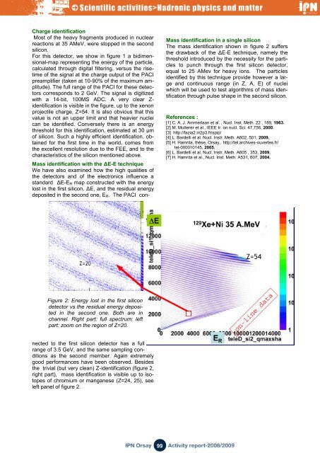
Mass identification with the ΔE-E technique<br />
We have also examined how the high qualities of<br />
the detectors <strong>and</strong> of the electronics influence a<br />
st<strong>and</strong>ard ΔE-E R map constructed with the energy<br />
lost in the first silicon, ΔE, <strong>and</strong> the residual energy<br />
deposited in the second one, E R . The PACI con-<br />
Mass identification in a single silicon<br />
The mass identification shown in figure 2 suffers<br />
the drawback of the ΔE-E technique, namely the<br />
threshold introduced by the necessity for the particles<br />
to punch through the first silicon detector,<br />
equal to 25 AMev for heavy ions. The particles<br />
identified by this technique provide however a large<br />
<strong>and</strong> continuous range (in Z, A, E) of <strong>nuclei</strong><br />
which will be used to test algorithms of mass identification<br />
through pulse shape in the second silicon.<br />
References :<br />
[1] C. A. J. Ammerlaan et al. , Nucl. Inst. Meth. 22 , 189, 1963.<br />
[2] M. Mutterer et al., IEEE tr. on nucl. Sci. 47,756, 2000.<br />
[3] http://fazia2.in2p3.fr/spip/<br />
[4] L. Bardelli et al. Nucl. Instr. Meth. A602, 501, 2009.<br />
[5] H. Hamrita, thèse, Orsay, http://tel.archives-ouvertes.fr/<br />
tel-000010145, 2005.<br />
[6] L. Bardelli et al. Nucl. Instr. Meth. A605 , 353, 2009.<br />
[7] H. Hamrita et al., Nucl. Inst. Meth. A531, 607, 2004.<br />
Figure 2: Energy lost in the first silicon<br />
detector vs the residual energy deposited<br />
in the second one. Both are in<br />
channel. Right part: full spectrum; left<br />
part: zoom on the region of Z=20.<br />
nected to the first silicon detector has a full<br />
range of 3.5 GeV, <strong>and</strong> the same sampling conditions<br />
as the second member. Again extremely<br />
good performances have been observed. Besides<br />
the trivial (but very clean) Z-identification (figure 2,<br />
right part), mass identification is visible up to isotopes<br />
of chromium or manganese (Z=24, 25), see<br />
left panel of figure 2.<br />
99