

PDF Version - Glidewell Dental Labs
PDF Version - Glidewell Dental Labs
PDF Version - Glidewell Dental Labs

You also want an ePaper? Increase the reach of your titles
YUMPU automatically turns print PDFs into web optimized ePapers that Google loves.
– ARTICLE and CLINICAL PHOTOS by<br />
Tarun Agarwal, DDS, PA<br />
Digital<br />
Imaging:<br />
An Important Visual Aid in<br />
Treatment Planning and Case Acceptance<br />
Photographic imaging has been available in dentistry<br />
for many years. Typically, it has been used for full-smile<br />
makeover simulations. Full-smile simulations can be very<br />
difficult and time consuming, however, and can often create<br />
unrealistic expectations or outcomes. This challenge, added<br />
to the expense of investing in traditional dental imaging<br />
software, leads many clinicians to completely avoid the use<br />
of digital imaging in their practice.<br />
For anterior cases, digital imaging can and should play a<br />
vital role in patient education and decision-making. In fact,<br />
in situations where a few teeth are being treated, its use<br />
may be even more important than for full-smile restorations.<br />
This case study will demonstrate how digital imaging can be<br />
used to communicate different treatment possibilities and<br />
assist in patient treatment acceptance. It will also detail the<br />
clinical technique used for achieving the patient’s desired<br />
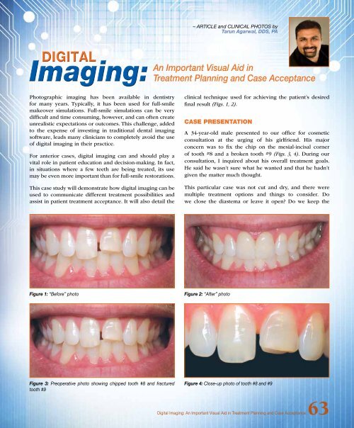
final result (Figs. 1, 2).<br />
Case Presentation<br />
A 34-year-old male presented to our office for cosmetic<br />
consultation at the urging of his girlfriend. His major<br />
concern was to fix the chip on the mesial-incisal corner<br />
of tooth #8 and a broken tooth #9 (Figs. 3, 4). During our<br />
consultation, I inquired about his overall treatment goals.<br />
He said he wasn’t sure what he wanted and that he hadn’t<br />
given the matter much thought.<br />
This particular case was not cut and dry, and there were<br />
multiple treatment options and things to consider. Do<br />
we close the diastema or leave it open? Do we keep the<br />
Figure 1: “Before” photo<br />
Figure 2: “After” photo<br />
Figure 3: Preoperative photo showing chipped tooth #8 and fractured<br />
tooth #9<br />
Figure 4: Close-up photo of tooth #8 and #9<br />
Digital Imaging: An Important Visual Aid in Treatment Planning and Case Acceptance63


