

PDF Download - Glidewell Dental Labs
PDF Download - Glidewell Dental Labs
PDF Download - Glidewell Dental Labs

You also want an ePaper? Increase the reach of your titles
YUMPU automatically turns print PDFs into web optimized ePapers that Google loves.
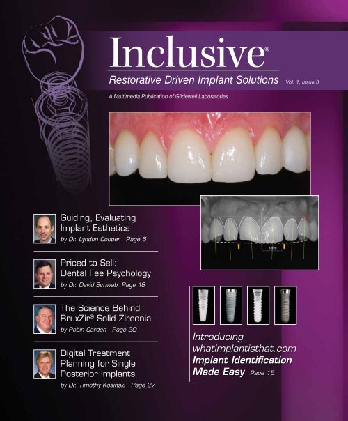
Inclusive®<br />
Restorative Driven Implant Solutions<br />
Vol. 1, Issue 3<br />
A Multimedia Publication of <strong>Glidewell</strong> Laboratories<br />
Guiding, Evaluating<br />
Implant Esthetics<br />
by Dr. Lyndon Cooper Page 6<br />
Priced to Sell:<br />
<strong>Dental</strong> Fee Psychology<br />
by Dr. David Schwab Page 18<br />
The Science Behind<br />
BruxZir ® Solid Zirconia<br />
by Robin Carden Page 20<br />
Digital Treatment<br />
Planning for Single<br />
Posterior Implants<br />
by Dr. Timothy Kosinski Page 27<br />
Introducing<br />
whatimplantisthat.com<br />
Implant Identification<br />
Made Easy Page 15
On the Web<br />
Read more at inclusivemagazine.com<br />
■ Original Video Presentations<br />
Learn to use BioTemps ® and the IOS FastScan Digital<br />
Impression System to sculpt the soft tissues and<br />
transfer those contours to the laboratory model.<br />
■ Online Lectures<br />
As part of the gIDE Lecture-on-Demand series, Dr.<br />
Joseph Kan presents the first part of a discussion on<br />
“Immediate Implant Placement in the Esthetic Zone.”<br />
■ Photo Essays<br />
View clinical photos from Dr. Timothy Kosinski’s presentation<br />
on the use of Digital Treatment Planning<br />
when restoring maxillary posterior teeth.<br />
■ Free CE credit<br />
Take short tests that will earn you 2 CE credits.<br />
■ PLUS ...<br />
Discover the benefits of whatimplantisthat.com. You’ll<br />
also find an eye-catching animation illustrating the<br />
properties of dental zirconia.<br />
Check out inclusivemagazine.com to see what<br />
the implant industry is buzzing about.<br />
When you see this icon, it means we have even more<br />
information on that topic available at inclusivemagazine<br />
.com. Take advantage of Inclusive magazine’s interactivity by<br />
logging on for in-depth coverage.<br />
– inclusivemagazine.com –
Contents<br />
Features<br />
15 Clinical Tip: Introducing<br />
whatimplantisthat.com<br />
16 Role of the Scan Appliance/<br />
Radiographic Index<br />
35 Evaluating Limited<br />
Vertical Space When<br />
Replacing Posterior Teeth<br />
6<br />
18<br />
20<br />
27<br />
Master of Esthetic Dentistry –– Objective Criteria:<br />
Guiding and Evaluating <strong>Dental</strong> Implant Esthetics<br />
Dr. Lyndon Cooper presents insights regarding an objective approach<br />
to planning, executing and evaluating the esthetic merit of<br />
single-tooth implant restorations. Meeting the goal of providing<br />
a single-tooth implant crown that equals or exceeds the esthetic<br />
value of the tooth it replaces requires identifying and addressing<br />
easily recognized anatomic constraints, Dr. Cooper writes.<br />
Priced to Sell: <strong>Dental</strong> Fee Psychology<br />
David Schwab, Ph.D, delves into case acceptance, explaining that<br />
the number of options offered to the patient and how those options<br />
are presented is critical. Schwab advocates a “good, better,<br />
best” method in which patients are given three choices for treatment<br />
at three different price points.<br />
R&D Corner –– BruxZir ® : Virtually Bulletproof<br />
BruxZir Solid Zirconia, the lab’s newest restorative material, is gaining<br />
popularity for a multitude of reasons. Robin Carden, senior director<br />
of Research & Development for <strong>Glidewell</strong> Laboratories, presents<br />
a material science overview of this unique zirconium dioxide<br />
material and discusses the four physical properties — high flexural<br />
strength, high fracture toughness, resistance to thermal shock and<br />
color/translucency — that make zirconia ideal for dental restorations.<br />
Digital Implant Treatment Planning: Restoration of<br />
Maxillary Posterior Single Teeth<br />
In this article and photo essay, Dr. Timothy Kosinski advocates the<br />
use of single dental implants to replace one or more teeth. Single<br />
dental implants prove functional and the final restoration esthetic,<br />
he says. Any previous reluctance to place implants in certain anatomic<br />
areas has been eliminated thanks to CAD/CAM technology,<br />
Kosinski illustrates.<br />
– Contents – 1
Letter from the Editor<br />
Welcome to the third issue of Inclusive. By now, I hope you have had a chance to view the expanded<br />
articles and videos, as well as take advantage of the CE opportunities at inclusivemagazine.com and<br />
glidewelldental.com.<br />
In our continued effort to provide you with milestone clinical articles, we are proud to include<br />
Dr. Lyndon Cooper’s article on guiding and evaluating implant esthetics. After reading this classic article,<br />
you will understand the importance of the “3:2 rule” and the gingival zenith when planning implants in<br />
the esthetic zone.<br />
Another vital aspect of treatment planning implant cases is the amount of prosthetic vertical space<br />
available. The patient may have adequate bone to accommodate an implant, but because of collapse of<br />
the bite or supra-eruption of the opposing dentition there may be limited restorative space. We present<br />
an overview that provides the average crown heights, lists diagnostic tools to determine the amount of<br />
space and illustrates the prosthetic height limitations from a laboratory standpoint.<br />
We’ll also introduce you to whatimplantisthat.com. Today, it is all too common for a patient to show<br />
up at your office with implants that already have been placed, and identifying these implants can be a<br />
challenge. Drs. Kent Howell and Nate Farley of The Ohio State University created this site to aid in the<br />
identification of implants based on common characteristics.<br />
On the material science side, we’ll also explain the unique properties of zirconia, which has become a<br />
very popular restorative material over the last several years. <strong>Glidewell</strong> Laboratories recently introduced<br />
BruxZir ® Solid Zirconia, a unique full-contoured all-zirconia restoration. Senior Director of R&D Robin<br />
Carden provides an overview of how this material can benefit you and your patients.<br />
You’ll also find a photo essay in which Dr. Tim Kosinski walks through the decision process and procedure<br />
to replace missing maxillary first molars, as well as a practice management article on pricing<br />
psychology when presenting treatment options from Dr. David Schwab.<br />
The Global Institute for <strong>Dental</strong> Education (gIDEdental.com) provides a tremendous amount of online<br />
educational opportunities. To introduce you to this site, we have a short online presentation by<br />
Dr. Joseph Kan of Loma Linda University School of Dentistry on immediate versus delayed implant<br />
placement.<br />
Also online is expanded coverage on the topics in the magazine, including a video that shows the utilization<br />
of BioTemps ® and the IOS FastScan Digital Impression System.<br />
As always, we welcome your feedback. Share your thoughts at inclusivemagazine@glidewelldental.com.<br />
Regards,<br />
Dr. Bradley C. Bockhorst<br />
Editor-in-Chief, Clinical Editor<br />
inclusivemagazine@glidewelldental.com<br />
– Letter from the Editor – 3
Publisher<br />
Jim <strong>Glidewell</strong>, CDT<br />
Editor-in-Chief<br />
Bradley C. Bockhorst, DMD<br />
Managing Editors<br />
Jim Shuck; Mike Cash, CDT<br />
Creative Director<br />
Rachel Pacillas<br />
Clinical Editor<br />
Bradley C. Bockhorst, DMD<br />
Contributing editors<br />
Dzevad Ceranic, Greg Minzenmayer<br />
senior Copy Editor<br />
Kim Watkins<br />
copy editors<br />
Melissa Manna, Jennifer Holstein<br />
Graphic Designers/Web Designers<br />
Jamie Austin, Deb Evans, Joel Guerra, Lindsey Lauria,<br />
Phil Nguyen, Gary O’Connell<br />
Photographers/Clinical Videographers<br />
Jennifer Brunst, RDAEF; Sharon Dowd;<br />
James Kwasniewski; Sterling Wright<br />
Illustrators<br />
Wolfgang Friebauer, MDT; Kevin Greene; Phil Nguyen<br />
magazine/Ad coordinator<br />
Vivian Tsang<br />
If you have questions, comments or suggestions, e-mail us at<br />
inclusivemagazine@glidewelldental.com. Your comments may be<br />
featured in an upcoming issue or on our website.<br />
© 2010 <strong>Glidewell</strong> Laboratories<br />
Neither Inclusive magazine nor any employees involved in its publication<br />
(“publisher”) makes any warranty, express or implied, or assumes<br />
any liability or responsibility for the accuracy, completeness, or usefulness<br />
of any information, apparatus, product, or process disclosed, or<br />
represents that its use would not infringe proprietary rights. Reference<br />
herein to any specific commercial products, process, or services by<br />
trade name, trademark, manufacturer or otherwise does not necessarily<br />
constitute or imply its endorsement, recommendation, or favoring<br />
by the publisher. The views and opinions of authors expressed<br />
herein do not necessarily state or reflect those of the publisher and<br />
shall not be used for advertising or product endorsement purposes.<br />
CAUTION: When viewing the techniques, procedures, theories and materials<br />
that are presented, you must make your own decisions about<br />
specific treatment for patients and exercise personal professional judgment<br />
regarding the need for further clinical testing or education and<br />
your own clinical expertise before trying to implement new procedures.<br />
Contributors<br />
■ Bradley C. Bockhorst, DMD<br />
After receiving his dental degree from<br />
Washington University School of <strong>Dental</strong><br />
Medicine, Dr. Bradley Bockhorst served<br />
as a Navy <strong>Dental</strong> Officer. Today, Dr. Bockhorst<br />
is Director of Clinical Technologies<br />
at <strong>Glidewell</strong> Laboratories, where he oversees<br />
Inclusive ® Digital Implant Treatment<br />
Planning Services and is editor-in-chief and clinical<br />
editor of Inclusive magazine. A member of the CDA,<br />
ADA, Academy of Osseointegration, International Congress<br />
of Oral Implantologists and American Academy of<br />
Implant Dentistry, Dr. Bockhorst lectures internationally<br />
on an array of dental implant topics. He maintains<br />
a private practice focused on implant prosthetics in Mission<br />
Viejo, Calif. Contact Dr. Bockhorst at 800-521-0576 or<br />
inclusivemagazine@glidewelldental.com.<br />
■ ROBIN A. CARDEN<br />
Robin Carden earned a bachelor’s degree<br />
from California State University, Long<br />
Beach in 1980. He is founder of Talon<br />
Composites, the manufacturer of Talbor ®<br />
— a composite material that uses advanced<br />
ceramics and metals. He holds more than<br />
25 patents, mostly related to metal and ceramic<br />
composites. In 1998, Robin won the Design Engineering<br />
Award from Design News. He is also inventor of<br />
the translucent orthodontic braces for 3M ESPE and Ceradyne<br />
Inc., the latter at which he worked for eight years as<br />
a senior engineer. Ceradyne Inc. awarded Robin the prestigious<br />
President’s Award for his work with Advanced Ceramics.<br />
Presently, Robin is Senior Director of <strong>Glidewell</strong> Laboratories’<br />
Research & Development Department. Contact him at<br />
inclusivemagazine.com.<br />
Inclusive is a registered trademark of <strong>Glidewell</strong> Laboratories.<br />
4<br />
– inclusivemagazine.com –
■ Lyndon F. Cooper, DDS, Ph.D<br />
Dr. Lyndon Cooper is the Stallings Distinguished<br />
Professor of Dentistry of the<br />
Department of Prosthodontics at the University<br />
of North Carolina at Chapel Hill.<br />
He is chairman and acting director of<br />
Graduate Prosthodontics and director of<br />
the Bone Biology and Implant Therapy<br />
Laboratory. Dr. Cooper is a Diplomate of the American<br />
Board of Prosthodontics and is the current president of<br />
the ACP Board of Directors. He received the ACP’s 2004<br />
Clinician/Researcher Award and the IADR’s 2009 Distinguished<br />
Scientist Award. His lab’s research findings have<br />
been presented in more than 70 publications. Contact him<br />
at lyndon_cooper@dentistry.unc.edu.<br />
■ Joseph Kan, DDS, MS<br />
Dr. Joseph Kan completed specialty training<br />
in prosthodontics and earned a master’s<br />
degree in implant surgery from Loma<br />
Linda University School of Dentistry in<br />
1997. He is a professor in the Department<br />
of Restorative Dentistry and research coordinator<br />
for the Implant Dentistry Program<br />
at LLUSD. In 1997, Dr. Kan was the recipient of the Best<br />
Research Award at the 12th annual meeting of the<br />
Academy of Osseointegration. He also received the Judson<br />
C. Hinckey Scientific Award in 2003 and the Robert James<br />
Achievement Award in 2005. He has been widely published<br />
in reference journals and has contributed chapters to textbooks.<br />
Contact him at info@gidedental.com.<br />
■ Nathanial Farley, DDS<br />
Dr. Nathanial “Nate” Farley received his<br />
dental degree from and is currently a<br />
prosthodontics resident at The Ohio State<br />
University. With 10 years of experience in<br />
Web design and development, he is passionate<br />
about putting dentistry topics on<br />
the Web utilizing current technologies and<br />
innovative thinking to make dental information<br />
easier to find and digest. This love led Dr. Farley to<br />
co-create the website whatimplantisthat.com. Dr. Farley is<br />
a member of the ADA, American College of Prosthodontics,<br />
American Academy of Fixed Prosthodontics, AAED and the<br />
Carl O. Boucher Prosthodontic Conference. Contact him at<br />
nfarley@gmail.com.<br />
■ TIMOTHY F. KOSINSKI, DDS, MAGD<br />
Dr. Timothy Kosinski graduated from<br />
the University of Detroit Mercy School<br />
of Dentistry and received an MS in biochemistry<br />
from Wayne State University<br />
School of Medicine. He is an adjunct assistant<br />
professor at the Mercy School of<br />
Dentistry and serves on the editorial<br />
review board of numerous dental journals.<br />
Dr. Kosinski is a Diplomate of ABOI/ID, ICOI and<br />
AO. He is a Fellow of the American Academy of Implant<br />
Dentistry and received his Mastership in the AGD, from<br />
which he received the 2009 Lifelong Learning and Service<br />
Recognition award. Contact Dr. Kosinski at 248-646-8651,<br />
drkosin@aol.com or smilecreator.net.<br />
■ Kent J. Howell, DMD<br />
Dr. Kent Howell graduated from Case<br />
School of <strong>Dental</strong> Medicine in Cleveland,<br />
Ohio, in 2008. He is currently a<br />
prosthodontics resident at The Ohio State<br />
University, where he will earn his MS<br />
and certificate in 2011. He is a member<br />
of the American College of Prosthodontists,<br />
American Academy of Fixed<br />
Prosthodontics and the Carl O. Boucher Prosthodontic<br />
Conference. He is co-creator of whatimplantisthat.com.<br />
The site was presented at this year’s AAFP meeting in<br />
Chicago, where it won the Table Clinic Award of Excellence<br />
for a Research Presentation. Contact Dr. Howell at<br />
howell.294@osu.edu or visit whatimplantisthat.com.<br />
■ David Schwab, Ph.D<br />
Dr. David Schwab presents practical, userfriendly<br />
seminars and in-office consulting<br />
sessions for the entire dental team. Fastpaced,<br />
filled with humor and overflowing<br />
with “pearls,” Dr. Schwab’s seminars are as<br />
popular as they are useful. An internationally<br />
known seminar speaker and practice<br />
management consultant who works exclusively<br />
with dental professionals, Dr. Schwab has served as<br />
Director of Marketing for the ADA and as Executive Director<br />
of the ACP. He currently works closely with Straumann<br />
to educate doctors and team members and to help them<br />
reach their full potential. Contact him at 888-324-1933 or<br />
davidschwab.com.<br />
– Contributors – 5
Master of Esthetic Dentistry<br />
Objective Criteria: Guiding and<br />
Evaluating <strong>Dental</strong> Implant Esthetics<br />
Go to inclusivemagazine.com to earn CE credit on this article.<br />
by Lyndon F. Cooper, DDS, Ph.D<br />
The evolution of dental implant therapies is fully apparent. From the introductory<br />
concepts of tissue-integrated prostheses with remarkable functional advantages,<br />
innovations have resulted in dental implant solutions spanning the spectrum of dental needs.<br />
Current discussions concerning the relative merit of an implant versus a 3-unit fixed partial<br />
denture fully illustrate the possibility that single implants represent a bona fide choice for tooth<br />
replacement. 1 Interestingly, when delving into the detailed comparisons between the outcomes<br />
of single-tooth implant versus fixed partial dentures or the intentional replacement of a failing<br />
tooth with an implant instead of restoration involving root canal therapy, little emphasis has been<br />
placed on the relative esthetic merits of one or another therapeutic approach to tooth replacement<br />
therapy. 2 An ideal prosthesis should fully recapitulate or enhance the esthetic features of<br />
the tooth or teeth it replaces. Although it is clearly beyond the scope of this article to compare<br />
the various methods of esthetic tooth replacement, there is, perhaps, sufficient space to share<br />
some insights regarding an objective approach to planning, executing and evaluating the esthetic<br />
merit of single-tooth implant restorations.<br />
Therapeutic success for dental implants has largely been described in terms of implant survival.<br />
Anterior single-tooth implant survival is high. 3 Further documentation provides implant success<br />
criteria, defined by the reporting of marginal bone level data. 4 Occasionally, prosthetic or<br />
restorative outcomes have been reported. Here, marginally less favorable data are reported for<br />
abutment complications of loosening or screw fracture. 3 Less often, biologic data concerning the<br />
peri-implant mucosal responses are provided. A biologic width develops around implant crowns,<br />
and the associated peri-implant connective tissue inflammatory cell infiltrate reacts to plaque<br />
accumulation. 5<br />
The incidence of peri-implantitis and its effect on implant esthetics may not be fully appreciated.<br />
Recently, two esthetic scoring systems have been described. 6,7 These or possibly other esthetic<br />
evaluations have not been widely deployed. Although Chang and colleagues 8 examined patientbased<br />
outcomes for anterior single-tooth implants, there remain many unanswered questions<br />
regarding the esthetic requirements and related patient satisfaction concerning anterior singletooth<br />
implants. In 2008, esthetic concerns dominated the discourse surrounding dental implants.<br />
An objective approach to planning, executing and evaluating therapy is warranted.<br />
6<br />
– inclusivemagazine.com –
Meeting the goal of providing a single-tooth implant crown that equals or exceeds the esthetic<br />
value of the tooth it replaces requires identifying and addressing easily recognized anatomic<br />
constraints. The hypothesis underscoring an objective approach to single-tooth dental implant<br />
esthetics is that the majority of unresolved esthetic problems are because of the discrepancies<br />
of implant crown dimension and orientation. Most often, these reflect improper clinical management<br />
of peri-implant and peri-coronal soft tissue architecture. 9 The application of time-proven<br />
and well-documented objective criteria for dental esthetics to the anterior single-tooth implant<br />
scenario can guide planning and ensure execution of implant placement, abutment design and<br />
crown formation to achieve the highest and most reproducible esthetic goals of the clinician and<br />
patient. The aim of this report is to describe how objective criteria can guide planning and execution<br />
of implant therapy and, more importantly, how a single aspect of dental implant planning<br />
and placement can negatively impact half of these objective criteria and lead to unacceptable<br />
implant-supported restorations.<br />
Objective Criteria for <strong>Dental</strong> Esthetics and the Implant Scenario<br />
In a classic (now out of print) textbook titled “Esthetic Guidelines for Restorative Dentistry,” 10 Dr.<br />
Urs Belser describes the objective criteria for dental esthetics. More recently, an updated list and<br />
illustration of these criteria were published<br />
as a chapter in the textbook “Bonded Porcelain<br />
Restorations in the Anterior Dentition.” 11<br />
These criteria (Table 1), together with the additional<br />
TABLE 1 — Objective Criteria for <strong>Dental</strong> Esthetics<br />
significance of identifying the midline<br />
and plane of occlusion as a prerequisite for<br />
ideal anterior dental esthetics, can provide an<br />
indelible guidance system for dental esthetics.<br />
In the process of evaluating single-tooth dental<br />
implant restorations in prospective and retrospective<br />
studies, 11–14 it became apparent that<br />
these criteria were equally valid to the dental<br />
implant restoration. The form of the dental<br />
implant-supported tooth requires careful consideration<br />
of these objective criteria (Fig. 1).<br />
■ Gingival health<br />
■ Balance of gingival levels<br />
■ Gingival zenith<br />
■ Interdental closure<br />
■ Interdental contact location<br />
■ Tooth axis<br />
■ Tooth characterization<br />
■ Surface texture<br />
■ Color<br />
■ Incisal edge configuration<br />
■ Lower lip line<br />
■ Smile symmetry<br />
<strong>Dental</strong> implant placement is neither fully intuited<br />
from the anatomy of the residual alveolar<br />
ridge nor can it be divined from the existing<br />
volume of bone. Desired tooth position dictates<br />
implant placement and informs the clinician<br />
■ Basic features of tooth<br />
form<br />
■ Relative tooth dimensions<br />
■ Midline and occlusal<br />
plane orientation<br />
Figure 1: Tooth form is objectively defined. The objective criteria<br />
for dental esthetics (Table 1) help guide decisions concerning<br />
ideal tooth form. The clinical photo of this implant crown replacing<br />
central incisor #8 reveals the significance of the many soft<br />
tissue items present as criteria defining dental esthetics. Note<br />
that much of the crown form is defined by the peri-implant mucosa.<br />
The lack of symmetry between the central incisors is due<br />
to the incorrect depth of implant placement and the 1 mm apical<br />
location of the gingival zenith. The incorrect soft tissue contour is<br />
represented by a more oval or triangular tooth form and a longer<br />
clinical crown when compared with the left central incisor. The<br />
more mesial location of the zenith has been compensated by the<br />
enhancement of the line angles and tooth character to correct<br />
the appearance of the tooth’s long axis. The loss of attachment<br />
at tooth #7 results in the absence of gingival closure and cannot<br />
be accommodated by modifications of the implant procedure or<br />
crown #8. These objective limitations reduce the overall esthetic<br />
value of this tooth display.<br />
– Master of Esthetic Dentistry — Objective Criteria: Guiding and Evaluating <strong>Dental</strong> Implant Esthetics – 7
egarding potential requirements for tissue augmentation. In considering the role of the objective<br />
criteria in planning for dental implant placement and recognizing that the depth of implant<br />
placement can dramatically affect one-half of these criteria, a potential objective strategy to esthetic<br />
planning for dental implant placement emerges. That strategy requires evaluation of the<br />
edentulous alveolar ridge and adjacent teeth in the context of the objective criteria for dental<br />
esthetics. Simply stated, dental implant placement can be guided by the location of the gingival<br />
zenith.<br />
The Gingival Zenith as a Guide for <strong>Dental</strong> Implant Placement<br />
The gingival zenith represents the most apical part of the clinical crown. It also represents both<br />
the faciolingual and the mesiodistal location of the crown in relationship to the edentulous ridge.<br />
As such, it has a remarkable influence on the morphology of the planned restoration. The gingival<br />
zenith affects other objective criteria, including the balance of gingival levels (too inferior<br />
or superior), the tooth axis (too distal or mesial), the tooth dimension (too inferior or superior),<br />
and the tooth form (triangular becomes ovoid if too inferior). Without the control of the gingival<br />
zenith, the clinician’s ability to define dental implant esthetics is vastly diminished (Fig. 2).<br />
Figure 2: In the left and right views, the retained “c” and “f” teeth reflect the absence of permanent cuspid teeth. The retained<br />
deciduous teeth have aided in the preservation of alveolar bone, but the location of the gingival contours are not correct and are<br />
unattractive. Using the present bone and gingival locations to guide implant placement would result in short clinical crowns. Redefining<br />
the gingival zenith of the permanent cuspid teeth is required.<br />
<strong>Dental</strong> Implant Control at the Zenith<br />
At least four factors affect the gingival zenith. First is the relative location of the tissues to the<br />
planned gingival zenith. Second is the depth of the dental implant placement. Third is the response<br />
of the buccal bone and mucosa to the implant procedure and components. Fourth is the<br />
prosthodontic management of the gingival zenith architecture.<br />
The Relative Locations of Tissues and the Planned Gingival Zenith<br />
Ideally, the planned gingival zenith is symmetric with the contralateral tooth and harmonious<br />
with the gingival levels of adjacent teeth. Unfortunately, most residual alveolar ridges are significantly<br />
resorbed. 15 Important objective classification 16 is useful and a Diagnostic Wax-Up permits<br />
the exact determination of the extent of resorption and permits planning to the key esthetic<br />
parameters. Interproximal tissue contours (papillae) appear to be supported by adjacent teeth<br />
connective tissue contacts, but peri-implant facial tissue contours are dependent on facial bone<br />
and co-dependent soft tissue morphology.<br />
8<br />
– inclusivemagazine.com –
Controlling the Depth<br />
of Implant Placement<br />
Decisions concerning the depth of implant<br />
placement should be based on the biologic<br />
understanding of the tissue responses to the<br />
implanted device. Assuming a steady state<br />
peri-implant bone level, it is well known that<br />
a biologic width forms at the dental implant 17<br />
and that the buccal dimension of the biologic<br />
width formed at an abutment is approximately<br />
3 mm. 18 The ideal depth of the implant placement<br />
is suggested to be 3 mm apical to the<br />
planned gingival zenith. The implant/abutment<br />
interface should also reside 2 mm palatal<br />
to the zenith to ensure adequate thickness<br />
of bone and mucosa to support tissue form. 19<br />
This “3:2 rule” further suggests to the clinician<br />
when bone grafting or soft tissue augmentation<br />
should be performed. If bone is not<br />
present at approximately this position from<br />
the gingival zenith, bone grafting procedures<br />
should be considered in preparation for ideal<br />
esthetics (Fig. 3).<br />
Figure 3: The location of the gingival zenith should be symmetrical<br />
with the contralateral tooth and in harmony with the adjacent<br />
teeth. As shown in this illustration, the gingival zenith should<br />
be located approximately 3 mm from the implant/abutment interface.<br />
This permits a subgingival crown margin at the facial<br />
aspect of the implant and provides at least 2.5 mm for the development<br />
of the biologic width in a supercrestal position. Placement<br />
of the implant/abutment interface in a deeper location will<br />
result in loss of bone and facial peri-implant mucosal recession.<br />
Without apology for the following circular logic, controlling the depth of placement is achieved<br />
by defining the gingival zenith. Managing the gingival zenith at the time of implant placement<br />
sets the stage for ideal anterior single-tooth esthetics. Whether William’s theory of tooth form has<br />
merit, 20 the characterization of teeth as square, ovoid or triangular is based on the peri-coronal<br />
architecture. An often unrecognized truth about dental implant esthetics is that tooth form is<br />
largely defined by the peri-implant mucosal architecture.<br />
Controlling Peri-Implant<br />
Mucosal Architecture<br />
A reproducible procedure should be imposed<br />
onto the artistic philosophy of each clinical<br />
exercise. For the single-tooth dental implant,<br />
this process begins with an esthetic diagnosis.<br />
The diagnosis is nothing more than the assessment<br />
of the objective criteria as displayed<br />
by the preoperative condition of the patient.<br />
Suggested is the use of clinical digital photographs<br />
upon which simple evaluations can be<br />
superimposed (Fig. 4).<br />
Figure 4: A simple photograph (representing the situation illustrated<br />
in Fig. 2) can be used to objectively evaluate the clinical<br />
situation to make a complete esthetic diagnosis. Note that the<br />
mirror image of the right and left gingival contours do not match.<br />
Note also that the orthodontist has provided good spacing for<br />
the central and lateral incisors; it is clear that relative to tooth<br />
#10, tooth #7 is distal in its location. The gingival zenith on tooth<br />
#11 has been marked to indicate how its position guides overall<br />
esthetic value of the implant restoration, presently represented<br />
by provisional crowns without occlusion.<br />
Perhaps the most prognostic indicator of<br />
eventual esthetic success through symmetry is<br />
gained by evaluation of the connective tissue<br />
attachment at the adjacent teeth. Careful assessment<br />
using a periodontal probe and diagnostic<br />
periapical radiograph are needed. Loss<br />
of attachment of greater than 1 mm is clinically<br />
discernible and difficult to regenerate.<br />
This step is essential because interproximal<br />
peri-implant mucosal contours (papillae) are<br />
– Master of Esthetic Dentistry — Objective Criteria: Guiding and Evaluating <strong>Dental</strong> Implant Esthetics – 9
Figure 5: Study casts of the interim situation and the diagnostically waxed cast. The location of the gingival zenith is directed by the<br />
process of diagnostic waxing. This is confirmed by the evaluation of the intraoperative study cast.<br />
greatly dependent on adjacent tooth contours. Together with study casts indicating the extent<br />
of alveolar ridge resorption, a thorough prognosis and treatment plan can be provided to the<br />
patient.<br />
For the situation of the single anterior missing tooth, it is not possible to fully appreciate these<br />
criteria unless a fully contoured crown is waxed in the edentulous space (Fig. 5). Following the<br />
diagnostic waxing, it is possible to understand the relationship between the proposed gingival<br />
zenith location and the existing mucosa. The relationship of the gingival zenith to the underlying<br />
bone can only be determined by bone sounding with a diagnostic template in place or, preferably,<br />
by use of volumetric imaging (e.g., Cone Beam Computed Tomography) with a radiopaque<br />
image of the gingival zenith in place (Fig. 6). This assessment is critical. Without underlying bone<br />
to support the buccal contour in full dimension, the esthetic volume of the edentulous space<br />
ultimately will be deficient (Fig. 7). Based on the location of the planned gingival zenith, therefore,<br />
decisions regarding the need for bone augmentation, socket preservation and/or soft tissue<br />
augmentation procedures can be prudently accessed.<br />
Figure 6: On the left, detailed evaluation of the diagnostically waxed cast shows that the concepts revealed by the objective esthetic<br />
evaluation have been translated to the cast. This includes the harmonious arrangement of the gingival zenith and the proper location<br />
of the cuspid zenith in the buccolingual as well as the apicoincisal direction. Bone should be present 3 mm apical to the gingival zenith.<br />
At right, an unrelated Cone Beam Computed Tomography image of a canine site exemplifies the examination of the required gingival<br />
zenith/bone relationship. In this example, insufficient bone for an esthetic restoration exists. The planned restoration’s zenith is 8 mm<br />
from the alveolar crest. The resulting crown would be approximately 14 to 15 mm in length (versus the average of 10 to 11 mm).<br />
Bone augmentation would be indicated.<br />
10<br />
– inclusivemagazine.com –
A<br />
B<br />
C<br />
Figure 7: (A) Intervening veneer preparations for teeth #7–10<br />
were performed in the enamel only. The provisional crowns are<br />
removed and impression copings are placed for fixture-level<br />
impressions of the Astra Tech dental implants. (B) ZirDesign <br />
abutment delivery in the well-formed residual alveolar mucosa.<br />
The provisional crown should aid in the creation of the gingival<br />
contours. The form of the provisional crown should reflect both<br />
the clinical crown marginal contours as well as provide ideal<br />
submucosal transition contour. In most cases, the interproximal<br />
surfaces of the abutment and crown should be concave or flat,<br />
whereas the buccal contours are slightly convex in support of the<br />
buccal architecture. The interproximal contours must accommodate<br />
sufficient interproximal tissue mass to support contours.<br />
(C) Provisional crowns reflect the contours of the diagnostic waxing<br />
and have been used to direct soft tissue changes at the<br />
implants as well as the mesial aspects of teeth #7 and #9.<br />
Prosthodontic Management of<br />
Peri-Implant Mucosal Architecture<br />
With an implant positioned properly in the alveolus,<br />
the control of peri-implant tissues is<br />
enhanced morphologically by enforcing the<br />
remodeling of tissues using properly contoured<br />
abutments and provisional crowns (Table<br />
2). To ensure proper healing and to limit<br />
inflammation, properly polished abutments<br />
of titanium or zirconia should be sculpted to<br />
support the soft tissue form, and thus, the cervical<br />
contour of the crown. Typically, the abutment<br />
will possess concave features, with the<br />
possible exception being a convexity of the<br />
buccal surface. This is particularly important<br />
in developing the contours of any provisional<br />
restoration for a dental implant. Morphologic<br />
refinement is established using the provisional<br />
crown and, again, the submucosal contours<br />
should be refined to be more root-like (concave<br />
interproximally) to support ideal tissue<br />
form. No particulate materials should be introduced<br />
into the sulcus, and all debris should be<br />
■ Initial presentation (Seibert classification)<br />
■ Implant position capability (relative to planned gingival zenith)<br />
■ Bone formation and resorption at the implant<br />
■ Peri-implant mucosa integration<br />
■ Character of the implant abutment interface<br />
■ Inflammation<br />
■ Local factors (plaque, etc.)<br />
■ Patient factors (biotype)<br />
■ Abutment form<br />
TABLE 2 –– Factors Controlling<br />
Buccal Peri-Implant Tissues<br />
■ Submucosal contour of the provisional crown<br />
■ Bone modeling/remodeling<br />
■ Potential adjacent tooth eruption<br />
– Master of Esthetic Dentistry — Objective Criteria: Guiding and Evaluating <strong>Dental</strong> Implant Esthetics – 11
A<br />
B<br />
Figure 8: (A) Facial view of final restorations on implants #6<br />
and #11, and teeth #7–10. All-ceramic crowns were bonded<br />
to ZirDesign implant abutments and veneers were bonded to<br />
#7–10. The photograph was made three months after delivery<br />
of the definitive prostheses. (B) One-year evaluation of the restoration/tissue<br />
relationships of #6–8 and (C) #9–11. The form<br />
and color of the peri-implant mucosa is in part due to the choice<br />
of the zirconia abutments and modification of the tissues during<br />
the immediate provisionalization period. The thick biotype contributes<br />
to the predictability of tissue responses illustrated here.<br />
The restorations were produced by Lee Culp, CDT.<br />
C<br />
carefully washed from the implant and sulcus prior to the delivery of the abutment and crown.<br />
The provisional crown should be highly polished, well adapted to the abutment margin and free<br />
of extruded cement (Fig. 7).<br />
Assessment at the Provisional Phase of Implant Restoration<br />
Excellent esthetics frequently involves iterative processes. For implant crowns, attempts to provide<br />
highly esthetic crowns to properly contoured peri-implant mucosa directly from a fixturelevel<br />
impression is not likely to achieve great expectations. It is important to provisionalize<br />
implants with provisional or definitive abutments and achieve the planned tissue architecture<br />
described earlier. After a period of tissue healing (6 to 8 weeks) or adaptation (3 to 4 weeks), objective<br />
assessment should be performed. Only after reviewing potential opportunities for refinement<br />
should the final impression of the implant or abutment be made. Several suggestions for<br />
capturing the form of the peri-implant mucosa include the placement of rigid materials into the<br />
sulcus. This is not recommended if the peri-implant tissues display little inflammation and tissue<br />
prolapse (Fig. 8). Regardless of the method chosen, the sulcus should be carefully examined and<br />
debrided after the impression is made. The provisional restoration should be replaced with little<br />
or no displacement or disruption of the peri-implant mucosa.<br />
Delivery and Assessment of the Final Prosthesis<br />
The goal of the laboratory procedures includes the preservation and possible directed enhancement<br />
of the peri-implant mucosal form created by the clinician, maintenance of the designated<br />
incisal edge position and incisal embrasures, and the creation of the designated abutment and<br />
crown. The prepared abutment and crown may be delivered to complete the restorative procedure.<br />
12<br />
– inclusivemagazine.com –
With a major goal being to preserve the peri-implant mucosal architecture with the gingival zenith<br />
as a reference point, it is important to evaluate possible tissue displacement when a final<br />
abutment is placed. Only modest, if any, blanching should be evident using this protocol following<br />
a careful provisionalization process. If tissues are displaced apically, it suggests that the abutment<br />
is improperly contoured and is most likely convex in form. The abutment can be modified<br />
and the tissue contours can be evaluated again. Abutment delivery is, therefore, a critical step in<br />
the control of the peri-implant mucosal form.<br />
Finally, the crown can be evaluated in the usual and customary manner. Included is a very useful<br />
checklist for this procedure that applies the objective criteria for dental esthetics. 6,7 It will focus<br />
attention beyond the issues of delivering an implant crown, and it reaffirms the maintenance of<br />
peri-implant mucosal architecture (Table 3).<br />
A Procedural Review<br />
Integration of the concepts discussed earlier<br />
indicates that for all anterior implants, there<br />
is a set of procedures that can ensure esthetic<br />
success (Table 3). The process begins with<br />
an esthetic diagnosis to reveal the limitations<br />
present and to suggest steps to overcome esthetic<br />
limitations before initiating implant<br />
therapy. The key features to observe include<br />
adjacent tooth connective tissue attachments.<br />
Further evaluation requires that a diagnostic<br />
waxing be performed to suggest the ideal restorative<br />
form. The designated gingival zenith<br />
can then be used to identify the critical crownto-bone<br />
relationship, today using volumetric<br />
radiographic imaging techniques. If the ideal<br />
gingival zenith is greater than 3 mm incisal<br />
and 2 mm buccal from the existing bone crest,<br />
bone augmentation procedures may be considered.<br />
The gingival zenith, therefore, becomes<br />
the therapeutic reference point. A positive esthetic<br />
result is suggested when the adjacent<br />
tooth attachment levels are intact and there is<br />
adequate bone relative to the reference point.<br />
Using a surgical guide, the implant can be<br />
accurately positioned to the zenith reference point. At the appropriate time (after immediate<br />
placement, one-stage surgery or two-stage surgery), an abutment can be placed to permit the formation<br />
of biologic width along the abutment and to begin to properly contour the peri-implant<br />
tissues. The provisional crown should be used to direct proper morphologic development of the<br />
peri-implant mucosa and control the crown’s ultimate form. Finally, the definitive restoration<br />
should impart color, translucency, contour and surface texture that embellish or match the adjacent<br />
and contralateral anterior teeth.<br />
Conclusion<br />
TABLE 3 –– Procedural Control of<br />
Peri-Implant Mucosal Architecture<br />
■ Esthetic diagnosis using objective criteria<br />
■ Determination of the adjacent connective tissue attachment<br />
■ Diagnostic Wax-Up with emphasis on peri-implant mucosal<br />
architecture (evaluation of the residual alveolar ridge)<br />
■ Assessment of bone-to-prosthesis relationship<br />
(CBCT/bone sounding)<br />
■ Possible bone and/or soft tissue augmentation<br />
to support objectively defined crown form<br />
■ Ideal placement of the implant relative to the<br />
planned gingival zenith<br />
■ Creating the ideal peri-implant mucosal architecture<br />
using well-formed provisional crowns and abutments<br />
■ Selection of abutment and crown materials<br />
to support peri-implant mucosal health<br />
■ Removal of cement from the sulcus<br />
An objective approach to dental implant therapy is warranted. Recent application of objective<br />
criteria suggests that further control of the anterior dental esthetics might be achieved. For example,<br />
the level of the peri-implant soft-tissue margin came to lie within 1 or 2 mm of the reference<br />
tooth in no more than 64 percent of the implant-supported replacements. The color of the<br />
peri-implant soft tissue matched that of the reference tooth in no more than just over one-third of<br />
cases. 6 More recently, Meijndert and colleagues 9 reported that only 66 percent of single-implant<br />
crowns in 99 patients were rated acceptable by a prosthodontist, despite high patient satisfaction.<br />
– Master of Esthetic Dentistry — Objective Criteria: Guiding and Evaluating <strong>Dental</strong> Implant Esthetics – 13
This may be the result of soft tissue changes. For example, the measured mean apical displacement<br />
of facial soft tissue was 0.6 mm one year after crown placement on abutments at flat-to-flat<br />
dental implants (Cardaropoli and colleagues). 21 In contrast, Cooper and colleagues 13 reported the<br />
stability of the facial soft tissue contour at conus design implant/abutment interfaces throughout<br />
a three-year period after dental implant placement and provisionalization.<br />
It may be possible to exert clinical control over the facial soft tissue contours that control singleimplant<br />
esthetics. Recognizing the initial limitations and guiding treatment planning by the use<br />
of the objective criteria for dental esthetics are essential to this process. Targeting the clinical<br />
and biologic factors affecting these criteria, particularly the buccal tissue contour, may improve<br />
single-dental implant esthetics. The influence of component selection is suggested but remains<br />
unproven. Nonetheless, the controlling depth of implant placement, managing peri-implant mucosal<br />
biology by limiting inflammation, and managing peri-implant mucosal morphology through<br />
ideal abutment selection and provisionalization extend the clinical control of single-tooth dental<br />
implant esthetics.<br />
ZirDesign is a trademark of Astra Tech Inc.<br />
References<br />
1. Salinas TJ, Block MS, Sadan A. Fixed partial denture or single-tooth implant restoration? Statistical considerations for sequencing<br />
and treatment. J Oral Maxillofac Surg 2004;62(Suppl 2):2–16.<br />
2. Tortopidis D, Hatzikyriakos A, Kokoti M, et al. Evaluation of the relationship between subjects’ perception and professional assessment<br />
of esthetic treatment needs. J Esthet Restor Dent 2007;19:154–62; discussion 163.<br />
3. Lindh T, Gunne J, Tillberg A, Molin M. A meta-analysis of implants in partial edentulism. Clin Oral Implants Res 1998;9:80–90.<br />
4. Roos J, Sennerby L, Lekholm U, et al. A qualitative and quantitative method for evaluating implant success: a 5-year retrospective<br />
analysis of the Brånemark implant. Int J Oral Maxillofac Implants 1997;12:504–14.<br />
5. Zitzmann NU, Berglundh T, Marinello CP, Lindhe J. Experimental peri-implant mucositis in man. J Clin Periodontol 2001;28:517–23.<br />
GUIDING AND EVALUATING DENTAL IMPLANT ESTHETICS 204 ©2008, COPYRIGHT THE AUTHOR JOURNAL COMPILATION<br />
©2008, WILEY PERIODICALS, INC.<br />
6.Fürhauser R, Florescu D, Benesch T, et al. Evaluation of soft tissue around single-tooth implant crowns: the pink esthetic score. Clin<br />
Oral Implants Res 2005;16:639–44.<br />
7. Meijer HJ, Stellingsma K, Meijndert L, Raghoebar GM. A new index for rating aesthetics of implant-supported single crowns and<br />
adjacent soft tissues — the Implant Crown Aesthetic Index. Clin Oral Implants Res 2005;16:645–9.<br />
8. Chang M, Wennström JL, Odman P, Andersson B. Implant supported single-tooth replacements compared to contralateral natural<br />
teeth. Crown and soft tissue dimensions. Clin Oral Implants Res 1999;10:185–94.<br />
9. Meijndert L, Meijer HJ, Stellingsma K, et al. Evaluation of aesthetics of implant supported single-tooth replacements using different<br />
bone augmentation procedures: a prospective randomized clinical study. Clin Oral Implants Res 2007;18:715–19.<br />
10. Kopp FR, Belser UC. Esthetic checklist for the fixed prosthesis. In: Sharer P, Rinn LA, Kopp FR, editors. Esthetic guidelines for<br />
restorative dentistry. Chicago (IL): Quintessence Publishing Co.; 1982. p. 187–92.<br />
11. Mange P, Belser U. Bonded porcelain restorations in the anterior dentition: a biomimetic approach. Chicago (IL): Quintessence<br />
Publishing Co.; 2002.<br />
12. Cooper L, Felton DA, Kugelberg CF, et al. A multicenter 12-month evaluation of single-tooth implants restored 3 weeks after 1-stage<br />
surgery. Int J Oral Maxillofac Implants 2001;16:182–92.<br />
13. Cooper LF, Ellner S, Moriarty J, et al. Three-year evaluation of single-tooth implants restored 3 weeks after 1-stage surgery. Int J<br />
Oral Maxillofac Implants 2007;22:791–800.<br />
14. De Kok IJ, Chang SS, Moriarty JD, Cooper LF. A retrospective analysis of peri-implant tissue responses at immediate load/provisionalized<br />
microthreaded implants. Int J Oral Maxillofac Implants 2006;21:405–12.<br />
15. Covani U, Cornelini R, Barone A. Buccolingual bone remodeling around implants placed into immediate extraction sockets: a case<br />
series. J Periodontol 2003;74:268–73.<br />
16. Seibert J, Salama H. Alveolar ridge preservation and reconstruction. Periodontol 2000 1996;11:69–84.<br />
17. Hermann JS, Buser D, Schenk RK, et al. Biologic width around titanium implants. A physiologically formed and stable dimension<br />
over time. Clin Oral Implants Res 2000;11:1–11.<br />
18. Kan JY, Rungcharassaeng K, Umezu K, Kois JC. Dimensions of peri-implant mucosa: an evaluation of maxillary anterior single<br />
implants in humans. J Periodontol 2003;74:557–62.<br />
19. Evans CD, Chen ST. Esthetic outcomes of immediate implant placements. Clin Oral Implants Res 2008;19:73–80.<br />
20. Bell RA. The geometric theory of selection of artificial teeth: is it valid? J Am Dent Assoc 1978;97:637–40.<br />
21. Cardaropoli G, Lekholm U, Wennström JL. Tissue alterations at implant-supported single-tooth replacements: a 1-year prospective<br />
clinical study. Clin Oral Implants Res 2006;17:165–71.<br />
Reprinted with permission of Blackwell Publishing Ltd. Cooper L. Master of Esthetic Dentistry Objective Criteria: Guiding and Evaluating<br />
<strong>Dental</strong> Implant Esthetics. Journal of Esthetic and Restorative Dentistry 2008;20(3):195–205.<br />
14<br />
– inclusivemagazine.com –
Clinical Tip<br />
whatimplantisthat.com<br />
The popularity of implant treatment has grown rapidly over<br />
the last 30 years. With today’s mobile society, it seems that<br />
every week a patient walks into the office with an implant<br />
that needs to be restored, repaired or redone. Many times,<br />
the patient’s implant case was done by another dentist. As<br />
experts, we want to do the best job possible for that patient,<br />
but all too often we lack the knowledge needed about the<br />
details of the implant. This presents many challenges in helping<br />
the patient attain his or her desired final result. With the<br />
hundreds of dental implants available, it can seem almost impossible<br />
to identify what you’re dealing with.<br />
That’s where whatimplantisthat.com comes in. This innovative<br />
website hosts a database of radiographs for quick identification<br />
of implants. The database is categorized by common<br />
characteristics, including whether the implant has a tapered<br />
or nontapered design and whether there are holes in the<br />
apex. This system allows you to quickly filter the radiographs,<br />
making it easier to find what you’re looking for and proceed<br />
with treatment.<br />
Created by Drs. Nate Farley and Kent Howell, the site evokes<br />
a community environment of camaraderie, relying on clinicians<br />
to upload images from their completed implant cases.<br />
Currently, the system has more than 150 images, and that<br />
number grows every day.<br />
n Your resource for quick implant identification<br />
n Never again wonder: What implant is that?<br />
n Practical aid for GPs and implantologists<br />
Other information you can find on the site includes: the implant<br />
company’s name, website and contact information; links<br />
to the company’s catalog; and driver size and torque values.<br />
– Clinical Tip: whatimplantisthat.com – 15
The Role of the Scan/Radiographic Index<br />
by Bradley C. Bockhorst, DMD<br />
A critical step in the Digital Treatment Planning process is ensuring the scan appliance remains completely<br />
seated and the upper and lower teeth are separated during the CT/CBCT scan. This can be accomplished<br />
with a scan index (also known as a Radiographic Index). It is basically a bite registration of the Scan Appliance<br />
related to the opposing dentition.<br />
We have found Capture ® Clear Bite from <strong>Glidewell</strong> Direct to be an excellent material for the fabrication of a scan index.<br />
It is a clear, radiolucent, medium-viscosity vinyl polysiloxane material (Fig. 1).<br />
Figure 1: Capture Clear Bite from <strong>Glidewell</strong><br />
Direct<br />
16<br />
– inclusivemagazine.com –
Lab Fabrication<br />
The Scan Appliance is seated on the articulated model. The pin is opened<br />
4 mm to 5 mm to separate the teeth. Capture Clear Bite is injected onto<br />
the occlusal surfaces of the entire arch and the articulator is closed<br />
(Fig. 2). Once the material has set, the Scan Appliance, scan index and<br />
models are shipped to your office.<br />
Chairside Fabrication<br />
If the lab does not have articulated models, the scan index can easily be<br />
fabricated chairside. First, ensure the Scan Appliance is fully seated in the<br />
patient’s mouth. For partially edentulous cases, inspection windows provide<br />
a means to verify seating (Fig. 3). Next, inject an ample amount of<br />
Clear Bite around the entire mandibular arch, as if you are making a standard<br />
bite registration. Guide patient into closure so the occlusal surfaces<br />
of the Scan Appliance and the opposing dentition are slightly separated.<br />
Figure 2: Lab-fabricated Scan Appliance or Radiographic<br />
Index<br />
If you are sending the patient to a radiology lab for his or her scan, ensure<br />
the patient and the radiology technician are familiar with the procedure<br />
for proper seating of the Scan Appliance and scan index.<br />
If a dual-scan protocol is being performed, the patient is scanned with<br />
the Scan Appliance and scan index (Fig. 4a). A second scan is done of the<br />
Scan Appliance alone (Fig. 4b). Don’t forget to remove the scan index!<br />
Figure 3: The cusp tip can be seen through the inspection<br />
window.<br />
conclusion<br />
Proper understanding and use of the Scan Appliance/Radiographic Index<br />
will ensure complete seating of the Scan Appliance and help your Digital<br />
Treatment Planning and guided surgery cases go smoothly.<br />
Figure 4a: The patient wears a Scan Appliance and scan<br />
index during a CT/CBCT scan.<br />
Figure 4b: A second scan is done of the Scan Appliance<br />
alone.<br />
– The Role of the Scan/Radiographic Index – 17
Priced to Sell<br />
<strong>Dental</strong> Fee Psychology<br />
by David Schwab, Ph.D<br />
When presenting fees to patients, case<br />
acceptance often hinges on how many<br />
options are presented, and the manner in which those<br />
options are presented.<br />
Consider the following scenarios:<br />
1. The patient is offered one fee — $5,000, for example.<br />
The patient has two choices: accept or decline the treatment.<br />
Even if this is a very reasonable fee for the proposed<br />
treatment, the presentation comes down to a “take<br />
it or leave it” offer.<br />
2. The patient is offered two options: $5,000 for the recommended<br />
treatment plan and $3,500 as another option,<br />
which, while not ideal, will still provide the patient with<br />
benefits. When presented with two options, most (but not<br />
all) patients will opt for the lower of the two fees. Hence,<br />
offering two options actually creates three choices: accept<br />
the ideal treatment plan, accept lesser but still salutary<br />
treatment or decline all treatment. The number of patients<br />
who choose to do nothing will decrease because some<br />
people who do not elect the $5,000 treatment plan will<br />
accept the $3,500 option. When it is clinically impractical<br />
to offer two options, consider phasing treatment. The<br />
patient is given the option of doing everything now for<br />
a fee of $5,000, or doing just Phase One now for a fee<br />
of $3,500. Phase Two can be completed later for about<br />
$1,500.<br />
It is not always the amount of the<br />
fee that makes the critical difference,<br />
but how the fee is presented in<br />
relation to other options.<br />
3. The patient is offered three options: good, better, best.<br />
The fees might look something like this: good ($3,000),<br />
better ($5,000) and best ($7,000). When offered three<br />
treatment options, many patients will talk themselves into<br />
the middle option. This strategy is what I call “Goldilocks”<br />
pricing. The patient might decide that the best option is<br />
too expensive, the least costly option might not totally<br />
solve the problem, but the middle option is “just right.”<br />
Keep in mind that because doing nothing is always an<br />
option for the patient, three options are actually four, and<br />
the more choices presented, the less likely it is that the<br />
null option will be chosen.<br />
The patient is offered three<br />
options: good, better, best.<br />
To further improve the odds that the patient will choose<br />
one of the treatment options presented (instead of the<br />
unspoken fourth option of declining treatment), take a<br />
page from author Dan Ariely, whose influential book<br />
“Predictably Irrational” offers many good insights. Ariely<br />
discusses hard and soft anchor prices. He states that consumers<br />
view prices ending in zero as soft anchors; people<br />
want to move off them. Prices that end in other numbers,<br />
however, are hard anchors, because, while everyone<br />
wants a deal, people are more accepting of odd-looking<br />
prices. For example, retailers know that an item priced<br />
at $19.95 is more likely to sell than the same item priced<br />
at $20. Consumers do not care about the nickel, but that<br />
very round number of $20 becomes an unacceptable<br />
price point to some. Ariely makes the point that because<br />
consumers are more likely to purchase an item priced at<br />
$19.95 than an identical one priced at $20, their behavior<br />
is at once predictable and irrational because consumers<br />
report in surveys that a five-cent price difference is insignificant.<br />
The lesson for dentistry is that fees need to be<br />
quoted using hard anchor prices. Using this model, the<br />
three options might be presented this way: good ($3,185),<br />
better ($5,273) and best ($7,183).<br />
18<br />
– inclusivemagazine.com –
GOOD<br />
BETTER<br />
BEST<br />
An extra service<br />
should not be treated as<br />
though it were an afterthought, but<br />
instead made an integral part of<br />
the overall pricing strategy.<br />
This example contains a double dose of psychology: three<br />
options, which inherently increase the chances that the<br />
patient will accept some form of treatment (often the midpriced<br />
choice); and odd-ball, hard anchor pricing to make<br />
all the fees seem more palatable than large, round numbers<br />
ending in zero.<br />
A real-life example illustrates the point. There was a doctor<br />
who presented a number of patients with the option<br />
of implant-retained overdentures. Many patients simply<br />
declined treatment, which is not surprising because they<br />
were given an all-or-nothing choice. This doctor then<br />
started offering his patients three options: good (conventional<br />
dentures), better (overdentures) and best (full fixed<br />
implant case). He used odd-ball pricing for each of the<br />
three options and explained to patients the limitations of<br />
conventional dentures, the benefits of overdentures, and<br />
the benefits but admittedly high cost of a full fixed implant<br />
case. He asked patients to make their own decision<br />
and said he would not be disappointed if the patient did<br />
not take the best option, as the overdenture option would<br />
provide a very satisfactory result. Patients were naturally<br />
drawn to the middle option, and when the doctor in effect<br />
gave the patient permission to choose overdentures, barriers<br />
dropped away and case acceptance soared.<br />
4. Another strategy also takes a page from Ariely’s book.<br />
Instead of offering three distinct options, present three<br />
choices where the third possibility is a value-added version<br />
of the second. For example, the patient is offered the<br />
following restorative choices: upper teeth only ($3,715);<br />
upper and lower teeth ($7,727); upper and lower teeth<br />
and free in-office whitening ($7,727). Note that the fee<br />
for the second and third options are exactly the same; the<br />
important difference is that the “free in-office whitening”<br />
is now a value add. Ariely argues persuasively that when<br />
consumers are presented with three options and the third<br />
is a value-added version of the second, a significant number<br />
of people choose the third option. In this example, I<br />
have used whitening as the value add because many restorative<br />
doctors have told me that when they do a fairly<br />
large restorative case, they “throw in” the whitening. It is<br />
fine to throw in the whitening; however, this extra service<br />
should not be treated as though it were an afterthought,<br />
but instead made an integral part of the overall pricing<br />
While everyone wants a deal,<br />
people are more accepting<br />
of odd-looking prices.<br />
strategy. By offering whitening (or some other service) as<br />
a third option in the form of a value add, the practice will<br />
end up closing larger cases.<br />
Doctors should analyze their case acceptance patterns<br />
and endeavor to use these templates. It is not always the<br />
amount of the fee that makes the critical difference, but<br />
how the fee is presented in relation to other options. By<br />
being aware of price psychology, doctors can create value<br />
in the patient’s mind for proposed treatment.<br />
– Priced to Sell: <strong>Dental</strong> Fee Psychology – 19
R&D Corner<br />
BruxZir®: Virtually Bulletproof<br />
What Is It? Why Does it Work?<br />
Go to inclusivemagazine.com to earn CE credit on this article.<br />
by Robin A. Carden<br />
Zirconia has been a popular dental material for the last several years for many reasons. It is<br />
used to create copings for crowns, frameworks for bridges and custom implant abutments for<br />
implant cases. <strong>Glidewell</strong> Laboratories has recently introduced BruxZir ® Solid Zirconia, a full-contour zirconia<br />
restoration with no porcelain overlay. Made from zirconium oxide powder, this advanced material has been<br />
refined to produce the strongest and most reliable all-ceramic to date. This article provides a material science<br />
overview of zirconium dioxide (ZrO2), one of the most studied ceramic materials in the world.<br />
Also known as zirconium oxide or zirconia, it is commercially available in two basic forms: naturally, as the<br />
mineral Baddeleyite, and synthetically, as derived from zircon sand (ZrSiO4). Zirconia powder (zirconium<br />
oxide, ZrO2) is synthesized from zircon sand (ZrO2·SiO2) using a solid-state reaction process. Several oxides<br />
are added to zirconia to stabilize the tetragonal and/or cubic phases: magnesium oxide (MgO), yttrium oxide<br />
(Y2O3), calcium oxide (CaO) and cerium (III) oxide (CE2O3), among others. Zirconia is a unique advanced<br />
ceramic, a chemical compound having the formula ZrO2. BruxZir is manufactured from yttria-stabilized<br />
20<br />
– inclusivemagazine.com –
(A)<br />
(B)<br />
The fracture toughness, or<br />
K1C value, for partially<br />
stabilized zirconia<br />
is high because of a<br />
unique event called phase<br />
transformation toughening.<br />
(A) Inclusive Titanium Abutment and BruxZir crown. (B) One piece screw-retained<br />
BruxZir with titanium insert.<br />
zirconia (YSZ) powder, which exhibits superior mechanical<br />
properties such as high strength and flexibility. A technological<br />
breakthrough, YSZ surpasses the strength limitations<br />
of traditional fine ceramics. The yttria-stabilized<br />
zirconia has potential for use in a wide variety of applications<br />
— everything from telecommunications to the new<br />
energy of the future to environmentally friendly products.<br />
Partially stabilized zirconia is an ideal material for dental<br />
restorations like BruxZir because of the four physical<br />
properties it exhibits.<br />
The first is high flexural strength. Typical zirconia materials<br />
have a flexural strength of more than 1,200 MPa.<br />
However, because of post-powder processing, BruxZir<br />
Solid Zirconia dental restorations are able to exceed that<br />
strength threshold, with flexural strengths up to 1,465 MPa.<br />
Unsintered BruxZir Solid Zirconia crowns (intaglio surface)<br />
The second is high fracture toughness, or K1C value. For<br />
example, a piece of lead or steel has high fracture toughness;<br />
glass or brittle materials have a low value. The fracture<br />
toughness for partially stabilized zirconia is high because<br />
of a unique event known as phase transformation<br />
toughening that occurs in the material. The toughening<br />
mechanism comes into play when a crack is encountered.<br />
The cubic grains are constraining the tetragonal precipitates<br />
that want to expand and release associated energy.<br />
When these grains are faced with a propagating crack tip,<br />
the tetragonal phase is released and allowed to change<br />
back to the more stable monoclinic phase. This results in<br />
the associated volumetric expansion, effectively closing<br />
the advancing crack. A kind of self-healing event occurs.<br />
This also means the material has high impact resistance.<br />
The third property is excellent resistance to thermal shock.<br />
Zirconia has relatively low thermal expansion numbers,<br />
which means it will remain very stable in the mouth.<br />
Unsintered BruxZir Solid Zirconia crowns (occlusal surface)<br />
– BruxZir: Virtually Bulletproof – 21
This patient presented with a failing amalgam restoration.<br />
BruxZir Solid Zirconia crown shown on a natural abutment<br />
Inclusive Custom Titanium Implant Abutment<br />
BruxZir Solid Zirconia crown<br />
Screw-retained BruxZir Solid Zirconia crown<br />
Screw access opening sealed<br />
22<br />
– inclusivemagazine.com –
In looking to create the<br />
strongest zirconia in the<br />
world, we found something<br />
else has happened: We<br />
created a zirconia with<br />
improved translucency.<br />
This Instron machine provides the measurement of a material’s mechanical properties,<br />
including flexural, fracture and tensile strength.<br />
This SEM machine photo exhibits the crystalline structure of zirconia.<br />
The fourth, and most innovative, is color and translucency.<br />
Zirconia has a natural opaque white hue, but <strong>Glidewell</strong><br />
Laboratories has recorded advancements that allow zirconia<br />
to be changed into a more desirable translucent natural<br />
ivory shade. This shade is much more lifelike than typical<br />
snow-white zirconia. The lab’s scientists start with the<br />
most pure powders available and create better chemistry<br />
by refining particulates via size reduction and blending.<br />
The laboratory then creates a green pre-form with very<br />
high pre-bisque firing density by using unique consolidation<br />
processes. These processes allow the smallest particulates<br />
to be as close as possible before the machining<br />
starts. By doing this, the lab also reduces the elongation<br />
factor, which means a more accurate crown dimension.<br />
After machining, the part is sintered to full density. By<br />
using these processes and refining the starting powder,<br />
we are able to create a material that has small grain size,<br />
which improves flexural strength and fracture toughness.<br />
As a crack moves through a material’s grain boundaries<br />
it is deflected by the material’s grains. If a material has<br />
many grains to deflect and take energy out of the force of<br />
the crack, it becomes inherently stronger. But in looking<br />
to create the strongest zirconia in the world, we found<br />
something else has happened: We created a zirconia with<br />
improved translucency. Focusing on smaller particulates<br />
created better translucency. And BruxZir Solid Zirconia<br />
has a higher translucency than other dental zirconias.<br />
Getting back to the workings of the material, in the field<br />
of mechanical properties, strength and toughness are related<br />
as follows. Brittle materials may exhibit significant<br />
tensile strength by supporting a static load. Toughness,<br />
however, indicates how much energy a material can absorb<br />
before mechanical failure. Fracture toughness is a<br />
property that describes the ability of a material with inherent<br />
microstructural flaws to resist fracture via crack<br />
growth and propagation. Methods have been devised to<br />
modify the yield strength, ductility and fracture toughness<br />
of both crystalline and amorphous materials. Fracture<br />
toughness is a quantitative way of expressing a brittle<br />
material’s resistance to fracture when a crack is present.<br />
This is one of the most important properties of any brittle<br />
material for virtually all design applications. If a material<br />
has a high value of fracture toughness, it will probably<br />
undergo ductile fracture. Brittle fracture is very characteristic<br />
of most ceramic and glass-ceramic materials, which<br />
typically exhibit low and inconsistent fracture toughness.<br />
Transformation toughening was a breakthrough in achieving<br />
high-strength ceramic materials with a high value for<br />
fracture toughness. For the first time, a ceramic material<br />
was available with an internal mechanism for actually<br />
inhibiting crack propagation. A crack in a traditional ceramic<br />
travels all the way through the ceramic with little<br />
inhibition, resulting in immediate and brittle fracture<br />
and catastrophic failure. The partially stabilized zirconia<br />
– BruxZir: Virtually Bulletproof – 23
exhibits a fracture toughness that is three to six times higher than normal zirconia and most other ceramics. Partially<br />
stabilized zirconia is so tough that it can be struck with a hammer or even fabricated into a hammer for driving nails.<br />
These innovations led to the development of BruxZir Solid Zirconia, which is indicated for bruxers and grinders as an<br />
esthetic posterior alternative to metal occlusal PFMs or cast-metal restorations. Designed and milled using CAD/CAM<br />
technology, BruxZir is sintered for 6.5 hours at 1,530 degrees Celsius. The final BruxZir crown or bridge emerges nearly<br />
chip-proof and is diamond polished and glazed to a smooth surface.<br />
Another beneficial physical characteristic of BruxZir is its wear properties. The <strong>Glidewell</strong> R&D team has determined that<br />
diamond polishing the BruxZir crown provides long-term life for opposing enamel surfaces. This wear compatibility has<br />
been validated in enamel wear “in-vitro” studies, and clinical studies are currently under way as well.<br />
To learn more about BruxZir ® Solid Zirconia or to find a lab that offers it, visit bruxzir.com or call 800-854-7256.<br />
A Scanning Electron Microscope is used for visual, highresolution<br />
analysis of the surface and grain size of the<br />
BruxZir material.<br />
24<br />
– inclusivemagazine.com –
Digital Implant Treatment Planning<br />
Restoration of Maxillary<br />
Posterior Single Teeth<br />
Go to inclusivemagazine.com for more photos and to earn CE credit on this article.<br />
Article and photos<br />
by Timothy F. Kosinski, DDS, MAGD<br />
<strong>Dental</strong> implants have undergone<br />
many positive advances in recent<br />
years. Our successes have dramatically increased,<br />
to the point where implant dentistry has become<br />
a constructive and simple alternative to conventional<br />
dental procedures. When a patient is<br />
missing one or more teeth, single dental implants<br />
prove functional and the final restorations esthetic.<br />
The newest materials can predictably match the structure,<br />
contour and lifelike qualities of natural dentition.<br />
Partial dentures and bridges have been the treatment of<br />
choice in dentistry for generations. The results may indeed<br />
be acceptable, but the newest implant alternatives are better<br />
and less traumatic to the patient. We no longer have to grind<br />
down healthy tooth structure to replace single missing teeth, and<br />
patients no longer have to bear the restraints of a removable appliance.<br />
Today, dental implants are a viable and important part of<br />
tooth replacement in our practices.<br />
The use of dental implants to support, retain and stabilize single<br />
crowns greatly improves the quality of life in patients who may have<br />
been deemed candidates for removable partial dentures. These partial<br />
appliances can be difficult to wear. Sore spots can make wearing them<br />
miserable, and they can move around the mouth while eating or speaking.<br />
It is intuitive that replacing missing teeth with a treatment that has proven to<br />
have an outstanding prognosis, is functionally strong and esthetically pleasing<br />
is a better option. <strong>Dental</strong> implants can help patients enjoy life again. The design<br />
of our modern dental implants, which follows basic engineering principles, has<br />
allowed the implant dentist to create beautiful, long-lasting solutions for patients’<br />
dental problems.<br />
– Digital Implant Treatment Planning: Restoration of Maxillary Posterior Single Teeth – 27
However, there are still concerns with any surgical procedure,<br />
especially in the sinus areas or in bone where<br />
nerves are located. These concerns have popularized the<br />
newest concepts in implant dentistry. We are now able<br />
to utilize CAD/CAM computer software to virtually place<br />
dental implants using CT scanning software to visualize<br />
the patient’s entire oral anatomy in three dimensions,<br />
which takes only a few minutes. The reluctance to place<br />
implants in certain anatomic areas is eliminated with the<br />
virtual evaluation of anatomy and placement of the implants<br />
in question. We are now certain of ideal placement<br />
or have the ability to abort the case or consider additional<br />
procedures before touching the patient.<br />
The information from a CT scan can be used by several<br />
scanning software programs. For this case we selected<br />
Simplant ® (Materialise <strong>Dental</strong> Inc.; Glen Burnie, Md.).<br />
This system has an open architecture, which allows us to<br />
virtually choose the desired implant and position it based<br />
on the vital anatomy. Digital treatment planning is proving<br />
to be a cost-effective solution to assist the implant<br />
dentist in planning an esthetic final result and minimize<br />
any surgical challenges he or she may face. Patient acceptance<br />
is improved because concerns the individual may<br />
have can be addressed in a precise manner.<br />
CT technology is based on planning algorithms used clinically<br />
for many years. CT scans and 3-D planning software<br />
can greatly improve our predictability and safety. This<br />
technique can be used for single-tooth edentulous spaces,<br />
like in the clinical case that follows, partially edentulous<br />
spaces, fully edentulous maxillary and mandibular overdenture<br />
cases, or fully edentulous maxillary and mandibular<br />
full-arch permanent restorations. The surgical cases<br />
are, therefore, driven by the final esthetic and functional<br />
result. It is critical to make sure that the final tooth contours<br />
are established prior to any surgical intervention.<br />
Placing the dental implants in the jaw before understanding<br />
tooth position and the final result is a big mistake.<br />
CT planning placement systems, like Simplant, provide a<br />
high level of comfort and safety for the patient by reducing<br />
surgical and restorative time. This is done by utilizing<br />
an accurate three-dimensional plan prior to implant<br />
placement. There are obvious advantages, including: easy<br />
visual understanding for clear case presentations to the<br />
patient, reduced surgical chairtime, reduced restorative<br />
chairtime in certain cases, reduced stress for the clinician<br />
and the patient, the avoidance of surprises during<br />
surgery, implants that are placed optimally for long-term<br />
implant and prosthetic success and, most importantly, an<br />
improved esthetic result.<br />
Typically, a scan appliance should be fabricated by the<br />
lab. This appliance shows the ideal prosthetic position<br />
of the teeth in the planning software. In a single implant<br />
case, a CT scan alone can be used to diagnose and<br />
virtually place the implant of choice. The planning software<br />
allows you to drop virtual teeth into the edentulous<br />
area.<br />
The surgical placement of dental implants can be done in<br />
a conventional manner using the information gathered in<br />
diagnosis using the CT image or a surgical guide created<br />
to help direct the implants in the ideal position. Based on<br />
the amount of attached gingiva, these cases can often be<br />
completed through a flapless procedure.<br />
Our patient in the case that follows is a 44-year-old female<br />
with several dental problems. The right and left maxillary<br />
first molars had been extracted years earlier. The mandibular<br />
arch will be restored with grafting and implants in<br />
the future. The main objective was to establish a correct<br />
occlusal plane relationship and improve the esthetics.<br />
Our choice of implants was SybronPro XRT dental implants<br />
(Sybron <strong>Dental</strong> Specialties; Orange, Calif.). The<br />
SybronPro XRT implant design incorporates innovative<br />
microthreads, a mount-free delivery system and selftaping<br />
threads. An internal octa or hex pattern allows<br />
for great stability of the platform-switching abutments.<br />
Here, a 4.8 mm crestal width, 4.1 mm body, 9 mm tall<br />
implant was used in the tooth #3 area. The determining<br />
factor in shape and size of the implant was based on the<br />
height and width of bone below the sinus. If less bone<br />
had been available, a sinus lift may have been necessary.<br />
The edentulous area of tooth #14 was an ideal place for<br />
a 4.1 mm by 9 mm internal hex implant. Two different<br />
implants were used in contralateral positions to describe<br />
the surgical technique and final implant restorations of<br />
each design.<br />
Final restoration consisted of a titanium abutment and cemented<br />
crown. Three different types of crowns were fabricated<br />
by the dental laboratory for comparison purposes,<br />
including esthetic and durable Prismatik CZ (zirconia<br />
coping with porcelain veneer), conventional porcelain<br />
fused to metal and BruxZir ® Solid Zirconia. It was determined<br />
that the Prismatik CZ crown was the most esthetic<br />
and would be durable. The abutment screws were tightened<br />
to 25 Ncm, the screw access openings sealed and<br />
the crowns cemented into place.<br />
Conclusion<br />
Implants provide an excellent option for restoring missing<br />
single teeth. CT scans and planning software prove<br />
invaluable in treatment planning. This case highlights the<br />
technique to restore missing posterior maxillary teeth utilizing<br />
a minimally invasive surgical procedure leading to<br />
an esthetic, functional prosthetic result.<br />
28<br />
– inclusivemagazine.com –
Figure 1: Periapical of edentulous maxillary right first molar area. How much<br />
vertical and height of bone do we really have?<br />
Figure 2: CT digital plan illustrates panoramic cross-sectional and axial views,<br />
as well as three-dimensional rendering of the patient’s maxilla. Simple panoramic<br />
radiographs or periapicals do not give the 3-D image achieved with CT scanning.<br />
Note: The patient had a large polyp in the maxillary right sinus. The sinus membrane<br />
is slightly thickened on the left side.<br />
Figure 3: Occlusal view of tooth #3 area. It appears clinically that we have adequate<br />
width of bone, but the CT gives us an exact interpretation of the amount<br />
of bone present.<br />
Figure 4: The Sybron implant system is simple and precise. The first drill used<br />
to initially determine angulation is the Lindemann Guide. This is a very sharp drill<br />
with a point. It also allows for lateral positions, as it also cuts on its side.<br />
Figure 5: A digital radiograph is taken to determine angulation of the primary<br />
drill.<br />
Figure 6: A sharp tissue punch blade removes soft tissue at the surgical site<br />
and eliminates the need for a full thickness flap. Sutures will not be required<br />
after implant placement.<br />
– Digital Implant Treatment Planning: Restoration of Maxillary Posterior Single Teeth – 29
Figure 7: The soft tissue is removed with a curette.<br />
Figure 8: The 2.2 mm diameter Twist Drill is used to establish depth, followed<br />
by the 3.3 mm and 4.1 mm Twist Drills. The black lines are clearly delineated:<br />
7 mm, 9 mm, 11 mm, 13 mm and 15 mm. Note the gingival was approximately<br />
3 mm in height, so in determining a visual of how deep to place the implant, the<br />
9 mm we want the implant to go into bone is added to the 3 mm of soft tissue<br />
height. Therefore, the line markings on the Twist Drill is visualized to 12 mm.<br />
Figure 9: Radiograph of 3.3 mm Twist Drill in site. Note the notches of the drill<br />
itself. The first break is at 7 mm, the second at 9 mm. This is intended to be our<br />
final depth, just at the floor of the sinus.<br />
Figure 10: The SybronPRO XRT Octa implant is picked up on the implant driver.<br />
Figure 11: The motor is turned down to record 25 Ncm of torque. The implant<br />
is driven into the osteotomy site and stops when 25 Ncm of torque is achieved.<br />
Figure 12: Final seating and the tightness of the implant in bone is accomplished<br />
with the torque wrench. The wrench is marked at 15, 25 and 35 Ncm.<br />
We easily achieved 25 Ncm of torque on this implant in the maxillary right first<br />
molar area.<br />
30<br />
– inclusivemagazine.com –
Figure 13: Either a cover screw or a taller healing abutment can be safely<br />
placed into the implant to allow for tissue healing.<br />
Figure 14: The healing abutment is tightened to 15 Ncm, which will prevent any<br />
loosening during the healing phase. Note there is no bleeding; no sutures were<br />
required. This is a very noninvasive therapy.<br />
Figure 15: A radiograph of the Sybron Octa implant shows the position immediately<br />
after surgical placement. Note the platform-switching design of the<br />
healing abutment.<br />
Figure 16: Tissue healed around the healing abutment after four months of integration.<br />
The patient had no symptoms and only took a Tylenol for discomfort the<br />
day of surgery. The healing abutment is removed from the implant. Note healthy<br />
gingival cuff created by the healing abutment.<br />
Figure 17: A direct impression is planned. The impression system is a two-piece<br />
system with an octagon base that engages the internal design of the Sybron<br />
implant and a screw that threads it into position.<br />
Figure 18: A hex driver is used to place the impression coping.<br />
– Digital Implant Treatment Planning: Restoration of Maxillary Posterior Single Teeth – 31
Figure 19: A radiograph is taken to ensure a complete seat of the impression<br />
coping. This is a mandatory protocol procedure to ensure the impression coping<br />
engages the implant completely.<br />
Figure 20: Note the clean contours of the impression. The impression coping<br />
must be retained properly in the impression to ensure a proper abutment and<br />
crown fabrication.<br />
Figure 21: The impression coping is removed from the implant and mounted<br />
onto a laboratory implant analogue.<br />
Figure 22: The head of the impression coping is reseated into the impression,<br />
a shade taken and the case sent to the lab.<br />
Figure 23: The healing abutment is replaced in the mouth while the dental laboratory<br />
makes a master cast using the implant analogue to fabricate the proper<br />
abutment and crown.<br />
Figure 24: The healing abutment is removed. The prepared abutment is seated<br />
and the abutment screw tightened to 25 Ncm.<br />
32<br />
– inclusivemagazine.com –
Figure 25: A radiograph is taken to ensure a complete seating of the abutment<br />
into the body of the implant.<br />
Figure 26: A piece of cotton or silicone is placed into the screw hole after tightening<br />
the abutment screw to the recommended torque. A little Cavit is used to<br />
cover the screw hole before crown cementation.<br />
Figure 27: Three different designs and types of crowns can be fabricated<br />
by the dental laboratory: esthetic Prismatik CZ , conventional PFM<br />
or durable BruxZir ® Solid Zirconia. A Prismatik CZ crown was chosen for its<br />
esthetics and strength.<br />
Figure 28: The Prismatik CZ crown is cemented into place.<br />
Figure 29: Final radiograph of implant-retained maxillary first molar cemented<br />
into place.<br />
Figure 30: Delivery of the final prosthesis for tooth #14: The healing abutment<br />
is removed, the hex abutment is seated and the abutment screw tightened to<br />
25 Ncm.<br />
– Digital Implant Treatment Planning: Restoration of Maxillary Posterior Single Teeth – 33
Figure 31: A radiograph is taken to ensure complete seating.<br />
Figure 32: The abutment screw opening is covered with silicone or cotton and<br />
Cavit before crown cementation.<br />
Figure 33: The Prismatik CZ crown is cemented onto the abutment.<br />
Figure 34: PA of the final restoration.<br />
34<br />
– inclusivemagazine.com –
Evaluating Limited Vertical Space<br />
When Replacing Posterior Teeth<br />
by Bradley C. Bockhorst, DMD<br />
As restorative dentists, we often are faced with cases in which the patient is missing one or more posterior<br />
teeth. If implant-supported crowns or bridges are being considered as one of the prosthetic options, the<br />
restorative space must be evaluated before the implant is placed (Table 1). If the bite has collapsed or the opposing<br />
teeth have supra-erupted, the vertical space may be limited. The table below lists the average crown lengths as a reference.<br />
Table 1: Average Length of Crown<br />
Tooth<br />
Central<br />
Incisor<br />
Lateral<br />
Incisor<br />
Canine<br />
First<br />
Premolar<br />
Second<br />
Premolar<br />
First<br />
Molar<br />
Second<br />
Molar<br />
Maxillary 10.5 mm 9 mm 10 mm 8.5 mm 8.5 mm 7.5 mm 7 mm<br />
Mandibular 9 mm 9.5 mm 11 mm 8.5 mm 8 mm 7.5 mm 7 mm<br />
Wheeler's <strong>Dental</strong> Anatomy, Physiology and Occlusion. Ash M & Nelson S. Saunders 8th Edition. 2003.<br />
■ Treatment Planning & Diagnostic Wax-Up<br />
• Initial assessment of the edentulous space should be<br />
done during the clinical examination. This would include<br />
evaluating the vertical space when the patient<br />
is closed into centric occlusion.<br />
• Articulated study models provide a standard diagnostic<br />
tool. The space can be easily measured and<br />
compared to the patient’s remaining teeth (Fig. 1–2).<br />
• A Diagnostic Wax-Up is an invaluable treatment planning<br />
tool, as well as a case-presentation and patienteducation<br />
aid (Fig. 3). If you are utilizing digital treatment<br />
planning, the scan appliance/Radiographic<br />
Guide is fabricated to illustrate the position and dimensions<br />
of the tooth (teeth) to be replaced (Fig. 4).<br />
• Because the restoration begins at the top of the implant,<br />
the planned position of the implant becomes<br />
critical. Appropriate radiography should be taken to<br />
evaluate the quantity of bone in the proposed implant<br />
site. These images can also be used as an aid<br />
in evaluating the restorative space (Fig. 5). CT scans<br />
and treatment planning software are excellent diagnostic<br />
tools to plan the case from the surgical and<br />
prosthetic perspectives (Fig. 6).<br />
Figure 1: The tooth #20 area with<br />
adequate space.<br />
Figure 2a, 2b: Supra-eruption of tooth #2 and 3 limits the vertical space for<br />
#30 and 31.<br />
Figure 3: Diagnostic Wax-Up.<br />
– Evaluating Limited Vertical Space When Replacing Posterior Teeth – 35
Figure 4: Radiographic Guide for<br />
replacement of tooth #19.<br />
Figure 5: PA showing ridge height and<br />
restorative space.<br />
Figure 6: Digital treatment planning from CBCT.<br />
■ Treatment Options<br />
The patient should be presented with the prosthetic options as well as the consequences of not restoring the space.<br />
There are many ways the options can be presented. One is the “good, better, best” approach. As detailed in the article<br />
by Dr. David Schwab on page 18 of this issue, if the patient is missing one or more posterior teeth, a partial denture<br />
may be a good option, a fixed partial denture a better option and an implant-supported crown or bridge the best option<br />
(because prepping of adjacent teeth is unnecessary, hygiene is simpler and bone height is more easily retained, etc.).<br />
If there is limited vertical space, the patient should be advised of the situation. Ideal treatment to create the appropriate<br />
space for the restoration should be presented. If the patient is unable or unwilling to undergo treatment, he or she<br />
should be aware of the compromise. It could eliminate the patient as a candidate for implant restoration in severe cases.<br />
■ Prosthetic Options<br />
From a laboratory standpoint, there are minimal height requirements for cemented as well as screw-retained restorations*:<br />
• For a cemented crown or bridge over an implant,<br />
a minimum of 7 mm is needed from the top of the<br />
implant to the opposing dentition (Fig. 7a, 7b). This<br />
provides space for the abutment and the crown. The<br />
abutment can be shortened to the head of the abutment<br />
screw. Approximately 2 mm of occlusal space<br />
is needed for the crown.<br />
• Because it is one piece, a screw-retained crown requires<br />
less vertical space than its cement-on counterpart.<br />
This restoration requires a minimum of 5 mm<br />
from the top of the implant to the opposing dentition<br />
(Fig. 8a, 8b). The head of the screw should be at least<br />
1 mm out of occlusion so the access opening can be<br />
sealed.<br />
Figure 7a, 7b: Ideal and minimum space for a cement-on crown.<br />
Figure 8a, 8b: Space requirements for a screw-retained crown.<br />
Note: If you are going to take an impression of an abutment in the mouth, the occlusal space must be at least 2 mm. An abutment reduction guide can be fabricated<br />
and used as an aid.<br />
* These are minimum heights to allow space for the components and restorative materials. They will result in short clinical crowns. Reduced retention for cemented<br />
crowns will also result. To achieve the desired esthetics, additional vertical space may be required.<br />
36<br />
– inclusivemagazine.com –


