

PDF Version - Glidewell Dental Labs
PDF Version - Glidewell Dental Labs
PDF Version - Glidewell Dental Labs

You also want an ePaper? Increase the reach of your titles
YUMPU automatically turns print PDFs into web optimized ePapers that Google loves.
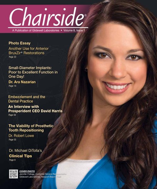
Chairside®<br />
A Publication of <strong>Glidewell</strong> Laboratories • Volume 8, Issue 1<br />
Photo Essay<br />
Another Use for Anterior<br />
BruxZir ® Restorations<br />
Page 22<br />
Small-Diameter Implants:<br />
Poor to Excellent Function in<br />
One Day!<br />
Dr. Ara Nazarian<br />
Page 14<br />
Embezzlement and the<br />
<strong>Dental</strong> Practice<br />
An Interview with<br />
Prosperident CEO David Harris<br />
Page 44<br />
The Viability of Prosthetic<br />
Tooth Repositioning<br />
Dr. Robert Lowe<br />
Page 55<br />
Dr. Michael DiTolla’s<br />
Clinical Tips<br />
Page 9<br />
COVER PHOTO<br />
Jennifer Folbigg, Customer Service Representative<br />
<strong>Glidewell</strong> Laboratories, Newport Beach, Calif.
Contents<br />
9 Dr. DiTolla’s Clinical Tips<br />
Showcased in this issue are a cloud-based platform I<br />
recently demoed from Smile Reminder that offers an<br />
impressive suite of personalized patient communication<br />
tools for growing your patient base, and an invaluable<br />
oral hygiene product from <strong>Dental</strong> Herb Company to<br />
aid in your practice’s fight against periodontal disease.<br />
Also featured are two innovative products designed<br />
to make your dentistry easier and more efficient: a<br />
sectional matrix system from Triodent for performing<br />
high-quality Class II restorations, and an LED curing<br />
light from Ivoclar Vivadent that features a compact,<br />
ergonomic shape to fit any dentist’s hands.<br />
14 Poor to Excellent Function in One Day!<br />
“Mini” or small-diameter implants offer many benefits<br />
for patients seeking maxillary and mandibular overdenture<br />
treatment. Dr. Ara Nazarian presents a case<br />
report featuring <strong>Glidewell</strong>’s Inclusive ® Mini Implants<br />
that demonstrates the protocol for the placement of<br />
these small-diameter implants, and the subsequent<br />
beneficial effects they have on the function and retention<br />
of the patient’s new prostheses.<br />
22 Photo Essay: Another Use for Anterior<br />
BruxZir ® Solid Zirconia Restorations<br />
In this photo essay, I address a difficult situation<br />
restorative dentists face in clinical practice: treating a<br />
patient with severe tetracycline staining. The patient<br />
in this case presented the additional challenges of<br />
severe bruxism and an edge-to-edge bite. I chose<br />
BruxZir crowns because I knew these high-strength<br />
restorations would not only withstand the destructive<br />
forces generated in his mouth, but would also have a<br />
better chance than a glass-ceramic material of completely<br />
blocking out the dark stump shades.<br />
Visit www.chairsidelive.com to view the latest episode of<br />
our weekly Web series “Chairside Live.” Also available on<br />
YouTube and iTunes.<br />
Contents 1
Contents<br />
38 Chairside Live Case of the Week:<br />
Episode 32 — A Disastrous Double-Arch<br />
Impression Tray<br />
This new column highlights a Case of the Week from<br />
a recent episode of our weekly Web series “Chairside<br />
Live.” The first case comes from Episode 32 and<br />
addresses one of my dental pet peeves: when our lab<br />
receives a bridge impression taken in a double-arch<br />
tray. While double-arch impressions can be suitable for<br />
a single-unit crown or two single-unit adjacent crowns,<br />
they are best avoided for multi-unit restorations.<br />
44 One-on-One with Dr. Michael DiTolla:<br />
Interview of David Harris<br />
According to Prosperident CEO and licensed private<br />
investigator David Harris, embezzlement will strike<br />
three in five dentists in their careers. While this statistic<br />
may surprise you, it doesn’t need to discourage you. In<br />
this issue’s featured interview, the man known in dental<br />
circles as “the dental fraud guru” shares his expert<br />
perspective on embezzlement in the dental office.<br />
Chairside Magazine Digital Edition<br />
Chairside magazine is now optimized for all popular<br />
desktop, tablet and smartphone platforms! To try out<br />
the new beta version of our digital magazine from<br />
your desktop computer or favorite mobile device, visit<br />
www.chairsidemagazine.com.<br />
55 Prosthetic Tooth Repositioning: A Viable<br />
Treatment Option for Select Cases<br />
For a select group of patients with minor tooth<br />
malposition, such as spacing, crowding, minor rotations<br />
and facial-lingual arch form displacement, esthetic and<br />
functional correction may be accomplished purely by<br />
restorative means, claims Dr. Robert Lowe. Three case<br />
reports demonstrate how prosthetic tooth repositioning<br />
can be a viable treatment option for these types of<br />
malocclusions when proper guidelines are followed.<br />
64 Biologic Shaping from a Restorative<br />
Perspective<br />
Dr. Daniel Melker focuses on the concept of biologic<br />
shaping in this article, outlining the numerous<br />
differences between this root-reshaping procedure<br />
and traditional crown lengthening. A case example<br />
illustrates how this periodontal corrective procedure<br />
can provide the restorative dentist with a predictable<br />
and successful method of restoring teeth.<br />
2<br />
www.chairsidemagazine.com
Contributors<br />
Michael C. DiTolla, DDS, FAGD<br />
Dr. Michael DiTolla is a graduate of the University of the Pacific Arthur A. Dugoni School of Dentistry. As director<br />
of clinical education and research at <strong>Glidewell</strong> Laboratories, he performs clinical testing on new products<br />
in conjunction with the company’s R&D department. <strong>Glidewell</strong> dental technicians have the privilege of rotating<br />
through his operatory and experiencing his commitment to excellence through his prepping and placement of<br />
their restorations. Dr. DiTolla is a CR evaluator and lectures nationwide on restorative and cosmetic dentistry.<br />
His clinical programs are available on DVD and online through <strong>Glidewell</strong> Laboratories. For more info on his<br />
articles or for a free copy of his clinical presentations, call 888-303-4221 or e-mail mditolla@glidewelldental.com.<br />
David Harris, MBA, CMA<br />
David Harris is a licensed private investigator, with a graduate business degree and a professional accounting<br />
designation. He is CEO of Prosperident, the world’s largest dental embezzlement investigation firm. Prosperident<br />
is consulted on hundreds of dental embezzlement matters annually, and David has frequently had the pleasure<br />
of hearing cell doors slam shut on perpetrators. David has lectured at several universities in the faculties of<br />
dentistry, business and law, and he has been interviewed on embezzlement by virtually every major North<br />
American dental magazine. David is a member of the Academy of <strong>Dental</strong> Management Consultants (ADMC)<br />
and the Speaking Consulting Network. Contact him at 888-398-2327 or www.dentalembezzlement.com.<br />
Robert A. Lowe, DDS, FAGD, FICD, FADI, FACD, FIADFE, FASDA<br />
Dr. Robert Lowe graduated magna cum laude from Loyola University School of Dentistry in 1982 and was<br />
a clinical professor in restorative dentistry at the school until its closure in 1993. Since January 2000,<br />
Dr. Lowe has maintained a private practice in Charlotte, N.C. He lectures internationally and his work<br />
is frequently published in well-known dental journals on esthetic and restorative dentistry. Dr. Lowe has<br />
earned Fellowship in the AGD, ICD, ADI, ACD and American Society for <strong>Dental</strong> Esthetics (ASDA), and<br />
received the Gordon Christensen Outstanding Lecturers Award and Diplomat status on the American Board<br />
of Esthetic Dentistry. Contact Dr. Lowe at 704-450-3321 or boblowedds@aol.com.<br />
Daniel J. Melker, DDS<br />
Dr. Daniel Melker graduated from the Boston University School of Graduate Dentistry in 1975 with specialty<br />
training in periodontics. Since then, he has maintained a private practice in periodontics in Clearwater,<br />
Fla. Dr. Melker lectures at the University of Florida Periodontic and Prosthodontic graduate programs on<br />
the periodontic-restorative relationship. He also presents at the University of Alabama at Birmingham,<br />
University of Houston, Baylor University and Louisiana State University’s graduate periodontal programs.<br />
Dr. Melker has published several articles in national dental magazines, and he has twice been honored with<br />
the Florida Academy of Cosmetic Dentistry Gold Medal. Contact him at 727-725-0100.<br />
Ara Nazarian, DDS, DICOI<br />
Dr. Ara Nazarian maintains a private practice in Troy, Mich., with an emphasis on comprehensive and<br />
restorative care. He is the director of the Reconstructive Dentistry Institute, a Diplomate of the ICOI, and has<br />
conducted lectures and hands-on workshops on esthetic materials and dental implants throughout the U.S.,<br />
Europe, New Zealand and Australia. Dr. Nazarian is also the creator of the DemoDent ® patient education<br />
model system. His articles have been published in many of today’s popular dental publications. Contact him<br />
at 248-457-0500 or www.aranazariandds.com.<br />
4<br />
www.chairsidemagazine.com
Publisher<br />
Jim <strong>Glidewell</strong>, CDT<br />
Editor-in-Chief and Clinical Editor<br />
Michael C. DiTolla, DDS, FAGD<br />
Managing Editors<br />
Jim Shuck; Mike Cash, CDT<br />
Creative Director<br />
Rachel Pacillas<br />
Copy Editors<br />
Jennifer Holstein, David Frickman,<br />
Chris Newcomb, Megan Strong<br />
Statistical Editor<br />
Darryl Withrow<br />
Digital Marketing Manager<br />
Kevin Keithley<br />
Graphic Designers<br />
Jamie Austin, Deb Evans,<br />
Joel Guerra, Audrey Kame, Phil Nguyen,<br />
Kelley Pelton, Makara You<br />
Web Designers<br />
Jamie Austin, Kevin Greene,<br />
Allison Newell, Melanie Solis, Ty Tran<br />
Photographer<br />
Sharon Dowd<br />
Videographers<br />
James Kwasniewski, Sam Lea<br />
Illustrator<br />
Wolfgang Friebauer, MDT<br />
Editor’s Letter<br />
The crowns & bridges produced at <strong>Glidewell</strong> Laboratories<br />
are now made using essentially 100 percent CAD/CAM<br />
technology, and I really notice the difference in the<br />
restorations I get back from the lab. The crowns just fit, and<br />
if I give them enough reduction, I can always get contours<br />
like a natural tooth. Before we started using CAD/CAM, the<br />
most frequent complaint we used to hear from dentists was<br />
about our consistency, so this technology really has been a<br />
game changer for our lab and our customers.<br />
More than a decade ago, new customers would tell me that<br />
they would get three great crowns from us and then two soso<br />
crowns, then another great one, then one ugly one, and so<br />
on. We were doing everything we could to fix those issues,<br />
but the underlying problem went deeper than our lab: there<br />
simply weren’t enough trained dental technicians available.<br />
There are currently only 18 accredited dental laboratory<br />
programs in the U.S. If that number seems shockingly low<br />
to you, it’s because it is. That number is down 62 percent<br />
since 1992 — a drastic decrease for that 20-year period. In<br />
fact, today these programs currently produce only about 300<br />
graduates annually for the entire U.S. To meet our demand,<br />
we had to hire people off the street and train them ourselves.<br />
But it takes time to develop as a technician, just as it does<br />
as a dentist.<br />
In 2007, Ivoclar Vivadent’s IPS e.max ® was introduced<br />
into our lab, and with this first high-strength, monolithic<br />
restoration came the day where a machine did most of<br />
the work. Ideal contours were found in CAD libraries, and<br />
dentists just had to give CAD technicians enough room to<br />
drop them in. Then in 2009, <strong>Glidewell</strong> launched BruxZir ®<br />
Solid Zirconia, signaling the next wave of the monolithic<br />
revolution. A year later, nearly all of our PFM crowns were<br />
produced using CAD/CAM as well.<br />
Coordinator and Ad Representative<br />
Thanks to our president and CEO’s unwavering commitment<br />
Teri Arthur<br />
to technology, we are able to give you, our dentists, the<br />
(teri.arthur@glidewelldental.com)<br />
consistency and predictability you’ve always wanted. Dentists<br />
often tell me that a BruxZir crown fits better than any<br />
If you have questions, comments or complaints regarding<br />
this issue, we want to hear from you. Please e-mail us at<br />
other crown they have prescribed. It’s a good time to be a<br />
chairside@glidewelldental.com. Your comments may be<br />
dentist, and it’s a great time to work with a lab that has fully<br />
featured in an upcoming issue or on our website:<br />
embraced the consistency of CAD/CAM dentistry.<br />
www.chairsidemagazine.com.<br />
Neither Chairside Magazine nor any employees involved in its publication<br />
(“publisher”), © makes 2013 any <strong>Glidewell</strong> warranty, Laboratories<br />
express implied, or assumes<br />
any liability or responsibility for the accuracy, completeness, or usefulness<br />
Chairside of any information, magazine apparatus, nor any employees product, involved or process in its disclosed, publication or<br />
Neither<br />
(“publisher”), represents makes that its any use would warranty, not express infringe proprietary or implied, rights. or assumes Reference any<br />
liability herein or to responsibility any specific for commercial the accuracy, products, completeness, process, or or services usefulness by<br />
Yours in quality dentistry,<br />
of trade any name, information, trademark, apparatus, manufacturer product, or otherwise or process does disclosed, not necessarily<br />
constitute that its or use imply would its endorsement, not infringe recommendation, proprietary rights. or Reference favoring<br />
or<br />
represents<br />
herein by the to publisher. any specific The commercial views and products, opinions of process, authors or expressed services<br />
by herein trade do name, not necessarily trademark, state manufacturer or reflect those or otherwise of publisher does and not<br />
necessarily shall not constitute be used for or advertising imply its endorsement, product endorsement recommendation, purposes. or<br />
favoring CAUTION: by the When publisher. viewing The the views techniques, and opinions procedures, of authors theories expressed and materials<br />
do that not are necessarily presented, state you or must reflect make those your of own the decisions publisher about and<br />
herein<br />
specific treatment for patients and exercise personal professional judgment<br />
regarding When viewing the need the for further techniques, clinical procedures, testing or education theories and<br />
Editor-in-Chief, Clinical Editor<br />
Dr. Michael C. DiTolla<br />
shall not be used for advertising or product endorsement purposes.<br />
CAUTION:<br />
materials your own that clinical are presented, expertise before you must trying make to implement your own new decisions procedures. about<br />
mditolla@glidewelldental.com<br />
specific treatment for patients and exercise personal professional<br />
judgment Chairside regarding ® Magazine the is need a registered for further trademark clinical of testing <strong>Glidewell</strong> or education Laboratories. and<br />
your own clinical expertise before trying to implement new procedures.<br />
5<br />
Chairside is a registered trademark of <strong>Glidewell</strong> Laboratories. Editor’s Letter
Letters to the Editor<br />
Dear Dr. DiTolla,<br />
Is BruxZir ® Solid Zirconia (<strong>Glidewell</strong> Laboratories)<br />
indicated for inlays/onlays as well as<br />
crowns? I only hear it mentioned for crowns.<br />
For patients that insist on tooth-colored restorations<br />
on second molars, what do you<br />
place, if anything? I love IPS e.max ® (Ivoclar<br />
Vivadent; Amherst, N.Y.), but I draw the line<br />
at the first molars forward.<br />
– Jeffrey L. Schultz, DDS, FAGD<br />
Bellaire, Texas<br />
Dear Jeff,<br />
BruxZir Solid Zirconia can be used for<br />
inlays and onlays, as well as crowns.<br />
We have dentists asking us for BruxZir<br />
veneers as well, which we can do, but I<br />
am waiting for some bond strength research<br />
to conclude before we make any<br />
recommendations. Veneers are essentially<br />
non-retentive preps, so we need<br />
to ensure that our cementation/bonding<br />
protocol is sufficient to retain them.<br />
For tooth-colored restorations on second<br />
molars, BruxZir Solid Zirconia is<br />
the only choice. However, you need<br />
to have at least 0.5 mm of occlusal reduction.<br />
I have a 0.6 mm depth-cutting<br />
bur in my kit that I use for these restorations,<br />
and by the time I finish the<br />
6<br />
www.chairsidemagazine.com<br />
reduction it will usually be at 0.7 mm.<br />
At 0.5 mm, you must reduce the opposing<br />
if the occlusion is high on the<br />
restoration; otherwise, the BruxZir restoration<br />
can fail. Cast gold still holds<br />
the title as the best second molar restoration,<br />
but you know as well as I do<br />
that most patients will not accept it.<br />
Hope that helps!<br />
– Mike<br />
Dear Dr. DiTolla,<br />
I’m totally blown away by “Chairside Live,”<br />
which I was intrigued to watch for the first<br />
time when you interviewed Gordon [Christensen]<br />
— I believe it was Part 3. First, let<br />
me congratulate you on the entire concept,<br />
which I found entertaining, informative and<br />
just plain fun to watch. You and Megan<br />
remind me of the old Dan Aykroyd-Jane<br />
Curtin SNL “Point/Counterpoint” parody. In<br />
any event, great job! I loved your retching<br />
skit at the end — hilarious!<br />
But you know you and your guest can’t<br />
spew out data without skeptical Michael<br />
(that’s me) chiming in. As far as Gordon’s<br />
claim that various drinks such as lemonade<br />
are 10-times more damaging to the external<br />
stain on BruxZir zirconia than Coca-Cola,<br />
a quick search (Yahoo Answers, NEWTON<br />
Ask-a-Scientist) found that the pH of Coke<br />
is 2.5, while lemonade is 3.8. On the other<br />
hand, another site (21st Century <strong>Dental</strong>) lists<br />
Country Time Lemonade as having a pH of<br />
2.5 and Coke Classic at 2.53. Gordon also<br />
mentioned energy drinks being worse than<br />
Coke, but this latter site found that Gatorade<br />
has a pH of 2.95. Bottom line: It’s very<br />
hard for me to believe that these drinks are<br />
worse than Coke when it comes to dissolving<br />
external ceramic stains, and 10-times<br />
worse? Nah! Even if pH is not the be-all and<br />
end-all factor, 10-times worse is still hard<br />
to believe.<br />
You also stated that Multilink ® Automix<br />
(Ivoclar Vivadent) was “self-etching,” but in<br />
fact, it’s the primers in the kit that are selfetching,<br />
not the cement itself. Minor point,<br />
perhaps, but your viewers could possibly<br />
have come away thinking that Multilink<br />
Automix is similar to RelyX Unicem (3M<br />
ESPE; St. Paul, Minn.), which, of course,<br />
it’s not.<br />
In any event, you again came up with a terrific<br />
idea, which I have to admit I’m jealous I<br />
didn’t think of first!<br />
– Michael Miller, DDS<br />
Houston, Texas<br />
Dear Michael,<br />
Wow, coming from you that is quite<br />
an honor! I have such respect for<br />
what you do at REALITY (www.<br />
realityesthetics.com), and it means<br />
a lot when one of my mentors takes<br />
the time to write a letter like this. You<br />
might even see your letter read on<br />
“Chairside Live,” which would earn<br />
you a signed picture of Megan and<br />
me. I’ll be sure to mark it with a dotted<br />
line so you can cut me out of the<br />
picture. Plus, addressing your letter on<br />
the show will give me the chance to<br />
prove I know the difference between<br />
self-etching resin cements and selfadhesive<br />
resin cements.<br />
Gordon was referring to an AGD study<br />
in their journal, General Dentistry (von<br />
Fraunhofer JA, Rogers MM. Effects of<br />
sports drinks and other beverages on<br />
dental enamel. Gen Dent. 2005 Jan-Feb;<br />
53(1):28-31).<br />
After that episode aired, a viewer sent<br />
me this link, http://fit4maui.com/water/<br />
pu/bottled_ph.html, which purports to<br />
measure the pH of different brands<br />
of bottled water. Could Aquafina and<br />
Dasani really have a pH of 4?<br />
Thanks again for the kind words,<br />
Michael! They mean the world to me.<br />
– Mike<br />
Dear Dr. DiTolla,<br />
I have followed some of your CE courses online.<br />
I see that you are a fan of SpeedCEM
(Ivoclar Vivadent). Do you use SpeedCEM<br />
to cement feldspathic porcelain veneers?<br />
Would you etch with hydrofluoric acid if the<br />
lab has already done so?<br />
– Marea White, DDS<br />
Bedford, Texas<br />
Dear Marea,<br />
Nice to hear from you! I am a fan of<br />
SpeedCEM, which is a self-adhesive<br />
resin cement similar to RelyX Unicem<br />
or Maxcem Elite (Kerr Corp.; Orange,<br />
Calif.). While these cements are strong<br />
enough for inlays, retentive onlays and<br />
retentive crown preps, they are not<br />
strong enough to bond low-retention<br />
restorations such as veneers.<br />
Every veneer manufacturer I have<br />
spoken with still recommends the<br />
total-etch (now called etch and rinse)<br />
technique for luting veneers, including<br />
higher strength veneers like IPS e.max.<br />
There is one lecturer I know of, Dr. Jose<br />
Luis-Ruiz, who mentioned to me in an<br />
interview for Chairside magazine that<br />
he is using self-etch to place veneers.<br />
However, he is doing it using a cement<br />
with a separate self-etch solution.<br />
PANAVIA F2.0 (Kuraray America; New<br />
York, N.Y.) and Multilink Automix are<br />
two good examples of self-etching<br />
resin cements with separate self-etch<br />
primers.<br />
The standard of care today is to use<br />
the total-etch (etch and rinse) technique<br />
with a light-cured resin cement<br />
to place veneers.<br />
The research I have seen does not<br />
show any improvement in bond<br />
strength if you re-etch the veneers<br />
with hydrofluoric acid in your office<br />
after try-in, although it is acceptable<br />
to clean the veneer with phosphoric<br />
acid.<br />
– Mike<br />
Dear Dr. DiTolla,<br />
I practice general dentistry in Petaluma,<br />
Calif. A few months ago, I attended one<br />
of your CE courses through our local<br />
dental society, Redwood Empire <strong>Dental</strong><br />
Society (REDS). I enjoyed your lecture and<br />
your sense of humor. Most importantly, I<br />
really liked all of your practical tips and<br />
information. I have been practicing since<br />
2000, and have taken many CE classes, but<br />
your lecture has made the biggest impact<br />
on my practice so far. Your preparation and<br />
impression techniques have helped me<br />
achieve perfect impressions and my crown<br />
cement appointments are so enjoyable now.<br />
My dental lab technician had always told me<br />
that my preps and impressions were very<br />
good, but the small changes I made since<br />
attending your course have helped me<br />
achieve excellent and consistent results. I<br />
wanted to thank you and let you know how<br />
useful your tips have been to my practice<br />
and to me. I hope you return to this area to<br />
lecture again.<br />
– Nadia Navid, DDS<br />
Petaluma, Calif.<br />
Dear Nadia,<br />
Thank you so much for your kind letter.<br />
I love hearing stories like yours,<br />
and I know your lab techs will be<br />
thrilled with your preps and impressions<br />
as well. They will love you even<br />
more if you send a digital photograph<br />
with all anterior cases! I keep playing<br />
with new products and techniques,<br />
looking for ways to help dentists get<br />
better results in a simple, predictable<br />
fashion. I will be sure to pass any of<br />
those your way, and I hope I get a<br />
chance to make it back to your neck<br />
of the (red)woods soon!<br />
– Mike<br />
CONNECT WITH CHAIRSIDE<br />
FOLLOW US ON TWITTER<br />
Find us @<strong>Glidewell</strong><strong>Dental</strong><br />
FIND US ON FACEBOOK<br />
“Like” us on Facebook to see<br />
what’s new.<br />
ITUNES WATCH AND<br />
LEARN<br />
Go the iTunes store and search<br />
“<strong>Glidewell</strong>.” Plus, download<br />
the free <strong>Glidewell</strong> Publications<br />
app for iPad.<br />
SHARE YOUR THOUGHTS<br />
Visit www.chairsidemagazine.com<br />
and select “Contact Us.” Or write to:<br />
<strong>Glidewell</strong> Laboratories<br />
ATTN: Chairside magazine<br />
4141 MacArthur Blvd.<br />
Newport Beach, CA 92660<br />
ACCESS OUR RESOURCES<br />
Clinical videos, product information<br />
and patient resources are a click<br />
away at www.glidewelldental.com.<br />
ADVERTISE/SUBMIT<br />
AN ARTICLE<br />
Call 888-303-4221<br />
Letters should include writer’s full name,<br />
address and daytime phone number. All<br />
correspondence may be published and<br />
edited for clarity and length.<br />
Letters to the Editor 7
Numbers<br />
by the<br />
2,827,512<br />
Total number of BruxZir ® crowns placed<br />
Source: <strong>Glidewell</strong> Laboratories internal data<br />
25<br />
Number of countries where<br />
BruxZir ® Solid Zirconia is sold<br />
Source: <strong>Glidewell</strong> Laboratories internal data<br />
10.6<br />
Dentistry personnel per 10,000 people in the U.S. Dentistry<br />
Source: Wolfram|Alpha, www.wolframalpha.com<br />
0.39<br />
personnel per 10,000 people in China<br />
Source: Infodent International magazine<br />
✓<br />
5,232<br />
Number of <strong>Glidewell</strong><br />
Laboratories customers<br />
in 2012 that had<br />
ZERO remakes<br />
(160,939 restorations<br />
were fabricated for<br />
these customers)<br />
Source: <strong>Glidewell</strong> Laboratories<br />
internal data<br />
Unemployment rate of U.S.<br />
1.5%<br />
dentists (one of the lowest<br />
of all U.S. professions)<br />
Source: U.S. Bureau of Labor Statistics<br />
8<br />
www.chairsidemagazine.com<br />
15%<br />
Percentage of anterior<br />
restorations fabricated at<br />
<strong>Glidewell</strong> Laboratories from<br />
BruxZir ® Solid Zirconia<br />
Source: <strong>Glidewell</strong> Laboratories internal data<br />
#8 & #9<br />
BruxZir ® Solid Zirconia is the second most<br />
requested restorative material at <strong>Glidewell</strong><br />
Laboratories for these upper front teeth<br />
Source: <strong>Glidewell</strong> Laboratories internal data
Dr. DiTolla’s<br />
CLINICAL TIPS<br />
PRODUCT........ Bluephase ® Style<br />
SOURCE........... Ivoclar Vivadent Inc. (Amherst, N.Y.)<br />
800-533-6825, www.ivoclarvivadent.com<br />
Design matters. Apple has taught me over the last<br />
few years that, regardless of how well an object<br />
does something, the look and feel of an item play<br />
an important role in the user’s personal connection<br />
to it. The original curing lights were gun-shaped, on<br />
the clunky side and struggled to reach the molars.<br />
The Bluephase Style LED curing light from Ivoclar<br />
Vivadent has won multiple design awards, and it<br />
is easy to see why. We just hired our first female<br />
dentist, and her hands are tiny compared to mine.<br />
Considering that more than 50 percent of dental<br />
school graduates are women, ergonomics is an<br />
important issue when we are sharing instruments<br />
such as electric handpieces and curing lights.<br />
Bluephase Style’s Polywave ® LED technology provides<br />
a broadband spectrum of 385–515 nm, and it<br />
will cure every dental material on the market today.<br />
The 10 mm light probe tip allows you to cure even<br />
the largest restorations because it can provide<br />
continuous curing for more than 10 minutes.<br />
Dr. DiTolla’s Clinical Tips 9
Dr. DiTolla’s<br />
CLINICAL TIPS<br />
PRODUCT........ Tooth & Gums Tonic ®<br />
SOURCE........... <strong>Dental</strong> Herb Company ® Inc.<br />
(Lancaster, N.H.)<br />
800-747-4372, www.dentalherb.com<br />
I started using Tooth & Gums Tonic more than 10 years<br />
ago when I started practicing at <strong>Glidewell</strong> Laboratories.<br />
Once we began filming all of my dentistry, I realized<br />
how bad bleeding tissue looked when we were making<br />
impressions or placing restorations. For some reason,<br />
I had been willing to tolerate this bleeding for the first<br />
13 years of my career, but now that I could see it onscreen,<br />
I was disgusted. While the potent and effective<br />
formulas of <strong>Dental</strong> Herb Company products remain unchanged,<br />
the impact of the company’s new ownership<br />
is evident. In addition to a fresh, new look, the company<br />
has vastly improved its customer service, updated its<br />
packaging and informational materials, and provided a<br />
more user-friendly Web presence with a “Find a Dentist”<br />
locator tool that patients can use to search for the nearest<br />
dental office carrying its products. So while the new<br />
owners continue to sell Tooth & Gums products through<br />
dental professionals, the company expects to have online<br />
ordering ready to go by this summer. It’s clear that<br />
<strong>Dental</strong> Herb Company wants to be a valued partner in<br />
your practice’s fight against periodontal disease, and<br />
in our lab’s fight against bloody impressions!
Dr. DiTolla’s<br />
CLINICAL TIPS<br />
PRODUCT........ Triodent V3 Sectional Matrix System<br />
SOURCE........... Ultradent Products Inc. (South Jordan, Utah)<br />
888-230-1420, www.ultradent.com<br />
Because I practice inside of a dental laboratory, I<br />
typically only do direct restorations when they are<br />
adjacent to other indirect restorations I am placing.<br />
I was introduced to this sectional matrix system while<br />
I was testing BruxZir ® inlays and found myself doing<br />
only inlay preps for a month. While I was able to<br />
rationalize that it was OK when I nicked an adjacent<br />
tooth while dropping the proximal box and extending<br />
it buccolingually, a fellow dentist told me about the<br />
WedgeGuard interproximal tooth shield, which is an<br />
integral part of the V3 Matrix. WedgeGuard is the<br />
standard Wave-Wedge interproximal wedge with a<br />
metal protector attached to it. Place the WedgeGuard<br />
between the teeth and prep the tooth safely with the<br />
metal protector in place. After you finish the prep,<br />
you simply grab the metal protector and pull it out<br />
with the wedge still in place. Then you place your<br />
matrix, place the V3 Ring, and place and cure the<br />
composite. Genius! Go to exclusive U.S. distributor<br />
Ultradent’s YouTube page to view an animation of<br />
the system in action.<br />
Dr. DiTolla’s Clinical Tips11
Dr. DiTolla’s<br />
CLINICAL TIPS<br />
PRODUCT........ Smile Reminder <br />
SOURCE........... Solutionreach (Lehi, Utah)<br />
866-605-6867, http://tinyurl.com/smilereminder<br />
FREE $10 STARBUCKS GIFT CARD! Yep, that is all<br />
it took to get me to take the online demo of Smile<br />
Reminder. Once a month I am asked to fill out an<br />
online survey for which the soliciting company will<br />
send me a $5 check. Don’t bother; I don’t want to walk<br />
it over to the bank. But a $10 Starbucks gift card?<br />
One of my staff members goes to one of their drivethru<br />
locations every day on her way to work — well<br />
played, Smile Reminder! What started off as a demo<br />
turned into a purchase just 10 minutes later when I<br />
saw the platform’s mind-boggling suite of features,<br />
which address everything from reducing no-shows<br />
with messaging and filling late cancellations, to<br />
sending targeted e-mail campaigns based on patient<br />
surveys and giving dentists and staff the ability to<br />
record custom video birthday greetings that get sent<br />
to patients. You can even track your online reputation<br />
by finding positive and negative reviews, as well as<br />
invite patients to write reviews and post them to the<br />
sites where your online reputation needs a boost.<br />
The electronic “recare” feature alone pays for the<br />
service because it automatically contacts patients<br />
who don’t have an appointment and are past their<br />
recall frequency, pulling the data from your practice<br />
management software. We use Henry Schein’s<br />
Dentrix ® , and the front office loves that it shows a live<br />
update of each patient’s pre-approved CareCredit<br />
amount. The Smile Reminder fixed price guarantee<br />
is also a refreshing approach I wish others would<br />
take. The dentists who signed up for Smile Reminder<br />
years ago are still paying the same price today. I like<br />
knowing what my monthly flat fee will always be and<br />
that there will not be any surprises down the road. I<br />
left out about 50 other functions where this powerful<br />
software also shines. Yes, you have my permission<br />
to put down the magazine and go get your Starbucks<br />
gift card. Go to http://tinyurl.com/smilereminder.
14 www.chairsidemagazine.com
Poor to Excellent<br />
Function in One Day!<br />
– ARTICLE by Ara Nazarian, DDS, DICOI<br />
Introduction<br />
Minimally invasive devices and procedures are fast becoming the largest growth<br />
segment of the medical and dental device industry. When compared to traditional<br />
approaches, they require less anesthesia, shorten surgical and recovery times,<br />
reduce patient risk, and can offer significant cost savings. Over time, we have<br />
witnessed the research and development of smaller and smaller components.<br />
Mini dental implants dramatically improve the quality of the outcomes for patients<br />
seeking maxillary and mandibular overdenture treatment.<br />
Small-diameter implants (1.8 mm to 3.0 mm) also differ from their full-sized<br />
counterparts in several ways. Their configuration allows for a more conservative<br />
placement protocol without involving tissue flaps or tapping procedures,<br />
resulting in minimal trauma for the bone and the gingival tissues. Mini dental<br />
implants’ size also allows the clinician to place them in ridges that might not<br />
otherwise be suitable for full-sized implants. Once these mini dental implants<br />
are firmly seated in place in intimate contact with bone, they can be immediately<br />
loaded with no lengthy waiting period or second-stage surgery.<br />
The following case report will demonstrate the protocol for the placement of<br />
small-diameter implants, and the subsequent effects on the new prostheses.<br />
Poor to Excellent Function in One Day!15
Case Report<br />
Diagnosis and Treatment Planning<br />
A male in his late 70s presented to our office; he was<br />
frustrated with the look and fit of his upper and lower<br />
dentures (Fig. 1). Most importantly, he complained that his<br />
lower denture was nonretentive and nonfunctional, always<br />
falling out when speaking or while eating. He had been a<br />
denture wearer for the last 25 years, resulting in excessive<br />
resorption of the mandible. The patient also suffered from<br />
hypertension, which was controlled with medication.<br />
The first phase of treatment would consist of having a new<br />
set of upper and lower dentures (Simply Natural Dentures <br />
[<strong>Glidewell</strong> Laboratories]) that would fit properly and occlude<br />
functionally. Utilizing recorded bases with corresponding<br />
wax rims, we analyzed the positioning of the teeth and<br />
proper proportions for an ideal smile. The patient desired<br />
to have his new set of teeth with shade B1 (VITA Classical<br />
Shade Guide [Vident; Brea, Calif.]). When the patient viewed<br />
the wax try-in, he quickly approved them for processing<br />
(Fig. 2).<br />
Palpation and radiographic examination revealed a<br />
moderately narrowed mandibular ridge (Fig. 3). Crestal bone<br />
and ridge height were sufficient to receive four 3 mm x 13 mm<br />
Inclusive ® Mini Implants (<strong>Glidewell</strong> Direct) (Fig. 4). The thread<br />
pattern and pitch of this implant are purposely designed<br />
to immediately maximize bone-to-implant thread contact.<br />
Others like it include: I-Mini (OCO Biomedical; Albuquerque,<br />
N.M.), I6B (AB <strong>Dental</strong> USA; Los Angeles, Calif.), HM Implant<br />
(Hiossen; Fairless Hills, Pa.), MILO ® (Intra-Lock; Boca Raton,<br />
Fla.), Midi ® (Basic <strong>Dental</strong> Implants; Albuquerque, N.M.),<br />
Intermezzo (MegaGen USA; Englewood Cliffs, N.J.) and<br />
miniMARK (ACE Surgical; Brockton, Mass.).<br />
After reviewing the patient’s panoramic radiograph, the<br />
mental foramina were also located, and it was confirmed<br />
that the four mini dental implants could be safely placed<br />
within the cuspid-to-cuspid area.<br />
Figure 1: Existing dentures were approximately 17 years old.<br />
Figure 2: New upper and lower dentures.<br />
Figure 3: Lower edentulous ridge, in preparation for mini dental<br />
implant placement.<br />
Outline of Clinical Treatment<br />
Before starting treatment, all the risks, benefits and<br />
alternatives were reviewed with the patient. A clean<br />
operating environment was established, the patient was<br />
draped and local anesthetic was administered. Then, an<br />
indelible marker was used to designate landmarks and<br />
areas of insertion.<br />
Keeping correct alignment, a 1.5 mm pilot drill from the<br />
Inclusive ® Surgical Kit (<strong>Glidewell</strong> Direct) was placed into<br />
the sites and advanced to a depth of 15 mm, measuring<br />
from the tissue surface using a surgical motor (AEU-7000E<br />
[Aseptico; Woodinville, Wash.]) with generous amounts of<br />
Figure 4: Inclusive Mini Implants (<strong>Glidewell</strong> Direct) in packages and<br />
ready for placement.<br />
16 www.chairsidemagazine.com
sterile water. This additional 2 mm was the same depth<br />
of the tissue height to bone. In other words, 13 mm for<br />
the osteotomy in bone and 2 mm for tissue thickness was<br />
created to place a 13 mm long implant. Paralleling pins<br />
(Salvin <strong>Dental</strong>; Charlotte, N.C.) were placed in the sites<br />
of the osteotomies and an X-ray was taken to check the<br />
angulations to ensure proper orientation among the implant<br />
sites. Using a rotary tissue punch, a 3.0 mm outline was<br />
created over the initial osteotomies and the tissue plugs<br />
removed with a serrated curette (Zoll <strong>Dental</strong>; Niles, Ill.). The<br />
osteotomies were completed with the final drill (2.4 mm)<br />
included in the Inclusive Surgical Kit. Once the osteotomies<br />
were completed, four 3 mm x 13 mm Inclusive Mini Implants<br />
were placed in the osteotomies, using an implant finger<br />
driver (Fig. 5) until increased torque became necessary. The<br />
ratchet wrench was then connected to the adapter and the<br />
implants were torqued to final depth, reaching a torque<br />
level of 65 Ncm (Fig. 6). A postoperative radiograph was<br />
taken of the implants before initiating the prosthetic phase<br />
of treatment.<br />
At that point, the location of each implant was transferred<br />
to the denture using bite registration material (Take 1 ®<br />
Advance [Kerr Corp.; Orange, Calif.]). These areas were<br />
relieved to a diameter of 5.0 mm, and the denture was then<br />
reseated passively, confirming adequate relief had been<br />
properly established.<br />
A covering silicone (Fit Test C&B [VOCO America; Briarcliff<br />
Manor, N.Y.]) was used to cover any undercuts or interface<br />
of the implants, allowing only the O-ball of the implant<br />
to be exposed. This step prevented problems of the pickup<br />
material locking around the implants. A female O-ring<br />
keeper cap (Inclusive) was then fitted over each implant.<br />
Retentive fit and mobility were again verified. Each O-ring<br />
would create a retentiveness of approximately 5 lbs. Since<br />
there were four implants with corresponding housings,<br />
the total amount of force needed to remove the prosthesis<br />
would be about 20 lbs.<br />
The cleaned and dried recesses in the lower denture were<br />
filled with cold-cure acrylic (Quick Up [VOCO America])<br />
(Fig. 7) and seated onto the implants, allowing it to<br />
polymerize. Upon setting, the lower denture was relieved of<br />
any excess flash, and the flange areas were relieved (Fig. 8).<br />
Finally, postoperative instructions were reviewed with the<br />
patient regarding denture placement, removal and oral<br />
hygiene. The patient was extremely excited and pleased that<br />
his new dentures were now very retentive and functional.<br />
Figure 5: The mini dental implant on the plastic insertion driver.<br />
Figure 6: Four mini dental implants in place in the mandible.<br />
Figure 7: Quick Up (VOCO America) was placed in recesses for<br />
housing pick-up.<br />
Figure 8: Retentive housings, as picked up in the lower denture.<br />
Poor to Excellent Function in One Day!17
Implants Are Easy to Code: The Question Is the Final Appliance!<br />
Tom M. Limoli, Jr.<br />
Gaining in rapid popularity are these so-called “mini<br />
implants.” They are sometimes referred to as “small” or<br />
“narrow diameter” depending on the precise dimensions as<br />
well as the specific manufacturer. From the coding, billing<br />
and reimbursement perspective, let’s separate the global<br />
procedure into its two major subcomponents. They are the<br />
implant and the existing denture that is now being modified<br />
to become an overdenture.<br />
Procedure code D6010 identifies the surgical placement<br />
of the implant body. In reviewing thousands of claims for<br />
multiple implant placements during the same surgical<br />
series, it is not uncommon to find documentation and<br />
billing based upon the premise of the “single incision<br />
rule.” This type of billing generally results in the first<br />
implant being billed at 100 percent of the fee, while each<br />
additional at the same surgical visit being billed at some<br />
variation of 80 percent, 60 percent, all the way down to<br />
40 percent of the fee for the first surgically placed implant.<br />
Implant Codes and Fees<br />
When the technique involved is the modification of an<br />
existing removable denture to one becoming an implantretained<br />
overdenture, they are globally identified with<br />
procedure code D5875. This code would only be applicable<br />
if we were simply going to be using the patient’s existing<br />
appliance with modifications. A weakness in the existing<br />
coding sequence is that code D5875 does not specify if<br />
the original removable appliance replaces either a partial<br />
or completely edentulous arch. Also the fact that this code,<br />
by ADA definition, does not require a “by report” clinical<br />
narrative makes the claim delay the inevitable. That is why<br />
I recommend that the code always be submitted with a<br />
description of the prosthetic modification along with the<br />
original date of placement and anticipated longevity.<br />
As concerns Dr. Nazarian’s specific technique and<br />
treatment plan for this individual patient code, D5875 would<br />
not apply since the completed “global” procedure is in fact<br />
an implant-retained overdenture identified by code D6053.<br />
Code Description Low Medium High<br />
National<br />
Average<br />
National<br />
RV<br />
D5875<br />
Modification of removable<br />
prosthesis following implant surgery<br />
$250 $324 $577 $334 6.68<br />
D6010 Surgical placement of implant body $1,532 $1,745 $2,398 $2,012 40.24<br />
D6053<br />
Implant/abutment-supported<br />
removable denture<br />
$2,117 $2,514 $3,500 $2,650 53.00<br />
CDT-2011/2012 copyright American <strong>Dental</strong> Association. All rights reserved. Fee data copyright Limoli and Associates/Atlanta <strong>Dental</strong> Consultants. This data<br />
represents 100 percent of the 90 th percentile. The relative value is based upon the national average and not the individual columns of broad-based data.<br />
The abbreviated code numbers and descriptors are not intended to be a comprehensive listing. Customized fee schedule analysis for your individual office<br />
is available for a charge from Limoli and Associates/Atlanta <strong>Dental</strong> Consultants at 800-344-2633 or www.limoli.com.<br />
Closing Comments<br />
The advent of the mini dental implant has given general<br />
dentists an efficient and more affordable way of solving<br />
many of the challenges associated with complete dentures.<br />
In providing mini dental implants that immediately improve<br />
denture function and retention, the clinician can rapidly<br />
restore a patient’s confidence and also yield positive economic<br />
benefits for the practice. In addition, the simplified protocols,<br />
conservative procedures and elimination of gingival surgery<br />
make mini dental implants ideal for medically, anatomically<br />
and financially compromised patients.<br />
It has been estimated that more than 36 million patients in<br />
the United States have lost their teeth; however, 0.5 percent<br />
have received implant therapy. This striking disparity signifies<br />
a huge untapped market for implants and dentures! CM<br />
Dr. Nazarian maintains a private practice in Troy, Mich., with an emphasis on<br />
comprehensive and restorative care. He can be reached at 248-457-0500 or at<br />
www.aranazariandds.com.<br />
Disclosure: Dr. Nazarian reports no disclosures.<br />
Reprinted by permission of Dentistry Today, © 2012 Dentistry Today.<br />
18 www.chairsidemagazine.com
22 www.chairsidemagazine.com
Photo Essay<br />
Another Use for ANTERIOR<br />
BruxZir ® Solid Zirconia Restorations<br />
– ARTICLE by Michael C. DiTolla, DDS, FAGD<br />
One of the most difficult clinical situations restorative dentists face in<br />
clinical practice is treating a patient with severe tetracycline staining.<br />
I treated one of these patients a few years ago with a set of veneers<br />
that were conservative but an esthetic compromise. The tetracycline stains<br />
showing through the veneers were still visible enough to bother the patient.<br />
When we removed the veneers six months later, I prepped 0.6 mm deeper to<br />
make the veneers thicker. But at the try-in appointment, it was clear that the<br />
esthetics hadn’t improved much from the first set. We ended up using lithium<br />
disilicate crowns, which provided an improved result, but there was still some<br />
minor show-through in the gingival third.<br />
Not long ago, the patient’s brother came to see me. He was already prepared<br />
to do crowns, so I wanted to see if BruxZir ® Solid Zirconia crowns (<strong>Glidewell</strong><br />
Laboratories) could block out the prep shade. While not as esthetic as lithium<br />
disilicate crowns, I hoped to get a more esthetic overall result by entirely<br />
blocking out the stump shade. Unlike his sister, this patient had fractured nearly<br />
every PFM in his mouth, and the wear in his mouth and his edge-to-edge<br />
bite clearly revealed a severe bruxing habit. Taking this into consideration,<br />
I knew BruxZir Solid Zirconia was the only ceramic material that would stand<br />
a chance of surviving in this hostile oral environment.<br />
Another Use for Anterior BruxZir Solid Zirconia Restorations23
Figure 2: With retractors, we get a much better picture of the challenge<br />
we are up against. When a young patient takes tetracycline<br />
while their primary or permanent teeth are forming, the tetracycline<br />
chelates, or binds, to calcium ions present on the hydroxyapatite<br />
crystals in the dentin and, to a lesser degree, in the enamel.<br />
Figure 1: My experience with tetracycline patients is that they have<br />
spent most of their lives trying not to smile. Even though smiling is<br />
an involuntary reaction to something that strikes us as funny, the<br />
majority of these patients become adept at smiling with stiff lips to<br />
cover as much of their teeth as possible.<br />
Figure 3: This view of the maxillary arch reveals the abuse that<br />
goes on in this patient’s mouth. The effect of his edge-to-edge<br />
bite is clear from the wear on the unrestored teeth. The strength<br />
of this patient’s musculature is also clear from the broken PFMs.<br />
Kudos to the dentist who put the cast metal crown on tooth #15;<br />
it’s doing fine!<br />
24 www.chairsidemagazine.com
Figure 4: This view of the mandibular arch shows the same type of<br />
destruction evident in the maxillary arch. The patient said no dentist<br />
had ever told him that he needed a nightguard, which surprised<br />
me. (Keep in mind, I’ve found patients to be wrong more than they<br />
are right when relaying clinical facts.) Again, the cast metal crown<br />
on the lower right is the only tooth — natural or restored — that is<br />
doing well in this oral environment.<br />
Figure 5: Just for fun, I take out my VITA Easyshade ® Compact<br />
(Vident; Brea, Calif.) and attempt to get a reading on the current<br />
shade of the patient’s teeth. I have to admit that I half expected<br />
smoke to come pouring out of the device as it attempted to match<br />
this shade. The device is programmed to give the closest shade<br />
as opposed to the exact shade, so it indicated a C4 as you might<br />
have guessed.<br />
Figure 6: When I place the C4 tab next to the teeth, it’s clear that<br />
the hue and the chroma are more intense, and the value is much<br />
lower. As the tetracycline staining is technically in the dentin, what<br />
we are seeing is the stains showing through the enamel. You know<br />
as well as I do that when we prep into the enamel, this discoloration<br />
will only intensify.<br />
Figure 7: A SeeMORE 4-way retractor (Discus <strong>Dental</strong>; Los Angeles,<br />
Calif.) is placed. I used to use these retractors only when filming,<br />
but then I realized they freed up a hand each for my assistant and<br />
me. The company stopped making them for a while, but rumor has<br />
it they are starting to again. Here I am placing PFG Light topical<br />
anesthetic (Steven’s Pharmacy; Costa Mesa, Calif.) onto moist<br />
mucosal tissue. (NOTE: Don’t dry the tissue beforehand.) We leave<br />
it in place for 45–60 seconds before rinsing. I love this topical gel<br />
because it is the only one I have found that keeps the patient from<br />
feeling the needle insertion.<br />
Another Use for Anterior BruxZir Solid Zirconia Restorations25
Figure 8: Next, I use my Wand ® STA ® device (Aseptico; Woodinville,<br />
Wash.) to give anesthetic. This anesthesia system has removed so<br />
much stress from my time spent in the operatory, especially when<br />
it comes to giving anterior infiltrations — a simple injection to give<br />
in a very sensitive area of the patient’s mouth. It never occurred to<br />
me that I was tensing up during these injections and concentrating<br />
on giving the anesthetic as slowly as possible. With the STA, I can<br />
set it to the slow speed and tell my assistant jokes, creating a lowstress<br />
environment for the patient.<br />
Figure 9: The first step in the Reverse Preparation Technique is to<br />
break the proximal contacts. We will be prepping all of the patient’s<br />
anterior teeth, so we start by simultaneously breaking the contact<br />
between tooth #8 & #9. Usually we use a #56 bur for this; however,<br />
by using an 856-025 bur (Axis <strong>Dental</strong>; Coppell, Texas), we not only<br />
break the contact, but we also begin to form our interproximal margins<br />
at the same time.<br />
Figure 10: Here we are starting to break through the contact,<br />
pushing the bur toward the palatal. With my KaVo ELECTROtorque<br />
handpiece (KaVo <strong>Dental</strong>; Charlotte, N.C.) spinning at 40,000 rpm,<br />
this big bur easily makes its way through the contact. The one thing<br />
to watch out for is nicking the gingival papilla. We also go to great<br />
lengths not to go subgingival, unless the existing crown has subgingival<br />
margins.<br />
Figure 11: I then move on to the rest of the interproximal contacts<br />
using the 856-025 bur to create separation and begin the formation<br />
of the interproximal margins. When I reach the most distal tooth I<br />
am going to prepare, I can still use the 856-025 bur on the mesial,<br />
but I must switch to the 856-016 bur (Axis <strong>Dental</strong>) on the distal<br />
to avoid over-preparing the tooth or damaging the adjacent tooth.<br />
26 www.chairsidemagazine.com
Figure 12: This is the Razor ® Carbide bur from Axis <strong>Dental</strong>, my<br />
favorite bur for cutting through PFM crowns. The Razor even cuts<br />
well on those metal substructures we see on patients who went<br />
to Mexico to have their dentistry done. It feels like you are cutting<br />
through a 1950’s Chevy bumper when replacing those types of restorations.<br />
The Razor cuts through porcelain as well, so it’s the only<br />
bur I ever need to get through a PFM.<br />
Figure 13: What’s not to love about the 90-degree angle of the<br />
Christensen Crown Remover (Hu-Friedy; Chicago, Ill.)? I’ve owned<br />
this one for at least 15 years and it still looks like it did the first time<br />
I used it. It’s a good thing Hu-Friedy’s scalers and explorers need to<br />
be replaced, otherwise they might put themselves out of business!<br />
Figure 14: This is a better shot of the Razor Carbide bur going<br />
though the porcelain of a PFM we are replacing. I used to use a<br />
diamond bur to cut through the porcelain and would then switch to<br />
a carbide to cut through the metal coping. But with the Razor, it’s<br />
one and done. It even has a reinforced shank to prevent breakage.<br />
Figure 15: Sometimes when I use the Christensen Crown Remover<br />
to open a crown, it still won’t come off because of the contacts.<br />
Rather than cutting through the lingual portion, I try to grab the<br />
coping with my hemostats and do my best to wiggle it off. Having<br />
cut off many high-strength, all-ceramic crowns, I will never again<br />
complain about cutting off a PFM.<br />
Another Use for Anterior BruxZir Solid Zirconia Restorations27
Figure 16: The next step of the Reverse Preparation Technique is<br />
to prepare the gingival margin. Unlike in dental school where I was<br />
taught to prep the margin as the last step of the preparation, I now<br />
do it as the second step. In my experience, doing it at the end feels<br />
10 times harder than doing it at this point. In fact, most dentists<br />
who try this never go back to placing the margin at the end of<br />
the procedure.<br />
Figure 17: The 801-021 bur (Axis <strong>Dental</strong>) is a round diamond, and<br />
when used parallel to the tooth, it cuts a half-circle into the gingival<br />
third. This ensures we have enough reduction in the gingival third<br />
for an esthetic restoration that won’t have an over-contoured<br />
emergence profile. An ugly gingival third is almost always the cause<br />
of ugly anterior crowns.<br />
Figure 18: With the gingival margin prepped on all teeth, we are<br />
now going to place incisal edge depth cuts. The three rings that<br />
are visible on the shank indicate that this is a 1.5 mm depth cutter,<br />
which will provide an adequate amount of reduction for the dental<br />
technician to build the desired incisal edge. This is especially true<br />
when working with IPS e.max ® crowns (Ivoclar Vivadent; Amherst,<br />
N.Y.), because they can be cut-back and layered for the best<br />
esthetic result.<br />
Figure 19: The benefit of using a self-limiting depth cutter is not<br />
having to guess like you do when using a #330 bur as a depth cutter.<br />
The shank is much wider than the cutting surface of the bur,<br />
making it impossible to go too deep. Once the shoulder of this bur<br />
is on the incisal edge, I can move the bur to the lingual to complete<br />
the depth cut.<br />
28 www.chairsidemagazine.com
Figure 20: On posterior teeth, this depth cutter is also used to<br />
establish reduction, which is typically 2 mm for bilayered restorations<br />
such as PFMs or Lava crowns (3M ESPE; St. Paul, Minn.).<br />
For lithium disilicate crowns, I prefer 1.5 mm of occlusal reduction.<br />
I prefer 1 mm for BruxZir crowns, but this material can be prepped<br />
as thin as 0.6 mm. On this cuspid, we are reducing the incisal edge<br />
1.5 mm, like we did for the other anterior teeth.<br />
Figure 21: I am now placing a 1 mm depth cut on the facial surface<br />
of the teeth to be prepped. I prefer to do this at the height<br />
of contour, or incisal to the height of contour, to ensure that I reduce<br />
enough in that area. One of the most frequent mistakes I used<br />
to make was under-reducing in this area, which leads to bulky,<br />
opaque, ugly crowns.<br />
Figure 22: This image shows the payoff of spending a little extra<br />
time up front to make these depth cuts. Now we can grab our 856-<br />
025 bur again and go to town, secure in the knowledge that we<br />
know exactly where we are going. These depth cuts are a road map<br />
that keeps us from under-prepping or over-prepping these teeth,<br />
allowing us to fly through this part of the prep sequence.<br />
Figure 23: Typically, I start this part of the prep sequence by reducing<br />
the incisal edges. I intentionally use the middle third of the bur<br />
to do this reduction because I want to save the tip of the bur for<br />
finishing the gingival margin. There is little chance that the tip will<br />
be dulled by then, but I’m prepping multiple teeth and I want it as<br />
new and as sharp as possible.<br />
Another Use for Anterior BruxZir Solid Zirconia Restorations29
Figure 24: Here I am using the 856-016 bur to do the occlusal<br />
reduction on the bicuspid. I use the bigger 856-025 bur on molars,<br />
but it is a little too big to use on bicuspids without accidentally<br />
nicking the adjacent teeth. You could also use a football bur to<br />
do this reduction (the convex shape of the bur will give you some<br />
“bonus” reduction).<br />
Figure 25: The 856-025 bur does a great job on the facial surface of<br />
anterior teeth as well. You can see that I already finished the facial<br />
reduction on tooth #10 as I reduce tooth #9 here. Notice how dark<br />
the staining is on tooth #10 compared to tooth #8. Tooth #9 is right<br />
in the middle in terms of shade because we have removed about<br />
half of the enamel. You can already see that the margin looks good<br />
on tooth #10 — that’s the beauty of the round bur.<br />
Figure 26: I use the 379-023 football bur (Axis <strong>Dental</strong>) to reduce<br />
the lingual surfaces of the anterior teeth. The convex shape of<br />
the bur helps to prepare a concave shape that will allow room<br />
for the incisal edges of the lower anteriors. Unlike other allceramic<br />
materials that require at least 1 mm of reduction, we<br />
can reduce just 0.6 mm on the lingual for a BruxZir crown.<br />
I have a 0.6 mm depth cutter in my bur kit to measure this precisely.<br />
Figure 27: Toward the end of the prep sequence, I like to start<br />
rounding things over, especially the junctions of the facial surfaces<br />
and the incisal edges. I also try to avoid leaving sharp corners on<br />
the mesial and distal corners of the incisal edges. While BruxZir<br />
crowns are strong enough to be placed on these sharp angles,<br />
CAD/CAM mills are not able to replicate those sharp angles with<br />
their round burs.<br />
30 www.chairsidemagazine.com
Figure 28: Even though we know we reduced the proper amount<br />
on the incisal, facial and gingival surfaces, there are interproximal<br />
areas where it is impossible to place depth cuts. This makes it<br />
prudent at this point to try on the BioTemps ® prep stent (<strong>Glidewell</strong><br />
Laboratories) for the BioTemps Provisionals we will be placing<br />
to make sure we have reduced enough in all dimensions. The<br />
BioTemps are prepped as thin as possible, so there should be<br />
plenty of clearance, except maybe interproximally.<br />
Figure 29: You can also try on the stent that was made with the<br />
BioTemps on the model. While the first prep stent serves to check<br />
interproximal reduction, this one shows your preps in relation to<br />
the BioTemps, which act as the proposed final restorations. Just<br />
as important, this stent can save you if something goes wrong<br />
with the BioTemps; simply fill this stent with Luxatemp ® Ultra<br />
(DMG America; Englewood, N.J.) and place it on the teeth for a<br />
direct temporary.<br />
Figure 30: The rubber really meets the road when you try in the<br />
BioTemps for the first time. If you have followed every step detailed<br />
so far, 9 times out of 10 the BioTemps will drop into place passively.<br />
This is our objective. If you skip the first stent that was made on<br />
the BioTemps prep model, the BioTemps may not passively seat.<br />
(For BioTemps techniques and troubleshooting tips, view the video<br />
“BioTemps Techniques for Indirect Temporization,” available in the<br />
Video Gallery at www.glidewelldental.com.)<br />
Figure 31: Here we have placed the Luxatemp Ultra into the<br />
BioTemps and are seating them on the preps. I insist on Luxatemp<br />
Ultra because it is the only bis-acryl temporary material that goes<br />
through a doughy stage, which allows me to pump the BioTemps up<br />
and down in it as though it were methyl methacrylate — other bisacryl<br />
materials go from soft to hard too quickly for this technique,<br />
potentially locking the BioTemps into place too soon.<br />
Another Use for Anterior BruxZir Solid Zirconia Restorations31
Figure 32: My assistant has trimmed the BioTemps with a thin,<br />
perforated diamond disc, taking extra care to make sure she<br />
opens the gingival embrasures. If anything, she will intentionally<br />
create black triangles on the temps so the patient can swish Tooth<br />
& Gums Tonic ® (<strong>Dental</strong> Herb Company; Lancaster, N.H.) through<br />
the spaces. This also avoids blunting the papilla with the temps,<br />
which can lead to real black triangles when we try in the permanent<br />
crowns. I have made that mistake too many times in the past and<br />
have had to drop the prep margins and re-impress.<br />
Figure 33: The BioTemps are now cemented with TempBond ®<br />
(Kerr Corp.; Orange, Calif.) and the temporary cement is cleaned<br />
up with an explorer and Thornton 3-in-1 Floss (Thornton International;<br />
Norwalk, Conn.). The proper overjet and overbite relationship<br />
has been re-established with the BioTemps, and the next two<br />
weeks will give us a good chance to see if the patient has any<br />
issues with this change. My assistant did a good job with the embrasures,<br />
but she over-trimmed the gingival margin on tooth #9. If<br />
I were concerned about gingival overgrowth on the margin, I could<br />
place some flowable composite, but I feel confident it will stay put.<br />
Figure 34: Two weeks later the patient returns, reporting no functional<br />
or phonetic issues with the BioTemps, so we can ask the<br />
dental technician assigned to the case to fabricate the final BruxZir<br />
crowns based on the digital scan of the BioTemps. Thanks to digital<br />
technology, we can now duplicate BioTemps in the contours of<br />
the final restorations by scanning them, storing the digital information<br />
and then using the stored digital file to mill the final crowns to<br />
match. (To request this “scan & save” service, simply note this preference<br />
on your BioTemps prescription.) Things look pretty good<br />
when we remove the BioTemps, and there are just a few spots of<br />
minor gingival irritation. It’s now time to take the final impression.<br />
Figure 35: Not taking the impression during the prep appointment<br />
for large anterior cases was a difficult lesson to learn, but now I<br />
won’t do it any other way. I have had too many cases of crowns<br />
having to be remade because the temps blunted the papilla. The<br />
first thing my assistant does is place a #00 Ultrapak ® cord (Ultradent;<br />
South Jordan, Utah). This cord does not have any hemostatic<br />
agent or epinephrine on it. Its purpose is to create vertical retraction<br />
of the tissue and sit against the inflamed base of the sulcus to<br />
prevent bleeding when I pull the top cord.<br />
32 www.chairsidemagazine.com
Figure 36: I prefer to use straight, non-serrated cord packers when<br />
placing the #00 cord. In this shot, you can see that this cord is<br />
braided and hollow. Its hollowness makes it easier to pack into the<br />
sulcus; however, even #00 solid cords are fairly easy to pack. Because<br />
it is braided, the #00 cord starts to expand once it is placed<br />
in the sulcus, absorbing any crevicular fluids. Trying to pack a #00<br />
cord when it is wet is frustrating, so we try to dry the sulcus as<br />
much as possible first, especially at the gingival margin.<br />
Figure 37: Now that the #00 cords are all in place, we can inspect<br />
the margins and see if any of them need to be dropped subgingivally.<br />
Our goal is to have slightly subgingival margins without taking<br />
a bur subgingival. We are able to achieve this because the #00 cord<br />
has vertically retracted the tissue approximately 0.5 mm. I typically<br />
drop the margins with the 856-025 bur, and most times I turn the<br />
water off and my electric handpiece down to 3,000 rpm. This way,<br />
I can clearly see what I’m doing.<br />
Figure 38: Even though I am a huge fan of the two-cord impression<br />
technique, I continue to try every non-cord retraction technique<br />
that comes on the market, hoping that one day I can stop packing<br />
cord. So far I haven’t found anything that works as well as cord,<br />
but the search continues. My common complaint about the paste<br />
retraction systems has been the difficulty in getting retraction<br />
material into the sulcus, so I ordered the 3M ESPE Retraction<br />
Capsule after seeing an ad about its narrow tip that the company<br />
claims fits directly in the sulcus. You can see us trying it out here.<br />
Figure 39: The retraction paste is left in place for a minimum<br />
of two minutes, but typically closer to eight minutes. Just<br />
like we do with the two-cord technique, we place ROEKO<br />
Comprecap Anatomic compression caps (Coltène/Whaledent;<br />
Cuyahoga Falls, Ohio) over the preps to help keep the retraction<br />
paste in place and the patient’s tongue away. The pressure<br />
also drives blood out of the capillaries, providing us with additional<br />
temporary hemostasis.<br />
Another Use for Anterior BruxZir Solid Zirconia Restorations33
Figure 40: I find that the 3M ESPE retraction paste rinses<br />
out more easily than other retraction pastes — another common<br />
complaint I have with them. As I examine the final impression,<br />
I am impressed with how good it looks. I still think I would<br />
have had more retraction with a second cord on top of the #00<br />
cord, but it might be the best cord-free impression I have taken.<br />
I’m not switching from retraction cord just yet, but this new<br />
product is a step in the right direction.<br />
Figure 41: Having practiced around dental technicians for the last<br />
12 years, I always hear them talking about what they want to see<br />
in a bite registration. They want the bite registration material to be<br />
only on the hard tissue. They also want the material to be between<br />
only the prepped and the opposing teeth; they don’t want any<br />
material between the unprepared teeth. It simply needs to capture<br />
the incisal thirds of the prepped teeth and the incisal thirds of the<br />
opposing teeth. After taking the bite registration, we put the temps<br />
back on and schedule the patient to come back one week later.<br />
Figure 42: Seven days later we remove the temps. To clean up the<br />
preps, I know no better way than with my KaVo SONICflex ® scaler<br />
(KaVo <strong>Dental</strong>). This scaler will blast any temporary cement — even<br />
Durelon ® (3M ESPE) — off the preps, leaving behind no trace of<br />
cement that could interfere with seating. The scaler doesn’t spin,<br />
so even if you accidently bump the tissue, it won’t cause bleeding.<br />
Figure 43: After trying in the crowns and getting the patient’s<br />
approval, we place two one-minute coats of G5 All-Purpose<br />
Desensitizer (CLINICIAN’S CHOICE; New Milford, Conn.) on the<br />
preps. We are going to use Ceramir ® Crown & Bridge cement<br />
(Doxa <strong>Dental</strong>; Newport Beach, Calif.) to place the BruxZir crowns,<br />
so we don’t need to use the Ivoclean ® solution (Ivoclar Vivadent)<br />
or Z-PRIME Plus (Bisco Inc.; Schaumburg, Ill.). Because Ceramir<br />
doesn’t rely on phosphates to bond to the zirconia, the salivary<br />
phosphates do not affect it, so there is no need to use a zirconia<br />
primer. As always, we place tooth #8 & #9 first to ensure proper<br />
seating, applying pressure apically with pinewood sticks.<br />
34 www.chairsidemagazine.com
Figure 44: Retracted facial view of the cemented BruxZir crowns. In<br />
addition to being the only permanent cement that bonds to BruxZir<br />
restorations without the use of a zirconia primer, Ceramir is also<br />
a breeze to clean up due to its gel state during set-up that allows<br />
for any excess cement to be peeled off in one piece. While these<br />
crowns likely won’t be mistaken for IPS Empress ® (Ivoclar Vivadent)<br />
or IPS e.max in terms of esthetics, these glass-ceramic materials<br />
would have resulted in show-through due to the dark stump shade<br />
color. This is definitely one case where the lower translucency of<br />
BruxZir Solid Zirconia is advantageous.<br />
Figure 45: Retracted left lateral view of the cemented BruxZir<br />
crowns. An interesting thing to note is the visible broken PFM in the<br />
lower left quadrant. We prescribed BruxZir Solid Zirconia for this<br />
case because we wanted to use a material that would completely<br />
mask the dark underlying stump shade. It’s just a coincidence that<br />
we can see a broken PFM, but broken restorations typically are<br />
my primary reason for prescribing BruxZir crowns. I don’t give a<br />
patient more than one chance to break restorations.<br />
Figure 46: Retracted right lateral view of the cemented BruxZir<br />
crowns. Here we see another broken PFM in lower right quadrant.<br />
My point in noting these broken PFMs is that, even if this patient<br />
didn’t have tetracycline staining and instead required replacement<br />
of all of these anterior crowns due to old, leaky composites and<br />
recurrent decay, BruxZir Solid Zirconia still would have been my<br />
restorative material of choice. PFMs have a pretty good track<br />
record over the last 50 years, but when I see a patient who breaks<br />
them, their two choices in my mind are cast gold and BruxZir<br />
Solid Zirconia.<br />
Figure 47: Occlusal view of the cemented BruxZir crowns. How are<br />
these restorations going to hold up against this patient’s difficult<br />
occlusal situation? There are no guarantees in dentistry, but singleunit<br />
BruxZir crowns have the lowest fracture rate of any restoration<br />
in our lab, with the exception of cast gold, but that material really<br />
wasn’t an option in this case. Because BruxZir Solid Zirconia is a<br />
monolithic material (solid zirconia with no porcelain overlay), I have<br />
a high degree of confidence that these crowns will be intact for<br />
years to come.<br />
Another Use for Anterior BruxZir Solid Zirconia Restorations35
GENERAL REFERENCES<br />
• Zarone F, Russo S, Sorrentino R. From porcelain-fused-to-metal to<br />
zirconia: clinical and experimental considerations. Dent Mater. 2011<br />
Jan;27(1):83-96.<br />
• Holt LR, Boksman L. Monolithic zirconia: minimizing adjustments.<br />
Dent Today. 2012 Dec;31(12):78, 80-1.<br />
• Janyavula S, Lawson N, Cakir D, Beck P, Ramp LC, Burgess JO.<br />
The wear of polished and glazed zirconia against enamel. J Prosthet<br />
Dent. 2013 Jan;109(1):22-9.<br />
• Rinke S, Schäfer S, Lange K, Gersdorff N, Roediger M. Practicebased<br />
clinical evaluation of metal-ceramic and zirconia molar<br />
crowns: 3-year results. J Oral Rehabil. 2013 Mar;40(3):228-37.<br />
• Shahin R, Kern M. Effect of air-abrasion on the retention of zirconia<br />
ceramic crowns luted with different cements before and after artificial<br />
aging. Dent Mater. 2010 Sep;26(9):922-8.<br />
• Kern M, Swift EJ Jr. Bonding to zirconia. J Esthet Restor Dent. 2011<br />
Apr;23(2):71-2.<br />
• Sasse M, Eschbach S, Kern M. Randomized clinical trial on single<br />
retainer all-ceramic resin-bonded fixed partial dentures: Influence<br />
of the bonding system after up to 55 months. J Dent. 2012 Sep;<br />
40(9):783-6.<br />
Figure 48: The final result — not a bad smile for a guy who told<br />
me he hasn’t smiled for the last 30 years. This type of patient really<br />
does need some coaching to learn to smile again, and I encourage<br />
them to practice in front of the mirror, as silly as that sounds. To me,<br />
it’s not that different from physical therapy, where a patient needs<br />
to re-learn a physical skill that they haven’t been able to do for an<br />
extended period of time. With this patient, I am already wondering<br />
what I will do if he wants to do the lower arch as well. I’m not a big<br />
fan of doing full crowns on lower anterior teeth and typically prefer<br />
veneers, but I’m not sure whether BruxZir veneers will block out<br />
the dark shades. If he opts for this treatment, you will see it here.<br />
Stay tuned! CM<br />
36 www.chairsidemagazine.com
38 www.chairsidemagazine.com
CASE OF THE WEEK: Episode 32<br />
A Disastrous Double-Arch Impression Tray<br />
– ARTICLE by Michael C. DiTolla, DDS, FAGD<br />
When dentists attend my lectures, they are often fascinated by the clinical cases I show of what other dentists<br />
are sending in to <strong>Glidewell</strong> Laboratories. “Chairside Live,” our weekly Web series, is a great opportunity for<br />
me to share these cases with dentists on an ongoing basis. Episodes can be viewed online and on demand at<br />
www.chairsidelive.com, or on YouTube and iTunes. If you aren’t already a viewer, I encourage you to start watching now<br />
for informative case examples from our lab and intriguing dentistry-related news stories.<br />
The video stills that follow highlight an interesting Case of the Week from Episode 32 that addresses what is probably<br />
my biggest dental pet peeve: when a double-arch tray is used for a bridge impression. While double-arch impressions<br />
can be suitable for one single-unit crown or two single-unit adjacent restorations, they should never be used for a bridge.<br />
A closer look at the case illustrates why.<br />
Figure 1: When walking through <strong>Glidewell</strong>’s crown & bridge department<br />
the other day, I stopped to ask a technician what one thing dentists do in<br />
cases they send to the lab that drives him crazy. “I’m glad you asked!” he<br />
said, and handed me this impression.<br />
Figure 2: Looking closer at this impression, we can see that it is for a 4-unit<br />
bridge, but it was taken in a double-arch tray. I learned from Dr. Gordon Christensen<br />
many years ago that this is a no-no, and now this technician wants me<br />
to know that he dislikes this technique just as much as Gordon does.<br />
A Disastrous Double-Arch Impression Tray39
Figure 3: Turning the impression, we can see that the prep was in contact<br />
with the tray — another no-no. As hard as it may be to believe, all it takes<br />
is one point of contact like this between tray and prep to prevent the entire<br />
bridge from seating properly.<br />
Figure 4: The impression itself around the splinted abutments is so-so;<br />
tooth #29 appears to have a void on the facial and the lingual margins,<br />
while tooth #28 has some very thin material on the facial and distolingual<br />
margins. This always makes me nervous as we pour the die stone because<br />
the material is heavy enough to bend those margins.<br />
Figure 5: The margins on tooth #31 also appear thin and friable, and it’s<br />
hard to tell definitively whether tissue retraction took place. Using the twocord<br />
impression technique, or to a lesser degree by using a diode laser, we<br />
can create enough lateral retraction to end up with a big, thick margin on<br />
the impression that won’t distort.<br />
Figure 6: As I flip the impression over, notice that we are missing the second<br />
molar opposing the bridge and that the first molar is the most distal<br />
tooth. You may recall that we are missing the first molar on the lower arch<br />
as well, which is going to make it more difficult to verify a correct bite.<br />
Figure 7: Here is the poured model of the impression. It looks like we have<br />
enough reduction for the BruxZir ® bridge (<strong>Glidewell</strong> Laboratories) the doctor<br />
prescribed, except for on tooth #28 perhaps. I would have prescribed<br />
a PFM bridge, but that is another story. I am still concerned about the bite<br />
because there aren’t any stops distal to the bridge.<br />
Figure 8: When I spin the articulator around and view the case from the<br />
anterior, my fears are confirmed. I have a hard time believing that the bite<br />
from the impression is correct. I cannot believe that the patient only bites<br />
on that cuspid. Without any unprepped teeth on the opposite side to hand<br />
articulate, the situation looks dicey.<br />
40 www.chairsidemagazine.com
Figure 9: As I look at the lower anterior teeth, I realize the bite problems<br />
are getting bigger because this patient spends some serious time with<br />
these teeth in contact with the uppers. Every once in awhile you will see a<br />
case like this with an anterior open bite, but if this isn’t one of these cases,<br />
this bite will drive the patient crazy.<br />
Figure 10: A little twist of the articulator brings the other two anterior<br />
teeth into contact, but now there is a huge gap between the posterior<br />
teeth. Again, there is no way to verify where the bite is correct. If only we<br />
had a full-arch impression on the upper and the lower, we could take an<br />
educated guess.<br />
Figure 11: Look at all these wonderful wear facets; usually, these make it a<br />
no-brainer for us to hand articulate a case. Even a separate bite registration<br />
over the preps could have saved this impression — if you ignore the fact<br />
that many bridges made from double-arch trays don’t fit. Bottom line: This<br />
case needs to go back to the doctor for new, full-arch impressions.<br />
Figure 12: As I was leaving the technician’s workstation, he also handed<br />
me these full-arch impressions. I was instantly suspicious when I saw the<br />
trays the dentist used. Do you recognize them? You do if you do Invisalign ®<br />
(Align Technology Inc.; San Jose, Calif.). These are the plastic trays you<br />
have to take Invisalign impressions in so that the company’s X-ray scanner<br />
can read through the trays.<br />
While double-arch impressions can be suitable for one<br />
single-unit crown or two single-unit adjacent restorations,<br />
they should never be used for a bridge.<br />
A Disastrous Double-Arch Impression Tray41
Impression errors are especially important to avoid when<br />
dealing with multiple-unit impressions because any mistakes<br />
will be multiplied across the entire length of the bridge.<br />
Figure 13: I thought we had seen it all when it comes to impressions, but<br />
this may be a first. It’s a 3-unit bridge impression on the lower, but the dentist<br />
took what looks like a half-arch impression with a full-arch tray. There<br />
is also some material placed on the other side of the tray to impress two<br />
molars and a bicuspid. Was this done purposely?<br />
Figure 14: Apparently, this was done intentionally. Even on the opposing<br />
model the doctor put a large amount of impression material on the side<br />
opposing the bridge — impressive! He then put some material on the other<br />
side to impress four additional teeth. How much money did the dentist<br />
save by not impressing that lateral and cuspid? Twelve cents? Pouring<br />
these impressions is going be a challenge and make excursions tougher<br />
to accurately replicate.<br />
Conclusion<br />
Using a double-arch tray looks so easy and seems so<br />
tempting when taking an impression on just one side of the<br />
mouth, but it very rarely makes for an accurate multiple-unit<br />
impression. Impression errors are especially important to<br />
avoid when dealing with multiple-unit impressions because<br />
any mistakes will be multiplied across the entire length of<br />
the bridge. Even if the bridge still fits the patient’s teeth,<br />
the bite will likely be off, which does not make for a happy<br />
patient. For any bridge case like this, you, the lab and your<br />
patient will be better served if you use a full-arch lower<br />
impression tray and a full-arch upper impression tray, as<br />
well as a bite registration between the opposing teeth and<br />
the preps. CM<br />
How to Watch<br />
To view past and current<br />
episodes visit<br />
www.chairsidelive.com.<br />
Also available on iTunes and YouTube.<br />
For clinical technique tips on taking a bridge impression, watch “Chairside Live<br />
Episode 36: The Do’s and Don’ts of Taking an Impression for a Bridge.”<br />
42 www.chairsidemagazine.com
44 www.chairsidemagazine.com
Interview with David Harris<br />
– INTERVIEW of David Harris, MBA, CMA<br />
by Michael C. DiTolla, DDS, FAGD<br />
David Harris is a licensed private investigator and the CEO of Prosperident, a<br />
company that specializes in the investigation of frauds and embezzlements<br />
committed against dentists. I first heard about David when I came across his<br />
seminar “How to Steal from a Dentist” listed in the program for a dental meeting<br />
where I was lecturing. The title of his lecture captured my fascination, especially<br />
when I saw that it was a course designed to help dentists detect and protect<br />
against dental-practice embezzlement. I wasn’t able to attend his lecture during<br />
the dental meeting, so I thought the next best thing would be to ask him to share<br />
his expertise on the subject in Chairside magazine.<br />
Interview with David Harris45
Dr. Michael DiTolla: For those of our readers who haven’t had<br />
the opportunity to see your lecture on dental-practice fraud yet,<br />
can you tell me a little bit about your background and how you<br />
got involved in dental embezzlement investigation?<br />
David Harris: I’ve been investigating dental embezzlement<br />
for about 22 years. Before that I did various things. I was<br />
in the Army for a while; I did investigation for a bank. After<br />
retiring from working for the bank, I was sitting at home<br />
not doing a whole lot when I got a call from a friend of<br />
mine who happened to be a dentist. He said, “I think my<br />
front-desk person is stealing from me, and you’re the only<br />
guy who I can think of to turn to on this.” So I went to his<br />
office that night, we found the fraudulent employee and we<br />
got rid of her. I went back to watching TV and really didn’t<br />
give it another thought.<br />
It was a coincidence when about three weeks later I went to<br />
my own dentist for a hygiene appointment and saw through<br />
the glass of the office door the same person who we had<br />
terminated from the other office three weeks earlier! So I<br />
ran away quickly hoping that she didn’t see me, went to<br />
the nearest pay phone — this story pre-dates me having<br />
a cell phone in my pocket — and phoned the dentist. I<br />
got put through to him on some pretext and I said, “I’m<br />
not coming in for my appointment today, but when I tell<br />
you why you’ll probably forgive me.” I told him about the<br />
time bomb he had sitting at the front desk, and he asked<br />
me what he should do next. Halfway through my second<br />
sentence he hired me. Things have changed a lot since then<br />
in a whole bunch of ways. I was doing this on my own then,<br />
and now I have a decent-sized company that helps me with<br />
investigations, but the basics haven’t changed.<br />
MD: That’s an amazing story. In terms of dentistry, I guess it’s<br />
not that surprising in the sense that in most of our communities,<br />
and even nationally, dentistry is a very tight-knit group where<br />
you know and see a lot of the same people. Even in corporate<br />
dentistry, with the dental product manufacturers, you’ll see<br />
somebody leave one company and then a new CEO gets hired<br />
at another company. It seems like the same people are shifting<br />
slots and moving around. So I guess it’s not shocking that<br />
somebody who gets fired from one dental office job turns up at<br />
another dental office.<br />
DH: It’s what they know. In the case of this particular<br />
woman, it was lucrative because she was getting paid her<br />
official salary and then her, shall we say, “unofficial” salary.<br />
MD: It’s not like when she got fired from the first practice<br />
that there was a scarlet letter put on her forehead to identify<br />
her as an embezzler on any interview she might go on after<br />
that, right?<br />
DH: Thieves are pretty good at doctoring their résumés<br />
enough to hide their backgrounds. One of the most common<br />
lines is simply telling the new employer that they’re still<br />
working at the previous place and saying, “My old employer<br />
doesn’t know I’m leaving, so please don’t call him.”<br />
MD: That’s an interesting line. I get the feeling that we’re going<br />
to hear about some slightly ingenious — albeit evil — things<br />
like that today. I guess these people have figured out how best to<br />
cover their tracks.<br />
DH: Thieves are pretty clever. One of the most interesting<br />
parts of my job is witnessing the sheer creativity that some<br />
of these folks show. I will now have to disappoint your<br />
readers a little bit because our policy in an uncontrolled<br />
forum like this one is not to talk specifics. My recurrent<br />
nightmare is to turn thieves into better thieves. We do talk<br />
about specifics in closed seminars, but in this interview,<br />
I feel a little bit constrained. Some of the stuff we see is<br />
almost spectacular in its ingenuity. You can’t help thinking<br />
“<br />
The serial embezzlers ... cater to what I sometimes call the ‘wet-fingered fantasy’<br />
some dentists have. A fantasy where they get into their office every<br />
morning, do high-quality dentistry on a relatively small number<br />
of patients and then go home, without having to<br />
get dragged into the messiness of<br />
managing their practice.<br />
”
about what these folks could accomplish if they put their<br />
minds to honest labor.<br />
MD: I guess what they’re doing on a small scale is what happens<br />
in big Wall Street firms when there is embezzlement. I don’t<br />
know if you have come across any studies or surveys on this, but<br />
what percent of dentists would you say will have embezzlement<br />
be an issue in their office at some point in their career?<br />
DH: In the published statistics, there are two or three<br />
surveys saying that somewhere between 50 and 60 percent<br />
of dentists will be victims. But there is a confounding factor<br />
to this because there is a fair amount of embezzlement that<br />
never gets detected by anybody and therefore won’t be in<br />
the statistics. So the true number is probably higher, but I<br />
think it’s safe to tell your audience that at least three in five<br />
dentists will be victims at some point in their careers.<br />
MD: Wow, that seems like a pretty high number. I wonder<br />
how much of that is from repeat offenders like the person you<br />
referenced in your first story where she goes from one office to<br />
another. Is that a common occurrence?<br />
DH: It definitely happens. We call them serial embezzlers.<br />
There was one woman who was working in the Toronto,<br />
Canada, area. Over a period of four years, she worked in<br />
13 different practices and stole from all of them. She was<br />
really good at getting hired, but as a thief — despite a fair<br />
amount of practice — she wasn’t all that skilled. So she<br />
would get caught fairly quickly and get terminated, then<br />
move to the next office.<br />
MD: If these so-called serial embezzlers can come up with<br />
creative schemes that continue to impress you, I would guess<br />
that they have decent verbal skills when it comes to lying.<br />
So couldn’t they show up at an office and seem to be a<br />
dream employee?<br />
DH: Absolutely. The serial embezzlers are very much takecharge<br />
people. They cater to what I sometimes call the<br />
“wet-fingered fantasy” some dentists have. A fantasy where<br />
they get into their office every morning, do high-quality<br />
dentistry on a relatively small number of patients and then<br />
go home, without having to get dragged into the messiness<br />
of managing their practice. The serial embezzlers cater to<br />
that. They know the computer systems really well; they’re<br />
organized and efficient. They look like they are working<br />
hard. It’s what every dentist wants. So it’s easy for them to<br />
get hired because when they’re in the door, they cater to<br />
this idea. They’re the people who will run personal errands<br />
for you on their lunch hours.<br />
MD: To back up the impression that they are somebody who<br />
would take a bullet for you, so how could they ever embezzle?<br />
DH: That’s right. Now, having said all that, the vast majority<br />
of embezzlement is not carried on by the serial embezzlers.<br />
It’s done by long-time employees. The big stuff that we<br />
investigate is usually from employees who have been in<br />
your office for 3, 5 or 12 years. Generally speaking, we think<br />
that these people had no plan to embezzle from you when<br />
they were hired. But then something happened to them<br />
that put their backs to the wall financially, and they decided<br />
that instead of going downtown and stealing people’s<br />
wallets, just sitting at the same desk where they work every<br />
day and handling the paperwork a little differently was a<br />
better answer.<br />
MD: Wow, so it’s often somebody who started off as a trusted<br />
employee and probably has a well-deserved good reputation?<br />
DH: Clean employment record, no blemishes on it at all.<br />
One morning they just woke up and said, “Today is the day<br />
I’m going to steal from my employer.”<br />
MD: Yeah, or something happens. Maybe they lose their house,<br />
a spouse loses a job, or they get divorced. There might be a<br />
situation that makes them desperate enough to steal from a<br />
person they might have previously held a lot of affection and<br />
trust for.<br />
DH: What I’ll suggest is that there are different definitions<br />
of desperation. There are some real hardship cases like<br />
the examples you mentioned; you know, somebody who<br />
is three months behind on their mortgage payment and is<br />
about to lose their house. We also find people who steal<br />
to get things that you and I probably wouldn’t consider<br />
necessities. We’re wrapping up an investigation now where<br />
the woman who was stealing was spending $800 a month<br />
on a personal trainer, and she also belonged to something<br />
called the Shoe of the Month Club. I wouldn’t consider her<br />
to be desperate. But of course what I think doesn’t matter;<br />
it’s her perception that governs her behavior.<br />
MD: Exactly. Do you think dentists are more prone to this type<br />
of embezzlement than other small businesses?<br />
DH: Probably. There is one differentiating characteristic<br />
between the way dentistry operates compared to, say, a<br />
plumbing business. The differentiation has nothing to do<br />
with the amount of business knowledge that each owner<br />
has, or the amount of attention that each spends on<br />
business versus the other things in their trade. What sets<br />
dentistry apart is that a lot of it is paid for by third parties.<br />
So we have this unstable situation where patients, for the<br />
most part, really don’t understand a whole lot about what<br />
just happened in their mouth, and somebody else is paying<br />
for it anyway. So the amount of attention that patients pay<br />
when leaving your office is minimal. If there is an extra<br />
charge in there or something that shouldn’t be, very few<br />
patients are going to notice it and object.<br />
MD: Especially if it’s an extra charge that is billed to the<br />
insurance company, right?<br />
Interview with David Harris47
DH: That’s right. So somebody gets extra soft tissue work<br />
done today, and it’s billed to their insurance company. Most<br />
of the time the patient won’t notice.<br />
MD: My original perception was that most of the embezzlement<br />
taking place in the dental office was from the cash patients as<br />
opposed to the insurance patients. The latter seems like a more<br />
difficult embezzlement because of the paper trail that is left<br />
with the insurance company. But you’re saying that it is just as<br />
likely to happen with the insurance people as the cash people?<br />
DH: Yes, it is. In fact, most embezzlers do both simultaneously.<br />
Dentists look at an insurance claim as a clinical document.<br />
To me, it’s a check requisition.<br />
MD: That’s a good point. Without giving too much away, are<br />
you saying that if a crown is done on a patient and the frontoffice<br />
person adds an extra buildup that wasn’t done, for<br />
example, that the employee is able to skim that amount off the<br />
top when the whole thing gets deposited?<br />
DH: That’s exactly right.<br />
MD: Interesting. Have you found that the vast majority of<br />
employees who embezzle are front-office staff? This seems like<br />
something that would be much more difficult for a hygienist or<br />
a chairside assistant to pull off.<br />
DH: I don’t think it’s more difficult; they just have to be a<br />
little bit more creative. We all know what has happened in the<br />
past three or four years to the price of gold. A lot of dentists I<br />
know have what they call a “gold jar” in the back of their lab.<br />
This is where they put the crowns they pull out of people’s<br />
mouths for various reasons. A lot of dentists jokingly refer to<br />
this as their retirement. Well, I’ve had a number of them say<br />
to me that since the price of gold has doubled, the gold jars<br />
don’t seem to fill up as quickly as they used to.<br />
MD: Wow, that’s an interesting one, but it seems a little<br />
tougher to prove. Are you able to catch people in those kinds of<br />
situations? Or is that just something that gives dentists a feeling<br />
that something funny may be going on in their offices?<br />
DH: You can catch them if you install cameras. And there<br />
are indicator powders that you can put in places that will<br />
turn people’s fingers purple if they touch it. If you want to<br />
catch them, you can.<br />
MD: I was noticing the other day that cameras seem to be<br />
everywhere. Almost everything we do is being recorded. You<br />
see cameras out on the street, you see them inside stores — you<br />
even see them on the air train that takes you from the airport<br />
terminal to the rental car lot. Do you suggest that dentists start<br />
putting cameras in their offices as well?<br />
DH: I’m trying to make up my mind about that, the<br />
usefulness of cameras with respect to embezzlement. In<br />
terms of catching most embezzlement, I think cameras are<br />
useless. Because you’d have to be the dumbest of thieves to<br />
visibly steal in front of a camera that you know is there. Let’s<br />
say you have four cameras in your office and your office<br />
is open 30 hours a week, your cameras are capturing 120<br />
hours of video a week. The practical issue is: When are you<br />
going to watch the footage? On the other hand, there have<br />
been dentists who have been accused of groping a sedated<br />
patient and things like that, and to me a camera would be<br />
a marvelous way for the dentist to defend against that kind<br />
of thing. So I can see the necessity of cameras in the clinical<br />
area perhaps more than in the administrative areas of the<br />
practice. But even with that, there are a lot of questions.<br />
Placement of the camera is critical to avoid ever being<br />
accused of placing it in a bad place, say in an area where<br />
you could look up women’s dresses or something like that.<br />
MD: With most of the embezzlement that goes on, do you get the<br />
feeling that it happens during working hours while everyone is<br />
there? Or does it happen during off-hours?<br />
DH: A lot of it happens off-hours. One of the things we<br />
frequently see with embezzlers is that they come and go at<br />
weird times. It does happen during office hours, but a lot of<br />
embezzlers want to be alone when they’re doing their stuff.<br />
MD: That also seems to tie in with what you said about the longterm<br />
employees. I would guess that if there are a few employees<br />
who have keys to the dental office that they are probably the<br />
longer-term employees versus the new employees.<br />
DH: Sure, and it will also be the ones who appear to be the<br />
hardest working. They’re the ones who are going to go to<br />
the dentist and say, “There’s some stuff I want to clean up<br />
on Saturdays, can I please have a key?” And then the dentist<br />
is going to think: “This is great, I’ve got a staff member who<br />
is super dedicated. I should give them an outlet for that.”<br />
MD: When you listen to practice management speakers, almost<br />
all of them emphasize that one of the key traits to having a very<br />
successful dental office is your ability to attract and retain longterm<br />
staff members and not have a lot of turnover. This really is<br />
the first time I’ve considered that long-term employees might be<br />
the ones who embezzle more often than the new employee who<br />
is the serial embezzler. Do you find that dentists are conflicted<br />
about this notion?<br />
DH: We can’t lose sight of the fact that the vast majority of<br />
dental office staff members are honest people who got into<br />
dentistry out of a genuine desire to help people. The bad<br />
apples are relatively few in number, but over the course of a<br />
30-year dental career, you’ll go through a lot of employees,<br />
so the chances of getting one of those bad apples at some<br />
point is high. That doesn’t mean that the vast majority of<br />
dental staff members are dishonest. I agree completely<br />
with the practice management consultants when they say<br />
long-term employees are part of your success. They don’t<br />
48 www.chairsidemagazine.com
steal because they’ve been there for a long time. If they<br />
act dishonestly, it’s their longevity that enables them to get<br />
away with it. Because they know the dentist, his habits, and<br />
what the dentist looks at and what he doesn’t, they can craft<br />
their fraud in a way that bypasses scrutiny. For example, if<br />
you’re a dentist who checks your day sheet every day — I<br />
think every dentist should do that — then someone who<br />
is going to embezzle from you knows that. So they’re not<br />
going to do something that leaves a mess on your day sheet.<br />
They’ll have to find a different way to steal.<br />
MD: I know we have a lot of staff members who read our<br />
magazine, so I’m glad you brought that up. Maybe a better way<br />
to state the practice management message is to say that a lot<br />
of a dental practice’s success comes from the dentist’s ability<br />
to find and retain honest, long-term employees. The long-term,<br />
dishonest employee is a counterintuitive thought, and I think<br />
most dentists would be flabbergasted to find out that a longterm<br />
employee is the one embezzling from them. But I think<br />
it’s a good point to make just because of the fact that those<br />
employees would probably be the last people a dentist would<br />
suspect in a situation like that.<br />
DH: A lot of dentists go through a period of disbelief. They’ll<br />
see some signs that somebody is stealing from them, and<br />
then they think about their employees and they’ll sort of<br />
rule everybody out — even those who they think have an<br />
opportunity to embezzle. They’ll convince themselves that<br />
the theft isn’t happening, and then they’ll go back to work.<br />
At some point the noise gets a little bit louder and something<br />
happens that they just can’t categorize as an innocent mistake<br />
anymore, and then they realize they have a problem. A lot of<br />
times there is a denial period that dentists go through when<br />
they have long-term employees because they have a lot of<br />
trust in those employees, whether it’s misplaced or not.<br />
MD: Have you come across instances of a family member working<br />
at the office and being responsible for the embezzlement?<br />
DH: Yes, we have. One scenario is when you have one<br />
spouse who is the dentist and one spouse who is the office<br />
manager. The office manager has decided to get divorced<br />
from the dentist, but hasn’t told the dentist that yet. So they<br />
need to build up a war chest in order to pay their attorney<br />
and find a place to live because their only source of<br />
income is employment income from their spouse, which is<br />
presumably going to be cut off when they drop the divorce<br />
bomb. The spouse knows they will need money under the<br />
mattress and that’s how they get it.<br />
MD: I was thinking more about kids coming to work in the<br />
office, or maybe an in-law. But that’s a great example that<br />
never occurred to me. Do you have a list of potential warning<br />
signs that dentists might see happening in their practice that<br />
could warrant an investigation?<br />
DH: We do. This is maybe where I have a slightly different<br />
view than a lot of people who write and speak about<br />
embezzlement. Many of them try to turn dentists into what<br />
I would call untrained, ill-equipped auditors in their own<br />
practices. These advisors give the dentists lists of things to<br />
“<br />
We see everything from stealing toilet paper at<br />
the office to frauds that exceed a million dollars.<br />
The average we see these days is probably a<br />
little over $100,000. I think last time we did the<br />
calculation, it came out to about $105,000.<br />
”
check for and to look at in order to stop embezzlement,<br />
or to find out if it’s happening. My approach is a little<br />
bit different. What I tell dentists is that there might be a<br />
thousand different ways to embezzle from their practice,<br />
but regardless of which of those thousand the thief is<br />
using, the way these thieves behave is very predictable. We<br />
already mentioned the people who are in the office alone<br />
at unusual times. You also might consider that employees<br />
who are reluctant to take vacations might have their finger<br />
in the till. So we have what we call the “Embezzlement Risk<br />
Assessment Questionnaire,” which is a scored questionnaire.<br />
If you score at a certain level, it tells you that you either<br />
have very little risk or, conceivably, that you are at high risk<br />
of embezzlement going on in your office.<br />
MD: So are you saying that one type of employee who might be<br />
suspicious is someone who gets two weeks’ paid vacation from<br />
the dentist but never uses it and cashes it out? Or maybe it’s the<br />
person who wants to stay in the office even when everybody else<br />
goes on vacation?<br />
DH: Yes, that’s a symptom. Whether they get cash for their<br />
vacation or not is irrelevant. To me, the real issue is that<br />
they do not want the office open when they are not there.<br />
MD: I see, so they want to be able to cover their trail at any<br />
moment if something irregular is discovered. They probably<br />
worry that if they are gone for a week and somebody starts<br />
digging through the computer that any irregularities could<br />
be noticed.<br />
DH: What uncovers a lot of fraud is patients asking questions<br />
about things. A very common scenario is when a patient<br />
says, “I was in two weeks ago and I paid by cash, but I just<br />
got my statement and it showed that I paid by check.” If that<br />
call comes to the thief, they can squelch it by saying: “Yes,<br />
I know. We just upgraded our computer system and there<br />
are a couple of bugs. The software vendor is working on<br />
it. We’re very sorry it happened.” It doesn’t matter whether<br />
there is one of those calls a day or a hundred, the thief can<br />
make them go away. On the other hand, if the thief is not<br />
in the office and there is someone else getting these calls,<br />
sooner or later that person is going to say to the doctor that<br />
something funny is going on. And then it unfolds. It’s about<br />
control of information in the practice, and the thief can only<br />
exert that control by being there.<br />
MD: That makes sense. They’d probably even insist on taking<br />
all phone calls, right?<br />
DH: That’s right. They’re often the ones who almost lunge for<br />
the phone when it rings. For a dentist who doesn’t suspect<br />
fraud, this looks like a very motivated, committed employee.<br />
MD: Might this employee work on having the best phone skills in<br />
the office, so it only makes sense to have them answer all calls?<br />
“<br />
You’ll see hand instruments and<br />
all kinds of consumables that are<br />
for sale online at a lower price<br />
than you can buy them from<br />
a supplier. Theoretically,<br />
I guess some of this stuff is<br />
gray market that somebody<br />
bought in some other country<br />
and imported. But I think a<br />
vast majority of it just kind of<br />
‘fell off the truck’ in one way<br />
or another.<br />
”
DH: Definitely.<br />
MD: From the different cases you’ve seen over the years, what<br />
would you say is the range or average of how much money is<br />
usually taken?<br />
DH: We see everything from stealing toilet paper at the office<br />
to frauds that exceed a million dollars. The average we see<br />
these days is probably a little over $100,000. I think last<br />
time we did the calculation it came out to about $105,000.<br />
MD: Have you actually caught somebody who was just stealing<br />
toilet paper?<br />
DH: It’s not one that we normally chase. But it certainly<br />
happens, and we do have dentists complaining to us about<br />
it. Sometimes it’s the tip of a bigger iceberg. But, yes, we<br />
do have lots of dentists who complain about things going<br />
missing when the staff members are probably the only<br />
people with the opportunity to steal. Another thing is, if<br />
you look on eBay, you’ll see all kinds of dental gear for sale.<br />
MD: Interesting. To my knowledge, I have never been embezzled<br />
from. But in preparing for this interview, I was trying to think<br />
like the criminal mind, and ask myself what I would do if I<br />
had the opportunity. A chairside assistant could maybe sell<br />
bleaching kits on eBay, the kind that don’t need custom trays,<br />
like the pre-made ones from Ultradent. Those could be sold on<br />
eBay directly to patients for a markup. Is that the kind of thing<br />
you’re talking about, or do you mean actual equipment?<br />
DH: Both. If a compressor is for sale on eBay, I highly doubt<br />
the dental assistant snuck it out of the office while nobody<br />
was watching. But you’ll see hand instruments and all kinds<br />
of consumables that are for sale online at a lower price than<br />
you can buy them from a supplier. Theoretically, I guess some<br />
of this stuff is gray market that somebody bought in some<br />
other country and imported. But I think the vast majority of<br />
it just kind of “fell off the truck” in one way or another.<br />
MD: Wow, and that’s not really something that anyone polices,<br />
or could even. It seems like a difficult thing to try to get a<br />
handle on.<br />
DH: I hate to say it, but I think most of the purchasers of<br />
this stuff aren’t end consumers buying bleach kits, but other<br />
dentists saying, “Wow, this stuff is really cheap on eBay.”<br />
MD: In a dental office where the dentist doesn’t pay a lot of<br />
attention to what arrives in the boxes from Patterson <strong>Dental</strong> or<br />
Henry Schein, you might have somebody ordering things at full<br />
price and then putting them on eBay. Three days later when it<br />
disappears, no one misses it because the dentist didn’t really<br />
need it or even order it in the first place, right?<br />
DH: Yes. Unless it’s enough to distort the ratio of consumables<br />
to productivity, which would have to be a whole lot of stuff<br />
going out the back door, nobody is ever going to notice.<br />
MD: I’ve heard stories about dental assistants, for example,<br />
coming into the office on a Saturday and making bleaching<br />
trays for people and charging for it. Obviously it’s illegal, but is<br />
that considered embezzlement as well?<br />
DH: I’m not sure it meets the formal definition of<br />
embezzlement, but it’s some kind of stealing, yes. What it<br />
really amounts to is practicing unlicensed dentistry. I saw<br />
something the other day about a dental assistant who would<br />
bring her friends in on Saturdays and do fillings on them.<br />
MD: The very first story you told was about a woman who<br />
was fired from one practice for embezzling, who you then ran<br />
into at another practice. Then you told me about the woman<br />
in Toronto who stole from 13 practices. It seems like at some<br />
point they would be prosecuted. Is it up to the dentist to decide<br />
whether they want to prosecute these employees?<br />
DH: Prosecution is the responsibility of the government,<br />
not the individual dentist. So when people say, “I’d like to<br />
press charges,” or “I’d like to not press charges,” they’re<br />
assuming a privilege that they really don’t have. It is the<br />
government that carries that responsibility and the financial<br />
and evidentiary burden that goes with it. Having said that,<br />
what a dentist can do is either communicate their interest in<br />
having somebody charged, or communicate that they really<br />
don’t want a person charged. Most of the time law enforcement<br />
and prosecuting agencies will give some weight to<br />
that. Also, if somebody hires us to investigate and we gather<br />
a fair amount of evidence, they can instruct us whether to<br />
share it with law enforcement. If we don’t share that evidence<br />
with law enforcement, in most cases they will have<br />
no interest in prosecuting because they don’t have the realistic<br />
means of gathering the same information themselves.<br />
MD: Have you seen any cases where it was not a full-time<br />
employee doing the embezzlement, but instead the dentist’s<br />
accountant or somebody who only comes in once a month, an<br />
auxiliary position like that?<br />
DH: The only cases where we’ve seen an appreciable amount<br />
of theft is with some kind of bookkeeper or accountant;<br />
somebody who has some level of control over the banking<br />
function, such as writing checks. A part-time bookkeeper<br />
is the only bookkeeper there, so even if that person only<br />
comes in three days a month, there is nobody else doing the<br />
job when they’re not there. So they can probably succeed<br />
there on a part-time basis. With somebody like a part-time<br />
receptionist, however, we really see very little stealing.<br />
Somebody who mans the front desk on Fridays is going to<br />
have a tough time getting away with much.<br />
MD: Might another warning sign be an employee who insists<br />
on doing all the insurance claims herself?<br />
Interview with David Harris51
DH: Yes, refusal to delegate is one thing. Another sort of<br />
related symptom is refusal to cross-train. A lot of these<br />
people come off as perfectionists. They tell the dentists<br />
that if somebody else does it and messes it up, then they<br />
have to fix it. In the meantime, your cash flow suffers<br />
because all these claims have been sent to the wrong<br />
place. The employee convinces the dentist that he or she<br />
is a perfectionist, which generally we consider a positive<br />
with employees rather than a negative characteristic. So the<br />
dentist tends to be receptive to this argument and the thief<br />
gets away with it.<br />
MD: It has to be even more confounding for a dentist to have an<br />
employee with all these fantastic traits that they wish all their<br />
employees had, and then to find a knife in their back with that<br />
employee’s fingerprints on it. Are you aware of some dentists<br />
who have been embezzled from multiple times?<br />
DH: Definitely. In fact, once you’ve been embezzled from<br />
once, the probability of you being a repeat victim is actually<br />
higher than the general dental population. About two-thirds<br />
of recorded embezzlement is from people who have already<br />
been a victim. The probability goes up from 50 to 60 percent<br />
to something closer to 70 percent.<br />
MD: How do you explain that?<br />
DH: I think the short answer is that some dentists are<br />
probably easier to steal from than others. What makes them<br />
easier to steal from could be anything from personality to<br />
how they run their office to who else is working in the<br />
office. There could be a lot of factors. Again, the chances<br />
of hiring a bad apple in your career are pretty good. The<br />
chances of hiring two are also pretty good.<br />
MD: Once somebody in the office is caught and nothing about<br />
the way the office is run changes, do you think it gives other<br />
people in the office the idea to do the same thing?<br />
DH: I don’t think that is what happens. I think five years<br />
goes by, somebody else gets hired and that person steals.<br />
The not checking the day sheet thing is a little bit of a red<br />
herring. But if I’m a nice, easygoing dentist, for example,<br />
the staff might get the idea that they can steal from me<br />
without me really doing anything, because I’m just way too<br />
nice. So I think if one staff member can form that opinion<br />
about a dentist, so can two or three more.<br />
MD: Let’s say I think I’m having an issue in my office and I give<br />
you a call. Can you tell me a little bit about what the process is<br />
like after that?<br />
DH: Sure. The first thing we do is have somebody reasonably<br />
senior at my company interview the dentist to see what the<br />
dentist is seeing, and just try to validate that there could<br />
be a problem. Sometimes we get dentists who don’t really<br />
think there is a problem, but they have an employee who<br />
did one thing to them once three years prior that they<br />
think could be symptomatic of stealing. We usually tell that<br />
doctor that if this person is embezzling, they’re going to see<br />
more manifestations than one instance three years ago. We<br />
try to help the dentist sort out what the employee is doing<br />
“<br />
One message I’ll give<br />
your readers is that it is<br />
really important to have<br />
individual logins for your<br />
practice management<br />
soft ware. Some offices<br />
have what I call the<br />
‘unicode,’ a single code<br />
that everybody uses to<br />
log in with, which makes<br />
it very tough for us to<br />
track who is doing the<br />
dirty stuff.<br />
”
that should give them concern. We probably have a better<br />
knowledge than the dentist of what embezzling behavior<br />
looks like.<br />
Once we mutually decide that an investigation should<br />
happen, the next thing we do is obtain their computer data.<br />
We don’t like to work on the dentists’ computers because<br />
they’re live systems and stuff is constantly changing. Plus,<br />
if we’re connected remotely to a dentist’s computer, there<br />
is a reasonable possibility that the staff member might<br />
realize what we are doing. One thing that we emphasize<br />
to every dentist we deal with is that an investigation has<br />
to be stealthy. The staff cannot know that you are doing an<br />
investigation until the process is complete and you have<br />
an answer. Because if you think there is fraud when there<br />
isn’t and you let the employees know that, you’ve destroyed<br />
the employment bond and rebuilding it will be close to<br />
impossible. On the other hand, if there is embezzlement<br />
going on, you want to spring a trap on the thief as opposed<br />
to the other way around. So stealth is important. What<br />
we do is we get a complete copy of someone’s practice<br />
management software data. So if you’re using Dentrix ®<br />
(Henry Schein; American Fork, Utah), for example, there<br />
is a folder on your server that has all the data. We get it<br />
and bring it into our computer lab, where we analyze it<br />
using our copy of Dentrix and look for patterns that are<br />
consistent with embezzlement.<br />
MD: Once you’ve identified that there might be some embezzlement<br />
going on, do you set the trap at that point? Or do you<br />
have to have another occasion or two to be able to make a<br />
strong case?<br />
DH: No, most of the time at that point we can see what<br />
has gone on. A lot of times we’re helped by third parties.<br />
For example, if we see a situation where there was money<br />
billed to an insurance company but the money didn’t come<br />
to the practice. Then we can go back to the insurance<br />
company and ask where the check went. If it went into the<br />
receptionist’s bank account, then we know.<br />
We also look at login names on the computer and who<br />
is logged into the practice management software. We also<br />
check if someone is coming and going at strange hours and<br />
if there is either an alarm system in the office or if there<br />
is some kind of building log that tracks access. If we can<br />
correlate transactions to a specific person’s access, then we<br />
have them. One message I’ll give your readers is that it is<br />
really important to have individual logins for your practice<br />
management software. Some offices have what I call the<br />
“unicode,” a single code that everybody uses to log in with,<br />
which makes it very tough for us to track who is doing the<br />
dirty stuff.<br />
DH: I highly doubt it will stop anybody from stealing, but<br />
it will make the job of pinning their hide to the wall far<br />
easier afterward. I’ll say the same thing about alarm systems<br />
in the office. I go into a lot of offices where there is one<br />
code that everybody in the office uses, including the office<br />
cleaners that were fired who used to work there three years<br />
ago. It’s important that everybody has their own unique<br />
login code for the alarm system, and that they are changed<br />
periodically. Because it stops employees from scooping up<br />
someone else’s code by watching over their shoulder when<br />
they’re entering it.<br />
MD: That is another great tip. I love your example about the<br />
office cleaners who were fired three years ago. I would present<br />
individual login codes to the staff as a protection measure<br />
against outside theft more than internal theft, but also suggest<br />
that they keep the codes to themselves regardless. That way<br />
people aren’t looking at one another wondering who is stealing<br />
from the office or thinking that is why the practice is going<br />
through all the security trouble.<br />
So if a dentist does think something funny is going on in their<br />
office and they want to give your company a call, what is the<br />
best way for them to contact you?<br />
DH: We have one e-mail address that we refer to as the<br />
“embezzlement hotline.” The e-mail address is emergency@<br />
dentalembezzlement.com. We have an on-duty fraud<br />
investigator 365 days a year, and that e-mail address<br />
is monitored by whoever is on duty. So if you send an<br />
e-mail to that address on a Sunday, you will typically get<br />
a response the same day from an investigator who will<br />
say, “Let’s find a time when you are able to speak freely,<br />
and go from there.” We also have a phone number and<br />
other e-mail addresses, but the absolute best way to get<br />
in touch with us if you have embezzlement concerns is<br />
emergency@dentalembezzlement.com.<br />
MD: Any tips about where they should be sending that e-mail<br />
from, just in case the embezzler is going through their e-mail?<br />
DH: If they’re not sure about their e-mail security, the best<br />
advice I can give your readers is to set up a new Hotmail<br />
or Gmail account and send it from there. Just because we’ll<br />
know that one is secure. CM<br />
For more information, contact David Harris at 888-398-2327 or by visiting<br />
www.dentalembezzlement.com. For immediate concerns about potential dental<br />
fraud being committed in your office, e-mail emergency@dentalembezzlement.com.<br />
MD: Individual logins seem like a good preemptive thing to<br />
have in place, so employees know that anything they do on the<br />
computer is going to be able to be traced back to them.<br />
Interview with David Harris53
Prosthetic Tooth Repositioning:<br />
A Viable Treatment Option for Select Cases<br />
– ARTICLE by Robert A. Lowe, DDS, FAGD, FICD, FADI, FACD, FIADFE, FASDA<br />
Introduction<br />
For many years, patients with esthetic and functional problems<br />
due to tooth malposition have had few treatment<br />
options. Orthodontics is the first option to correct tooth<br />
malposition; however, not all patients are willing to follow<br />
through due to the length of time it takes to complete treatment.<br />
In a certain percentage of these cases, orthognathic<br />
surgery is also suggested to correct maxillary and mandibular<br />
jaw position prior to orthodontic therapy. Often, the<br />
patient is still faced with the prospect of restorative dentistry<br />
when these therapies are completed to gain a full esthetic and<br />
functional correction. Therefore, many patients never have<br />
the opportunity to receive the treatment they seek unless<br />
they agree to this lengthy regimen.<br />
With the advent of dentin bonding and advancements in<br />
dental porcelains, elective esthetic dentistry has never before<br />
been in such high demand. For a select group of these<br />
patients with minor tooth malposition, such as spacing (diastemata),<br />
crowding (mesial and distal overlapping), minor<br />
rotations and facial-lingual arch form displacement, esthetic<br />
and functional correction may be accomplished purely by<br />
restorative means. The patient, however, must understand that<br />
correction of these malpositions will require a more aggressive<br />
preparation of the teeth involved to align the arch form.<br />
A diagnostic wax-up is absolutely necessary to help determine<br />
the amount of tooth preparation that will be required.<br />
A silicone or plastic preparation guide, or stent, is fabricated<br />
from the diagnostic wax-up that is approved by the<br />
patient. In some cases, intentional endodontics is required<br />
to gain the proper space for the correction of tooth position.<br />
It is imperative that the patient be aware of this possibility<br />
before any treatment is started. However, as long as the<br />
patient is fully informed of all treatment options, the patient<br />
should have the opportunity to pursue this type of elective<br />
treatment, if that is what the patient desires.<br />
Case Preparation<br />
In order to determine if a patient is a candidate for prosthetic<br />
tooth repositioning, mounted study casts are<br />
required. It is recommended to duplicate the models so a<br />
preoperative model can be kept as part of the permanent<br />
record. The second model is prepared to assess how much<br />
tooth reduction is required to gain an optimal result. Depth<br />
cuts and preparation dimensions can be recorded for use<br />
during the operative phase of treatment. Once the teeth are<br />
prepared, a wax-up is done to correct tooth contour and<br />
position. Keep in mind proper tooth length and width when<br />
designing the esthetics, or “Golden Proportion,” of the case.<br />
When preparing crowded dentition, the first step is to<br />
perform an enameloplasty on teeth that are outside of the<br />
proposed arch form to bring them into better alignment. Next,<br />
the proximal contacts between the teeth are broken. Crowded<br />
or overlapped teeth will require wrap-around veneers<br />
Prosthetic Tooth Repositioning: A Viable Treatment Option for Select Cases55
or full-coverage crowns. It is recommended to use a very thin<br />
diamond instrument, such as a 30-micron interproximal composite<br />
finishing diamond, or mosquito diamond, to shape<br />
opposing proximal surfaces and vertically break the contact<br />
between the roots. These surfaces can later be highly polished<br />
with 50-micron finishing burs, discs and fine curettes.<br />
One key to achieving good results is having adequate<br />
interradicular space for development of a healthy gingival<br />
papilla that can easily be cleaned by the patient. Teeth<br />
that are out of line in the buccolingual dimension must be<br />
corrected by over-preparing the side of the tooth that is out<br />
of alignment. The opposite side of the tooth, in most cases,<br />
will only need slight preparation in the marginal area. It is<br />
important to mention that so-called “no-prep techniques”<br />
cannot possibly correct misalignment of functional surfaces<br />
without adding thickness to the tooth form, resulting in<br />
overcontoured teeth.<br />
Figure 1: A preoperative, full-arch, retracted view showing the amount<br />
of crowding present in this Class II Division 1 patient.<br />
Figure 2: A preoperative incisal view of the maxillary arch showing the<br />
rotation and crowding of the maxillary anterior segment.<br />
Figure 3: This incisal view of the preoperative cast shows the areas<br />
in black that need to be reduced to create proper arch form before<br />
reducing for the restorative material.<br />
Figure 4: The completed maxillary and mandibular composite mockup<br />
for the patient.<br />
Case Report #1<br />
The patient in this case has a Class II Division 1 malocclusion<br />
with normal overjet and crowding of the maxillary<br />
and mandibular anterior segments (Figs. 1, 2). The areas of<br />
tooth structure outside the proposed arch form are marked<br />
on the preoperative study model (Fig. 3). For labiolingual<br />
malpositions, the proposed arch form will be positioned<br />
halfway between the most facially positioned tooth and the<br />
most lingually positioned tooth. This will allow for more<br />
conservation of tooth structure by avoiding a full correction<br />
on any one malpositioned tooth.<br />
It is important to inform the patient that this type of case<br />
may require correction of both arches because, with normal<br />
overjet, the mandibular malpositioned teeth will get in the<br />
way of correcting the maxillary teeth in the lingual direction<br />
if only a maxillary arch alignment correction is attempted.<br />
This must first be verified by preoperative cast preparation<br />
and composite mock-up (Fig. 4).<br />
56 www.chairsidemagazine.com
Figure 5: The areas that need to be reshaped as determined on the<br />
preoperative cast are marked prior to preparation.<br />
Figure 6: The provisional stents in place to be used as preparation<br />
guides to evaluate for proper tooth reduction.<br />
Figure 7: A maxillary arch incisal view of the completed case. Compare<br />
the postoperative arch form to the preoperative view shown in Figure 1.<br />
Figure 8: A retracted facial view of the completed case.<br />
Figure 9: An eight-year postoperative view of the completed case.<br />
For a select group of<br />
patients with minor tooth<br />
malposition … esthetic and<br />
functional correction may<br />
be accomplished purely by<br />
restorative means.<br />
If the case is determined to be reasonable to perform, the<br />
patient must then approve the mock-up to ensure that the<br />
proposed correction will meet his or her expectations. If desired,<br />
the actual teeth can be marked in the same fashion as<br />
the study models using a sterile marker to show where the<br />
teeth need to be reshaped prior to depth-cut placement and<br />
tooth preparation for the restorative material (Fig. 5). Clear<br />
provisional stents made from the composite mock-ups can<br />
also serve as three-dimensional preparation guides to verify<br />
proper tooth reduction (Fig. 6).<br />
A completed incisal view and full-smile retracted view are<br />
shown in Figures 7 and 8. Compare these to the preoperative<br />
views (Figs. 1, 2) to visualize prosthetic corrections.<br />
Figure 9 is an eight-year postoperative, full-arch, retracted<br />
facial view. This case has been esthetically and functionally<br />
stable over this period of time.<br />
Prosthetic Tooth Repositioning: A Viable Treatment Option for Select Cases57
Figure 10: A full-arch retracted preoperative view of a patient in<br />
centric relation with no posterior tooth contact. He has no TMJ<br />
symptoms at this time.<br />
Figure 11: As the incisal edges of teeth #7–10 are reduced out of<br />
contact, the posterior teeth begin to come into contact.<br />
Figure 12: Following the incisal reduction of teeth #7–10, contacts of<br />
the posterior maxillary teeth can be seen from this maxillary arch view.<br />
Figure 13: Tooth preparation of the maxillary incisors completed for<br />
all-ceramic restorations.<br />
Case Report #2<br />
The patient shown in a preoperative, retracted view in<br />
Figure 10 is positioned in centric relation. It is evident<br />
that this patient is occluding on the anterior only and has<br />
no posterior tooth contact. He has been told that his only<br />
option is to have jaw surgery followed by orthodontics and<br />
restorative therapy. After mounting the preoperative study<br />
models in centric relation on a semi-adjustable articulator,<br />
it was determined that if the maxillary anterior arch form<br />
could be slightly expanded facially (increasing the overjet),<br />
it would likely allow the mandible to close and the posterior<br />
teeth to contact.<br />
When the maxillary teeth #7–10 are reduced incisally, the<br />
posterior teeth will come into contact. Therefore, the operative<br />
plan will be to prepare teeth #7–10 and place 360-degree<br />
ceramic restorations to correct the arch form in the facial<br />
direction and tilt the long axis of the crowns slightly toward<br />
the facial aspect, creating overbite and overjet. As the incisal<br />
edges are shortened, the posterior teeth come into contact<br />
(Figs. 11, 12). Once this occurs, the teeth must then be depth<br />
cut on the facial and palatal aspects to allow for the thickness<br />
of the ceramic material (Fig. 13).<br />
Figure 14 shows the case completed after the four maxillary<br />
incisor restorations are delivered. Note the functional<br />
contact that now exists in centric occlusion for the patient.<br />
Although the crossbite cannot be addressed without restoration<br />
of the posterior teeth and a full-mouth reconstruction,<br />
the patient has gained a stable occlusal situation by the restoration<br />
of four teeth without invasive orthognathic surgery.<br />
58 www.chairsidemagazine.com
Figure 14: After placement of the ceramic restorations, contacts in<br />
the posterior region can now be seen back to the first molar region,<br />
giving this patient a more stable intercuspation in centric occlusion.<br />
Figure 15: A preoperative smile view of a dentally compensated<br />
Class II malocclusion.<br />
Figure 16: From this preoperative incisal/occlusal view, there is a<br />
great deal of crowding and rotation because the teeth are tipped lingually,<br />
constricting the arch form and pushing teeth out of the arch.<br />
Figure 17: An incisal view of teeth #5–8 after breaking the proximal<br />
contacts and separating the teeth (interproximal reduction, or IPR). It<br />
is important to separate the root forms at the gingival crest interproximally<br />
with a mosquito diamond, allowing retraction cord to be placed.<br />
This will ensure proper space for the emergence profiles and healthy<br />
interproximal gingival tissue.<br />
Case Report #3<br />
The patient shown in Figure 15 presented with a dentally<br />
compensated Class II malocclusion. He had never pursued<br />
esthetic dental treatment because he was consistently told<br />
that his functional and esthetic dental problems could not<br />
be corrected without orthognathic surgery and orthodontics<br />
prior to restorative therapy. In his opinion, the cure was<br />
worse than the disease.<br />
After working up the case on study models as previously described,<br />
it was determined that this patient could be helped<br />
prosthetically without surgical intervention. Figure 16 is an<br />
occlusal/incisal view of the preoperative maxillary arch. The<br />
orthodontic approach to unraveling this crowded arch would<br />
be expansion, or tipping the teeth in the labial direction. This<br />
would increase the arch length and allow for proper tooth<br />
alignment. It would also increase the overjet, resulting in a<br />
more Class II-like appearance.<br />
When planning to orthodontically prepare these teeth, it is<br />
important to note that correcting the lingual inclination of<br />
the clinical crowns will have the same effect in gaining arch<br />
length. There will be very little need to prepare these teeth<br />
on the facial surfaces. The majority of the tooth reduction<br />
will be on the proximal and lingual surfaces to orthodontically<br />
correct the clinical crown angulation with the bur<br />
(“diamond-driven orthodontics”).<br />
Prosthetic Tooth Repositioning: A Viable Treatment Option for Select Cases59
Figure 18: A facial view of teeth #5–8 after selective reduction on the<br />
facial and lingual surfaces to remove excess tooth structure outside<br />
the proposed arch form.<br />
Figure 19: An incisal view of the preparations for teeth #5–8 after<br />
depth cutting and two-plane reduction. This process is referred to as<br />
“orthodontic tooth preparation.” Note that the prepared incisal edges<br />
now follow a nice arch form that will be followed in the definitive restorations.<br />
Compare this to the varied directions of the incisal edges in<br />
the preoperative condition in Figure 16.<br />
Figure 20: A facial view of the completed preparations. These<br />
orthodontically prepared teeth are now ready to accept restorations<br />
that will not only correct the clinical crown positions, but will also<br />
be structurally sound and esthetic due to the space created for the<br />
proper thickness of restorative material.<br />
Figure 21: A facial view of the correction of the maxillary arch after<br />
provisionalization with a rubberized urethane provisional material<br />
(Tuff-Temp [Pulpdent Corporation; Watertown, Mass.]).<br />
Interproximal reduction, or IPR, is performed in a similar<br />
fashion to that of conventional orthodontics. The goal is to<br />
separate the prepared teeth at the free gingival crest (Fig. 17).<br />
The facial surfaces of the rotated teeth are reshaped in areas<br />
that are facial to the proposed completed arch form (Fig. 18).<br />
Lastly, depth cutting and two-plane reduction is performed<br />
to allow for the proper positioning and thickness of the<br />
definitive restorations (Fig. 19).<br />
Figure 20 shows the completed preparations from the facial<br />
view. Looking only at the final shape of the final preparations<br />
of the teeth prior to master impression-making, one<br />
would be hard-pressed to know how severe the preoperative<br />
crowding and rotations were.<br />
After making the master impression, taking a facebow<br />
transfer and making interocclusal records, the preparations<br />
60 www.chairsidemagazine.com
Figure 22: The delivered maxillary restorations and the provisionalized<br />
mandibular arch.<br />
Figure 23: An incisal/occlusal view of the definitive restorations on<br />
the maxillary arch after delivery. Compare this to the preoperative<br />
view in Figure 16. Note that the crown forms have normal anatomic<br />
contours and incisal edge thickness even after prosthetic correction<br />
of the original misalignment.<br />
Figure 24: An incisal/occlusal view of the definitive restorations on<br />
the mandibular arch after delivery.<br />
Figure 25: A postoperative smile view of the completed case. This<br />
patient chose A1 as the final shade of the ceramics (IPS e.max ®<br />
[Ivoclar Vivadent; Amherst N.Y.]). Compare this to the preoperative<br />
smile view in Figure 15. An amazing transformation in esthetics and<br />
arch form accomplished without surgery or braces was achieved for<br />
this patient with careful planning and precise clinical execution from<br />
preparation and provisionalization to delivery.<br />
are provisionalized using a rubberized urethane provisional<br />
material and a clear plastic stent made from a diagnostic<br />
mock-up of the case (Fig. 21).<br />
At the following appointment, the maxillary restorations are<br />
delivered and the mandibular arch is prepared in the same<br />
fashion as previously described, and then provisionalized<br />
(Fig. 22). Figure 23 shows the completed maxillary arch<br />
from the occlusal/incisal view after delivery of the definitive<br />
restorations on teeth #5–13. Figure 24 shows the completed<br />
mandibular restoration from the occlusal/incisal. The completed<br />
smile view is shown in Figure 25. Compare this final<br />
result to the preoperative smile view in Figure 15 to see the<br />
remarkable transformation!<br />
Prosthetic Tooth Repositioning: A Viable Treatment Option for Select Cases61
Conclusion<br />
Prosthetic tooth repositioning is a viable treatment option<br />
for select malocclusions that require esthetic and functional<br />
correction. The stability of these cases has been shown<br />
clinically when proper guidelines have been followed. Some<br />
specialists have editorialized that this type of treatment is a<br />
“quick-fix cop-out,” arguing that patients should be talked<br />
into the ortho/surgical approach for these types of cases. It<br />
is important to note that crowded dentition is very difficult<br />
to clean, which can pose a challenge to maintaining proper<br />
periodontal health, so it is very common to see these patients<br />
with chronic marginal and interproximal gingivitis and, in<br />
later years, full-blown periodontitis when these problems<br />
are not addressed and corrected. Therefore, it is important<br />
to emphasize to patients that these corrections, no matter<br />
how they are accomplished, are needed for dental health<br />
reasons as much as they are for esthetic correction. Just ask<br />
these patients if the sacrifice of a little more tooth structure<br />
versus the more “conservative” surgical approach was worth<br />
it for them. It is always best to present all of the treatment<br />
options and let patients help decide the course of treatment<br />
that best suits their needs. CM<br />
Prosthetic tooth<br />
repositioning is a viable<br />
treatment option for select<br />
malocclusions that require<br />
esthetic and functional<br />
correction. The stability<br />
of these cases has been<br />
shown clinically when<br />
proper guidelines have<br />
been followed.<br />
Dr. Robert Lowe is in private practice in Charlotte, N.C. He also lectures internationally<br />
and publishes on esthetic and restorative dentistry. Contact him at<br />
boblowedds@aol.com or 704-450-3321.<br />
Acknowledgement<br />
The author would like to acknowledge the ceramic artistry of William “CK”<br />
Kim, CDT, of Yes <strong>Dental</strong> Lab in Case #1; Mike Felgenhauer, CDT, of <strong>Dental</strong> Arts<br />
Precision Laboratory in Case #2; and Nadar Hedeshi, CDT, in Case #3.<br />
62 www.chairsidemagazine.com
Biologic Shaping from a<br />
Restorative Perspective<br />
– ARTICLE by Daniel J. Melker, DDS<br />
64 www.chairsidemagazine.com
In today’s world of advanced dental procedures and<br />
technology, traditional or classic dental principles can<br />
easily be lost. This may especially be true with the<br />
decision-making process of saving teeth. Implants are<br />
wonderful options when appropriate, but they should not<br />
be selected when a tooth can be saved using a predictable<br />
perio or restorative protocol that yields excellent long-term<br />
prognoses. Too often today, good teeth are being removed<br />
in favor of implant placement that is occurring in a clinical<br />
environment of inadequate bone and soft tissue, as well<br />
as biomechanical compromise. Biologic shaping and soft<br />
tissue grafting offer a classic, proven methodology for<br />
treating teeth with absolute predictability.<br />
Often our restorative treatment plans lead us to subgingival<br />
margins, furcation involvement, root flutes and concavities,<br />
in addition to a multitude of complex issues. Many of the<br />
issues we face are in the subgingival environment and require<br />
periodontal corrective procedures to return the foundation<br />
to a healthy state. Traditionally, crown lengthening was<br />
indicated for deep subgingival margins, not only to facilitate<br />
impression making but also to correct biologic width<br />
infringements. Biologic shaping is a periodontal corrective<br />
procedure reported in the literature 1 that may complement<br />
traditional crown lengthening, yet it differs from traditional<br />
crown lengthening in the following ways:<br />
5.<br />
6.<br />
calculus and caries formation. Biologic shaping<br />
leaves the subgingival area as smooth as glass; there<br />
are no areas for plaque, calculus or caries to hide.<br />
Traditional crown lengthening worsens crown-toroot<br />
ratio. Biologic shaping maintains crown-to-root<br />
ratio.<br />
Traditional perio is about pockets and probing. Biologic<br />
shaping is about preserving bone, smoothing<br />
out the rough spots, and making restorative dentistry<br />
predictable and a joy to perform.<br />
The concept of biologic shaping is presented in the case<br />
that follows. The procedure stresses a 360-degree removal<br />
of tooth surface irregularities as well as all cementoenamel<br />
junctions (CEJs) and existing margins. An important aspect<br />
of the procedure is to remove any concavities or furcation<br />
involvements. Once the root surfaces are perfectly smooth,<br />
the flap is placed just coronal to the osseous surface and<br />
sutured in place. After 12 to 14 weeks of healing, the<br />
restorative dentist simply places a new margin just coronal<br />
to the gingival collar, which allows for a perfect impression<br />
to be taken. This case also features the specific correction of<br />
a mesial concavity on an upper first bicuspid.<br />
1.<br />
2.<br />
3.<br />
4.<br />
Traditional crown lengthening moves the bone away<br />
from the margin. Biologic shaping moves the margin<br />
away from the bone.<br />
Traditional crown lengthening requires osseous<br />
surgery to re-establish the biologic width. Biologic<br />
shaping may require minor osseous surgery, but<br />
it generally avoids major osseous surgery and still<br />
re-establishes biologic width because you have the<br />
choice to locate your restorative margin coronal to<br />
the old restorative margin (0.5 mm apical to the core<br />
is the coronal extent).<br />
Traditional crown lengthening may open furcations<br />
and render a poor prognosis. Biologic shaping<br />
preserves the integrity of the furcation because<br />
aggressive osseous surgery was not needed.<br />
Traditional crown lengthening does not eliminate<br />
flutes, concavities or root clefts, leaving the<br />
postoperative lengthened crown at risk for disease<br />
recurrence due to increased susceptibility for plaque,<br />
Case Presentation<br />
Figure 1: This patient will undergo a maxillary full-arch restoration to<br />
correct occlusal issues and mild periodontal disease. When performing<br />
definitive restorative procedures, it is critical to have an ideal periodontal<br />
foundation to restore. There was an initial discussion on whether to restore<br />
the bicuspids. After review of occlusal issues, it was decided to include the<br />
bicuspids in the provisional phase of treatment.<br />
Biologic Shaping from a Restorative Perspective65
Figure 2: Upon reflection of the tissue with a full-thickness flap due to<br />
the existing thick bone, the tooth surfaces exhibited calculus located in<br />
concavities.<br />
Figure 3: From a slightly different angle, the irregular contours of the bone<br />
can be seen. Osseous contouring will be necessary to create contours that<br />
will be compatible with the soft tissue when it is replaced. Once the flap is<br />
reflected, a split-thickness dissection is used to preserve the periosteum<br />
for suturing of the flap and for stability.<br />
Figure 4: Using a C847-016 diamond bur (Axis <strong>Dental</strong>; Coppell, Texas), the<br />
tooth surface is gently smoothed to remove any irregularities of the root<br />
surface, as well as all CEJs. The concavity on the upper first bicuspid is also<br />
removed by gently blending the line angles approximating the concavity.<br />
Removal of the middle tooth surface of the bicuspid was avoided so as<br />
not to deepen the concavity.<br />
Figure 5: Once the gross removal of tooth structure is completed, an<br />
F847-016 diamond bur (Axis <strong>Dental</strong>) is used to smooth the root surface.<br />
Biologic shaping and soft tissue grafting offer a ... proven<br />
methodology for treating teeth with absolute predictability.<br />
66 www.chairsidemagazine.com
Figure 6: A C801L-023 diamond round bur (Axis <strong>Dental</strong>) is then used to<br />
properly contour the bone to mimic the soft tissue. The term for this procedure<br />
is “creating a parabolic architecture,” and it is the key to forming an<br />
ideal interface between bone, tooth and tissue. This phase of the surgery<br />
helps to avoid the formation of pockets between the bone and soft tissue<br />
when the tissue is replaced.<br />
Figure 7: Upon completion of the biologic shaping and osseous contouring,<br />
an ideal foundation is created over which the soft tissue can be sutured<br />
in place.<br />
Figure 8: 5-0 chromic gut suture material is used to replace the flap just<br />
coronal to the osseous underlying foundation. An important aspect of<br />
suturing the flap is to involve the periosteum as an attachment apparatus<br />
for the suture. The suture grabs the periosteum apically to allow for perfect<br />
placement of the flap so that no movement or displacement of the flap can<br />
occur. There is no need for any dressing to be placed.<br />
Figure 9: An occlusal view showing as much primary closure of the flaps<br />
as possible. This allows for decreased discomfort in the healing phase.<br />
Also note that no CEJs are present on any of the teeth. A recent article<br />
by Rapley and Cobb, et al. 2 demonstrated with electron microscopy that<br />
the CEJs tend to hold biofilm and that these areas can be a source of<br />
periodontal breakdown. It is the belief of the author that by removing the<br />
CEJs, we are treating a cause of future breakdown, thus changing the<br />
environment for long-term maintenance.<br />
The procedure stresses a 360-degree removal of tooth<br />
surface irregularities ... all CEJs and existing margins.<br />
Biologic Shaping from a Restorative Perspective67
Biologic shaping is about preserving bone,<br />
smoothing out the rough spots, and making<br />
restorative dentistry predictable and a joy to perform.<br />
Figure 10: The day of the reline appointment after four weeks of healing.<br />
The provisionals will be closed to fit the teeth, leaving 1 mm of space<br />
between the provisional and the tooth surface to allow for future biologic<br />
width growth in a coronal direction. No prepping of the tooth surface is<br />
done at this appointment.<br />
Figure 11: Impressions day, 12 weeks post-op. All margins are placed<br />
just coronal to the gingival collars. A size 7/00 SilTrax ® cord (Pascal<br />
International; Bellevue, Wash.) is placed in the sulcus to allow for the lab<br />
technicians to trim the dies.<br />
Figures 12–14: Final restorations placed. All are IPS e.max ® crowns<br />
(Ivoclar Vivadent; Amherst, N.Y.) with the exception of full-coverage gold<br />
on the second molars. All margins are supragingival. Ideal health exists<br />
between the crowns and the soft tissue with no inflammation present.<br />
(Restorations courtesy of Dr. Howard Chasolen of Sarasota, Fla.) CM<br />
Dr. Daniel Melker is in private practice in Clearwater, Fla., and lectures nationwide<br />
on periodontics and prosthodontics. Contact him at 727-725-0100.<br />
REFERENCES<br />
1. Melker DJ, Richardson CR. Root reshaping: an integral component of periodontal<br />
surgery. Int J Periodontics Restorative Dent. 2001 Jun;21(3):296-304.<br />
2. Satheesh K, MacNeill SR, Rapley JW, Cobb CM. The CEJ: a biofilm and calculus<br />
trap. Compend Contin Educ Dent. 2011 Mar;32(2):30, 32-7.<br />
68 www.chairsidemagazine.com


