

PDF Download - Glidewell Dental Labs
PDF Download - Glidewell Dental Labs
PDF Download - Glidewell Dental Labs

You also want an ePaper? Increase the reach of your titles
YUMPU automatically turns print PDFs into web optimized ePapers that Google loves.
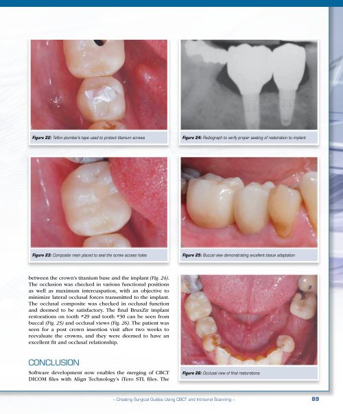
Figure 22: Teflon plumber’s tape used to protect titanium screws<br />
Figure 24: Radiograph to verify proper seating of restoration to implant<br />
Figure 23: Composite resin placed to seal the screw access holes<br />
Figure 25: Buccal view demonstrating excellent tissue adaptation<br />
between the crown’s titanium base and the implant (Fig. 24).<br />
The occlusion was checked in various functional positions<br />
as well as maximum intercuspation, with an objective to<br />
minimize lateral occlusal forces transmitted to the implant.<br />
The occlusal composite was checked in occlusal function<br />
and deemed to be satisfactory. The final BruxZir implant<br />
restorations on tooth #29 and tooth #30 can be seen from<br />
buccal (Fig. 25) and occlusal views (Fig. 26). The patient was<br />
seen for a post crown insertion visit after two weeks to<br />
reevaluate the crowns, and they were deemed to have an<br />
excellent fit and occlusal relationship.<br />
Conclusion<br />
Software development now enables the merging of CBCT<br />
DICOM files with Align Technology’s iTero STL files. The<br />
Figure 26: Occlusal view of final restorations<br />
– Creating Surgical Guides Using CBCT and Intraoral Scanning – 89


