THE FINGERPRINT SOURCEBOOK - Crime Scene Investigator ...
THE FINGERPRINT SOURCEBOOK - Crime Scene Investigator ...
THE FINGERPRINT SOURCEBOOK - Crime Scene Investigator ...

Create successful ePaper yourself
Turn your PDF publications into a flip-book with our unique Google optimized e-Paper software.
C H A P T E R<br />
C O N T E N T S<br />
3 3.1 Introduction<br />
EMBRYOLOGY AND<br />
MORPHOLOGY OF<br />
FRICTION RIDGE SKIN<br />
Kasey Wertheim<br />
4 3.2 Embryology: Establishing<br />
Uniqueness and Pattern Formation<br />
in the Friction Ridge Skin<br />
5 3.3 Limb Development<br />
7 3.4 Differentiation of the Friction<br />
Ridge Skin<br />
8 3.5 Primary Ridge Formation<br />
11 3.6 Secondary Ridge<br />
Formation<br />
12 3.7 Pattern Formation<br />
18 3.8 Genetics<br />
21 3.9 Uniqueness:<br />
Developmental Noise<br />
22 3.10 Summary: Keys to<br />
Uniqueness and<br />
Pattern Formation<br />
22 3.11 Reviewers<br />
24 3.12 References<br />
3–1
CHAPTER 3<br />
EMBRYOLOGY AND<br />
MORPHOLOGY OF<br />
FRICTION RIDGE SKIN<br />
Kasey Wertheim<br />
Embryology and Morphology of Friction Ridge Skin C H A P T E R 3<br />
3.1 Introduction<br />
Friction ridge skin has unique features that persist from<br />
before birth until decomposition after death. Upon contact<br />
with a surface, the unique features of friction ridge skin may<br />
leave an impression of corresponding unique details. Two<br />
impressions can be analyzed, compared, and evaluated,<br />
and if sufficient quality and quantity of detail is present (or<br />
lacking) in a corresponding area of both impressions, a competent<br />
examiner can effect an individualization or exclusion<br />
(identify or exclude an individual). The analysis, comparison,<br />
evaluation, and verification (ACE-V) methodology, combined<br />
with the philosophy of quantitative–qualitative examinations,<br />
provide the framework for practical application of the friction<br />
ridge examination discipline. But at the heart of the discipline<br />
is the fundamental principle that allows for conclusive<br />
determinations: the source of the impression, friction ridge<br />
skin, is unique and persistent.<br />
Empirical data collected in the medical and forensic communities<br />
continues to validate the premises of uniqueness<br />
and persistence. One hundred years of observations and<br />
statistical studies have provided critical supporting documentation<br />
of these premises. Detailed explanations of the<br />
reasons behind uniqueness and persistence are found in<br />
specific references that address very small facets of the<br />
underlying biology of friction ridge skin. This chapter brings<br />
together these references under one umbrella for the latent<br />
print examiner to use as a reference in understanding<br />
why friction ridge skin is unique and persistent.<br />
The basis of persistence is found in morphology and<br />
physiology; the epidermis faithfully reproduces the threedimensional<br />
ridges due to physical attachments and<br />
constant regulation of cell proliferation and differentiation.<br />
But, the basis of uniqueness lies in embryology; the unique<br />
features of the skin are established between approximately<br />
10.5 and 16 weeks estimated gestational age (EGA) due to<br />
developmental noise.<br />
3–3
C H A P T E R 3 Embryology and Morphology of Friction Ridge Skin<br />
3.2 Embryology: Establishing<br />
Uniqueness and Pattern Formation<br />
in the Friction Ridge Skin<br />
3.2.1 Introduction to Embryology<br />
The uniqueness of friction ridge skin falls under the larger<br />
umbrella of biological uniqueness. No two portions of any<br />
living organism are exactly alike. The intrinsic and extrinsic factors<br />
that affect the development of any individual organ, such<br />
as human skin, are impossible to duplicate, even in very small<br />
areas. The uniqueness of skin can be traced back to the late<br />
embryological and early fetal development periods.<br />
3.2.2 Early Embryological Development:<br />
0–2 Weeks EGA (Raven and Johnson, 1992,<br />
pp 1158–1159)<br />
The process of embryological development begins with fertilization<br />
of the egg and continues through a period of rapid<br />
cell division called “cleavage”. In mammalian eggs, an inner<br />
cell mass is concentrated at one pole, causing patterned alterations<br />
during cleavage. Although egg cells contain many<br />
different substances that act as genetic signals during early<br />
embryological development, these substances are not distributed<br />
uniformly. Instead, different substances tend to be<br />
clustered at specific sites within the growing embryo. During<br />
growth, signal substances are partitioned into different<br />
daughter cells, endowing them with distinct developmental<br />
instructions. In this manner, the embryo is prepatterned to<br />
continue developing with unique cell orientation.<br />
3.2.3 Late Embryological Development:<br />
3–8 Weeks EGA (Raven and Johnson, 1992,<br />
pp 1160–1164)<br />
The first visible results of prepatterning can be seen<br />
immediately after completion of the cleavage divisions<br />
as different genes are activated. Certain groups of cells<br />
move inward toward the center of the sphere in a carefully<br />
orchestrated migration called “gastrulation”. This process<br />
forms the primary tissue distinctions between ectoderm,<br />
endoderm, and mesoderm. The ectoderm will go on to<br />
form epidermis, including friction ridge skin; the mesoderm<br />
will form the connective tissue of the dermis, as well as<br />
muscle and elements of the vascular system; and the<br />
endoderm goes on to form the organs.<br />
3–4<br />
Once specialized, the three primary cell types begin their<br />
development into tissue and organs. The process of tissue<br />
differentiation begins with neurulation, or the formation of<br />
the notochord (the precursor to the spinal cord and brain)<br />
as well as the neural crest (the precursor to much of the<br />
embryo’s nervous system). Segmented blocks of tissue<br />
that become muscles, vertebrae, and connective tissue<br />
form on either side of the notochord. The remainder of the<br />
mesoderm moves out and around the inner endoderm,<br />
forming a hollow chamber that will ultimately become the<br />
lining of the stomach and intestines.<br />
During late embryological development, the embryo<br />
undergoes “morphogenesis”, or the formation of shape.<br />
Limbs rapidly develop from about 4 weeks EGA, and the<br />
arms, legs, knees, elbows, fingers, and toes can all be<br />
seen in the second month. During this time, the hand<br />
changes from a paddlelike form to an adult form, including<br />
the formation of the fingers and rotation of the thumb. Also<br />
during this time, swellings of mesenchyme called “volar<br />
pads” appear on the palms of the hands and soles of the<br />
feet. Within the body cavity, the major organs such as the<br />
liver, pancreas, and gall bladder become visible. By the end<br />
of week 8, the embryo has grown to about 25 millimeters<br />
in length and weighs about 1 gram.<br />
3.2.4 Fetal Growth: 9–12 Weeks EGA<br />
During the third month, the embryo’s nervous system and<br />
sense organs develop, and the arms and legs begin to<br />
move. Primitive reflexes such as sucking are noticed, and<br />
early facial expressions can be visualized. Friction ridges<br />
begin to form at about 10.5 weeks EGA and continue to<br />
mature in depth as the embryo passes into the second trimester.<br />
From this point on, the development of the embryo<br />
is essentially complete, and further maturation is referred<br />
to as fetal growth rather than embryonic development.<br />
3.2.5 Second Trimester<br />
The second trimester is marked by significant growth to<br />
175 millimeters and about 225 grams. Bone growth is<br />
very active, and the body becomes covered with fine hair<br />
called lanugo, which will be lost later in development. As<br />
the placenta reaches full development, it secretes numerous<br />
hormones essential to support fetal bone growth and<br />
energy. Volar pads regress and friction ridges grow until<br />
about 16 weeks EGA, when the minutiae become set.
Sweat glands mature, and the epidermal–dermal ridge<br />
system continues to mature and grow in size. By the end<br />
of the second trimester, sweat ducts and pores appear<br />
along epidermal ridges, and the fetus begins to undergo<br />
even more rapid growth.<br />
3.2.6 Third Trimester<br />
In the third trimester, the fetus doubles in weight several<br />
times. Fueled by the mother’s bloodstream, new brain cells<br />
and nerve tracts actively form. Neurological growth continues<br />
long after birth, but most of the essential development<br />
has already taken place in the first and second trimesters.<br />
The third trimester is mainly a period for protected growth.<br />
3.3 Limb Development<br />
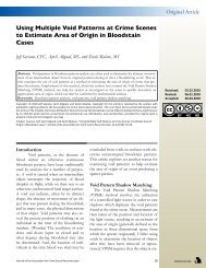
3.3.1 Hand Development<br />
During the initial phases of formation, the hand undergoes<br />
significant changes in topography. Until approximately 5–6<br />
weeks EGA, the hand appears as a flat, paddlelike structure<br />
with small protrusions of tissue that will become fingers.<br />
From 6 to 7 weeks EGA, these finger protrusions in the<br />
hand plate begin to form muscle and cartilage that will<br />
become bone at later stages of hand growth (Figure 3–1).<br />
From 7 to 8 weeks EGA, the fingers begin to separate and<br />
the bone begins to “ossify” or harden. By 8 weeks EGA,<br />
the joints begin to form between the bones of the hand,<br />
and the external hand morphology appears similar in proportion<br />
to that of an infant.<br />
Embryology and Morphology of Friction Ridge Skin C H A P T E R 3<br />
FIGURE 3–1<br />
Growth of the hand progresses from (A) a paddlelike<br />
form (magnification = 19.5 X), (B) continues as<br />
the fingers separate (magnification = 17.3 X) and<br />
(C) the volar pads become prominent (magnification<br />
= 7.7 X), and (D) achieves infantlike appearance<br />
by 8 weeks EGA (magnification = 4.2 X). (Reprinted<br />
with permission from Cummins (1929).)<br />
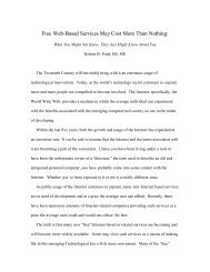
3.3.2 Volar Pad Development<br />
Volar pads (Figure 3–2) are transient swellings of tissue<br />
called mesenchyme under the epidermis on the palmar<br />
surface of the hands and soles of the feet of the human<br />
fetus (Figure 3–3).<br />
The interdigital pads appear first, around 6 weeks EGA,<br />
followed closely in time by the thenar and hypothenar<br />
pads. At approximately 7–8 weeks EGA, the volar pads<br />
begin to develop on the fingertips, starting with the thumb<br />
and progressing toward the little finger in the same radioulnar<br />
gradient that ridge formation will follow. Also at<br />
about 8 weeks EGA, the thenar crease begins to form in<br />
the palm, followed by the flexion creases in the fingers at<br />
around 9 weeks EGA (Kimura, 1991).<br />
3.3.3 Volar Pad “Regression”<br />
The pads remain well rounded during their rapid growth<br />
around 9–10 weeks EGA, after which they begin to demonstrate<br />
some individual variation in both shape and position<br />
(Babler, 1987; Burdi et al., 1979; Cummins, 1926, 1929).<br />
During the period from 8 to 10 weeks EGA, thumb rotation<br />
is achieved (Lacroix et al., 1984, p 131). Also at about 10<br />
weeks EGA, the flexion creases of the toes begin formation,<br />
followed at about 11 weeks EGA by the distal transverse<br />
flexion crease in the palm, and at about 13 weeks<br />
EGA by the proximal transverse flexion crease in the palm<br />
(Kimura, 1991).<br />
As a result of the volar pads’ slowing growth, their contour<br />
becomes progressively less distinct on the more rapidly<br />
growing surface (Figure 3–4). This process has been<br />
defined as “regression” (Lacroix et al., 1984, pp 131–133),<br />
3–5
C H A P T E R 3 Embryology and Morphology of Friction Ridge Skin<br />
3–6<br />
FIGURE 3–2<br />
A low-power scanning electron microscope<br />
view of a fetal hand displaying prominent<br />
digital and palmar volar pads. (Reprinted with<br />
permission from Carlson (1999), p 152.)<br />
FIGURE 3–3<br />
Normally, 11 volar pads develop and<br />
regress on each limb (one on each digit<br />
and six on the larger surface of the palm<br />
or sole). The hypothenar pad of the palm<br />
is divided into distal (Hd) and proximal<br />
(Hp) portions. The first (I) interdigital<br />
volar pad is also divided into two<br />
portions, making a total of 13 potential<br />
elevations on each surface. On plantar<br />
surfaces, the proximal portions of the<br />
hypothenar pad (Hp) and the thenar<br />
pad (Thp) are absent, leaving 11 distinct<br />
plantar elevations. (Reprinted with<br />
permission from Cummins (1929), p 114.)<br />
FIGURE 3–4<br />
Drawings that represent a volar pad from<br />
initial formation until complete regression,<br />
excluding growth of the size of the finger.<br />
Actual EGA values are highly variable and<br />
are included only as approximations in this<br />
figure. (Reprinted with permission from<br />
Wertheim and Maceo (2002), p 61.)
ut it is important to understand that the pad is not actually<br />
shrinking; rather, the volar pads are overtaken by the faster<br />
growth of the larger surrounding surface. The volar pads of<br />
the palm begin to regress as early as 11 weeks EGA,<br />
followed closely by the volar pads of the fingers. By 16<br />
weeks EGA, volar pads have completely merged with the<br />
contours of the fingers, palms, and soles of the feet<br />
(Cummins, 1929, p 117).<br />
3.4 Differentiation of the<br />
Friction Ridge Skin<br />
3.4.1 Development of the Epidermis<br />
The primitive epidermis is established at approximately<br />
1 week EGA, when ectoderm and endoderm are separately<br />
defined. A second layer of epidermis forms at about<br />
4–5 weeks EGA. The outermost of the three layers is the<br />
periderm. The middle layer, which is the actual epidermis,<br />
is composed of basal keratinocytes (named because of the<br />
keratins these cells manufacture). At about 8 weeks EGA,<br />
the basal cells between the epidermis and the dermis<br />
begin to consistently divide and give rise to daughter cells<br />
that move vertically to form the first of the intermediate<br />
cell layers (Holbrook, 1991b, p 64). At this point, the embryonic<br />
epidermis is three to four cell layers thick, but it is still<br />
smooth on its outer and inner surfaces. Keratinocytes are<br />
tightly bound to each other by desmosomes, and the cells<br />
of the basal layer are attached to the basement membrane<br />
by hemidesmosomes (Holbrook, 1991a, p 5).<br />
3.4.2 Development of the Dermis<br />
The first dermal components to originate from the mesoderm<br />
are fibroblasts. These irregular branching cells<br />
secrete proteins into the matrix between cells. Fibroblasts<br />
synthesize the structural (collagen and elastic)<br />
Embryology and Morphology of Friction Ridge Skin C H A P T E R 3<br />
FIGURE 3–5<br />
Scanning electron micrograph of a resin cast<br />
of the fine vascularature of the finger of an<br />
85-year-old man shows a complex pattern of<br />
capillary loops in dermal ridges. Approximate<br />
magnification = 150 X (left) and 700 X (right).<br />
(Reprinted with permission from Montagna<br />
et al. (1992).)<br />
components that form the connective tissue matrix of<br />
the dermis. During the period 4–8 weeks EGA, many of<br />
the dermal structures begin formation. Elastic fibers first<br />
appear around 5 weeks EGA at the ultrastructural level in<br />
small bundles of 20 or fewer fibrils (Holbrook, 1991b, pp<br />
64–101). Nerve development occurs in different stages<br />
from 6 weeks EGA onwards. Neurovascular bundles and<br />
axons with growth cones are seen in the developing dermis<br />
as early as 6 weeks EGA (Moore and Munger, 1989,<br />
pp 128–130). In fact, axons can be traced to the superficial<br />
levels of the dermis, and in some cases they almost abut<br />
the basal lamina of the epidermis. By 9 weeks EGA, innervation<br />
(the appearance of nerve endings) of the epidermis<br />
has begun to occur, although there are some Merkel cells<br />
in the epidermis that are not yet associated with axons.<br />
In embryos older than 10 weeks EGA, Merkel cells are predominant<br />
in the developing epidermis, and their related<br />
axons and neurofilaments are present in the dermis<br />
(Moore and Munger, 1989, p 127; Smith and Holbrook,<br />
1986).<br />
The dermis becomes distinguishable from deeper subcutaneous<br />
tissue due largely to a horizontal network of<br />
developing blood vessels. From 8 to 12 weeks EGA, vessels<br />
organize from dermal mesenchyme and bring muchneeded<br />
oxygen and hormones to the underside of the<br />
developing epidermis. Unlike other epidermal structures,<br />
blood vessels continue to alter with aging, as some capillary<br />
loops are lost and new ones arise from the interpapillary<br />
network. This continues into late adulthood (Figure<br />
3–5) (Smith and Holbrook, 1986).<br />
A second vascular network forms deep in the reticular dermis<br />
by about 12 weeks EGA. Unlike the developing primary<br />
ridges, the vascular network is not a permanent structure.<br />
There is significant reorganization of capillary beds during<br />
the period 8–20 weeks EGA to keep pace with skin growth;<br />
even after birth, microcirculation continues to form and remodel<br />
(Holbrook, 1991b, p 100; Smith and Holbrook, 1986).<br />
3–7
C H A P T E R 3 Embryology and Morphology of Friction Ridge Skin<br />
3.5 Primary Ridge Formation<br />
FIGURE 3–6<br />
A reconstruction of the first<br />
three-dimensional undulations that<br />
occur on the underside of the fetal<br />
volar epidermis at the epidermal–<br />
dermal junction. (Artwork by<br />
Brandon Smithson. Re-drawn<br />
from Hale (1952), p 152.)<br />
FIGURE 3–7<br />
A histological cross section of 10.5-week<br />
EGA fetal volar skin at the onset of rapid<br />
localized cellular proliferation. (Image<br />
provided by William Babler.)<br />
3.5.1 Initiation of Primary Ridge Formation<br />
At around 10–10.5 weeks EGA, basal cells of the epidermis<br />
begin to divide rapidly (Babler, 1991, p 98; Holbrook and<br />
Odland, 1975, p 17). As volar epidermal cells divide, shallow<br />
“ledges” (Hale, 1952) can be seen on the bottom of<br />
the epidermis. These ledges delineate the overall patterns<br />
that will become permanently established on the volar surfaces<br />
several weeks later (Babler, 1991, p 101; Evatt, 1906).<br />
Primary ridges are the first visual evidence of interaction<br />
between the dermis and epidermis and are first seen forming<br />
as continuous ridges (Figure 3–6).<br />
The prevailing theory of events before the visualization<br />
of primary ridge structure involves centers of active cell<br />
proliferation (Figure 3–7), which will become the centers of<br />
sweat gland development (Babler, 1991, p 98).<br />
According to this theory, the “units” of rapidly multiplying<br />
cells increase in diameter, somewhat randomly, growing<br />
into one another (Figure 3–8) along lines of relief perpendicular<br />
to the direction of compression.<br />
Furthermore, according to this theory, as the series of<br />
localized proliferations “fuse” together, the resulting<br />
linear ridges of rapidly dividing epidermal cells fold into<br />
3–8<br />
the dermis, creating the first visible ridge structure at the<br />
epidermal–dermal junction (Ashbaugh, 1999, p 79). Another<br />
plausible theory is that developing nerves may interact<br />
with epidermal cells to stimulate clustered interactions that<br />
blend together in the early stages of ridge development.<br />
At the time of embryonic friction ridge formation, the<br />
central nervous and cardiovascular systems are undergoing<br />
a critical period of development (Hirsch, 1964). Researchers<br />
have reported innervation at the sites of ridge formation<br />
immediately preceding the appearance of friction ridges<br />
and suggest that innervation could be the trigger mechanism<br />
for the onset of proliferation (Bonnevie, 1924; Dell<br />
and Munger, 1986; Moore and Munger, 1989). Several<br />
researchers even postulate that the patterning of the<br />
capillary–nerve pairs at the junction of the epidermis and<br />
the dermis is the direct cause of primary ridge alignment<br />
(Dell and Munger, 1986; Hirsch and Schweichel, 1973;<br />
Moore and Munger, 1989; Morohunfola et al., 1992).<br />
Early research on pattern distribution established “developmental<br />
fields”, or groupings of fingers on which patterns<br />
had a greater tendency to be similar (Meier, 1981; Roberts,<br />
1982; Siervogel et al., 1978). Later discoveries confirmed<br />
the neurological relation of spinal cord sections C–6, C–7,<br />
and C–8 to innervation of the fingers (Heimer, 1995).<br />
Specifically, Kahn and colleagues (2001) reported that a
large ridge-count difference between C–8-controlled fingers<br />
4 and 5 may predict a larger waist-to-thigh ratio and,<br />
therefore, an increased risk of some major chronic diseases<br />
such as heart disease, cancer, and diabetes. Other<br />
interesting hypotheses have been published regarding the<br />
connection between innervation and friction ridge patterning,<br />
but the main consideration for the purposes of friction<br />
ridge formation is that specific parts of the nervous system<br />
are undergoing development at the same time that ridges<br />
begin to appear on the surface of the hands.<br />
The presence of nerves and capillaries in the dermis before<br />
friction ridge formation may be necessary for friction ridge<br />
proliferation. It would seem that complex simultaneous<br />
productions such as friction ridge formation would benefit<br />
from being in communication with the central nervous<br />
system or the endocrine and exocrine (hormone) systems<br />
(Smith and Holbrook, 1986). However, it is doubtful that<br />
nerves or capillaries independently establish a map that<br />
directly determines the flow of the developing friction<br />
ridges. It seems more likely that the alignment of the<br />
nerves and capillaries is directed by the same stresses<br />
and strains on the developing hand that establish ridge<br />
alignment (Babler, 1999; Smith and Holbrook, 1986). It is<br />
well recognized in cell biology that physical pressure on a<br />
cellular system can trigger electrochemical changes within<br />
Embryology and Morphology of Friction Ridge Skin C H A P T E R 3<br />
FIGURE 3–8<br />
These drawings represent the theory<br />
that just before ridge formation, localized<br />
cellular proliferations grow together into<br />
what will appear as ridges at around 10.5<br />
weeks EGA. (Reprinted with permission<br />
from Wertheim and Maceo (2002), p 49.)<br />
that system. Merkel cells occupy the epidermis just prior<br />
to innervation along those pathways (Holbrook, 1991a),<br />
suggesting that even before ridge formation, the stresses<br />
created by the different growth rates of the dermis and<br />
epidermis are causing differential cell growth along invisible<br />
lines that already delineate pattern characteristics (Loesch,<br />
1973). Regardless of the trigger mechanism controlling the<br />
onset of the first primary ridge proliferations, the propagation<br />
of primary ridges rapidly continues.<br />
3.5.2 Propagation of Primary Ridge Formation<br />
Primary ridges mature and extend deeper into the dermis<br />
(Figure 3–9) for a period of approximately 5.5 weeks, from<br />
their inception at 10.5 weeks EGA until about 16 weeks<br />
EGA. The cell growth during this phase of development<br />
is along the primary ridge, in what has been labeled the<br />
“proliferative compartment”. The proliferative compartment<br />
encompasses basal and some suprabasal cells, ultimately<br />
governed by stem cells, and is responsible for new skin cell<br />
production of the basal layer of skin (Lavker and Sun, 1983).<br />
3.5.3 Minutiae Formation<br />
Although the exact mechanisms for formation of minutiae<br />
are unclear, the separate accounts of many researchers<br />
3–9
C H A P T E R 3 Embryology and Morphology of Friction Ridge Skin<br />
FIGURE 3–9<br />
Histological cross section of fetal volar<br />
skin between 10.5 and 16 weeks EGA. During<br />
this time, primary ridges (as marked by the<br />
arrow) increase in depth and breadth.<br />
(Image provided by William Babler.)<br />
FIGURE 3–10<br />
Drawings that illustrate the theoretical formation<br />
of minutiae arising from expansion of the volar<br />
surface during the critical stage (frames 1–10)<br />
and continuing to increase in size after secondary<br />
ridge formation (frames 11–16). (Reprinted with<br />
permission from Wertheim and Maceo (2002), p 51.)<br />
who have examined fetal tissue allow for a fairly accurate<br />
reconstruction of the morphogenesis of friction ridges in<br />
successive stages of the development process. Figure<br />
3–10 illustrates the process of minutiae formation as<br />
hypothesized by a general consensus of the literature.<br />
Many events happen during this rapid period of primary<br />
ridge growth. The finger rapidly expands, new primary<br />
ridges form across the finger, and the existing primary<br />
ridges begin to separate because of growth of the digit.<br />
As existing ridges separate, the tendency of the surface to<br />
be continually ridged creates a demand for new ridges.<br />
Hale reports that new ridges pull away from existing<br />
primary ridges to fill in these gaps, creating bifurcations by<br />
3–10<br />
mechanical separation. Ending ridges form when a developing<br />
ridge becomes sandwiched between two established<br />
ridges. According to this theory, “fusion between adjacent<br />
ridges [which have already formed] seems improbable,<br />
although there is no evidence for or against this process”<br />
(Hale, 1952, p 167).<br />
Other models explain ridge detail in nature as a chemical<br />
reaction–suppression scheme in which morphogens react<br />
and diffuse through cells, causing spatial patterns (Murray,<br />
1988, p 80). According to these models, hormones circulate<br />
first through newly formed capillaries just before ridge formation<br />
in the epidermis, offering another potential factor in<br />
the genesis of ridge formation (Smith and Holbrook, 1986).
upper layer of the epidermis<br />
pressure pressure<br />
dermis<br />
“bed” of springs<br />
A recent model of the process of friction ridge morphogenesis<br />
has been likened to mechanical instability (Kücken<br />
and Newell, 2005). Building on the folding hypothesis of<br />
Kollmann (1883) and Bonnevie (1924), Kücken and Newell<br />
(2005) consider the basal layer as “an overdamped<br />
elastic sheet trapped between the neighboring tissues of<br />
the intermediate epidermis layer and the dermis”, which<br />
they mathematically model as “beds of weakly nonlinear<br />
springs” (Figure 3–11).<br />
Their computer program models the results of forcing<br />
enough compressive stress to cause a buckling instability<br />
on a virtual three-dimensional elastic sheet constrained by<br />
fixed boundaries on two sides. The resulting ridge patterns<br />
are similar to all three major fingerprint pattern types oriented<br />
by the upper fixed boundary of the nailbed and the<br />
lower fixed boundary of the distal interphalangeal flexion<br />
crease (Figure 3–12).<br />
Regardless of the exact mechanism of minutiae formation<br />
(mechanical or static; fusion or chemical), the exact<br />
location of any particular bifurcation or ridge ending within<br />
the developing ridge field is governed by a random series<br />
of infinitely interdependent forces acting across that<br />
particular area of skin at that critical moment. Slight differences<br />
in the mechanical stress, physiological environment,<br />
or variation in the timing of development could significantly<br />
affect the location of minutiae in that area of skin.<br />
Embryology and Morphology of Friction Ridge Skin C H A P T E R 3<br />
FIGURE 3–11<br />
A drawing that represents the state of the<br />
epidermal–dermal boundary just before<br />
ridge formation. (Reprinted with permission<br />
from Kücken and Newell (2005), p 74.)<br />
FIGURE 3–12<br />
Computer simulations demonstrating<br />
that bounded stress fields across a threedimensional<br />
spherical surface produce<br />
fingerprintlike patterns. (Reprinted with<br />
permission from Kücken and Newell<br />
(2005), p. 79.)<br />
3.6 Secondary Ridge Formation<br />
3.6.1 Initiation of Secondary Ridge Formation<br />
By 15 weeks EGA, the primary ridges are experiencing<br />
growth in two directions: the downward penetration of the<br />
sweat glands and the upward push of new cell growth.<br />
Generally, the entire volar surface is ridged by 15 weeks<br />
EGA. Okajima (1982) shows a fully ridged palm of a<br />
14-week-old fetus (Figure 3–13).<br />
Between 15 and 17 weeks EGA, secondary ridges appear<br />
between the primary ridges on the underside of the epidermis<br />
(Babler, 1991, p 98). Secondary ridges are also cell<br />
proliferations resulting in downfolds of the basal epidermis.<br />
At this time in fetal development, the randomly located<br />
minutiae within the friction ridge pattern become permanently<br />
set (Hale, 1952, pp 159–160), marking the end of new<br />
primary ridge formation (Figure 3–14) (Babler, 1990, p 54).<br />
3.6.2 Propagation of Secondary<br />
Ridge Formation<br />
As the secondary ridges form downward and increase<br />
the surface area of attachment to the dermis, the primary<br />
ridges are pushing cells toward the surface to keep pace<br />
with the growing hand. These two forces, in addition to cell<br />
adhesion, cause infolding of the epidermal layers above the<br />
attachment site of the secondary ridges (Hale, 1952). As<br />
3–11
C H A P T E R 3 Embryology and Morphology of Friction Ridge Skin<br />
FIGURE 3–13<br />
Image of a 14-week EGA fetal palm stained with<br />
toluidine blue. (Reprinted with permission from<br />
Okajima (1982), p 185 (no magnification given).)<br />
FIGURE 3–14<br />
A histological cross section of fetal volar skin<br />
representing the onset of secondary ridge<br />
formation between maturing primary ridges<br />
(as marked by the arrows) at about 16 weeks<br />
EGA. (Image provided by William Babler.)<br />
secondary ridges continue to mature from 16 to 24 weeks<br />
EGA, this structure is progressively mirrored on the surface<br />
of friction ridge skin as the furrows (Burdi et al., 1979, pp<br />
25–38) (Figure 3–15).<br />
3.6.3 Formation of Dermal Papillae<br />
Dermal papillae are the remnants of dermis left projecting<br />
upward into the epidermis when anastomoses bridge primary<br />
and secondary ridges (Figures 3–16 and 3–17). They<br />
begin to form at approximately 23 weeks EGA (Okajima,<br />
3–12<br />
1975) and continue to become more complex throughout<br />
fetal formation and even into adulthood (Chacko and Vaidya,<br />
1968; Misumi and Akiyoshi, 1984).<br />
3.7 Pattern Formation<br />
3.7.1 Shape of the Volar Pad<br />
It is observed throughout the physical world that ridges<br />
tend to align perpendicularly to physical compression<br />
across a surface (Figure 3–18).
Ridges also form transversely to the lines of growth stress<br />
in friction skin. The predominant growth of the hand is<br />
longitudinal (lengthwise) and ridges typically cover the volar<br />
surface transversely (side to side). This phenomenon is<br />
seen in the ridge flow across the phalanges.<br />
Bonnevie first hypothesized in 1924 that volar pad height<br />
affects friction ridge patterns (Bonnevie, 1924, p 4). Disruptions<br />
in the shape of the volar surfaces of the hands and<br />
feet create stresses in directions other than longitudinal.<br />
The ridges flow in a complex manner across these threedimensional<br />
structures.<br />
Embryology and Morphology of Friction Ridge Skin C H A P T E R 3<br />
FIGURE 3–15<br />
A reconstruction of the secondary ridges<br />
continuing to form on the underside of the<br />
fetal volar epidermis between existing primary<br />
ridges with sweat ducts. (Artwork by Brandon<br />
Smithson. Re-drawn from Hale (1952), p 153.)<br />
FIGURE 3–16<br />
A reconstruction of the underside of the<br />
epidermis of fetal volar skin that represents<br />
anastomoses bridging primary and secondary<br />
ridges and cordoning off sections of dermis<br />
that remain protruding upward as “dermal papillae”<br />
or “papillae pegs”. (Artwork by Brandon<br />
Smithson. Re-drawn from Hale (1952), p 154.)<br />
FIGURE 3–17<br />
A scanning electron microscope view of the<br />
complex understructure of human epidermis<br />
as the dermis has been removed (inverted).<br />
Magnification (approximate) = 8 X (left) and 80<br />
X (right). (Reprinted with permission from<br />
Montagna and Parakkal (1974), pp 34–35.)<br />
The distinction between the size, height, and shape of the<br />
volar pad, and the effects of differences in each of these<br />
elements on a friction ridge pattern, is a difficult topic to<br />
study (Chakraborty, 1991; Jamison, 1990; Mavalwala et al.,<br />
1991). However, almost all research points to the conclusion<br />
that the shape of the volar pad influences the stress<br />
across the skin that directs ridge alignment. One contrary<br />
viewpoint to this conclusion exists. In 1980, Andre G. de<br />
Wilde proposed a theory that pattern formation is directed<br />
much earlier in fetal life, before volar pads form, while the<br />
hand is still in a paddlelike shape (De Wilde, 1980). He<br />
3–13
C H A P T E R 3 Embryology and Morphology of Friction Ridge Skin<br />
FIGURE 3–18<br />
When tension is applied across the<br />
top of a semiflexible membrane, forces<br />
of compression occur on the bottom. The<br />
natural relief of compression forces<br />
creates ridges forming transversely to the<br />
stress. (Reprinted with permission from<br />
Wertheim and Maceo (2002), p 57.)<br />
FIGURE 3–19<br />
The loxodrome results when an elastic film<br />
is stretched evenly over a hemisphere. Ridges<br />
form concentrically around the apex of the<br />
membrane disruption. The mathematical<br />
formula for this pattern can be found in tensor<br />
calculus, a field that offers much promise<br />
in predicting ridge formation across volar<br />
surfaces. (Reprinted with permission from<br />
Wertheim and Maceo (2002), p 62.)<br />
hypothesized that ridges direct the size and shape of the<br />
volar pads. However, no other theoretical or empirical<br />
support for this theory could be found. All other research<br />
indicates that friction ridges align according to volar pad<br />
shape and symmetry at approximately 10.5 weeks EGA.<br />
3.7.1.1 Symmetrical Volar Pad. The growth and regression<br />
of the volar pads produce variable physical stresses across<br />
the volar surface that affect the alignment of the ridges<br />
as the ridges first begin to form. Whether ridge flow will<br />
conform to a whorl or a loop pattern appears highly correlated<br />
with the symmetry of the stress across the surface<br />
of the finger. If the volar pad and other elements of finger<br />
growth are symmetrical during the onset of primary ridge<br />
formation, then a symmetrical pattern (a whorl or an arch)<br />
will result. Ridges will form concentrically around the apex<br />
of a volar pad that is high and round when the generating<br />
layer of friction ridge skin first begins to rapidly produce<br />
skin cells. The ridge flow from a symmetrical volar pad conforms<br />
to the navigational pattern of the loxodrome (Figure<br />
3–19) (Mulvihill and Smith, 1969; Elie, 1987). Research in<br />
both the medical and mathematical fields suggests that<br />
this same physical model applies across the entire volar<br />
surface of the hands and feet (Cummins, 1926, 1929;<br />
Loesch, 1973; Penrose and O’Hara, 1973).<br />
3–14<br />
3.7.1.2 Asymmetrical Volar Pad. The degree of asymmetry<br />
of the finger volar pad when ridges first begin to form<br />
determines the asymmetry of the pattern type. Many researchers<br />
have reported that asymmetrical “leaning” pads<br />
form looping patterns and that low or absent volar pads<br />
form arch patterns (Cummins, 1926, p 138). Babler perhaps<br />
conducted the most scientific validation of the correlation<br />
between pad symmetry and pattern type through extensive<br />
examination of fetal abortuses (Babler, 1978).<br />
Cummins published an extensive analysis of malformed<br />
hands to demonstrate the effect of the growth and topology<br />
of the hand on ridge direction (Cummins, 1926).<br />
Cummins also concluded that ridge direction is established<br />
by the contours of the hands and feet at the time of ridge<br />
formation. Penrose examined friction ridge pattern formation<br />
from a mathematical perspective, arriving at the same<br />
conclusion (Loesch, 1973; Penrose and Plomley, 1969).<br />
More recently, Kücken and Newell (2005) modeled stress<br />
fields across bounded three-dimensional, spherical virtual<br />
surfaces, creating relatively accurate-appearing ridge patterns<br />
(Figure 3–20).<br />
If the volar pad and other growth factors of the finger are<br />
asymmetrical during the critical stage, then that same
degree of asymmetry will be reflected in the ridge flow<br />
of the resulting pattern. This biological process cannot be<br />
thought of as limited to the extremes of volar pad regression,<br />
occurring either completely symmetrically or asymmetrically<br />
(leaning all the way to one side). In fact, there is a continuum<br />
involved from whorl patterns to loop patterns. Figure 3–21<br />
illustrates several patterns from different individuals whose<br />
volar pads were theoretically the same approximate size<br />
at the critical stage (i.e., the volar pads had similar ridge<br />
counts), but differed in the degree of their symmetry.<br />
Subtle variations in the symmetry of a volar pad could<br />
affect the formation of a whorl pattern versus a central<br />
pocket loop whorl pattern, or a central pocket loop whorl<br />
pattern versus a loop pattern. Any one of the numerous<br />
genetic or environmental factors present during the critical<br />
stage could cause a slight deviation in the normal developmental<br />
symmetry of the volar pad and, therefore, affect the<br />
resulting pattern type.<br />
3.7.2 Size of the Volar Pad<br />
3.7.2.1 Pattern Size. The size, particularly the height, of the<br />
volar pad during primary ridge formation affects the ridge<br />
count from the core to the delta of normal friction ridge<br />
Embryology and Morphology of Friction Ridge Skin C H A P T E R 3<br />
FIGURE 3–20<br />
Computer models demonstrating directional<br />
field points (tic marks) stretched in the direction<br />
of stress. The white spot illustrates the degree<br />
of compressive stress and the location where<br />
ridge formation takes place first (center of the<br />
white portion represents the apex of the pad).<br />
(Reprinted with permission from Kücken and<br />
Newell (2005), p 79.)<br />
FIGURE 3–21<br />
Six different fingerprint patterns from different<br />
individuals, representing the continuum of volar<br />
pad symmetry at the onset of friction ridge<br />
proliferation, ranging from (1) nearly symmetrical<br />
to (6) very displaced. (Reprinted with permission<br />
from Wertheim and Maceo (2002), p 69.)<br />
patterns (Bonnevie, 1924; Mulvihill and Smith, 1969; Siervogel<br />
et al., 1978). Researchers have observed that ridges<br />
that form on high, pronounced volar pads conform to the<br />
surface as high-count whorl patterns. Conversely, ridges<br />
that form on a finger with a low or absent volar pad create<br />
low-count or arch-type patterns (Babler, 1987, pp 300–301).<br />
Holt (1968) reported that the total finger ridge count (TFRC)<br />
of all 10 fingers, taken by adding the ridge counts from<br />
the core to the delta in loops, or the core toward the radial<br />
delta in whorls, is the most inheritable feature in dermatoglyphics.<br />
This combined information points directly to the<br />
conclusion that timing events related to volar pad and friction<br />
ridge formation affect friction ridge patterns.<br />
3.7.2.2 Timing Events. The ridge count of a friction ridge<br />
pattern is related to two different events: the timing of the<br />
onset of volar pad regression and the timing of the onset<br />
of primary ridge formation. Differences in the timing of<br />
either event will affect the ridge count of that particular<br />
pattern. For example, early onset of volar pad regression<br />
would lead to a volar pad that was in a more regressed<br />
state at the time of the onset of primary ridge formation,<br />
and a relatively low-ridge-count pattern (or arch) would<br />
likely result. Conversely, overall late onset of volar pad regression<br />
would mean that the pad was still relatively large<br />
3–15
C H A P T E R 3 Embryology and Morphology of Friction Ridge Skin<br />
FIGURE 3–22<br />
Chart A illustrates the effects of two<br />
independent timing events on the<br />
resulting ridge count of a friction ridge<br />
pattern. Chart B illustrates their<br />
combined effects on pattern ridge count.<br />
(Reprinted with permission from<br />
Wertheim and Maceo (2002), p 65.)<br />
when primary ridges began forming, and a high-ridge-count<br />
pattern would more likely result (Figure 3–22). This theory<br />
is supported by a study that found that “late maturers” had<br />
higher-than-average ridge counts, and “early maturers” had<br />
lower-than-average ridge counts (Meier et al., 1987).<br />
If the onset of volar pad regression occurred at the normal<br />
time, then earlier-than-average onset of primary ridge<br />
formation would occur on a larger-than-average volar pad,<br />
leading to a higher-than-average ridge count. Likewise,<br />
later-than-average onset of primary ridge formation would<br />
occur on a smaller-than-average volar pad, leading to a<br />
lower-than-average ridge count (Figure 3–22A). When both<br />
early and late timing of both factors are taken into account,<br />
the results become even more complex (Figure 3–22B).<br />
To make matters even more complex, the size of the volar<br />
pad with respect to the finger is also affected by many<br />
factors. Diet and chemical intake of the mother (Holbrook,<br />
1991b), hormone levels (Jamison, 1990), radiation levels<br />
(Bhasin, 1980), and any other factors that affect the growth<br />
rate of the fetus during the critical stage could all indirectly<br />
affect the ridge counts of the developing friction ridges on<br />
the finger. It is important to remember that anything that<br />
affects the tension across the surface of the finger could<br />
affect the resulting ridge alignment and pattern type. However,<br />
Holt’s findings seem to indicate that timing events,<br />
rather than environmental factors, play the dominant role<br />
in determining TFRC (Holt, 1968).<br />
3.7.2.3 Delta Placement. The onset of cellular proliferation,<br />
which begins primary ridge formation, occurs first in<br />
three distinct areas: (1) the apex of the volar pad (which<br />
corresponds to the core of the fingerprint pattern); (2) the<br />
distal periphery, or tip of the finger (near the nailbed); and<br />
3–16<br />
High<br />
Ridge<br />
Count<br />
Low<br />
Early Late<br />
Timing<br />
Onset of<br />
Volar Pad<br />
Regression<br />
Onset of<br />
Primary<br />
Ridge<br />
Formation<br />
Late<br />
Onset of<br />
Volar Pad<br />
Regression<br />
Early<br />
Larger than<br />
Average Ridge Count<br />
Average Ridge Count<br />
Smaller than Average<br />
Ridge Count<br />
Early Late<br />
Onset of Friction<br />
Ridge Proliferation<br />
(3) the distal interphalangeal flexion crease area (below the<br />
delta(s) in a fingerprint) (Figure 3–23).<br />
As ridge formation continues, new proliferation occurs on<br />
the edges of the existing ridge fields in areas that do not<br />
yet display primary ridge formation. These three “fields” of<br />
ridges converge as they form, meeting in the delta area of<br />
the finger. This wavelike process of three converging fields<br />
allows for the visualization of how deltas most likely form<br />
(Figure 3–24).<br />
The concept of “converging ridge fields” also offers a way<br />
to visualize the difference between the formation of highversus<br />
low-ridge-count patterns. If ridges begin forming<br />
on the apex (center) of the pad first and proceed outward<br />
before formation begins on the tip and joint areas, then<br />
by the time the fields meet, a relatively large distance will<br />
have been traversed by the field on the apex of the pad; in<br />
that instance, a high-count pattern will be formed (Figure<br />
3–25). However, if the ridges form first on the two outermost<br />
portions and proceed inward, and formation begins<br />
at the last instant on the apex of the pad, then only a few<br />
ridges may be formed by the time the fields meet; in that<br />
instance, a very low-count pattern is observed (Figure<br />
3–26). The combined observations of different researchers<br />
examining friction ridges on the finger during the critical<br />
stage of development further support the validity of this<br />
model (Babler, 1991, 1999; Dell and Munger, 1986; Hirsch<br />
and Schweichel, 1973).<br />
3.7.3 Combined Effect of Timing and<br />
Symmetry on Ridge Formation<br />
When it is understood that timing and symmetry control<br />
two very different elements of ridge flow, it becomes easy
to see how both small and large loop and whorl patterns<br />
form. A finger pad that regresses symmetrically will form<br />
a whorl pattern, regardless of early or late timing of friction<br />
ridge formation with respect to volar pad regression. If the<br />
timing of the onset of primary ridge formation in this situation<br />
is early in fetal life, then the volar pad will still be high<br />
on the finger, and the whorl pattern will have a high ridge<br />
count. If timing is later in fetal life, after the pad has almost<br />
completely been absorbed into the contours of the finger,<br />
then a low-count whorl pattern will result. With further<br />
regression, an arch pattern will form (Figure 3–27).<br />
Likewise, asymmetrical finger pads will form loop patterns<br />
and will also be affected by timing. If ridges begin forming<br />
early with respect to volar pad regression on an asymmetrical<br />
pad, then the pad will be large, and a high-count<br />
loop will result. Later timing leads to a low-count loop or<br />
arch-type pattern (Figure 3–28). Again, volar pad placement<br />
Embryology and Morphology of Friction Ridge Skin C H A P T E R 3<br />
FIGURE 3–23<br />
A drawing depicting the normal starting<br />
locations of ridge formation and subsequent<br />
coverage across the surface of a<br />
finger. (Reprinted with permission from<br />
Wertheim and Maceo (2002), p 66.)<br />
FIGURE 3–24<br />
A drawing that depicts an easy way to<br />
visualize how deltas form from three<br />
converging ridge fields. (Reprinted with<br />
permission from Wertheim and Maceo<br />
(2002), p 66.)<br />
FIGURE 3–25<br />
A drawing that depicts the likely progression<br />
of ridges on a high-ridge-count pattern.<br />
(Reprinted with permission from Wertheim<br />
and Maceo (2002), p 67.)<br />
FIGURE 3–26<br />
A drawing that depicts the likely progression<br />
of ridges on a low-ridge-count pattern.<br />
(Reprinted with permission from Wertheim<br />
and Maceo (2002), p 67.)<br />
is not simply symmetrical or asymmetrical; a continuum of<br />
volar pad symmetry occurs and accounts for the variety of<br />
pattern types observed.<br />
A regression scheme seems to exist whereby the volar pad<br />
is symmetrical at the onset and becomes progressively<br />
more asymmetrical as it regresses. This is supported by<br />
general fingerprint pattern statistics that show that more<br />
than one-half of all fingerprint patterns are ulnar loops. More<br />
specifically, this scheme is supported by fetal research that<br />
has determined that early timing of primary ridge formation<br />
leads to a higher percentage (95 percent) of whorls (Babler,<br />
1978, p 25). Also, low- and high-ridge-count patterns occur<br />
less frequently than average-count patterns (Cowger, 1983).<br />
All research tends to indicate that volar pads regress from<br />
an early symmetrical position to an asymmetrical position<br />
later in fetal life. Although this is the norm, it is certainly not<br />
without exception, because whorl patterns with extremely<br />
3–17
C H A P T E R 3 Embryology and Morphology of Friction Ridge Skin<br />
FIGURE 3–27<br />
These different fingerprint patterns (bottom)<br />
were formed on completely different, but<br />
symmetrical, volar pads (top). The drawings on<br />
the top illustrate the likely fetal condition of the<br />
symmetrical volar pad that produced the resulting<br />
print below it. From left to right, the images<br />
show the results of the combined timing of the<br />
onset of friction ridge proliferation versus volar<br />
pad regression. (Reprinted with permission<br />
from Wertheim and Maceo (2002), p 71.)<br />
FIGURE 3–28<br />
These different fingerprint patterns (bottom)<br />
were formed on different, asymmetrical volar<br />
pads (top). The drawings on the top illustrate<br />
the likely fetal condition of the asymmetrical<br />
volar pad that produced the resulting print<br />
below it. From left to right, the images show<br />
the results of the combined timing of the onset<br />
of friction ridge proliferation versus volar pad<br />
regression. (Reprinted with permission from<br />
Wertheim and Maceo (2002), p 71.)<br />
low ridge counts and loop patterns with extremely high<br />
ridge counts can both be found with relative ease in even<br />
small collections of recorded fingerprints.<br />
3.8 Genetics<br />
3.8.1 Introduction to Genetic Diversity<br />
and Friction Ridge Skin<br />
In 1904, Inez Whipple presented research that provided<br />
a detailed theory of evolutionary progression of the volar<br />
surface (Whipple, 1904). Ashbaugh succinctly summarizes<br />
Whipple’s proposition of the evolutionary genesis of friction<br />
ridges:<br />
3–18<br />
Early mammals were covered with a scale-like<br />
skin surface. Each scale had one hair protruding<br />
from it and an accompanying oil or sebaceous<br />
gland. On volar areas, which are the bottoms of<br />
the hands and feet, hairs slowly disappeared due<br />
to surface use. The pore that was related to the<br />
hair changed from a sebaceous gland to a sweat<br />
gland. Its purpose, to keep the surface skin damp<br />
which enhanced the grip of the volar surface.<br />
Starting in all likelihood as a mutation, scales<br />
started to line up in rows and fuse together. This<br />
further assisted the grip of the skin surface by<br />
increasing friction. Through natural selection, this<br />
mutation became prevalent. Scales slowly evolved<br />
into wart-like units with pore openings near the<br />
centre. The fusing of these wart formations into<br />
rows is the predecessor to the friction ridge, the<br />
individual wart being the equivalent of a ridge dot<br />
(Ashbaugh, 1991, p 27).<br />
Fourteen years after Whipple’s phylogenetic (evolutionary<br />
history) theory was presented, researchers diverged<br />
from her theory and presented an ontogenetic (individual<br />
developmental or embryonic history) model, suggesting<br />
that fusion of warts into ridges occurs during embryonic<br />
development (Wilder and Wentworth, 1918). In 1926, Cummins<br />
refuted the ontogenetic scheme (Cummins, 1926, p<br />
134). However, Hale later included the ontogenetic model<br />
in his conclusions (Hale, 1952). Literature since that time<br />
has been mixed. Multiple researchers have demonstrated<br />
that the first visual evidence of interaction between the<br />
dermis and the epidermis is ridges, not a series of units,<br />
protruding into the dermis (Figure 3–6, p 3-8). Perhaps
with advances in technology, the theory that localized<br />
cell proliferations grow together into linear ridges before<br />
the appearance of the ridge as a structure will be demonstrated.<br />
Until then, fusion of units into ridges remains a<br />
possible model of development that could provide individuality<br />
before the appearance of the first ridge structures.<br />
The term “ridge unit” might be limited to a description<br />
of an adult sweat pore and surrounding ridge (Ashbaugh,<br />
1999, pp 25, 35), with the term “localized proliferation” being<br />
used to describe theoretical events of fetal formation<br />
(Babler, 1987, p 298).<br />
3.8.2 The Role of Genetics<br />
Every aspect of the growth and development of a single<br />
cell into a fully formed human is initiated by a genetic blueprint.<br />
The capacity to form friction ridges is inherent within<br />
the developing embryo. The patterns that these ridges<br />
form, however, are limited by nature and are defined by the<br />
fingerprint community as whorls, loops, arches, combinations<br />
and transitions of these basic patterns, or lack of a<br />
pattern (Hirsch, 1964). Although genetics may direct when<br />
and where ridges will form by providing the blueprint for<br />
proteins, nature provides the boundaries for patterning<br />
through physical mechanisms (Ball, 1999).<br />
Proteins direct cellular activity by facilitating biochemical<br />
processes within the cell. These processes depend not<br />
only on the protein derived from the gene but also on the<br />
many other nonprotein components of the cell such as<br />
sugars, lipids, hormones, inorganic elements (e.g., oxygen),<br />
inorganic compounds (e.g., nitric oxide), and minerals. Additionally,<br />
the physical environment around and within cells,<br />
including surface tension, electrical charge, and viscosity,<br />
contributes to the way the cell functions (Ball, 1999).<br />
Genetic information directs cellular function, serves as a<br />
link between generations, and influences an individual’s<br />
appearance. Some aspects of appearance are similar for<br />
each individual of that species (i.e., those characteristics<br />
that define the species). However, within the species, for<br />
each aspect of an individual’s appearance, many genes and<br />
external factors affect the final outcome of physical appearance.<br />
The genes involved with a specific attribute (e.g., skin<br />
color) produce the appropriate proteins, which in turn react<br />
with each other and with the many nongenetic components<br />
of the cell in complex biochemical pathways during<br />
the growth and development of the fetus (Ball, 1999).<br />
These biochemical pathways proceed under the omnipresent<br />
influence of external factors.<br />
Embryology and Morphology of Friction Ridge Skin C H A P T E R 3<br />
Although DNA is crucial for providing the blueprint for the<br />
development of a particular model, there are so many<br />
steps between the genesis of the DNA-encoded protein<br />
and the final product that even two individuals who originated<br />
from the same DNA would produce two completely<br />
unique models.<br />
Perhaps Jamison best describes the interplay between<br />
genes and the environment in friction ridge skin:<br />
Since dermatoglyphic formation cannot be derived<br />
solely from either genetic or environmental factors,<br />
it must result from an interaction of the two<br />
types of factors. This interaction is probably far<br />
from being simple and it most likely involves a<br />
multiple step reciprocal positive feedback relationship<br />
(Maruyama, 1963) in which either a genetically<br />
or an environmentally-based factor causes<br />
a change in the uterine environment, leading<br />
to a genetic response (perhaps in the form of a<br />
“switch mechanism”, as in Roberts (1986)), which<br />
then leads to an increasingly complex series of<br />
genetic-environmental interactive responses<br />
(Jamison, 1990, p 103).<br />
The ultimate example of the role of the environment in<br />
friction ridge formation is monozygotic twins, who share<br />
identical genetic information and very similar intrauterine<br />
environments, but on many occasions have very different<br />
patterns. The role of genetics is currently understood by<br />
the indication that several main genes, in conjunction with<br />
a number of modifying genes, may be responsible for volar<br />
patterning, but it is well established that friction ridge patterning<br />
is also affected by the environment (Chakraborty,<br />
1991; Hirsch, 1964; Loesch, 1982, 1983; Slatis et al., 1976;<br />
Weninger et al., 1976).<br />
Like many traits, genetics influences pattern formation indirectly<br />
by contributing to the timing of the onset of friction<br />
ridge skin, the timing of the onset of volar pad regression,<br />
the growth rate of the fetus, and other factors. Stresses<br />
across small areas of skin are not inherited, but rather they<br />
represent one of many environmental factors that influence<br />
pattern formation.<br />
Until recently (Chakraborty, 1991; Mavalwala et al., 1991),<br />
most researchers in the field of genetics and physical<br />
anthropology have traditionally viewed TFRC as evidence<br />
of direct genetic control of fingerprint pattern formation<br />
(Bonnevie, 1924; Holt, 1968). The research of Sara Holt<br />
(1968) regarding the inheritability of TFRC is a significant<br />
3–19
C H A P T E R 3 Embryology and Morphology of Friction Ridge Skin<br />
finding that supports the two-tiered development scheme<br />
suggested by this and other literary reviews of fingerprint<br />
pattern formation. Logic also supports this scheme. Genetically<br />
controlled timed events would be less susceptible to<br />
environmental variations, and, therefore, TFRC would be<br />
more inheritable than pattern type. Additionally, the wide<br />
range of patterns found on the palms (Malhotra, 1982)<br />
demonstrates the complex nature of factors that affect<br />
ridge alignment. Patterning and ridge counts are indirectly<br />
inherited and are not affected by only one developmental<br />
factor. However, ridge flow and ridge count are both affected<br />
by tension across the surface of growing fetal skin.<br />
3.8.3 Familial Studies<br />
3.8.3.1 Ethnic Variation. Thousands of anthropological studies<br />
have been conducted on distinct populations to identify<br />
trends in fingerprint pattern formation. Perhaps one of the<br />
most comprehensive reviews of this tremendous body of<br />
research was conducted by Jamshed Mavalwala, resulting<br />
in a 300-page bibliography of dermatoglyphic references<br />
(Mavalwala, 1977). The major result from this body of work<br />
was the demonstration that intratribal variations in friction<br />
ridge pattern frequencies were greater than intertribal<br />
variations. Likewise, intraspecies variations in primates<br />
were greater than interspecies variations. The body of<br />
literature on ethnic variation suggests that multiple genes<br />
affect pattern formation and that those genes interact with<br />
respect to final pattern characteristics.<br />
3.8.3.2 Abnormalities. The medical community has been,<br />
and continues to be, interested in dermatoglyphics (Durham<br />
et al., 2000; Kahn et al., 2001; Schaumann and Opitz,<br />
1991) and creases (Kimura, 1991) as indicators of abnormal<br />
fetal development during the critical stage. Although there<br />
is evidence that interest has waned in recent decades<br />
(Reed, 1991), it was reported in 1991 that significantly<br />
more than 3,500 articles in the international literature dealt<br />
with different aspects of dermatoglyphics (Mavalwala,<br />
1977). Although many articles relate certain medical conditions<br />
to statistically significant occurrences of abnormal<br />
ridge pattern combinations, many researchers still heed<br />
the warning that “dermatoglyphics may be of uncertain, if<br />
any, diagnostic value due to the lack of a specific dermatoglyphic<br />
stereotype in individual patients” (Schaumann,<br />
1982, pp 33–34).<br />
Harold Cummins was perhaps one of the most prominent<br />
researchers on the specific reasons behind abnormal friction<br />
ridge pattern development (Cummins, 1923, 1926).<br />
3–20<br />
From dozens of developmental-defect case studies, he<br />
concluded that “whatever the nature of the defect, the<br />
[ridge] configurations occur as systems partly or wholly<br />
unlike the normal, but obviously conforming to the irregularities<br />
of the part” (Cummins, 1926, p 132). Later in his<br />
career, Cummins established that the absence of dermal<br />
ridges can be caused by chromosomal abnormalities (Figure<br />
3–29) (Cummins, 1965). Other research (Schaumann<br />
and Alter, 1976) has attributed a more pronounced condition,<br />
dysplasia, to localized deviation in normal nerve<br />
branching during fetal development (Figure 3–30).<br />
A third and much more extreme (and rare) condition<br />
involves the complete lack of ridge features on the fingers<br />
and palms of the hands as well as the toes and soles of the<br />
feet. Cummins hypothesizes that in epidermolysis, or the<br />
death and dissolution of the epidermis, the disintegrated<br />
epidermis sloughs, and the denuded surface is gradually<br />
covered by a growth of skin cells arising from the dermis<br />
after the capacity has gone for the epidermal–dermal junction<br />
to produce ridges (Cummins, 1965). Other researchers<br />
indicate that this condition, also known as aplasia, appears<br />
to stem from a chromosomal abnormality linked to the<br />
complete lack of nerve development in the epidermis at the<br />
time ridges are supposed to form. In a 1965 article, Cummins<br />
postulates that epidermolysis can be inherited, citing<br />
three generations of a family, 13 of whom lacked ridges<br />
over fingers, palms, toes, and soles (Cummins, 1965).<br />
Schaumann and Alter (1976) reproduce a family tree showing<br />
16 of 28 family members from four generations having<br />
congenital ridge aplasia, and go on to reference other evidence<br />
of the inheritance of ridge anomalies (Figure 3–31).<br />
Goradia and colleagues (1979) make a convincing argument<br />
that there is a continuum between normal epidermal ridges,<br />
disassociated ridges, and aplasia. They cite cases of overlap<br />
in the same person between normal and disassociated<br />
ridges as well as overlap between disassociated ridges and<br />
areas with no discernible pattern. Additionally, the authors<br />
bring to light that certain chromosomal abnormalities have<br />
been found to be associated with both disassociation and<br />
aplasia.<br />
Although not a typical abnormality, incipient ridges, also<br />
described as “rudimentary”, “interstitial”, or “nascent”<br />
ridges, are not present in the majority of friction ridge impressions.<br />
When they are present on an individual, studies<br />
have shown them to be hereditary (Penrose and Plomley,<br />
1969). In 1979, Okajima examined incipient ridges and affirmed<br />
earlier research indicating that these structures are
permanent, although they carry no sweat glands (Okajima,<br />
1979) (Figure 3–32).<br />
3.9 Uniqueness: Developmental<br />
Noise<br />
3.9.1 Ridge Path<br />
The uniqueness of friction skin is imparted from the permanent<br />
base structure through a myriad of random forces,<br />
which, themselves, are affected by a seemingly infinite<br />
Embryology and Morphology of Friction Ridge Skin C H A P T E R 3<br />
FIGURE 3–29<br />
An impression showing normal ridges<br />
(top right), mildly disassociated ridges<br />
(middle), and severely disassociated ridges<br />
(bottom left) in a patient with a chromosomal<br />
abnormality. (Reprinted with permission from<br />
Schaumann and Alter (1976), p 98.)<br />
FIGURE 3–30<br />
Impressions of epidermis displaying<br />
mild (left) and severe (right) dysplasia.<br />
(Reprinted with permission from<br />
Schaumann and Alter (1976), pp 94–96.)<br />
FIGURE 3–31<br />
Impressions (left) of fragmented or<br />
absent ridges from a subject with aplasia<br />
(Reprinted with permission of the March of<br />
Dimes from Goradia et al.,1979). Overall<br />
image (right) of the hands of a mother<br />
and daughter with aplasia. (Reprinted<br />
with permission from Schaumann<br />
and Alter (1976), p 91.)<br />
number of factors. The fetal volar pads play a major role in<br />
affecting the tensions that directly influence pattern formation<br />
(volar pad symmetry) and ridge count (volar pad size),<br />
but minutiae formation occurs on a much smaller level.<br />
Localized stresses (tensions and compressions), resulting<br />
from growth of the tissue layers of the digit and interactions<br />
with existing ridge fields, create the foundations for<br />
second-level uniqueness.<br />
3.9.2 Ridge Morphology<br />
Ridge morphology (third-level detail) is the surface manifestation<br />
of a unique heterogeneous cellular community<br />
3–21
C H A P T E R 3 Embryology and Morphology of Friction Ridge Skin<br />
FIGURE 3–32<br />
A photograph (magnification = 13 X) of incipient<br />
ridges (left) and the dermal surface under<br />
the ridges (right) showing double-row<br />
arrangement of dermal papillae, marking them<br />
as permanent features of friction ridge skin.<br />
(Reprinted with permission of the March<br />
of Dimes from Okajima (1979), p 191.)<br />
along the basement membrane, which constantly feeds the<br />
epidermis a three-dimensional portrait of its uniqueness. It<br />
is completely inconceivable that the physical stresses and<br />
cellular distributions that create that community could be<br />
exactly duplicated, on any level, in two different areas of<br />
developing fetal tissue. Each individual section of every<br />
ridge is unique. Therefore, any ridge arrangement, regardless<br />
of quantity, cannot be replicated. Wide variations in<br />
the amount of detail that is recorded from the threedimensional<br />
skin to the two-dimensional impression during<br />
any given contact may result in the impossibility of individualization<br />
of some latent impressions, but the arrangement<br />
of features on the skin and the resulting details in the<br />
impression on a surface are still unique.<br />
3.9.3 Maturation of the Skin<br />
After maturation of the primary and secondary ridges at<br />
24 weeks EGA, anastomoses begin to cross through the<br />
dermis (Hale, 1952), linking primary and secondary ridges<br />
and molding the upper portion of the dermis into papillae<br />
pegs. Papillae continue to change form even into late<br />
adulthood and become complex (Misumi and Akiyoshi,<br />
1984). Although the shape of the epidermal–dermal boundary<br />
may change over time, the rate of skin cell production<br />
in the basal layer of skin does not become spatially<br />
incongruent. It is for this reason that changes in the shape<br />
of the basal layer “sheet” do not produce features that appear<br />
significantly different on the surface (Figure 3–33). The<br />
consistent rate of basal skin cell proliferation in neighboring<br />
areas of skin provides consistent unique detail to the<br />
surface of skin. The pattern increases many times over in<br />
size, but the sequence of ridges never changes throughout<br />
fetal and adult life, barring injury or disease that affects the<br />
basal layer of skin.<br />
3–22<br />
3.10 Summary: Keys to Uniqueness<br />
and Pattern Formation<br />
3.10.1 Uniqueness<br />
As the skin progresses through the entire process of ridge<br />
formation (Figure 3–34), many factors contribute to the end<br />
result: complete structural uniqueness, from ridge path to<br />
ridge shape. Although genetics has been shown to play a<br />
role in pattern formation, it does not determine the arrangement<br />
of minutiae or ridge shapes within the pattern. The morphogenesis<br />
of these finer details is a product of the unique<br />
developmental noise that occurs in that area of skin during<br />
the critical period of friction ridge formation.<br />
3.10.2 Pattern Formation<br />
The fetal volar pads play a major role in influencing pattern<br />
formation (volar pad symmetry) and ridge count (volar pad<br />
size), but the volar pads do not directly cause ridge alignment.<br />
Instead, the volar pads affect the topology of the<br />
surface and the overall tension and compression across<br />
the developing epidermal–dermal junction, which in turn<br />
directly affects friction ridge alignment during the critical<br />
stage of ridge development. Any stress or strain on the<br />
developing finger during the critical stage (Figure 3–35) of<br />
friction ridge formation could affect ridge alignment.<br />
3.11 Reviewers<br />
The reviewers critiquing this chapter were Jeffrey G.<br />
Barnes, Patti Blume, Mary Ann Brandon, Brent T. Cutro,<br />
Sr., Lynne D. Herold, Michelle L. Snyder, and John R.<br />
Vanderkolk.
Embryology and Morphology of Friction Ridge Skin C H A P T E R 3<br />
FIGURE 3–33<br />
An illustration of the progression of the structure<br />
of volar skin from fetal life (left) through late<br />
adulthood (right). (Reprinted with permission from<br />
Wertheim and Maceo (2002), p 39.)<br />
FIGURE 3–34<br />
Drawings representing volar skin before (A), during<br />
(B–E), and after (F–H) the critical stage of friction<br />
ridge formation: (A) undifferentiated friction ridge<br />
skin; (B) initiation of primary ridge formation at<br />
the epidermal–dermal border; (C) primary ridges<br />
increasing in depth; (D) skin growth separating<br />
existing primary ridges; (E) new primary ridge<br />
growth between existing primary ridges (sweat<br />
ducts are forming); (F) initiation of secondary ridge<br />
growth between primary ridges; (G) secondary<br />
ridge maturation combined with surface ridge<br />
appearance; (H) entire system begins maturation<br />
process (approximately 24 weeks EGA). (Reprinted<br />
with permission from Wertheim and Maceo (2002),<br />
p 56.)<br />
FIGURE 3–35<br />
A chart showing the consensus of the literature<br />
regarding estimated time frames for the onset<br />
(becoming larger) and regression (becoming<br />
smaller) of the volar pads, as well as the onset<br />
and growth of the primary and secondary ridges.<br />
3–23
C H A P T E R 3 Embryology and Morphology of Friction Ridge Skin<br />
3.12 References<br />
Ashbaugh, D. R. Ridgeology. J. Forensic Ident. 1991, 41 (1),<br />
16–64.<br />
Ashbaugh, D. R. Quantitative–Qualitative Friction Ridge<br />
Analysis: An Introduction to Basic and Advanced Ridgeology;<br />
CRC Press: Boca Raton, FL, 1999.<br />
Babler, W. J. Prenatal Selection and Dermatoglyphic Patterns.<br />
Am. J. Physical Anthropol. 1978, 48 (1), 21–28.<br />
Babler, W. J. Prenatal Development of Dermatoglyphic<br />
Patterns: Associations with Epidermal Ridge, Volar Pad, and<br />
Bone Morphology. Collegium Anthropologicum 1987, 11 (2),<br />
297–303.<br />
Babler, W. J. Prenatal Communalities in Epidermal Ridge<br />
Development. In Trends in Dermatoglyphic Research;<br />
Durham, N., Plato, C., Eds.; Kluwer Academic Press:<br />
Dordrecht, Netherlands, 1990; pp 54–68.<br />
Babler, W. J. Embryologic Development of Epidermal Ridges<br />
and Their Configurations. In Dermatoglyphics: Science in<br />
Transition; Plato, C., Garruto, R., Schaumann, B., Eds.; Birth<br />
Defects Original Article Series; March of Dimes: New York,<br />
1991; pp 95–112.<br />
Babler, W. J. Marquette University, Milwaukee, WI.<br />
Personal communication, 1999.<br />
Ball, P. The Self-Made Tapestry: Pattern Formation in<br />
Nature; Oxford University Press: New York, 1999.<br />
Bhasin, M. Effect of Natural Background Radiation on<br />
Dermatoglyphic Traits. Acta Anthropogenetica 1980, 4<br />
(1–2), 1–27.<br />
Bonnevie, K. Studies on Papillary Patterns on Human<br />
Fingers. J. Genetics 1924, 15, 1–112.<br />
Burdi, A.R., Babler, W. J., Garn, S.M. Monitoring Patterns of<br />
Prenatal Skeletal Development. In Dermatoglyphics—Fifty<br />
Years Later; Birth Defects Original Article Series 15(6);<br />
March of Dimes: Washington, DC, 1979; pp 25–38.<br />
Carlson, B. Human Embryology and Development Biology;<br />
Mosby: New York, 1999.<br />
Chacko, S; Vaidya, M. The Dermal Papillae and Ridge Patterns<br />
in Human Volar Skin. Acta Anatomica (Basel) 1968,<br />
70 (1), 99–108.<br />
3–24<br />
Chakraborty, R. The Role of Heredity and Environment on<br />
Dermatoglyphic Traits. In Dermatoglyphics: Science in Transition;<br />
March of Dimes: Washington, DC, 1991; pp 151–191.<br />
Cowger, J. F. Friction Ridge Skin: Comparison and Identification<br />
of Fingerprints; Elsevier Science: New York, 1983.<br />
Cummins, H. The Configurations of Epidermal Ridges in a<br />
Human Acephalic Monster. Anatomical Record 1923, 26<br />
(1), 1–13.<br />
Cummins, H. Epidermal Ridge Configurations in Developmental<br />
Defects, with Particular References to the Ontogenetic<br />
Factors Which Condition Ridge Direction. Am. J.<br />
Anatomy 1926, 38 (1), 89–151.<br />
Cummins, H. The Topographic History of the Volar Pads<br />
(Walking Pads; Tastballen) in the Human Embryo. Contributions<br />
to Embryol. 1929, 20, 105–126.<br />
Cummins, H. Loss of Ridged Skin Before Birth. Finger Print<br />
Ident. Mag. 1965, 46, 3–7, 23.<br />
Dell, D.; Munger, B. The Early Embryogenesis of Papillary<br />
(Sweat Duct) Ridges in Primate Glabrous Skin:<br />
The Dermatotopic Map of Cutaneous Mechanoreceptors<br />
and Dermatoglyphics. J. Comp. Neurol. 1986, 244 (4),<br />
511–532.<br />
De Wilde, A. G. A Theory Concerning Ridge Pattern Development.<br />
Bull. Int. Dermatoglyphics Assoc. 1980, 8 (1),<br />
2–18.<br />
Durham, N., Fox, K., Plato, C., Eds. The State of Dermatoglyphics:<br />
The Science of Finger and Palm Prints; Edwin<br />
Mellen Press: New York, 2000.<br />
Elie, J. A New Methodological Approach to Dermatoglyphic<br />
Variability. Can. Rev. Physical Anthropol. 1987, 6 (1), 54–63.<br />
Evatt, E. J. The Development and Evolution of the Papillary<br />
Ridges and Patterns of the Volar Surfaces of the Hand. J.<br />
Anatomy 1906, 41, 66–70.<br />
Goradia, R.; Davis, B.; DeLeon, R. Familial Ridge<br />
Dissociation-Aplasia and X-Chromosome Aneuploidy.<br />
In Dermatoglyphics—Fifty Years Later; Birth Defects Original<br />
Article Series; March of Dimes: Washington, DC, 1979;<br />
pp 591–607.<br />
Hale, A. Morphogenesis of Volar Skin in the Human Fetus.<br />
Am. J. Anatomy 1952, 91 (1), 147–173.
Heimer, L. The Human Brain and Spinal Cord: Functional<br />
Neuroanatomy and Dissection Guide, 2nd ed.; Springer-<br />
Verlag: New York, 1995.<br />
Hirsch, W. Biological Aspects of Finger Prints, Palms, and<br />
Soles. Fingerprint and Ident. Mag. 1964, 3–17.<br />
Hirsch, W.; Schweichel, J. U. Morphological Evidence Concerning<br />
the Problem of Skin Ridge Formation. J. Mental<br />
Deficiency Res. 1973, 17 (1), 58–72.<br />
Holbrook, K. A. Structure and Development of the Skin. In<br />
Pathophysiology of Dermatologic Diseases, 2nd ed.; Soter,<br />
M., Baden, H., Eds.; McGraw-Hill: New York, 1991a; pp 3–43.<br />
Holbrook, K. A. Structure and Function of the Developing<br />
Human Skin. In Biochemistry and Physiology of the Skin;<br />
Goldsmith, L., Ed.; Oxford University Press: New York,<br />
1991b; pp 64–101.<br />
Holbrook, K. A.; Odland, G. F. The Fine Structure of Developing<br />
Human Epidermis: Light Scanning, and Transmission<br />
Electron Microscopy of the Periderm. J. Invest. Dermatol.<br />
1975, 65 (1), 16–38.<br />
Holt, S. B. The Genetics of Dermal Ridges; Charles C.<br />
Thomas: Springfield, IL, 1968.<br />
Jamison, C. Dermatoglyphics and the Geschwind Hypothesis<br />
I: Theoretical Background and Palmar Results of<br />
Dyslexia II. Digital Results of Dyslexia and Developmental<br />
Implications. In Trends in Dermatoglyphic Research; Durham,<br />
N., Plato, C., Eds.; Kluwer Academic Press: Dordrecht,<br />
Netherlands, 1990; pp 99–135.<br />
Kahn, H.; Ravindranath, R.; Valdez, R.; Venkat Narayan, K.<br />
M. Fingerprint Ridge-Count Difference between Adjacent<br />
Fingertips (dR45) Predicts Upper-Body Distribution:<br />
Evidence for Early Gestational Programming. Am. J.<br />
Epidemiol. 2001, 153 (4), 338–344.<br />
Kimura, S. Embryological Development of Flexion Creases.<br />
In Dermatoglyphics Science in Transition; March of Dimes:<br />
Washington, DC, 1991; pp 113–129.<br />
Kollmann, A. Der Tastapparat der Hand der menschlichen<br />
Rassen und der Affen in seiner Entwickelung und Gliederung<br />
(The Tactile Apparatus of the Hand of the Human<br />
Races and Apes in Its Development and Structure); Voss<br />
Verlag: Hamburg, Germany, 1883.<br />
Kücken, M.; Newell, A. Fingerprint Formation. J. Theoretical<br />
Biol. 2005, 235 (1), 71–83.<br />
Embryology and Morphology of Friction Ridge Skin C H A P T E R 3<br />
Lacroix, B.; Wolff-Wuenot, M.; Haffen, K. Early Human<br />
Hand Morphology: An Estimation of Fetal Age. Early Human<br />
Development 1984, 9 (2), 127–136.<br />
Lavker, R. M.; Sun, T. T. Epidermal Stem Cells. J. Invest.<br />
Dermatol. 1983, 81 (1) (Suppl.), 121s–127s.<br />
Loesch, D. The Contributions of L.S. Penrose to Dermatoglyphics.<br />
J. Mental Deficiency Res. 1973, 17 (1), 1–17.<br />
Loesch, D. Genetic Studies of Dermatoglyphics—Advances<br />
and Limitations. Progress in Dermatoglyphic Res. 1982, 84,<br />
45–77.<br />
Loesch, D. Quantitative Dermatoglyphics: Classification,<br />
Genetics, and Pathology; Oxford University Press: New<br />
York, 1983.<br />
Malhotra, K. Progress in Genetics of Palmar Pattern Ridge<br />
Counts in Man. Progress in Dermatoglyphic Res. 1982, 84,<br />
111–128.<br />
Maruyama, M. The Second Cybernetics: Deviation-<br />
Amplifying Mutual Causal Processes. Am. Scientist<br />
1963, 5 (2), 164–179.<br />
Mavalwala, J. Dermatoglyphics: An International Bibliography;<br />
Mouton: Chicago, 1977.<br />
Mavalwala, J.; Mavalwala, P.; Kamali, S. Issues of Sampling<br />
and of Methodologies in Dermatoglyphics. In Dermatoglyphics:<br />
Science in Transition; March of Dimes: Washington,<br />
DC, 1991; pp 291–303.<br />
Meier, R. J. Sequential Developmental Components of Digital<br />
Dermatoglyphics. Human Biol. 1981, 53 (4), 557–573.<br />
Meier, R. J.; Goodson, C. S.; Roche, E. Dermatoglyphic<br />
Development and Timing of Maturation. Human Biol. 1987,<br />
59 (2), 357–373.<br />
Misumi, Y.; Akiyoshi, T. Scanning Electron Microscopic<br />
Structure of the Finger Print as Related to the Dermal Surface.<br />
The Anatomical Record 1984, 208 (1), 49–55.<br />
Montagna, W.; Parakkal, P. The Structure and Function of<br />
Skin, 3rd ed.; Academic Press: New York, 1974.<br />
Montagna, W.; Kligman, A.; Carlisle, K. Atlas of Normal<br />
Human Skin; Springer-Verlag: New York, 1992.<br />
Moore, S. J.; Munger, B. The Early Ontogeny of the Afferent<br />
Nerves and Papillary Ridges in Human Digital Glabrous<br />
Skin. Dev. Brain Res. 1989, 48 (1), 119–141.<br />
3–25
C H A P T E R 3 Embryology and Morphology of Friction Ridge Skin<br />
Morohunfola, K.; Munger, B.; Jones, T. The Differentiation<br />
of the Skin and its Appendages. I. Normal Development<br />
of Papillary Ridges. The Anatomical Record 1992, 232 (4),<br />
587–598.<br />
Mulvihill, J. J.; Smith, D. W. The Genesis of Dermatoglyphics.<br />
J. Pediatr. 1969, 75 (4), 579–589.<br />
Murray, J. D. How the Leopard Gets Its Spots. Scientific<br />
American 1988, 80.<br />
Okajima, M. Development of Dermal Ridges in the Fetus.<br />
J. Med. Genet. 1975, 12 (3), 243–250.<br />
Okajima, M. Dermal and Epidermal Structures of the Volar<br />
Skin. In Dermatoglyphics—Fifty Years Later; Birth Defects<br />
Original Article Series; March of Dimes: Washington, DC,<br />
1979; pp 179–188.<br />
Okajima, M. A Methodological Approach to the Development<br />
of Epidermal Ridges Viewed on the Dermal Surface<br />
of Fetuses. In Progress in Dermatoglyphic Research; Alan<br />
R. Liss, Inc.: New York, 1982; pp 175–188.<br />
Penrose, L.; O’Hara, P. The Development of Epidermal<br />
Ridges. J. Med. Genet. 1973, 10 (3), 201–208.<br />
Penrose, L.; Plomley, N. Structure of Interstitial Epidermal<br />
Ridges. Zeitschrift für Morphologie und Anthropologie<br />
1969, 61 (1), 81–84.<br />
Raven, P.; Johnson, G. Biology, 3rd ed.; Mosby Year Book:<br />
St. Louis, MO, 1992.<br />
Reed, T. Impact of Changes in Medical Genetics on Teaching<br />
and Disseminating Information on Dermatoglyphics. In<br />
Dermatoglyphics: Science in Transition; March of Dimes:<br />
Washington, DC, 1991; pp 305–319.<br />
Roberts, D. Population Variation in Dermatoglyphics: Field<br />
Theory. Progress in Dermatoglyphic Res. 1982, 84, 79–91.<br />
3–26<br />
Roberts, D. The Genetics of Human Fetal Growth. In Human<br />
Growth, A Comprehensive Treatise; Falkner, F.; Tanner,<br />
J., Eds.; Plenum Press: New York, 1986; vol. 3, pp 113–143.<br />
Schaumann, B. Medical Applications of Dermatoglyphics.<br />
Progress in Dermatoglyphic Res. 1982, 84, 33–34.<br />
Schaumann, B.; Alter, M. Dermatoglyphics in Medical Disorders;<br />
Springer-Verlag: New York, 1976.<br />
Schaumann, B.; Opitz, J. Clinical Aspects of Dermatoglyphics.<br />
In Dermatoglyphics: Science in Transition; March of<br />
Dimes: Washington, DC, 1991; pp 193–228.<br />
Siervogel, R. M.; Roche, A.; Roche, E. Developmental<br />
Fields for Dermatoglyphic Traits as Revealed by Multivariate<br />
Analysis. Human Biol. 1978, 50 (4), 541–556.<br />
Slatis, H.; Katznelson, M.; Bonne-Tamir, B. The Inheritance<br />
of Fingerprint Patterns. Am. J. Hum. Genet. 1976, 28 (3),<br />
280–289.<br />
Smith, L. T.; Holbrook, K. A. Embryogenesis of the Dermis<br />
in Human Skin. Pediatr. Dermatol. 1986, 3 (4), 271–280.<br />
Weninger, M.; Aue-Hauser, G.; Scheiber, V. Total Finger<br />
Ridge-Count and the Polygenic Hypothesis: A Critique.<br />
Human Biol. 1976, 48 (4), 713–725.<br />
Wertheim, K.; Maceo, A. The Critical Stage of Friction<br />
Ridge Pattern Formation. J. Forensic Ident. 2002, 52 (1),<br />
35–85.<br />
Whipple, I., The Ventral Surface of the Mammalian Chiridium,<br />
With Special Reference to the Conditions Found in<br />
Man. Zeitschrift für Morphologie und Anthropologie 1904,<br />
7, 261–368.<br />
Wilder, H. H.; Wentworth, B. Personal Identification; The<br />
Gorham Press: Boston, 1918.