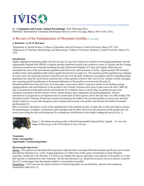
A Review of the Endoparasites of Mountain Gorillas by J. Rothman ...
A Review of the Endoparasites of Mountain Gorillas by J. Rothman ...
A Review of the Endoparasites of Mountain Gorillas by J. Rothman ...

You also want an ePaper? Increase the reach of your titles
YUMPU automatically turns print PDFs into web optimized ePapers that Google loves.
In: Companion and Exotic Animal Parasitology, D.D. Bowman (Ed.)<br />
Publisher: International Veterinary Information Service (www.ivis.org), Ithaca, New York, USA.<br />
A <strong>Review</strong> <strong>of</strong> <strong>the</strong> <strong>Endoparasites</strong> <strong>of</strong> <strong>Mountain</strong> <strong>Gorillas</strong> (31-Jan-2003)<br />
J. <strong>Rothman</strong> 1 and D. D. Bowman 2<br />
1 Department <strong>of</strong> Animal Science, College <strong>of</strong> Agriculture and Life Sciences, Cornell University, Ithaca, NY, USA.<br />
2 Department <strong>of</strong> Veterinary Microbiology and Immunology, College <strong>of</strong> Veterinary Medicine, Cornell University, Ithaca, NY,<br />
USA.<br />
Introduction<br />
Highly endangered mountain gorillas (Gorilla beringei beringei) are found in two distinct non-breeding populations: Bwindi<br />
Impenetrable National Park (BINP), a tropical montane rainforest located in <strong>the</strong> southwest corner <strong>of</strong> Uganda, and <strong>the</strong> Virunga<br />
Volcanoes located on <strong>the</strong> mountains bordering Rwanda, Democratic Republic <strong>of</strong> Congo and Uganda. Both areas are<br />
surrounded <strong>by</strong> some <strong>of</strong> <strong>the</strong> most densely populated and intensively cultivated areas in Africa. Approximately 600 mountain<br />
gorillas remain, <strong>the</strong>ir population split almost equally between <strong>the</strong> two parks [1]. The mountain gorilla population has enlarged<br />
in recent years, but increasing amounts <strong>of</strong> guerilla activities near <strong>the</strong> parks, bushmeat consumption, and <strong>the</strong> expanding human<br />
population has made <strong>the</strong> conservation and protection <strong>of</strong> <strong>the</strong>se gorillas critical to <strong>the</strong>ir survival [2]. Ano<strong>the</strong>r recently emerging<br />
area requiring careful consideration is <strong>the</strong> human habituation <strong>of</strong> <strong>the</strong> gorillas as tourists enter <strong>the</strong> park [3].<br />
<strong>Mountain</strong> gorillas have been <strong>the</strong> focus <strong>of</strong> several major conservation efforts. Long-term studies on <strong>the</strong> feeding ecology,<br />
ranging patterns, and social behavior <strong>of</strong> <strong>the</strong> gorillas in <strong>the</strong> Virunga Volcanoes have been in place since <strong>the</strong> early 1960s [4].<br />
Due to ecotourism in both parks and <strong>the</strong> encroachment <strong>of</strong> civilization, <strong>the</strong>re is concern that, as gorillas become more<br />
intimately associated with <strong>the</strong>ir human visitors, human diseases may compromise <strong>the</strong> gorilla populations. Disease<br />
transmission is recognized as an important area <strong>of</strong> conservation for <strong>the</strong>se species, but to date has been very little studied. The<br />
<strong>Mountain</strong> Gorilla Veterinary Project has assisted in monitoring <strong>the</strong> health <strong>of</strong> <strong>the</strong> mountain gorillas in recent years and <strong>the</strong>ir<br />
primary objective is to provide emergency care to injured and seriously sick gorillas, and monitor <strong>the</strong> health <strong>of</strong> mountain<br />
gorilla groups [5].<br />
Here we present a descriptive review <strong>of</strong> <strong>the</strong> endoparasites <strong>of</strong> <strong>the</strong> mountain gorilla, in hopes that we have provided a resource<br />
for primatologists, ecologists, veterinarians, park managers and all o<strong>the</strong>rs involved in <strong>the</strong> protection <strong>of</strong> <strong>the</strong> gorillas that will<br />
assist in providing <strong>the</strong> tools and background for endoparasite identification in this highly endangered species (Fig. 1).<br />
Figure 1. Silverback mountain gorilla in Bwindi Impenetrable National Park, Uganda. - To view this<br />
image in full size go to <strong>the</strong> IVIS website at www.ivis.org . -<br />
Nematodes<br />
Order: Strongylidia -<br />
Superfamily: Trichostrongyloidea<br />
Hyostrongylus kigeziensis<br />
History - This species was described from specimens collected from a necropsied Bwindi mountain gorilla [6], and was later<br />
described <strong>by</strong> Sleeman et al., in <strong>the</strong> Virunga population [7]. O<strong>the</strong>r hosts <strong>of</strong> this genus <strong>of</strong> nematodes include Ethiopian<br />
Leporidae, Suidae and a few Bovidae. It is hard to say whe<strong>the</strong>r this species represents a true parasite <strong>of</strong> <strong>the</strong> gorilla or a worm<br />
that typically is maintained in o<strong>the</strong>r mammals. The fact that Sleeman et al., found Hyostrongylus larvae in cultures <strong>of</strong> gorilla<br />
feces [7] would suggest that this parasite might be a true parasite <strong>of</strong> gorillas.<br />
Morphology - This worm has <strong>the</strong> typical structure <strong>of</strong> trichostrongylidae: an uncoiled body, adorned with continuous<br />
longitudinal ridges (Fig. 2).
Figure 2. Head <strong>of</strong> Hyostrongylus rubidis. - To view this image in full size go to <strong>the</strong> IVIS website at<br />
www.ivis.org . -<br />
Living worms are <strong>of</strong>ten bright red in color. Females are about 1 cm long, males slightly shorter (Fig. 3). The male has a<br />
distinct copulatory bursa and short stout spicules (Fig. 4).<br />
Figure 3. Female tail <strong>of</strong> Hyostrongylus rubidis. - To view this image in full size go to <strong>the</strong> IVIS website at<br />
www.ivis.org . -<br />
Figure 4. Male tail <strong>of</strong> Hyostrongylus rubidis. - To view this image in full size go to <strong>the</strong> IVIS website at<br />
www.ivis.org . -<br />
Buccal cavity lacking, <strong>the</strong> excretory pore lies midway along <strong>the</strong> esophagus, with <strong>the</strong> deirids slightly anterior posterior to<br />
excretory pore. Hyostrongylus can be differentiated from <strong>the</strong> o<strong>the</strong>r stomach worms <strong>of</strong> gorilla’s, Parali<strong>by</strong>ostrongylus kalinae,<br />
because <strong>the</strong> latter has large medial ridges on <strong>the</strong> female at level <strong>of</strong> <strong>the</strong> vulva. Also, <strong>the</strong> deirids <strong>of</strong> Hyostrongylus are relatively<br />
larger than those <strong>of</strong> Parali<strong>by</strong>ostrongylus kalinae.<br />
Diagnosis at Necropsy - The worms live on <strong>the</strong> mucosa <strong>of</strong> <strong>the</strong> stomach or in small, ulcerated areas on <strong>the</strong> mucosal surface.<br />
LiveHyostrongylus tend to be brown or bright red in color.<br />
Diagnosis in Fecal Examination - The eggs are typical strongylid eggs (Fig. 5). Based on <strong>the</strong> drawing <strong>by</strong> Durette-Desset et al.,<br />
[6], <strong>the</strong> eggs are about 70 to 75 µm in length.<br />
Figure 5. Hyostrongylus egg, recovered upon fecal analysis. - To view this image in full size go to <strong>the</strong><br />
IVIS website at www.ivis.org . -<br />
Hosts - <strong>Mountain</strong> gorilla; no o<strong>the</strong>r hosts are known.<br />
Mode <strong>of</strong> Transmission - <strong>Gorillas</strong> would be infected <strong>by</strong> <strong>the</strong> ingestion <strong>of</strong> larval contaminated foodstuffs.<br />
Life Cycle - The life cycle <strong>of</strong> Hyostrongylus kigeziensis has not been described. The life cycle <strong>of</strong> a swine parasite, H. rubidus,<br />
has been reported in detail [8]. The adults live in <strong>the</strong> stomach. Eggs are passed in <strong>the</strong> feces. First-stage larvae are about<br />
0.3 mm long. Under optimal conditions, <strong>the</strong> first molt occurs in about 3 days and <strong>the</strong> second molt about 2 days later. The firststage<br />
and second-stage larvae have long filamentous tails and tubular buccal cavities. The third-stage infective larvae<br />
produced <strong>by</strong> <strong>the</strong> second molt are about 0.7 mm long and retain <strong>the</strong> second-stage larval cuticle as a protective sheath. The<br />
infective larvae cannot penetrate <strong>the</strong> skin, and <strong>the</strong>refore enter a new host <strong>by</strong> ingestion. After ingestion, larvae exsheath in <strong>the</strong><br />
stomach, and penetrate <strong>the</strong> epi<strong>the</strong>lial folds <strong>of</strong> <strong>the</strong> gastric mucosa. The third molt occurs 5 days after infection. The final molt<br />
to <strong>the</strong> adult stage occurs in 13 days. The prepatent period is 17 days [9]. Maximum egg output was observed at 24 days after<br />
infection.<br />
Clinical Signs - Descriptions based on H. rubidus suggest that larvae develop in <strong>the</strong> gastric mucosa where <strong>the</strong>y destroy <strong>the</strong><br />
epi<strong>the</strong>lium and cause <strong>the</strong> formation <strong>of</strong> lentil-sized nodules. Adult worms induce a chronic catarrhal gastritis leading to <strong>the</strong><br />
formation <strong>of</strong> ulcers [10].<br />
Parali<strong>by</strong>ostrongylus kalinae<br />
History - This species was first described <strong>by</strong> Durette-Dusset et al., during a necropsy <strong>of</strong> <strong>the</strong> same female mountain gorilla <strong>of</strong>
<strong>the</strong> Bwindi Impenetrable Forest in 1989 from which <strong>the</strong>y recovered and described <strong>the</strong> specimens <strong>of</strong> Hyostrongylus kigeziensis<br />
[6]. This species was named after conservation biologist Jan Kalina, who with her husband Tom Butynski, facilitated <strong>the</strong><br />
creation <strong>of</strong> <strong>the</strong> Bwindi Impenetrable National Park. This genus has been reported in, primitive rodents, hyracoids, and<br />
porcupines [11-12].<br />
Lane [12] reported <strong>the</strong> recovery <strong>of</strong> Parali<strong>by</strong>ostrongylus hebrenicutus from <strong>the</strong> stomach and duodenum <strong>of</strong> a necropsied gorilla<br />
provided <strong>by</strong> <strong>the</strong> College <strong>of</strong> Surgeons. Dian Fossey recorded Parali<strong>by</strong>ostrongylus hebrenicutus as being recovered during<br />
necropsy from <strong>the</strong> liver <strong>of</strong> an adult female gorilla <strong>of</strong> <strong>the</strong> Rwandan Virungas [13]; <strong>the</strong> stage <strong>of</strong> <strong>the</strong> worm recovered was not<br />
given, and <strong>the</strong> hepatic location seems very unusual for <strong>the</strong>se worms. It seems that both Parali<strong>by</strong>ostrongylus kalinae and<br />
Parali<strong>by</strong>ostrongyulus hebrenicutus are both rodent or lagomorph parasites that are capable <strong>of</strong> developing in gorillas. The<br />
rodent host <strong>of</strong> Parali<strong>by</strong>ostrongylus hebrenicutus has been found to be members <strong>of</strong> <strong>the</strong> genus A<strong>the</strong>rurus. The rodent or<br />
lagomorph host <strong>of</strong> Parali<strong>by</strong>ostrongylus kalinae has yet to be described.<br />
Morphology - This genus has <strong>the</strong> typical structure <strong>of</strong> <strong>the</strong> trichostrongylidae, i.e., body uncoiled and adorned with continuous<br />
longitudinal ridges. Living worms are <strong>of</strong>ten bright red. There is a large dorsal esophageal tooth present. Females are about<br />
1 cm long, males slightly shorter. The male has a distinct copulatory bursa, and short stout spicules. Buccal cavity lacking,<br />
excretory pore lies midway along <strong>the</strong> esophagus, with deirids slightly anterior posterior to excretory pore.<br />
Parali<strong>by</strong>ostrongylus kalinae, has large medial ridges on <strong>the</strong> female at level <strong>of</strong> <strong>the</strong> vulva and deirids that are smaller than those<br />
<strong>of</strong> Hyostrongylus kigeziensis.<br />
Parali<strong>by</strong>ostrongylus kalinae differs from Parali<strong>by</strong>ostrongylus hebrenicutus in that it has a short dorsal ray on <strong>the</strong> bursa<br />
similar to that on two o<strong>the</strong>r species <strong>of</strong> Parali<strong>by</strong>ostrongylus that have been found in west African lagomorphs.<br />
Diagnosis at Necropsy - Worms were identified in stomach contents on one occasion. There was no report <strong>of</strong> any signs<br />
associated with <strong>the</strong> infection because <strong>the</strong> gorilla was found dead and <strong>the</strong> necropsy was performed several days after recovery<br />
[6]. At necropsy, it would be expected that <strong>the</strong> fresh worms would appear red in color.<br />
Diagnosis in Fecal Examination - The eggs are typical strongylid eggs, and would be very similar to those <strong>of</strong> Hyostrongylus.<br />
The eggs <strong>of</strong> <strong>the</strong>se two species need to be more carefully described.<br />
Hosts - <strong>Mountain</strong> gorilla.<br />
Mode <strong>of</strong> Transmission - The host is infected <strong>by</strong> <strong>the</strong> ingestion or skin penetration <strong>of</strong> third-stage larvae.<br />
Life Cycle - The life cycle <strong>of</strong> <strong>the</strong> related species P. hebrenicutus is presented because that <strong>of</strong> P. kalinae has not been<br />
described. With P. hebrenicutus, when <strong>the</strong> host is infected through <strong>the</strong> skin, <strong>the</strong> larvae enter <strong>the</strong> lymphatic system and reach<br />
<strong>the</strong> lungs and heart within 8 hours <strong>of</strong> infection. They reach <strong>the</strong> stomach within 2 days. If <strong>the</strong>y are ingested through<br />
contaminated food or soil, <strong>the</strong>y reach <strong>the</strong> stomach within 24 hours post infection. Larvae localize within <strong>the</strong> stomach mucosa.<br />
The fifth day postinfection, regardless <strong>of</strong> mode, <strong>the</strong> larvae become imbedded in <strong>the</strong> gastric mucosa where <strong>the</strong>y undergo <strong>the</strong><br />
third molt followed <strong>by</strong> <strong>the</strong> fourth molt during <strong>the</strong> nineteenth day. Eggs appear on <strong>the</strong> twenty-eighth day after infection [14].<br />
Clinical Signs - None have been described.<br />
Impalaia sp.<br />
History - This species was first described <strong>by</strong> Monnig in 1923 in impalas from South Africa. The species comprising this<br />
genus typically parasitize large, herbivorous antelope, such as <strong>the</strong> impala. Members <strong>of</strong> this genus have also been found in<br />
camels, giraffe, okapi, and sheep. The parasites found in <strong>the</strong> gorilla were not identified to species. The first report <strong>of</strong> this<br />
genus in <strong>the</strong> Virunga population <strong>of</strong> mountain gorillas was from Redmond [15], and <strong>the</strong> worm was later found during <strong>the</strong><br />
necropsy <strong>of</strong> a Virunga gorilla [16].<br />
Morphology - The adults recovered from ruminants are about 1 to 2 cm long. The anterior end <strong>of</strong> both sexes bears a slight<br />
inflation with marked transverse striae. The female is readily identified <strong>by</strong> <strong>the</strong> monodelphic uterus with <strong>the</strong> vulva being found<br />
just anteriad to <strong>the</strong> anus. The dorsal ray <strong>of</strong> <strong>the</strong> male is very long compared to o<strong>the</strong>r members <strong>of</strong> <strong>the</strong> family. The synlophe is<br />
composed <strong>of</strong> 16 evenly spaced longitudinal cuticular ridges. The eggs, typical <strong>of</strong> strongylid nematodes, are around 75 µm <strong>by</strong><br />
35 µm.<br />
Diagnosis at Necropsy - The worms live in <strong>the</strong> small intestine or caecum <strong>of</strong> <strong>the</strong> host.<br />
Diagnosis in Fecal Examination - The worms recovered from <strong>the</strong> gorilla have not been described as to stage. Adult worms<br />
would produce typical strongylid eggs in <strong>the</strong> feces.<br />
Hosts - The reports from mountain gorillas include <strong>the</strong> collection <strong>of</strong> Impalaia sp. from 1 <strong>of</strong> 6 necropsied animals [16]. O<strong>the</strong>r<br />
hosts <strong>of</strong> Impalaia include herbivores: giraffe, okapi, sheep, impala, reedbuck, Thomson’s gazelle, sable antelope, gerenuk,<br />
blesblock, oryx, tsessebe, camels [17-18].<br />
Mode <strong>of</strong> Transmission - The host ingests third-stage larvae that have developed in <strong>the</strong> soil or on vegetation.<br />
Life Cycle - The life cycle for Impalaia tuberculata has been described relative to its development in <strong>the</strong> impala [19-20].<br />
After ingestion <strong>of</strong> <strong>the</strong> enshea<strong>the</strong>d third-stage larvae, <strong>the</strong> exshea<strong>the</strong>d third-stage larvae probably do not migrate deeply into <strong>the</strong><br />
mucosa. The fourth-stage larvae are found in <strong>the</strong> proximal portion <strong>of</strong> <strong>the</strong> small intestine and <strong>the</strong> young adults are found<br />
throughout <strong>the</strong> intestine. The adult worms tend to be found mainly in <strong>the</strong> distal small intestine. The female passes numerous
eggs in <strong>the</strong> feces. The eggs develop to a first stage larva in <strong>the</strong> feces where <strong>the</strong>y feed <strong>of</strong>f <strong>of</strong> <strong>the</strong> microorganisms and fecal<br />
matter. They continue to <strong>the</strong> second stage in a few days and <strong>the</strong>n to <strong>the</strong> third infective stage. At <strong>the</strong> third stage <strong>the</strong>y are<br />
enshea<strong>the</strong>d, and migrate from <strong>the</strong> fecal matter to soil and vegetation using stored food as energy. They can survive for months<br />
on <strong>the</strong>ir stored food supply until <strong>the</strong>y are consumed <strong>by</strong> <strong>the</strong> host.<br />
Clinical Signs - Clinical signs would probably be similar to those <strong>of</strong> an overwhelming infection with Cooperia, a related<br />
parasite <strong>of</strong> ruminants that can cause watery diarrhea when present in large numbers.<br />
Trichostrongylus sp.<br />
History - The genus was first described <strong>by</strong> Looss in 1905. Over thirty species have been reported in mammals and birds [20].<br />
This genus was first recorded in mountain gorillas, from necropsies <strong>of</strong> gorillas in <strong>the</strong> Virunga population [16] and <strong>the</strong>n <strong>by</strong><br />
identification <strong>of</strong> Trichostrongyle-like eggs in fecal examinations in both <strong>the</strong> Virunga and Bwindi populations (Fig. 6) [7,21-<br />
25].<br />
Figure 6. Strongylid egg recovered from <strong>the</strong> feces <strong>of</strong> Bwindi mountain gorillas. - To view this image in<br />
full size go to <strong>the</strong> IVIS website at www.ivis.org . -<br />
Most species in this genus have been described from birds, ruminants and primitive rodents around <strong>the</strong> world. Species have<br />
also been found in human and non-human primates, camel, pigs and equids. <strong>Gorillas</strong> are probably not <strong>the</strong> normal host <strong>of</strong> <strong>the</strong>se<br />
parasites. However, without more careful descriptions, it is not possible to identify <strong>the</strong> actual host, which is likely a ruminant<br />
or a lagomorph.<br />
Morphology - These are small worms (less than 1 cm long) without a buccal cavity. The bursa <strong>of</strong> <strong>the</strong> male has large lateral<br />
lobes with a relatively short dorsal lobe (Fig. 7). The vulva <strong>of</strong> <strong>the</strong> female is located slightly posterior to midbody, and <strong>the</strong> two<br />
branches <strong>of</strong> <strong>the</strong> uterus are amphidelphic. The uterus is filled with eggs that are thin shelled and <strong>of</strong> <strong>the</strong> typical strongylid type<br />
(Fig. 8).<br />
Figure 7. Trichostrongylus male. - To view this image in full size go to <strong>the</strong> IVIS website at<br />
www.ivis.org . -<br />
Figure 8. Trichostrongylus female. - To view this image in full size go to <strong>the</strong> IVIS website at<br />
www.ivis.org . -<br />
Diagnosis at Necropsy - These small brownish worms are difficult to discern at necropsy without special care to examine <strong>the</strong><br />
mucosal scrapings <strong>of</strong> <strong>the</strong> small intestine. If worms are discovered, care must be taken to recover both males and females if a<br />
specific diagnosis is desired.<br />
Diagnosis in Fecal Examination - The eggs in feces are typical <strong>of</strong> o<strong>the</strong>r strongylid nematodes, and may be confused with<br />
o<strong>the</strong>r parasitic nematodes common in <strong>the</strong> mountain gorilla. There is indication based on measurements made on eggs in feces<br />
that <strong>the</strong> eggs <strong>of</strong> Trichostrongylus may be longer than those <strong>of</strong> Oesophagostomum (Kalema, personal communication). To<br />
identify eggs as being those <strong>of</strong> Trichostrongylus, it is necessary to culture <strong>the</strong> eggs and to examine hatched and developed<br />
third-stage larvae. The sheath extending beyond <strong>the</strong> tip <strong>of</strong> <strong>the</strong> tail is shorter than that <strong>of</strong> most o<strong>the</strong>r trichostrongylid parasites.<br />
Hosts - Livestock (ruminants, pigs, fowl), humans and o<strong>the</strong>r primates, camels, rodents, wild ruminants and equids.<br />
Mode <strong>of</strong> Transmission - The host ingests <strong>the</strong> infective third stage larvae.<br />
Life Cycle - The adult worms produce eggs that are passed in <strong>the</strong> feces. The eggs hatch under suitable environmental<br />
conditions and develop to <strong>the</strong> first stage larvae in <strong>the</strong> feces where <strong>the</strong>y feed on <strong>the</strong> microorganisms and fecal matter in <strong>the</strong><br />
feces and surrounding soil. They develop to <strong>the</strong> second stage in a few days and <strong>the</strong>n to <strong>the</strong> third-stage, enshea<strong>the</strong>d, infective<br />
larva. Migration from <strong>the</strong> fecal matter to soil and vegetation occurs using stored food as energy. Larvae can survive for<br />
months on <strong>the</strong>ir stored food supplies until consumed <strong>by</strong> a host. Larvae can generally withstand harsh temperatures above<br />
freezing. When larvae are consumed, <strong>the</strong>y develop in <strong>the</strong> small intestine and adults inhabit <strong>the</strong> anterior part <strong>of</strong> <strong>the</strong> small<br />
intestine. Some species invade and remain in <strong>the</strong> intestinal mucosa as larvae for some time before re-entering <strong>the</strong> lumen to<br />
mature [20].<br />
Clinical Signs - There have been no clinical signs <strong>of</strong> disease associated with Trichostrongylus sp. reported from gorillas,
however, <strong>the</strong> disease manifestations associated with trichostrongylosis is diarrhea that may sometimes be quite severe. In<br />
humans infected with Trichostrongylus, slight abdominal discomfort is occasionally reported, but usually patients are<br />
asymptomatic [26].<br />
Order: Strongylidia<br />
Superfamily: Strongyloidea<br />
Murshidia devians<br />
History - This genus <strong>of</strong> strongyloid nematodes was first described <strong>by</strong> Lane (1914) and represents species that are typically<br />
found to infect elephants and rhinoceros. Campana-Rouget described <strong>the</strong> species Murshidia devians from a lowland gorilla in<br />
<strong>the</strong> Republic <strong>of</strong> Congo, suggesting that <strong>the</strong> parasite described was an accidental parasite <strong>of</strong> <strong>the</strong> gorilla for three reasons:<br />
1. Murshidia was never previously recovered from primates, having been found only in elephants and rhinoceros.<br />
2. The adults <strong>of</strong> this species are typically found in <strong>the</strong> large intestine, and in <strong>the</strong> gorilla, adult worms were found in <strong>the</strong><br />
large intestine, but <strong>the</strong>y were also found ectopically in <strong>the</strong> skin and muscles <strong>of</strong> <strong>the</strong> back and thorax.<br />
3. The species Murshidia devians is very close morphologically to two species commonly infecting elephants [27].<br />
Consequently, it is likely that <strong>the</strong> elephant is probably <strong>the</strong> natural host <strong>of</strong> <strong>the</strong> parasites found in <strong>the</strong> gorilla. Hastings et al.,<br />
report finding Murshidia devians in <strong>the</strong> large intestine <strong>of</strong> a mountain gorilla at necropsy [16]. Ashford et al., identified <strong>the</strong><br />
worms during <strong>the</strong> necropsy <strong>of</strong> a mountain gorilla in <strong>the</strong> Bwindi forest [22].<br />
Morphology - The genus, as is typical <strong>of</strong> members <strong>of</strong> <strong>the</strong> Strongyloidea, has a large buccal capsule. The buccal capsule is<br />
anteriorly directed, has a single external leaf crown, and sometimes bears two or more teeth at its base. In <strong>the</strong> case <strong>of</strong><br />
Murshidia devians, <strong>the</strong> oral opening is elongated dorso-ventrally, <strong>the</strong> external leaf crown has about 80 elements, and <strong>the</strong>re are<br />
no teeth figured at <strong>the</strong> base <strong>of</strong> <strong>the</strong> buccal capsule [27]. The recovered worms were about 20 mm long. The vulva <strong>of</strong> <strong>the</strong> female<br />
is located just anterior to <strong>the</strong> anus, and leads into a prodelphic uterus with two branches. The bursa <strong>of</strong> <strong>the</strong> male has a welldeveloped<br />
dorsal lobe. Males have spicules and a gubernaculum.<br />
Diagnosis at Necropsy - The adults would typically be found free within <strong>the</strong> lumen <strong>of</strong> <strong>the</strong> large intestine [16]. Fossey reported<br />
finding <strong>the</strong> worms in <strong>the</strong> small intestine <strong>of</strong> a necropsied older female mountain gorilla [13]. The report <strong>by</strong> Campana-Rouget<br />
found that <strong>the</strong> worms were present in ectopic sites [27]. Thus, it is possible that worms will be found in sites o<strong>the</strong>r than <strong>the</strong><br />
intestine.<br />
Diagnosis in Fecal Examination - Eggs <strong>of</strong> <strong>the</strong> typical strongylid type may be detected in <strong>the</strong> feces; <strong>the</strong> eggs are 60 µm <strong>by</strong><br />
30 µm. When third stage larvae are cultured in feces, <strong>the</strong> larva has a long sheath that extends beyond <strong>the</strong> tip <strong>of</strong> <strong>the</strong> tail [28].<br />
Hosts - Murshidia devians was described from <strong>the</strong> lowland gorilla and reported from both populations <strong>of</strong> mountain gorillas.<br />
In all cases, Murshidia is probably an accidental parasite [22]. The members <strong>of</strong> this genus are typically in elephants and<br />
rhinoceros.<br />
Mode <strong>of</strong> Transmission - The host ingests <strong>the</strong> infective third stage larvae.<br />
Life Cycle - There is no information on <strong>the</strong> life cycle <strong>of</strong> any Murshidia sp. It is assumed that this parasite has a direct life<br />
cycle like o<strong>the</strong>r members <strong>of</strong> <strong>the</strong> subfamily Cyathostominae. The female lays eggs in <strong>the</strong> intestine and <strong>the</strong> host passes <strong>the</strong>m in<br />
its feces. The eggs hatch on <strong>the</strong> ground to release <strong>the</strong> first-stage larvae, which feeds on bacteria. The first-stage larvae molt to<br />
second-stage larvae that also feed on bacteria. The second stage molts to <strong>the</strong> third-stage larvae that are enshea<strong>the</strong>d. The<br />
development in <strong>the</strong> final host has not been studied. The finding <strong>of</strong> <strong>the</strong> adults <strong>of</strong> Murshidia in ectopic sites in <strong>the</strong> gorilla would<br />
indicate that <strong>the</strong>re might be some form <strong>of</strong> extraintestinal development associated with <strong>the</strong> development <strong>of</strong> <strong>the</strong>se worms in<br />
<strong>the</strong>ir normal hosts.<br />
Clinical Signs - No information available.<br />
Oesophagostomum stephanostomum<br />
History - This species was first described from gorillas in 1904 [29]. The worms were from <strong>the</strong> zoology museum <strong>of</strong><br />
Cambridge University with <strong>the</strong> simple designation "from large intestines <strong>of</strong> gorilla", so <strong>the</strong>re is no indication as to where <strong>the</strong><br />
host originated. Rouselot and Pellissier reported on <strong>the</strong> presence <strong>of</strong> Oesophagostomum stephanostomum in western lowland<br />
gorillas from <strong>the</strong> Republic <strong>of</strong> Congo [30]. Specimens <strong>of</strong> this genus were later found at necropsy <strong>of</strong> Virunga gorillas [16], in<br />
Bwindi gorillas [22] and in larval cultures [7]. Surveys found eggs in <strong>the</strong> feces <strong>of</strong> gorillas from <strong>the</strong> Bwindi population and<br />
identified <strong>by</strong> <strong>the</strong>m as Oesophagostomum [21,22,31].<br />
Morphology - Adult females are 18 to 30 mm in length; males are 18 to 24 mm long. The worm has a relative small buccal<br />
cavity compared to o<strong>the</strong>r members <strong>of</strong> <strong>the</strong> strongyloidea and a marked transverse cervical groove at <strong>the</strong> excretory pore (Fig. 9).
Figure 9. Tail <strong>of</strong> male Oesophagostomum. - To view this image in full size go to <strong>the</strong> IVIS website at<br />
www.ivis.org . -<br />
The stoma has both external and internal leaf crowns, with <strong>the</strong> external crown having 30 to 38 petals. There are two to three<br />
times as many petals on <strong>the</strong> internal leaf crown. The base <strong>of</strong> <strong>the</strong> buccal capsule contains six ra<strong>the</strong>r large chitinous plates. The<br />
male has a prominent bursa and two ra<strong>the</strong>r long spicules (Fig. 10). The vulva <strong>of</strong> <strong>the</strong> female is near <strong>the</strong> anus and <strong>the</strong> vagina<br />
connects to <strong>the</strong> kidney shaped ovejector (Fig. 11) [29].<br />
Figure 10. Tail <strong>of</strong> female Oesophagostomum. - To view this image in full size go to <strong>the</strong> IVIS website at<br />
www.ivis.org . -<br />
Figure 11. Mouth <strong>of</strong> Oesophagostomum radiata. - To view this image in full size go to <strong>the</strong> IVIS website<br />
at www.ivis.org . -<br />
Diagnosis at Necropsy - Adults are found free within <strong>the</strong> lumen <strong>of</strong> <strong>the</strong> large intestine. Worms <strong>of</strong> this genus form nodules as<br />
larvae in <strong>the</strong> intestinal wall (Fig. 12).<br />
Figure 12. Oesophagostomum nodules in <strong>the</strong> small intestine in a necropsied laboratory primate. - To view<br />
this image in full size go to <strong>the</strong> IVIS website at www.ivis.org . -<br />
These are small, raised areas about 1 mm in diameter in <strong>the</strong> small and large intestine. The lesions can grow into small<br />
abscesses and eventually <strong>the</strong> entire intestine may be inflamed and edematous. Nodules can reach 4 - 5 mm in diameter, and<br />
contain eosinophils and leukocytes. There can be a significant number <strong>of</strong> nodules at necropsy; 40 nodules were found in 1 <strong>of</strong><br />
9 gorillas in <strong>the</strong> Congo [30].<br />
Diagnosis in Fecal Examination - Eggs are 60 - 80 µm <strong>by</strong> 40 - 55 µm (Fig. 13). When cultured to <strong>the</strong> infective stage, <strong>the</strong><br />
third-stage larva is about 1 mm long, has 16 to 24 triangular intestinal cells, and a sheath that extends 150 µm beyond <strong>the</strong> tip<br />
<strong>of</strong> <strong>the</strong> larva.<br />
Figure 13. Egg <strong>of</strong> Oesophagostomum. - To view this image in full size go to <strong>the</strong> IVIS website at<br />
www.ivis.org . -<br />
Hosts - <strong>Gorillas</strong>, chimpanzees, and humans (reported from Uganda, Senegal, and surprisingly, Brazil) [32].<br />
Mode <strong>of</strong> Transmission - The host ingests infective third-stage larvae.<br />
Life Cycle - There is no information on <strong>the</strong> life cycle <strong>of</strong> O. stephanostomum. The life cycle is considered to be similar to<br />
related species such as O. bifurcum, ano<strong>the</strong>r species found in Old World primates. After larvae are ingested, it takes<br />
approximately 3 months until eggs appear in <strong>the</strong> feces [33]. The molt from third to fourth stage occurs sometime prior to Day
19 to 22 after infection. Some young adult worms were still present in nodules within <strong>the</strong> bowel wall after almost a year from<br />
<strong>the</strong> time <strong>of</strong> infection. Animals can shed eggs for almost a year.<br />
Clinical Signs - Disease was first associated with gorillas captured for zoological collections in Gabon and <strong>the</strong> Republic <strong>of</strong><br />
Congo [30]. The gorillas died within a few months <strong>of</strong> capture following ill health. Fecal examinations revealed <strong>the</strong> eggs <strong>of</strong><br />
strongylid nematodes, and necropsies later revealed that <strong>the</strong> gorillas were infected with both hookworms and had nodular<br />
disease due to oesophagostomins. The clinical signs manifested as anorexia and lack <strong>of</strong> grooming with an unkempt<br />
appearance. There was mucoid diarrhea similar to amebic dysentery in humans. The gorillas remained lying or sitting holding<br />
<strong>the</strong>ir heads with both hands in an attitude <strong>of</strong> desperation. In free-ranging mountain gorillas, nodules <strong>of</strong> oesophagostomiasis<br />
have been observed at necropsy <strong>by</strong> <strong>the</strong> <strong>Mountain</strong> Gorilla Veterinary Center (cited in [7]) and Hastings et al., speculate that<br />
feces containing blood and mucus may have been caused <strong>by</strong> Oesophagostomum [34].<br />
Order: Ascaridida<br />
Superfamily: Cosmocercoidea<br />
Probstmayria gorillae<br />
History - In 1955, Kreis described Probstmayria gorillae based on specimens collected from a gorilla that died in a zoo in<br />
Basel, Switzerland [35]. Specimens belonging to <strong>the</strong> genus Probstmayria have been described from mountain gorillas in both<br />
<strong>the</strong> Virunga region [7,15] and in groups <strong>of</strong> gorillas in Bwindi Impenetrable National Park [22,25,31]. In 1990, Ashford and<br />
o<strong>the</strong>rs found that gorillas had a 100% prevalence [31], while <strong>Rothman</strong> and o<strong>the</strong>rs found a 13% prevalence in a single group<br />
sampled over 7 weeks [25]. No o<strong>the</strong>r surveys <strong>of</strong> Bwindi gorilla gastro-intestinal parasites found any Cosmocercoidea [21,23].<br />
Specimens collected from <strong>the</strong> feces <strong>of</strong> lowland gorillas in Gabon have been described as two species distinct from<br />
Probstmayria gorillae; <strong>the</strong>se two species were named Probstmayria goodallae and Probstmayria gabonensis [36].<br />
Morphology - These are small worms with an esophagus characteristic <strong>of</strong> <strong>the</strong> Cosmocercoidea, i.e., with a large valved bulb<br />
at <strong>the</strong> base and a vestibule between <strong>the</strong> stoma and <strong>the</strong> beginning <strong>of</strong> <strong>the</strong> muscular esophagus proper (Fig. 14 and Fig. 15).<br />
Figure 14. Full body <strong>of</strong> Probstmayria. - To view this image in full size go to <strong>the</strong> IVIS website at<br />
www.ivis.org . -<br />
Figure 15. Head <strong>of</strong> Probstmayria. - To view this image in full size go to <strong>the</strong> IVIS website at<br />
www.ivis.org . -<br />
The female is characterized <strong>by</strong> giving birth to large larvae that are almost one-third her total length. Waerebeke et al., in <strong>the</strong>ir<br />
description <strong>of</strong> P. gabonensis [36] stated that because Kreis cited both <strong>the</strong> gorilla and gibbon as <strong>the</strong> host <strong>of</strong> this species [35], it<br />
was complicated as to whe<strong>the</strong>r this species was originally from a gorilla or a gibbon. The fact that <strong>the</strong>re have been no reports<br />
<strong>of</strong> Probstmayria from gibbons would suggest that <strong>the</strong> original origin <strong>of</strong> <strong>the</strong> species is most likely from <strong>the</strong> gorilla. P.<br />
gabonensis is <strong>the</strong> most similar to P. gorillae in that <strong>the</strong> lips are similar in shape. P. goodallae differs form <strong>the</strong> o<strong>the</strong>r two<br />
described species from <strong>the</strong> gorilla in that <strong>the</strong>y have an inverse symmetry (one ventral lip and two latero-dorsal lips). The sizes<br />
<strong>of</strong> all three species are similar, and <strong>the</strong>re have been no careful comparisons <strong>of</strong> characters that could be used for rapid<br />
identification to <strong>the</strong> species level upon routine examination.<br />
Diagnosis at Necropsy - Larvae and adults will be found in <strong>the</strong> lumen <strong>of</strong> <strong>the</strong> cecum and colon. The adults are not large<br />
(females are only about 2 mm long), and <strong>the</strong> larvae that are newly born will be almost half <strong>the</strong> length <strong>of</strong> <strong>the</strong> mo<strong>the</strong>r.<br />
Diagnosis in Fecal Examination - Fecal examination can reveal both <strong>the</strong> adults and larvae <strong>of</strong> Probstmayria. The fact that <strong>the</strong><br />
mo<strong>the</strong>r gives birth to highly precocious larvae and that <strong>the</strong>re is no intermediate host required means that very large<br />
populations can develop within an infected animal. Eggs will not be seen in <strong>the</strong> feces.<br />
Hosts - <strong>Gorillas</strong>, o<strong>the</strong>r species in chimpanzees, African and Asian pigs and horses, and Central American tapirs.<br />
Mode <strong>of</strong> Transmission - The life cycle is direct (Fig. 16); <strong>the</strong> host ingests <strong>the</strong> third stage larvae, usually through infected<br />
foodstuffs or <strong>by</strong> fecal-oral contamination.<br />
Figure 16. Life cycle <strong>of</strong> Probstmayria: Direct life cycle passed in fresh feces as a larval stage, <strong>by</strong> <strong>the</strong><br />
ingestion <strong>of</strong> feces containing larvae. The adults live in <strong>the</strong> lumen <strong>of</strong> <strong>the</strong> colon, and <strong>the</strong> female produces<br />
larvae ra<strong>the</strong>r than eggs. They are capable <strong>of</strong> internal autoinfection with <strong>the</strong> development <strong>of</strong> very large<br />
numbers <strong>of</strong> parasitic worms. They tend to be non-pathogenic even at very heavy infections. - To view<br />
this image in full size go to <strong>the</strong> IVIS website at www.ivis.org . -
Life Cycle - The life cycle is direct. Females contain very few (1 to 3) larvae that are quite large compared to her body size.<br />
The larvae that are born provide <strong>the</strong> means for <strong>the</strong> development <strong>of</strong> internal autoinfection and for this reason, very large<br />
numbers <strong>of</strong> worms can build up within <strong>the</strong> intestine <strong>of</strong> <strong>the</strong> host. Transmission is probably assisted <strong>by</strong> <strong>the</strong> fact that larvae can<br />
remain alive in feces for 4 or 5 days.<br />
Clinical Signs - This species has not been found to be pathogenic.<br />
Order: Strongylidia<br />
Superfamily: Ascaridoidea<br />
Ascaris lumbricoides<br />
History - Ascaris lumbricoides was first described <strong>by</strong> Linnaeus in 1758, as a parasite observed, and recorded <strong>by</strong> ancient<br />
peoples. This is one <strong>of</strong> <strong>the</strong> most common parasites <strong>of</strong> people around <strong>the</strong> world and is believed to be present in one-sixth <strong>of</strong> <strong>the</strong><br />
world’s human population. In 1995, Kalema found Ascaris lumbricoides eggs in <strong>the</strong> Bwindi population <strong>of</strong> mountain gorillas;<br />
she found eggs in <strong>the</strong> feces <strong>of</strong> two infants and two adult female gorillas [21]. In 1990, a study <strong>of</strong> <strong>the</strong> feces <strong>of</strong> people living<br />
around Bwindi forest revealed high loads <strong>of</strong> Ascaris, but <strong>the</strong>re was no evidence <strong>of</strong> Ascaris in gorilla populations [31].<br />
Morphology - Ascaris lumbricoides is a large cream-colored or white worm that will reach lengths <strong>of</strong> up to 30 cm or greater<br />
(Fig. 17). The worms have 3 large lips on <strong>the</strong> anterior end that surround <strong>the</strong> buccal opening. The tail is relatively short and<br />
comes to a ra<strong>the</strong>r abruptly pointed tip. The mail tail tends to curl ventrally and has no bursa.<br />
Figure 17. Adult Ascaris suum recovered at necropsy from pigs. - To view this image in full size go to<br />
<strong>the</strong> IVIS website at www.ivis.org . -<br />
Diagnosis at Necropsy - Ascaris lumbricoides are quite distinct worms that would be difficult to confuse with any <strong>of</strong> <strong>the</strong> o<strong>the</strong>r<br />
parasites that might be found in <strong>the</strong> small intestine <strong>of</strong> gorillas at necropsy. Occasionally, senescent worms will be found in <strong>the</strong><br />
cecum or colon at necropsy; <strong>the</strong>se worms are dead or dying and in <strong>the</strong> process <strong>of</strong> being expelled from <strong>the</strong> body. There are no<br />
reports <strong>of</strong> necropsied gorillas infected with Ascaris sp.<br />
Diagnosis in Fecal Examination - Ascaris lumbricoides eggs are ovoid and about 60 µm long. The eggs have a thick shell and<br />
a dark-brown external coat that appear to have a rough outer coat typically stained <strong>by</strong> bile to a golden brown color (Fig. 18).<br />
When passed in feces, <strong>the</strong> eggs usually contain a single undivided cell.<br />
Figure 18. Ascaris egg. - To view this image in full size go to <strong>the</strong> IVIS website at www.ivis.org . -<br />
Hosts - Humans and o<strong>the</strong>r primates.<br />
Mode <strong>of</strong> Transmission - Infected soil/ foodstuff is ingested <strong>by</strong> <strong>the</strong> host (Fig. 19). The eggs persist in <strong>the</strong> soil for long periods<br />
and can remain infectious in soil for years. It takes about two weeks under optimal conditions for eggs to become infectious<br />
through <strong>the</strong> embryonation <strong>of</strong> <strong>the</strong> larval to <strong>the</strong> infective stage.
Life Cycle - After ingestion <strong>of</strong> an infective egg, larvae hatch. Recent work has suggested that <strong>the</strong> eggs hatch in <strong>the</strong> large<br />
intestine and that <strong>the</strong> larvae actually enter <strong>the</strong> circulation through <strong>the</strong> wall <strong>of</strong> <strong>the</strong> large bowel (at least, with <strong>the</strong> closely related<br />
parasite <strong>of</strong> swine, Ascaris suum). The hatched larvae will migrate into <strong>the</strong> intestinal wall, and travel through <strong>the</strong> portal<br />
circulation to <strong>the</strong> liver, heart and through <strong>the</strong> pulmonary vessels to <strong>the</strong>ir destination, <strong>the</strong> interalveolar tissues <strong>of</strong> <strong>the</strong> lung. The<br />
larvae <strong>the</strong>n make <strong>the</strong>ir way up <strong>the</strong> respiratory escalator, and <strong>the</strong>y are <strong>the</strong>n swallowed. The remainder <strong>of</strong> <strong>the</strong> development takes<br />
place in <strong>the</strong> lumen <strong>of</strong> <strong>the</strong> small intestine. The prepatent period is about 2 months, and <strong>the</strong> worms live about 1 year. The<br />
females produce prodigious amounts <strong>of</strong> eggs, having been shown to produce about 200,000 eggs per day.<br />
Clinical Signs - There are no clinical signs reported in mountain gorillas. In humans, light infections usually produce little in<br />
<strong>the</strong> way <strong>of</strong> signs or symptoms. Heavy infections can cause intestinal obstruction and related signs. It has been shown in<br />
people that Ascaris lumbricoides infections do have some effect on <strong>the</strong> nutrition <strong>of</strong> <strong>the</strong>ir human hosts.<br />
Figure 19. Life cycle <strong>of</strong> Ascaris: Direct life cycle. These large nematodes live in <strong>the</strong> small intestine. Eggs<br />
are passed in <strong>the</strong> feces, and after several weeks in <strong>the</strong> soil, <strong>the</strong> eggs are em<strong>by</strong>ronated and infectious. The<br />
gorillas become infected <strong>by</strong> ingesting infectious soil, or soil contaminated vegetation. In <strong>the</strong> gorilla, <strong>the</strong><br />
larval Ascaris make a liver- lung migration before returning to <strong>the</strong> small intestine where <strong>the</strong>y complete<br />
<strong>the</strong>ir development. The prepatent period is about 6 weeks. Light infections are typically without<br />
pathology, although <strong>the</strong> large adult worm can occasionally cause intestinal perforation or migration into and blockage <strong>of</strong> <strong>the</strong><br />
bile duct. Very heavy infections that might occur in young animals could cause significant disease. - To view this image in<br />
full size go to <strong>the</strong> IVIS website at www.ivis.org . -<br />
Order: Spirurida<br />
Superfamily: Physalopteroidea<br />
Chitwoodspirura sp.<br />
History - In 1956, Chabaud and Rousselot described Chitwoodspirura wehri from a lowland gorilla from Congo (Braizeville)<br />
[37]. Only immature specimens were found. It was fur<strong>the</strong>r described from specimens collected from a gorilla at <strong>the</strong> Hirakata<br />
Zoological Park soon after its arrival from <strong>the</strong> Congo [38]. A nematode found during <strong>the</strong> necropsy <strong>of</strong> mountain gorillas in<br />
Rwanda was identified as Chitwoodspirura sp., but a specific determination was not made [16].<br />
Morphology - The worms are large: males are 5 cm and females are 10 cm, in length. The esophagus is typical <strong>of</strong> spirurids<br />
(divided into a muscular and glandular portion). There is a large buccal cavity, and <strong>the</strong> anterior end possesses a pair <strong>of</strong> lateral<br />
pseudolabia that close over mouth. Each <strong>of</strong> <strong>the</strong> two pseudolabia possesses approximately nine large denticles. The vulva is<br />
about one-third <strong>of</strong> <strong>the</strong> body length behind <strong>the</strong> anterior extremity. In <strong>the</strong> male, <strong>the</strong> right spicule is about 1 mm long; <strong>the</strong> left is<br />
about 5 mm long. The tails <strong>of</strong> both <strong>the</strong> males and females come toge<strong>the</strong>r in a relatively rounded tip. The egg is small (23 x 45<br />
µm), larvated, thick shelled, and bears what appear to be seated opercula on each end.<br />
Diagnosis at Necropsy - The worm is found in <strong>the</strong> stomach [16,37].<br />
Diagnosis in Fecal Examination - Characteristic small, thick-shelled, larvated spirurid eggs would be found in <strong>the</strong> feces.<br />
Hosts - Lowland gorilla, possibly a mountain gorilla.<br />
Mode <strong>of</strong> Transmission - Based on o<strong>the</strong>r spirurid life cycles, it is expected that <strong>the</strong> gorilla would become infected <strong>by</strong> <strong>the</strong><br />
accidental ingestion <strong>of</strong> a corprophagous arthropod on vegetation.<br />
Life Cycle - There is no known information about <strong>the</strong> life cycle <strong>of</strong> Chitwoodspirura sp. Spirurid nematodes go through an<br />
arthropod intermediate host. The thick-shelled nature <strong>of</strong> <strong>the</strong> egg passed in <strong>the</strong> feces would suggest that <strong>the</strong> intermediate host is<br />
likely to be a coprophagous arthropod <strong>of</strong> some type that becomes infected <strong>by</strong> ingesting <strong>the</strong> embryonated egg.<br />
Clinical Signs - No information available.<br />
Order: Rhabditida<br />
Superfamily: Rhabditoidea<br />
Strongyloides fulleborni<br />
History - This species was described in 1905 <strong>by</strong> von Linstow using specimens collected from Anthroppi<strong>the</strong>cus troglodytes<br />
and Cynocephalus babuin. Man was first recorded as a host <strong>of</strong> this parasite in Zimbabwe [39]. Strongyloides fulleborni eggs<br />
have been reported in <strong>the</strong> feces <strong>of</strong> mountain gorillas in both Bwindi and <strong>the</strong> Virungas [7,21-24,31].<br />
Morphology - The parasitic par<strong>the</strong>nogenetic female is about 3.5to 4.5mm long. Like o<strong>the</strong>r Strongyloides spp., she is very<br />
slender and has an esophagus that is very long, one-fourth to one-fifth <strong>of</strong> <strong>the</strong> total body length. The ovary <strong>of</strong> Strongyloides<br />
fulleborni, like o<strong>the</strong>r species that produce eggs in <strong>the</strong> feces ra<strong>the</strong>r than larvae, spirals around <strong>the</strong> intestine. These eggs<br />
produced <strong>by</strong> <strong>the</strong> par<strong>the</strong>nogenetic female appear clear, contain a larva, and measure 50to 60µm x 25 to 35µm.<br />
Diagnosis at Necropsy - These worms are very small and very easy to overlook at necropsy unless techniques are used that
are designed specifically for <strong>the</strong>ir collection. It is best to take a small portion <strong>of</strong> fresh bowel (proximal small intestine) and<br />
suspend it with weights in a graduated cylinder <strong>of</strong> saline at 37ºC overnight. The very small worms will migrate out <strong>of</strong> <strong>the</strong><br />
tissue and drop to <strong>the</strong> bottom <strong>of</strong> <strong>the</strong> cylinder. The next morning, <strong>the</strong> intestine can be removed from <strong>the</strong> cylinder, and <strong>the</strong><br />
sediment examined for <strong>the</strong> presence <strong>of</strong> <strong>the</strong> small Strongyloides.<br />
Diagnosis in Fecal Examination - The eggs <strong>of</strong> Strongyloides fulleborni are ovoid, clear, and embryonated when passed in <strong>the</strong><br />
feces with a relatively thin shell (Fig. 20).<br />
Figure 20. Strongyloides egg. - To view this image in full size go to <strong>the</strong> IVIS website at www.ivis.org . -<br />
The eggs measure 50 to 60 µm x 25 to 35 µm. It is easy to miss <strong>the</strong> eggs in fecal samples because <strong>the</strong>y are transparent. If <strong>the</strong><br />
feces sit for any length <strong>of</strong> time at room temperature, <strong>the</strong> larvae are liable to hatch. The larvae <strong>of</strong> Strongyloides can be<br />
identified <strong>by</strong> <strong>the</strong>ir possession <strong>of</strong> a short esophagus (rhabditiform with a distinct corpus, isthmus, and bulbus) and a genital<br />
primordium that is quite large, being longer than <strong>the</strong> body is wide.<br />
Hosts - Humans and o<strong>the</strong>r primates. The species Strongyloides fulleborni is much more common in non-human primates than<br />
in humans even where <strong>the</strong> ranges overlap; it is believed that typically non-human primates are <strong>the</strong> major host <strong>of</strong> this parasite.<br />
Mode <strong>of</strong> Transmission - The most common mode <strong>of</strong> infection is penetration <strong>of</strong> <strong>the</strong> skin <strong>by</strong> infective third-stage larvae. In<br />
people <strong>of</strong> <strong>the</strong> Democratic Republic <strong>of</strong> Congo, 26 <strong>of</strong> 76 infants less than 200 days <strong>of</strong> age were infected with this parasite [40].<br />
Examination <strong>of</strong> milk from nursing mo<strong>the</strong>rs revealed three Strongyloides larvae; in one 2 ml sample <strong>of</strong> milk from a nursing<br />
mo<strong>the</strong>r 2 weeks post-partum [40]. It is expected that Strongyloides fulleborni would also be transmitted in non-human<br />
primates in <strong>the</strong> milk <strong>of</strong> nursing mo<strong>the</strong>rs.<br />
Life Cycle - The infective larvae that develop in soil are 575 - 640 µm and penetrate <strong>the</strong> skin. When larvae penetrate <strong>the</strong> skin,<br />
<strong>the</strong>y enter <strong>the</strong> blood stream and are carried to <strong>the</strong> lungs. From <strong>the</strong> lungs, <strong>the</strong> larvae make <strong>the</strong>ir way to <strong>the</strong> intestinal tract <strong>by</strong><br />
entering <strong>the</strong> respiratory openings, being coughed up, and swallowed. It takes 3.5 days for <strong>the</strong> larvae to reach <strong>the</strong> small<br />
intestine and 9 days before eggs are passed in <strong>the</strong> feces. Larvae that penetrate <strong>the</strong> skin may also enter into muscle and o<strong>the</strong>r<br />
parts <strong>of</strong> <strong>the</strong> body where <strong>the</strong>y are capable <strong>of</strong> persisting as larvae for extended periods. These larvae form <strong>the</strong> reservoir <strong>of</strong> larvae<br />
that cause transmammary transmission, which accounts for infection in newborn animals.<br />
The life cycle <strong>of</strong> S. fulleborni includes a free-living portion that has been termed <strong>the</strong> "heterogonic cycle". The heterogonic<br />
cycle is one wherein <strong>the</strong> stages that develop from <strong>the</strong> egg passed in <strong>the</strong> feces develop in <strong>the</strong> soil to free-living male and<br />
female worms. The eggs produced <strong>by</strong> <strong>the</strong> free-living female <strong>the</strong>n go onto produce larvae that are <strong>the</strong> same as <strong>the</strong> infective<br />
third-stage larvae that penetrate <strong>the</strong> skin. This is in contrast to <strong>the</strong> "homogonic cycle", wherein <strong>the</strong> larvae develop directly<br />
from <strong>the</strong> stage passed in <strong>the</strong> feces into <strong>the</strong> stage that penetrates <strong>the</strong> skin. This relatively academic point surrounding <strong>the</strong> life<br />
cycle <strong>of</strong> this parasite is important because it provides a means <strong>of</strong> <strong>the</strong> worm increasing its genetic diversity and <strong>the</strong> number <strong>of</strong><br />
potentially infective stages when conditions are appropriate for heterogonic development.<br />
Clinical Signs - The clinical sign <strong>of</strong> most significance in humans is <strong>the</strong> "swollen belly" syndrome that has been reported in<br />
neonate humans in New Guinea. It appears that <strong>the</strong> infection is well tolerated <strong>by</strong> adult humans. In <strong>the</strong> case <strong>of</strong> primates, <strong>the</strong>re<br />
appear to be no reports dealing with <strong>the</strong> presentation <strong>of</strong> disease in infected animals.<br />
Order: Enoplida<br />
Superfamily: Trichinelloidea<br />
Trichuris trichiura<br />
History - Linnaeus was <strong>the</strong> first to describe this worm. Ashford et al., reported that <strong>the</strong> game guards at Bwindi Impenetrable<br />
National Park were infected with Trichuris trichiura and that it did not appear to be infecting <strong>the</strong> gorillas at that time [31].<br />
However, a later report [21] found that <strong>the</strong> gorillas in <strong>the</strong> same area where Ashford et al., conducted <strong>the</strong>ir study were infected.<br />
In Nkurunungi’s study conducted between 1993 and 1994, Bwindi gorillas were found to have an 80% prevalence <strong>of</strong><br />
Trichuris [23]. Nkurunungi claimed that <strong>the</strong> eggs found were morphologically dissimilar to Trichuris trichuria [23]. <strong>Rothman</strong><br />
et al., did not find any Trichuris sp. in <strong>the</strong> research group <strong>of</strong> gorillas located in <strong>the</strong> same area <strong>of</strong> Bwindi forest in 2000 [25].<br />
Mudakikawa et al., and Sleeman et al., found 1 out <strong>of</strong> 74 fecal samples from <strong>the</strong> Virunga population <strong>of</strong> mountain gorillas<br />
positive for Trichuris eggs [7,24]. They were <strong>of</strong> <strong>the</strong> opinion that it was <strong>the</strong> same as Trichuris trichiura found in man, and<br />
were concerned that it was a case <strong>of</strong> transfer from humans to habituated gorillas [7,24].<br />
Morphology - The adult parasites live with <strong>the</strong>ir anterior ends threaded through <strong>the</strong> mucosa <strong>of</strong> <strong>the</strong> large bowel. The worms are<br />
about 2 to 3 cm long and have a very thin anterior end that encloses <strong>the</strong> stichosome esophagus characteristic <strong>of</strong> this group <strong>of</strong><br />
worms (Fig. 21). The eggs contain a single cell when passed in <strong>the</strong> feces and are brown, lemon shaped, with polar pugs; <strong>the</strong>
eggs are about 50 µm long and 22 µm in widest diameter.<br />
Figure 21. Male Trichuris. - To view this image in full size go to <strong>the</strong> IVIS website at www.ivis.org . -<br />
Diagnosis at Necropsy - These worms will be found as small cream-colored worms attached to <strong>the</strong> wall <strong>of</strong> <strong>the</strong> cecum.<br />
Uncommonly, worms in humans are found in <strong>the</strong> wall <strong>of</strong> <strong>the</strong> appendix and in <strong>the</strong> colon, probably when present in large<br />
numbers. The exact location <strong>of</strong> <strong>the</strong> worms in gorillas at necropsy has not been described.<br />
Diagnosis in Fecal Examination - The eggs <strong>of</strong> Trichuris trichiura are characteristic and would be difficult to confuse with<br />
o<strong>the</strong>r eggs passed in <strong>the</strong> feces <strong>of</strong> gorillas (Fig. 22). The eggs contain a single cell when passed in <strong>the</strong> fees and are brown,<br />
lemon shaped, with polar pugs; <strong>the</strong> eggs are about 50 µm long and 22 µm in widest diameter.<br />
Figure 22. Trichuris egg. - To view this image in full size go to <strong>the</strong> IVIS website at www.ivis.org . -<br />
Hosts - Humans and o<strong>the</strong>r primates. Trichuris trichiura is a common parasite <strong>of</strong> people, and <strong>the</strong>re is a good chance that it<br />
could move into <strong>the</strong> gorilla population if <strong>the</strong> gorillas were to become infected and if <strong>the</strong> soils were <strong>of</strong> an appropriate type to<br />
support <strong>the</strong> transmission <strong>of</strong> this parasite.<br />
Mode <strong>of</strong> Transmission - Transmission is <strong>by</strong> <strong>the</strong> ingestion <strong>of</strong> eggs containing an infective-stage larva (Fig. 23). Eggs can<br />
persist in <strong>the</strong> soil for many years.<br />
Figure 23. Life Cycle <strong>of</strong> Trichuris: Direct life cycle through <strong>the</strong> ingestion <strong>of</strong> soil or soil contaminated<br />
vegetation containing eggs. Adults live with <strong>the</strong>ir anterior ends threaded through <strong>the</strong> mucosa <strong>of</strong> <strong>the</strong> colon<br />
and rectum. Eggs are passed in <strong>the</strong> feces and require several weeks to become infectious. All stages <strong>of</strong><br />
maturation occur within <strong>the</strong> mucosa <strong>of</strong> <strong>the</strong> posterior bowel. The prepatent period is 3 months. Light<br />
infections typically cause no disease. Heavy infections can cause diarrhea, anemia and in people, stunted<br />
growth. - To view this image in full size go to <strong>the</strong> IVIS website at www.ivis.org . -<br />
Life Cycle - Eggs passed in <strong>the</strong> feces enter <strong>the</strong> soil environment where <strong>the</strong>y embryonate and become infectious, under<br />
appropriate conditions and temperatures, it will take <strong>the</strong> eggs several weeks to embryonate. Infection is obtained <strong>by</strong> <strong>the</strong><br />
ingestion <strong>of</strong> an egg ei<strong>the</strong>r in soil or soil-contaminated foodstuffs. Once ingested, <strong>the</strong> eggs hatch within <strong>the</strong> large intestine, and<br />
<strong>the</strong> larvae penetrate <strong>the</strong> wall where <strong>the</strong>y develop (<strong>the</strong>re is some controversy as to whe<strong>the</strong>r any development takes place in <strong>the</strong><br />
wall <strong>of</strong> <strong>the</strong> small intestine [41]). The adult worms will lay eggs 2 to 3 months after eggs are ingested. Worms probably live<br />
somewhere between 1 to 4 years.<br />
Clinical Signs - Clinical signs have not been reported in gorillas. Signs in people include anemia, growth stunting, and in<br />
massive infections, prolapse <strong>of</strong> <strong>the</strong> rectum.<br />
Callodium hepaticum<br />
History - This worm is <strong>of</strong>ten described under <strong>the</strong> synonym, Capillaria hepatica; <strong>the</strong> accepted designation at this time is<br />
Callodium hepaticum. This species was first described <strong>by</strong> Bancr<strong>of</strong>t in 1893 in <strong>the</strong> rat. This worm was described in <strong>the</strong><br />
Virunga mountain gorilla during necropsies [16,42].<br />
Morphology - Callodium hepaticum is a small worm, several mm long, which is threaded through <strong>the</strong> liver parenchyma. The<br />
female, like most o<strong>the</strong>r members <strong>of</strong> this superfamily, are characterized <strong>by</strong> having an esophagus composed <strong>of</strong> a change <strong>of</strong><br />
glandular cells, i.e., a stichosome esophagus. Males have a terminal anus and a single spicule within a protrucible<br />
membranous sheath. The eggs are typical <strong>of</strong> <strong>the</strong> group, being brown with polar plugs. The eggs have discrete small pits over<br />
<strong>the</strong>ir surface.<br />
Diagnosis at Necropsy - Callodium hepaticum is found in <strong>the</strong> liver parenchyma, <strong>the</strong>refore diagnosis is made at necropsy or<br />
via biopsy (Fig. 24 and Fig. 25). The lesions tend to be small and discrete. In massive infections <strong>the</strong>re may be hepatitis with
eosinophilia.<br />
Figure 24. Callodium eggs in an infected liver. - To view this image in full size go to <strong>the</strong> IVIS website at<br />
www.ivis.org . -<br />
Figure 25. Section <strong>of</strong> a Callodium female worm in <strong>the</strong> liver. - To view this image in full size go to <strong>the</strong><br />
IVIS website at www.ivis.org . -<br />
Diagnosis in Fecal Examination - Eggs are not found in <strong>the</strong> feces <strong>of</strong> infected individuals. Sometimes eggs are seen in <strong>the</strong><br />
feces <strong>of</strong> primates or o<strong>the</strong>r animals that have ingested <strong>the</strong> liver <strong>of</strong> a host containing adult forms (Fig. 26).<br />
Figure 26. Callodium egg that was passed in <strong>the</strong> feces. - To view this image in full size go to <strong>the</strong> IVIS<br />
website at www.ivis.org . -<br />
Hosts - Typically found in rodents, but also found in dogs, cats, hyraxes, humans and o<strong>the</strong>r primates.<br />
Mode <strong>of</strong> Transmission - Rodents are <strong>the</strong> main transmitters <strong>of</strong> <strong>the</strong> parasite while cats and dogs are <strong>the</strong> principle hosts releasing<br />
<strong>the</strong> eggs into <strong>the</strong> soil where <strong>the</strong>y undergo embryonation to <strong>the</strong> infective stage (Fig. 27). Ingesting embryonated eggs infects<br />
<strong>the</strong> final host. After ingestion, <strong>the</strong> larva from <strong>the</strong> embryonated egg hatches, penetrates <strong>the</strong> intestinal mucosa, and makes its<br />
way to <strong>the</strong> liver where it develops to <strong>the</strong> adult stage.<br />
Figure 27. Life cycle <strong>of</strong> Callodium: Normal parasite <strong>of</strong> rats and o<strong>the</strong>r small rodents. Unusual life cycle in<br />
that female lives and lays eggs within <strong>the</strong> liver parenchyma. For eggs to become infectious, <strong>the</strong>y require a<br />
period <strong>of</strong> incubation in aerated soil. Thus <strong>the</strong> eggs are free to <strong>the</strong> environment when a rat dies and<br />
decomposes, or when ingested <strong>by</strong> a predator such as a cat. The eggs in <strong>the</strong> soil from a decomposed rat or<br />
passed unharmed in cat feces will embryonate, and become infectious to most mammalian hosts. The<br />
host becomes infected when <strong>the</strong>y accidentally ingest eggs. The pathology is minimum to nil. - To view<br />
this image in full size go to <strong>the</strong> IVIS website at www.ivis.org . -<br />
Life Cycle - The female worm moves into <strong>the</strong> liver and deposits small groups <strong>of</strong> uncleaved eggs that become encapsulated <strong>by</strong><br />
host tissue. The eggs become encapsulated and are not released from <strong>the</strong> original host, so <strong>the</strong> next host or a disseminator<br />
species must consume <strong>the</strong> host ei<strong>the</strong>r <strong>by</strong> cannibalism, predation or scavenging. If a disseminator species ingests <strong>the</strong> original<br />
host, <strong>the</strong> eggs pass through <strong>the</strong> gastrointestinal tract and are dispersed into <strong>the</strong> environment with <strong>the</strong> feces. The eggs develop<br />
at 25ºC in moist soil conditions to become infective in about a month. After ingestion, first stage larvae (140 - 190 µm) are<br />
found in <strong>the</strong> liver two days post infection and it is suggested that <strong>the</strong>y follow <strong>the</strong> portal system from <strong>the</strong> intestine to <strong>the</strong> liver.<br />
Third-stage larvae appear on <strong>the</strong> 5th day and fourth-stage larvae appear on <strong>the</strong> 9th day. By day 13, lesions are found on <strong>the</strong><br />
liver. Males reach <strong>the</strong> adult stage in 18 days and females reached <strong>the</strong> adult stage in 20 days. Males die <strong>by</strong> day 40 and females<br />
remain alive for 59 days after depositing several groups <strong>of</strong> eggs [20,43].<br />
Clinical Signs - Acute or subactute hepatitis with hypereosinophilia, probably only in very heavy infections [41].
Cestodes<br />
Family: Anoplocephalidae<br />
Anoplocephala gorillae<br />
History - Nybelin described Anoplocephala gorillae from a mountain gorilla inhabiting Mount Sabinio, Kivu Volcano<br />
(Virunga Region) [44]. Ashford et al., found that Bwindi gorillas had an 85% prevalence <strong>of</strong> <strong>the</strong>se cestodes in <strong>the</strong> populations<br />
sampled [31]. This finding was confirmed through fecal examination <strong>of</strong> Bwindi gorilla groups in later surveys [21-23].<br />
<strong>Gorillas</strong> in <strong>the</strong> Virunga region were infected as well, and that prevalence between age-sex classes differed, with infants<br />
containing <strong>the</strong> lowest prevalence (57%) compared with adults (77% – 100%) [7,24]. In a study in 1992-1993, Nkurunungi<br />
found that all Bwindi gorillas sampled were infected with Anoplocephala gorillae, with <strong>the</strong> exception <strong>of</strong> a lactating infant<br />
[22]. Recently, in <strong>the</strong> research group <strong>of</strong> mountain gorillas in Bwindi forest, no Anoplocephalidae were found [25].<br />
Morphology - These are relative large cestodes with <strong>the</strong> typical appearance <strong>of</strong> an anoplocephalid, being thick-bodied with<br />
very wide segments and a scolex with large muscular suckers and no rostellum. The tapeworms that have been described are<br />
about 10 cm long and about 1.5 cm wide.<br />
Diagnosis at Necropsy - Large white to cream colored worms recovered from <strong>the</strong> small intestine (Fig. 28).<br />
Figure 28. Whole anoplocepahlid tapeworms from <strong>the</strong> intestine <strong>of</strong> a horse. - To view this image in full<br />
size go to <strong>the</strong> IVIS website at www.ivis.org . -<br />
Fossey recovered <strong>the</strong>se worms from <strong>the</strong> large intestine in one necropsied gorilla, and recovered worms from <strong>the</strong> small<br />
intestine in ano<strong>the</strong>r necropsied gorilla. Six <strong>of</strong> eight gorillas necropsied in <strong>the</strong> Virunga mountain gorillas were found to harbor<br />
this parasite [45].<br />
Diagnosis in Fecal Examination - The egg <strong>of</strong> this species was presented as a drawing <strong>by</strong> Ian Redmond [15]. In sedimentations<br />
or direct smears, <strong>the</strong> eggs <strong>of</strong> anoplocephalid tapeworms appear ra<strong>the</strong>r dark with a clear central area containing <strong>the</strong> hexacanth<br />
embryo. The shape tends to be that <strong>of</strong> an irregular spheroid. In sugar flotations, <strong>the</strong> eggs appear clearer, but <strong>the</strong> hexacanth<br />
embryo will be more evident (Fig. 29).<br />
Figure 29. Eggs <strong>of</strong> an anoplocephalid tapeworm from <strong>the</strong> feces <strong>of</strong> a sheep. - To view this image in full<br />
size go to <strong>the</strong> IVIS website at www.ivis.org . -<br />
Hosts - <strong>Gorillas</strong><br />
Mode <strong>of</strong> Transmission - The gorilla is probably infected <strong>by</strong> <strong>the</strong> accidental ingestion <strong>of</strong> infected orabatid mites present in<br />
vegetation or soil (Fig. 30).<br />
Figure 30. Life cycle <strong>of</strong> Anoplocephala: Indirect life cycle. Adult tapeworms live in <strong>the</strong> small intestine.<br />
Tapeworm segments and eggs are shed in <strong>the</strong> feces. Eggs are ingested <strong>by</strong> orabatid mites where <strong>the</strong>y<br />
develop to <strong>the</strong> infectious cysticercoid stage. <strong>Gorillas</strong> become infected <strong>by</strong> <strong>the</strong> accidental ingestion <strong>of</strong> <strong>the</strong>se<br />
mites on soil or on foodstuffs. All development within <strong>the</strong> gorillas occurs within <strong>the</strong> intestinal tract. - To<br />
view this image in full size go to <strong>the</strong> IVIS website at www.ivis.org . -<br />
Life Cycle - The life cycle <strong>of</strong> this species has not been studied, and <strong>the</strong> details for o<strong>the</strong>r species have not been worked out<br />
[46]. In Anoplocephala perfoliata, a very similar parasite that uses <strong>the</strong> horse as a final host, soil-dwelling oribatid mites are<br />
<strong>the</strong> intermediate hosts. The mite ingests <strong>the</strong> egg passed in <strong>the</strong> feces, and a procercoid larval tapeworm matures and grows<br />
within <strong>the</strong> body cavity <strong>of</strong> <strong>the</strong> mite. The horse is infected <strong>by</strong> <strong>the</strong> accidental ingestion <strong>of</strong> <strong>the</strong> infected oribatid mite. All<br />
development takes place within <strong>the</strong> lumen <strong>of</strong> <strong>the</strong> horse’s intestinal tract [43]. It is expected that <strong>the</strong> life cycle <strong>of</strong><br />
Anoplocephala gorillae would be similar.<br />
Clinical Signs - None described, and based on related species, no significant pathology would be expected.
Protozoa<br />
Phylum: Sarcodina<br />
Entamoeba histolytica<br />
History - This species is mainly a cosmopolitan parasite <strong>of</strong> humans and is found in all populations around <strong>the</strong> world,<br />
especially in unsanitary communities. The first description <strong>of</strong> this species in wild mountain gorillas was <strong>by</strong> Hastings et al.,<br />
in 1992 [16]. E. histolytica was later described in <strong>the</strong> feces <strong>of</strong> Rwandan Virunga gorillas [7,24]. Amoebae identified as E.<br />
histolytica have been found in <strong>the</strong> Virunga population <strong>of</strong> mountain gorillas; but to date, Entamoeba histolytica has not been<br />
found in <strong>the</strong> Bwindi population [21-25].<br />
Morphology - There are two stages in <strong>the</strong> life history <strong>of</strong> this parasitic amoeba, <strong>the</strong> trophozoite and <strong>the</strong> cyst. The trophozoite is<br />
a nonflagellate form that moves <strong>by</strong> <strong>the</strong> protrusion <strong>of</strong> blunt pseudopods that extend out from <strong>the</strong> surface <strong>of</strong> <strong>the</strong> cell.<br />
Trophozoites are uninucleate and have a nuclear structure that is characteristic, i.e., a large endosome (central dot) and<br />
clumped nuclear material in a relatively regular pattern along <strong>the</strong> inner surface <strong>of</strong> <strong>the</strong> nuclear membrane (Fig. 31).<br />
Figure 31. Trophozoite <strong>of</strong> E. histolytica. - To view this image in full size go to <strong>the</strong> IVIS website at<br />
www.ivis.org . -<br />
Amoebae feed <strong>by</strong> encircling bits <strong>of</strong> food or tissue with <strong>the</strong>ir protoplasm that are <strong>the</strong>n endocytosed and digested. Invasive<br />
forms <strong>of</strong> Entamoeba histolytica are <strong>the</strong> only intestinal amoeba trophozoites that will be seen to contain red blood cells. The<br />
stage that is transmitted between hosts is <strong>the</strong> cyst stage. Mature cysts contain 4 nuclei (Fig. 32).<br />
Figure 32. Cyst <strong>of</strong> E. histolytica. - To view this image in full size go to <strong>the</strong> IVIS website at<br />
www.ivis.org . -<br />
It has been recognized that <strong>the</strong>re are two distinct species <strong>of</strong> Entamoeba in humans, Entamoeba histolytica and Entamoeba<br />
dispar [47]. The two species are identical, although <strong>the</strong>y can be distinguished <strong>by</strong> various molecular markers that have been<br />
incorporated in various diagnostic test kits for humans. It is believed that Entamoeba histolytica is <strong>the</strong> invasive species while<br />
Entamoeba dispar lives as a commensal. The species in gorillas have been identified as E. histolytica, but <strong>the</strong>re has not been<br />
any attempt to distinguish <strong>the</strong> parasite in <strong>the</strong> gorillas from E. dispar.<br />
Diagnosis at Necropsy - In <strong>the</strong> wild gorillas, all findings have been ei<strong>the</strong>r in healthy animals in which case cysts have been<br />
found in fecal samples, or where organisms have been found in <strong>the</strong> feces at necropsy. There do not seem to be any signs <strong>of</strong><br />
large bowel ulceration or ectopic abscesses in wild gorillas that are attributable to infection with this parasite. If gorillas were<br />
to develop disease due to E. histolytica, it is expected that it would mimic that seen in humans with erosive ulceration <strong>of</strong> <strong>the</strong><br />
large intestine [41].<br />
Diagnosis in Fecal Examination - The cysts are observed in formed stools. The trophozoites are seen in diarrheic stool. The<br />
trophozoites measure from 8 to 30 µm and can be highly active in saline preparations made with fresh feces. Cysts measure<br />
from 10 to 20 µm, mature cysts contain 4 nuclei and may contain chromatoidal bodies with blunt ends. E. histolytica can be<br />
differentiated from E. hartmanni <strong>by</strong> <strong>the</strong> size <strong>of</strong> its cysts. E. histolytica has larger cysts compared to E. hartmanni.<br />
Hosts - Humans and o<strong>the</strong>r primates.<br />
Mode <strong>of</strong> Transmission - The host ingests cysts in contaminated water or food.<br />
Life Cycle - The trophozoites (motile, feeding stage) are located in <strong>the</strong> large intestine and reproduce <strong>by</strong> binary fission. They<br />
vary in size according to <strong>the</strong>ir degree <strong>of</strong> activity and conditions within <strong>the</strong> host but are normally about 8 to 30 µm in diameter.<br />
The trophozoites exhibit significant movement, using <strong>the</strong>ir pseudopodia to propel <strong>the</strong>m in different directions. Trophozoites<br />
can cause ulcers in <strong>the</strong> intestinal wall through <strong>the</strong> secretion <strong>of</strong> enzymes that disrupt <strong>the</strong> mucosa. Trophozoites feed on<br />
sloughed cells and intestinal material, but when <strong>of</strong> <strong>the</strong> invasive variety, can destroy living tissue and will ingest red blood<br />
cells. If <strong>the</strong> wall <strong>of</strong> <strong>the</strong> cecum and colon become ulcerated, invasive trophozoites can be carried to o<strong>the</strong>r tissues, most<br />
typically <strong>the</strong> liver, where <strong>the</strong>y can establish abscesses that can become quite large.<br />
In <strong>the</strong> lumen <strong>of</strong> <strong>the</strong> colon, <strong>the</strong> amoebae go through a process <strong>of</strong> encystation, where <strong>the</strong> trophozoite expels all <strong>of</strong> its undigested<br />
food and condenses into a precyst. Then, a thin cell wall is secreted so that <strong>the</strong> premature cyst is formed. The maturation <strong>of</strong><br />
<strong>the</strong> cyst consists <strong>of</strong> 2 consecutive mitotic divisions <strong>of</strong> <strong>the</strong> nucleus. The cysts are <strong>the</strong>n infective and pass out with <strong>the</strong> hosts’
feces. Cysts may survive in <strong>the</strong> external environment for at least 12 days and can tolerate temperatures up to 50ºC. After<br />
being ingested, <strong>the</strong> cysts reach <strong>the</strong> digestive tract and excystation occurs to initiate <strong>the</strong> next infection.<br />
Clinical Signs - There have been no clinical signs reported from gorillas. In humans, we would not expect clinical signs with<br />
E. dispar, and cysts would be present in <strong>the</strong> stools. When diarrhea occurs for o<strong>the</strong>r reasons, trophozoites may be passed in <strong>the</strong><br />
more fluid feces due to <strong>the</strong> increased motility <strong>of</strong> <strong>the</strong> intestine. With tissue invasive E. histolytica, <strong>the</strong> infected individual has<br />
intermittent diarrhea which progresses to dysentery with fever and abdominal discomfort. Trophozoites and blood cells in<br />
mucoid feces are <strong>of</strong>ten passed.<br />
Entamoeba hartmanni<br />
History - This amoeba is cosmopolitan in distribution and for a long time was considered to be a smaller type <strong>of</strong> E.<br />
histolytica. In 1959, Burrows redescribed it, noting several important morphological differences that warranted a separate<br />
species [41]. It was first reported in <strong>the</strong> mountain gorilla in 1989 <strong>by</strong> Hastings et al., in 1992 and later <strong>by</strong> Mudakikwa et al.,<br />
1998 and Sleeman et al., in 1995-1997. It has only been found in <strong>the</strong> Virunga population.<br />
Morphology - Very similar in all aspects to E. histolytica. The trophozoites measure 4 to 12 µm and <strong>the</strong> cysts measure 5 to 10<br />
µm.<br />
Diagnosis at Necropsy - No signs <strong>of</strong> pathogenicity. The presence <strong>of</strong> cysts and/or trophozoites in <strong>the</strong> intestinal tract indicates<br />
infection.<br />
Diagnosis in Fecal Examination - See E. histolytica. The smaller distinguishes this species from E. histolytica. Vacuoles do<br />
not contain erythrocytes because this species is not pathogenic.<br />
Hosts - Humans and o<strong>the</strong>r primates.<br />
Mode <strong>of</strong> Transmission - The cyst is <strong>the</strong> transmission stage that moves between hosts ei<strong>the</strong>r <strong>by</strong> direct fecal oral contact or<br />
through contaminated food or water.<br />
Life Cycle - This species feeds on bacteria in <strong>the</strong> intestinal tract and does not invade <strong>the</strong> intestinal wall <strong>of</strong> <strong>the</strong> host.<br />
Clinical Signs - non-pathogenic<br />
Entamoeba coli<br />
History - This is a worldwide non-pathogenic parasite <strong>of</strong> man. It was first discovered <strong>by</strong> Grassi in 1879 [41]. It was first<br />
described in mountain gorillas <strong>by</strong> Hastings et al., in 1992 in <strong>the</strong> Virunga population [16] but has not yet been found in <strong>the</strong><br />
Bwindi population.<br />
Morphology - The trophozoites <strong>of</strong> Entamoeba coli are <strong>the</strong> largest amoeboid trophozoites (15 to 50 µm) that will be found in<br />
<strong>the</strong> gorilla. The trophozotes have a nucleus that is similar to that <strong>of</strong> Entamoeba histolytica, but <strong>the</strong> karyosome is larger (Fig.<br />
33).<br />
Figure 33. Trophozoite <strong>of</strong> E. coli. - To view this image in full size go to <strong>the</strong> IVIS website at<br />
www.ivis.org . -<br />
Trophozoites <strong>of</strong> E. coli will never be seen to contain red blood cells. The mature cysts <strong>of</strong> Entamoeba coli will contain 8 nuclei<br />
(Fig. 34). Cysts may also contain chromatoidal bodies that will have jagged, splinter-like ends.<br />
Figure 34. Cyst <strong>of</strong> E. coli. - To view this image in full size go to <strong>the</strong> IVIS website at www.ivis.org . -<br />
Diagnosis at Necropsy - Trophozoites and cysts may be found in <strong>the</strong> fecal matter<br />
Diagnosis in Fecal Examination - The cysts <strong>of</strong> E. coli have 1 to 8 nuclei while those <strong>of</strong> E. histolytica have only 1 to 4 nuclei.<br />
There are numerous textbooks on human parasitology that can be used to distinguish <strong>the</strong> different species <strong>of</strong> amoebae found<br />
in <strong>the</strong> stools <strong>of</strong> gorillas. There are various immunodiagnostic tests that are specific for E. histolytica that can be used to<br />
distinguish <strong>the</strong> antigens in <strong>the</strong> feces and species on slide <strong>by</strong> immun<strong>of</strong>luorescence.
Hosts - Humans and o<strong>the</strong>r primates.<br />
Mode <strong>of</strong> Transmission - Direct fecal oral transmission <strong>of</strong> <strong>the</strong> cyst stage.<br />
Life Cycle - The cyst is ingested and trophozoites are released within <strong>the</strong> large bowel. This organism is a commensal and <strong>the</strong><br />
trophozoites live on <strong>the</strong> intestinal mucosa. Some trophozoites encyst and are passed in <strong>the</strong> feces.<br />
Clinical Signs - non-pathogenic<br />
Iodamoeba buetschlii<br />
History - This parasite is widespread all over <strong>the</strong> world, but is less prevalent compared with E. coli or E. histolytica. This<br />
species was first described in mountain gorillas <strong>by</strong> Hastings et al., [16] in <strong>the</strong> Virunga population but has not yet been<br />
described for <strong>the</strong> Bwindi population. It was later found in a survey <strong>of</strong> <strong>the</strong> Virunga gorillas in 1995-1997 [7,24].<br />
Morphology - The trophozoites and cysts both contain a single nucleus. The nucleus is distinctive in that it has a very large<br />
central karyosome that almost fills <strong>the</strong> entire nucleus except for a small halo. The cyst and trophozoites may contain a large<br />
glycogen granule that will stain dark brown with Lugol’s iodine. The mature trophozoites are 6-25 µm in diameter (Fig. 35).<br />
Figure 35. Trophozoite <strong>of</strong> Iodamoeba. - To view this image in full size go to <strong>the</strong> IVIS website at<br />
www.ivis.org . -<br />
Diagnosis at Necropsy - Trophozoites and cysts can be found in <strong>the</strong> fecal matter.<br />
Diagnosis in Fecal Examination - The glycogen vacuole can be seen in an iodine preparation from fresh stool; in ironhematoxylin-stained<br />
and trichrome-stained preparations, <strong>the</strong> glycogen reserve will appear as an empty vacuole.<br />
Hosts - The host ingests <strong>the</strong> trophozoites or cysts in contaminated water or food.<br />
Mode <strong>of</strong> Transmission - Humans, non-human primates, domesticated animals.<br />
Life Cycle - I. butschlii is transmitted <strong>by</strong> a cyst that can be differentiated from o<strong>the</strong>r cysts because it contains a large, easily<br />
visible glycogen reserve. The amoeba escapes through a pore in <strong>the</strong> cyst wall and moves rapidly in <strong>the</strong> host.<br />
Clinical Signs - non-pathogenic<br />
Endolimax nana<br />
History - This is a worldwide non- pathogenic parasite <strong>of</strong> man. It was first described as a separate species in 1917.<br />
Mudakikawa et al., and Sleeman et al., found 31 out <strong>of</strong> 70 fecal samples from <strong>the</strong> Virunga population <strong>of</strong> mountain gorillas<br />
positive for Endolimax nana cysts, and 63 <strong>of</strong> 73 samples were positive for trophozoites that were identified as ei<strong>the</strong>r<br />
Endolimax nana or Iodamoeba buetschlii [7,24].<br />
Morphology - The trophozoites <strong>of</strong> Entalimax nana are small (6 to 15 µm). The nucleus is distinctive in that it has a very large<br />
central karyosome that almost fills <strong>the</strong> entire nucleus except for a small halo (Fig. 36). The cysts have four nuclei when<br />
mature.<br />
Figure 36. Trophozoite <strong>of</strong> Endolimax nana. - To view this image in full size go to <strong>the</strong> IVIS website at<br />
www.ivis.org . -<br />
Diagnosis at Necropsy - Trophozoites and cysts may be found in <strong>the</strong> fecal matter.<br />
Diagnosis in Fecal Examination - The cysts <strong>of</strong> Endolimax nana have 1 to 4 nuclei and can <strong>the</strong>refore be readily distinguished<br />
from those <strong>of</strong> Iodamoeba butschlii that have only 1 nucelus. There are numerous textbooks on human parasitology that can be<br />
used to distinguish <strong>the</strong> different species <strong>of</strong> amoebae found in <strong>the</strong> stools <strong>of</strong> gorillas.<br />
Hosts - Humans and o<strong>the</strong>r primates.<br />
Mode <strong>of</strong> Transmission - Direct fecal oral transmission <strong>of</strong> <strong>the</strong> cyst stage.<br />
Life Cycle - The cyst is ingested and trophozoites are released within <strong>the</strong> large bowel. This organism is a commensal and <strong>the</strong><br />
trophozoites live on <strong>the</strong> intestinal mucosa. Some trophozoites encyst, and are passed in <strong>the</strong> feces.<br />
Clinical Signs - non-pathogenic.<br />
Giardia lamblia<br />
History - Giardia lamblia is a common temperate and tropical parasite. Hasting et al., (1992) first described Giardia in <strong>the</strong>
feces <strong>of</strong> necropsied Virunga gorillas. Mudadikwa et al., and Sleeman et al., found that 2/70 Virunga gorillas had Giardia cysts<br />
in <strong>the</strong>ir feces [7,24]. Nizeyi et al., found Giardia in two fecal samples, with an overall prevalence in <strong>the</strong> population being 2%<br />
from non-habituated Bwindi gorillas [48]. The prevalence <strong>of</strong> Giardia in cattle living outside park boundaries in Bwindi can<br />
reach 35%, and may be a significant means <strong>of</strong> transmission to <strong>the</strong> gorillas [49].<br />
Morphology - The trophozoite is a 12 to 15 µm bilaterally symmetrical pear-shaped flagellate (Fig. 37).<br />
Figure 37. Trophozoite <strong>of</strong> Giardia. - To view this image in full size go to <strong>the</strong> IVIS website at<br />
www.ivis.org . -<br />
There are two nuclei with central karyosomes, two axonemes, two blepharoplasts, two parabasal bodies, and four pairs <strong>of</strong><br />
flagella. An ovoid sucking disc occupies approximately 3/4 <strong>of</strong> <strong>the</strong> surface <strong>of</strong> <strong>the</strong> ventral surface. This luminal flagellate’s<br />
trophozoite is sometimes considered to resemble a "tennis racket with eyes" [50]. The cyst is ellipsoidal, 9 to 12 µm, and<br />
contains 2 to 4 nuclei with several <strong>of</strong> <strong>the</strong> structures <strong>of</strong> <strong>the</strong> trophozoite (Fig. 38). The flagellate inhabits <strong>the</strong> small intestine<br />
(duodenum and upper jejunum) and at times <strong>the</strong> bile duct and gall bladder.<br />
Figure 38. Giardia cyst. - To view this image in full size go to <strong>the</strong> IVIS website at www.ivis.org . -<br />
Diagnosis at Necropsy - At necropsy, fecal samples can be assessed for presence <strong>of</strong> trophozoites and cysts.<br />
Diagnosis in Fecal Examination - Trophozoites may be detected in direct smears <strong>of</strong> diarrheal feces. Cysts may be found<br />
through fecal flotation in zinc sulfate.<br />
Hosts - Humans and o<strong>the</strong>r primates, domesticated pets and livestock.<br />
Mode <strong>of</strong> Transmission - It is probable that mountain gorillas are infected <strong>by</strong> ingesting contaminated food, water or through<br />
accidental contact with infected feces <strong>of</strong> man and/or o<strong>the</strong>r gorillas (Fig. 39).<br />
Figure 39. Life Cycle <strong>of</strong> Giardia: Direct life cycle. <strong>Gorillas</strong> become infected <strong>by</strong> ingesting fecal<br />
contaminated soil, vegetation, or water containing <strong>the</strong> infectious cyst. In <strong>the</strong> small intestine, cysts open to<br />
release two trophozoites. Tophozoites multiply <strong>by</strong> binary fission and can increase to very large numbers.<br />
With <strong>the</strong> aid <strong>of</strong> <strong>the</strong>ir sucking disc, <strong>the</strong> trophozoites attach <strong>the</strong>mselves to <strong>the</strong> surface <strong>of</strong> <strong>the</strong> intestinal<br />
mucosa. Periodically, trophozoites will pass in <strong>the</strong> bowel and excrete cyst wall. Within <strong>the</strong> cyst, <strong>the</strong><br />
trophozoite undergoes one last division, so <strong>the</strong> infectious cyst passed in <strong>the</strong> feces contains two tropohozites. The prepatent<br />
period is 6 - 7 days. Often infections are without signs. There may be intermittent loose stools, and on occasion it is<br />
associated with diarrhea that can become severe. - To view this image in full size go to <strong>the</strong> IVIS website at www.ivis.org . -<br />
Life Cycle - Like o<strong>the</strong>r protozoa described above, Giardia has a direct life cycle with a cystic and trophozoite stage. After <strong>the</strong><br />
infective cyst is ingested, Giardia excysts and trophozoites attach to <strong>the</strong> epi<strong>the</strong>lial cells <strong>of</strong> <strong>the</strong> small intestine <strong>by</strong> <strong>the</strong> use <strong>of</strong> its<br />
sucking disc. Trophozoites probably feed <strong>by</strong> absorbing nutrients through <strong>the</strong> surface opposite to <strong>the</strong> disc. Trophozoites divide<br />
<strong>by</strong> longitudinal binary fission. Encystation occurs as trophozoites move into <strong>the</strong> colon. Recently formed cysts have two<br />
nuclei, but <strong>the</strong> trophozoite inside will divide so that <strong>the</strong> mature cyst contains two trophozoites. Cysts are <strong>the</strong> infective stage<br />
passed in <strong>the</strong> feces. The prepatent period ranges from 6 - 15 days and infection can last months to years. Giardia lamblia is<br />
<strong>the</strong> only flagellated parasitic protozoa found in <strong>the</strong> small intestine, all o<strong>the</strong>rs are found in <strong>the</strong> cecum and colon [44,50].<br />
Clinical Signs - There are no published reports <strong>of</strong> clinical signs <strong>of</strong> giardiasis in wild or captive gorillas. Clinical signs in<br />
infected humans include diarrhea, malabsorbtion syndrome, nausea, flatulence and weight loss. Nizeyi et al., report that <strong>the</strong><br />
stool <strong>of</strong> gorillas harboring Giardia was normal (i.e., containing no blood or mucous) [48].<br />
Chilomastix mesnili<br />
History - Chilomastix mesnili is a cosmopolitan, world-wide non-pathogenic protozoan commensal <strong>of</strong> man, first seen <strong>by</strong>
Davaine in 1854 who called it Cercomonas hominis. It was later renamed in 1912. In a survey in 1995-1997, it was found that<br />
31/70 Virunga mountain gorillas contained ei<strong>the</strong>r trophozoites or cysts in <strong>the</strong>ir feces [7,24]. It has been suggested that this<br />
commensal could have been introduced into <strong>the</strong> Virunga population from humans, or may not have been detected in o<strong>the</strong>r<br />
surveys because <strong>of</strong> differential technique [7]. Monkeys harbor a species <strong>of</strong> Chilomastix that is morphologically<br />
indisinguishable from C. mesnili, and this species has been found in surveys <strong>of</strong> primates.<br />
Morphology - The trophozoite is asymmetrically pear shaped with a spiral groove extending through <strong>the</strong> middle half <strong>of</strong> <strong>the</strong><br />
body, measuring 6 - 20 µm in length and 3 to 10 µm in breadth. Iron hematoxylin staining will show a nucleus near <strong>the</strong><br />
anterior pole, a small central karyosome, and a well- defined cytosome situated near <strong>the</strong> nucleus. The colorless cysts are<br />
lemon shaped, measuring 7 to 10 µm in length and 4 to 6 µm in breadth (Fig. 40). A cytosome, and 1 - 2 nuclei are visible.<br />
Figure 40. Chilomastix cyst. - To view this image in full size go to <strong>the</strong> IVIS website at www.ivis.org . -<br />
Diagnosis at Necropsy - Chilomastix mesnili lives commensally in <strong>the</strong> cecal region <strong>of</strong> <strong>the</strong> large intestine. Fecal examination<br />
will indicate infection.<br />
Diagnosis in Fecal Examination - Iron hemotoxylin staining <strong>of</strong> a fecal film will demonstrate <strong>the</strong> trophozoites and/ or cysts. It<br />
is hard to see flagella in iron-hematoxylin stained preparations and diagnosis is most easily made in fresh feces where <strong>the</strong><br />
trophozoites can be observed to move.<br />
Hosts - Humans and o<strong>the</strong>r primates.<br />
Mode <strong>of</strong> Transmission - Chilomastix mesnili has a direct life cycle; infection results through fecal-oral contamination, or<br />
through infected food/water supplies.<br />
Life Cycle - Chilomastix mesnili is considered to be a normal commensal protozoan <strong>of</strong> humans. The trophozoites live on<br />
enteric bacteria in <strong>the</strong> lumen <strong>of</strong> <strong>the</strong> cecal region <strong>of</strong> <strong>the</strong> large intestine, and multiply <strong>by</strong> binary fission. Both <strong>the</strong> cysts and<br />
trophozoites are passed in <strong>the</strong> feces.<br />
Clinical Signs - None, Chilomastix is a harmless commensal.<br />
Phylum: Apicomplexa<br />
Cryptosporidium parvum<br />
History - Nizeyi et al., were <strong>the</strong> first to describe <strong>the</strong> presence <strong>of</strong> Cryptosporidium oocysts in <strong>the</strong> feces <strong>of</strong> wild gorillas [48].<br />
Using immun<strong>of</strong>luorescence staining, 11/100 Bwindi gorilla fecal samples were positive for cysts [48]. Molecular analysis<br />
revealed that <strong>the</strong>se samples contained Cryptosporidium parvum, Genotype 2, which can be transmitted zoonotically between<br />
animals and humans [51]. Captive populations <strong>of</strong> lowland gorillas are also known to rarely harbor Cryptosporidium [52].<br />
Cattle and people living outside park boundaries in Bwindi may be a reservoir <strong>of</strong> Cryptosporidium [49].<br />
Morphology - The oocysts are one <strong>of</strong> <strong>the</strong> smallest protozoan stages present in <strong>the</strong> feces <strong>of</strong> animals. The typical oocyst is<br />
round and approximately 5 µm in diameter. The oocysts when passed in <strong>the</strong> feces contain 4 sporozoites and a residual body<br />
(Fig. 41). The wall <strong>of</strong> oocysts will become red on acid-fast staining. Nizeyi et al., gave <strong>the</strong> dimensions <strong>of</strong> oocysts reported<br />
from gorillas as being 4.3 to 5.6 µm with a mean <strong>of</strong> 5.1 µm [48].<br />
Figure 41. Cryptosporidium oocyst with sporozoites. - To view this image in full size go to <strong>the</strong> IVIS<br />
website at www.ivis.org . -<br />
Diagnosis at Necropsy - Oocysts can be found in <strong>the</strong> feces <strong>of</strong> animals at necropsy. The life-cycle stages <strong>of</strong> Cryptosporidium<br />
parvum are found just under <strong>the</strong> surface <strong>of</strong> enterocytes. These very small stages can be observed with routine histopathology<br />
<strong>of</strong> <strong>the</strong> intestinal mucosa, although <strong>the</strong> probability <strong>of</strong> sectioning <strong>the</strong> appropriate area within <strong>the</strong> intestine is small.<br />
Diagnosis in Fecal Examination - The detection <strong>of</strong> Cryptosporidium oocysts in <strong>the</strong> feces is difficult because <strong>of</strong> <strong>the</strong>ir small<br />
size. In sugar flotations, <strong>the</strong> oocysts will <strong>of</strong>ten appear somewhat pink due to chromatic aberration that occurs with more
inexpensive lenses, on research grade microscopes <strong>the</strong> oocysts will <strong>of</strong>ten appear colorless and be harder to detect. Various<br />
fluorescent antibody test kits have been developed for <strong>the</strong> detection <strong>of</strong> this parasite in human feces, and <strong>the</strong>re are also ELISA<br />
kits that can be used to detect free antigen in <strong>the</strong> fecal matter <strong>of</strong> infected hosts. More recently, PCR techniques have been used<br />
to distinguish genotypes <strong>of</strong> oocysts present in fecal matter using extracted DNA or RNA. Immun<strong>of</strong>lourescent antigen assays<br />
and staining techniques have been developed that cause fecal preparations containing cysts to be easily detected <strong>by</strong> use <strong>of</strong> an<br />
flourescent light microscope.<br />
Hosts - Humans and o<strong>the</strong>r primates, o<strong>the</strong>r mammals including livestock, and a wide range <strong>of</strong> o<strong>the</strong>r vertebrates<br />
Mode <strong>of</strong> Transmission - Direct fecal-oral transmission <strong>of</strong> <strong>the</strong> oocyst stage.<br />
Life Cycle - Infection is acquired <strong>by</strong> <strong>the</strong> ingestion <strong>of</strong> infective oocysts that contain 4 sporozoites. Excystation occurs in <strong>the</strong><br />
upper gastrointestinal tract, and <strong>the</strong> developmental stages are found in <strong>the</strong> brush border <strong>of</strong> <strong>the</strong> mucosal epi<strong>the</strong>lium <strong>of</strong> <strong>the</strong><br />
intestine. Sporozoites develop into trophozoites, which undergo schizogony to produce Type I schizonts (containing 8<br />
merozoites each) and <strong>the</strong>n TypeII schizonts (containing 4 merozoites each). Trophozoites and schizonts are found within <strong>the</strong><br />
cells just underneath <strong>the</strong> cell membrane and bulge into <strong>the</strong> intestinal lument. The Type II schizonts initiate a gametogenous<br />
cycle, which, <strong>by</strong> forming micro and macro gametocytes, form mature gametes. Fusion <strong>of</strong> <strong>the</strong> gametes occurs, and 80% <strong>of</strong> <strong>the</strong><br />
progeny become thick walled infective oocysts, and <strong>the</strong> remaining 20% become thin walled oocysts that autoinfect <strong>the</strong> host’s<br />
intestine <strong>by</strong> opening and starting <strong>the</strong> asexual reproductive cycle again.<br />
Clinical Signs - Nyzeyi et al., report that 3/11 mountain gorillas found to be infected with Cryptosporidium had blood and<br />
mucous in <strong>the</strong>ir stool, nei<strong>the</strong>r sign <strong>of</strong> which is typically associated with cryptosporidiosis [48]. In humans, acute diarrhea is<br />
<strong>of</strong>ten a clinical sign <strong>of</strong> cryptosporidiasis, and in some cases, nausea, vomiting, abdominal cramps, and fever. In infants and<br />
young children, dehydration from diarrhea and vomiting can have a fatal outcome. Fortunately, <strong>the</strong>re have been no reports <strong>of</strong><br />
infant gorillas dying <strong>of</strong> signs associated with cryptosporidiosis.<br />
Summary<br />
This review <strong>of</strong> <strong>the</strong> literature identified 13 helminths and 8 protozoa (Table 1). A diagnostic chart for examining fecal samples<br />
for protozoa is present in Table 2a and Table 2b.<br />
Table 1. The endoparasites identified in mountain gorillas.<br />
Parasite Virunga <strong>Gorillas</strong> Bwindi <strong>Gorillas</strong> References*<br />
Helminths<br />
Hyostrongylus kigeziensis X X [6,7]<br />
Parali<strong>by</strong>ostrongylus kalinae X [6]<br />
Impalaia sp. X [15,16]<br />
Trichostrongylus sp. X X [16,21-25]<br />
Murshidia devians X X [16,22]<br />
Oesophagastomum stephanostomum X X [7,16,21-22,31]<br />
Probstmayria gorillae X X [15]<br />
Ascaris lumbricoides X [31]<br />
Chitwoodspirura sp. X [16]<br />
Strongyloides fuelleborni X X [7,21-24,31]<br />
Trichurus sp. X X [7,21,23,24]<br />
Capillaria hepatica X [16,42]<br />
Anoplocephala gorillae X X [7,21-25,31]
Parasite Virunga <strong>Gorillas</strong> Bwindi <strong>Gorillas</strong> References*<br />
Protozoa<br />
Entamoeba histolytica X [7,16,24]<br />
Entamoeba hartmanni X [7,16,24]<br />
Entamoeba coli X [7,16,24]<br />
Iodamoeba buetschelli X [7,24]<br />
Chilomastix mesnili X [7,24]<br />
Endolimax nana X [7,24]<br />
Giardia lamblia X X [7,24,48]<br />
Cryptosporidium sp. X [48]<br />
(* only references relating specifically to <strong>the</strong> subspecies <strong>of</strong> mountain gorillas are reported, for a full list refer to text)<br />
Table 2a. Characteristics <strong>of</strong> <strong>the</strong> cysts (unstained) <strong>of</strong> intestinal amoebas and<br />
flagellates found in mountain gorillas.<br />
Protozoa<br />
Cysts<br />
Amoeba Size (µm) Shape Nuclei<br />
Endolimax nana 5 - 14 spherical 1 - 4<br />
Entamoeba coli ** 10 - 33 spherical 1 - 8<br />
Entamoeba hartmanni 5 - 10 spherical 1 - 4<br />
Entamoeba histolytica** 10 - 20 spherical 1 - 4<br />
Iodamoeba buetschelli 5 - 20 irregular 1<br />
Flagellates<br />
Giardia lamblia<br />
Chilomastix mesnili<br />
8 - 12 length x<br />
7 - 10 width<br />
7 - 10 length x<br />
4 - 6 width<br />
oval 2 or 4<br />
pear shaped 1 - 2<br />
* adapted from Beaver et al., [41]<br />
** Entamoeba coli and Entamoeba histolytica may be differentiated <strong>by</strong> nuclear morphology when stained. In Entamoeba coli,<br />
<strong>the</strong> peripheral chromatin is irregular in size and arrangement, and is more abundant. The karyosome is large and is surrounded<br />
<strong>by</strong> a halo <strong>of</strong> unstaining material. In Entamoeba coli, chromatoidal bodies that have jagged, splinter-like edges are observed.
Table 2b. Characteristics <strong>of</strong> <strong>the</strong> trophozoites (unstained) <strong>of</strong> intestinal amoebas and flagellates found in mountain gorillas.<br />
Protozoa<br />
Trophozoites<br />
Amoeba Size (µm) Nucleus Inclusions Karyosomes<br />
Endolimax nana 6 - 15 rarely visible bacteria large; central or eccentric<br />
Entamoeba coli ** 15 - 50 rarely visible<br />
Entamoeba hartmanni 4 - 12 invisible<br />
bacteria and o<strong>the</strong>r<br />
material<br />
bacteria and o<strong>the</strong>r<br />
material<br />
usually large; variable<br />
location<br />
usually large; variable<br />
location<br />
Entamoeba histolytica ** 8 - 30 usually invisible red blood cells minute; location is central<br />
Iodamoeba buetschelli 8 - 20 invisible bacteria<br />
large and granular; location<br />
is central or eccentric<br />
Flagellates<br />
Giardia lamblia<br />
9 - 12 length x<br />
5 - 15 width<br />
pair <strong>of</strong> nuclei, one on<br />
each side <strong>of</strong> <strong>the</strong> midline<br />
organelles, sucking<br />
disc, four flagella<br />
central<br />
Chilomastix mesnili<br />
6 - 20 length x<br />
3 - 10 width (pear<br />
shaped)<br />
one, medial, near <strong>the</strong><br />
anterior pole<br />
organelles, three<br />
flagella, cystome<br />
small but distinct, central<br />
* adapted from Beaver et al., [41]<br />
** Entamoeba coli and Entamoeba histolytica may be differentiated <strong>by</strong> nuclear morphology when stained. In Entamoeba coli,<br />
<strong>the</strong> peripheral chromatin is irregular in size and arrangement, and is more abundant. The karyosome is large and is surrounded<br />
<strong>by</strong> a halo <strong>of</strong> unstaining material. In Entamoeba coli, chromatoidal bodies that have jagged, splinter-like edges are observed.<br />
References<br />
1. McNeilage A, Plumptre AJ, Brock Doyle A et al. Bwindi Impenetrable National Park, Uganda gorilla and large mammal<br />
census 1997. Wildlife Cons Soc, Working Paper 1998; 14:52 pp.<br />
2. Plumptre AJ, Williamson EA. Conservation- oriented research in <strong>the</strong> Virunga region. In: Robbins MM, Sicotte P, Stewart<br />
KJ, eds. <strong>Mountain</strong> gorillas: Three decades <strong>of</strong> research at Karisoke. Cambridge: Cambridge University Press, 2001; 362-389.<br />
3. Butynski TM, Kalina J. Gorilla tourism: A critical look. In: Milner- Gulland EJ, Mace R, eds. Conservation <strong>of</strong> Biological<br />
Resources. Oxford: Blackwell Scientific Publications, 1998; 294-313.<br />
4. Stewart KJ, Sicotte P, Robbins MM. <strong>Mountain</strong> gorillas <strong>of</strong> <strong>the</strong> Virungas: a short history. In: Robbins MM, Sicotte P,<br />
Stewart KJ, eds. <strong>Mountain</strong> gorillas: Three decades <strong>of</strong> research at Karisoke. Cambridge: Cambridge University Press, 2001; 1-<br />
26.<br />
5. Cranfield M. Conservation medicine activities <strong>of</strong> <strong>the</strong> mountain gorilla veterinary program. In: Proceedings 24th Annu<br />
Meeting Am Soc Primatol. 2001; 54:110.<br />
6. Durette-Desset MC, Chabaud AG, Ashford RW, et al. Two new species <strong>of</strong> <strong>the</strong> Trichostrongylidae (Nematoda:<br />
Tichostrongyloidea), parasitic in Gorilla gorilla beringei in Uganda. Syst Parasitol 1992; 23:159-166.<br />
7. Sleeman JM, Meader LL, Mudakikwa AB, et al. Gastrointestinal parasites <strong>of</strong> mountain gorillas (Gorilla gorilla beringei) in<br />
<strong>the</strong> Parc National des Volcans, Rwanda. J Zoo Wildl Med 2000; 31:322-8.<br />
8. Alicata JE. Early developmental stages <strong>of</strong> nematodes occurring in swine. USDA Technical Bulletin 1935; 489:96 pp.<br />
9. Kendall SB, Small AJ, Phipps LP. Oesophagostomum species in pigs in England. 1. Oesophagostomum quadrispinulatum:<br />
description and life-history. J Comp Pathol 1977; 87:223-229.<br />
10. Kotlan A. On <strong>the</strong> histotropic phase <strong>of</strong> parasitic larvae <strong>of</strong> Hyostrongylus rubidus. Acta Vet Hung 1949; 1:76-82.<br />
11. Durette-Desset MC. Keys to <strong>the</strong> genera <strong>of</strong> <strong>the</strong> Superfamily Trichostrongyloidea. No.10. In: Anderson RC, Chabaud AG,<br />
eds. CIH keys to <strong>the</strong> nematode parasites <strong>of</strong> vertebrates. Slough (England): CAB, 1974; 1-86.<br />
12. Lane C. Some Strongylata. Parasitol 1923; 348-364.<br />
13. Fossey D. <strong>Gorillas</strong> in <strong>the</strong> mist. New York: Houghton Mifflin Company, 1983; 326 pp.<br />
14. Cassone J, Vuong PN, Durette-Desset MC. Cycle biologique de Parali<strong>by</strong>ostrongylus hebrinicutus (Nematoda:<br />
Strongylidae). Ann Parasitol Hum Comp 1992; 2:33-41.<br />
15. Redmond I. Summary <strong>of</strong> Parasitology Research, November 1976 to April 1978. In: Fossey D. <strong>Gorillas</strong> in <strong>the</strong> mist. New
York: Houghton Mifflin Company, 1983; 271-286.<br />
16. Hastings BE, Gibbons LM, Williams J. Parasites <strong>of</strong> free ranging mountain gorillas: survey and epidemiological factors.<br />
In: Proceedings Joint Meeting Am Assoc Zoo Vet and Am Assoc Wildlife Vet 1992; 301-302.<br />
17. Boomker J. A revision <strong>of</strong> <strong>the</strong> genus Impalaia Monnig 1924. Onderstepoort J Vet Res 1977; 44:131-138.<br />
18. Gibbons LM, Durette-Desset MC, Daynes P. A review <strong>of</strong> <strong>the</strong> genus Impalaia Monnig, 1923 (Nematoda:<br />
Trichostrongyloidea). Ann Parasitol Hum Comp 1977; 52:435-446.<br />
19. Anderson IG. Observations on <strong>the</strong> life-cycles and larval morphogenesis <strong>of</strong> Haemonchus bedfordi, Impalaia tuberculata<br />
and Longistrongylus sabie (Nematoda: Trichostrongyloidea) parasite in impala, Aepyceros melampus. S A J Zool 1995;<br />
30:37-45.<br />
20. Levine ND. Nematode parasites <strong>of</strong> domestic animals and man. 2nd ed. Minneapolis: Burgess Intl Group, 1980; 477 pp.<br />
21. Kalema G. Epidemiology <strong>of</strong> <strong>the</strong> intestinal parasite burden <strong>of</strong> mountain gorilla, Gorilla gorilla beringei, in Bwindi<br />
Impenetrable National Park, Southwest Uganda. Unpublished report. Zebra Found Vet Zool Edu 1995; 19-34.<br />
22. Ashford RW, Lawson H, Butynski TM, et al. Patterns <strong>of</strong> intestinal parasitism in <strong>the</strong> mountain gorilla Gorilla gorilla in <strong>the</strong><br />
Bwindi Impenetrable Forest, Uganda. J Zool (London) 1996; 239:507-514.<br />
23. Nkurunungi JB. A survey <strong>of</strong> <strong>the</strong> gastro-intestinal helminths <strong>of</strong> <strong>the</strong> wild mountain gorilla (Gorilla gorilla beringei<br />
Matschie) and man in Bwindi Impenetrable National Park-Southwestern Uganda. In: Proceedings <strong>of</strong> <strong>the</strong> ecological<br />
monitoring programme workshop: Research as an important tool <strong>of</strong> ecological monitoring in Bwindi Impenetrable National<br />
Park, Uganda, 1999.<br />
24. Mudakikwa AB, Sleeman J, Foster J, et al. An indicator <strong>of</strong> human impact: Gastrointestinal parasites <strong>of</strong> mountain gorillas<br />
(Gorilla gorilla beringei) from <strong>the</strong> Virunga Volcanoes Region, Central Africa. In: Proceedings Joint Meeting Am Assoc Zoo<br />
Vet and Am Assoc Wildlife Vet 1992; 436-437.<br />
25. <strong>Rothman</strong> JM, Bowman DD, Pell AN. Intestinal parasites in <strong>the</strong> research group <strong>of</strong> mountain gorillas in Bwindi<br />
Impenetrable National Park, Uganda: Preliminary results. Ann N Y Acad Sci 2002; 969:346-349.<br />
26. Boreham RE, McCowen MJ, Ryan AE, et al. Human trichostrongyliasis in Queensland. Pathology 1995; 27:182-185.<br />
27. Campana-Rouget Y. Un nouveau Murshidia (Strongylidae, Nematoda) parasite accidentel du gorille. Bull Inst Royal Sci<br />
Nat Béelgique 1959; 40:1-8.<br />
28. Bhat MN, Manickam R. Corproculture and demonstration <strong>of</strong> third stage larvae <strong>of</strong> Murshidia sp. in elephants (Elephas<br />
maximus). Indian J Vet Med 1998; 1140-1142.<br />
29. Stossich M. Sopra alcuni nematodi. Annuario del Museo Zoologico Della R Universita Di Napoli 1904; 1:1-5.<br />
30. Rousselot R, Pellissier A. III. Esophagostomose nodulaire à Esophagostomum stephanostomum Pathologie du gorille et<br />
du chimpanzee. Bull Soc Pathol Exot 1952; 9:569-574.<br />
31. Ashford RW, Reid GD, Butynski TM, et al. The intestinal faunas <strong>of</strong> man and mountain gorillas in a shared habitat. Ann<br />
Trop Med Parasitol 1990; 84:337-340.<br />
32. Chabaud AG, Lariviere M. Sur les Oesophagostomes parasites de l’homme. Bull Soc Pathol Exot 1958; 3:384-393.<br />
33. Eberhard ML, Kovacs-Nace E, Blotkamp J, et al. Experimental Oesophagostomum bifurcum in monkeys. J Helminthol<br />
2001; 75:51-56.<br />
34. Hastings BE, Condiotti M, Sholley C, et al. Clinical signs <strong>of</strong> disease in wild mountain gorillas. In: Proceedings Joint<br />
Meeting Am Assoc Zoo Vet and Am Assoc Wildlife Vet 1988; 107.<br />
35. Kreis VHA. Ibid XVIII Das genus Probstmayria. Ransom 1907. Schweiz Archiv Tierheik. 1955; 97:422- 433.<br />
36. Van Waerebeke D, Chabaud AG, Collet JY. New atractid nematode parasites <strong>of</strong> chimpanzee and gorilla in Gabon. Ann<br />
Parasitol Hum Comp 1988; 63:37-47.<br />
37. Chaubaud AG, Rousselot R. Un nouveau spiruridae du gorille Chitwoodspirura gorille Chitwoodspirura wehri. Bull Soc<br />
Pathol Exot 1956; 467-472.<br />
38. Noda R, Yamada H. On two species <strong>of</strong> nematodes, Necator gorillae sp. nov. (Ancylostomidae) and Chitwoodspirura<br />
wheri. Chabaud and Rousselot, 1956. Bull Univ Osaka Prefect Series B. 1964; 15:176-180.<br />
39. Blackie WK. A helmithological survey <strong>of</strong> Sou<strong>the</strong>rn Rhodesia. Med. Mem. Ser. No. 5, Lond School Hyg Trop Med 1932,<br />
p. 91.<br />
40. Brown RC, Girardeau HF. Transmammary passage <strong>of</strong> Strongyloides sp. larvae in <strong>the</strong> human host. Am J Trop Med Hyg<br />
1977; 26:215-219.<br />
41. Beaver PC, Jung RC, Cupp EW. Clinical Parasitology. Philidelphia: Lea & Febiger, 1984; 825 pp.<br />
42. Graczyk TK, Lowenstein LJ, Cranfield MR. Capillaria hepatica (Nematoda) infections in human-habituated mountain<br />
gorillas (Gorilla gorilla beringei) <strong>of</strong> <strong>the</strong> Parc National de Volcans, Rwanda. J Parasitol 1999; 85:1168-1170.<br />
43. Anderson RC. Nematode parasites <strong>of</strong> vertebrates: <strong>the</strong>ir development and transmission. 2nd Edition.Oxon: CAB<br />
International, 2000; 650 pp.<br />
44. Nybelin O. Anaplocephala gorillae n. sp. Arkiv for Zool, 1924;19B:1-3.<br />
45. Mudadkikwa A, Cranfield MR, Sleeman JM, et al. Clinical medicine, preventative health care and research on mountain<br />
gorillas in <strong>the</strong> Virunga Volcanos region. In: Robbins MM, Sicotte P, Stewart KJ, eds. <strong>Mountain</strong> gorillas: Three decades <strong>of</strong><br />
research at Karisoke. Cambridge: Cambridge University Press, 2001; 342-360.<br />
46. Denegri G, Bernadina W, Perez-Serrano J, et al. Anoplocephalid cestodes <strong>of</strong> veterinary and medical significance: a<br />
review. Folia Parasitol 1998; 45:1-8.<br />
47. Diamond LS, Clark CG. A redescription <strong>of</strong> Entamoeba histolytica Schaudinn, 1903 (Emended Walker, 1911) separating
it from Entamoeba dispar Brumpt, 1925. J Eukar Microbiol 1993; 40:340-344.<br />
48. Nizeyi JB, Mwebe R, Nanteza A, et al. Cryptosporidium sp. and Giardia sp. infections in mountain gorillas (Gorilla<br />
gorilla beringei) <strong>of</strong> <strong>the</strong> Bwindi Impenetrable National Park, Uganda. J Parasitol 1999; 85:1084-1088.<br />
49. Nizeyi JB, Cranfield MR, Graczyk TK. Cattle near <strong>the</strong> Bwindi Impenetrable National Park, Uganda, as a reservoir <strong>of</strong><br />
Cryptosporidium parvum and Giardia duodenalis for local community and free-ranging gorillas. Parasitol Res 2002; 88:380-<br />
385<br />
50. Bowman DD. Georgis’ Parasitology for Veterinarians. 7th Edition. Philadelphia, PA: WB Saunders, 1999; 414 pp.<br />
51. Graczyk TK, DaSilva AJ, Cranfield MR, et al. Cryptosporidium parvum genotype 2 infections in free-ranging mountain<br />
gorillas (Gorilla gorilla beringei) <strong>of</strong> <strong>the</strong> Bwindi Impenetrable National Park, Uganda. Parasitol Res 2001; 87:368-370.<br />
52. Soledad Gomez M, Gracenea M, Montoliu I, et al. Intestinal parasitism – Protozoa and helminths – in primates at <strong>the</strong><br />
Barcelona Zoo. J Med Primatol 1996; 25:419-423.<br />
All rights reserved. This document is available on-line at www.ivis.org. Document No. A0314.0103.