Boyer diss 2009 1046..
Boyer diss 2009 1046.. Boyer diss 2009 1046..
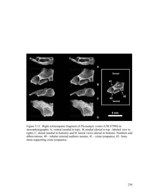
Figure 3.11. Right ectotympanic fragment of Plesiadapis cookei (UM 87990) in stereophotographic A, ventral (medial to top); B, medial (dorsal to top - labeled view to right); C, dorsal (medial to bottom); and D, lateral views (dorsal to bottom). Numbers and abbreviations: 40 – tubular external auditory meatus, 41 – crista tympanica, 42– bony struts supporting crista tympanica. 236
Figure 3.12. Right dentary of Plesiadapis cookei (UM 87990) in A, buccal; B, lingual; and C, stereophotograph occlusal views. On A and B, note lack of margoconid on I 1 . On C, note trigonid basin of P 4 . 237
- Page 213 and 214: SYSTEMATIC PALEONTOLOGY Class MAMMA
- Page 215 and 216: Premaxilla and premaxillary dentiti
- Page 217 and 218: nerve and vessels in life (Fig. 3.5
- Page 219 and 220: identifiable. No ethmoid foramina c
- Page 221 and 222: process is quite large, projecting
- Page 223 and 224: vestibuli. This groove’s point of
- Page 225 and 226: 9: 40). The right side reveals an a
- Page 227 and 228: e seen as a wedge-shaped, rugose de
- Page 229 and 230: process appears as solid bone. Admi
- Page 231 and 232: 16) for P. tricuspidens and Rose (1
- Page 233 and 234: DENTAL FUNCTIONAL MORPHOLOGY OF P.
- Page 235 and 236: Lower premolar molarization As indi
- Page 237 and 238: SUMMARY AND CONCLUSION The skull of
- Page 239 and 240: REFERENCES Bloch, J.I., Boyer, D.M.
- Page 241 and 242: TABLES Table 3.1. List of anatomica
- Page 243 and 244: Table 3.2. Anatomical abbreviations
- Page 245 and 246: Table 3.3. Size comparison among pl
- Page 247 and 248: Table 3.4 continued. European plesi
- Page 249 and 250: Figure 3.1. Cranium of Plesiadapis
- Page 251 and 252: Figure 3.3. Right maxillary teeth (
- Page 253 and 254: Figure 3.4. Cranium of Plesiadapis
- Page 255 and 256: Figure 3.5. Cranium of Plesiadapis
- Page 257 and 258: Figure 3.6. Cranium of Plesiadapis
- Page 259 and 260: Figure 3.8. Fragment from right nuc
- Page 261 and 262: Figure 3.9. Right promontorium of P
- Page 263: Figure 3.10. Cranium of Plesiadapis
- Page 267 and 268: Figure 3.14. A, Plot of relief inde
- Page 269 and 270: CHAPTER 4: THE FIRST KNOWN SKELETON
- Page 271 and 272: among plesiadapiforms (e.g., Szalay
- Page 273 and 274: Institutional and collections abbre
- Page 275 and 276: CaL - capitulum (of humerus) antero
- Page 277 and 278: HSV - head shape variable = ln(DEW/
- Page 279 and 280: MSD - mid-shaft dorsoventral or ant
- Page 281 and 282: Ry - ray (as in “digit ray”) S-
- Page 283 and 284: History of descriptive study of the
- Page 285 and 286: illustrations of this material, exc
- Page 287 and 288: astragalus and calcaneum was highly
- Page 289 and 290: discussion of the femur indicates t
- Page 291 and 292: supinator crests. He also noted tha
- Page 293 and 294: that it may not even be an archonta
- Page 295 and 296: unstudied material. Specifically, h
- Page 297 and 298: 5321), some metapodials (MNHN R 529
- Page 299 and 300: Gingerich and Gunnell (1992) publis
- Page 301 and 302: prehensility they provide, is an in
- Page 303 and 304: euarchontans (Fig. 1.1). Their anal
- Page 305 and 306: for comparison. These include isola
- Page 307 and 308: plesiadapid samples have the same m
- Page 309 and 310: Organization of results Each bone i
- Page 311 and 312: Bloch and Boyer (2002) and N. inter
- Page 313 and 314: clavicle reflects some basic aspect
Figure 3.11. Right ectotympanic fragment of Plesiadapis cookei (UM 87990) in<br />
stereophotographic A, ventral (medial to top); B, medial (dorsal to top - labeled view to<br />
right); C, dorsal (medial to bottom); and D, lateral views (dorsal to bottom). Numbers and<br />
abbreviations: 40 – tubular external auditory meatus, 41 – crista tympanica, 42– bony<br />
struts supporting crista tympanica.<br />
236


