- Page 1 and 2:
NEW CRANIAL AND POSTCRANIAL REMAINS
- Page 3 and 4:
Stony Brook University The Graduate
- Page 5 and 6:
postcranial anatomy more accurately
- Page 7 and 8:
Table of Contents List of Figures .
- Page 9 and 10:
Zygomatic. . . . . . . . . . . . .
- Page 11 and 12:
Zygomatic. . . . . . . . . . . . .
- Page 13 and 14:
Humerus . . . . . . . . . . . . . .
- Page 15 and 16:
Description . . . . . . . . . . . .
- Page 17 and 18:
Description . . . . . . . . . . . .
- Page 19 and 20:
Craniodental material of Plesiadapi
- Page 21 and 22:
Figure 2.25. MNHN CR 965 Plesiadapi
- Page 23 and 24:
Figure 4.24. UM 87990 Plesiadapis c
- Page 25 and 26:
Table List Chapter 2 Table 2.1. Num
- Page 27 and 28:
Table 4.30. Caudal vertebrae measur
- Page 29 and 30:
CHAPTER 1: INTRODUCTION Plesiadapif
- Page 31 and 32:
primates, sharing many dental featu
- Page 33 and 34:
were acquired. Specifically, the
- Page 35 and 36:
of the plesiadapiform skeleton. Ext
- Page 37 and 38:
Gervais, M.P., 1877. Enumération d
- Page 39 and 40:
Szalay, F.S., 1972. Cranial morphol
- Page 41 and 42:
CHAPTER 2: A REEVALUATION OF CRANIA
- Page 43 and 44:
Information on the cranium of basal
- Page 45 and 46:
petrosal bulla predicts that a sutu
- Page 47 and 48:
History of descriptive study of ple
- Page 49 and 50:
Gingerich (1971) rebutted Szalay (1
- Page 51 and 52:
9c, Gingerich (1976) labeled a groo
- Page 53 and 54:
portion of the bulla medial to the
- Page 55 and 56:
Bloch and Silcox (2006) described t
- Page 57 and 58:
MATERIALS AND METHODS Material exam
- Page 59 and 60:
whitening, dark and light areas on
- Page 61 and 62:
SYSTEMATIC PALEONTOLOGY Class MAMMA
- Page 63 and 64:
efore meeting a large, anteroposter
- Page 65 and 66:
The canine is a simple, single-root
- Page 67 and 68:
existence and/or nature of contacts
- Page 69 and 70:
outlined. This includes description
- Page 71 and 72:
eneath it while also extending post
- Page 73 and 74:
y a pair of parallel grooves (Fig.
- Page 75 and 76:
the skull (Fig. 2.1). The length of
- Page 77 and 78:
margin clearly had a posteriorly pr
- Page 79 and 80:
groove measures about 0.29 mm in di
- Page 81 and 82:
2.13: 50). The suture with the supr
- Page 83 and 84:
preserved (Fig. 2.15: 55). In fact,
- Page 85 and 86:
the posterior septum, a deeply inci
- Page 87 and 88:
Plesiadapis tricuspidens MNHN CR 12
- Page 89 and 90:
is roughly 2.8 mm long. Medial to t
- Page 91 and 92:
is not convoluted like many other s
- Page 93 and 94:
110) and the only squamosal/alisphe
- Page 95 and 96:
two regions for the internal jugula
- Page 97 and 98:
ventral to the sinuous suture (132)
- Page 99 and 100:
provides measurements of these and
- Page 101 and 102:
of the promontorium of the Pellouin
- Page 103 and 104:
seen on the HRxCT scan is expressed
- Page 105 and 106:
primates, as well as treeshrews and
- Page 107 and 108:
canaliculus are present on the sept
- Page 109 and 110:
it is missing from the other side o
- Page 111 and 112:
observations and interpretations ma
- Page 113 and 114:
REFERENCES Beard, K.C., 1993. Phylo
- Page 115 and 116:
Russell, D.E., 1959. Le crâne de P
- Page 117 and 118:
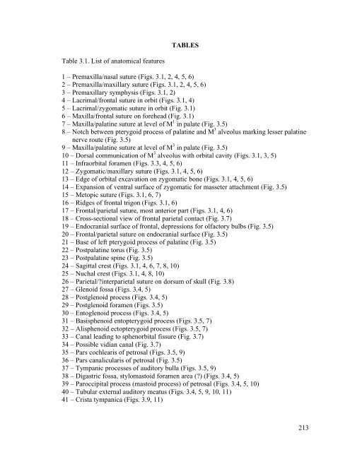
TABLES Table 2.1. Numerical list of
- Page 119 and 120:
81 - Occipital/petrosal suture (Fig
- Page 121 and 122:
Table 2.2. Abbreviations for crania
- Page 123 and 124:
Table 2.3a. Petrosal features of pl
- Page 125 and 126:
Table 2.4. List of cranial measurem
- Page 127 and 128:
Shape variables (Table 2.6) ac/GM -
- Page 129 and 130:
Table 2.5. continued Specimen MNHN
- Page 131 and 132:
Appendix Table 2.1. Specimens scann
- Page 133 and 134:
Fig. 2.39 - bs, Ptr, rtp, s1, s2 Fi
- Page 135 and 136:
Figure 2.1. UALVP 46685 Pronothodec
- Page 137 and 138:
Figure 2.2. UALVP 46685 Pronothodec
- Page 139 and 140:
Figure 2.3. UALVP 46685 Pronothodec
- Page 141 and 142:
Figure 2.4. UALVP 46685 Pronothodec
- Page 143 and 144:
Figure 2.5. UALVP 46685 Pronothodec
- Page 145 and 146:
Figure 2.6. UALVP 49105 Pronothodec
- Page 147 and 148:
Figure 2.7. UALVP 46687 Pronothodec
- Page 149 and 150:
Figure 2.8. USNM 309902 Nannodectes
- Page 151 and 152:
Figure 2.9. USNM 309902 Nannodectes
- Page 153 and 154:
Figure 2.10. USNM 309902 Nannodecte
- Page 155 and 156:
Figure 2.11. USNM 309902 Nannodecte
- Page 157 and 158:
Figure 2.13. USNM 309902 Nannodecte
- Page 159 and 160:
Figure 2.15. AMNH 17388 Nannodectes
- Page 161 and 162:
Figure 2.16. AMNH 17388 Nannodectes
- Page 163 and 164:
Figure 2.18. MNHN CR 125 Plesiadapi
- Page 165 and 166:
Figure 2.20. MNHN CR 125 Plesiadapi
- Page 167 and 168:
Figure 2.22. MNHN CR 965, Plesiadap
- Page 169 and 170:
Figure 2.23. MNHN CR 965 Plesiadapi
- Page 171 and 172:
Figure 2.24. MNHN CR 965 Plesiadapi
- Page 173 and 174:
Figure 2.25. MNHN CR 965, Plesiadap
- Page 175 and 176:
Figure 2.26. Pellouin skull Plesiad
- Page 177 and 178:
Figure 2.27. Pellouin skull Plesiad
- Page 179 and 180:
Figure 2.28. Pellouin skull Plesiad
- Page 181 and 182:
Figure 2.29. Pellouin skull Plesiad
- Page 183 and 184:
Figure 2.30. MaPhQ 33y Adapis paris
- Page 185 and 186:
Figure 2.31. MNHN CR 126, Plesiadap
- Page 187 and 188:
Figure 2.32. SBU MRd-12 Sciurus car
- Page 189 and 190: Figure 2.33. UMMZ 58983 Tupaia glis
- Page 191 and 192: Figure 2.34. Boyer coll. Marmota mo
- Page 193 and 194: Figure 2.35. UMMZ TS13 Lagostomus m
- Page 195 and 196: Figure 2.36. AMNH 41527 Lagostomus
- Page 197 and 198: Figure 2.37. AMNH 185638 Indri indr
- Page 199 and 200: Figure 2.38. USNM 482353 Ignacius c
- Page 201 and 202: Figure 2.39. UM 108207 Acidomomys h
- Page 203 and 204: Figure 2.40. Reconstruction of ples
- Page 205 and 206: CHAPTER 3: DESCRIPTION OF THE FIRST
- Page 207 and 208: these species. Changing ecological
- Page 209 and 210: Institutional abbreviations AMNH, A
- Page 211 and 212: Methods of examination and document
- Page 213 and 214: SYSTEMATIC PALEONTOLOGY Class MAMMA
- Page 215 and 216: Premaxilla and premaxillary dentiti
- Page 217 and 218: nerve and vessels in life (Fig. 3.5
- Page 219 and 220: identifiable. No ethmoid foramina c
- Page 221 and 222: process is quite large, projecting
- Page 223 and 224: vestibuli. This groove’s point of
- Page 225 and 226: 9: 40). The right side reveals an a
- Page 227 and 228: e seen as a wedge-shaped, rugose de
- Page 229 and 230: process appears as solid bone. Admi
- Page 231 and 232: 16) for P. tricuspidens and Rose (1
- Page 233 and 234: DENTAL FUNCTIONAL MORPHOLOGY OF P.
- Page 235 and 236: Lower premolar molarization As indi
- Page 237 and 238: SUMMARY AND CONCLUSION The skull of
- Page 239: REFERENCES Bloch, J.I., Boyer, D.M.
- Page 243 and 244: Table 3.2. Anatomical abbreviations
- Page 245 and 246: Table 3.3. Size comparison among pl
- Page 247 and 248: Table 3.4 continued. European plesi
- Page 249 and 250: Figure 3.1. Cranium of Plesiadapis
- Page 251 and 252: Figure 3.3. Right maxillary teeth (
- Page 253 and 254: Figure 3.4. Cranium of Plesiadapis
- Page 255 and 256: Figure 3.5. Cranium of Plesiadapis
- Page 257 and 258: Figure 3.6. Cranium of Plesiadapis
- Page 259 and 260: Figure 3.8. Fragment from right nuc
- Page 261 and 262: Figure 3.9. Right promontorium of P
- Page 263 and 264: Figure 3.10. Cranium of Plesiadapis
- Page 265 and 266: Figure 3.12. Right dentary of Plesi
- Page 267 and 268: Figure 3.14. A, Plot of relief inde
- Page 269 and 270: CHAPTER 4: THE FIRST KNOWN SKELETON
- Page 271 and 272: among plesiadapiforms (e.g., Szalay
- Page 273 and 274: Institutional and collections abbre
- Page 275 and 276: CaL - capitulum (of humerus) antero
- Page 277 and 278: HSV - head shape variable = ln(DEW/
- Page 279 and 280: MSD - mid-shaft dorsoventral or ant
- Page 281 and 282: Ry - ray (as in “digit ray”) S-
- Page 283 and 284: History of descriptive study of the
- Page 285 and 286: illustrations of this material, exc
- Page 287 and 288: astragalus and calcaneum was highly
- Page 289 and 290: discussion of the femur indicates t
- Page 291 and 292:
supinator crests. He also noted tha
- Page 293 and 294:
that it may not even be an archonta
- Page 295 and 296:
unstudied material. Specifically, h
- Page 297 and 298:
5321), some metapodials (MNHN R 529
- Page 299 and 300:
Gingerich and Gunnell (1992) publis
- Page 301 and 302:
prehensility they provide, is an in
- Page 303 and 304:
euarchontans (Fig. 1.1). Their anal
- Page 305 and 306:
for comparison. These include isola
- Page 307 and 308:
plesiadapid samples have the same m
- Page 309 and 310:
Organization of results Each bone i
- Page 311 and 312:
Bloch and Boyer (2002) and N. inter
- Page 313 and 314:
clavicle reflects some basic aspect
- Page 315 and 316:
Humerus Description.—The right an
- Page 317 and 318:
epicondyle actually projects somewh
- Page 319 and 320:
cookei is absolutely longer than an
- Page 321 and 322:
tuberosity. This crest probably del
- Page 323 and 324:
olecranon process to estimate its t
- Page 325 and 326:
distinct, convex distal radial face
- Page 327 and 328:
of the midcarpal joint), and its pr
- Page 329 and 330:
(there is no evidence for more than
- Page 331 and 332:
matches the opposing facet on the t
- Page 333 and 334:
mobility at the trapezoid-trapezium
- Page 335 and 336:
Function.—The three proximal carp
- Page 337 and 338:
the bone presently being described:
- Page 339 and 340:
the “set 2” MC II is a larger,
- Page 341 and 342:
differs from MC II and III in havin
- Page 343 and 344:
even more pronounced. The distal en
- Page 345 and 346:
etween the distal carpals and the
- Page 347 and 348:
have stouter shaft diameters for th
- Page 349 and 350:
difference makes them more like kno
- Page 351 and 352:
antipronograde clinging postures, o
- Page 353 and 354:
foramina, and faces slightly proxim
- Page 355 and 356:
spine at the superior tip of the il
- Page 357 and 358:
the thigh (Gambaryan, 1974). The ha
- Page 359 and 360:
The femoral shaft is smooth, lackin
- Page 361 and 362:
lacking a lateral extension of its
- Page 363 and 364:
The anteromedial side of the tibial
- Page 365 and 366:
fibular notch and the strong crest
- Page 367 and 368:
to the peroneal surface. The perone
- Page 369 and 370:
could even be described as having t
- Page 371 and 372:
Function.—The functional features
- Page 373 and 374:
flexor fibularis groove surface. Ex
- Page 375 and 376:
tubercle, which is centrally locate
- Page 377 and 378:
Cuboid Description.—The right cub
- Page 379 and 380:
Ectocuneiform Description.—A left
- Page 381 and 382:
Metatarsals Hallucal metatarsal des
- Page 383 and 384:
articulation with the entocuneiform
- Page 385 and 386:
medial side facet on MT IV from a v
- Page 387 and 388:
Vertebral column Vertebral column d
- Page 389 and 390:
measurements, see caption of Fig. 4
- Page 391 and 392:
transverse processes, and the poste
- Page 393 and 394:
taxa appear to have slightly more p
- Page 395 and 396:
dorsoventrally than craniocaudally.
- Page 397 and 398:
The zygapophyses increase in size b
- Page 399 and 400:
vertebrae. It is also similar to th
- Page 401 and 402:
identifications have been reversed.
- Page 403 and 404:
preserved. The ribs are slender and
- Page 405 and 406:
Carpolestes simpsoni (UM 101963) an
- Page 407 and 408:
third metacarpal, similar to arbore
- Page 409 and 410:
autapomorphy, because it appears th
- Page 411 and 412:
Jenkins (1974) found that Tupaia gl
- Page 413 and 414:
Nannodectes and other plesiadapids,
- Page 415 and 416:
are consistent with more frequent u
- Page 417 and 418:
differences generated from informat
- Page 419 and 420:
Perry, M. Silcox, R. Secord and man
- Page 421 and 422:
ubriventer: implications for the fu
- Page 423 and 424:
Linnaeus, C., 1758. Systema naturae
- Page 425 and 426:
Stern, J.T.,Jr., 1988. Essentials o
- Page 427 and 428:
Table 4.3A. Measurements of the pro
- Page 429 and 430:
Table 4.3C. Comparative shape varia
- Page 431 and 432:
Table 4.5A. Measurements and shape
- Page 433 and 434:
Table 4.10. Measurements and shape
- Page 435 and 436:
Table 4.13. Measurements of plesiad
- Page 437 and 438:
Table 4.15. Measurements of plesiad
- Page 439 and 440:
Table 4.17B. Measurements of the di
- Page 441 and 442:
Table 4.18B. Measurements of the di
- Page 443 and 444:
Table 4.20B. Measurements 11-18 of
- Page 445 and 446:
Table 4.21A. Measurements 1-10 and
- Page 447 and 448:
Table 4.23. Measurements and shape
- Page 449 and 450:
Table 4.26. Measurements of plesiad
- Page 451 and 452:
Table 4.28. Measurements of plesiad
- Page 453 and 454:
Table 4.32. Measurements of plesiad
- Page 455 and 456:
Table 4.35. Body segment lengths (m
- Page 457 and 458:
Table 4.37A. Parameters for Gingeri
- Page 459 and 460:
Table 4.38C. Summary of plesiadapid
- Page 461 and 462:
Appendix Table 4.1B. Plesiadapis co
- Page 463 and 464:
Appendix Table 4.3. Other plesiadap
- Page 465 and 466:
Figure 4.2. Plesiadapis cookei (UM
- Page 467 and 468:
Figure 4.4. Plot of principal coord
- Page 469 and 470:
Figure 4.6. Surface reconstructions
- Page 471 and 472:
Figure 4.8. Surface reconstructions
- Page 473 and 474:
Figure 4.9. Surface reconstructions
- Page 475 and 476:
Figure 4.10. Surface reconstruction
- Page 477 and 478:
Figure 4.12. Surface reconstruction
- Page 479 and 480:
Figure 4.13. Surface reconstruction
- Page 481 and 482:
Figure 4.14. Plesiadapis cookei or
- Page 483 and 484:
Figure 4.15. Surface reconstruction
- Page 485 and 486:
Figure 4.17. Plot of principal coor
- Page 487 and 488:
Figure 4.19. Plesiadapis cookei (UM
- Page 489 and 490:
Figure 4.21. Plesiadapis cookei (UM
- Page 491 and 492:
Figure 4.23. Plesiadapis cookei (UM
- Page 493 and 494:
Figure 4.24. Plesiadapis cookei (UM
- Page 495 and 496:
Figure 4.25. Surface reconstruction
- Page 497:
Figure 4.27. Plesiadapis cookei (UM
- Page 500 and 501:
Figure 4.30 472
- Page 502 and 503:
Figure 4.31. Measurements of astrag
- Page 504 and 505:
Figure 4.33 476
- Page 506 and 507:
Figure 4.34. Measurements of calcan
- Page 508 and 509:
Figure 4.36. Plesiadapis cookei (UM
- Page 510 and 511:
Figure 4.38. Plesiadapis cookei (UM
- Page 512 and 513:
Figure 4.40. Stereophotographic vie
- Page 514 and 515:
Figure 4.42. Plesiadapis cookei (UM
- Page 516 and 517:
Figure 4.44. Plesiadapis cookei (UM
- Page 518 and 519:
Figure 4.46. 490
- Page 520 and 521:
Figure 4.47 492
- Page 522 and 523:
Figure 4.48. Plesiadapis cookei (UM
- Page 524 and 525:
Figure 4.50. 496
- Page 526 and 527:
Figure 4.51. Surface reconstruction
- Page 528 and 529:
INTRODUCTION Bloch et al. (2007) an
- Page 530 and 531:
have a lacrimal bone that retains i
- Page 532 and 533:
Institutional abbreviations AMNH, A
- Page 534 and 535:
level cladogram. A total of 33 cran
- Page 536 and 537:
plesiadapiform Ignacius graybullian
- Page 538 and 539:
RESULTS Phylogenetic reconstruction
- Page 540 and 541:
Optimization of postcranial traits
- Page 542 and 543:
Therefore, character optimization r
- Page 544 and 545:
carpolestid bulla is not split into
- Page 546 and 547:
2008). I therefore changed the codi
- Page 548 and 549:
Re-coding and optimization of crani
- Page 550 and 551:
and paromomyids. This, however, is
- Page 552 and 553:
REFERENCES Beard, K.C., 1989. Postc
- Page 554 and 555:
Novacek, M.J., 1986. The skull of l
- Page 556 and 557:
TABLES Table 5.1. Dental characters
- Page 558 and 559:
Table 5.2. Dental character matrix.
- Page 560 and 561:
asisphenoid and basioccipital bones
- Page 562 and 563:
111 (p3). Deltopectoral crest of hu
- Page 564 and 565:
158 (p50). Metatarsal I facet on en
- Page 566 and 567:
Table 5.4C. Postcranial characters
- Page 568 and 569:
Table 5.7. Posterior carotid forame
- Page 570 and 571:
Figure 5.2. 542
- Page 572 and 573:
Figure 5.3 544
- Page 574 and 575:
Figure 5.4. Plot of posterior carot
- Page 576 and 577:
ostral end of the nasals) and prema
- Page 578 and 579:
its body size would fit predictions
- Page 580 and 581:
vertically-to-caudally projecting t
- Page 582 and 583:
More generally speaking, this disse
- Page 584 and 585:
Figure 6.1. CT reconstruction of Pl
- Page 586 and 587:
BIBLIOGRAPHY Alexander, R.M., Jayes
- Page 588 and 589:
Coleman, M. N. and Boyer, D.M. 2008
- Page 590 and 591:
Godinot, M., Beard, K.C., 1991. Fos
- Page 592 and 593:
MacPhee, R.D.E., 1981. Auditory Reg
- Page 594 and 595:
Savage, D.E., Russell, D.E., 1983.
- Page 596 and 597:
Szalay, F.S., Drawhorn, G., 1980. E


