Uterus retroverted with KOH Ring visible - Medical Dynamics
Uterus retroverted with KOH Ring visible - Medical Dynamics
Uterus retroverted with KOH Ring visible - Medical Dynamics

You also want an ePaper? Increase the reach of your titles
YUMPU automatically turns print PDFs into web optimized ePapers that Google loves.
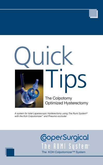
Quick<br />
Tips<br />
The Colpotomy<br />
Optimized Hysterectomy<br />
A system for total Laparascopic Hysterectomy using The Rumi System ®<br />
<strong>with</strong> the Koh Colpotomizer and Pneumo-occluder<br />
The <strong>KOH</strong> Colpotomizer System
Technique Objective:<br />
To provide the surgeon performing<br />
laparoscopic hysterectomy procedures <strong>with</strong> the<br />
proper tools to move <strong>with</strong> greater accuracy,<br />
reproducibility, and safety.
(<strong>Uterus</strong> <strong>retroverted</strong> <strong>with</strong> <strong>KOH</strong> <strong>Ring</strong> <strong>visible</strong>)<br />
Upper Pedicles (Not pictured)<br />
<strong>Uterus</strong> Position: Anteverted<br />
Key Structures: Fallopian Tubes, Ovarian Ligament,<br />
Round Ligament, and Broad Ligament<br />
Note: The tubo-ovarian pedicle consists of the fallopian tubes<br />
and ovarian ligament.<br />
• Divide the tubo-ovarian pedicle<br />
• Divide the round ligament<br />
• Divide the posterior leaf of the broad ligament<br />
Purpose:<br />
1. To provide lateral movement of the uterus<br />
2. To increase distance from the ureters
Uterovesical Peritoneum<br />
(Bladder Flap)<br />
<strong>Uterus</strong> Position: Retroverted<br />
Key Structures: Uterovesical Peritoneum, Bladder,<br />
Pubocervical Fascia, Bladder Pillars<br />
Note: Uterovesical pertains to the uterus and bladder<br />
• Push the colpotomizer against the cervix, to<br />
ensure proper location to create bladder flap<br />
• Elevate peritoneum, make horizontal incision,<br />
and push flap against colpotomizer towards vagina<br />
• Continue dissection laterally across the plane<br />
Purpose<br />
1. To move bladder back off of cervix to allow<br />
anterior colpotomy<br />
2. To ensure optimal vaginal canal length
Anterior Colpotomy<br />
(10 o’clock to 2 o’clock )<br />
<strong>Uterus</strong> Position: Retroverted<br />
Key Structures: Vaginal Fornices<br />
Note: Be sure to inflate Pneumo-occluder<br />
• Push the colpotomizer against vaginal fornices to<br />
stretch vagina<br />
• Make incision along the cup rim to the anterior<br />
vaginal wall<br />
Purpose:<br />
1. To preserve optimal vaginal length<br />
2. To increase distance from ureters
Posterior Colpotomy<br />
(4 o’clock to 8 o’ clock)<br />
<strong>Uterus</strong> Position: Anteverted<br />
Key Structures: Vaginal Fornices, Uterosacral<br />
Ligaments<br />
• Maintain pressure against the vaginal fornices<br />
<strong>with</strong> the colpotomizer<br />
• Palpate to locate the upper rim of the colpotomizer<br />
• Make incision along the cup rim to the posterior<br />
vaginal wall<br />
Purpose:<br />
1. To preserve optimal vaginal length<br />
2. To preserve uterosacral ligaments, thereby,<br />
preserving existing uterine support and nerve supply
Uterine Vessel/<br />
Cardinal Ligament Pedicle<br />
(2 to 4 o’clock and 8 to 10 o’clock)<br />
<strong>Uterus</strong> Position: Lateral ( Right and Left)<br />
Key Structures: Uterine Vessels, Cardinal Ligament,<br />
Ureters, Lateral Fornices<br />
• Expose the ( right, left ) vaginal fornix<br />
• Push colpotomizer against fornix which pushes<br />
the uterine vessels upward away from ureters<br />
• Secure and dessicate ( right, left ) uterine vessels<br />
• Divide the ( right, left ) vaginal fornix to complete<br />
colpotomy incision<br />
Purpose:<br />
1. To increase distance from ureters<br />
2. To preserve optimal vaginal length<br />
3. To totally free uterus <strong>with</strong>in abdominal cavity
Removal of <strong>Uterus</strong><br />
• Deflate the pneumo-occluder<br />
• Attach a tenaculum to the cervix<br />
• Attempt to <strong>with</strong>draw RUMI from vagina<br />
• Remove uterus vaginally OR if uterus is enlarged<br />
morcellate or deflate RUMI balloon for vaginal<br />
removal
Closure of Vagina<br />
• Laparoscopic vaginal cuff closure is done by<br />
re-inserting the pneumo-occluder into the vagina<br />
and inflating it <strong>with</strong> 150 cc’s of saline OR by leaving<br />
the uterus wedged in the vaginal canal<br />
• Vaginal cuff closure can also be accomplished<br />
vaginally
Images provided by<br />
Charles H. Koh, M.D., FRCOG, FACOG,<br />
Associate Clinical Professor, Department of OB/GYN,<br />
<strong>Medical</strong> College of Wisconsin<br />
95 Corporate Drive, Trumbull, CT 06611 • 203.601.5200 • 800.243.297<br />
www.coopersurgical.com • ©2007CooperSurgical, Inc. 81187 Rev. 12/07