Early osseointegration to hydrophilic and hydrophobic ... - Straumann
Early osseointegration to hydrophilic and hydrophobic ... - Straumann
Early osseointegration to hydrophilic and hydrophobic ... - Straumann

You also want an ePaper? Increase the reach of your titles
YUMPU automatically turns print PDFs into web optimized ePapers that Google loves.
<strong>Early</strong> <strong>osseointegration</strong> <strong>to</strong> <strong>hydrophilic</strong> <strong>and</strong> <strong>hydrophobic</strong> implant surfaces in humans<br />
Scientific source<br />
Lang NP, Salvi GE, Huynh-Ba G, Ivanovski S, Donos N, Bosshardt DD.<br />
Clin. Oral Implants Res. 2011;22:349–356.<br />
Introduction<br />
The surface characteristics of titanium implants influence the rate<br />
<strong>and</strong> degree of <strong>osseointegration</strong>. Moderately rough surfaces such as<br />
SLA ® have demonstrated superior bone-<strong>to</strong>-implant contact (BIC)<br />
than surfaces such as titanium plasma-sprayed (TPS), Al 2<br />
O 3<br />
-blasted<br />
or machined surfaces 1,2 . Chemical modification, such as with the <strong>hydrophilic</strong><br />
SLActive ® surface, can further enhance the <strong>osseointegration</strong><br />
process.<br />
Investigations comparing <strong>osseointegration</strong> with various implant surfaces<br />
have been performed, but tend <strong>to</strong> be in vivo animal studies.<br />
No data are available from human studies, <strong>and</strong> the healing sequence<br />
of the early <strong>osseointegration</strong> process in human <strong>and</strong> how it<br />
compares <strong>to</strong> the process – seen in other in vivo investigations – is<br />
relatively unknown.<br />
The aim of this investigation, therefore, was <strong>to</strong> evaluate the rate <strong>and</strong><br />
degree of <strong>osseointegration</strong> at two different implant surfaces (SLA ®<br />
<strong>and</strong> SLActive ® ) during the early phases of healing in a human<br />
model.<br />
Materials <strong>and</strong> methods<br />
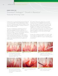
A <strong>to</strong>tal of 49 specially designed titanium implants (length 4 mm, outer<br />
diameter 2.8 mm) with either a SLA ® or SLActive ® surface were<br />
placed in the retromolar region of 28 healthy volunteers. A healing<br />
cap with an internal screw assembly was attached <strong>to</strong> the coronal<br />
part of the implant. After submerged healing periods of 7, 14, 28<br />
<strong>and</strong> 42 days, the implants were removed using a specially designed<br />
trephine, which removed the implant <strong>and</strong> circumferential tissue<br />
of 1 mm thickness.<br />
His<strong>to</strong>logical sections were prepared <strong>and</strong> his<strong>to</strong>metric analyses performed<br />
for amounts of new bone, old bone, bone debris, soft tissue<br />
<strong>and</strong> BIC.<br />
Results<br />
Healing was uneventful at all sites. Of the 49 implants placed, 30<br />
were available for his<strong>to</strong>logical/his<strong>to</strong>metric analysis; difficulty in<br />
harvesting the biopsies resulted in the loss of some specimens.<br />
% of new bone on the implant surface<br />
Artifacts were present on a number of specimens – these areas were<br />
excluded from analysis so that only artifact-free regions were evaluated.<br />
The percentages of new bone-<strong>to</strong>-implant contact after 7, 14,<br />
28 <strong>and</strong> 42 days are shown in table 1.<br />
% mean<br />
value<br />
7 days 14 days 28 days* 42 days<br />
SLActive ® 6.0 14.8 48.3 62.0<br />
SLA ® 6.0 12.2 32.4 62.0<br />
Table 1: Percentage of BIC after 7, 14, 28 <strong>and</strong> 42 days<br />
*Statistically significant (p=0.033)<br />
After 7 days, no differences were observed between the SLA ® <strong>and</strong><br />
SLActive ® specimens. BIC was approximately 6 %, <strong>and</strong> some early<br />
bone apposition was noted in places where existing bone was in<br />
close contact with the implant surface; bone therefore bridged a<br />
gap between old bone <strong>and</strong> implant in these situations. The majority<br />
of the space between bone <strong>and</strong> implant was filled with soft tissue<br />
comprising primitive matrix with various bone debris particles.<br />
BIC increased <strong>to</strong> 12.2 % <strong>and</strong> 14.8 % for SLA ® <strong>and</strong> SLActive ® , respectively,<br />
after 14 days. Bone formation was noted on the existing bone,<br />
extending partly on<strong>to</strong> the implant surface. The beginning of new<br />
bone apposition was evident over large areas of the surface of the<br />
SLActive ® implants. Larger bone particles were seen <strong>to</strong> be surrounded<br />
by osteoid, which helped trabecula formation.
BIC increased in both sample types by day 28, but was significantly<br />
higher with SLActive ® (48.3 %) than with SLA ® (32.4 %). A bony<br />
coating was observed with both specimen types, but almost complete<br />
BIC was observed within some threads of the SLActive ® implants,<br />
<strong>and</strong> new mineralized bone trabeculae were observed<br />
extending in<strong>to</strong> the provisional matrix.<br />
After 42 days, BIC increased further <strong>to</strong> 62 % for both SLA ® <strong>and</strong><br />
SLActive ® . An advanced stage of bone maturation was observed<br />
with both surfaces, <strong>and</strong> the formation of osteons was observed<br />
away from the implant surface. The osteocoating was noted <strong>to</strong> be<br />
thick <strong>and</strong> extensive, <strong>and</strong> was frequently connected via trabeculae,<br />
extending on<strong>to</strong> new bone.<br />
Conclusions<br />
■ Similar healing patterns were observed for both SLA ® <strong>and</strong><br />
SLActive ® implants<br />
■ Osseointegration (BIC) was greater after 14 days <strong>and</strong> significantly<br />
greater after 28 days for SLActive ®<br />
■ The rate of <strong>osseointegration</strong> was substantially slower (approximately<br />
double the healing time) in humans than that observed<br />
in animal studies<br />
■ This is the first study <strong>to</strong> demonstrate his<strong>to</strong>logically the <strong>osseointegration</strong><br />
process with SLActive ® in humans<br />
References<br />
1<br />
Buser, D., Schenk, R.K., Steinemann, S., Fiorellini, J.P., Fox, C.H., & Stich, H. (1991) Influence of surface characteristics on bone integration of<br />
titanium implants. A his<strong>to</strong>morphometric study in miniature pigs. Journal of Biomedical Materials Research 25: 889-902.<br />
2<br />
Cochran, D.L., Schenk, R.K., Lussi, A., Higginbot<strong>to</strong>m, F.L., & Buser, D. (1998) Bone response <strong>to</strong> unloaded <strong>and</strong> loaded titanium implants with<br />
a s<strong>and</strong>blasted <strong>and</strong> acid-etched surface: a his<strong>to</strong>metric study in the canine m<strong>and</strong>ible. Journal of Biomedical Materials Resarch 40: 1-11.<br />
International Headquarters<br />
Institut <strong>Straumann</strong> AG<br />
Peter Merian-Weg 12<br />
CH-4002 Basel, Switzerl<strong>and</strong><br />
Phone +41 (0)61 965 11 11<br />
Fax +41 (0)61 965 11 01<br />
www.straumannusa.com<br />
<strong>Straumann</strong> USA<br />
<strong>Straumann</strong> USA, LLC<br />
60 Minuteman Road<br />
Andover, MA 01810<br />
Phone 800/448 8168<br />
978/747 2500<br />
Fax 978/747 2490<br />
www.straumannusa.com<br />
<strong>Straumann</strong> Canada<br />
<strong>Straumann</strong> Canada Limited<br />
3115 Harvester Road, 1st Floor<br />
Burling<strong>to</strong>n, ON L7N 3N8<br />
Phone 800/363 4024<br />
905/319 2900<br />
Fax 905/319 2911<br />
www.straumann.ca<br />
USLIT 419 4/12