Download - Straumann
Download - Straumann
Download - Straumann

Create successful ePaper yourself
Turn your PDF publications into a flip-book with our unique Google optimized e-Paper software.
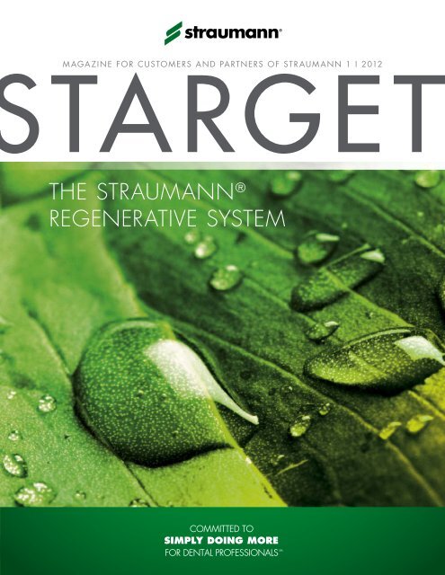
MAGAzINE fOR CUSTOMERS AND PARTNERS Of STRAUMANN 1 I 2012<br />
the straumann ®<br />
regenerative system
Imprint starget – magazine for Customers and Partners of straumann i © straumann usa i 60 minuteman road i andover, ma 01810 i Phone 800/448 8168<br />
Fax 978/747 2490 i Editors roberto gonzález i mildred Loewen i E-Mail info.usa@straumann.com i Internet www.straumann.us/starget<br />
Legal Notice exclusion of liability for articles by external authors: articles by external authors published in starget have been systematically assessed and carefully<br />
selected by the publisher of starget (straumann usa). such articles in every case reflect the opinion of the author(s) concerned and therefore do not necessarily<br />
coincide with the publisher’s opinion. nor does the publisher guarantee the completeness or accuracy and correctness of articles by external authors published in<br />
starget. the information given in clinical case descriptions, in particular, cannot replace a dental assessment by an appropriately qualified dental specialist in an<br />
individual case. any orientation to articles published in starget is therefore on the dentist’s responsibility. articles published in starget are protected by copyright<br />
and may not be reused, in full or in part, without the express consent of the publisher and the author(s) concerned. straumann ® and all other trademarks and logos are<br />
registered trademarks of straumann usa and/or of its affiliates. third party corporate names and brand names that may be mentioned may be registered or otherwise<br />
protected marks even if this is not specially indicated. the absence of such an indication shall not therefore be interpreted as allowing such a name to be freely used.
editorial<br />
STARGET 1 I 12<br />
3<br />
A New Era in Oral Tissue<br />
Regeneration<br />
Dear Valued Customer,<br />
It is my pleasure to introduce myself as the new head of <strong>Straumann</strong> North America,<br />
and also to introduce this first installment of STARGET for 2012. The focus of this issue,<br />
"The <strong>Straumann</strong> Regenerative System," is particularly significant for me as I come<br />
to <strong>Straumann</strong> USA as the former Head of <strong>Straumann</strong> Global Regenerative Sales.<br />
As you know, <strong>Straumann</strong> provides solutions for both tissue regeneration and GBR<br />
including Emdogain, BoneCeramic, <strong>Straumann</strong> AlloGraft, and most recently<br />
MembraGel ® , an innovative liquid membrane that can be precisely applied to the<br />
surgical site.<br />
Andy Molnar<br />
<strong>Straumann</strong> Executive Vice President<br />
North America<br />
You’ll find several interesting articles on tissue regeneration in this issue of<br />
STARGET: “Why Repair When You Can Regenerate?“ on page 6, “Redefining<br />
the Membrane“ on page 12, and an interview with Prof. Dr. Christoph Hämmerle<br />
on the innovation and distinctiveness of MembraGel on page 16. You’ll also see<br />
an article on our Esthetic Case Book, which documents 11 cases illustrating the<br />
dramatic results that can be achieved through Emdogain, on page 18.<br />
Restorative dentistry has seen breakthroughs over the past few years. This STARGET<br />
features articles on the latest development of <strong>Straumann</strong> CARES ® digital workflow.<br />
We hope you will enjoy the clinical case on <strong>Straumann</strong>'s screw-retained hybrid<br />
solution from your peers, and an article based on the interviews with three key<br />
opinion leaders on global trends in dentistry.<br />
Enjoy this issue of STARGET and please let us know how we can continue to<br />
improve upon it.<br />
Sincerely,<br />
Andy Molnar<br />
<strong>Straumann</strong> Executive Vice President, North America
4 STARGET 1 I 12<br />
overvieW<br />
Overview<br />
STARGET 1 I 2012<br />
6<br />
The <strong>Straumann</strong> ® Regenerative System<br />
offers solutions for oral tissue regeneration<br />
– ranging from conservative dentistry to<br />
dental restoration. New to the portfolio:<br />
<strong>Straumann</strong> ® regenerative S y S tem<br />
<strong>Straumann</strong> ® MembraGel ® .<br />
28<br />
<strong>Straumann</strong> ® CareS ® digital S olutionS<br />
<strong>Straumann</strong>, together with 3M ESPE,<br />
has introduced a streamlined digital<br />
workflow that connects the Lava C.O.S.<br />
Intra-Oral Scanner to the <strong>Straumann</strong> ®<br />
CARES ® Digital Solutions platform.<br />
Simply doing more<br />
54<br />
Global Trends – Where is the final<br />
destination? We asked three prominent<br />
dentists to give us their perspective<br />
on things: Lyndon Cooper, Kenneth<br />
Malament and Daniel Wismeijer.
C ontent<br />
STARGET 1 I 12<br />
5<br />
CONTENT<br />
FOCAL POINT 6 Why Repair When You Can Regenerate Instead?<br />
STRAUMANN ® REGENERATIVE SYSTEM 12 <strong>Straumann</strong> MembraGel ® – Re-Defining the Membrane<br />
16 Interview with Christoph Hämmerle<br />
18 An Esthetic Case Selection on <strong>Straumann</strong> Emdogain<br />
20 <strong>Straumann</strong> Allograft Portfolio Expands<br />
STRAUMANN ® CARES ® DIGITAL SOLUTIONS 22 Evaluating the Precision of <strong>Straumann</strong> ® CARES ® Guided Surgery<br />
Based on a Clinical Case<br />
28 Digital Workflow<br />
30 The Intraoral Workflow<br />
32 3M ESPE Lava Ultimate Restorative<br />
38 Interview with Mike Rynerson<br />
RESTORATIVE 43 <strong>Straumann</strong> ® Anatomic IPS e.max ® Abutment<br />
45 Immediate Full Mouth Restoration Using Implant-Supported Fixed<br />
Hybrid Prosthetics<br />
SURGICAL 50 Updated SLActive ® Scientific Evidence Brochure<br />
ITI / EDUCATION 52 <strong>Straumann</strong> & Baylor University Launch the First Interdisciplinary<br />
Digital Dentistry Course<br />
53 ITI Membership Tops 10,000<br />
SIMPLY DOING MORE 54 Global Trends<br />
60 <strong>Straumann</strong> AID: Access to Implant Dentistry<br />
64 Literature Alerts<br />
67 Upcoming 2012 Education Events
6 STARGET 1 I 12<br />
foC al point<br />
STrAUMANN ® reGeNerATive SYSTeM<br />
Why Repair When You Can Regenerate Instead?<br />
The More Complex an Organism is, the Lesser the Capacity of its Body to Perform Regenerative<br />
Processes<br />
The biological process, or self-sufficient capability to restore deficient tissue is referred to as the ability<br />
for regeneration. In contrast to repair, i.e., wound healing, whereby the original biological structure is<br />
not fully rebuilt, the goal of regeneration is to completely restore the structure and function of tissue that<br />
has been lost or injured. This ability has continuously diminished as creatures have evolved and the<br />
complexity of these organisms has increased. Compared to the “champions” of regeneration, present<br />
day cnidarians, which can re-grow severed extremities and internal organs that have been lost, this<br />
capability in humans, without additional support, is limited to a few types of external tissue.<br />
When the Body’s Own Healing Process “Overshoots its Target“<br />
When a person’s tissue is injured, the body’s repair process, rather than regenerative process, begins.<br />
Here, the body does not primarily attempt to restore the original state and function of the tissue,<br />
but rather to close the wound as quickly as possible. This spontaneous healing process involves the<br />
formation of connective tissues that penetrate deeply into the original tissue and also remove “good”<br />
tissue in the process. During such a “radical” healing process, it is possible that even important functions<br />
are permanently destroyed.<br />
Regeneration Can be Guided with Medical Treatment<br />
To prevent this type of overcompensation in the healing process wherever possible, strong antiinflammatory<br />
drugs are administered today, such as in cases of back injuries. This prevents this destructive<br />
process from occurring, in turn making it possible to preserve part of the nerve tissue and mitigating<br />
deficits such as paralyses. In medical terms, regeneration entails assisting desirable tissue formation<br />
processes, and guiding and limiting the repair mechanisms involved in wound healing as to not impede<br />
the regeneration of intact, functioning tissue.
foC al point<br />
STARGET 1 I 12<br />
7<br />
Fig. 1: The cnidarians (pictured here, a green anemone, anthopleura xanthogrammica) are equipped with astounding regenerative<br />
capabilities that far surpass those of humans.
8 STARGET 1 I 12<br />
<strong>Straumann</strong> ® regenerative S y S tem<br />
The Insertion of Implants Requires Healthy Bone Tissue<br />
Great advances in regeneration have been achieved in dentistry over that last 20 years. We can<br />
now regenerate the bone tissue of the jaw, opening the door for implants as a treatment for replacing<br />
teeth. This has required scientific evidence documenting the physiological processes involved in tissue<br />
formation and the necessary aids for controlling these processes. Today, for example, we know that<br />
lost or missing bone can only be regenerated by the body when the bone-forming cells can perform<br />
their work undisturbed. Without external intervention, this process would have to compete with the rapid<br />
natural wound repair mechanism, which would result in the growth of new connective tissue instead of<br />
the desired bone tissue.<br />
Fig. 2: The high porosity of <strong>Straumann</strong> ® BoneCeramic (90 %) allows blood vessels and vital bone to vascularize into the material.<br />
Regeneration Requires Matrices and Membranes<br />
Today, one means of guiding the regeneration process requires two aids: a matrix and a membrane.<br />
The matrix keeps the space available for the bone to grow while a membrane serves as a barrier to<br />
prevent the connective tissue infiltration of the gingiva.
<strong>Straumann</strong> ® regenerative S y S tem<br />
STARGET 1 I 12<br />
9<br />
Various types of these matrices and membranes are used today for regenerative purposes. Some of<br />
these “placeholders” remain in the body, integrated into the newly formed bone for the rest of the person’s<br />
life. Applying membranes for this purpose is also quite complex and time-consuming. <strong>Straumann</strong> ®<br />
MembraGel ® was developed to deal with the disadvantages of the types of membranes used in the<br />
past. The time-consuming cutting and fitting of the membrane is unnecessary since MembraGel is<br />
applied in liquid form and polymerizes to form a solid film in less than a minute. The resulting hydrogel<br />
is biocompatible and completely biodegradable.<br />
Periodontitis – The Greatest Threat to the Periodontium<br />
For implantology, the regeneration of the periodontium is equally as important as the new formation<br />
of bone, which is destroyed by periodontitis and can ultimately result in the loss of the tooth. Despite<br />
numerous attempts to regenerate the destroyed tissue, a breakthrough to achieve complete tissue<br />
reformation remains to be seen. <strong>Straumann</strong> ® Emdogain can be used in cases to undo some of this<br />
destruction, making it possible to prevent loss of the tooth. Emdogain contains the key components<br />
required for building up the cementum and periodontal ligament, important parts of the periodontium.<br />
These elements, or proteins, tap into the body’s own natural ability to rebuild the surrounding tooth<br />
structures, using nature’s help.<br />
“Simply Doing More“ – During the Regeneration Process<br />
<strong>Straumann</strong>’s successes in the field of regeneration are promising and we have yet to fully realize our<br />
dreams. We are already working on further innovations to guide regenerative processes in an even<br />
simpler and more effective manner. We are inspired by two great visions:<br />
» A matrix for bone augmentation that breaks down fully and can be used without requiring a<br />
membrane.<br />
» Guided tissue regeneration that allows for complete preservation of the tooth.<br />
Now making its first entrance into the dental market with <strong>Straumann</strong> MembraGel, we view PEG technology<br />
as the foundation for achieving these goals. Beyond MembraGel, there are numerous combinations and<br />
degrees of cross-linking these biocompatible hydrogels, which present an exciting range of possible<br />
options. This innovative technology is only in the early phases of trials and development and has<br />
enormous potential for the future.
10 STARGET 1 I 12<br />
<strong>Straumann</strong> ® regenerative S y S tem<br />
The <strong>Straumann</strong> ® Regenerative System: Everything from a Single Source<br />
The <strong>Straumann</strong> ® Regenerative System offers you a variety of solutions for oral tissue regeneration –<br />
ranging from conservative dentistry to dental restoration. Our goal is to offer you a variety of predictable<br />
and scientifically proven regenerative treatment solutions – all from a single source and in the tried and<br />
tested quality that is <strong>Straumann</strong>’s hallmark.<br />
Tissue Regeneration<br />
By mimicking the natural process of odontosis,<br />
<strong>Straumann</strong> ® emdogain forms an insoluble,<br />
three-dimensional extracellular matrix that<br />
remains on the root surface for 2 – 4 weeks<br />
and enables a selective cell population,<br />
proliferation and differentiation.<br />
Bone Formation<br />
<strong>Straumann</strong> ® allograft is a wide range of<br />
bone allograft solutions, allowing you the<br />
flexibility to choose the treatment that’s right for<br />
your case. Through a commercial partnership<br />
with LifeNet Health ® , <strong>Straumann</strong>’s allograft<br />
options provide confidence in the safety of the<br />
material you use to treat your patients.<br />
<strong>Straumann</strong> ® BoneCeramic is a fully synthetic<br />
bone substitute material with excellent<br />
morphology that promotes the new formation<br />
of vital bone. It can be used for a series of<br />
procedures used for dental bone regeneration.<br />
Bone Healing<br />
<strong>Straumann</strong> ® membragel ® is a membrane<br />
of the latest generation. It combines unique<br />
material properties that have been developed<br />
to promote undisturbed bone healing and to<br />
simplify the surgical procedure.
STARGET 1 I 12<br />
11<br />
straumann ® emdogain <br />
IS TRUE PERIODONTAL REGENERATION<br />
IMPORTANT TO YOU?<br />
Photos courtesy of Dr. G.<br />
Zuchelli, Bologna, Italy<br />
before<br />
after<br />
More than 100 clinical publications in peer-reviewed journals demonstrate<br />
<strong>Straumann</strong> ® Emdogain to be safe and effective in stimulating the formation of new<br />
periodontal soft and hard tissue. These clinical studies involve more than<br />
3000 defects in over 2500 patients.<br />
Contact <strong>Straumann</strong> Customer Service at 800/448 8168 to learn more<br />
about <strong>Straumann</strong> solutions or to locate a representative in your area.<br />
www.straumann.us<br />
excellent clinical results 1-4<br />
Long-term clinical benefit 5,6<br />
Less patient discomfort 3<br />
1<br />
Tonetti MS, et al. Enamel matrix proteins in the regenerative therapy of deep intrabony defects.J Periodontol.<br />
2002;29:317–325.<br />
2<br />
Froum SJ, et al. A comparative study utilizing open flap debridement with and without enamel matrix derivative in<br />
the treatment of periodontal intrabony defects: A 12-month re-entry.J Periodontol. 2001;72:25–34.<br />
3<br />
Jepsen S, et al. A randomized clinical trial comparing enamel matrix derivative and membrane treatment of<br />
buccal class II furcation involvement in mandibular molars. Part I: study design and results for primary outcomes.<br />
J Periodontol. 2004; 75:1150 –1160.<br />
4<br />
McGuire MK, et al. Evaluation of human recession defects treated with coronally advanced flaps and either enamel<br />
matrix derivative or connective tissue. Part 1: comparison of clinical parameters.J Periodontol. 2003;74:1110 –1125.<br />
5<br />
Sculean A, et al. Ten-year results following treatment of intra-bony defects with enamel matrix proteins and guided<br />
tissue regeneration. J Clin Periodontol. 2008; 35:817-824.<br />
6<br />
Data on file (McGuire 10 year)
12 STARGET 1 I 12<br />
<strong>Straumann</strong> ® regenerative S y S tem<br />
STrAUMANN ® MeMbraGel ®<br />
Re-Defining the Membrane<br />
In 2010, <strong>Straumann</strong> introduced <strong>Straumann</strong> MembraGel, an advanced technology membrane that<br />
is one of the most significant innovations in guided bone regeneration in recent history.<br />
Created with PEG (polyethylene glycol) hydrogel technology, <strong>Straumann</strong> MembraGel is applied in<br />
liquid form and molds to the defect. Shortly after application, the liquid components solidify, stabilizing<br />
the bone graft and providing an effective barrier to tissue infiltration. <strong>Straumann</strong> MembraGel then<br />
biodegrades over time. It is designed to achieve undisturbed bone regeneration, which is a prerequisite<br />
for achieving ideal clinical and esthetic results.<br />
“<strong>Straumann</strong> MembraGel is a key innovation in Guided Bone Regeneration. With the<br />
PEG technology we are on the verge of something new, entering a new era in oral<br />
tissue regeneration.“ Christoph Hämmerle, University of Zurich<br />
Designed for Improving GBR Procedures<br />
<strong>Straumann</strong> MembraGel provides effective support in bone formation for GBR (guided bone regeneration)<br />
cases due to its optimized barrier properties. It facilitates undisturbed bone healing as a basis for the<br />
optimal clinical outcome achieved by stabilization of the bone graft material. The precise and easy<br />
application simplifies the surgical procedure.
<strong>Straumann</strong> ® regenerative S y S tem<br />
STARGET 1 I 12<br />
13<br />
Stabilization of the Bone Graft<br />
The gel-like consistency of <strong>Straumann</strong> ® MembraGel<br />
® allows for precise application to the<br />
surgical site and adaptation to various types<br />
and sizes of bone defects.<br />
Solidification and Stabilization<br />
Once solidified in situ (20 – 50 seconds after<br />
application), <strong>Straumann</strong> ® MembraGel is<br />
designed to stabilize the underlying bone graft<br />
to facilitate undisturbed bone regeneration.<br />
Backed by Scientific Documentation<br />
<strong>Straumann</strong> MembraGel is backed by preclinical 1,2,3,4,5 and clinical 6 documentation including one- and<br />
three-year follow-up data 7 presented in 2010 and submitted for publication. The ongoing clinical<br />
program with <strong>Straumann</strong> MembraGel includes over 40 centers and more than 200 patients in Europe<br />
and North America.<br />
<strong>Straumann</strong> ® MembraGel Exclusive Education Program<br />
<strong>Straumann</strong> ® MembraGel was launched in conjunction with a well-received specialized education<br />
program that provides in-depth information on pre-clinical and clinical evidence, hands-on product<br />
training and the surgical techniques of this new application. Herbert Früh, Head of Business Unit<br />
Regenerative at <strong>Straumann</strong> explained, “MembraGel has allowed <strong>Straumann</strong> to bring a brilliant idea to
14 STARGET 1 I 12<br />
<strong>Straumann</strong> ® regenerative S y S tem<br />
fruition. It is an innovative technology that requires training. Customers’ initial experience is that it saves<br />
time during intraoral application but it is different to handle. For this reason, it was the right decision to<br />
combine the launch of the new product with an intensive education program.”<br />
Availability<br />
<strong>Straumann</strong> ® MembraGel ® was introduced in Europe and North America toward the end of 2010, with<br />
roll-outs to other markets planned in the future.<br />
Hands-on workshop at a <strong>Straumann</strong> MembraGel education event.<br />
1<br />
Humber CC, Sándor GK, Davis JM et al. Bone healing with an in situ formed bioresorbable polyethylene glycol hydrogel membrane<br />
in rabbit calvarial defects. Oral Surg Oral Med Oral Pathol Oral Radiol Endod 2010;109:372–384. Jung RE, Lecloux G,<br />
2<br />
Rompen E et al. A feasibility study evaluating an in situ formed synthetic biodegradable membrane for guided bone regeneration<br />
3<br />
in dogs. Clin Oral Implants Res 2009;20:151–161. Thoma DS, Halg G-A, Dard MM et al. Evaluation of a new biodegradable<br />
4<br />
membrane to prevent gingival ingrowth into mandibular bone defects in minipigs. Clin Oral Implants Res 2009;20:7–16. Herten<br />
M, Jung RE, Ferrari D et al. Biodegradation of different synthetic hydrogels made of polyethylene glycol hydrogel/RGD-peptide
<strong>Straumann</strong> ® regenerative S y S tem<br />
STARGET 1 I 12<br />
15<br />
PeG – The TechNOLOGY BehiNd STrAUMANN ® MeMbraGel ®<br />
<strong>Straumann</strong> MembraGel is a synthetic, biodegradable in situ-formed membrane made of polyethylene<br />
glycol (PEG) which forms a molecular network. The material is biocompatible, is applied in liquid form<br />
and sets quickly. Due to its gel-like consistency, it can be applied to cover the exact shape of the defect<br />
or augmented area and does not require cutting and trimming to shape before application – unlike<br />
traditional GBR membranes.<br />
What exactly is PEG, and what makes it suited to this purpose? PEG is a polymer (a large molecule<br />
comprised of repeating structural units). It is produced through the reaction between ethylene oxide and<br />
water or the organic compound ethylene glycol, which is often used as a precursor to polymer materials.<br />
PEG is available in a wide range of molecular weights. The molecular weight also determines the<br />
consistency of the material, which can vary greatly from waxy-solid to water-soluble liquid states. PEG<br />
has a number of properties that are particularly desirable in a number of biological, pharmaceutical<br />
and chemical applications: it is highly flexible, non-toxic and non-immunogenic. It is also hydrophilic,<br />
meaning that its attachment to biomolecules can increase solubility and decrease aggregation of the<br />
molecules.<br />
Thanks to these very advantageous properties, PEG has been extensively used in many medical,<br />
pharmaceutical and medical device applications. For instance, it is used as a spray-on adhesion barrier<br />
and as a main ingredient in laxatives; as an agent for bowel irrigation prior to surgery or colonoscopy;<br />
as an addition to protein medications to extend their effect and increase dosing intervals; and as a<br />
carrier in various medications, such as soft capsules, ointments, tablets, and lubricants. Preliminary<br />
research is also underway to investigate the potential of PEG as a component in many other exciting<br />
applications, such as gene therapy, spinal cord injuries and the suppression of carcinogenesis. The<br />
use of PEG as a customizable, liquid-applied membrane is therefore another milestone in a long line of<br />
applications for this useful, versatile and extensively researched material.<br />
5<br />
modifications: an immunohistochemical study in rats. Clin Oral Implants Res 2009;20:116–125. Jung RE, Zwahlen M, Weber FE<br />
et al. Evaluation of an in situ formed hydrogel as a biodegradable membrane for guided bone regeneration. Clin Oral Implants Res<br />
6<br />
2006;17:426–433. Jung RE, Hälg GA, Thoma DS, Hämmerle CHF. A randomized, controlled clinical trial to evaluate a new<br />
7<br />
membrane for guided bone regeneration around dental implants. Clin Oral Implants Res 2009;20:162–168. Ramel C, Halg G,<br />
Thoma D et al. A randomized clinical trial to evaluate a synthetic gel membrane for GBR around dental implants – 1- and 3-year<br />
results. European Association for Osseointegration 19 th Annual Scientific Meeting, Glasgow, UK, 6–9 October 2010; Abs 055.
16 STARGET 1 I 12<br />
<strong>Straumann</strong> ® regenerative S y S tem<br />
iNTerview<br />
“Being Part of this Network is Something Truly Exciting –<br />
Something We’ve Never Done in this Way Before.“<br />
Andy Molnar, <strong>Straumann</strong> Executive Vice President, North America, interviews<br />
Professor Dr. Christoph Hämmerle of the University of Zurich about<br />
<strong>Straumann</strong> ® MembraGel ® .<br />
professor Hämmerle, our company is introducing membragel by way of an<br />
international education-based program. this is a product which has been longawaited<br />
in the market; so many people would like to get their hands on it as<br />
Prof. Dr. Christoph Hämmerle<br />
Chairman of the Department of Fixed and<br />
Removable Prosthodontics and Dental Material<br />
Science, University of Zurich/Switzerland.<br />
soon as possible. What are your thoughts on this educational approach to the<br />
launch?<br />
I think it’s very important to use an educational approach. What we’re dealing<br />
with here is a new technology, a new kind of membrane, a new way to apply<br />
the membrane. There are a lot of differences from all the membranes we’ve had<br />
before. I think it’s very important to launch the product in a well-structured way so<br />
that we know what experience we can rely on. It’s also an opportunity to learn<br />
about new things we need to become familiar with before we can use a new<br />
technology like this successfully and predictably.<br />
“We treated over thirty defects and had minimal complications.<br />
Based on this experience and learning, we moved on to larger defects<br />
with a very good success rate once again.“ Christoph Hämmerle<br />
i know you’re excited about membragel and the progress the product is making.<br />
How would you describe your involvement from the start, from inception all the<br />
way through the development of the project?<br />
It has been very exciting for us. It’s really special to be there when the original<br />
idea is born and then to participate in the development of a new product, which<br />
is eventually able to solve problems and improve patient care. With all the sharing,<br />
teaching, and feedback we’re getting from different people all over the world,<br />
being part of this network is something really exciting, something we’ve never<br />
done this way before. For this reason, we’re looking forward to continuing with the
<strong>Straumann</strong> ® regenerative S y S tem<br />
STARGET 1 I 12<br />
17<br />
development of this scientific and educational network and<br />
really benefiting from the process. I think it’s great!<br />
from our learning so far, what are the key points that we<br />
need to pass on to our educators?<br />
There are three key points. One is that you have to change<br />
some of your surgical habits to adapt to the new technology,<br />
the new way to apply the membrane. The second is to adhere<br />
to published surgical guidelines. And the third is to begin with<br />
less complex cases until you have had around ten successful<br />
cases, and then move on to more difficult cases.<br />
When we conducted our initial study, in which we used a<br />
collagen membrane as a control, we did not see any more<br />
difficulties with the membrane using the new technology than<br />
with the membrane using the older technology. I think this was<br />
the case because we were careful to use small dehiscence<br />
defects in non-esthetic areas. We treated over thirty defects<br />
and had minimal complications. Based on that experience<br />
and learning, we began to expand to larger defects, again,<br />
with very good success.<br />
Professor Hämmerle, thank you very much for your time.<br />
It’s much appreciated.<br />
“I think it’s very important to launch the product<br />
in a well-structured way so that we know what<br />
experience we can rely on.“ Christoph Hämmerle<br />
Returning to the topic of changing habits, this is a new<br />
technology, and there is a great temptation to use it in all<br />
kinds of different ways because it is rather easy to use and<br />
very new and exciting. We need to exercise restraint and<br />
really adhere to new practices in aspects of our surgical<br />
work.<br />
The second point pertains to the surgical guidelines. We<br />
have seen that the surgical guidelines are not always<br />
followed accurately. Following the surgical guidelines could<br />
improve the practical outcomes when it comes to using this<br />
new product.<br />
The third aspect is the size of the defects we tackle first.
18 STARGET 1 I 12<br />
<strong>Straumann</strong> ® regenerative S y S tem<br />
GrOwTh iN receSSiON<br />
An Esthetic Case Selection on <strong>Straumann</strong> ® Emdogain<br />
Clinicians have been predictably treating gingival recession with <strong>Straumann</strong> ®<br />
Emdogain for years and talking about the beautiful smiles they gave back<br />
to their patients – we gave them the chance to prove it.<br />
The best cases from the 2009 “Growth in Recession” Esthetic Case Competition<br />
were chosen by a renowned international jury to appear in <strong>Straumann</strong>’s Growth in<br />
Recession Esthetic Case Book. This casebook, published in 2011, features eleven<br />
cases presented together with the patients’ histories and images of each step to<br />
facilitate a detailed understanding of the surgical procedures involved.<br />
This valuable tool is complimentary to you with your next order of <strong>Straumann</strong><br />
Emdogain. Please reference order code “RECESSION” when placing your order.<br />
Designed to help achieve excellent esthetic results<br />
Esthetic Casebook: <strong>Straumann</strong> ® Emdogain<br />
The following clinicians contributed their Case reports to this publication:<br />
p Brazil: Dr. Robert Carvalho da Silva, DDS, MS, PH.D. (co-authors: Dr. Julio<br />
Cesar Joly, DDS, MS, PH.D., and Dr. Paulo Fernando Mesquita de Carvalho,<br />
DDS, MS)<br />
p USA: Dr. Robert Levine, DDS – Dr. Ken Akimoto, DDS, MSD – Dr. Mark I. Gutt,<br />
DMD– Dr. Eunseok Eugene Oh, DDS (co-author: Dr. Vincent Iacono, DDS) – Dr.<br />
Paul G. Luepke, DDS, MS<br />
p Canada: Dr. Ira Paul Sy, DDS, MS, Dip. Periodontics<br />
p Germany: Dr. med. dent. Andreas Hofmann, M.Sc. – Dr. Bjørn Greven (coauthor:<br />
Dr. Bernd Heinz)<br />
p Spain: Dr. Ion Zabalegui, MD
<strong>Straumann</strong> ® regenerative S y S tem<br />
STARGET 1 I 12<br />
19<br />
“BOTH THE SCIENTIFIC EVIDENCE AND MY PERSONAL ExPERIENCE<br />
SUPPORT THAT WITH THE APPROPRIATE CASE STRAUMANN ® EMDOGAIN<br />
SIGNIFICANTLY IMPROVES ROOT COVERAGE COMPARED TO THE<br />
CORONALLY ADVANCED FLAP ALONE.”<br />
DR. MICHAEL K. MCGUIRE, DDS<br />
Treatment of recession defects with <strong>Straumann</strong> Emdogain<br />
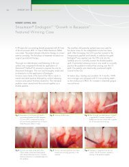
Courtesy of Dr. Paul G. Luepke, DDS, MSD<br />
A referred 23-year-old female patient was presenting with multiple gingival recessions<br />
(teeth #8 to #3). The prominent canine showed a Miller Class III recession, the<br />
other teeth presented with Miller Class I recessions. The treatment procedure began<br />
with a thorough cleaning and scaling of the exposed root surfaces with hand<br />
and sonic instruments and was followed by a split thickness flap preparation of a<br />
Zucchelli-style flap (without vertical releasing incisions). A dissection into the vestibular<br />
mucosa allowed for further mobilization.<br />
Dr. Paul G. Luepke, DDS, MS<br />
1996 Master’s degree in Periodontics, University<br />
of Texas at San Antonio Health Science Center, Tx,<br />
USA • 1997 Diplomate of the American Board<br />
<strong>Straumann</strong> ® PrefGel ® (EDTA) was applied for two minutes on the root surface.<br />
Subsequently, the surgical area was rinsed with sterile saline and <strong>Straumann</strong> ®<br />
Emdogain was applied to the root surfaces. Connective tissue graft (CTG) was<br />
harvested from the palate with the single incision technique. The graft was then<br />
split. The CTG was fixed on the root surface and the flap coronally positioned and<br />
fixed with sling sutures.<br />
of Periodontology • 2008 Assistant Professor of<br />
Surgical Services Periodontics Division, Marquette<br />
University School of Dentistry in Milwaukee, WI,<br />
USA • 2009 Interim Department Chair of Surgical<br />
Sciences at Marquette University School of Dentistry<br />
in Milwaukee, WI, USA<br />
Mechanical tooth cleaning in the surgical area was avoided during the first 4<br />
weeks and a chlorhexidine solution was prescribed. Sutures were removed 10<br />
days after surgery.<br />
Initial situation<br />
6 week follow-up<br />
11 month follow-up
20 STARGET 1 I 12<br />
<strong>Straumann</strong> ® regenerative S y S tem<br />
ALLOGrAfT LiNe exPANSiON<br />
Building A Foundation for Success with More Options –<br />
<strong>Straumann</strong> AlloGraft Portfolio Expands<br />
<strong>Straumann</strong> is pleased to expand the options available for you through our commercial partnership with<br />
LifeNet Health, ® allowing you the flexibility of choice when treating your patient. Processed with LifeNet<br />
Health’s proprietary and patented Allowash xG ® technology, <strong>Straumann</strong> AlloGraft gives you confidence<br />
that your graft is safe and effective.<br />
<strong>Straumann</strong> AlloGraft C/C mix, Mineralized<br />
Ground Cortical/Cancellous mix<br />
A mix of the strength of cortical bone with the<br />
structure of cancellous bone to support bony<br />
ingrowth in one product<br />
<strong>Straumann</strong> AlloGraft OCAN (top) and <strong>Straumann</strong><br />
AlloGraft GC (bottom), electron microscope<br />
image, magnification 20x<br />
<strong>Straumann</strong> AlloGraft OCAN 0.25 cc, Mineralized Ground Cancellous<br />
<strong>Straumann</strong> AlloGraft GC 0.25 cc, Mineralized Ground Cortical<br />
<strong>Straumann</strong> AlloGraft DGC 0.25 cc, Demineralized Ground Cortical<br />
For smaller defects, when extensive grafting is not needed<br />
<strong>Straumann</strong> AlloGraft OCAN (left), <strong>Straumann</strong><br />
AlloGraft GC (middle), <strong>Straumann</strong> AlloGraft DGC<br />
(right), electron microscope image, magnification 20x<br />
SIMPLIFy WITH CONFIDENCE<br />
From crown to root, <strong>Straumann</strong> provides you with the<br />
convenience of ordering solutions from one provider – all<br />
from the scientifically driven company you know and trust.
STARGET 1 I 12<br />
21<br />
straumann ® Cares ® digitaL soLutions<br />
SEAMLESS CONNECTIONS<br />
Pave your way to success. Covering a full product range from temporary restorations to<br />
esthetic crown and bridge restorations, <strong>Straumann</strong> ® CARES ® Digital Solutions is now featuring:<br />
new generation scanner new cAd software<br />
new applications leading range of materials<br />
<strong>Straumann</strong> ® CARES ® Digital Solutions brings modern digital dentistry to dental professionals as a complete system –<br />
reliable, precise, and dedicated to your needs.<br />
intra-oraL sCan<br />
guided surgery<br />
CadCam<br />
Please contact us at 800/448 8168. More information on www.straumann-cares-digital-solutions.com
22 STARGET 1 I 12<br />
<strong>Straumann</strong> ® C areS ® digital S olutionS<br />
STefANO STOreLLi, LeONArdO AMOrfiNi, MAUriziO cAMANdONA ANd eUGeNiO rOMeO<br />
Evaluating the Precision of <strong>Straumann</strong> ® CARES ® Guided<br />
Surgery Based on a Clinical Case<br />
Introduction<br />
The use of guided surgery is paving the way for the future of implant surgery. Softwarebased<br />
pre-operative planning differs considerably from traditional planning with casts<br />
and x-ray printouts. The following case report involves a restoration for a partially<br />
edentulous woman with a fixed prosthesis preceded by pre-operative planning<br />
with the <strong>Straumann</strong> ® Guided Surgery System using coDiagnostix (software) and<br />
gonyx (scan and surgical template fabrication device).<br />
Dr. Stefano Storelli<br />
Graduation in Dentistry and Dental Prosthetics at the<br />
University of Milan/Italy. PhD in Implant Dentistry.<br />
Postgraduate in Oral Surgery (class of 2012).<br />
Lecturer at the Department of Implant Prosthetics at<br />
the University of Milan. Author of various national<br />
and international publications and collaboration on<br />
various transcriptions. ITI and SIO member. Private<br />
practice in Milan.<br />
Patient History<br />
An 87-year-old Caucasian woman referred to our clinic, asked for a solution for her<br />
faulty fixed bridge which was causing pain and difficulty in eating. She had never<br />
worn a removable prosthesis and was willing to do anything possible to keep her<br />
fixed dentition. The patient suffered from an unspecified choreia with symptoms of<br />
involuntary movements, and was on aspirin treatment for her high blood pressure.<br />
Despite these restrictions, she was in a good physical and mental health.<br />
Clinically, the following situation was diagnosed: (1) a faulty fixed bridge (7 to<br />
12), still anchored on the left side, where an old implant was still in use, (2) two<br />
Fig. 1 Fig. 2<br />
Fig. 3<br />
Fig. 4
<strong>Straumann</strong> ® C areS ® digital S olutionS<br />
STARGET 1 I 12<br />
23<br />
remaining teeth (8 and 11) which were fractured, and (3) a resorption on site 8<br />
and a fracture of 11 revealed by the OPG as well as the inclination of the distal<br />
implant placed in 12 (figs. 1 – 3). Under these circumstances the clinician decided<br />
to remove the bridge and to restore the patient with a removable prosthesis.<br />
After a couple of weeks the patient stated that it was impossible for her to wear<br />
the temporary denture and she returned to the dental practice several times due<br />
to fractures to the prosthesis. Therefore, it became apparent that a removable<br />
restoration was not suitable for this patient and that an implant solution had to<br />
be taken into consideration. Since minimally invasive surgery was intended, the<br />
clinician opted for a guided implant insertion in the post-extractive sites.<br />
Dr. Leonardo Amorfini<br />
Graduation in Dentistry and Dental Prosthetics at the<br />
University of Milan/Italy. Author of various national<br />
and international publications and collaboration<br />
on various transcriptions. ITI, AO, SICOI and SIO<br />
Treatment Planning<br />
member. Private practice in Gallarate (VA).<br />
A mock-up of the future teeth was evaluated, followed by the preparation of the<br />
diagnostic template with the <strong>Straumann</strong> ® gonyx table. The plate was attached<br />
to the barium teeth and the three titanium pins were placed according to the<br />
manufacturer’s instructions (figs. 4, 5). The diagnostic guide was tested for<br />
Fig. 5 Fig. 6<br />
Fig. 7 Fig. 8
24 STARGET 1 I 12<br />
<strong>Straumann</strong> ® C areS ® digital S olutionS<br />
stability in the mouth of the patient before performing the Cone Beam CT scan<br />
(fig. 6). The CT showed a remarkable resorption on site 8 and confirmed the<br />
inclination of the implant placed in 12. Therefore, the implant to be placed<br />
immediately in position 8 was moved to 7 and a miniflap was raised. The<br />
implant in position 11 had to deal with the position on the other implant.<br />
The final treatment decided upon was a fixed implant supported<br />
Dental Technician Maurizio Camandona<br />
Teacher of post-graduate courses in dental implant<br />
technologies at the University of Milan.<br />
Author and co-Author of professional articles and<br />
reference books. Private practice in Lomazzo<br />
(Como)/Italy, specialized in implant prosthetics,<br />
ceramics and state-of-the-art technologies (CADCAM<br />
prosthesis on 3 implants in positions 7-11-12. The <strong>Straumann</strong> ®<br />
coDiagnostix software was used to plan the treatment (figs. 7, 8). The<br />
implant positions were defined in the software and the resulting template plan<br />
was sent to the lab. The implant in position 7 was planned to be a Roxolid ®<br />
implant with small diameter (<strong>Straumann</strong> ® Bone Level implant, NC Ø 3.3 mm,<br />
SLActive ® 12.0 mm); for the implant in 11, a <strong>Straumann</strong> ® Bone Level implant<br />
(RC Ø 4.1 mm, SLActive 12.0 mm) was chosen.<br />
etc.). Speaker at national congresses on the topics<br />
listed above.<br />
Fig. 9<br />
Fig. 10<br />
Fig. 11 Fig. 12
<strong>Straumann</strong> ® C areS ® digital S olutionS<br />
STARGET 1 I 12<br />
25<br />
It was possible to identify the implant in 12 as a CoreVent1 Ø 4.0 mm<br />
implant placed about 15 years ago and, after some research, some prosthetic<br />
component of the respective implant manufacturer was found that was<br />
compatible with the internal connection to this implant. After drilling the holes<br />
into the scan template according to the template plan, the lab provided the<br />
surgical guide (figs. 9 – 11).<br />
Surgical Procedure<br />
The two teeth were removed together with the granulation tissue around the<br />
root of tooth 8 under local anesthesia. The considerable resorption needed to<br />
be treated with regenerative material (bovine bone substitute material covered<br />
by a resorbable collagen membrane) to avoid major alteration of the contour.<br />
The surgical guide was placed on the remaining teeth and on the healing<br />
cap of the distal implant (fig. 12). The surgical procedure was performed<br />
according to the surgical plan. The implant in position 7 was positioned after<br />
raising a mini-flap and by using the extra-long drill (Ø 2.8 mm) through the<br />
Prof. Eugenio Romeo<br />
Graduation in Medicine and Surgery in 1984 at the<br />
University of Milan/Italy. Director of the Department<br />
of Implant Prosthetics at the University of Milan since<br />
1992. Associate Professor since 2005. Author<br />
of various educational books and national and<br />
international publications. Chairman of the Advanced<br />
Oral Implantology course at the University of Milan.<br />
ITI fellow.<br />
Ø 2.8 mm sleeve (figs. 13, 14).<br />
Fig. 13 Fig. 14<br />
Fig. 15<br />
Fig. 16
26 STARGET 1 I 12<br />
<strong>Straumann</strong> ® C areS ® digital S olutionS<br />
The implant was positioned after the guide had been removed, since the implant, having a diameter of 3.3 mm, cannot<br />
be inserted through the Ø 2.8 mm sleeve. The implant in position 11 was placed after using the extra-long drill series<br />
(Ø 2.2/2.8/3.5 mm) with the 1 mm reduction handle through the Ø 5 mm diameter sleeve (figs. 15, 16). The implant<br />
was positioned through the surgical guide. Transmucosal healing caps were positioned and the bone substitute material<br />
was inserted into the extraction sockets. Both sites were covered with the resorbable collagen membrane.<br />
Final Prostheses and Follow-up<br />
After 6 weeks, the OPG showed correct healing with no radiolucencies (fig. 17). Clinically, the implants sounded<br />
correct and were stable (fig. 18). The healing abutments were removed and screw-retained transfer parts were placed<br />
Fig. 17 Fig. 18<br />
Fig. 19<br />
Fig. 20<br />
Fig. 21<br />
Fig. 22<br />
Fig. 23 Fig. 24<br />
Fig. 25
<strong>Straumann</strong> ® C areS ® digital S olutionS<br />
STARGET 1 I 12<br />
27<br />
in order to take the impression. The three impression copings were splinted<br />
with a bridge of acrylic resin that had been prepared a few days before (figs.<br />
21, 22). The three abutments were placed in position (fig. 22) and the metalceramic<br />
prosthesis (fig. 24) was cemented with a removable cement (fig. 23).<br />
After one month of loading, no complications were registered or stated by<br />
the patient. By comparing the values obtained by computer planning with the<br />
x-ray taken after implant placement (figs. 19, 20), the precision of the system<br />
became visible, with the postextractive implant being about 1 mm deeper than<br />
planned and without much variability in the other dimensions.<br />
Conclusion<br />
The use of computer-guided implant placement allowed expanded treatment<br />
options and a fast, minimal invasive surgery for a patient who was not able to<br />
withstand long procedures. Producing the guide in such a manner is efficient<br />
and it fits well on natural teeth because it has been customized on the patient's<br />
impressions. The implant placement in the case reported here demonstrates the<br />
reliability of the <strong>Straumann</strong> ® Guided Surgery system.<br />
With software-based treatment planning, the implants could be placed in<br />
the same practiced manner as with traditional techniques – but with higher<br />
precision and peace of mind. However, the learning curve for software-based<br />
planning should not be underestimated; in addition to familiarization with the<br />
software, new surgical techniques have to be applied and the surgeon needs<br />
to get accustomed to visualizing the surgical procedure through the software.
28 STARGET 1 I 12<br />
<strong>Straumann</strong> ® C areS ® digital S olutionS<br />
diGiTAL wOrkfLOw<br />
Seamlessly Connected with <strong>Straumann</strong> ® CARES ®<br />
Digital Solutions<br />
1. MULTiPLe dATA SOUrceS 2. STrAUMANN ® cAreS ® viSUAL deSiGN<br />
Surgical planning<br />
<strong>Straumann</strong> ® CARES ® Visual<br />
Digital impression taking<br />
Scan master model <strong>Straumann</strong> ® CARES ®<br />
Scan CS2<br />
A digital workflow to thousands of scanners<br />
<strong>Straumann</strong>, together with 3M ESPE, has introduced a<br />
streamlined digital workflow that connects the Lava C.O.S.<br />
Intra-Oral Scanner to the <strong>Straumann</strong> ® CARES ® Digital Solutions<br />
platform. Parallel to the Cadent iTero ® intraoral scanner,<br />
dentists using the Lava C.O.S. scanner are now able to<br />
transfer digital scan data of the patient’s oral geometry to<br />
the dental lab using the <strong>Straumann</strong> ® CARES ® system. The<br />
CARES ® platform offers seamless connectivity to thousands<br />
of scanners in dental practices worldwide.<br />
CARES ® 7.0: the open standard software platform<br />
<strong>Straumann</strong> ® CARES ® Visual 7.0 offers a wide range of<br />
benefits: the advantages of a flexible, open software<br />
standard – through the Dental Wings Open Software<br />
(DWOS ® ) software core – and the quality and predictability<br />
of the validated workflow of <strong>Straumann</strong> ® CARES ® – through<br />
specific <strong>Straumann</strong> software applications. Powered by the<br />
combined resources of the partners, the CARES ® platform<br />
strives for the leading role in dentistry and provides you<br />
with access to future high-class developments of the digital<br />
dental industry.
<strong>Straumann</strong> ® C areS ® digital S olutionS<br />
STARGET 1 I 12<br />
29<br />
3. vArieTY Of MANUfAcTUriNG OPTiONS 4. vArieTY Of PrOSTheTic OPTiONS<br />
VALIDATED STRAUMANN WORKFLOWS<br />
<strong>Straumann</strong> milling centers<br />
HIgH qUALITy RESTORATIONS<br />
For modern implant and restorative dentistry:<br />
Customized <strong>Straumann</strong> ® CARES ® Abutments (Ti, ZrO 2<br />
),<br />
<strong>Straumann</strong> ® CARES ® Screw-retained bars and bridges<br />
(CoCr, Ti)<br />
Copings, crowns and bridges, inlays, onlays and veneers<br />
Resin nano ceramic:<br />
3M ESPE Lava Ultimate Restorative Ceramics: zerion ®<br />
(ZrO 2<br />
), IPS e.max ® CAD, IPS Empress ® CAD, VITA Mark II,<br />
ExTERNAL WORKFLOWS<br />
Via open STL format<br />
VITA TriLuxe<br />
Metals: ticon ® (Ti), coron ® , (CoCr)<br />
Polymers: polyamide, polycon ® ae, polycon ® cast 2<br />
<strong>Straumann</strong> milling centers: specialists in prosthetics<br />
<strong>Straumann</strong> has a strong and long-time expertise in CADCAM<br />
manufacturing of prosthetic restorations in industrialgrade<br />
precision and quality. The high reliability of these<br />
restorations is based on <strong>Straumann</strong>’s strategy of validated<br />
design software and manufacturing that are compatible<br />
with each other. Via the <strong>Straumann</strong> milling centers, design<br />
expertise is offered as a service for complex restorations<br />
A leading material and application range<br />
The <strong>Straumann</strong> ® CARES ® Digital Solutions portfolio provides<br />
a leading range of CADCAM materials and applications –<br />
according to your needs of serving your customers’ requests<br />
and of working cost-effectively without compromising on<br />
quality: from single-tooth restorations to 16-element bridges<br />
and from well-known to innovative materials like the new<br />
3M ESPE Lava Ultimate Restorative.<br />
such as screw-retained bars and bridges, and as a scan<br />
service for customized abutments 1 .<br />
1 2<br />
Scan service available in Germany only burn out resin, not for clinical use<br />
IPS Empress ® and IPS e.max ® are registered trademarks of Ivoclar Vivadent AG, Liechtenstein. 3M, ESPE, Lava are trademarks of 3M or 3M ESPE AG.<br />
Used under license in Canada.
30 STARGET 1 I 12<br />
<strong>Straumann</strong> ® C areS ® digital S olutionS<br />
The iNTrAOrAL wOrkfLOw<br />
The <strong>Straumann</strong> Digital Workflow for Implant Restorations:<br />
Designed to be Simple, Accurate and Efficient<br />
The conventional prosthetic workflow using traditional impression taking, casting and waxing techniques<br />
can lead to inconsistent impression quality due to human errors. This can result in poor clinical and<br />
esthetic outcomes and time-consuming adjustments during seating. Digitalizing these processes can<br />
improve this situation from both a professional and business perspective.<br />
From the intra-oral scan to the final restoration<br />
<strong>Straumann</strong> offers a new and complete digital workflow for implant restorations. Starting with an intra-oral<br />
scan of the implant site, the customized <strong>Straumann</strong> ® CARES ® Abutment or full contour crown is designed<br />
to provide accuracy together with time and cost efficiency through the whole restorative procedure.<br />
This kind of digital workflow for implant restorations eliminates cumbersome and time-consuming manual<br />
steps in dental practice and in the laboratory. Digital impressions allow immediate quality control by<br />
the dentist, and result in an excellent impression being sent to the laboratory. The workflow therefore<br />
eliminates or reduces impression retakes and restoration remakes, ensuring that seating appointments<br />
are efficient due to the excellent occlusion and contact-points of the restoration.
<strong>Straumann</strong> ® C areS ® digital S olutionS<br />
STARGET 1 I 12<br />
31<br />
deNTiST<br />
Scan the scanbody directly on the implant<br />
with iTero intra-oral scanning and send<br />
the digital data to your partner laboratory.<br />
LABOrATOrY<br />
Design the customized <strong>Straumann</strong> ® CARES ®<br />
Abutment in <strong>Straumann</strong> CARES Visual and<br />
send data to the <strong>Straumann</strong> milling center<br />
for production.<br />
LABOrATOrY<br />
Finalize the restoration using the highprecision<br />
<strong>Straumann</strong> ® CARES ® Abutment,<br />
iTero model, <strong>Straumann</strong> Repositionable<br />
implant analog, and full contour crown.<br />
deNTiST<br />
Serve patient with high-quality customized<br />
restoration designed to provide optimal<br />
function and esthetics.
32 STARGET 1 I 12 <strong>Straumann</strong> ® C areS ® digital S olutionS<br />
3M eSPe LAvA ULTiMATe reSTOrATive<br />
A New Dimension for Dental Materials<br />
3M ESPE and <strong>Straumann</strong> have partnered up to offer a<br />
new CADCAM restorative material, 3M ESPE Lava<br />
Ultimate Restorative, through <strong>Straumann</strong> ® CARES ® Digital<br />
Solutions. The material is based on Resin Nano Ceramic<br />
(RNC) technology, defining a new material class that combines<br />
the benefits of ceramic based on true nano technology<br />
and highly cross-linked resin. 1<br />
Entering the field of nanotechnology<br />
The field of nanotechnology has expanded dramatically as<br />
nanostructured materials exhibit unique properties on the macroscale<br />
that offer high-potential technological benefits. Typically,<br />
the critical properties of nanomaterials are attributable<br />
to internal structures between 1 and 100 nanometers in dimension,<br />
defining the nano world. In comparison, a human<br />
hair is about 200,000 nanometers in diameter, and a typical<br />
virus is about 100 nanometers long, a size which is at the<br />
outer boundaries of nanotechnology. As size is decreased to<br />
nanoscale dimensions, physical properties, e.g., optical characteristics,<br />
get altered, especially when size nears the molecular<br />
scale, meaning < 5 nm. These unique properties are in<br />
the focus when research starts its innovative work to achieve<br />
materials with greatest efficiencies. In the dental field, 3M<br />
ESPE Lava Ultimate Restorative offers an advanced dental<br />
material designed and engineered to be tooth-like and to<br />
deliver workflow advances.<br />
Human hair: Ø 200,000 nm<br />
Virus: Ø 100 nm<br />
Nano particle: Ø 1-100 nm<br />
1<br />
3M, ESPE, Lava, Ultimate Restorative is available with release of <strong>Straumann</strong> ® CARES ® Visual 6.2
<strong>Straumann</strong> ® C areS ® digital S olutionS<br />
STARGET 1 I 12<br />
33<br />
<strong>Straumann</strong> ® CARES ® Restoration made of 3M ESPE Lava Ultimate Restorative. Courtesy of 3M ESPE Ag.
34 STARGET 1 I 12<br />
<strong>Straumann</strong> ® C areS ® digital S olutionS<br />
MATeriAL deScriPTiON<br />
3M ESPE Lava Ultimate Restorative is a Resin Nano Ceramic containing approximately 79 %<br />
surface-modified nanoceramic particles. The ceramic particles are made up of three different ceramic fillers<br />
(of silica and zirconia) ranging between 4 and 20 nm that reinforce a highly cross-linked polymeric matrix.<br />
The ceramic fillers are a combination of:<br />
Silica filler: non-agglomerated/non-aggregated, 20 nm<br />
Zirconia filler: non-agglomerated/non-aggregated 4 to 11 nm<br />
Zirconia/silica cluster filler: aggregated, comprised of 20 nm silica and 4 to 11 nm zirconia particles<br />
According to 3M ESPE<br />
Bonded zirconia/silica nano particles clustered and surface treated in a proprietary process. Courtesy of 3M ESPE AG.
<strong>Straumann</strong> ® C areS ® digital S olutionS<br />
STARGET 1 I 12<br />
35<br />
rNc – A New MATeriAL cLASS<br />
3M ESPE Lava Ultimate Restorative is a new CADCAM material based on Resin Nano Ceramic<br />
(RNC) technology, which is defined as a new material class. RNCs consist of nano ceramic components<br />
embedded in a highly cross-linked polymeric matrix. The true nanotechnology imparts excellent esthetics,<br />
strength and wear resistance. Using RNC technology 3M ESPE Lava Ultimate Restorative is designed<br />
to support a streamlined, flexible workflow.<br />
» Brilliant esthetics – Resin Nano Ceramic for lasting polish<br />
The physical properties of 3M ESPE Lava Ultimate Restorative make this material very similar to<br />
the natural translucency and fluorescence of the teeth in its behavior. The <strong>Straumann</strong> ® CARES ® Restorations<br />
made of it have a glossy appearance when delivered. An easy polishing step taking less than 4 minutes<br />
makes the restoration highly brilliant.<br />
» 10-year limited warranty* – Designed to be durable for reliable restorations<br />
The high flexural strength and fracture toughness of the 3M ESPE Lava Ultimate Restorative material<br />
make it a strong one-piece restoration. It is not brittle, allowing for chipping-free restorations. The material<br />
properties make it possible for the <strong>Straumann</strong> milling centers to produce thin and minimally invasive<br />
restorations, which also open up new treatment possibilities.<br />
» Maintains functional balance – Absorption of chewing forces and less wear to opposing enamel<br />
The nanoceramic technology makes it very kind to the opposing tooth regarding abrasion. 3M<br />
ESPE Lava Ultimate Restorative absorbs chewing forces and brings a new quality to all single tooth<br />
restorations.<br />
iMPrOved wOrkfLOw ThrOUGh AdvANTAGeS iN PrePArATiON ANd hANdLiNG<br />
The high efficiency of the workflow made possible with 3M ESPE Lava Ultimate Restorative is of<br />
special interest for both dental labs and dental practices.<br />
*If all the conditions of the <strong>Straumann</strong> Guarantee ® are fulfilled and if the material is used in strict compliance with approved<br />
indications and instructions for use of 3M ESPE.
36 STARGET 1 I 12<br />
<strong>Straumann</strong> ® C areS ® digital S olutionS<br />
» No further processing or firing<br />
Once the <strong>Straumann</strong> ® CARES ® Restorations made of 3M ESPE Lava Ultimate Restorative are milled<br />
and delivered to the dental professional – no firing is required before being seated. The restoration can<br />
be polished, characterized with light-cured restoratives, and the anatomy can be changed by addingon<br />
or build-up. The abolition of the firing step specially emphasizes the new material category RNC of<br />
3M ESPE Lava Restorative.<br />
» Easy adjustment. Adjustments and customizations can be carried out extra-orally or intra-orally with<br />
light-cured composite, such as 3M Filtek Supreme xTE Universal Restorative / 3M Filtek Ultimate<br />
Universal Restorative, for excellent esthetic match.<br />
» Benefits for dentists, dental labs and patients. <strong>Straumann</strong> ® CARES ® restorations made of 3M<br />
ESPE Lava Ultimate Restorative deliver esthetics without the requirement of further processing steps.<br />
They offer advantages across the dental workflow which are of true benefit to dentists, dental labs and<br />
patients. 3M ESPE Lava Ultimate Restorative offers high control and efficiency.<br />
STrAUMANN – SPeciALiST iN cAdcAM PrOSTheTicS<br />
The new 3M ESPE Lava Ultimate Restorative is indicated for single tooth restorations and can only<br />
be processed by using modern CADCAM technology. 3M ESPE and <strong>Straumann</strong> have partnered up<br />
to offer 3M ESPE Lava Ultimate Restorative, through <strong>Straumann</strong> ® CARES ® Digital Solutions. The<br />
backing of <strong>Straumann</strong> as a reliable provider supports the opportunities this material offers through highlevel<br />
milling expertise and qualitative prosthetic outcomes. New treatment options are possible using<br />
3M ESPE Lava Ultimate Restorative via the <strong>Straumann</strong> milling centers – specialists in CADCAM<br />
prosthetics.<br />
For more detailed information on 3M ESPE LAVA Ultimate Restorative, please go to website http://solutions.3m.com/wps.<br />
portal/3M/en_US/3M-ESPE-NA/dental-professionals/products/category/digital-materials/lava-ultimate/.<br />
3M, ESPE, Lava, Filtek are trademarks of 3M or 3M ESPE AG.
<strong>Straumann</strong> ® C areS ® digital S olutionS<br />
STARGET 1 I 12<br />
37<br />
BeNefiTS fOr deNTiSTS, deNTAL LABS ANd PATieNTS<br />
1. DATA DIgITALIzATION 4. PROCESSINg / POLISHINg<br />
Multiple data sources with <strong>Straumann</strong> ® CARES ®<br />
Digitalization of patient situation with various scanners, the<br />
intraoral scanners iTero ® or Lava C.O.S. and the <strong>Straumann</strong> ®<br />
CARES ® Scan CS2 desktop scanner.<br />
No firing required after milling<br />
An additional polish makes the restoration highly brilliant.<br />
2. DESIgN 5. SEATINg<br />
Validated workflow through <strong>Straumann</strong> CARES Visual<br />
Quality and predictability of the validated workflow via the<br />
specific <strong>Straumann</strong> software applications.<br />
Easy adjustment<br />
Adjustments and customizations can be carried out with lightcured<br />
composite.<br />
3. PRODUCTION 6. PATIENT<br />
<strong>Straumann</strong> CARES restorations in high <strong>Straumann</strong> quality<br />
Thin and minimally invasive restorations made of 3M<br />
ESPE Lava Ultimate Restorative via the <strong>Straumann</strong><br />
milling centers.<br />
Less wear to opposing enamel<br />
3M ESPE Lava Ultimate is not brittle, allowing for chipping<br />
free restorations. It shows no abrasion or opposite tooth<br />
damage.<br />
ShAdeS AvAiLABLe<br />
Eight shades available, four of which include high translucency<br />
HIGH<br />
Transluscency<br />
LOW<br />
Transluscency<br />
Courtesy of 3M ESPE AG<br />
A1 A2 A3 A3.5 B1 C2 D2 Bleach
38 STARGET 1 I 12<br />
<strong>Straumann</strong> ® C areS ® digital S olutionS<br />
iNTerview<br />
Scanning 2.0: Digital Impression-Taking<br />
with iTero
<strong>Straumann</strong> ® C areS ® digital S olutionS<br />
STARGET 1 I 12<br />
39<br />
An interview with Michael Rynerson, <strong>Straumann</strong> Global Head<br />
of iTero Sales, on the iTero intraoral scanning system.<br />
How do you view the market acceptance for a system such<br />
as itero?<br />
Interest in digital impression-taking is truly tremendous. In<br />
particular, we have experienced this at congresses where the<br />
system is demonstrated live. Commercially, the iTero system<br />
with its highly developed 3D technology has gained a leading<br />
position in the USA. We also enjoy widespread interest from<br />
dentists and dental laboratories across Europe, where we<br />
have achieved a leading position in intraoral scanning in<br />
some markets. There are a number of good reasons for<br />
deciding in favor of iTero: user-friendliness, precision, and<br />
efficiency – also in terms of time and costs. Of course there<br />
is an emotional element too – many of our customers are<br />
passionate about adopting great new technologies for the<br />
benefit of their patients.<br />
What role does itero play in the <strong>Straumann</strong> portfolio?<br />
Intraoral scanning is an integral part of our digital portfolio<br />
because it is the most direct link between the restorative<br />
dentist, the laboratory, and our CARES ® production centers.<br />
Our focus is on solutions which enable our customers to offer<br />
their patients the best possible treatment available on the<br />
market. The advantages of iTero make intraoral scanning a<br />
key technology in dentistry and, thus, of interest to all dental<br />
professionals.<br />
<strong>Straumann</strong> implants. True to the motto of our digital portfolio<br />
“Seamless Connections,” <strong>Straumann</strong> will now take the next<br />
step together with our customers. The new digital workflow<br />
is the basis for increasing efficiency and quality, and<br />
facilitates the cooperation between surgeons, dentists, and<br />
dental laboratories significantly. We view digitalization as<br />
a pronounced added value for customers at every stage of<br />
the workflow.<br />
“The role as a center of digital competence makes<br />
the laboratory an important partner for the dentist.”<br />
Michael Rynerson<br />
Can you explain digital workflow for implants in more detail?<br />
Following the recovery period, a scanbody is screwed onto<br />
the implant. The dentist scans the scanbody and the adjacent<br />
teeth in sequence and immediately sees on the screen the<br />
completed digital impression. The scanbody provides the<br />
necessary data on the position of the implant in the scan.<br />
Then the data is forwarded to the laboratory for the design of<br />
the customized <strong>Straumann</strong> CARES abutment in CARES Visual.<br />
The laboratory sends the production data to <strong>Straumann</strong><br />
for fabrication of a CARES custom abutment, and orders a<br />
precision-milled iTero model from an iTero Regional Milling<br />
Center. The laboratory orders a <strong>Straumann</strong> Repositional<br />
Implant Analog that fits into the iTero model.<br />
are there any innovations especially for implantologists?<br />
As an innovative company, <strong>Straumann</strong> has developed a<br />
complete digital workflow, from intraoral scans to <strong>Straumann</strong> ®<br />
CARES ® copings and crowns, to customized abutments for<br />
are dental laboratories also a target group for itero?<br />
In most cases modern dental laboratories are already centers<br />
of competence for digital technology – with CADCAM<br />
being a top subject for some time now. This role as a center
40 STARGET 1 I 12<br />
<strong>Straumann</strong> ® C areS ® digital S olutionS<br />
of digital competence makes the laboratory an important<br />
partner for the dentist. Thus, the iTero system also offers the<br />
dental laboratory new service opportunities. This is of special<br />
value because cooperation is closely linked via the intraoral<br />
scan workflow.<br />
are there other possibilities for using itero as part of the<br />
digital workflow?<br />
We are presently uniting the various workflows to provide<br />
optimum compatibility. For example, our surgical planning<br />
software, the <strong>Straumann</strong> ® Guided Surgery System, combines<br />
the DVT and CT information on the bone situation to a 3D<br />
image for implant planning. Even today, we can already<br />
combine such data sets with intraoral scanning information<br />
to provide better documentation on the bone, soft tissue and<br />
tooth situation. This allows improved surgical and prosthetic<br />
planning.<br />
Cadent, the manufacturer of itero, is part of align<br />
technology as of april 2011. What are the implications?<br />
Align Technology is a heavyweight in the orthodontic industry<br />
and includes invisalign in its portfolio, a well-known and<br />
widely used digital service for orthodontic treatments.<br />
Therefore Align truly understands digital dentistry, as well<br />
as knowing the needs of dental professionals, making them<br />
an ideal new home for the iTero technology. Furthermore,<br />
because <strong>Straumann</strong> is the exclusive distribution partner for<br />
iTero in Europe, and, since February of this year, also official<br />
distribution partner for iTero in North America, <strong>Straumann</strong> is<br />
one of the closest and most important partners of Align. Our<br />
cooperation to date with Align has been superb and we are<br />
enthusiastic about continuing our partnership.<br />
What does the future hold in your opinion?<br />
For me it is not a question of will the majority of dentists<br />
work predominantly with intraoral scanners, but when will<br />
this become reality. We have all seen a similar revolution<br />
in photography. Who still uses film cameras these days?<br />
Popular smartphones are another good example of how<br />
digital impression technology will evolve. iTero is comparable<br />
to smartphones in so far as it is a platform for a range of<br />
dental services that can be expanded through the installation<br />
of new software applications. If you will, iTero is an<br />
extremely precise digital 3D camera with which dentists can<br />
already cover a wide range of restorative and orthodontic<br />
indications today. Future developments of the software and<br />
connected services will generate added value. In short,<br />
if somebody purchases an iTero scanner today, they<br />
should have confidence that it will continue to gain in value<br />
through software innovations. The system already generates<br />
considerable added value, we are only really at the beginning<br />
of this technology – the development potential for the next<br />
few years is enormous. For example, in future one could well<br />
imagine taking 3D “snapshots” of patients at their first visit<br />
and after subsequent treatments. Thus, dentists could not only<br />
document tooth situations in a digital manner, but would be<br />
better able to assess future developments in the patient’s oral<br />
condition. Today, dentistry is still very reactive in many cases<br />
– using digital tools dental professionals will be able to be<br />
much more proactive.
<strong>Straumann</strong> ® C areS ® digital S olutionS<br />
STARGET 1 I 12<br />
41<br />
iNTrA-OrAL ScAN viA iTero<br />
The highly advanced intraoral scanner iTero<br />
utilizes parallel-confocal 3D imaging and is<br />
thus 100 % powder-free and autofocus. Both<br />
features allow the dentist to place the scanning<br />
head directly on the teeth to take a series of<br />
3D images which are combined into a precise<br />
3D representation of the patient’s teeth. This<br />
provides stable handling as well as high precision<br />
and is often more pleasant for the patients<br />
than conventional methods. iTero guides the<br />
treating professional from tooth to tooth, similar<br />
to an automobile navigation system. The<br />
scanner is not only easy to operate, but also<br />
inspires patients as they can follow every step<br />
on the screen.
DID yOU KNOW?<br />
<strong>Straumann</strong> is the #1 dental implant system worldwide
eS torative<br />
STARGET 1 I 12<br />
43<br />
ALL-cerAMic reSTOrATiONS<br />
<strong>Straumann</strong> ® Anatomic IPS e.max ® Abutment<br />
by <strong>Straumann</strong> ® and Ivoclar Vivadent ®<br />
Combining Outstanding Properties – Now Available for NC<br />
<strong>Straumann</strong> is the partner of choice for innovations in implant<br />
and restorative dentistry. Ivoclar Vivadent is the established<br />
specialist in ceramic materials and final restorations. The<br />
synergy of technologies has resulted in a premium esthetic<br />
solution: the <strong>Straumann</strong> Anatomic IPS e.max Abutment, now<br />
also available for Narrow CrossFit ® (NC).<br />
Esthetics by Design<br />
Designed by <strong>Straumann</strong> and produced by Ivoclar Vivadent<br />
to fit a range of patient needs, this abutment is specifically<br />
intended for reliable use and esthetic results and offers great<br />
flexibility in its application. It gives you the high-end esthetics<br />
of a custom restoration in combination with the flexibility and<br />
predictability of a stock abutment.<br />
High-end esthetics: all-ceramic zirconium dioxide (ZrO 2 )<br />
material in two shades for natural-looking restorations.<br />
Effectiveness: pre-shaped abutment, straight and angled<br />
design in two gingival heights for cost-efficient restoration.<br />
Predictability: <strong>Straumann</strong> Anatomic IPS e.max Abutment –<br />
strong connection between the implant and the restoration.<br />
Flexibility: modifiable abutment (chair-side and lab-side) for<br />
screw-retained and cemented all-ceramic restorations.<br />
Now also available for <strong>Straumann</strong> ® Narrow CrossFit ® (NC) Bone Level implants<br />
Color: white (MO 0)<br />
Article No. 022.2812 022.2814 022.2822 022.2824<br />
Color: Shaded (MO 1)<br />
Article No. 022.2832 022.2834 022.2842 022.2844<br />
GH 2 mm GH 3.5 mm GH 2 mm GH 3.5 mm
»<br />
»<br />
44 STARGET 1 I 12<br />
reS torative<br />
Screw-retained full ceramic restorations<br />
Cemented full ceramic restorations<br />
»<br />
directly veneered<br />
individualized emergence profile<br />
»<br />
overpressed<br />
cemented<br />
The abutment serves as an excellent basis for cost-efficient<br />
restorations for those clinicians looking to transition into the<br />
all-ceramic world of dental restorations.<br />
» Different shades<br />
» Different gingival heights<br />
» Straight and angled<br />
» RC and NC<br />
shaded (MO 1) white (MO 0)<br />
IPS e.max ® is a registered trademark of Ivoclar Vivadent AG, Liechtenstein.
eS torative<br />
STARGET 1 I 12<br />
45<br />
cOrBiN PArTridGe ANd BreNT GArriSON<br />
Immediate Full Mouth Restoration Using Implant-Supported<br />
Fixed Hybrid Prosthetics<br />
Initial Situation<br />
A 49-year-old woman with an unremarkable medical history<br />
presented for a full mouth extraction. Severe periodontal<br />
disease was present in addition to mobile teeth and noted<br />
bone loss (figs. 1, 2). She indicated that she wanted to<br />
have an implant-supported fixed prosthesis in order to avoid<br />
having to wear traditional dentures long term.<br />
maxilla using the implant planning software. Four <strong>Straumann</strong> ®<br />
Bone Level implants 1 were planned for the maxilla and four<br />
<strong>Straumann</strong> ® Soft Tissue Level implants 2 were planned for the<br />
mandible (figs. 3, 4). The posterior implants in the areas<br />
of #14 and #25 were angled to avoid the sinus and still<br />
provide for first molar occlusion in the final prosthesis. A<br />
guided surgical stent was then ordered through the software<br />
for the maxilla. The referring office supplied the immediate<br />
Treatment Plan<br />
A CT scan was performed and converted into implant<br />
planning software. Upon examination of the CT scan and<br />
denture prior to surgery. The patient was scheduled for surgery<br />
approximately one month after the second consultation to<br />
allow for creation of the stent and immediate dentures.<br />
reconsultation with the patient, it was determined that four<br />
implants in each arch would be placed to support a fixed<br />
prosthesis. Using the converted scan, the implant sizes and<br />
locations were planned for both the mandible and the<br />
Surgical Procedure<br />
The initial phase of the surgery involved removing all of the<br />
existing teeth with the exception of the #38 and #48, due<br />
Fig. 1 Fig. 2 Fig. 3<br />
Fig. 4 Fig. 5 Fig. 6
46 STARGET 1 I 12<br />
reS torative<br />
to nerve involvement. The patient was sedated and the teeth were extracted as<br />
atraumatically as possible. Once the teeth were removed, the maxillary arch was<br />
exposed and the surgical stent was secured to the maxilla. The osteotomies were<br />
performed through the guide using a <strong>Straumann</strong> ® Guided Surgical Kit with a<br />
final drill diameter of 3.5 mm (fig. 5). Stabilization pins were used to secure the<br />
stent while other osteotomy sites were prepared. The four <strong>Straumann</strong> ® Bone Level<br />
implants were then placed with primary stability using a hand piece at 35 Ncm<br />
(fig. 6). Sutures were used for ridge closure in a continuous and interrupted fashion.<br />
Corbin G. Partridge, DMD<br />
Oral and maxillofacial surgeon. Full-time private<br />
practice at Northeast Oral and Maxillofacial<br />
Surgery in Indianapolis, Indiana. He has published<br />
several papers in both the Journal of Oral and<br />
Maxillofacial Surgery and the Journal of the Indiana<br />
Dental Association. He served in the U.S. Army as<br />
Attention was then directed to the mandible, where osteotomies were performed<br />
in the areas of #36, #33, #43 and #46. The Standard Plus implants were placed<br />
in the anterior sites #33 and #43. The Regular Neck Tapered Effect implant<br />
was placed in the area of #36 and the Wide Neck Tapered Effect implant was<br />
placed in the area of #46 (fig. 7). All implants were placed with primary stability<br />
using a hand piece at 35 Ncm. No sutures were required as the mandibular ridge<br />
was not exposed.<br />
the Executive Officer of the Head and Neck Surgery<br />
Team with the 47 th Combat Support Hospital in<br />
Mosul, Iraq during Operation “Iraqi Freedom“<br />
2005 – 2007. He is an ITI member.<br />
cpartridge@neomsindy.com<br />
Fig. 7 Fig. 8<br />
Fig. 9<br />
Fig. 10
eS torative<br />
STARGET 1 I 12<br />
47<br />
Prosthetic Procedure<br />
Immediately after the implants were placed, impression posts were attached and<br />
impressions of both arches were taken. After the impressions were taken, healing<br />
caps were placed on all implants, the impressions and immediate dentures were<br />
sent to the lab, and the patient left the office. Using the impressions, the lab<br />
converted the immediate dentures into screw retained immediate prostheses, which<br />
were heat cured overnight (figs. 8 – 10). The patient returned to the office the next<br />
day for placement of the provisional prostheses. The healing caps were removed<br />
and the appropriate abutments were placed. The maxillary prosthesis was placed<br />
over the abutments and attached using four screws, with the mandibular prosthesis<br />
fixed in a similar fashion (figs. 11, 12). The patient’s bite was adjusted using a<br />
handpiece with a denture bur. Once the adjustments were finished and the patient<br />
was satisfied with her bite, a temporary filling material was placed in the screw<br />
holes of the prostheses and final x-rays were taken (figs. 13 – 17). The patient was<br />
given instructions for post-op hygiene and told not to chew for eight weeks to allow<br />
Brent T. Garrison, DDS, MSD<br />
Oral and maxillofacial surgeon. Full-time private<br />
practice at Northeast Oral and Maxillofacial Surgery<br />
in Indianapolis, Indiana. He has published several<br />
articles and given numerous presentations on all<br />
aspects of oral and maxillofacial surgery. He has<br />
served terms as President of the Great Lakes Society<br />
of Oral and Maxillofacial Surgery, the Indiana<br />
Society of Oral and Maxillofacial Surgeons and the<br />
Indianapolis District Dental Society. He is Assistant<br />
Clinical Professor of Oral and Maxillofacial Surgery<br />
at the Indiana University Medical Center. He is an<br />
ITI member.<br />
bgarrison@neomsindy.com<br />
Fig. 11 Fig. 12<br />
Fig. 13 Fig. 14
48 STARGET 1 I 12<br />
reS torative<br />
for proper integration, after which a limited soft-chew diet was recommended. This<br />
is recommended due to the limited strength of the provisional prostheses, which<br />
serve a more esthetic rather than functional purpose.<br />
Outcome<br />
The patient returned to the office for her one week check, and was healing<br />
well. She will wear the provisional fixed prostheses for approximately six months,<br />
allowing the ridges to form fully and heal. At this time, she will return to the office<br />
for final impressions, which will be used by the laboratory to create the permanent<br />
bar retained prostheses. Combining the milled bar-retained prostheses with the<br />
splinted <strong>Straumann</strong> ® SLActive ® implants will result in a strong and permanent<br />
alternative to traditional dentures.<br />
1<br />
4x <strong>Straumann</strong> ® Bone Level Implant RC Ø 4.1, 12 mm SLActive. 2 2x Standard Plus RN Ø 3.3, 12 mm<br />
SLActive/1 x Tapered Effect WN Ø 4.8, 12 mm WN/1 x Tapered Effect RN Ø 4.1 x 10 mm)<br />
Fig. 15<br />
Fig. 16<br />
Fig. 17
50 STARGET 1 I 12<br />
S urgiC al<br />
STrAUMANN ® SLactive ®<br />
Updated SLActive Scientific Evidence Brochure<br />
The <strong>Straumann</strong> SLActive implant surface offers confidence in all treatments thanks to its unique properties of hydrophilicity<br />
and chemical activity, which help to accelerate the osseointegration process. 1 The excellent osseoconductive properties of<br />
the SLActive surface have been supported by numerous preclinical and clinical studies.<br />
Now clinicians can access the updated SLActive Scientific Evidence Brochure to review some of the key scientific studies<br />
supporting the SLActive implant surface. The newly available and updated version of the SLActive Scientific Evidence Brochure<br />
features summaries of 14 preclinical and 7 clinical studies on SLActive. A few new clinical studies from this information-rich<br />
brochure are highlighted below. If you wish to have an SLActive Scientific Evidence Brochure sent to you, please contact your<br />
local <strong>Straumann</strong> territory manager or contact the <strong>Straumann</strong> Customer Service team at 800/448 8168.<br />
A prospective study on 3 weeks loading of chemically modified<br />
titanium implants in the maxillary molar region: 1-year results<br />
M. Roccuzzo, T.G. Wilson Int J Oral Maxillofac Implants 2009;24:65–72.<br />
Abstract: SLActive ® implants were placed in the posterior<br />
maxilla, which tends to have lower bone density, and loaded<br />
after 3 weeks. Preliminary results suggest no complications<br />
and no early implant failures in this challenging indication.<br />
Conclusions<br />
• Successful functional loading is possible in the maxillary<br />
molar region after 3 weeks with SLActive implants<br />
• Implant survival was 100 % after 12 months in low<br />
density bone<br />
• The procedure represents an important step toward faster<br />
healing and increased treatment predictability<br />
Early loading of nonsubmerged titanium implants with a<br />
chemically modified sand-blasted and acid-etched surface:<br />
6-month results of a prospective case series study in the<br />
posterior mandible focusing on peri-implant crestal bone<br />
changes and implant stability quotient (ISQ) values<br />
Bornstein MM, Hart CN, Halbritter SA, Morton D, Buser D.<br />
Clin Implant Dent Relat Res 2009;11(4):338-347.<br />
Abstract: Forty patients received 56 SLActive ® implants,<br />
which were functionally loaded after 3 weeks. Implant<br />
stability (ISQ) was measured at various time points for up<br />
to 26 weeks and showed a steady increase from implant<br />
placement to week 26.<br />
Conclusions<br />
• Early loading with SLActive implants 3 weeks after placement<br />
in the posterior mandible has a low risk for early<br />
failures<br />
• Definitive functional restoration after 3 weeks is possible<br />
1<br />
Compared to SLA in an animal model
S urgiC al<br />
STARGET 1 I 12<br />
51<br />
Early loading at 21 days of non-submerged titanium<br />
implants with a chemically modified sandblasted and<br />
acid-etched surface: 3-year results of a prospective stu dy<br />
in the posterior mandible<br />
Bornstein MM, Wittneben J-G, Brägger U, Buser D. J Periodontol<br />
2010;81(6):809-818.<br />
A multicenter prospective ‘non-interventional’ study to<br />
document the use of and success of <strong>Straumann</strong> ® SLActive<br />
implants in daily dental practice<br />
Luongo G, Oteri G. 24th Annual Meeting of the Academy of Osseointegration,<br />
February 26-28, San Diego, CA, USA; poster P220. J Oral Implantol 2010;<br />
36(4):305-314.<br />
Abstract: SLActive ® implants were placed in patients and<br />
functionally loaded after 21 days; clinical and radiographic<br />
parameters were evaluated for up to 36 months. No implants<br />
were lost and clinical attachment levels and probing depths<br />
were improved versus historical SLA ® controls.<br />
Conclusions<br />
• Early loaded SLActive implants can achieve and maintain<br />
successful tissue integration over 3 years<br />
• The procedure offers rehabilitation with a definitive restoration<br />
after 3 weeks, increasing cost-effectiveness for the<br />
patient<br />
• Loading after 3 weeks can be recommended in defined<br />
clinical situations for standard sites without bone defects<br />
Abstract: A multicenter non-interventional study was<br />
conducted, in which 276 SLActive implants were placed and<br />
documented in 218 patients according to situations where<br />
implants would normally be placed. After 1 year the survival<br />
and success rate was 98.2%, similar to that observed in<br />
strictly controlled clinical trials.<br />
Conclusions<br />
• The 1-year cumulative survival and success rate was 98.2%<br />
• All failed implants were associated with a simultaneous<br />
sinus floor augmentation procedure<br />
• The success rate of SLActive implants in daily practice is<br />
similar to that observed in formal clinical trials with strictly<br />
controlled patient populations
52 STARGET 1 I 12<br />
international team for implantology<br />
<strong>Straumann</strong> & Baylor University Launch the<br />
First Interdisciplinary Digital Dentistry Course<br />
On July 29-30, 2011, twenty-four dental professionals braved the Dallas, Texas drought and 100+<br />
degree temperatures to learn about the digital dentistry workflow at Baylor University’s College<br />
of Dentistry.<br />
Over the course of two days, the participants—comprised of clinicians and lab technicians—learned<br />
about where the dental world is headed and how interdisciplinary digital dentistry will continue to play<br />
a larger part in their practices and labs. The course, cosponsored by <strong>Straumann</strong>, was designed for<br />
the interdisciplinary team (surgeon, restorative dentist and lab technician) to attend and learn about<br />
the different digital technologies available to each member of the team, and to understand how each<br />
team member and their technology plays a role in achieving optimal patient care and esthetic results.<br />
Participants were exposed to all of the links in the digital dentistry chain, including <strong>Straumann</strong> Guided<br />
Surgery Planning Software, the gonyx table, the iTero scanner, and the CS2 scanner.<br />
It was a jam-packed two days, filled with lectures, case presentations and hands-on sessions.<br />
Participants learned how to treatment plan implant cases with the <strong>Straumann</strong> Guided Surgery treatment<br />
planning software and how a radiographic template and surgical guide can be made with precise<br />
accuracy on a gonyx table. They also experienced firsthand how to take an intra-oral impression with<br />
an iTero system and how to open an iTero file into the CS2 CADCAM scanner and design a coping,<br />
abutment or crown.<br />
It was an excellent course, and many of the attendees left excited to start incorporating digital dentistry<br />
into their practices.<br />
The advice from previous attendees is to register as a team- many who came alone said they wished<br />
they had brought their interdisciplinary team members.<br />
Baylor will host this course again July 27-28, 2012. If you are interested in learning more about<br />
the Interdisciplinary Digital Dentistry Courses, please contact the <strong>Straumann</strong> Education Department at<br />
800/448 8168 and press 5, or register on the Baylor CE website at: http://www.bcdce.com/courses/<br />
course/category.php?id=2
international team for implantology<br />
STARGET 1 I 12<br />
53<br />
ITI Membership Tops 10,000<br />
Basel, Switzerland – The International Team for Implantology<br />
(ITI), a leading academic organization dedicated to the<br />
promotion of evidence-based education and research in the<br />
field of implant dentistry, announced on October 17, 2011<br />
that it had welcomed its 10,000th member. This achievement<br />
marks another milestone in the 31-year history of the ITI.<br />
This growth is due in large part to the recently launched<br />
ITI Study Club concept, which has evoked an overwhelming<br />
response around the globe. Exceeding its own expectations,<br />
the ITI to date has established more than 500 Study Clubs on<br />
every continent. Free participation in ITI Study Clubs as part<br />
of the annual benefits package has proven to be a significant<br />
membership value. ITI Study Clubs are characterized by an<br />
approach to learning through presentations and interactive<br />
discussion in relatively small groups at a local level. They<br />
represent an efficient platform from which to disseminate and<br />
exchange knowledge on the latest treatment approaches in<br />
implant dentistry.<br />
The 10,000th member of the ITI is Dr. Jocelyn Shand, a<br />
well respected Oral & Maxillofacial Surgeon from the Royal<br />
Children’s Hospital, University of Melbourne and President<br />
of the Australian & New Zealand Association of Oral &<br />
Maxillofacial Surgeons. She was welcomed personally by<br />
the ITI President Prof. Dr. Daniel Buser and the leadership<br />
team of the ITI Section Australasia.<br />
“We are very pleased with the current membership<br />
development and the opportunity to welcome our 10,000th<br />
member,“ said Prof. Buser. “The continued growth of the<br />
ITI, which is a result of the systematic implementation of our<br />
strategic Vision 2017 developed in 2007, is a clear sign of the<br />
strong demand by colleagues for evidence-based education<br />
and treatment guidelines that are delivered independently of<br />
commercial interests.“<br />
“I heard about the ITI several years ago and since then traced<br />
its activities,“ said Dr. Shand. “After attending several ITI<br />
education courses in Melbourne and working with a number<br />
of esteemed ITI Fellows in Australia including Dr. Anthony<br />
Dickinson and Dr. Stephen Chen, I was immediately drawn<br />
to becoming a member of this global organization with its<br />
wealth of benefits that will significantly enhance both my<br />
professional activities and international perspectives.“<br />
Established in 1980 by a small group of visionary pioneers<br />
led by Prof. Dr. André Schroeder and Dr. Fritz <strong>Straumann</strong>, the<br />
ITI has championed the cause of implant dentistry since the<br />
early days, playing a continuous role in its development to<br />
the present day. Today, the organization has a true global<br />
presence through its 27 national or regional Sections. The<br />
ITI takes a leading role all over the world in continuing<br />
dental education and the development of standard treatment<br />
methods to ensure reliable outcomes. In addition, the ITI is the<br />
largest non-governmental organization worldwide to award<br />
research grants in implant dentistry.<br />
The ITI welcomes all appropriately qualified professionals with<br />
an interest in implant dentistry as members of the organization.<br />
Membership of the ITI offers a wealth of benefits and the<br />
highest quality educational support as well as the opportunity<br />
to meet likeminded professionals through the extensive event<br />
schedule. Dental practitioners who are interested in finding<br />
out more or joining the ITI should go to www.iti.org.
54 STARGET 1 I 12<br />
Simply doing more<br />
deNTiSTrY ON The MOve<br />
Global Trends – Setting the Pace of Innovation<br />
and Progress in Dentistry
Simply doing more<br />
STARGET 1 I 12<br />
55<br />
Where are We Headed?<br />
How will the world’s demographics, technologies and economies develop in the future? Will these<br />
developments impact implantology, dentistry and the other related medical disciplines? The answers<br />
to these questions determine how <strong>Straumann</strong> designs our products and services, and how we drive<br />
innovations. Perhaps the most globally influential trend is the aging population and the attendant need<br />
for health services, including restorative dentistry. Practices of the future will look different than those<br />
of today.<br />
In the Beginning, There Was Titanium<br />
In the 1970s, the discovery of titanium’s osseointegrative properties and high compatibility with the<br />
human body led to the development of dental implants for the replacement of lost teeth and anchoring<br />
of prostheses. Key advantages of implants over conventional denture techniques allowed manufacturers<br />
and specialized dentists to develop a new discipline of dentistry, which has since evolved into today’s<br />
standard treatments with restorative dentistry.<br />
The Excitement Continues<br />
Nearly forty years after discovering the osseointegration of titanium, we are entering the next quantum<br />
leap in dentistry: the digitalization of restorative dentistry. This will have tangible effects on the way<br />
we look at various occupations and, in turn, how dental experts work together. New products and<br />
innovations are constantly developed, potentially leading to new treatment opportunities.<br />
STARGET interviewed three prestigious dental specialists on trends and new technologies in restorative<br />
dentistry: lyndon Cooper, Kenneth malament and daniel Wismeijer.
56 STARGET 1 I 12<br />
Simply doing more<br />
restorations on a metal frame? Zirconia? What if the patient<br />
only has enough bone for 6 mm implants? Should we graft<br />
LYNdON cOOPer<br />
Stallings Distinguished Professor,<br />
Professor & Chair, Dept. of<br />
Prosthodontics; University<br />
of North Carolina School of Dentistry<br />
bone and place 8 mm or 10 mm implants, or is 6 mm enough?<br />
I’m not suggesting there is only one answer, but together the<br />
profession and industry can use evidence to determine which<br />
therapies provide maximum return on patient investment with<br />
a minimal number of interventions and components.<br />
What about standards for products?<br />
How can the implant industry serve its customers better?<br />
Implant therapies are evolving rapidly, yet dentists with<br />
mature practices learned little about implants in dental school<br />
10 years ago. The industry must continue to help educate<br />
dentists. There needs to be an ethical discussion between<br />
the industry and the profession about standards in product<br />
testing, documentation and training. Finally, the industry truly<br />
serves both patient and dentist by providing clinical data<br />
about the products. How many implants failed? We clinicians<br />
care and need to know.<br />
“The industry must continue to help educate dentists.<br />
There needs to be an ethical discussion between<br />
the industry and the profession about standards<br />
in product testing, documentation and training.“<br />
Lyndon Cooper<br />
One implant system rarely covers everything, so you may<br />
need at least two different systems. There may be some value<br />
in diversity. However, I also see value in having evidence<br />
based standards and shared criteria that reflect clinical<br />
outcomes. It would be good to have efficacy data for implants<br />
tested six to 12 months before a product is sold. The drug<br />
industry gathers data after the product has been introduced.<br />
That model might work for our very fast product evolution.<br />
Have we reached the limits in osseointegration?<br />
We continue to improve the osseointegration potential of<br />
implants, but we still must learn to identify high risk patients<br />
and develop successful strategies for them. From a broader<br />
perspective, our knowledge of how soft tissues integrate<br />
with the abutment is very immature. We need new scientific<br />
understanding so we can get healthy soft tissue integration<br />
that protects the underlying bone, stays free of inflammation<br />
and looks natural.<br />
Will dentistry develop standards of care?<br />
Simplification and standardization can improve the quality of<br />
care. For example, what possibilities exist for an edentulous<br />
maxilla – four, six or eight implants? Ceramic or acrylic<br />
is peri-implantitis a latent epidemic?<br />
We don’t know; we don’t fully appreciate its incidence or<br />
prevalence. We must acknowledge its presence and dentists<br />
must evaluate implants regularly for inflammation and act<br />
appropriately. Does the inflammation begin at the implant
Simply doing more<br />
STARGET 1 I 12<br />
57<br />
surface, at the implant-abutment interface, or at the abutment<br />
itself? The solutions could be very different.<br />
How has the economic crisis affected dentistry?<br />
US laboratories are making fewer crowns and veneers, while<br />
removable and complete denture volume is less affected,<br />
suggesting that people still need teeth but they have adopted<br />
a cost-conscious attitude. The growth of implant sales has<br />
been slower since 2008. The growing debate about<br />
nationalized healthcare in the US is fueling new discussion<br />
about access to dental care.<br />
complex restoration of defects. These three developments<br />
really open the door for tremendous growth in implant<br />
dentistry.<br />
What will the relevance of prosthodontics be in the future?<br />
As we move to smarter, more capable technologies, we need<br />
better understanding of how to create occlusion, form, color,<br />
and natural esthetic appearance within the whole mouth.<br />
These are the concerns of the specialty of prosthodontics,<br />
and I am very confident in its future.<br />
“Preventive dentistry is more successful than ever<br />
before, but many people don’t go to a dentist regularly,<br />
and their mouths show the usual levels of<br />
decay and periodontal problems.“ Kenneth Malament<br />
keNNeTh MALAMeNT<br />
Clinical Professor of Postdoctoral<br />
Prosthodontics, Tufts School of Dental<br />
Medicine, Boston<br />
What have been the most significant developments in<br />
prosthodontics in the past two to three years?<br />
Digital scanning equipment and software continues to improve.<br />
Then, for the very first time a material, lithium disilicate, has<br />
shown no signs of breakage over 2½ years. This monolithic<br />
material can be CADCAM’d to full contour in one step,<br />
unlike the bilayer materials. Finally, bone grafting materials<br />
and techniques have significantly improved, allowing more<br />
Will better prevention reduce the need for prosthodontics?<br />
I don’t think so. Preventive dentistry is more successful than ever<br />
before, but many people don’t go to a dentist regularly, and<br />
their mouths show the usual levels of decay and periodontal<br />
problems. Also, people are still affected by accidents<br />
and disease. Dental restorations break and cause serious<br />
problems. In my practice, I haven't seen any diminution of<br />
required care.<br />
are the products coming onto the market adequately tested?<br />
No. Look at the whole cosmetic dentistry concept, where people<br />
were putting plastics on everything and considering it conventional<br />
and thorough and long-term successful dentistry. Bacteria grow<br />
on and within resinous materials, and can cause serious<br />
breakdown when they attack tooth structure. Recent research<br />
on how ceramic materials flex and fracture has completely
58 STARGET 1 I 12<br />
Simply doing more<br />
changed the way we understand the mechanics and strength<br />
of ceramic materials. As we develop new materials, we must<br />
understand and test them better.<br />
are high-end esthetics out of reach for the average patient?<br />
Right now, access to great esthetics is limited by the supply of<br />
great dentist/technician teams. As we move to smart systems,<br />
the expertise of the best practitioners will be built into the<br />
software. I think CADCAM dentistry will eventually drop the<br />
price of dentistry, but I’m not sure when.<br />
“As we move to smart systems, the expertise of the<br />
best practitioners will be built into the software.“<br />
Kenneth Malament<br />
What major advances do you expect in the near to mid<br />
term?<br />
Low-radiation, noninvasive CT scanning for all dental<br />
impressions while the patient sits in a chair. Spectrophotometric<br />
color analysis will help design the whole mouth on a giant<br />
computer monitor.<br />
Has the economic crisis had an effect on prosthodontics?<br />
No. The companies that will win in the next 10 years are<br />
those that best integrate the technologies and educate<br />
dentists and technicians. The real explosion will come from<br />
well-funded industrial giants who are very active players<br />
within the story.<br />
dANieL wiSMeijer<br />
Professor & Chairman, Dept. Oral<br />
Function & Restorative Dentistry;<br />
Head, Oral Implantology & Prosthetic<br />
Dentistry Section, Academic Center<br />
for Dentistry, Amsterdam (ACTA)<br />
do you think all of dentistry is going to become digital?<br />
A dentist’s work is two-fold: handicraft and oral medicine. It<br />
will be a while before we digitize interaction with the patient,<br />
looking at the medical and psychological background,<br />
diagnosis, and individual treatment design. On the handicraft<br />
side, digital radiography improves diagnostics and reduces<br />
chemical waste. Starting with oral scanning, digital<br />
articulators and planners will replace physical models,<br />
and the whole workflow will become digital, ending in<br />
CADCAM production. Once we get good light images of<br />
the teeth, a machine will also produce the prosthesis in the<br />
right color.<br />
is digitalization the most important trend in dentistry now?<br />
It’s where things are happening. Virtual implant planning<br />
is getting more precise, and computer manufacturing of<br />
prosthetics is getting simpler than the casting method. We<br />
can design the treatment digitally (site, position, implant type,<br />
superstructure) and insert the implants plus superstructure<br />
in one treatment sequence with a fault margin of 40 μm<br />
– the width of the thinnest human hair. Everything is more<br />
controllable, and if something doesn’t fit during the digital<br />
design phase, we can correct it before it reaches the patient.<br />
So the digital environment is going to be very important.
Simply doing more<br />
STARGET 1 I 12<br />
59<br />
What do you see as the biggest risks in digital dentistry?<br />
We will become more dependent on commercial software.<br />
Some jobs will be lost. In order to rely on machines and<br />
software, the safety standard must be very high. And in order to<br />
rely on internet communication, the transmission, security, format<br />
and integrity of private medical information must be ensured.<br />
Finally, the machines are not cheap, and it may be a long time<br />
before we can deliver digital dentistry at a lower price.<br />
What sort of innovations can we expect in the next years?<br />
Improved navigation, so the dentist can monitor inside the<br />
bone and teeth as he or she works. Apparatuses that convert<br />
hand movements into very small precise machine movements<br />
will make micro treatment possible. Finally, I hope companies<br />
agree on data standards, so all the machines and software<br />
can communicate with each other.<br />
How will technological changes affect current dentists?<br />
Young graduates will want to practice digital dentistry. Mature<br />
dentists close to retirement face the choice of investing in<br />
new technologies to attract young dentists who will then buy<br />
their practices, or continuing the old way and losing some<br />
of their original investment. There is no choice in between. If<br />
they want to stay in dentistry they will have to invest in new<br />
technologies.<br />
does digitalization get ample attention in dental education?<br />
Students leaving ACTA in a few years will be used to working<br />
with intra-oral scanning, cone beam CT scans, digital<br />
planning, drill guides, and CADCAM. As they join practices,<br />
they will show other dentists the new techniques. We are<br />
lucky that our school is new and so are all the fittings. Many<br />
other schools are looking at what we are doing, but it will<br />
take them time to convert.
60 STARGET 1 I 12<br />
Simply doing more<br />
STrAUMANN Aid: AcceSS TO iMPLANT deNTiSTrY<br />
A Meaningful Contribution to a Different Quality of Life<br />
effecTive ANd PrAcTicAL cONTriBUTiONS<br />
TO SOcieTY<br />
Providing high quality, safe, effective, and lasting solutions that<br />
restore dental function and enhance the quality of life is perhaps<br />
the greatest contribution we can make to society. In 2009,<br />
around a million patients spread across 70 countries invested<br />
in a <strong>Straumann</strong> dental solution, and we have simply done<br />
more to make certain that it will meet their expectations for<br />
many years to come.<br />
At the same time, many people around the world are unable to<br />
benefit from dental care due to geographic and economic<br />
limitations. As a leader in our industry and as a responsible<br />
corporate citizen, we believe we have a duty to help in<br />
a practical, meaningful way. Our aim is to ensure that our<br />
contributions benefit not only the recipients but also, indirectly,<br />
our other stakeholders.<br />
AcceSS TO deNTAL SOLUTiONS<br />
‘<strong>Straumann</strong> AID’ (Access to Implant Dentistry) is our structured<br />
global program to assist patients who need implant treatment<br />
but are unable to afford it. Through this program, we donate<br />
all the <strong>Straumann</strong> products necessary, relying on dentists in<br />
the ITI network to provide the clinical services without charge.<br />
SPONSOriNG deNTAL cAre ArOUNd The GLOBe<br />
Our strategy for corporate sponsorship is to concentrate<br />
primarily on charitable activities where we can have the<br />
greatest impact, based on our own competencies and the<br />
collaboration of our partners. For this reason, we focus on<br />
dentistry-related programs that help people who do not have<br />
access to dental care or cannot afford implant dentistry.<br />
Basic dental care and oral hygiene programs<br />
We continued our tradition of sponsoring charitable missions<br />
and activities that bring basic dental care and education<br />
to the underprivileged. Our contributions in 2009 included<br />
support for the following:<br />
p SDI Secours Dentaire International, treating patients<br />
in Tanzania<br />
p A team of young dentists from the University of Connecticut<br />
Health Center, School of Dental Medicine to provide dental<br />
treatment and oral hygiene to underprivileged children<br />
in Chile<br />
p The ‘Hope for All’ project to sponsor the education of two<br />
dental students in Cambodia<br />
p A team of young dentists from the Dental Institute of<br />
Basel University, to provide dental treatment to orphaned<br />
children in Cambodia<br />
Providing dental treatment and oral hygiene to underprivileged children.
Simply doing more<br />
STARGET 1 I 12<br />
61
62 STARGET 1 I 12<br />
p Three teams of young dentists from the Dental Institute of<br />
Zurich University, to provide dental treatment to children<br />
in the Dominican Republic, Guatemala and the Ukraine<br />
p The Smile Charity Foundation/Korean Universities and<br />
Schools of Dentistry, to improve the oral health of disabled<br />
persons and to provide scholarships in Korea.<br />
In each case the sponsorship goals were met. Thanks to the<br />
assistance of our dental partners, these projects have been<br />
an extremely efficient use of funding and have made a real<br />
difference to hundreds of people, most of whom are children.<br />
We intend to continue our global charitable activities with the<br />
goal of providing access to dental treatment to disadvantaged<br />
people. Although economic downturns frequently result in<br />
reduced charitable giving, we plan to continue our <strong>Straumann</strong><br />
The joy of dentistry! One of several hundred children treated through dental<br />
projects sponsored by <strong>Straumann</strong>.<br />
AID program and to support the National Foundation for<br />
Ectodermal Dysplasia (NFED) and other relief projects. We<br />
will continue to focus our charitable sponsoring on fields<br />
related to dentistry, where we feel we have most to offer and<br />
where our involvement is appreciated by our stakeholders.<br />
Ectodermal dysplasia<br />
Ectodermal Dysplasia (ED) refers to a group of genetic<br />
disorders characterized by abnormal development of the skin<br />
Ectodermal dysplasia patients are typically stigmatized by severely malformed<br />
or missing teeth in addition to other debilitating symptoms.<br />
and associated structures. Patients typically have few teeth and<br />
the teeth they do develop are usually severely malformed. On<br />
average, each ED patient in the US faces dental charges of<br />
$28,000. This is a considerable burden because more than<br />
50% of patients are not covered by insurance. The NFED,<br />
based in Mascoutah, Illinois, is committed to providing help<br />
and hope to families around the globe who are affected by<br />
this condition.<br />
This picture demonstrates the tremendous difference that treatment can make.
STARGET 1 I 12<br />
63<br />
WE CONTINUE OUR TRADITION OF SPONSORING CHARITABLE<br />
MISSIONS AND ACTIVITIES THAT BRING BASIC DENTAL CARE AND<br />
EDUCATION TO THE UNDERPRIVILEGED.<br />
Today, several thousand individuals affected by ED in more<br />
than 65 countries receive services from the Foundation,<br />
which has provided financial assistance and information, in<br />
addition to connecting affected families with each other and<br />
with practitioners.<br />
Once patients reach adulthood, they can be treated with dental<br />
implants, which make a significant difference. <strong>Straumann</strong><br />
provides free implants to ED patients, but these represent a<br />
small portion of the overall treatment costs. We work with<br />
the NFED to expand its network of dental professionals who<br />
provide free services to ED patients; we also support the<br />
foundation with financial aid.<br />
Cleft lips and palates (Clefts)<br />
Cleft lips and palates (clefts) are one of the most common<br />
We contribute to dental relief projects to provide treatment and instruction on<br />
oral hygiene to orphans.<br />
major birth defects in developing countries and occur in<br />
1 in 600 to 1000 births. Despite the fact that surgery is<br />
straightforward, it is not widely available, with the result that<br />
millions of children suffer from unoperated clefts. Although<br />
the condition is not usually life-threatening, patients are<br />
stigmatized by their appearance and have great difficulty<br />
in eating and speaking. <strong>Straumann</strong> contributes to non-profit<br />
organizations providing free surgery to children with clefts in<br />
developing countries.<br />
On-the-spot treatment in a <strong>Straumann</strong>-supported dental relief project in Gambia.
64 STARGET 1 I 12<br />
Simply doing more<br />
LiTerATUre ALerTS<br />
Selected Literature of Potential Interest from Recently<br />
Published Journals<br />
STRAUMANN ® EMDOGAIN TM<br />
Casarin RCV, Ribeiro Edel P, Nociti FH Jr, Sallum AW, Ambrosano<br />
GMB, Sallum EA, Casati MZ. Enamel matrix derivative<br />
proteins for the treatment of proximal class II furcation<br />
involvements: a prospective 24-month randomized<br />
clinical trial. J Clin Periodontol 2010;37(12):1100-1109.<br />
A total of 12 patients with bilateral proximal furcation defects<br />
≥ 5 mm and bleeding on probing were treated with open<br />
flap debridement and EDTA alone (control) or in combination<br />
with EMD (test). Clinical parameters were evaluated prior<br />
to treatment and after 6, 12 and 24 months. No significant<br />
differences were observed after 24 months. Probing depth<br />
reduction was 1.9 ± 1.6 mm and 1.0 ± 1.3 mm in the test<br />
and control groups, respectively, and the gain in relative<br />
horizontal clinical attachment level was 1.4 ± 0.9 mm and<br />
0.7 ± 1.3 mm in the test and control groups, respectively.<br />
After 24 months, only five class II furcations remained in the<br />
test group, compared to 10 in the control group (p < 0.05).<br />
STRAUMANN ® DENTAL IMPLANT SySTEM<br />
Bilhan H, Kutay O, Arat S, Çekici A, Cehreli MC. Astra<br />
Tech, Branemark, and ITI implants in the rehabilitation<br />
of partial edentulism: two-year results. Implant Dent<br />
2010;19(5):437-446.<br />
In 26 patients, Astra Tech implants (42), Brånemark implants<br />
(36) and <strong>Straumann</strong> ® implants (29) were placed; abutment<br />
connection was performed for the Astra Tech and Brånemark<br />
implants after 3 months and all implants received fixed prostheses<br />
after 4 months. Clinical parameters were recorded<br />
after 6, 12 and 24 months and radiographic bone level was<br />
recorded after 2 years. Implant survival was 100 % for all systems.<br />
Plaque index and peri-implant inflammation scores were<br />
higher for the Brånemark implants in the first year. Marginal<br />
bone loss around ITI and Astra Tech implants was similar at 2<br />
years (p > 0.05). The marginal bone loss around Branemark<br />
implants was higher than Astra Tech implants (p < 0.05) but<br />
similar to ITI implants at 2-year recall appointment (p > 0.05).<br />
Nedir R, Nurdin N, Vazquez L, Szmukler-Moncler S,<br />
Bischof M, Bernard J-P. Osteotome sinus floor elevation<br />
technique without grafting: a 5-year prospective study. J<br />
Clin Periodontol 2010;37(11):1023-1028.<br />
Osteotome sinus floor elevation was performed without grafting<br />
in 17 patients (mean residual bone height 5.4 ± 2.3 mm)<br />
and a total of 25 <strong>Straumann</strong> SLA ® implants (10 mm in length)<br />
were placed. Implant survival after 5 years was 100 % and<br />
the mean increase in peri-implant bone was 3.2 ± 1.3 mm,<br />
while implant protrusion into the sinus decreased from 4.9 ±<br />
1.9 mm at baseline to 1.5 ± 0.9 mm. Mean crestal bone loss<br />
was 0.8 ± 0.8 mm, which stabilized over 5 years; bone gain<br />
after 1 year was noted at 20 implants. Grafting is therefore<br />
not required for bone gain of at least 3 mm.<br />
Bosshardt DD, Salvi GE, Huynh-Ba G, Ivanovski S, Donos<br />
N, Lang NP. The role of bone debris in early healing adjacent<br />
to hydrophilic and hydrophobic implant surfaces in<br />
man. Clin Oral Implants Res 2011;22(4):357-364.<br />
<strong>Straumann</strong> experimental implants (4.0 mm long and 2.8 mm<br />
diameter) with either an SLA or SLActive surface were placed<br />
in the retromolar region in 28 volunteers and retrieved by
Simply doing more<br />
STARGET 1 I 12<br />
65<br />
trephine after 7, 14, 28 and 42 days. All surfaces were partially<br />
coated with bone debris and new bone formation was<br />
observed after 7 days. New bone gradually increased over<br />
time while fractions of old bone, soft tissue and bone debris<br />
gradually decreased. New bone was higher with SLActive<br />
after 2 and 4 weeks. The change in bone debris:soft tissue<br />
ratio changed significantly from 7 to 42 days for both SLActive<br />
and SLA. The bone debris:soft tissue ratio suggested that<br />
bone debris had a significant influence on the initiation of<br />
bone deposition.<br />
Buser D, Wittneben J, Bornstein MM, Grütter L, Chappuis<br />
V, Belser UC. Stability of contour augmentation and esthetic<br />
outcomes of implant-supported single crowns in the<br />
esthetic zone: 3-year results of a prospective study with<br />
early implant placement postextraction. J Periodontol<br />
2011;82(3):342-349.<br />
A total of 20 patients received implant-supported crowns on<br />
<strong>Straumann</strong> Bone Level implants in the esthetic zone, and clinical,<br />
radiologic, and esthetic parameters were recorded for<br />
3 years follow-up. All implants were successfully osseointegrated<br />
and stable, with good peri-implant soft tissues after<br />
3 years. Good results were obtained for the pink and white<br />
esthetic scores, with white esthetic scores being slightly superior.<br />
Mean crestal bone loss was 0.18 mm after 3 years;<br />
bone loss from 0.5 to 1.0 mm was observed at only two<br />
implants, one of which showed minor recession of the facial<br />
mucosa (< 1 mm).<br />
Zupnik J, Kim SW, Ravens D, Karimbux N, Guze K. Factors<br />
associated with dental implant survival: a 4-year retrospective<br />
analysis. J Periodontol 2011;82:1390-1395.<br />
A retrospective chart review of patients at the Harvard School<br />
of Dental Medicine (HSDM) who had one of two types of<br />
rough-surface implants (group A: <strong>Straumann</strong> SLA ® , group B:<br />
Nobel TiUnite ® ) placed by periodontology residents from<br />
2003 to 2006 was performed. Demographic, health, and<br />
implant data were collected and analyzed by multi-model<br />
analyses to determine failure rates and any factors that may<br />
have increased the likelihood of an implant failure. The study<br />
cohort included 341 dental implants. The odds ratio for an<br />
implant failure was most clearly elevated for diabetes (2.59)<br />
and implant surface group B (7.84), and male groups (4.01).<br />
There was no significant difference regarding the resident<br />
experience. The success rate for HSDM periodontology residents<br />
was 96.48% during the 4-year study period.
66 STARGET 1 I 12<br />
Simply doing more<br />
Cochran DL, Jackson JM, Bernard J-P, ten Bruggenkate<br />
CM, Buser D, Taylor TD, Weingart D, Schoolfield JD, Jones<br />
AA, Oates TW Jr. A 5-year prospective multicenter study<br />
of early loaded titanium implants with a sandblasted<br />
and acid-etched surface. Int J Oral Maxillofac Implants<br />
2011;26(6):1324-1332.<br />
A total of 439 implants were placed in 135 partially and fully<br />
edentulous patients, with abutments connected after 6 weeks<br />
in type II and III bone and after 12 weeks in type IV bone.<br />
Evaluations were performed for up to 5 years, after which<br />
the cumulative implant survival and success rates were 99.1%<br />
and 98.8%, respectively; all implant failures were between<br />
surgery and 1 year. SLA-surfaced implants can therefore be<br />
loaded after 6 weeks in type II and III bone and maintain<br />
high survival and success over 5 years.
Simply doing more<br />
STARGET 1 I 12<br />
67<br />
STrAUMANN NOrTh AMericA edUcATiON cOUrSeS<br />
Upcoming 2012 Education Events<br />
For more information on programs in the US contact the<br />
<strong>Straumann</strong> US Education Department at 978/747 2553<br />
or education.us@straumann.com or visit us on the website:<br />
www.straumann.us and click on the courses tab.<br />
March 30-21, 2012<br />
The Art, Science and Business of Clinical Implant Practice<br />
las vegas, nv<br />
Lecturers: Dr. Paul Fugazzotto, Dr. Kanyon Keeney<br />
For more information on programs in Canada contact the<br />
<strong>Straumann</strong> Canada Department at 905/319 2900 or<br />
education.ca@straumann.com or visit us on the website:<br />
www.straumann.ca.com and click on the courses tab.<br />
April 20-21, 2012<br />
The Art, Science and Business of Clinical Implant Practice<br />
Washington, dC<br />
Lecturers: Dr. Paul Fugazzotto, Dr. Kanyon Keeney<br />
March 23, 2012<br />
Dental Implant Complications Symposium: Etiology,<br />
Prevention & Treatment<br />
new york, ny<br />
Lecturers: Dr. Stuart Froum et. al.<br />
Target Audience: Dental Clinicians<br />
Products: Implants<br />
Registration: <strong>Straumann</strong>.cvent.com/event/march23<br />
April 20-21, 2012<br />
Digital Interdisciplinary Dentistry<br />
louisville, Ky – university of louisville<br />
Lecturers: Dr. Dean Morton, Dr. Jay Beagle, Travis Roy<br />
Target Audience: Interdisciplinary Team consisting of<br />
Surgeon, Restorative Dentist and Lab Technician<br />
*must attend in a team<br />
Product: SGS, CADCAM, Implants, Restorative Components<br />
March 30-31, 2012<br />
Digital Interdisciplinary Dentistry<br />
farmington, Ct – university of Connecticut<br />
Lecturers: Dr. Don Somerville, Dr. David Shafer<br />
and Charlie Meneguzzo<br />
Target Audience: Interdisciplinary Team consisting of<br />
Surgeon, Restorative Dentist and Lab Technician<br />
*must attend in a team<br />
Product: SGS, CADCAM, Implants, Restorative Components<br />
For more information on this program or to register:<br />
http://smile.uthscsa.edu<br />
April 23, 2012<br />
"Are you Referring to Me?" Strategies for Marketing the<br />
Oral and Maxillofacial Surgery Practice<br />
JaWS Society annual meeting<br />
Dr. David Schwab
68 STARGET 1 I 12<br />
Simply doing more<br />
April 27-28, 2012<br />
Understanding the Basics of Surgical and Restorative<br />
Dental Implant Therapy<br />
detroit, mi – university of detroit mercy<br />
Lecturers: Ahmad M. Fard DDS, MS and Anthony Neely,<br />
DDS, PhD<br />
Target Audience: Clinicians<br />
Products: Implants<br />
For more information on this program or to register:<br />
http://dental.udmercy.edu/ce<br />
May 18, 2012<br />
Advanced Implant Surgery & Tissue Grafting<br />
dallas, tX at Baylor university<br />
Lecturer: Dr. Thomas Wilson<br />
May 19, 2012<br />
Advanced Implant Surgery & Tissue Grafting –<br />
Cadaver Course<br />
dallas, tX at Baylor university<br />
Lecturers: Dr. Thomas Wilson and Dr. Jim Ruskin<br />
June 4-8, 2011<br />
ITI Education Week<br />
Boston, ma<br />
July 27-28, 2012<br />
Digital Interdisciplinary Dentistry<br />
Baylor university, dallas, tX<br />
Lecturers: Dr. Frank Higginbottom and Dr. Thomas Wilson<br />
Target Audience: Interdisciplinary Team consisting of<br />
Surgeon, Restorative Dentist and Lab Technician<br />
*must attend in a team<br />
Product: SGS, CADCAM, Implants, Restorative Components<br />
For more information on this program or to register:<br />
http://www.bcdce.com/courses/course/category.php?id=2<br />
August 5-12, 2012<br />
Simplification and Predictability of Implant Dentistry<br />
alaska Cruise<br />
Lecturer: Dr. Robert Vogel<br />
Target Audience: Entire Implant Team<br />
Products: Implants<br />
To register please go through www.kennedyseminars.com<br />
and click on “REGISTER NOW“<br />
November 2-3, 2012<br />
Temple University 2nd Annual <strong>Straumann</strong> Lecture –<br />
Current Trends and Techniques in Planning and Restoring<br />
Implants in the Esthetically Demanding Patient<br />
philadelphia, pa<br />
Lecturer: Dr. Will Martin<br />
December 14-15, 2012<br />
Digital Interdisciplinary Dentistry<br />
Baylor university, dallas, tX<br />
Lecturers: Dr. Frank Higginbottom and Dr. Thomas Wilson<br />
Target Audience: Interdisciplinary Team consisting of<br />
Surgeon, Restorative Dentist and Lab Technician<br />
*must attend in a team<br />
Product: SGS, CADCAM, Implants, Restorative Components<br />
For more information on this program or to register:<br />
http://www.bcdce.com/courses/course/category.php?id=2
Simply doing more<br />
STARGET 1 I 12<br />
69<br />
ITI Education Week Boston – June 4-8, 2012<br />
Comprehensive Implant Dentistry: From Treatment Plan to Clinical Implementation
70 STARGET 1 I 12
WORLD-WIDE NEAR TO CUSTOMERS<br />
Subsidiary companies<br />
Distributors<br />
HQ Switzerland<br />
institut straumann ag<br />
Peter merian-Weg 12<br />
4002 Basel<br />
tel. +41/61 965 11 11<br />
Fax +41/61 965 11 01<br />
Subsidiary companies:<br />
Australia/New Zealand<br />
straumann Pty. Ltd.<br />
7 gateway Court<br />
Port melbourne 3207<br />
victoria<br />
tel. +61/39 64 67 060<br />
Fax +61/39 64 67 232<br />
Austria/Hungary<br />
straumann gmbh austria<br />
Florido tower<br />
Floridsdorfer hauptstr. 1<br />
1210 Wien<br />
tel. +43/12 94 06 60<br />
Fax +43/12 94 06 66<br />
Belgium<br />
straumann<br />
Belgicastraat 3<br />
1930 Zaventem<br />
tel. +32/27 90 10 00<br />
Fax +32/27 90 10 20<br />
Brazil<br />
straumann Brasil Ltda<br />
rua Funchal 263<br />
04551-060 são Paulo<br />
tel. +55/11 30 89 66 83<br />
Fax +55/11 30 89 66 84<br />
Canada<br />
straumann Canada Ltd.<br />
3115 harvester road<br />
suite 100<br />
Burlington/On-L7n 3n8<br />
tel. +1/905 319 29 00<br />
Fax +1/905 319 29 11<br />
Czech Republic<br />
straumann s.r.o.<br />
na Žertvách 2196<br />
180 00 Prague 8<br />
tel. +420/284 094 650<br />
Fax +420/284 094 659<br />
Denmark<br />
straumann Danmark aps<br />
hundige strandvej 178<br />
2670 greve<br />
tel. +45/46 16 06 66<br />
Fax +45/43 61 25 81<br />
Finland<br />
straumann Oy<br />
Fredrikinkatu 48a 7 krs.<br />
00100 helsinki<br />
tel. +358/96 94 28 77<br />
Fax +358/96 94 06 95<br />
France<br />
straumann France<br />
3, rue de la galmy – Chessy<br />
77701 marne-la-vallée cedex 4<br />
tel. +33/164 17 30 00<br />
Fax +33/164 17 30 10<br />
Germany<br />
straumann gmbh<br />
Jechtinger straße 9<br />
79111 Freiburg<br />
tel. +49/76 14 50 10<br />
Fax +49/76 14 50 11 49<br />
Great Britain<br />
straumann Ltd.<br />
3 Pegasus Place, gatwick road<br />
Crawley rh109ay,<br />
West sussex<br />
tel. +44/12 93 65 12 30<br />
Fax +44/12 93 65 12 39<br />
Italy<br />
straumann italia s.r.l.<br />
viale Bodio 37a<br />
20158 milano<br />
tel. +39/02 39 32 831<br />
Fax +39/02 39 32 8365<br />
Japan<br />
straumann Japan K.K.<br />
sapia tower 16F, 1-7-12<br />
marunouchi, Chiyoda-ku,<br />
tokyo, 100-0005 Japan<br />
tel. +81/352 18 26 00<br />
Fax +81/352 18 26 01<br />
Mexico<br />
straumann méxico sa de Cv<br />
rubén Darío # 281 int. 1702<br />
Piso 17<br />
Col. Bosque de Chapultepec<br />
11580 méxico DF.<br />
tel. +52/55 5282 6262<br />
Fax +52/55 5282 6289<br />
Netherlands<br />
straumann B.v.<br />
Postbus 338<br />
3400 ah iJsselstein<br />
tel. +31/30 60 46 611<br />
Fax +31/30 60 46 728<br />
Norway<br />
straumann as<br />
P.O.Box 1751 vika<br />
0122 Oslo<br />
tel. +47/23 35 44 88<br />
Fax +47/23 35 44 80<br />
South Korea<br />
straumann south Korea<br />
(formerly: B.i. trading Co. Ltd.)<br />
1467-75, seocho3 -Dong,<br />
seocho-gu, seoul<br />
tel. +82/72 265 8777<br />
Fax +82/72 265 8797<br />
Spain/Portugal<br />
straumann s.a.<br />
edificio arroyo - a<br />
avda. de Bruselas, 38<br />
Planta 1<br />
28108 alcobendas (madrid)<br />
tel. +34/902 400 979<br />
Fax +34/913 449 517<br />
Sweden<br />
straumann aB<br />
Fabriksgatan 13<br />
41250 göteborg<br />
tel. +46/31 708 75 00<br />
Fax +46/31 708 75 29<br />
USA<br />
straumann usa, LLC<br />
60 minuteman road<br />
andover, ma 01810<br />
tel. +1/800 448 8168<br />
+1/978 747 2500<br />
Fax +1/978 747 2490
GROWTH<br />
IN RECESSION<br />
AN ESTHETIC CASE SELECTION ON STRAUMANN ® EMDOGAIN<br />
This book, which presents 11 prominent clinical cases, is the product<br />
of an international competition organized by <strong>Straumann</strong>. The objective<br />
was to illustrate the esthetic results that can be achieved when using<br />
<strong>Straumann</strong> ® Emdogain in combination with conventional surgical<br />
methods in the treatment of gingival recession. See page 18 of this<br />
issue of STARGET for more information.<br />
Please contact your local <strong>Straumann</strong> sales team member if you would<br />
like to order a copy of this case book.<br />
03/12 USLIT 395