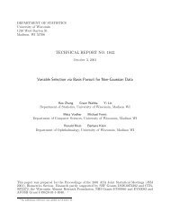
On the Analysis of Optical Mapping Data - University of Wisconsin ...
On the Analysis of Optical Mapping Data - University of Wisconsin ...
On the Analysis of Optical Mapping Data - University of Wisconsin ...

You also want an ePaper? Increase the reach of your titles
YUMPU automatically turns print PDFs into web optimized ePapers that Google loves.
5<br />
for microbial and o<strong>the</strong>r small genomes. In <strong>the</strong> early stages <strong>of</strong> a sequencing project, a genomewide<br />
restriction map provides ordering and orientation information for sequence contigs. In<br />
later stages, optical mapping can be used to validate ordering and orientation, estimate<br />
sequence gap sizes, and identify potential misassemblies. Recently, <strong>the</strong> focus <strong>of</strong> optical<br />
mapping has changed in two important ways. First, <strong>the</strong> ability to automate image processing<br />
and much <strong>of</strong> <strong>the</strong> subsequent analysis has made it practical to collect and analyze very large<br />
data sets. This allows <strong>the</strong> study <strong>of</strong> large genomes. Second, as more and more high quality<br />
sequence information has become available, <strong>the</strong> detection <strong>of</strong> genomic variation has emerged<br />
as a major goal <strong>of</strong> optical mapping. The construction <strong>of</strong> restriction maps to aid sequencing is<br />
still important for organisms where sequence information is absent or incomplete. This is a<br />
particularly challenging task for large genomes, with mixed success so far. In this <strong>the</strong>sis, we<br />
will largely restrict our attention to <strong>the</strong> case where a high quality reference copy is available.<br />
1.2 Example<br />
Throughout <strong>the</strong> <strong>the</strong>sis, we use optical map data recently collected and reported by<br />
Reslewic et al. (unpublished) to illustrate specific ideas. The data were obtained from two human<br />
cell sources. <strong>On</strong>e was a normal diploid male lymphoblastoid cell line GM07535 (Coriell<br />
Cell Repositories, Camden, NJ). The o<strong>the</strong>r was a complete hydatidiform mole (CHM), artificially<br />
created to be homozygous (Fan et al., 2002). The restriction enzyme SwaI was used<br />
in both cases. Table 1.1 gives some basic numerical summaries <strong>of</strong> <strong>the</strong> two data sets.<br />
Source CHM GM07535<br />
Number <strong>of</strong> maps 416284 206796<br />
Avg. molecule size (Kb) 436.5 441.9<br />
Avg. fragment size (Kb) 21.3 20.2<br />
Total map mass (Mb) 187386 91915<br />
Approximate coverage 62.1 29.9<br />
Table 1.1 Summary <strong>of</strong> <strong>the</strong> CHM and GM07535 data sets