On the Analysis of Optical Mapping Data - University of Wisconsin ...
On the Analysis of Optical Mapping Data - University of Wisconsin ...
On the Analysis of Optical Mapping Data - University of Wisconsin ...

Create successful ePaper yourself
Turn your PDF publications into a flip-book with our unique Google optimized e-Paper software.
ON THE ANALYSIS OF OPTICAL MAPPING DATA<br />
by<br />
Deepayan Sarkar<br />
A dissertation submitted in partial fulfillment <strong>of</strong><br />
<strong>the</strong> requirements for <strong>the</strong> degree <strong>of</strong><br />
Doctor <strong>of</strong> Philosophy<br />
(Statistics)<br />
at <strong>the</strong><br />
UNIVERSITY OF WISCONSIN–MADISON<br />
2006
Copyright by Deepayan Sarkar 2006<br />
All Rights Reserved
To my parents.<br />
i
ii<br />
ACKNOWLEDGMENTS<br />
I would like to thank <strong>the</strong> many people without whom this work would not have been<br />
possible. The optical mapping system, which forms <strong>the</strong> basis for this <strong>the</strong>sis, was pioneered by<br />
Pr<strong>of</strong> David Schwartz and continues to be developed under his leadership at <strong>the</strong> Laboratory for<br />
Molecular and Computational Genomics at <strong>the</strong> <strong>University</strong> <strong>of</strong> <strong>Wisconsin</strong> – Madison. Several<br />
people at <strong>the</strong> lab have helped me understand various aspects <strong>of</strong> <strong>the</strong> system: primarily Steve<br />
Goldstein, as well as Rod Runnheim, Chris Churas, Shiguo Zhou and Gus Potamousis. The<br />
optical map data used in this <strong>the</strong>sis were collected by Alex Lim, Susan Reslewic and Jill<br />
Herschleb. Pr<strong>of</strong> Paul Lizardi’s lab at Yale provided some supplementary data and analysis.<br />
Finally, my advisor Pr<strong>of</strong> Michael Newton provided many important insights throughout <strong>the</strong><br />
last few years and helped immensely in shaping <strong>the</strong> final manuscript.
DISCARD THIS PAGE
iii<br />
TABLE OF CONTENTS<br />
Page<br />
LIST OF TABLES . . . . . . . . . . . . . . . . . . . . . . . . . . . . . . . . . . . .<br />
LIST OF FIGURES . . . . . . . . . . . . . . . . . . . . . . . . . . . . . . . . . . .<br />
ABSTRACT . . . . . . . . . . . . . . . . . . . . . . . . . . . . . . . . . . . . . . .<br />
v<br />
vi<br />
viii<br />
1 Overview <strong>of</strong> <strong>Optical</strong> <strong>Mapping</strong> . . . . . . . . . . . . . . . . . . . . . . . . . . 1<br />
1.1 Background . . . . . . . . . . . . . . . . . . . . . . . . . . . . . . . . . . . . 1<br />
1.2 Example . . . . . . . . . . . . . . . . . . . . . . . . . . . . . . . . . . . . . . 5<br />
1.3 Elements <strong>of</strong> optical mapping . . . . . . . . . . . . . . . . . . . . . . . . . . . 6<br />
1.3.1 Image processing . . . . . . . . . . . . . . . . . . . . . . . . . . . . . 6<br />
1.3.2 <strong>Optical</strong> map data . . . . . . . . . . . . . . . . . . . . . . . . . . . . . 10<br />
1.3.3 Goals and challenges . . . . . . . . . . . . . . . . . . . . . . . . . . . 11<br />
1.3.4 Algorithms . . . . . . . . . . . . . . . . . . . . . . . . . . . . . . . . 13<br />
1.3.5 Example (continued) . . . . . . . . . . . . . . . . . . . . . . . . . . . 16<br />
1.4 Outline . . . . . . . . . . . . . . . . . . . . . . . . . . . . . . . . . . . . . . . 20<br />
2 Modeling <strong>Optical</strong> Map <strong>Data</strong> . . . . . . . . . . . . . . . . . . . . . . . . . . . 21<br />
2.1 A stochastic model . . . . . . . . . . . . . . . . . . . . . . . . . . . . . . . . 21<br />
2.1.1 Origin . . . . . . . . . . . . . . . . . . . . . . . . . . . . . . . . . . . 21<br />
2.1.2 Errors . . . . . . . . . . . . . . . . . . . . . . . . . . . . . . . . . . . 23<br />
2.2 Parameter estimation . . . . . . . . . . . . . . . . . . . . . . . . . . . . . . . 26<br />
2.2.1 Desorption . . . . . . . . . . . . . . . . . . . . . . . . . . . . . . . . . 27<br />
2.2.2 Length errors . . . . . . . . . . . . . . . . . . . . . . . . . . . . . . . 31<br />
2.2.3 Cut errors . . . . . . . . . . . . . . . . . . . . . . . . . . . . . . . . . 31<br />
2.3 Diagnostics . . . . . . . . . . . . . . . . . . . . . . . . . . . . . . . . . . . . 33<br />
3 Significance <strong>of</strong> <strong>Optical</strong> Map Alignments . . . . . . . . . . . . . . . . . . . . 37<br />
3.1 Introduction . . . . . . . . . . . . . . . . . . . . . . . . . . . . . . . . . . . . 37<br />
3.2 Methods . . . . . . . . . . . . . . . . . . . . . . . . . . . . . . . . . . . . . . 39
iv<br />
Page<br />
3.3 Results . . . . . . . . . . . . . . . . . . . . . . . . . . . . . . . . . . . . . . . 40<br />
3.3.1 Exploration . . . . . . . . . . . . . . . . . . . . . . . . . . . . . . . . 40<br />
3.3.2 Simplifications . . . . . . . . . . . . . . . . . . . . . . . . . . . . . . . 43<br />
3.3.3 Simulation . . . . . . . . . . . . . . . . . . . . . . . . . . . . . . . . . 47<br />
3.3.4 Improving assembly . . . . . . . . . . . . . . . . . . . . . . . . . . . . 47<br />
3.4 Discussion . . . . . . . . . . . . . . . . . . . . . . . . . . . . . . . . . . . . . 49<br />
3.4.1 Uses . . . . . . . . . . . . . . . . . . . . . . . . . . . . . . . . . . . . 49<br />
3.4.2 Information measure . . . . . . . . . . . . . . . . . . . . . . . . . . . 51<br />
3.4.3 O<strong>the</strong>r topics . . . . . . . . . . . . . . . . . . . . . . . . . . . . . . . . 56<br />
3.4.4 Conclusion . . . . . . . . . . . . . . . . . . . . . . . . . . . . . . . . . 58<br />
4 Detecting Copy Number Polymorphism . . . . . . . . . . . . . . . . . . . . 60<br />
4.1 Introduction . . . . . . . . . . . . . . . . . . . . . . . . . . . . . . . . . . . . 60<br />
4.2 Methods . . . . . . . . . . . . . . . . . . . . . . . . . . . . . . . . . . . . . . 62<br />
4.3 Results . . . . . . . . . . . . . . . . . . . . . . . . . . . . . . . . . . . . . . . 67<br />
4.4 Discussion . . . . . . . . . . . . . . . . . . . . . . . . . . . . . . . . . . . . . 72<br />
5 Future Work . . . . . . . . . . . . . . . . . . . . . . . . . . . . . . . . . . . . . 76<br />
5.1 Alignment . . . . . . . . . . . . . . . . . . . . . . . . . . . . . . . . . . . . . 76<br />
5.1.1 Score function . . . . . . . . . . . . . . . . . . . . . . . . . . . . . . . 76<br />
5.1.2 Scale errors . . . . . . . . . . . . . . . . . . . . . . . . . . . . . . . . 77<br />
5.2 O<strong>the</strong>r topics . . . . . . . . . . . . . . . . . . . . . . . . . . . . . . . . . . . . 80<br />
LIST OF REFERENCES . . . . . . . . . . . . . . . . . . . . . . . . . . . . . . . 82<br />
APPENDICES<br />
Appendix A: Score functions for alignment . . . . . . . . . . . . . . . . . . . . . 85<br />
Appendix B: Hidden Markov Model calculations . . . . . . . . . . . . . . . . . . 87
DISCARD THIS PAGE
v<br />
LIST OF TABLES<br />
Table<br />
Page<br />
1.1 Summary <strong>of</strong> two optical map data sets . . . . . . . . . . . . . . . . . . . . . . . 5<br />
1.2 Summary <strong>of</strong> variations detected in CHM and GM07535 . . . . . . . . . . . . . 19<br />
3.1 Percentage <strong>of</strong> GM07535 maps declared significant . . . . . . . . . . . . . . . . . 46<br />
3.2 Percentage <strong>of</strong> GM07535 maps declared significant by <strong>the</strong> SOMA and LR scores . 50<br />
4.1 AIC and BIC values for HMM fits . . . . . . . . . . . . . . . . . . . . . . . . . 70
DISCARD THIS PAGE
vi<br />
LIST OF FIGURES<br />
Figure<br />
Page<br />
1.1 Diagrammatic overview <strong>of</strong> optical mapping . . . . . . . . . . . . . . . . . . . . 4<br />
1.2 Close-up <strong>of</strong> optical map image . . . . . . . . . . . . . . . . . . . . . . . . . . . 7<br />
1.3 Intensity pr<strong>of</strong>iles . . . . . . . . . . . . . . . . . . . . . . . . . . . . . . . . . . . 7<br />
1.4 Estimation <strong>of</strong> scale across channel surface . . . . . . . . . . . . . . . . . . . . . 9<br />
1.5 Visualization <strong>of</strong> optical map alignments . . . . . . . . . . . . . . . . . . . . . . 17<br />
1.6 Visualization <strong>of</strong> optical map assembly . . . . . . . . . . . . . . . . . . . . . . . 18<br />
2.1 Distribution <strong>of</strong> restriction fragment lengths . . . . . . . . . . . . . . . . . . . . 22<br />
2.2 Quantile plot illustrating desorption . . . . . . . . . . . . . . . . . . . . . . . . 29<br />
2.3 Non-parametric Grenander estimator <strong>of</strong> desorption rate . . . . . . . . . . . . . 30<br />
2.4 Variance models for observed fragments sizes . . . . . . . . . . . . . . . . . . . 32<br />
2.5 Distribution <strong>of</strong> fragment length errors . . . . . . . . . . . . . . . . . . . . . . . 32<br />
2.6 Diagnostic plot: quantiles <strong>of</strong> fragment lengths . . . . . . . . . . . . . . . . . . . 34<br />
2.7 Diagnostic plot: mean difference plot . . . . . . . . . . . . . . . . . . . . . . . . 35<br />
2.8 Diagnostic plot: hanging rootogram for number <strong>of</strong> fragments . . . . . . . . . . . 36<br />
3.1 Independence <strong>of</strong> in silico fragment lengths . . . . . . . . . . . . . . . . . . . . . 41<br />
3.2 Dependence <strong>of</strong> spurious scores on optical map . . . . . . . . . . . . . . . . . . . 42<br />
3.3 Null distribution <strong>of</strong> difference between optimal scores . . . . . . . . . . . . . . . 44<br />
3.4 Variance <strong>of</strong> errors . . . . . . . . . . . . . . . . . . . . . . . . . . . . . . . . . . 44
vii<br />
Figure<br />
Page<br />
3.5 Parametric models for µ(M) . . . . . . . . . . . . . . . . . . . . . . . . . . . . . 45<br />
3.6 Comparison <strong>of</strong> significance strategies using iterative assembly . . . . . . . . . . 48<br />
3.7 LR scores for ungapped global alignment . . . . . . . . . . . . . . . . . . . . . . 50<br />
3.8 Optimal scores vs self-alignment score . . . . . . . . . . . . . . . . . . . . . . . 52<br />
3.9 Self-score plot for simulated data . . . . . . . . . . . . . . . . . . . . . . . . . . 54<br />
3.10 Thinning <strong>of</strong> coverage in optical map alignments . . . . . . . . . . . . . . . . . . 55<br />
3.11 Estimated thinning rates . . . . . . . . . . . . . . . . . . . . . . . . . . . . . . 56<br />
3.12 Effect <strong>of</strong> permutations on test statistics . . . . . . . . . . . . . . . . . . . . . . 59<br />
4.1 Power function . . . . . . . . . . . . . . . . . . . . . . . . . . . . . . . . . . . . 64<br />
4.2 Results <strong>of</strong> one simulation run . . . . . . . . . . . . . . . . . . . . . . . . . . . . 68<br />
4.3 Summary <strong>of</strong> several simulation runs . . . . . . . . . . . . . . . . . . . . . . . . 69<br />
4.4 The GM07535 genome compared to CHM . . . . . . . . . . . . . . . . . . . . . 71<br />
4.5 Results for MCF-7 chromosomes 17 and 20 . . . . . . . . . . . . . . . . . . . . 73<br />
5.1 Correlation <strong>of</strong> sizing errors within map . . . . . . . . . . . . . . . . . . . . . . . 78<br />
5.2 Improvement in optimal alignment score . . . . . . . . . . . . . . . . . . . . . . 79
ON THE ANALYSIS OF OPTICAL MAPPING DATA<br />
Deepayan Sarkar<br />
Under <strong>the</strong> supervision <strong>of</strong> Pr<strong>of</strong>essor Michael A. Newton<br />
At <strong>the</strong> <strong>University</strong> <strong>of</strong> <strong>Wisconsin</strong>-Madison<br />
Whole genome analysis <strong>of</strong> variation is becoming possible with improved biotechnology,<br />
and this is anticipated to have pr<strong>of</strong>ound implications for biology and medicine. Ideally,<br />
one would like to record a sampled genome at <strong>the</strong> nucleotide level, but this goal remains<br />
beyond our reach in spite <strong>of</strong> <strong>the</strong> fact that we now have a finished reference copy for several<br />
species. Using well-defined genomic markers, physical maps represent a genome at a lower<br />
resolution than nucleotide sequence. In particular, optical mapping produces physical maps<br />
based on coordinates <strong>of</strong> recognition sites <strong>of</strong> specific restriction enzymes. <strong>Optical</strong> mapping is<br />
well developed for small (e.g. microbial) genomes, and recent advances have enabled optical<br />
mapping <strong>of</strong> mammalian-sized genomes as well. This development, however, raises important<br />
new computational and statistical questions. The availability <strong>of</strong> reference genomes has been<br />
instrumental in <strong>the</strong> development <strong>of</strong> methods based on optical mapping to detect withinspecies<br />
variation, by serving as <strong>the</strong> basis for comparison with a sampled genome. Reference<br />
copies also open up o<strong>the</strong>r, less obvious, possibilities that impact <strong>the</strong> understanding and<br />
statistical analysis <strong>of</strong> optical mapping data. In this <strong>the</strong>sis we explore some such possibilities,<br />
particularly in <strong>the</strong> context <strong>of</strong> large genomes. In particular, we address parameter estimation<br />
in optical map models, <strong>the</strong> assessment <strong>of</strong> significance <strong>of</strong> optical map alignments, and <strong>the</strong> use<br />
<strong>of</strong> optical map data to detect copy number alterations.<br />
Michael A. Newton
viii<br />
ABSTRACT<br />
Whole genome analysis <strong>of</strong> variation is becoming possible with improved biotechnology,<br />
and this is anticipated to have pr<strong>of</strong>ound implications for biology and medicine. Ideally,<br />
one would like to record a sampled genome at <strong>the</strong> nucleotide level, but this goal remains<br />
beyond our reach in spite <strong>of</strong> <strong>the</strong> fact that we now have a finished reference copy for several<br />
species. Using well-defined genomic markers, physical maps represent a genome at a lower<br />
resolution than nucleotide sequence. In particular, optical mapping produces physical maps<br />
based on coordinates <strong>of</strong> recognition sites <strong>of</strong> specific restriction enzymes. <strong>Optical</strong> mapping is<br />
well developed for small (e.g. microbial) genomes, and recent advances have enabled optical<br />
mapping <strong>of</strong> mammalian-sized genomes as well. This development, however, raises important<br />
new computational and statistical questions. The availability <strong>of</strong> reference genomes has been<br />
instrumental in <strong>the</strong> development <strong>of</strong> methods based on optical mapping to detect withinspecies<br />
variation, by serving as <strong>the</strong> basis for comparison with a sampled genome. Reference<br />
copies also open up o<strong>the</strong>r, less obvious, possibilities that impact <strong>the</strong> understanding and<br />
statistical analysis <strong>of</strong> optical mapping data. In this <strong>the</strong>sis we explore some such possibilities,<br />
particularly in <strong>the</strong> context <strong>of</strong> large genomes. In particular, we address parameter estimation<br />
in optical map models, <strong>the</strong> assessment <strong>of</strong> significance <strong>of</strong> optical map alignments, and <strong>the</strong> use<br />
<strong>of</strong> optical map data to detect copy number alterations.
1<br />
Chapter 1<br />
Overview <strong>of</strong> <strong>Optical</strong> <strong>Mapping</strong><br />
1.1 Background<br />
Completion <strong>of</strong> <strong>the</strong> Human Genome Project (International Human Genome Sequencing<br />
Consortium, 2004, Build 35) stands as a critical landmark in science and medicine. All kinds<br />
<strong>of</strong> biological mysteries may be unlocked with keys buried in <strong>the</strong> full genomic sequence. Of<br />
course, <strong>the</strong> particular sequence documented online is a single reference copy. Its structure<br />
and basic content are shared by all humans, but plasticity and variation in <strong>the</strong> genome<br />
underlie both normal biology and disease. Such variations include insertions, deletions, rearrangements,<br />
single nucleotide polymorphisms (SNPs) and copy number changes. Measuring<br />
how much and in what manner one human genome varies from ano<strong>the</strong>r is a central problem<br />
<strong>of</strong> <strong>the</strong> genomic sciences. A direct approach to detect all <strong>the</strong> differences between two genomes<br />
would be to sequence each genome separately, but this is not feasible with current technology.<br />
Sequencing costs are high and <strong>the</strong> effort required to construct even a single genome<br />
is considerable. In any case, differences are likely to represent only a tiny fraction <strong>of</strong> <strong>the</strong><br />
genome. Research on various alternative methods to study genome-wide structural variation<br />
is ongoing, some <strong>of</strong> which are summarized by Eichler (2006).<br />
Physical maps: Information about genomic variation can also be obtained from lower<br />
resolution representations <strong>of</strong> <strong>the</strong> genome that do not record <strong>the</strong> full nucleotide sequence, such<br />
as physical maps. A physical map is a listing <strong>of</strong> <strong>the</strong> locations along <strong>the</strong> genome where certain<br />
markers occur. Typically, each marker is a short, well defined nucleotide sequence, such as
2<br />
<strong>the</strong> recognition sequence <strong>of</strong> a restriction enzyme (more below). The ordered sequence <strong>of</strong><br />
distances in base pairs between successive marker positions summarizes <strong>the</strong> genome sequence<br />
and can be viewed as a sort <strong>of</strong> bar code <strong>of</strong> <strong>the</strong> genome. Genomic differences can affect <strong>the</strong><br />
presence or absence <strong>of</strong> markers, <strong>the</strong> distances between <strong>the</strong>m and <strong>the</strong>ir orientation, inducing<br />
analogous changes in <strong>the</strong> bar code. Thus, by comparing physical maps instead <strong>of</strong> <strong>the</strong> full<br />
nucleotide sequences, we can detect an important subset <strong>of</strong> structural genomic variation,<br />
especially indels and translocations that are difficult to detect with many o<strong>the</strong>r technologies.<br />
In this <strong>the</strong>sis we consider statistics for <strong>Optical</strong> <strong>Mapping</strong>, a system to study a particular class<br />
<strong>of</strong> physical maps known as restriction maps.<br />
Restriction maps: A restriction map is a physical map induced by restriction enzymes.<br />
These DNA scissors, as <strong>the</strong>y have been called, occur naturally in bacteria, where <strong>the</strong>y act as<br />
a defense mechanism by cutting up foreign (usually virus) DNA molecules at any occurrence<br />
<strong>of</strong> a certain recognition sequence. Sites on <strong>the</strong> molecule where this sequence occurs are<br />
known as restriction sites or recognition sites and stretches <strong>of</strong> DNA between successive sites<br />
are called restriction fragments. For instance, <strong>the</strong> enzyme SwaI, denoted 5 ′ -ATTT|AAAT-<br />
3 ′ , recognizes <strong>the</strong> sequence ATTTAAAT, cutting between <strong>the</strong> 4 th and 5 th nucleotides. This<br />
restriction site appears more than 225000 times in Build 35 <strong>of</strong> <strong>the</strong> human genome, with more<br />
or less random fragment sizes (see Figures 2.1 and 3.1). Different enzymes have different<br />
recognition sequences and thus provide different physical maps. Note that having a reference<br />
copy <strong>of</strong> <strong>the</strong> human genome allows us to perform such in silico 1 experiments. The availability<br />
<strong>of</strong> in silico reference maps can be extremely helpful, and <strong>the</strong>ir use is a recurrent <strong>the</strong>me in<br />
this <strong>the</strong>sis.<br />
In early experiments with restriction maps, <strong>the</strong> problem was to reconstruct <strong>the</strong> underlying<br />
map from data on only <strong>the</strong> sizes <strong>of</strong> restriction fragments, without direct information on <strong>the</strong>ir<br />
ordering (Waterman, 1995; Setubal and Meidanis, 1997). Examples <strong>of</strong> such experiments<br />
include <strong>the</strong> double digest and partial digest problems. These problems are computationally<br />
1 in <strong>the</strong> computer
3<br />
hard, do not always have a unique solution, and may not scale well. Additionally, <strong>the</strong>se<br />
methods typically require measurements from multiple copies <strong>of</strong> <strong>the</strong> target DNA, usually<br />
through <strong>the</strong> creation <strong>of</strong> clone libraries.<br />
<strong>Optical</strong> mapping: <strong>Optical</strong> mapping (Schwartz et al., 1993; Dimalanta et al., 2004) produces<br />
ordered restriction maps from single DNA molecules. Briefly, DNA from hundreds <strong>of</strong><br />
thousands <strong>of</strong> cells in solution is randomly sheared to produce pieces that are around 500 Kb<br />
long. The solution is <strong>the</strong>n passed through a micro-channel, where <strong>the</strong> DNA molecules are<br />
stretched and <strong>the</strong>n attached to a positively charged glass support. A restriction enzyme is<br />
<strong>the</strong>n applied, cleaving <strong>the</strong> DNA at corresponding restriction sites. The DNA molecules remain<br />
attached to <strong>the</strong> surface, but <strong>the</strong> elasticity <strong>of</strong> <strong>the</strong> stretched DNA pulls back <strong>the</strong> molecule<br />
ends at <strong>the</strong> cleaved sites. The surface is photographed under a microscope after being stained<br />
with a fluorochrome. The cleavage sites show up in <strong>the</strong> image as tiny gaps in <strong>the</strong> fluorescent<br />
line <strong>of</strong> <strong>the</strong> molecule, giving an snapshot <strong>of</strong> <strong>the</strong> full restriction map. Even though <strong>the</strong>se<br />
molecules are large by many standards, <strong>the</strong>y may still represent only a small fraction <strong>of</strong> <strong>the</strong><br />
chromosome <strong>the</strong>y come from. Naturally, <strong>the</strong> amount <strong>of</strong> information in an optical map data<br />
set is related to <strong>the</strong> size <strong>of</strong> <strong>the</strong> underlying genome. It is common to measure <strong>the</strong> effective<br />
size <strong>of</strong> a data set by its coverage, which is <strong>the</strong> ratio <strong>of</strong> <strong>the</strong> accumulated lengths <strong>of</strong> all optical<br />
maps and <strong>the</strong> estimated length <strong>of</strong> <strong>the</strong> genome.<br />
Several types <strong>of</strong> noise affect optical map data, and a reliable picture <strong>of</strong> <strong>the</strong> true map can<br />
only be obtained by combining information from multiple optical maps that redundantly tile<br />
<strong>the</strong> genome. Most <strong>of</strong> <strong>the</strong> algorithmic challenges in optical mapping stem from trying to model<br />
<strong>the</strong> various kinds <strong>of</strong> noise, which are not all completely understood, and making inferences<br />
about <strong>the</strong> underlying map. Figure 1.1 outlines <strong>the</strong> basic steps <strong>of</strong> data collection, image<br />
processing and data analysis that toge<strong>the</strong>r form <strong>the</strong> cornerstones <strong>of</strong> <strong>the</strong> optical mapping<br />
system.<br />
Uses: <strong>Optical</strong> mapping has various applications. It has been successfully used to assist in<br />
sequence assembly and validation efforts (Ivens et al., 2005; Armbrust et al., 2004), usually
Figure 1.1 Diagrammatic overview <strong>of</strong> optical mapping. DNA from many thousands <strong>of</strong> cells,<br />
each representing a copy <strong>of</strong> <strong>the</strong> genome being studied, is passed through micr<strong>of</strong>luidic channels<br />
(a.k.a. groups) where <strong>the</strong>y are stretched and attached to a positively charged glass surface.<br />
After a restriction enzyme is applied, <strong>the</strong> surface is imaged using fluorescence microscopy.<br />
DNA molecules appear as bright fluorescent pixels, which are subsequently identified and<br />
converted into candidate restriction maps in <strong>the</strong> image processing step. Statistical analysis<br />
is concerned largely with calculations post image processing, although it does inform <strong>the</strong><br />
previous steps as well.<br />
4
5<br />
for microbial and o<strong>the</strong>r small genomes. In <strong>the</strong> early stages <strong>of</strong> a sequencing project, a genomewide<br />
restriction map provides ordering and orientation information for sequence contigs. In<br />
later stages, optical mapping can be used to validate ordering and orientation, estimate<br />
sequence gap sizes, and identify potential misassemblies. Recently, <strong>the</strong> focus <strong>of</strong> optical<br />
mapping has changed in two important ways. First, <strong>the</strong> ability to automate image processing<br />
and much <strong>of</strong> <strong>the</strong> subsequent analysis has made it practical to collect and analyze very large<br />
data sets. This allows <strong>the</strong> study <strong>of</strong> large genomes. Second, as more and more high quality<br />
sequence information has become available, <strong>the</strong> detection <strong>of</strong> genomic variation has emerged<br />
as a major goal <strong>of</strong> optical mapping. The construction <strong>of</strong> restriction maps to aid sequencing is<br />
still important for organisms where sequence information is absent or incomplete. This is a<br />
particularly challenging task for large genomes, with mixed success so far. In this <strong>the</strong>sis, we<br />
will largely restrict our attention to <strong>the</strong> case where a high quality reference copy is available.<br />
1.2 Example<br />
Throughout <strong>the</strong> <strong>the</strong>sis, we use optical map data recently collected and reported by<br />
Reslewic et al. (unpublished) to illustrate specific ideas. The data were obtained from two human<br />
cell sources. <strong>On</strong>e was a normal diploid male lymphoblastoid cell line GM07535 (Coriell<br />
Cell Repositories, Camden, NJ). The o<strong>the</strong>r was a complete hydatidiform mole (CHM), artificially<br />
created to be homozygous (Fan et al., 2002). The restriction enzyme SwaI was used<br />
in both cases. Table 1.1 gives some basic numerical summaries <strong>of</strong> <strong>the</strong> two data sets.<br />
Source CHM GM07535<br />
Number <strong>of</strong> maps 416284 206796<br />
Avg. molecule size (Kb) 436.5 441.9<br />
Avg. fragment size (Kb) 21.3 20.2<br />
Total map mass (Mb) 187386 91915<br />
Approximate coverage 62.1 29.9<br />
Table 1.1 Summary <strong>of</strong> <strong>the</strong> CHM and GM07535 data sets
6<br />
1.3 Elements <strong>of</strong> optical mapping<br />
We now describe in more depth elements <strong>of</strong> a typical optical mapping experiment. We<br />
start with image processing and go on to discuss <strong>the</strong> structure <strong>of</strong> optical map data and<br />
<strong>the</strong> goals and challenges we face in data analysis. We describe two basic computational<br />
tasks, alignment and assembly, that are fundamental in addressing many o<strong>the</strong>r problems.<br />
We end with a summary <strong>of</strong> <strong>the</strong> analysis <strong>of</strong> <strong>the</strong> GM07535 and CHM data sets reported by<br />
Reslewic et al..<br />
1.3.1 Image processing<br />
Intensity pr<strong>of</strong>iles: For a typical optical mapping experiment, hundreds <strong>of</strong> raw images<br />
need to be processed to obtain useful data (Figure 1.2). The first step in this process is to<br />
identify <strong>the</strong> collections <strong>of</strong> pixels in an image that toge<strong>the</strong>r represent a single DNA molecule.<br />
This is a complicated task that falls in <strong>the</strong> domain <strong>of</strong> computer vision and will not be<br />
discussed fur<strong>the</strong>r. The end product <strong>of</strong> this step is an intensity pr<strong>of</strong>ile for each molecule<br />
(Figure 1.3) giving <strong>the</strong> measured fluorescent intensity as a function <strong>of</strong> distance along <strong>the</strong><br />
“backbone”. There are two ways to proceed. We may consider <strong>the</strong>se pr<strong>of</strong>iles as our primary<br />
data, and retain <strong>the</strong> information <strong>the</strong>y contain in subsequent analyses. Alternatively, we may<br />
immediately convert <strong>the</strong>m into putative restriction maps, i.e. to an ordered sequence <strong>of</strong><br />
fragment lengths. The second approach is simpler because it separates <strong>the</strong> problem into two<br />
parts that can be refined independently. Also, many standard techniques in computational<br />
biology apply, with suitable adaptations, in this formulation. The first approach has a certain<br />
appeal, but presents difficult challenges and we do not investigate it fur<strong>the</strong>r. The rest <strong>of</strong> this<br />
discussion assumes <strong>the</strong> second, two-step approach.<br />
Cleavage sites: To convert intensity pr<strong>of</strong>iles to restriction maps, one has to first identify<br />
<strong>the</strong> cleavage sites or cut sites in <strong>the</strong> map, indicated by ‘dips’ in <strong>the</strong> intensity pr<strong>of</strong>ile. The<br />
approaches traditionally used to identify cut sites are largely heuristic, although formal
7<br />
Figure 1.2 Close-up <strong>of</strong> a typical optical map image with (bottom) and without (top) optical<br />
maps marked up. The colors are reversed for legibility, so DNA molecules are represented<br />
by dark pixels against a light background. The image processing step is complex, and is<br />
summarized only briefly in <strong>the</strong> text.<br />
Figure 1.3 Examples <strong>of</strong> intensity pr<strong>of</strong>iles from 5 different molecules. Cuts are indicated by<br />
dips in <strong>the</strong> intensity.
8<br />
statistical techniques may also be used. Naturally, not all cuts present are always detected,<br />
nor are all reported cuts real.<br />
Lengths: <strong>On</strong>ce cut sites are identified, <strong>the</strong> intensity pr<strong>of</strong>ile between cuts is integrated to<br />
obtain a total intensity <strong>of</strong> <strong>the</strong> corresponding restriction fragment. These measurements <strong>the</strong>n<br />
need to be scaled appropriately to convert <strong>the</strong>m to base pair units. Due to variability in<br />
experimental conditions, <strong>the</strong> scale factor is usually different in different surfaces and channels,<br />
and possibly even within a channel. To deal with this, DNA molecules with known length<br />
and restriction pattern, called standards, are placed in <strong>the</strong> sample along with <strong>the</strong> DNA<br />
being mapped. These standards are identified in <strong>the</strong> images based only on <strong>the</strong>ir pattern.<br />
Their measured fluorescent lengths and known base-pair lengths are <strong>the</strong>n used to scale o<strong>the</strong>r<br />
fragments. Naturally, <strong>the</strong> measured intensities <strong>of</strong> <strong>the</strong> standards are <strong>the</strong>mselves subject to<br />
noise, and <strong>the</strong> scale is usually estimated as a smooth function <strong>of</strong> position on <strong>the</strong> image.<br />
Figure 1.4 shows <strong>the</strong> distribution <strong>of</strong> standards on a typical surface along with <strong>the</strong> estimated<br />
scale.<br />
Quality: A critical <strong>the</strong>me in <strong>the</strong> optical mapping system is <strong>the</strong> automated processing <strong>of</strong><br />
massive amounts <strong>of</strong> data, beginning with image processing. <strong>On</strong>ly a fraction <strong>of</strong> <strong>the</strong> fluorescent<br />
material seen in raw images is ever marked up by <strong>the</strong> image processing step and reported as<br />
optical maps. This filtering is important to ensure a certain minimal quality in <strong>the</strong> optical<br />
map data one works with. Of course, <strong>the</strong> automated processing is not perfect and certain<br />
maps are marked up wrongly; subsequent methods need to be able to deal robustly with<br />
<strong>the</strong>se errors. Most existing methods treat all maps equally once <strong>the</strong>y are reported by <strong>the</strong><br />
image processing s<strong>of</strong>tware. However, it is likely that some weight or measure <strong>of</strong> confidence<br />
reported along with every map would be useful in fur<strong>the</strong>r analysis. This is an area that could<br />
benefit from fur<strong>the</strong>r research.
9<br />
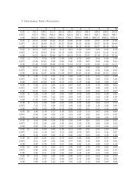
0.96 0.98 1.00 1.02 1.04<br />
Offset across channel (pixels)<br />
200<br />
0<br />
−200<br />
−400<br />
20000 40000 60000 80000 100000<br />
Offset along channel (pixels)<br />
Figure 1.4 Estimated scale factor (relative to mean) across <strong>the</strong> surface <strong>of</strong> a typical channel,<br />
showing spatial dependence. The estimated scale here is based on a LOESS smooth <strong>of</strong><br />
measured intensities <strong>of</strong> ‘standards’, which are housekeeping molecules <strong>of</strong> known genomic<br />
length and restriction pattern. The locations <strong>of</strong> <strong>the</strong>se standards, identified by <strong>the</strong>ir pattern,<br />
are indicated by white dots on <strong>the</strong> figure above. Black dots represent locations <strong>of</strong> identified<br />
optical map fragments.
10<br />
1.3.2 <strong>Optical</strong> map data<br />
Representation: An optical map identified by image processing is essentially an ordered<br />
sequence <strong>of</strong> fragment lengths. Thus, an optical map with n fragments may be denoted as<br />
x = (x 1 , . . .,x n )<br />
where x i is <strong>the</strong> measured length <strong>of</strong> <strong>the</strong> i th fragment. Ano<strong>the</strong>r natural representation <strong>of</strong> an<br />
optical map is as a sequence <strong>of</strong> recognition sites. An optical map x is easily converted into a<br />
sequence <strong>of</strong> cut sites by accumulating <strong>the</strong> lengths, noting that <strong>the</strong> cut sites are only defined<br />
up to location. Denoting <strong>the</strong> conversion from fragment lengths to cut site locations by S,<br />
we may write<br />
S(x) =<br />
{0 = s 0 < s 1 < · · · < s n = ∑ }<br />
x i<br />
where x i = s i −s i−1 for i = 1, . . ., n are fragment lengths and s i = ∑ i<br />
j=0 x j are locations <strong>of</strong> cut<br />
sites. The endpoints s 0 and s n are not treated as cut site locations since <strong>the</strong>y represent breaks<br />
that define <strong>the</strong> original molecule as a segment <strong>of</strong> <strong>the</strong> whole genome (from shearing) ra<strong>the</strong>r<br />
than breaks created by <strong>the</strong> restriction enzyme. The first representation, being invariant to<br />
origin, has <strong>the</strong> advantage <strong>of</strong> being unambiguous, but <strong>the</strong> second is <strong>of</strong>ten more useful, e.g.<br />
for defining alignments between two or more optical maps. Of course, both representations<br />
apply to any physical map. <strong>Optical</strong> maps may have additional meta-data associated with<br />
<strong>the</strong>m (e.g. confidence scores from image processing), but most existing algorithms ignore<br />
such attributes.<br />
Characteristics: <strong>Optical</strong> map molecules are generally regarded as random snapshots obtained<br />
from <strong>the</strong> underlying genome, i.e., <strong>the</strong>ir locations are assumed to be uniformly distributed<br />
within <strong>the</strong> genome. Their orientation is not known a priori. The lengths <strong>of</strong> <strong>the</strong><br />
molecules vary; a typical molecule may be around 500 Kb long, and 1000 Kb molecules are<br />
not uncommon. Unlike sequence reads that are obtained as averages over many copies <strong>of</strong> a<br />
clone, optical maps represent single molecules derived from genomic DNA, providing a more
11<br />
direct glimpse at <strong>the</strong> underlying structure <strong>of</strong> <strong>the</strong> genome. Unfortunately, this also means<br />
that raw optical maps can be fairly noisy. In particular,<br />
• not all true restriction sites are observed, i.e. some cuts are missing, due to imperfect<br />
digestion by <strong>the</strong> restriction enzyme<br />
• breakage <strong>of</strong> DNA may cause spurious cuts to appear in a map<br />
• measurement <strong>of</strong> fluorescent intensities and conversion to base pairs is inaccurate, causing<br />
sizing errors in fragment lengths<br />
• relatively small fragments (say 5 Kb or less) may lose adhesion to <strong>the</strong> surface and desorb,<br />
in which case <strong>the</strong>y are not included in <strong>the</strong> final map. Some <strong>of</strong> <strong>the</strong>se fragments may<br />
re-attach <strong>the</strong>mselves near o<strong>the</strong>r fragments, potentially causing length overestimation<br />
in <strong>the</strong> latter.<br />
All <strong>the</strong>se noises are confounded with image processing errors. Mistakes in image processing<br />
may also cause optical chimeras, where unrelated maps are marked up as one because <strong>the</strong>y<br />
overlap on <strong>the</strong> image. O<strong>the</strong>r less systematic errors are also present. These errors, along<br />
with <strong>the</strong> choice <strong>of</strong> restriction enzyme and genome, affect <strong>the</strong> typical size <strong>of</strong> an optical map<br />
fragment. The average fragment size, <strong>of</strong>ten used to summarize an optical map data set, is<br />
usually between 5 and 40 Kb.<br />
1.3.3 Goals and challenges<br />
Goals: A typical optical mapping experiment begins with <strong>the</strong> collection <strong>of</strong> data followed by<br />
image processing to identify individual optical maps. The goal <strong>of</strong> subsequent analysis depends<br />
partly on <strong>the</strong> genome being mapped. Although <strong>the</strong> goal <strong>of</strong> optical mapping is always to make<br />
inferences about <strong>the</strong> underlying restriction map, it is important to distinguish between cases<br />
where a draft reference sequence <strong>of</strong> <strong>the</strong> organism is available and ones where it is not. In <strong>the</strong><br />
latter case, <strong>the</strong> goal <strong>of</strong> optical mapping is de novo assembly, i.e. to reconstruct <strong>the</strong> underlying<br />
restriction map, <strong>of</strong>ten to assist in sequencing efforts. In <strong>the</strong> former case, a possible candidate
12<br />
restriction map can be derived in silico by identifying <strong>the</strong> enzyme recognition pattern in<br />
<strong>the</strong> reference sequence, and <strong>the</strong> primary goal <strong>of</strong> optical mapping is to determine how <strong>the</strong><br />
genome under study differs from <strong>the</strong> reference copy in terms <strong>of</strong> <strong>the</strong>ir respective restriction<br />
maps. Such differences can be due to errors in <strong>the</strong> sequence, especially in <strong>the</strong> early stages<br />
<strong>of</strong> sequencing, but more importantly, <strong>the</strong>y can reflect real biological variation. In ei<strong>the</strong>r<br />
case, <strong>the</strong>se broad goals are <strong>of</strong>ten tackled by breaking <strong>the</strong>m down into smaller, more tractable<br />
problems.<br />
Algorithmic challenges: <strong>Optical</strong> mapping has been very successful in obtaining restriction<br />
maps <strong>of</strong> relatively small genomes (e.g. microbes). A critical component <strong>of</strong> this success<br />
has been algorithmic research in <strong>the</strong> 1990’s specifically aimed at optical mapping data, notably<br />
<strong>the</strong> work <strong>of</strong> Anantharaman et al. (1999) leading to <strong>the</strong> Gentig assembly s<strong>of</strong>tware. With<br />
recent technological advances, <strong>the</strong> focus has shifted to larger genomes. The primary challenge<br />
introduced by this shift is scalability. Computational methods that work well for microbial<br />
genomes may fail for large genomes due to memory and speed limits <strong>of</strong> existing computational<br />
systems. Since mammalian genomes differ in size from microbial genomes by several orders<br />
<strong>of</strong> magnitude, <strong>the</strong> relative coverage may be far less. Careful statistical analysis is thus critical<br />
in making full use <strong>of</strong> <strong>the</strong> available data. New methods are also required to take advantage <strong>of</strong><br />
in silico maps when <strong>the</strong>y are available. It should be noted that restriction maps have many<br />
fundamental similarities with sequence data, and algorithms developed for sequence analysis<br />
can <strong>of</strong>ten be adapted to work with optical maps (e.g. Huang and Waterman, 1992).<br />
Validation: Due to <strong>the</strong> nature <strong>of</strong> optical mapping data, it is rarely possible to know <strong>the</strong><br />
true answer except in very special circumstances. It is <strong>the</strong>refore natural to use simulation to<br />
validate algorithmic techniques. While this has been implicitly acknowledged in much <strong>of</strong> <strong>the</strong><br />
algorithmic work on optical mapping, we think that <strong>the</strong> stochastic model used in simulation<br />
itself deserves closer attention. With <strong>the</strong> large data sets that are now available, we can also<br />
hope to use <strong>the</strong> data to validate models, at least in some limited ways. In particular, we<br />
have found graphical diagnostics to be particularly useful in model checking (see Section 2.3),
13<br />
which is not surprising since well designed graphs can usually convey complex information<br />
more effectively than numerical summaries.<br />
1.3.4 Algorithms<br />
Problems in optical mapping are <strong>of</strong>ten approached indirectly by trying to answer simpler,<br />
more specific ones. This is not uncommon in computational biology, where <strong>the</strong> complexity<br />
<strong>of</strong> a problem may make a holistic solution difficult. Two algorithmic questions that play a<br />
recurrent role in many <strong>of</strong> <strong>the</strong>se approaches are alignment and assembly. Each tries to answer<br />
a particular problem; however, it is <strong>of</strong>ten more useful to think <strong>of</strong> <strong>the</strong>se as tools ra<strong>the</strong>r than<br />
solutions. Here, we give an overview <strong>of</strong> <strong>the</strong>se two fundamental computational tasks.<br />
Alignment<br />
The problem <strong>of</strong> alignment is to detect association or overlap between two or more restriction<br />
maps. Such association is measured by a score function which assigns a numerical<br />
measure <strong>of</strong> goodness to any potential alignment. Of course different score functions may<br />
be used and much rests on choosing a suitable score function. Waterman et al. (1984) presented<br />
a score function for restriction map comparison, which was subsequently extended by<br />
Huang and Waterman (1992). Valouev et al. (2006) have developed scores functions for <strong>the</strong><br />
comparison problem specifically in <strong>the</strong> context <strong>of</strong> optical mapping. These score functions<br />
have been derived as model-based likelihood ratio test statistics, although this is not strictly<br />
necessary (Appendix A).<br />
Given a suitable score function, dynamic programming is used to efficiently search for<br />
optimal alignments. In <strong>the</strong> context <strong>of</strong> alignment against a reference, for example, every individual<br />
optical map must be scored across <strong>the</strong> genome. Alignment algorithms for nucleotide<br />
sequence data, such as <strong>the</strong> Needleman-Wunsch and Smith-Waterman algorithms, can be<br />
adapted to work with restriction maps. Certain modifications are required to enable such<br />
use; <strong>the</strong>se are described by Valouev et al. (2006).
14<br />
Significance: An optimal alignment exists in any map comparison problem, irrespective<br />
<strong>of</strong> any actual association. In order to minimize <strong>the</strong> potential effects <strong>of</strong> misaligned maps, it is<br />
essential to limit alignments by some additional criterion. This is <strong>the</strong> problem <strong>of</strong> assessing<br />
<strong>the</strong> significance <strong>of</strong> a given alignment. The significance problem in optical map alignment<br />
is more difficult than in sequence alignment, because <strong>of</strong> a greater degree <strong>of</strong> noise and also<br />
because <strong>of</strong> differences in <strong>the</strong> nature <strong>of</strong> <strong>the</strong> data. We find deficiencies in <strong>the</strong> current state <strong>of</strong><br />
<strong>the</strong> art, and in Chapter 3 we introduce and evaluate an alternative approach to measuring<br />
<strong>the</strong> significance <strong>of</strong> optical map alignments. Here, we give a general overview <strong>of</strong> <strong>the</strong> mechanics<br />
<strong>of</strong> map alignment.<br />
Notation: We restrict our attention to pairwise alignments, i.e. those between two restriction<br />
maps. Let x = (x 1 , . . .,x m ) and y = (y 1 , . . .,y n ) denote two restriction maps with<br />
m and n fragments respectively. Let <strong>the</strong> corresponding representations in terms <strong>of</strong> cut sites<br />
be S(x) = {s 0 < s 1 < · · · < s m } and S(y) = {t 0 < t 1 < · · · < t n }. An alignment between x<br />
and y can be represented by an ordered set <strong>of</strong> index pairs<br />
C = (( i 1<br />
j1<br />
)<br />
,<br />
( i2<br />
j2<br />
)<br />
, . . .,<br />
( ik<br />
jk<br />
))<br />
indicating a correspondence between <strong>the</strong> cut sites s il and t jl for l = 1, . . .,k, where 0 < i 1 <<br />
· · · < i k < m and 0 < j 1 < · · · < j k < n. To allow missing fragments in <strong>the</strong> alignment, this<br />
last condition can be modified to allow successive indices to be equal, as long as successive<br />
index pairs are not identical. For non-trivial alignments k ≥ 2, in which case <strong>the</strong> alignment<br />
consists <strong>of</strong> k −1 aligned chunks. The l th chunk (l = 1, . . ., k −1) has lengths ˜x l = s il −s il−1 ,<br />
and ỹ l = t jl − t jl−1 involving m l = i l − i l−1 and n l = j l − j l−1 fragments respectively in<br />
<strong>the</strong> original maps x and y. To be used successfully in a dynamic programming algorithm, a<br />
score function must be additive, in <strong>the</strong> sense that <strong>the</strong> score <strong>of</strong> a complete alignment must be<br />
<strong>the</strong> sum <strong>of</strong> <strong>the</strong> scores for its component chunks.
15<br />
Gapped alignments: The above description implicitly assumes that given any two cut<br />
sites involved in <strong>the</strong> alignment, all intermediate cut sites will also be involved. Such alignments<br />
are known as ungapped alignments. <strong>On</strong>e may wish to relax this assumption and allow<br />
gaps, e.g. to represent deletions or insertions. The above notation can be easily generalized<br />
to include such gapped alignments by allowing some index pairs to attain a special value<br />
(<br />
representing a boundary, e.g.<br />
il<br />
) (<br />
j l = NA)<br />
. In principle <strong>the</strong> requirement that il ’s and j l ’s<br />
be increasing can also be relaxed to allow change in orientation within an alignment (e.g. to<br />
represent inversion) but this is rarely allowed in practice due to difficulty in implementation.<br />
The true orientation <strong>of</strong> raw optical maps are unknown, so both must be considered during<br />
analysis.<br />
Map types: x and y above denote generic restriction maps. In practice, <strong>the</strong>y can be one<br />
<strong>of</strong> three types; individual optical maps, reference maps derived in silico from sequence and<br />
intermediate consensus maps derived by combining multiple optical maps. This distinction is<br />
important when comparing two maps. For example, optical maps are noisy whereas in silico<br />
reference maps are generally considered error free. Consensus maps lie somewhere in between,<br />
since <strong>the</strong>y contain information averaged over individual optical maps. Thus, comparing an<br />
optical map with ano<strong>the</strong>r optical map is a symmetric problem, whereas comparing an optical<br />
map with an in silico reference or a consensus map is not.<br />
Alignment types: Most types <strong>of</strong> sequence alignment problems have a corresponding map<br />
alignment problem. Terminology regarding <strong>the</strong> various types <strong>of</strong> alignment are not standard,<br />
so we refrain from giving a full list and refer <strong>the</strong> reader to <strong>the</strong>ir favorite book on sequence<br />
alignment, e.g. Waterman (1995). Two variants <strong>of</strong> global alignment have been particularly<br />
useful in recent work: overlap alignment, where a suffix <strong>of</strong> one map is aligned to a prefix <strong>of</strong><br />
ano<strong>the</strong>r, and fit alignment, where an alignment is desired for a map so that it is completely<br />
contained in ano<strong>the</strong>r, usually much larger, map. Local alignments are ano<strong>the</strong>r important<br />
class <strong>of</strong> alignments that are potentially useful in identifying structural variation, but have<br />
not been studied extensively in this context.
16<br />
S<strong>of</strong>tware: The SOMA s<strong>of</strong>tware suite can be used to perform restriction map alignments.<br />
As in sequence alignment, one is <strong>of</strong>ten interested in sub-optimal alignments as well, i.e. highscoring<br />
alignments in addition to <strong>the</strong> top-scoring one. SOMA is able to find such alignments.<br />
Genspect can be used to visualize alignments reported by SOMA. Figure 1.5 shows a typical<br />
visualization <strong>of</strong> optical map alignments.<br />
Assembly<br />
The assembly problem can be viewed as a multiple alignment problem, with an additional<br />
step <strong>of</strong> producing an inferred consensus map. The most successful optical map assembly s<strong>of</strong>tware<br />
to date is Gentig, based on ideas described in Anantharaman et al. (1997) (for clones)<br />
and Anantharaman et al. (1999) (for genomic DNA). Briefly, <strong>the</strong>y develop a Bayesian approach<br />
where a prior model for <strong>the</strong> unknown restriction map and a conditional distribution<br />
for optical maps given <strong>the</strong> true map are used to derive <strong>the</strong> posterior density for an hypo<strong>the</strong>sized<br />
map. The inferred restriction map is, in principle, <strong>the</strong> one that maximizes this posterior<br />
density. Due to <strong>the</strong> complexities <strong>of</strong> <strong>the</strong> problem, a complete search is infeasible, and various<br />
heuristics are employed to enable an efficient implementation. We have little to add on <strong>the</strong><br />
assembly problem, and refer <strong>the</strong> reader to <strong>the</strong> original papers for fur<strong>the</strong>r details. Gentig<br />
results can also be visualized using Genspect, as shown in Figure 1.6.<br />
1.3.5 Example (continued)<br />
The goal <strong>of</strong> an optical mapping project is to infer <strong>the</strong> underlying restriction map <strong>of</strong> <strong>the</strong><br />
genome being studied. For small genomes, Gentig serves this purpose well. However, for<br />
large genomes such as GM07535 and CHM (Table 1.1), <strong>the</strong> sizes <strong>of</strong> <strong>the</strong> data sets exceeds its<br />
capacity, and new algorithms are required. Fortunately, additional information is available<br />
for <strong>the</strong>se data sets in <strong>the</strong> form <strong>of</strong> an in silico reference map, derived from <strong>the</strong> human genome<br />
sequence by locating instances <strong>of</strong> <strong>the</strong> SwaI recognition pattern. The genomes being studied<br />
are largely similar to this reference, so we are primarily interested in how <strong>the</strong>ir restriction<br />
maps differ from <strong>the</strong> reference.
Figure 1.5 A visualization <strong>of</strong> alignments <strong>of</strong> optical maps to an in silico reference. The<br />
alignments were done using SOMA, and Genspect was used to visualize <strong>the</strong> results. The<br />
top row represents a segment <strong>of</strong> <strong>the</strong> in silico map derived from <strong>the</strong> human genome, to<br />
which optical maps were aligned. The optical map fragments are color coded to indicate cut<br />
differences and jittered vertically to emphasize fragment boundaries.<br />
17
Figure 1.6 A visualization, using Genspect, <strong>of</strong> an assembled consensus map, along with<br />
optical maps that support it. The assembly was produced by Gentig. The visualization is<br />
similar to that in Figure 1.5, with <strong>the</strong> exception that <strong>the</strong> top row represents <strong>the</strong> assembled<br />
consensus map ra<strong>the</strong>r than <strong>the</strong> predefined alignment target.<br />
18
19<br />
Assembly: For <strong>the</strong>se examples, <strong>the</strong> assembly problem was approached using a two-step<br />
procedure. In <strong>the</strong> first step, each individual optical map was aligned to <strong>the</strong> reference map.<br />
The reference genome was <strong>the</strong>n tiled by overlapping “windows” and maps that aligned were<br />
grouped toge<strong>the</strong>r according to membership in <strong>the</strong>se windows. In <strong>the</strong> second step, <strong>the</strong> maps<br />
in each group were assembled using Gentig, giving a local snapshot <strong>of</strong> <strong>the</strong> target map. This<br />
strategy can be expected to work in regions where <strong>the</strong> differences are minor, and use <strong>of</strong><br />
gapped alignments can reveal certain larger-scale variations. For regions <strong>of</strong> more severe<br />
differences, an initial consensus map can be extended into its flanks by iteratively aligning<br />
optical maps to it, allowing partial overlap at <strong>the</strong> boundaries, followed by assembly. This<br />
procedure is revisited in Section 3.3.4.<br />
Differences: The next task was to identify <strong>the</strong> differences between <strong>the</strong> assembled consensus<br />
maps (contigs) and <strong>the</strong> reference map. <strong>On</strong>ce again, this was approached in two steps,<br />
starting with alignments <strong>of</strong> <strong>the</strong> consensus contigs to <strong>the</strong> reference. This induces inferred<br />
alignments <strong>of</strong> single optical maps to <strong>the</strong> reference. Individual differences between <strong>the</strong> assembled<br />
consensus and <strong>the</strong> reference, specifically in restriction sites and fragment lengths,<br />
can <strong>the</strong>n be assigned confidence in <strong>the</strong> form <strong>of</strong> p-values <strong>of</strong> simple hypo<strong>the</strong>sis tests. In practice,<br />
<strong>the</strong> initial alignment is <strong>of</strong>ten problematic in regions with small fragments, and some<br />
automated and manual curation is currently required. Larger indels and translocations are<br />
usually identified manually. Table 1.2 summarizes <strong>the</strong> structural variations identified in <strong>the</strong><br />
CHM and GM07535 genomes. See Reslewic et al. for more details <strong>of</strong> <strong>the</strong> analysis.<br />
Genome Insertions Deletions Extra cuts Missing cuts O<strong>the</strong>rs<br />
CHM 221 217 449 466 14<br />
GM07535 109 52 132 254 10<br />
Table 1.2 Summary <strong>of</strong> “<strong>Optical</strong> Structural Variations” (OSV) identified in <strong>the</strong> CHM and<br />
GM07535 data sets. The events included are those that were significant at a nominal False<br />
Discovery Rate <strong>of</strong> 90%.
20<br />
1.4 Outline<br />
<strong>Optical</strong> mapping is a fast, low-cost, single-molecule system for producing whole genome<br />
restriction maps. Its potential applications for studies <strong>of</strong> normal and disease biology are<br />
manifold, but computational and statistical challenges created by large genomes must be<br />
met in order for optical mapping to achieve this potential. Existing algorithms have been<br />
effective on optical map data from small genomes. These algorithms do not easily extend<br />
to <strong>the</strong> much larger data sets that are now being collected from larger genomes, and we are<br />
as yet unable to completely mine <strong>the</strong> wealth <strong>of</strong> information contained in <strong>the</strong>m. In part, this<br />
is due to unavoidable computational bottlenecks. However, new avenues <strong>of</strong> analysis have<br />
opened up with <strong>the</strong> availability <strong>of</strong> more and more sequence information. In <strong>the</strong> following<br />
chapters, we present some new ideas on how to deal with optical map data. These ideas share<br />
a common <strong>the</strong>me in that <strong>the</strong>y all take advantage <strong>of</strong> <strong>the</strong> availability <strong>of</strong> in silico reference maps<br />
derived from sequence. They do not, by any means, resolve all outstanding questions, but<br />
hopefully <strong>the</strong>y contribute to <strong>the</strong> understanding <strong>of</strong> optical map data and provide a reference<br />
for future work in this area. In Chapter 2, we discuss stochastic models for optical map<br />
errors and present some new approaches to parameter estimation in that setting. In Chapter<br />
3, we propose a new method to determine significance <strong>of</strong> alignments <strong>of</strong> optical maps to a<br />
reference, which is an important prerequisite in many analyses. In Chapter 4, we use <strong>the</strong>se<br />
alignments as <strong>the</strong> basis for an assembly-free method to detect copy number polymorphisms.<br />
Especially in cancer biology, <strong>the</strong> ability to detect gains and losses <strong>of</strong> DNA is critical, as<br />
frequently deleted sites may harbor tumor suppressor genes, and frequently amplified regions<br />
may harbor oncogenes.
21<br />
Chapter 2<br />
Modeling <strong>Optical</strong> Map <strong>Data</strong><br />
The first step in <strong>the</strong> analysis <strong>of</strong> optical mapping data is to understand its inherent variability.<br />
Unlike traditional restriction mapping techniques, optical mapping obviates <strong>the</strong> need<br />
to reconstruct <strong>the</strong> order <strong>of</strong> restriction fragments. However, <strong>the</strong> orientations <strong>of</strong> optical maps<br />
are unknown, fragment lengths are not measured accurately, not all cuts are correctly identified,<br />
and small fragments may desorb and not be seen at all. Fur<strong>the</strong>r, some maps identified<br />
by image processing may not represent any real restriction maps; e.g., chimeric maps caused<br />
by crossing over <strong>of</strong> maps in <strong>the</strong> image, marked up as one. In this chapter we discuss how<br />
<strong>the</strong>se sources <strong>of</strong> noise can be modeled. Section 2.1, which describes models for optical map<br />
errors, is mostly a review. Later sections consider <strong>the</strong> estimation <strong>of</strong> model parameters from<br />
optical map data. Many <strong>of</strong> <strong>the</strong> ideas presented <strong>the</strong>re are new and <strong>of</strong>ten take advantage <strong>of</strong><br />
an in silico reference map. In particular, we outline a non-parametric approach to estimate<br />
desorption rate, use alignments <strong>of</strong> optical maps to a reference to estimate sizing and scaling<br />
error parameters, and discuss <strong>the</strong> use <strong>of</strong> simulation to develop diagnostic plots that can be<br />
used to assess goodness <strong>of</strong> fit.<br />
2.1 A stochastic model<br />
2.1.1 Origin<br />
Underlying restriction map: It is natural to model optical maps as being generated<br />
from an underlying ‘true’ restriction map associated with <strong>the</strong> genome under study. This<br />
restriction map can be thought <strong>of</strong> as a fixed but unknown (high-dimensional) parameter.
22<br />
0 1 2 3 4 5<br />
0 1 2 3 4 5<br />
0 1 2 3 4 5<br />
0 1 2 3 4 5<br />
150<br />
17 (0.13)<br />
18 (0.07) 19 (0.2)<br />
20 (0.08) 21 (0.13)<br />
22 (0.13) X (0.08)<br />
Y (0.08)<br />
100<br />
50<br />
Quantiles <strong>of</strong> insilico fragment lengths<br />
0<br />
150<br />
9 (0.1) 10 (0.09) 11 (0.1) 12 (0.11) 13 (0.06) 14 (0.1) 15 (0.08) 16 (0.15)<br />
1 (0.12) 2 (0.08) 3 (0.09) 4 (0.06) 5 (0.09) 6 (0.08) 7 (0.09) 8 (0.09)<br />
150<br />
100<br />
50<br />
0<br />
100<br />
50<br />
0<br />
0 1 2 3 4 5<br />
0 1 2 3 4 5<br />
0 1 2 3 4 5<br />
Quantiles <strong>of</strong> exponential<br />
0 1 2 3 4 5<br />
Figure 2.1 Exponential Q-Q plot <strong>of</strong> SwaI restriction fragment lengths, grouped by chromosome,<br />
in <strong>the</strong> in silico map derived from Build 35 <strong>of</strong> <strong>the</strong> human genome sequence. The<br />
paren<strong>the</strong>sized values in <strong>the</strong> strip labels indicate rank autocorrelations. It is common to model<br />
restriction site locations by a homogeneous Poisson process, or equivalently, <strong>the</strong> fragment<br />
lengths as i.i.d. exponential variates. The Q-Q plots are roughly linear (although <strong>the</strong> mild but<br />
systematic curvature is intriguing), and <strong>the</strong> rank autocorrelations are low, suggesting only<br />
mild lack <strong>of</strong> fit. Interestingly, <strong>the</strong> slopes are not <strong>the</strong> same for all chromosomes, suggesting<br />
different rates.
23<br />
Alternatively, it can be thought <strong>of</strong> as <strong>the</strong> realization <strong>of</strong> a random process; in particular,<br />
recognition sites along <strong>the</strong> genome have been modeled as <strong>the</strong> realizations <strong>of</strong> a homogeneous<br />
Poisson point process, or equivalently <strong>the</strong> fragment lengths as i.i.d. exponential variates. This<br />
model is supported by Figure 2.1, derived from Build 35 <strong>of</strong> <strong>the</strong> human genome sequence.<br />
The rate <strong>of</strong> this process depends on <strong>the</strong> restriction enzyme being used, as well as <strong>the</strong> genome<br />
being mapped. In some cases, it may vary across, or even within, chromosomes. Genomic<br />
differences within a species usually involve only a fraction <strong>of</strong> <strong>the</strong> genome, and corresponding<br />
restriction maps are expected to be largely similar. In any case, we are chiefly interested in<br />
modeling <strong>the</strong> generative process <strong>of</strong> data conditional on <strong>the</strong> underlying restriction map. It<br />
should be noted that <strong>the</strong> notion <strong>of</strong> a ‘true’ map is somewhat simplified. Diploid genomes<br />
have two versions <strong>of</strong> <strong>the</strong> map, largely similar but not identical. Cancer samples are usually<br />
a mixture <strong>of</strong> several cell populations that each contribute a slightly different genome.<br />
Shotgun breaks: Before <strong>the</strong>y are passed into micro-channels, chromosomal DNA is randomly<br />
broken up into smaller molecules, usually by subjecting <strong>the</strong> DNA to vibration. This<br />
shearing is <strong>of</strong>ten referred to as a whole genome shotgun process. The origin <strong>of</strong> each observed<br />
optical map molecule is characterized by its location in <strong>the</strong> coordinate system defined by <strong>the</strong><br />
underlying true (unknown) restriction map, as well as its length. The distribution <strong>of</strong> <strong>the</strong><br />
location (e.g. midpoint) is assumed to be uniform over <strong>the</strong> underlying genome. It is typical<br />
to consider only optical maps longer than a predetermined threshold, usually 300 Kb. The<br />
distribution <strong>of</strong> lengths <strong>of</strong> <strong>the</strong> filtered maps is usually consistent with a truncated exponential<br />
distribution.<br />
2.1.2 Errors<br />
Cut site errors: A restriction site in <strong>the</strong> true restriction map may fail to show up in a<br />
corresponding optical map. These missing cuts can be due to ei<strong>the</strong>r incomplete digestion<br />
by <strong>the</strong> restriction enzyme or noise in <strong>the</strong> optical map image. Whe<strong>the</strong>r true cut sites are<br />
identified (success) or not (failure) is modeled as independent Bernoulli trials, with some
24<br />
unknown probability p, say, <strong>of</strong> success. It is possible to argue that <strong>the</strong> probability should<br />
depend on proximity to o<strong>the</strong>r cuts, but <strong>the</strong> idea is difficult to formalize. Instead, <strong>the</strong> issue<br />
is dealt with using a desorption model for small fragments (see below). An optical map can<br />
also contain false cuts, i.e. apparent restriction sites that correspond to no restriction site<br />
in <strong>the</strong> true map, perhaps due to random breakage <strong>of</strong> DNA or image errors. The locations<br />
<strong>of</strong> such spurious cut sites in optical maps may be modeled as <strong>the</strong> realizations <strong>of</strong> a homogeneous<br />
Poisson process, with rate ζ, say, per Kb <strong>of</strong> DNA. These models have been used by<br />
Anantharaman et al. (1999) and Valouev et al. (2006).<br />
Length measurement errors: Consider an optical map with n fragments <strong>of</strong> measured<br />
lengths X 1 , . . .,X n . Assuming no cut errors, each fragment has a corresponding true but<br />
unobserved length, which we denote by µ i , i = 1, . . .,n. Recall that each X i is calculated as<br />
<strong>the</strong> product Y i R i , where Y i is <strong>the</strong> total fluorescent intensity <strong>of</strong> <strong>the</strong> pixels that constitute <strong>the</strong><br />
fragment, and R i is a scale factor to convert fluorescent intensities to base pairs, estimated<br />
using standards. Restricting our attention to <strong>the</strong> marginal distribution <strong>of</strong> X i , we may treat<br />
Y i and R i as independent latent variables within a given image. We can assume without loss<br />
<strong>of</strong> generality that <strong>the</strong> true scale factor is 1. It is natural to assume that <strong>the</strong> distribution <strong>of</strong><br />
Y i depends only on µ i . Valouev et al. (2006) note that Y i is <strong>the</strong> sum <strong>of</strong> intensities <strong>of</strong> several<br />
pixels. Assuming <strong>the</strong>se terms to be i.i.d., <strong>the</strong> expected number <strong>of</strong> terms is proportional to<br />
µ i . Invoking <strong>the</strong> Central Limit Theorem, <strong>the</strong>y postulate that for some σ,<br />
Y i ∼ N ( )<br />
µ i , σ 2 µ i<br />
This model additionally has <strong>the</strong> following desirable property: denoting <strong>the</strong> N (µ, σ 2 µ) density<br />
by f µ , if Y i ∼ f µi , Y j ∼ f µj and Y i and Y j are independent, <strong>the</strong>n Y i + Y j ∼ f µi +µ j<br />
. This<br />
is relevant when adjacent fragments are reported as one due to a missing cut. Valouev et al.<br />
(2006) ignore scaling and postulate that <strong>the</strong> observed lengths X i = Y i . If we instead assume<br />
that <strong>the</strong> mean E(R i ) = 1 and variance V (R i ) = τ 2 > 0 without making any fur<strong>the</strong>r<br />
assumptions about <strong>the</strong> distribution <strong>of</strong> R i , we have<br />
E(X i ) = E(E(Y i R i |R i )) = µ i
25<br />
and<br />
V (X i ) = E(V (Y i R i |R i )) + V (E(Y i R i |R i ))<br />
= σ ( 2 τ 2 + 1 ) µ + τ 2 µ 2<br />
In o<strong>the</strong>r words, <strong>the</strong> true variance is <strong>the</strong> sum <strong>of</strong> terms linear and quadratic in µ. Fur<strong>the</strong>r,<br />
since Y i is multiplied by a random quantity, normality <strong>of</strong> Y i may not translate to X i . Note<br />
that <strong>the</strong>se arguments apply to <strong>the</strong> marginal distribution <strong>of</strong> X i ’s. As can be seen in Figure<br />
1.4, fragments within a map are <strong>of</strong>ten much closer to each o<strong>the</strong>r on <strong>the</strong> surface compared to<br />
nearby standards. Consequently, <strong>the</strong> values <strong>of</strong> R i are likely to vary much less within maps<br />
than between maps. In o<strong>the</strong>r words, fragments <strong>of</strong> an optical map are possibly correlated,<br />
being oversized or undersized toge<strong>the</strong>r.<br />
Small fragments: Fragments that are relatively small add various complications to <strong>the</strong><br />
optical map model. Adhesion <strong>of</strong> DNA molecules to <strong>the</strong> glass surface is not overly strong,<br />
which means that small fragments may sometimes detach and float away. This phenomenon<br />
is referred to as desorption. It is fairly natural to model <strong>the</strong> probability <strong>of</strong> a fragment being<br />
desorbed as a decreasing function <strong>of</strong> its length. Controlled experiments suggest that this<br />
probability reduces to 0 for fragments around 10 Kb or longer. Even when small fragments<br />
are observed, <strong>the</strong>y are <strong>of</strong>ten balled up instead <strong>of</strong> being clearly stretched out as longer fragments.<br />
Whatever <strong>the</strong> reasons, this has <strong>the</strong> effect that <strong>the</strong> sizing error distribution described<br />
above breaks down for smaller fragments. Generally speaking, measured lengths <strong>of</strong> smaller<br />
fragments are believed to be more variable than <strong>the</strong> model for larger fragments would imply.<br />
O<strong>the</strong>r errors: The sources <strong>of</strong> noise described above encapsulate much <strong>of</strong> <strong>the</strong> systematic<br />
variability observed in optical maps. There are o<strong>the</strong>r errors that are difficult to model, but<br />
are present in <strong>the</strong> data none<strong>the</strong>less. For example, two unrelated molecules may be mistakenly<br />
combined; <strong>the</strong>se optical chimeras are particularly troublesome as <strong>the</strong>y may falsely suggest<br />
translocation in <strong>the</strong> sampled genome. Ano<strong>the</strong>r common occurrence is for stray pieces <strong>of</strong><br />
fluorescent material or an intersecting map to be mistakenly considered part <strong>of</strong> a fragment,
26<br />
resulting in an unusually large sizing error for that particular fragment. The image processing<br />
step attempts to control such errors, but <strong>the</strong>y can not be eliminated entirely.<br />
2.2 Parameter estimation<br />
Estimation <strong>of</strong> parameters in <strong>the</strong> stochastic model described above is difficult, but it is<br />
important for several reasons. First, estimates <strong>of</strong> <strong>the</strong> parameters are required in certain<br />
fundamental procedures. For example, likelihood ratio based score functions are expressed<br />
in terms <strong>of</strong> model parameters, and exact values <strong>of</strong> <strong>the</strong> parameters are required to completely<br />
define <strong>the</strong> score. Parameter values are also required for null distributions used in determining<br />
p-values for potential genomic variations (Reslewic et al.). Second, estimates are necessary<br />
in order to simulate optical maps. Due to <strong>the</strong> complex nature <strong>of</strong> <strong>the</strong> data, simulation is<br />
<strong>of</strong>ten <strong>the</strong> only reasonable approach to investigate <strong>the</strong> operating characteristics <strong>of</strong> various<br />
inferential procedures, despite <strong>the</strong> fact that <strong>the</strong> model may not capture all <strong>the</strong> variability in<br />
real data. Simulation can also be a useful tool in directing laboratory research, since it can<br />
provide guidance about which aspects <strong>of</strong> <strong>the</strong> experiment have <strong>the</strong> maximum impact on <strong>the</strong><br />
final results.<br />
Difficulty: The difficulty in estimation arises primarily because <strong>the</strong> true restriction map<br />
is rarely known. Even for optical maps from genomes whose sequence (and hence restriction<br />
map) is completely known, <strong>the</strong> correspondence between cut sites in observed optical maps<br />
and recognition sites in <strong>the</strong> true restriction map are never known with certainty. In fact,<br />
inferring this correspondence is precisely <strong>the</strong> goal <strong>of</strong> alignment. <strong>On</strong>e possibility is to assume<br />
<strong>the</strong> correctness <strong>of</strong> alignments that are declared to be statistically significant, and <strong>the</strong>n use<br />
<strong>the</strong>se alignments for estimation. We will briefly discuss such methods, noting that <strong>the</strong><br />
resulting estimates are likely to be biased. A secondary difficulty in estimation is due to <strong>the</strong><br />
fact that <strong>the</strong> parameters may not remain constant over <strong>the</strong> course <strong>of</strong> an experiment. This is<br />
difficult to address, and we can only assume that <strong>the</strong> changes are not substantial enough to
27<br />
affect inference. If necessary, <strong>the</strong> degree <strong>of</strong> change can be assessed by dividing <strong>the</strong> data by<br />
time <strong>of</strong> collection and compare estimates across time periods.<br />
To illustrate estimation techniques, we use <strong>the</strong> GM07535 data set and <strong>the</strong> in silico reference<br />
map derived from <strong>the</strong> human genome sequence (Build 35).<br />
2.2.1 Desorption<br />
Random truncation: We begin with <strong>the</strong> estimation <strong>of</strong> desorption rates, since this can<br />
be achieved without alignments, under certain assumptions. For a fragment in <strong>the</strong> true<br />
restriction map spanned by an observed optical map, let Z be a random variable indicating<br />
whe<strong>the</strong>r that particular fragment was observed, and let Y be its measured length had it been<br />
observed. The desorption rate is quantified by <strong>the</strong> probability that a fragment is observed<br />
(not truncated), given by<br />
π(y) = P (Z = 1|Y = y)<br />
Suppose that <strong>the</strong> marginal density <strong>of</strong> <strong>the</strong> unobserved (pre-truncation) random variable Y is<br />
g. Let X represent <strong>the</strong> length <strong>of</strong> an observed (truncated version <strong>of</strong> Y ) fragment, i.e. X = Y<br />
if Z = 1. Then, <strong>the</strong> marginal density <strong>of</strong> X is given by<br />
h(x) = 1 K<br />
π(x) g(x)<br />
where K is <strong>the</strong> normalizing constant<br />
K =<br />
∫ ∞<br />
0<br />
π(t) g(t) dt<br />
As formulated, π(·) only identifiable up to scale since π ′ (y) := α π(y), 0 < α ≤ 1 induces <strong>the</strong><br />
same h from g. Desorption is known to affect small fragments only, so we may additionally<br />
assume that lim y→∞ π(y) = 1, making π(·) identifiable. Empirically, π(y) = 1 for y > 15 Kb.<br />
Length distribution: Let us consider <strong>the</strong> distribution <strong>of</strong> <strong>the</strong> unobserved random variable<br />
Y . Suppose <strong>the</strong> true recognition sites are realizations <strong>of</strong> a homogeneous Poisson process with<br />
rate θ, true recognition sites are observed independently with probability p, and false cuts
28<br />
are realizations <strong>of</strong> a homogeneous Poisson process with rate ζ. If we assume independence <strong>of</strong><br />
<strong>the</strong>se errors and no error in sizing, <strong>the</strong>n <strong>the</strong> observed cut sites are realizations <strong>of</strong> a homogeneous<br />
Poisson process with rate p θ+ζ. Consequently, <strong>the</strong> fragment sizes Y are exponentially<br />
distributed. The assumption <strong>of</strong> no sizing error is <strong>of</strong> course unrealistic; Valouev et al. (2006)<br />
show that <strong>the</strong> exponential distribution holds approximately even with reasonable sizing error<br />
models.<br />
Exponential rate: The rate <strong>of</strong> <strong>the</strong> relevant exponential distribution depends on unknown<br />
parameters. Fortunately, this rate can be estimated directly from <strong>the</strong> data, thanks to <strong>the</strong><br />
memoryless property <strong>of</strong> <strong>the</strong> exponential distribution, namely that<br />
P(Y > t + s | Y > t) = P(Y > s)<br />
when Y is exponentially distributed, or equivalently, Y |Y > t has <strong>the</strong> same distribution as<br />
Y + t. In o<strong>the</strong>r words, left truncation <strong>of</strong> exponential variates is equivalent to an additive<br />
shift. Since it is known empirically that π(y) = 1 for y > 15 Kb, <strong>the</strong> truncated observations<br />
X|X > 15 has <strong>the</strong> same distribution as Y |Y > 15, i.e., an exponential truncated at 15 Kb.<br />
A robust estimate <strong>of</strong> <strong>the</strong> rate can be obtained from <strong>the</strong> interquartile range <strong>of</strong> <strong>the</strong> truncated<br />
observations. Empirical evidence is provided by a Q-Q plot <strong>of</strong> <strong>the</strong> observed values <strong>of</strong> X in<br />
Figure 2.2.<br />
Non-parametric estimation: A naive non-parametric estimate <strong>of</strong> π is given by<br />
̂π(t) ∝ ĥ(t)<br />
g(t)<br />
where ĥ is <strong>the</strong> estimated density <strong>of</strong> observed fragment lengths X and g is <strong>the</strong> known density <strong>of</strong><br />
Y . X is a positive random variable, so usual kernel density estimates are inappropriate, but<br />
alternatives such as zero-truncated kernel density estimates and log-spline density estimates<br />
exist. More interestingly, <strong>the</strong> non-parametric MLE <strong>of</strong> π can be obtained under <strong>the</strong> additional<br />
assumption that π is increasing. This is reasonable since longer fragments are less likely to<br />
desorb. The MLE follows from <strong>the</strong> existence <strong>of</strong> <strong>the</strong> MLE <strong>of</strong> a monotone density, given by
29<br />
Quantiles <strong>of</strong> fragment lengths (Kb)<br />
25<br />
20<br />
15<br />
10<br />
5<br />
0<br />
Rate = 1 / 18.8<br />
0 5 10 15 20 25<br />
difference<br />
2.5<br />
2.0<br />
1.5<br />
1.0<br />
0.5<br />
0.0<br />
Rate = 1 / 18.8<br />
0 5 10 15 20 25<br />
Quantiles <strong>of</strong> Exponential<br />
mean<br />
Figure 2.2 Plot <strong>of</strong> quantiles <strong>of</strong> observed fragment lengths (from a subset <strong>of</strong> GM07535 optical<br />
maps) against quantiles <strong>of</strong> an exponential distribution. The rate is chosen so that <strong>the</strong> curve<br />
is parallel to <strong>the</strong> diagonal except near <strong>the</strong> origin. This is clearer in <strong>the</strong> second panel, which is<br />
obtained by rotating <strong>the</strong> first clockwise by 45 ◦ . This additive shift in <strong>the</strong> Q-Q plot caused by<br />
truncation is a feature unique to <strong>the</strong> exponential distribution that follows from its memoryless<br />
property.
30<br />
1.0<br />
0.8<br />
0.6<br />
π^<br />
0.4<br />
0.2<br />
0.0<br />
0 5 10 15 20 25<br />
Fragment length<br />
Figure 2.3 The non-parametric MLE <strong>of</strong> π based on <strong>the</strong> Grenander estimator. The smooth<br />
grey curve is based on a naive zero truncated kernel density estimate <strong>of</strong> h.<br />
<strong>the</strong> so called Grenander estimator (see van der Vaart, 1998, Chapter 24). To see this, let G<br />
be <strong>the</strong> (known) CDF <strong>of</strong> Y . This happens to be exponential in this case, but any monotone<br />
decreasing density is sufficient. Consider <strong>the</strong> quantities <strong>of</strong> interest in a scale transformed by<br />
G, i.e., Ỹ = G(Y ), ˜X = G(X) and<br />
)<br />
˜π (ỹ) = P<br />
(Z = 1|Ỹ = ỹ = π(G −1 (ỹ))<br />
Let ˜h be density <strong>of</strong> ˜X. Since Ỹ ∼ U (0, 1) with constant density, ˜h(˜x) ∝ ˜π(˜x). Since G and<br />
hence G −1 are monotone increasing transformations,<br />
π ↑ =⇒ ˜π = π ◦ G −1 ↑ =⇒ ˜h ↑<br />
Hence, <strong>the</strong> MLE <strong>of</strong> ˜h, based on ˜X i ’s, is given by <strong>the</strong> Grenander estimator. The MLE <strong>of</strong> ˜π is<br />
proportional to that <strong>of</strong> ˜h, with <strong>the</strong> constant <strong>of</strong> proportionality obtained from <strong>the</strong> fact that<br />
lim ey→1 ˜π(ỹ) = 1. The estimator is inconsistent at ỹ = 1, i.e. y = ∞, but that is not <strong>of</strong><br />
interest to us. The MLE <strong>of</strong> π in <strong>the</strong> original scale is given by ̂π = ̂˜π ◦ G.<br />
Parametric estimation: Parametric forms <strong>of</strong> π are naturally easier to work with in practice.<br />
Obtaining maximum likelihood estimates in that case is straightforward in principle;<br />
most <strong>of</strong> <strong>the</strong> difficulty arises in obtaining <strong>the</strong> normalizing constant<br />
K(π) =<br />
∫ ∞<br />
0<br />
π(t) g(t) dt
31<br />
as a function <strong>of</strong> <strong>the</strong> parameters. An analytical solution exists for some simple families,<br />
including one that is commonly used, given by π α (t) = 1 − e −αt , α > 0.<br />
Caveats: Although <strong>the</strong> analysis described above is appealing, it depends critically on <strong>the</strong><br />
assumption that Y is exponentially distributed, which in turn depends on <strong>the</strong> sizing error<br />
model used. Unfortunately, sizing error is less stable for small fragments, which is precisely<br />
<strong>the</strong> region <strong>of</strong> interest. It should also be noted that even if <strong>the</strong> exponential distribution<br />
holds marginally, fragment lengths can be considered independent only conditional on <strong>the</strong><br />
underlying restriction map. The marginal dependence is weak for large genomes, but cannot<br />
be ignored for smaller ones.<br />
2.2.2 Length errors<br />
Parameters: Recall that <strong>the</strong> marginal distribution <strong>of</strong> <strong>the</strong> measured length <strong>of</strong> an optical<br />
map fragment <strong>of</strong> true length µ is characterized by mean µ and variance σ 2 (τ 2 + 1)µ+τ 2 µ 2 . σ<br />
and τ can be estimated indirectly given a reasonable alignment scheme, provided we assume<br />
highly significant alignments to be true. The values <strong>of</strong> σ and τ used in Figure 2.4 were<br />
derived using an informal method <strong>of</strong> moments estimator, with <strong>the</strong> observed variances for<br />
subsets <strong>of</strong> <strong>the</strong> data defined by various ranges <strong>of</strong> µ used as <strong>the</strong> response in a linear regression<br />
with terms µ and µ 2 . It should be noted that <strong>the</strong>se estimates are based on maps with<br />
significant alignments and are thus likely to be biased to some extent.<br />
Normality: Figure 2.4 suggests that <strong>the</strong> distribution <strong>of</strong> <strong>the</strong> standardized lengths have<br />
heavier tails than normal. This is clearer in <strong>the</strong> normal Q-Q plot in Figure 2.5. Empirically,<br />
a t distribution appears to be a much better fit, which is not surprising since <strong>the</strong> calculation<br />
<strong>of</strong> X i involves an estimated scale (see Section 2.1.2).<br />
2.2.3 Cut errors<br />
The probability p <strong>of</strong> a true cut being observed and <strong>the</strong> rate ζ <strong>of</strong> spurious cuts can be<br />
similarly estimated using significant alignments. However, <strong>the</strong> amount <strong>of</strong> bias introduced
32<br />
µ<br />
(52.1,448]<br />
(37.3,52.1]<br />
(29.9,37.3]<br />
(24.9,29.9]<br />
(21.2,24.9]<br />
(18.2,21.2]<br />
(15.6,18.2]<br />
(13.6,15.6]<br />
(11.7,13.6]<br />
(10,11.7]<br />
(X − µ) µ<br />
(X − µ) µ<br />
(X − µ) τ 2 µ 2 + σ 2 µ(τ 2 + 1)<br />
−4 −2 0 2<br />
−0.6 −0.4 −0.2 0.0 0.2 0.4<br />
−6 −4 −2 0 2 4 6<br />
Figure 2.4 Variance models for observed fragments sizes. The dependence <strong>of</strong> <strong>the</strong> variance<br />
on <strong>the</strong> true fragment length µ is difficult to study because <strong>the</strong> true fragment lengths are unknown.<br />
However, given a reasonable alignment scheme, <strong>the</strong> true lengths are known if highly<br />
significant alignments are assumed to be true. Here, we use significant alignments <strong>of</strong> <strong>the</strong><br />
GM07535 data, leaving out all fragments less than 10 Kb since smaller fragments may have<br />
a different variance model. To remove possible effects <strong>of</strong> within-map dependence, only one<br />
fragment is randomly selected from each map. The plots are box and whisker plots <strong>of</strong> lengths<br />
standardized according to different variance models, compared across different ranges <strong>of</strong> µ.<br />
Clearly, <strong>the</strong> first two variance models do not completely capture <strong>the</strong> systematic dependence<br />
on µ, but <strong>the</strong> last one does. The large proportion <strong>of</strong> ‘outliers’ suggests non-normality, which<br />
is explored fur<strong>the</strong>r in Figure 2.5.<br />
−4 −2 0 2 4<br />
Normal<br />
t with 5 d.f.<br />
X − µ<br />
τ 2 µ 2 + σ 2 µ(τ 2 + 1)<br />
2<br />
0<br />
−2<br />
−4 −2 0 2 4<br />
Theoretical Quantiles<br />
Figure 2.5 Distribution <strong>of</strong> standardized fragment length errors. The data used in Figure<br />
2.4 are used again in Q-Q plots to illustrate that <strong>the</strong> observed lengths are non-normal.<br />
Empirically, <strong>the</strong> t distribution seems to be a better fit.
33<br />
by rejecting maps that do not align, as well as assuming that <strong>the</strong> significant alignments are<br />
completely correct, is uncertain. Often it is instructive instead to assess a model by some<br />
diagnostic plots, as described next.<br />
2.3 Diagnostics<br />
Due to <strong>the</strong> complexity <strong>of</strong> <strong>the</strong> model and <strong>the</strong> interplay between its various aspects, it is<br />
next to impossible to estimate all <strong>the</strong> parameters separately. However, given a particular<br />
set <strong>of</strong> parameter values, maps simulated from that model can be used to indirectly test<br />
goodness <strong>of</strong> fit. Specifically, simulated maps should have characteristics that are similar to<br />
observed maps, be <strong>the</strong>y numerical summaries or graphical diagnostics. In Figures 2.6, 2.7<br />
and 2.8, we present three diagnostic plots based on <strong>the</strong> marginal distributions <strong>of</strong> observed<br />
restriction fragment lengths, and <strong>the</strong> number <strong>of</strong> fragments in a map. The data being modeled<br />
is <strong>the</strong> set <strong>of</strong> GM07535 optical maps; maps are simulated from <strong>the</strong> in silico reference map<br />
with a combination <strong>of</strong> values for p (0.70, 0.75 and 0.80) and ζ (0.001, 0.003 and 0.005),<br />
keeping all o<strong>the</strong>r components fixed. The rate <strong>of</strong> desorption is determined by <strong>the</strong> function<br />
π α (t) = 1 −e −αt . The first plot suggests that <strong>the</strong> effect <strong>of</strong> desorption has been well modeled.<br />
Considered toge<strong>the</strong>r, <strong>the</strong> three plots suggest that p = 0.7 and ζ = 0.005 come closest to<br />
modeling <strong>the</strong> observed data. It is important to note that <strong>the</strong>se are only a few examples,<br />
and o<strong>the</strong>r similar diagnostic plots could be useful for similar purposes. None <strong>of</strong> <strong>the</strong>se plots<br />
require alignments, but plots analogous to Figure 2.4 that do depend on alignment may also<br />
be useful.
34<br />
250<br />
200<br />
0.700 − 0.005<br />
0 50 100 150 200 250<br />
0.750 − 0.005 0.800 − 0.005<br />
150<br />
100<br />
50<br />
Quantiles <strong>of</strong> GMO7535 fragment lengths<br />
0<br />
250<br />
0.700 − 0.003 0.750 − 0.003<br />
0.700 − 0.001 0.750 − 0.001<br />
0.800 − 0.003<br />
0.800 − 0.001<br />
250<br />
200<br />
150<br />
100<br />
50<br />
0<br />
200<br />
150<br />
100<br />
50<br />
0<br />
0 50 100 150 200 250<br />
Quantiles <strong>of</strong> fragment lengths in simulated data<br />
0 50 100 150 200 250<br />
Figure 2.6 Diagnostic plots based on <strong>the</strong> distribution <strong>of</strong> observed optical map fragment<br />
lengths. This is similar in principle to Figure 2.2, and in fact has <strong>the</strong> same quantities on<br />
<strong>the</strong> vertical axis, namely <strong>the</strong> quantiles <strong>of</strong> observed fragment lengths <strong>of</strong> GM07535 optical<br />
maps. However, instead <strong>of</strong> quantiles <strong>of</strong> exponential, <strong>the</strong> horizontal axis here has quantiles<br />
<strong>of</strong> fragment lengths in optical map sets simulated using various combinations <strong>of</strong> parameter<br />
values. The effect <strong>of</strong> desorption appears to have been modeled fairly well. The spurious<br />
cut rate ζ appears to have little effect (at least for <strong>the</strong> values used here), but <strong>the</strong> digestion<br />
probability p certainly does.
35<br />
30<br />
0.700 − 0.005<br />
0 50 100 150 200 250<br />
0.750 − 0.005 0.800 − 0.005<br />
20<br />
10<br />
0<br />
−10<br />
0.700 − 0.003 0.750 − 0.003<br />
0.800 − 0.003<br />
30<br />
20<br />
difference<br />
10<br />
0<br />
30<br />
0.700 − 0.001 0.750 − 0.001<br />
0.800 − 0.001<br />
−10<br />
20<br />
10<br />
0<br />
−10<br />
0 50 100 150 200 250<br />
mean<br />
0 50 100 150 200 250<br />
Figure 2.7 As we saw in Figure 2.2, it is <strong>of</strong>ten helpful to look at rotated Q-Q plots so that<br />
deviations from <strong>the</strong> diagonal are emphasized. In this mean-difference plot, which effectively<br />
rotates each panel in Figure 2.6 clockwise by 45 ◦ , systematic patterns are apparent that were<br />
not obvious in <strong>the</strong> earlier plot. In particular, this plot gives more insight into <strong>the</strong> subtler<br />
effect <strong>of</strong> <strong>the</strong> spurious cut rate. Recall that <strong>the</strong> distribution <strong>of</strong> fragment lengths is roughly<br />
comparable to an exponential distribution with mean 20 Kb, so more than 99% <strong>of</strong> fragments<br />
are shorter than 100 Kb.
36<br />
0.20<br />
0.15<br />
0.10<br />
0.05<br />
0.00<br />
0.700 − 0.005<br />
10 20 30 40 50 60<br />
0.750 − 0.005 0.800 − 0.005<br />
0.700 − 0.003 0.750 − 0.003<br />
0.800 − 0.003<br />
0.20<br />
P(X = x)<br />
0.15<br />
0.10<br />
0.05<br />
0.00<br />
0.20<br />
0.15<br />
0.10<br />
0.05<br />
0.00<br />
0.700 − 0.001 0.750 − 0.001<br />
0.800 − 0.001<br />
10 20 30 40 50 60<br />
x<br />
10 20 30 40 50 60<br />
Figure 2.8 A hanging rootogram comparing <strong>the</strong> observed distribution <strong>of</strong> <strong>the</strong> number <strong>of</strong><br />
fragments in GM07535 optical maps to various simulated map sets. The rootogram, an<br />
innovation due to John Tukey, is intended to compare <strong>the</strong> distribution <strong>of</strong> a discrete random<br />
variable to a reference distribution. Here, <strong>the</strong> continuous reference curve represents <strong>the</strong><br />
relative frequencies <strong>of</strong> number <strong>of</strong> fragments observed in <strong>the</strong> GM07535 data and is <strong>the</strong> same<br />
in each panel. The vertical lines represent corresponding frequencies in simulated data, but<br />
<strong>the</strong>y ‘hang’ from <strong>the</strong> reference ra<strong>the</strong>r than starting from <strong>the</strong> origin. Systematic departures<br />
from <strong>the</strong> reference are indicated by patterns <strong>of</strong> <strong>the</strong> lower endpoints relative to <strong>the</strong> origin.<br />
Also, <strong>the</strong> vertical axis plots <strong>the</strong> square root <strong>of</strong> <strong>the</strong> proportions (hence <strong>the</strong> name rootogram)<br />
to emphasize smaller probabilities.
37<br />
Chapter 3<br />
Significance <strong>of</strong> <strong>Optical</strong> Map Alignments<br />
3.1 Introduction<br />
Physical maps describe <strong>the</strong> locations <strong>of</strong> one or more markers on a genome and can be<br />
viewed as a coarse summary <strong>of</strong> <strong>the</strong> full DNA sequence. Restriction maps are physical maps<br />
induced by restriction enzymes, naturally produced by bacteria to defend <strong>the</strong>mselves by<br />
cutting up (or restricting) foreign DNA. The marker associated with a restriction enzyme<br />
is a specific pattern it recognizes and cleaves; typically a palindromic DNA sequence 4 to 8<br />
base pairs long. <strong>Optical</strong> mapping (Schwartz et al., 1993; Dimalanta et al., 2004) is a single<br />
molecule approach for <strong>the</strong> construction <strong>of</strong> ordered restriction maps <strong>of</strong> genomic DNA. DNA<br />
molecules broken apart using a shotgun process are stretched and attached to a positively<br />
charged glass support. When a restriction enzyme is applied, it cleaves <strong>the</strong> DNA at sites<br />
recognized by <strong>the</strong> enzyme. The DNA molecules remain attached to <strong>the</strong> surface, but <strong>the</strong><br />
elasticity <strong>of</strong> <strong>the</strong> stretched DNA pulls back <strong>the</strong> molecule ends at <strong>the</strong> cleaved sites. After<br />
being stained with a fluorochrome, <strong>the</strong>se sites can be identified under a microscope as tiny<br />
gaps in <strong>the</strong> fluorescent line <strong>of</strong> <strong>the</strong> molecule, giving a local snapshot <strong>of</strong> <strong>the</strong> full restriction<br />
map.<br />
Noise: Unlike o<strong>the</strong>r restriction mapping techniques, optical mapping bypasses <strong>the</strong> problem<br />
<strong>of</strong> reconstructing <strong>the</strong> order <strong>of</strong> <strong>the</strong> restriction fragments. However, optical map data are not<br />
perfect: <strong>the</strong> inter-site fragment sizes are not measured exactly, some true recognition sites<br />
may go undigested by <strong>the</strong> enzyme or unidentified by image processing, some spurious cuts
38<br />
may appear where <strong>the</strong>re should have been none and small fragments may not be represented<br />
because <strong>the</strong>y float away or merge with neighboring fragments. See Chapter 1 for a detailed<br />
overview <strong>of</strong> <strong>the</strong> optical mapping system and Chapter 2 for more on <strong>the</strong> inherent errors and<br />
statistical features <strong>of</strong> optical map data.<br />
Alignment: A fundamental computational problem in optical mapping is alignment, i.e.,<br />
given an optical map, trying to identify whe<strong>the</strong>r it overlaps with o<strong>the</strong>r restriction maps, and if<br />
so, where. Alignments are not particularly valuable individually, but used en masse <strong>the</strong>y are<br />
important components in many procedures. Dynamic Programming (DP) algorithms have<br />
been used extensively in DNA and protein sequence alignment (Durbin et al., 1998), and can<br />
be used to align restriction maps with suitable modifications (Huang and Waterman, 1992).<br />
Dynamic programming is a generic approach to alignment, and its usefulness depends on <strong>the</strong><br />
details <strong>of</strong> how it is applied. There are two important components in such alignment schemes.<br />
The first is a score function, which is <strong>the</strong> objective function that <strong>the</strong> algorithm maximizes<br />
(see Appendix A). The second is <strong>the</strong> strategy for detecting significance, i.e., whe<strong>the</strong>r or not<br />
<strong>the</strong> alignment with <strong>the</strong> optimum score, which exists even if <strong>the</strong>re is no true alignment, should<br />
be considered a real alignment as opposed to a spurious one. The nature <strong>of</strong> optical mapping<br />
data makes this problem harder than for sequence alignment.<br />
Significance: Prior to <strong>the</strong> present work, <strong>the</strong> detection <strong>of</strong> significance in optical map alignments<br />
has not been systematically studied. Conceptually, <strong>the</strong> problem is a test for <strong>the</strong> null<br />
hypo<strong>the</strong>sis that <strong>the</strong> maps being aligned are independent, with <strong>the</strong> optimal score as <strong>the</strong> test<br />
statistic. Unfortunately, <strong>the</strong> null distribution, i.e. <strong>the</strong> distribution <strong>of</strong> <strong>the</strong> optimal score under<br />
independence, is not easy to obtain. Rules based on simulated optical maps are possible;<br />
however, <strong>the</strong>y are predicated on <strong>the</strong> accuracy <strong>of</strong> <strong>the</strong> simulation model, which may not truly<br />
reflect all <strong>the</strong> complexities <strong>of</strong> optical mapping. Our main contribution, as described in Section<br />
3.2, is to phrase <strong>the</strong> significance problem in a way that allows us to naturally sample<br />
from <strong>the</strong> null distribution <strong>of</strong> optimal scores avoiding any explicit model for optical maps. In<br />
Section 3.3, this framework is used to investigate <strong>the</strong> properties <strong>of</strong> a particular score function
39<br />
using optical mapping data from a human genome. Finally, we consider <strong>the</strong> implications <strong>of</strong><br />
<strong>the</strong> proposed approach in Section 3.4. Among o<strong>the</strong>r things, we develop conditional significance<br />
tests with control over error rates, outline a scheme to compare various score functions<br />
and provide explanations for several empirical observations.<br />
3.2 Methods<br />
Alignment problems are primarily <strong>of</strong> two kinds; first, where an optical map is aligned<br />
against ano<strong>the</strong>r optical map, and second, where an optical map is aligned against an errorfree<br />
reference map, typically derived in silico from sequence. The first kind might be a<br />
component in assembly algorithms. The second kind can be used to divide a large assembly<br />
problem, which are at least <strong>of</strong> quadratic complexity, into smaller tractable ones (see Section<br />
1.3.5). Here, we restrict our attention to alignment problems <strong>of</strong> <strong>the</strong> second kind. Such<br />
alignments are also used in Chapter 4 to investigate copy number alterations.<br />
Formulation: Denote an optical map by M and <strong>the</strong> in silico reference map by ˜G. Consider<br />
scores for all possible alignments <strong>of</strong> M to ˜G and choose <strong>the</strong> alignment for which <strong>the</strong> score is<br />
maximized. Denote <strong>the</strong> corresponding optimal score by S. We are interested in determining<br />
whe<strong>the</strong>r this optimal alignment is statistically significant. It is convenient to phrase this<br />
question as a test <strong>of</strong> independence. Specifically, it is assumed that M and ˜G are random<br />
quantities generated by some stochastic mechanism, and <strong>the</strong> statistic S is used to test <strong>the</strong><br />
null hypo<strong>the</strong>sis H 0 : M ⊥ ˜G. The distribution <strong>of</strong> S under H 0 is determined by <strong>the</strong> marginal<br />
distributions <strong>of</strong> M and ˜G.<br />
Conditional permutation test: We avoid specification <strong>of</strong> <strong>the</strong> distribution <strong>of</strong> M, which<br />
is complex, by conditioning on M. Let P G denote <strong>the</strong> marginal distribution <strong>of</strong> ˜G. For any<br />
random G ∼ P G , let S(G|M) be <strong>the</strong> optimal score obtained by aligning <strong>the</strong> fixed M against<br />
G. P G induces a distribution <strong>of</strong> <strong>the</strong> scalar random variable S(G|M), represented by <strong>the</strong><br />
)<br />
corresponding CDF, denoted F 0 (·|M). Using <strong>the</strong> observed optimal score S = S<br />
(˜G|M , H 0
40<br />
is rejected at level α if S > c α where F 0 (c α |M) = 1 − α. The advantage <strong>of</strong> this formulation<br />
is that given a choice <strong>of</strong> P G , we can in principle simulate from F 0 (·|M) to obtain a suitable<br />
cut<strong>of</strong>f, without requiring any probabilistic model for <strong>the</strong> optical map M. An effective choice<br />
<strong>of</strong> P G is given by random permutations <strong>of</strong> <strong>the</strong> reference ˜G. This preserves characteristics <strong>of</strong><br />
<strong>the</strong> reference that are known to affect <strong>the</strong> spurious score distribution, namely <strong>the</strong> number<br />
and lengths <strong>of</strong> fragments. Permuting <strong>the</strong> order <strong>of</strong> fragments is also reasonable given <strong>the</strong><br />
additive nature <strong>of</strong> score functions, which essentially reward matches in order. Formally, if we<br />
assume that <strong>the</strong> fragment lengths defining G are i.i.d. from some distribution in a family F,<br />
permutation can be viewed as sampling from P G conditional on <strong>the</strong> set <strong>of</strong> fragment lengths in<br />
˜G, which is sufficient for F. Such tests are <strong>of</strong>ten called permutation tests (Cox and Hinkley,<br />
1979, Chapter 6). See Figure 3.1 for a graphical justification <strong>of</strong> <strong>the</strong> i.i.d. assumption.<br />
3.3 Results<br />
3.3.1 Exploration<br />
We use optical map data from GM07535, a diploid normal human lymphoblastoid cell line,<br />
for illustration. The data consists <strong>of</strong> 206796 optical maps longer than 300 Kb. These maps<br />
are aligned against an in silico reference map derived from Build 35 <strong>of</strong> <strong>the</strong> human genome<br />
sequence (International Human Genome Sequencing Consortium, 2004), with sequence gaps<br />
replaced by <strong>the</strong>ir estimated length. We use a score function implemented in <strong>the</strong> SOMA<br />
s<strong>of</strong>tware suite with parameters that have been extensively used with optical map data. The<br />
actual score function, henceforth referred to as <strong>the</strong> SOMA score, is described in Appendix<br />
A. In addition to <strong>the</strong> best alignment scores against <strong>the</strong> in silico reference, we consider best<br />
scores for each map against several independent random permutations <strong>of</strong> <strong>the</strong> reference. The<br />
permutations are done separately for every chromosome, thus retaining <strong>the</strong> total length and<br />
number <strong>of</strong> fragments within each. For <strong>the</strong> most part, we restrict our attention to ungapped<br />
global alignments.<br />
In <strong>the</strong>ory, we can approximate <strong>the</strong> conditional null distribution F 0 (·|M) by sampling from<br />
it an arbitrary number <strong>of</strong> times. In practice, each such sample involves a permutation <strong>of</strong>
41<br />
1.0<br />
0.8<br />
0.6<br />
0.4<br />
0.2<br />
0.0<br />
1.0<br />
0.8<br />
0.6<br />
0.4<br />
0.2<br />
0.0<br />
Pairs <strong>of</strong> successive ranks<br />
0.0 0.2 0.4 0.6 0.8 1.0<br />
0.0 0.2 0.4 0.6 0.8 1.0<br />
0.0 0.2 0.4 0.6 0.8 1.0<br />
chr19 ( 0.2 ) chr20 ( 0.08 ) chr21 ( 0.13 ) chr22 ( 0.13 ) chrX ( 0.08 ) chrY ( 0.08 )<br />
chr13 ( 0.06 ) chr14 ( 0.1 ) chr15 ( 0.08 ) chr16 ( 0.15 ) chr17 ( 0.13 ) chr18 ( 0.07 )<br />
chr7 ( 0.09 ) chr8 ( 0.09 ) chr9 ( 0.1 ) chr10 ( 0.09 ) chr11 ( 0.1 ) chr12 ( 0.11 )<br />
chr1 ( 0.12 ) chr2 ( 0.08 ) chr3 ( 0.09 ) chr4 ( 0.06 ) chr5 ( 0.09 ) chr6 ( 0.08 )<br />
1.0<br />
0.8<br />
0.6<br />
0.4<br />
0.2<br />
0.0<br />
1.0<br />
0.8<br />
0.6<br />
0.4<br />
0.2<br />
0.0<br />
0.0 0.2 0.4 0.6 0.8 1.0<br />
0.0 0.2 0.4 0.6 0.8 1.0<br />
0.0 0.2 0.4 0.6 0.8 1.0<br />
Figure 3.1 Independence <strong>of</strong> in silico fragment lengths. Our method presumes that restriction<br />
fragment lengths in <strong>the</strong> reference copy are i.i.d.. Here, we plot successive pairs <strong>of</strong><br />
ranks separately for each chromosome. The scatter is fairly uniform, supporting <strong>the</strong> i.i.d.<br />
assumption. The figures in paren<strong>the</strong>ses indicate rank autocorrelation. The average fragment<br />
length is known to differ across chromosomes, so we restrict all permutations to within<br />
chromosomes.
42<br />
Replication 2<br />
5<br />
0<br />
−5<br />
−10<br />
−15<br />
Spurious scores<br />
−15 −10 −5 0 5<br />
Replication 1<br />
Counts<br />
1842<br />
1624<br />
1420<br />
1229<br />
1052<br />
889<br />
740<br />
604<br />
482<br />
374<br />
280<br />
199<br />
132<br />
78<br />
39<br />
13<br />
1<br />
Figure 3.2 Dependence <strong>of</strong> spurious scores on optical map. For each map, <strong>the</strong> optimal scores<br />
for ungapped global alignment against two independent permutations <strong>of</strong> <strong>the</strong> reference are<br />
plotted against each o<strong>the</strong>r. The scores are highly correlated, suggesting that a significant<br />
component <strong>of</strong> <strong>the</strong> distribution <strong>of</strong> <strong>the</strong> best spurious score for an optical map is determined by<br />
<strong>the</strong> map itself.<br />
<strong>the</strong> reference map followed by alignment. The large number <strong>of</strong> maps and <strong>the</strong> computational<br />
cost <strong>of</strong> alignment makes this approach undesirable, and leads us to search for methods in<br />
which a limited number <strong>of</strong> permutations suffice. Figure 3.2, which uses hexagonal binning<br />
(Carr et al., 1987) to plot <strong>the</strong> optimal scores <strong>of</strong> each map against two independent permutations<br />
<strong>of</strong> <strong>the</strong> reference, suggests that F 0 (·|M) depends strongly on <strong>the</strong> map. This insight<br />
provides ways to substantially simplify <strong>the</strong> map-specific assessment <strong>of</strong> significance. We can<br />
express <strong>the</strong> optimal “spurious” scores S(G|M) as<br />
S(G|M) = µ(M) + ǫ(G|M) (3.1)<br />
where µ(M) is a map-specific mean and ǫ(G|M)’s are independent deviations with mean 0.<br />
As specified, this is a trivial model which is only useful with fur<strong>the</strong>r assumptions. In view<br />
<strong>of</strong> Figure 3.2, it is not unreasonable to suppose that <strong>the</strong> distribution <strong>of</strong> ǫ(G|M) is free <strong>of</strong> M,<br />
or perhaps depends on it only through µ(M). We can proceed ei<strong>the</strong>r by estimating µ(M)<br />
separately for each map, or by postulating a functional form for µ(M). Both approaches are<br />
useful and are discussed in turn.
43<br />
3.3.2 Simplifications<br />
Direct approach: If <strong>the</strong> deviations ǫ(G|M) are i.i.d., <strong>the</strong>n for G 1 and G 2 independent<br />
realizations from P G , <strong>the</strong> difference T(M) = S(G 1 |M) − S(G 2 |M) has distribution free <strong>of</strong><br />
M. This allows us to indirectly check distributional assumptions on ǫ(G|M). For example, if<br />
ǫ(G|M) ∼ N (0, σ 2 ) <strong>the</strong>n T ∼ N (0, 2σ 2 ). Since <strong>the</strong> scores are extreme values, a more natural<br />
model for ǫ(G|M)’s is a scale and location shifted Gumbel distribution, in which case T has<br />
a logistic distribution (Gumbel, 1961). Figure 3.3 shows Q-Q plots <strong>of</strong> observed differences<br />
against both distributions, suggesting that <strong>the</strong> latter model is more plausible. More generally,<br />
with optimal scores for each map against n independent permutations from P G , we can define<br />
)<br />
<strong>the</strong> test statistics S<br />
(˜G|M − ̂µ (M) with mean 0 and variance ( 1 + n) 1 V ar(ǫ(G|M)) under<br />
<strong>the</strong> null, where<br />
̂µ (M) = 1 n<br />
n∑<br />
S (G i |M)<br />
i=1<br />
In practice, <strong>the</strong> i.i.d. assumption is not entirely justified. Ra<strong>the</strong>r, as Figure 3.4 suggests, a<br />
model with standard deviation linear in µ(M) is more appropriate, e.g.:<br />
V ar(ǫ(G|M)) = σ 2 (δ 1 − µ(M)) 2<br />
δ 1 can be estimated using Iteratively Reweighted Least Squares to fit a suitable generalized<br />
least squares model, to give standardized test statistics<br />
)<br />
S<br />
(˜G|M − ̂µ (M)<br />
T 1 (M) =<br />
̂δ 1 − ̂µ (M)<br />
Cut<strong>of</strong>fs can be estimated empirically; each optical map aligned to a fur<strong>the</strong>r independent<br />
permutation <strong>of</strong> <strong>the</strong> reference provides a single sample from <strong>the</strong> null distribution.<br />
Regression: A second approach is to model µ(M) as a parametric function <strong>of</strong> M. A<br />
simple scheme that appears to work well is multiple linear regression with <strong>the</strong> number <strong>of</strong><br />
fragments in a map and its length in base-pairs as predictors. For each map, ̂µ (M) is an<br />
unbiased estimator <strong>of</strong> µ(M) and can serve as <strong>the</strong> response in <strong>the</strong> regression model. Figure
44<br />
Quantiles <strong>of</strong> difference<br />
10<br />
5<br />
0<br />
−5<br />
−10<br />
Normal<br />
Logistic<br />
−4 −2 0 2 4 −10 −5 0 5 10<br />
Theoretical quantiles<br />
Figure 3.3 The distribution <strong>of</strong> ǫ(G) induces a distribution <strong>of</strong> <strong>the</strong> difference between two<br />
independent realizations <strong>of</strong> <strong>the</strong> best spurious score for a map. This distribution can be<br />
compared to observed data to indirectly check models for ǫ(G). The Q-Q plots here suggest<br />
that a logistic distribution for <strong>the</strong> differences (induced by an extreme value distribution for<br />
ǫ) is a better fit that normal (induced when ǫ’s are normal).<br />
Absolute deviation from average<br />
6<br />
5<br />
4<br />
3<br />
2<br />
1<br />
0<br />
−40 −30 −20 −10 0<br />
Average spurious score<br />
Counts<br />
1443<br />
1273<br />
1113<br />
964<br />
826<br />
698<br />
582<br />
475<br />
380<br />
295<br />
221<br />
158<br />
105<br />
63<br />
32<br />
11<br />
1<br />
Figure 3.4 Variance <strong>of</strong> errors. µ(M) is estimated by <strong>the</strong> average spurious score against four<br />
permutations from P G . Absolute deviations <strong>of</strong> scores against a fifth permutation is plotted<br />
against <strong>the</strong>se averages. The LOESS smooth suggests that <strong>the</strong> standard deviation <strong>of</strong> <strong>the</strong><br />
errors is a linear function <strong>of</strong> <strong>the</strong> average spurious score.
45<br />
Mean spurious score<br />
0<br />
−10<br />
−20<br />
Number <strong>of</strong> fragments<br />
20 40 60<br />
Length (Kb)<br />
400 600 800 1000<br />
Regression Fit<br />
−30 −20 −10 0<br />
Counts<br />
6630<br />
5837<br />
5094<br />
4402<br />
3760<br />
3169<br />
2628<br />
2138<br />
1698<br />
1309<br />
971<br />
683<br />
445<br />
259<br />
122<br />
36<br />
1<br />
Figure 3.5 Parametric models for µ(M). The average <strong>of</strong> four spurious scores for each map is<br />
plotted against <strong>the</strong> number <strong>of</strong> fragments N, <strong>the</strong> length L, and <strong>the</strong> fitted values from a linear<br />
model with terms N, L and <strong>the</strong>ir product NL. The multiple regression model explains more<br />
<strong>of</strong> <strong>the</strong> variability, and also suggests better symmetry.<br />
3.5 demonstrates <strong>the</strong> utility <strong>of</strong> this approach. As before, a generalized least squares model<br />
with standard deviation linear in <strong>the</strong> fitted values is more appropriate, giving standardized<br />
test statistics<br />
)<br />
S<br />
(˜G|M − ˜µ (M)<br />
T 2 (M) =<br />
̂δ 2 − ˜µ (M)<br />
where ˜µ (M) are <strong>the</strong> fitted responses.<br />
Comparison: Table 3.1 summarizes <strong>the</strong> results from both approaches. Specifically, <strong>the</strong><br />
mean spurious scores for each <strong>of</strong> <strong>the</strong> 206796 GM07535 maps were estimated using n = 4<br />
permutations <strong>of</strong> <strong>the</strong> reference. A fifth permutation was used for parameter estimation: δ 1<br />
in <strong>the</strong> direct approach, δ 2 and <strong>the</strong> regression coefficients in <strong>the</strong> regression approach. A sixth<br />
permutation was used to sample from <strong>the</strong> null distributions, and 99% and 99.9% cut<strong>of</strong>fs<br />
were determined by <strong>the</strong> appropriate quantiles <strong>of</strong> <strong>the</strong>se samples <strong>of</strong> size 206796. The two<br />
approaches largely agree in both cases. For aligning a future map, <strong>the</strong> regression method<br />
is <strong>of</strong> more practical value, as it would require only one alignment to ˜G, whereas <strong>the</strong> direct<br />
method would require additional alignments to several permuted references to estimate µ(M).
46<br />
Nominal specificity: 99.0%<br />
Regression<br />
Direct Not significant Significant<br />
Not significant 63.3 2.3<br />
Significant 2.9 31.6<br />
Nominal specificity: 99.9%<br />
Regression<br />
Direct Not significant Significant<br />
Not significant 72.9 1.1<br />
Significant 2.8 23.2<br />
Table 3.1 Percentage <strong>of</strong> GM07535 maps (out <strong>of</strong> 206796) declared significant by <strong>the</strong> two<br />
methods. The direct approach makes fewer assumptions, although its precision can be fur<strong>the</strong>r<br />
improved by using more permutations. The regression approach is an approximation that<br />
seems to perform well, which is welcome news as <strong>the</strong> latter is <strong>of</strong> more practical value. See<br />
Figure 3.12 and <strong>the</strong> accompanying discussion for more on this topic.
47<br />
3.3.3 Simulation<br />
Given a generative model, such as <strong>the</strong> one described in Chapter 2, we can simulate optical<br />
maps and align <strong>the</strong>m using <strong>the</strong> test developed above. Since we know <strong>the</strong> origin <strong>of</strong> <strong>the</strong>se maps,<br />
we can estimate <strong>the</strong> true sensitivity and specificity. Such models are <strong>of</strong> limited use since <strong>the</strong>y<br />
do not capture all <strong>the</strong> sources <strong>of</strong> noise in real data, nor do <strong>the</strong>y reflect <strong>the</strong> fluctuations in<br />
parameter values that occur in <strong>the</strong> course <strong>of</strong> a real experiment. None<strong>the</strong>less, simulation is<br />
a valuable diagnostic tool. We applied <strong>the</strong> regression test derived above with a nominal<br />
specificity <strong>of</strong> 99.9% to 50,000 maps simulated from <strong>the</strong> human reference. 73.42% <strong>of</strong> correct<br />
alignments were declared as significant, 0.39% had at least one spurious alignment declared<br />
to be significant in addition to <strong>the</strong> correct one and 0.27% had only spurious significant<br />
alignments. It should be noted that some incorrect alignments are likely to be legitimate<br />
due to repeat structures in <strong>the</strong> genome.<br />
3.3.4 Improving assembly<br />
Alignments <strong>of</strong> optical maps to a reference is an important component <strong>of</strong> <strong>the</strong> iterative assembly<br />
scheme described in Section 1.3.5. Previously, a constant cut<strong>of</strong>f (determined heuristically)<br />
has been used to assess significance with <strong>the</strong> SOMA score. Figure 3.2 clearly suggests<br />
that this can be improved. By replacing <strong>the</strong> constant cut<strong>of</strong>f with <strong>the</strong> rules derived above and<br />
comparing <strong>the</strong> results, we can get quantitative validation <strong>of</strong> our methods indirectly through<br />
assembly. Figure 3.6 compares <strong>the</strong> results <strong>of</strong> using various significance strategies, including<br />
<strong>the</strong> previously used cut<strong>of</strong>f and two regression cut<strong>of</strong>fs with different nominal specificities.<br />
Chromosome 2 was assembled using <strong>the</strong> CHM data set (Section 1.2), which was not used in<br />
determining <strong>the</strong> significance rules. A simple measure <strong>of</strong> success is <strong>the</strong> contig rate, i.e., <strong>the</strong><br />
proportion <strong>of</strong> maps passing <strong>the</strong> alignment step that are eventually included in <strong>the</strong> assembly.<br />
By this measure, <strong>the</strong> new strategy clearly performs better. It is less clear how to compare<br />
<strong>the</strong> quality <strong>of</strong> <strong>the</strong> resulting alignments, but <strong>the</strong> few measures we use in Figure 3.6 also favor<br />
<strong>the</strong> new significance strategy.
48<br />
Constant (4.5 / 10)<br />
Constant (2.75 / 10)<br />
Regression (99.9 %)<br />
Regression (99.0 %)<br />
14000<br />
Number Input<br />
2<br />
10000<br />
Number in assembly<br />
2<br />
0.95<br />
Ratio<br />
2<br />
12000<br />
9000<br />
0.90<br />
0.85<br />
10000<br />
8000<br />
0.80<br />
8000<br />
7000<br />
0.75<br />
0.70<br />
Bases Covered (Mb)<br />
2<br />
Number <strong>of</strong> Gaps<br />
2<br />
Proportion Unaligned<br />
2<br />
232<br />
30<br />
0.012<br />
0.010<br />
230<br />
25<br />
0.008<br />
228<br />
20<br />
0.006<br />
0.004<br />
226<br />
15<br />
1 2 3<br />
1 2 3<br />
Iteration<br />
1 2 3<br />
Figure 3.6 Comparison <strong>of</strong> various significance strategies through <strong>the</strong> iterative assembly<br />
procedure <strong>of</strong> Reslewic et al., where pairwise alignment is used as a filtering step before assembly.<br />
Chromosome 2 is assembled using <strong>the</strong> CHM data and <strong>the</strong> SOMA score. Two versions<br />
<strong>of</strong> <strong>the</strong> regression-based cut<strong>of</strong>f, with nominal specificities <strong>of</strong> 99.9% and 99.0%, are compared<br />
with a previously used scheme <strong>of</strong> declaring significance when <strong>the</strong> alignment has more than<br />
10 aligned restriction sites and score above 4.5. To investigate whe<strong>the</strong>r performance is only<br />
affected by <strong>the</strong> number <strong>of</strong> maps allowed in by <strong>the</strong> filter, a similar scheme with <strong>the</strong> constant<br />
cut<strong>of</strong>f lowered to 2.75 is also used, where <strong>the</strong> cut<strong>of</strong>f is selected to allow roughly <strong>the</strong> same<br />
number <strong>of</strong> maps in <strong>the</strong> first step as <strong>the</strong> 99.0% regression cut<strong>of</strong>f. To allow partial alignments<br />
at <strong>the</strong> boundary <strong>of</strong> <strong>the</strong> reference, “aligned length” and “count” are used as surrogates for<br />
length and number <strong>of</strong> fragments, which effectively make <strong>the</strong> regression cut<strong>of</strong>fs more conservative<br />
than <strong>the</strong>ir nominal specificities would suggest. The first row reports <strong>the</strong> number <strong>of</strong><br />
maps fed into <strong>the</strong> assembly step and <strong>the</strong> number (and proportion) <strong>of</strong> <strong>the</strong>se assembled into<br />
contigs. In <strong>the</strong> second row, we attempt to assess <strong>the</strong> quality <strong>of</strong> <strong>the</strong> assembly by aligning <strong>the</strong><br />
consensus contigs to <strong>the</strong> original in silico reference. The first two panels graph <strong>the</strong> number<br />
<strong>of</strong> bases in <strong>the</strong> reference covered and <strong>the</strong> numbers <strong>of</strong> gaps. The third panel shows a crude<br />
measure <strong>of</strong> <strong>the</strong> false positive rate, namely <strong>the</strong> proportion <strong>of</strong> bases in <strong>the</strong> assembled contigs<br />
that do not align to <strong>the</strong> reference.
49<br />
3.4 Discussion<br />
3.4.1 Uses<br />
Alignment is a fundamental problem in optical mapping. From a statistical perspective,<br />
optical map alignment is more challenging than DNA sequence alignment because optical<br />
map data are more noisy. Prior work in this area has mostly focused on developing score<br />
functions that can be used in DP algorithms. Here we have proposed a general framework to<br />
study <strong>the</strong> null distributions <strong>of</strong> optimal scores for an arbitrary score function. Its most obvious<br />
use is to derive significance tests with direct control over error rates. We have demonstrated<br />
<strong>the</strong> usefulness <strong>of</strong> such tests in improving assembly <strong>of</strong> large genomes.<br />
Evaluating score functions: The methods described above are applicable to any score<br />
function and provide a natural mechanism to evaluate <strong>the</strong>m. We consider here <strong>the</strong> modelbased<br />
likelihood ratio (LR) score proposed by Valouev et al. (2006) for aligning optical maps<br />
to an in silico reference. Figure 3.7 plots <strong>the</strong> best spurious ungapped global alignment score<br />
against two replications from P G using this score. The correlation is weaker, but a map<br />
specific cut<strong>of</strong>f is still more appropriate than a constant cut<strong>of</strong>f. We apply <strong>the</strong> direct approach<br />
as before with n = 4 replications to estimate µ(M). The results, shown in table 3.2, indicate<br />
that at least for <strong>the</strong> particular sets <strong>of</strong> parameters used, <strong>the</strong> SOMA score is more sensitive at a<br />
comparable specificity. This is somewhat surprising, since <strong>the</strong> LR score is based on a formal<br />
likelihood ratio test whereas <strong>the</strong> SOMA score is largely heuristic. Informal experiments<br />
suggest that this is at least in part due to <strong>the</strong> sizing model used by Valouev et al. (2006),<br />
which does not consider scaling errors and consequently underestimates <strong>the</strong> marginal sizing<br />
variance for large fragments.<br />
More generally, this framework can be used for exploratory purposes, e.g. to compare <strong>the</strong><br />
performance <strong>of</strong> different scores, or to guide <strong>the</strong> choice <strong>of</strong> parameters for a given score. It is<br />
helpful, particularly for overlap alignments (required in iterative assembly to extend flanks<br />
<strong>of</strong> a contig), if <strong>the</strong> distribution <strong>of</strong> <strong>the</strong> optimal score under <strong>the</strong> null does not depend strongly<br />
on <strong>the</strong> map, since o<strong>the</strong>rwise significant alignments can be masked by spurious alignments.
50<br />
Spurious score<br />
Replication 2<br />
70<br />
60<br />
50<br />
40<br />
30<br />
20<br />
20 30 40 50 60 70<br />
Replication 1<br />
Counts<br />
1581<br />
1394<br />
1219<br />
1056<br />
904<br />
764<br />
636<br />
520<br />
415<br />
322<br />
241<br />
172<br />
114<br />
68<br />
34<br />
12<br />
1<br />
Figure 3.7 LR scores for ungapped global alignment, after Valouev et al. (2006). Optimal<br />
scores for GM07535 optical maps aligned against two independent permutations <strong>of</strong> <strong>the</strong> in<br />
silico reference are plotted against each o<strong>the</strong>r.<br />
Nominal Specificity<br />
Score function 99.0% 99.9%<br />
SOMA 34.47 26.01<br />
LR 26.09 18.84<br />
Table 3.2 Percentage <strong>of</strong> GM07535 maps declared as significant by <strong>the</strong> SOMA and LR scores<br />
using <strong>the</strong> direct approach.
51<br />
The ability to simulate from <strong>the</strong> null distribution allows us to try out and choose from<br />
among various sets <strong>of</strong> parameters. Note that an appropriate choice may depend on <strong>the</strong> task;<br />
for example, <strong>the</strong> best score for gapped alignment is always larger than that for ungapped<br />
alignment, so <strong>the</strong> same set <strong>of</strong> parameters may not be optimal for both. Indeed, it is tempting<br />
to try and ‘improve’ <strong>the</strong> scores we have used in our examples and consider ones o<strong>the</strong>r than<br />
those reported in Table 3.2; however, we will refrain from doing so since a proper study<br />
requires a systematic effort that is beyond <strong>the</strong> scope <strong>of</strong> this discussion.<br />
3.4.2 Information measure<br />
Location-specific cut<strong>of</strong>f: It is empirically known that different cut<strong>of</strong>fs for <strong>the</strong> SOMA<br />
score seem appropriate for alignments to different parts <strong>of</strong> a reference map, but a formal approach<br />
incorporating this idea has been difficult to formulate. Map-specific cut<strong>of</strong>fs provide<br />
a perfectly natural explanation for this observation, since an optical map is largely determined<br />
by its origin. However, this does not guard against spurious alignments at similar<br />
(homologous) regions in <strong>the</strong> genome, which are also a potential concern.<br />
Information measure: A related construct that proves useful in fur<strong>the</strong>r understanding<br />
optical map score functions is <strong>the</strong> score obtained by aligning a map with itself, which we<br />
henceforth denote by ψ(M). Given a score function, this can be thought <strong>of</strong> as an information<br />
measure for <strong>the</strong> map: if <strong>the</strong> map had no errors, this would be <strong>the</strong> score for <strong>the</strong> correct alignment.<br />
Errors normally reduce <strong>the</strong> correct alignment score from this perfect score. ψ(M) is <strong>of</strong><br />
course higher for longer maps, but is also affected by <strong>the</strong> lengths <strong>of</strong> <strong>the</strong> component fragments<br />
since most score functions reward matches involving longer fragments, which are rarer. Maps<br />
with lower information content are naturally harder to align successfully. However, Figure<br />
3.8 shows that even for maps with high information content, <strong>the</strong> distributions <strong>of</strong> spurious<br />
and real SOMA scores are not well separated.<br />
Simulation: In general, any optical map dataset and score function can be summarized<br />
by a plot analogous to Figure 3.8. Figure 3.9 shows such a plot for a set <strong>of</strong> simulated optical
52<br />
Optimal score<br />
20<br />
10<br />
0<br />
−10<br />
−20<br />
Real<br />
Permuted<br />
10 20 30 40<br />
self−alignment score<br />
10 20 30 40<br />
Counts<br />
1236<br />
1090<br />
954<br />
827<br />
708<br />
599<br />
499<br />
409<br />
327<br />
254<br />
191<br />
136<br />
91<br />
55<br />
28<br />
10<br />
1<br />
Figure 3.8 Optimal scores with <strong>the</strong> real and a permuted reference map are plotted for each<br />
optical map against alignment score with itself. The solid diagonal line represents <strong>the</strong> ideal<br />
score for a map, had it been completely error free.
53<br />
maps, where <strong>the</strong> separation between spurious and real scores is much more clear. Comparison<br />
with Figure 3.8 reveals an interesting point, namely that for a fair proportion <strong>of</strong> real optical<br />
maps with high information content, <strong>the</strong> optimal score with <strong>the</strong> real reference is more likely<br />
to have risen from <strong>the</strong> spurious score distribution than <strong>the</strong> real one. This could be due to<br />
<strong>the</strong> maps being <strong>of</strong> low quality, but could also be a reflection <strong>of</strong> real differences between <strong>the</strong><br />
reference map and <strong>the</strong> actual genome. Maps <strong>of</strong> <strong>the</strong> latter type are <strong>of</strong> particular interest as<br />
<strong>the</strong>y contain possibly novel information about <strong>the</strong> underlying genome. This fact can be used<br />
to develop a filter to obtain a smaller subset <strong>of</strong> maps relatively richer in “interesting” maps.<br />
<strong>On</strong>e possible approach to calibrate such a filter is described below. The usefulness <strong>of</strong> such<br />
filters is yet to be explored.<br />
Thinning: Even if all declared alignments were correct, <strong>the</strong> set <strong>of</strong> inferred locations would<br />
only be a subset <strong>of</strong> <strong>the</strong> full set <strong>of</strong> true shotgun locations because not all maps are successfully<br />
aligned. The probability that a map will be successfully aligned depends on <strong>the</strong> origin <strong>of</strong> <strong>the</strong><br />
map, its length and <strong>the</strong> errors involved (Figure 3.10). Averaging out <strong>the</strong> length and error<br />
distributions, this probability can be expressed as a location specific truncation probability.<br />
This random truncation can be thought <strong>of</strong> as a thinning <strong>of</strong> <strong>the</strong> true coverage process, which is<br />
usually modeled as a homogeneous Poisson process (Lander and Waterman, 1988). A good<br />
estimate <strong>of</strong> <strong>the</strong> thinning rate is necessary to normalize observed coverage, which can, for<br />
example, be used to study copy number alterations in <strong>the</strong> underlying genome (Chapter 4).<br />
This estimation has traditionally been done by Monte Carlo simulation <strong>of</strong> noisy maps from<br />
a normal reference map, followed by alignment, thus replicating <strong>the</strong> pipeline actual optical<br />
maps go through. The most time consuming step in this process is alignment. In view <strong>of</strong> <strong>the</strong><br />
discussion above, we may expect to be able to model <strong>the</strong> probability <strong>of</strong> a map being aligned<br />
as a function <strong>of</strong> ψ(M). In particular, for <strong>the</strong> SOMA score and ungapped global alignment,<br />
we fit <strong>the</strong> following logistic regression model to <strong>the</strong> alignments <strong>of</strong> simulated maps used in<br />
Figure 3.9:<br />
P ( aligned | M ) =<br />
eα+β<br />
log ψ(M)<br />
1 + e α+β log ψ(M) (3.2)
54<br />
Optimal score<br />
20<br />
10<br />
0<br />
−10<br />
−20<br />
Real<br />
Permuted<br />
10 20 30 40<br />
self−alignment score<br />
10 20 30 40<br />
Counts<br />
279<br />
247<br />
217<br />
189<br />
163<br />
139<br />
117<br />
97<br />
78<br />
62<br />
47<br />
35<br />
24<br />
16<br />
9<br />
4<br />
1<br />
Figure 3.9 Analogue <strong>of</strong> Figure 3.8 for 50000 simulated optical maps, where <strong>the</strong> best alignment<br />
score for real and permuted reference maps are plotted against ψ(M). Possible explanations<br />
for <strong>the</strong> differences between this and <strong>the</strong> previous plot are discussed in <strong>the</strong> text.
Figure 3.10 Schematic representation <strong>of</strong>“thinning”in optical map alignments. The horizontal<br />
axis represents <strong>the</strong> underlying genome, with vertical lines indicating restriction sites. <strong>On</strong><br />
<strong>the</strong> left, optical maps are represented as intervals, while on <strong>the</strong> right <strong>the</strong>y are viewed as<br />
point events represented by <strong>the</strong> midpoint <strong>of</strong> <strong>the</strong> optical maps. The top panel in both plots<br />
represent <strong>the</strong> true shotgun random sample <strong>of</strong> optical maps that originated from this region.<br />
Actual optical maps obtained by image processing will have noise, including sizing errors,<br />
missing cuts and false cuts, so not all <strong>the</strong>se maps will be successfully aligned. Fur<strong>the</strong>r, <strong>the</strong><br />
chance <strong>of</strong> being aligned may depend on <strong>the</strong> location <strong>of</strong> <strong>the</strong> map; for example, maps with<br />
fewer fragments (from regions with fewer recognition sites) may be less likely to align than<br />
maps <strong>of</strong> similar length with more fragments. In <strong>the</strong> bottom panels, which represent <strong>the</strong><br />
results <strong>of</strong> alignment, unaligned maps are indicated in grey. We ignore any possible errors<br />
in <strong>the</strong> alignment step, assuming that <strong>the</strong>y would be mild. Since <strong>the</strong> probability <strong>of</strong> being<br />
successfully aligned depends on <strong>the</strong> origin, <strong>the</strong> locations <strong>of</strong> aligned maps, which is what we<br />
actually observe, are no longer uniformly distributed.<br />
55
56<br />
Actual alignments<br />
Predicted by model<br />
Density<br />
0.015<br />
0.010<br />
0.005<br />
0.000<br />
0.015<br />
0.010<br />
0.005<br />
0.000<br />
75 80 85 90 95 100 105<br />
50 55 60 65 70 75<br />
20 25 30 35 40 45 50<br />
Location (Mb)<br />
0.015<br />
0.010<br />
0.005<br />
0.000<br />
Figure 3.11 Estimated thinning rates. The data are approximately 10,000 simulated maps<br />
from human chromosome 14. The first curve is <strong>the</strong> kernel density estimate <strong>of</strong> locations<br />
obtained from alignments declared significant. The second curve is <strong>the</strong> density <strong>of</strong> <strong>the</strong> true<br />
locations <strong>of</strong> <strong>the</strong> same simulated maps, but with weights given by model (3.2).<br />
The fitted model was <strong>the</strong>n used to estimate P ( aligned | M ) for a new set <strong>of</strong> maps simulated<br />
from chromosome 14, which were actually aligned as well. Figure 3.11 compares <strong>the</strong> kernel<br />
density estimate obtained from aligned locations with <strong>the</strong> estimated density <strong>of</strong> <strong>the</strong> true<br />
locations <strong>of</strong> all simulated maps, but with weights given by model (3.2). The estimated<br />
densities estimated by <strong>the</strong> two methods are very close, suggesting that we can do away with<br />
<strong>the</strong> alignment step without substantial drawbacks.<br />
The calibration provided by (3.2) can also help in preliminary filtering <strong>of</strong> optical maps.<br />
Currently, it is common to entirely remove maps shorter than a certain length (typically 300<br />
Kb) from analysis as <strong>the</strong>y are expected to have little information. Our observations would<br />
suggest that ψ(M) is a better quantity on which to base this decision. This is also related to<br />
our earlier discussion motivated by a comparison <strong>of</strong> Figures 3.8 and 3.9. The subset <strong>of</strong> maps<br />
that have a high probability <strong>of</strong> being aligned based on ψ(M) but are not actually aligned to<br />
<strong>the</strong> reference are likely to contain a higher proportion <strong>of</strong> maps that originate from regions <strong>of</strong><br />
<strong>the</strong> genome not represented in <strong>the</strong> reference copy.<br />
3.4.3 O<strong>the</strong>r topics<br />
Choice <strong>of</strong> Null hypo<strong>the</strong>sis: Independence <strong>of</strong> M and ˜G is not necessarily <strong>the</strong> obvious<br />
hypo<strong>the</strong>sis to test when determining significance. It is not unlikely for an optical map,
57<br />
especially a short noisy one, to originate from somewhere in <strong>the</strong> reference but have its<br />
optimal alignment somewhere else. The null hypo<strong>the</strong>sis <strong>of</strong> independence is not true in such<br />
a case, yet we would not want to declare <strong>the</strong> optimal alignment significant. Thus, it may be<br />
reasonable to define <strong>the</strong> best spurious score <strong>of</strong> M against ˜G as <strong>the</strong> maximum score among<br />
alignments that are not <strong>the</strong> true alignment. This is <strong>of</strong> course not observable, since we<br />
have no way <strong>of</strong> knowing <strong>the</strong> true alignment, or even whe<strong>the</strong>r it exists at all. There are<br />
o<strong>the</strong>r problems with this definition; e.g. what makes an alignment sufficiently different from<br />
<strong>the</strong> true alignment? Should alignments to incorrect but homologous regions be considered<br />
spurious? By formulating <strong>the</strong> problem as a test <strong>of</strong> independence, <strong>the</strong>se issues are avoided.<br />
O<strong>the</strong>r methods: Valouev et al. (2006) suggest an approach to determine significance that<br />
is similar to ours in principle, but is completely model-based. They postulate that <strong>the</strong><br />
fragment lengths in <strong>the</strong> reference genome ˜G are i.i.d. exponential variates, and describe a<br />
conditional model for optical maps given <strong>the</strong> reference. These are <strong>the</strong>n used to formally derive<br />
<strong>the</strong> marginal distribution <strong>of</strong> optical maps, which reduces to an i.i.d. exponential distribution<br />
for <strong>the</strong> optical map fragment lengths, but with a different rate. Cut<strong>of</strong>fs are obtained by<br />
simulating both reference and optical maps under <strong>the</strong> null hypo<strong>the</strong>sis <strong>of</strong> independence. This<br />
is a perfectly valid approach, but may be sensitive to parameter estimates as well as model<br />
misspecification, which is a legitimate concern since <strong>the</strong>ir conditional model excludes certain<br />
known sources <strong>of</strong> noise, namely desorption and scaling (see Chapter 2). Our conditional<br />
non-parametric approach bypasses <strong>the</strong>se concerns.<br />
Direct approach vs regression: Estimating <strong>the</strong> mean spurious score µ(M) separately<br />
for each map is usually feasible and more powerful than regression. However, for alignments<br />
involving only part <strong>of</strong> an optical map, a cut<strong>of</strong>f based on <strong>the</strong> full map is not appropriate. This<br />
is a concern particularly for overlap matches, where alignments overhanging at <strong>the</strong> boundary<br />
<strong>of</strong> <strong>the</strong> reference map are allowed. The regression approach can still be used in such cases by<br />
considering only <strong>the</strong> aligned portion <strong>of</strong> <strong>the</strong> map. The regression on N and L as used above<br />
is <strong>of</strong> course not <strong>the</strong> only possible model, but Table 3.1 suggests that it explains most <strong>of</strong> <strong>the</strong>
58<br />
map-specific variation in <strong>the</strong> SOMA score. Generally speaking, <strong>the</strong> direct non-parametric<br />
approach is an important exploratory tool, e.g. when comparing scores or deciding what<br />
parameters to use, but <strong>the</strong> regression approach is more practical for regular use.<br />
Effect <strong>of</strong> permutations: Our decision to use a limited number <strong>of</strong> permutations makes<br />
our approach somewhat unusual, in <strong>the</strong> sense that <strong>the</strong> test statistics <strong>the</strong>mselves are not<br />
entirely data-dependent, but involve random permutations <strong>of</strong> <strong>the</strong> reference. This raises <strong>the</strong><br />
question: how many permutations are sufficient and how do <strong>the</strong>y affect <strong>the</strong> inference? In<br />
our examples, 6 permutations <strong>of</strong> <strong>the</strong> reference define <strong>the</strong> test; 4 to estimate µ (M), one to<br />
estimate model parameters and ano<strong>the</strong>r to obtain empirical cut<strong>of</strong>fs. Figure 3.12 shows <strong>the</strong><br />
effect <strong>of</strong> using two separate sets <strong>of</strong> <strong>the</strong>se 6 permutations. In <strong>the</strong> direct approach, even with<br />
this small number <strong>of</strong> permutations, <strong>the</strong> variability in <strong>the</strong> observed statistics is mild compared<br />
to <strong>the</strong> variability inherent in <strong>the</strong> null distribution. This variability can be fur<strong>the</strong>r reduced by<br />
using more permutations to estimate <strong>the</strong> mean spurious scores. It is even less <strong>of</strong> a concern<br />
in <strong>the</strong> regression method, which is <strong>the</strong> approach used in practical tasks.<br />
3.4.4 Conclusion<br />
In this chapter, we have addressed <strong>the</strong> question <strong>of</strong> significance <strong>of</strong> optical map alignments<br />
to a reference map. Significance <strong>of</strong> alignments are determined by <strong>the</strong>ir scores. Our primary<br />
goal was to obtain <strong>the</strong> null distribution, with as few assumptions as possible, <strong>of</strong> <strong>the</strong> optimal<br />
alignment score <strong>of</strong> a map given any score function. We achieved this using alignments to permutations<br />
<strong>of</strong> <strong>the</strong> reference map, and developed conditional permutation tests for significance<br />
with control over error rates. This approach was fur<strong>the</strong>r simplified to obtain simple map<br />
specific score cut<strong>of</strong>fs that have been validated using simulation and through use in iterative<br />
assembly. We have outlined ways to use this approach to compare different score functions.<br />
Our investigations have also provided new insight into <strong>the</strong> nature <strong>of</strong> optical map data and<br />
led to a map-specific summary score that may help simplify certain aspects <strong>of</strong> optical map<br />
analysis.
59<br />
Test statistics: Variability due to permutations<br />
0 10 20<br />
Null / Direct<br />
Observed / Direct<br />
Counts<br />
3546<br />
20<br />
3124<br />
2728<br />
10<br />
2359<br />
2017<br />
Second set <strong>of</strong> permutations<br />
20<br />
Null / Regression<br />
Observed / Regression<br />
0<br />
1702<br />
1413<br />
1151<br />
917<br />
708<br />
527<br />
372<br />
10<br />
245<br />
143<br />
0<br />
69<br />
22<br />
1<br />
0 10 20<br />
First set <strong>of</strong> permutations<br />
Figure 3.12 Variability in test statistics due to permutations. Two separate sets <strong>of</strong> permutations<br />
are used to derive <strong>the</strong> test statistics T 1 (M) and T 2 (M). The left panel represents<br />
realizations <strong>of</strong> T 1 and T 2 from <strong>the</strong> null distribution, and <strong>the</strong> right panel shows <strong>the</strong>ir observed<br />
values. Ideally, <strong>the</strong> observed values should not depend on <strong>the</strong> permutations used. Not surprisingly,<br />
this holds for <strong>the</strong> regression approach but not <strong>the</strong> direct approach. However, even<br />
with only 4 permutations to estimate µ (M), <strong>the</strong> variability in <strong>the</strong> latter is mild compared<br />
to <strong>the</strong> variability inherent in <strong>the</strong> null distribution. The panels corresponding to <strong>the</strong> null<br />
distributions indicate that unlike T 1 , T 2 retains some map-specific component.
60<br />
Chapter 4<br />
Detecting Copy Number Polymorphism<br />
4.1 Introduction<br />
<strong>Optical</strong> mapping: A physical map describes <strong>the</strong> locations <strong>of</strong> certain markers on a genome.<br />
Restriction maps are physical maps induced by restriction enzymes, naturally produced<br />
by bacteria to defend <strong>the</strong>mselves by cutting up, or restricting, foreign DNA. The marker<br />
associated with a restriction enzyme is <strong>the</strong> specific pattern it recognizes and cleaves; typically<br />
a palindromic DNA sequence 4 to 8 base pairs long. <strong>Optical</strong> mapping (Schwartz et al.,<br />
1993; Dimalanta et al., 2004) is a single molecule approach for <strong>the</strong> construction <strong>of</strong> ordered<br />
restriction maps <strong>of</strong> genomic DNA. Briefly, hundreds <strong>of</strong> thousands <strong>of</strong> DNA molecules, sheared<br />
using a shotgun process, are stretched by passing <strong>the</strong>m through a micro-channel and attached<br />
to a positively charged glass support. A restriction enzyme is <strong>the</strong>n applied, cleaving <strong>the</strong> DNA<br />
at sites recognized by <strong>the</strong> enzyme. The DNA molecules remain attached to <strong>the</strong> surface, but<br />
<strong>the</strong> elasticity <strong>of</strong> <strong>the</strong> DNA recoils <strong>the</strong> molecule ends at <strong>the</strong> cleaved sites. The surface is<br />
photographed under a microscope after being stained with a fluorochrome. Cleavage sites<br />
can be identified as tiny gaps in <strong>the</strong> fluorescent line <strong>of</strong> <strong>the</strong> molecule, giving a local snapshot <strong>of</strong><br />
<strong>the</strong> complete restriction map. Unlike o<strong>the</strong>r restriction mapping techniques, optical mapping<br />
bypasses <strong>the</strong> problem <strong>of</strong> reconstructing <strong>the</strong> order <strong>of</strong> <strong>the</strong> restriction fragments. Figure 1.1<br />
gives a diagrammatic overview <strong>of</strong> optical mapping.<br />
Goals: The goals <strong>of</strong> optical mapping are varied, but much <strong>of</strong> its usefulness arises from<br />
being a fast and low cost surrogate to sequencing. <strong>Optical</strong> mapping has been used to assist
61<br />
in sequence assembly and validation (Ivens et al., 2005; Armbrust et al., 2004). The study<br />
<strong>of</strong> within-species structural genomic variation has recently emerged as a novel application<br />
<strong>of</strong> optical mapping. Structural variation can be <strong>of</strong> many types, including single nucleotide<br />
polymorphism (SNP), insertion, deletion, inversion, translocation and copy number polymorphism<br />
(CNP). Reslewic et al. report <strong>the</strong> use <strong>of</strong> optical mapping to identify several types<br />
<strong>of</strong> structural variation in two human genomes (see Table 1.2). This is <strong>the</strong> first report addressing<br />
<strong>the</strong> structural variation problem using optical map data. An important step in this<br />
process was to assemble <strong>the</strong> optical maps into a full restriction map <strong>of</strong> <strong>the</strong> genome. For large<br />
mammalian genomes, assembly is a challenging task and an area <strong>of</strong> active research. Copy<br />
number alterations were not addressed by this method.<br />
CNP: Copy number changes usually reflect complex structural variation. For example,<br />
lowered (but non-zero) copy number in a region indicates aberration in some but not all<br />
copies <strong>of</strong> <strong>the</strong> genome. The differences could be in one copy <strong>of</strong> a diploid genome, or <strong>the</strong><br />
sample could be a mixture <strong>of</strong> several genome populations, some fraction <strong>of</strong> which contain <strong>the</strong><br />
differences. Regions <strong>of</strong> elevated copy number are presumably repeated multiple times in <strong>the</strong><br />
genome, but <strong>the</strong> copy number by itself does not specify where <strong>the</strong> extra copies are located.<br />
Unfortunately, we are far from having optical map assembly algorithms that are sophisticated<br />
enough to detect such structural nuances. <strong>On</strong> <strong>the</strong> o<strong>the</strong>r hand, copy number alterations can<br />
be important, particularly for genomes from cancer cells, where losses may occur at tumor<br />
suppressor genes and gains may occur at oncogenes (Newton, 2002). Simple copy number<br />
analysis is valuable in such situations even without information on <strong>the</strong> underlying structural<br />
changes. In this chapter, we describe a method that uses optical maps to study CNP. It uses<br />
optical map alignments, but bypasses <strong>the</strong> assembly step. Our method is illustrated using<br />
both simulated data and optical map data from a breast cancer cell line (MCF-7).
62<br />
4.2 Methods<br />
Motivation: We start with optical maps obtained from a sampled genome and a reference<br />
map representing a ‘normal’ genome, usually derived in silico from a reference sequence. If<br />
a region within <strong>the</strong> reference has increased (resp. decreased) copy number in <strong>the</strong> sample,<br />
more (fewer) maps will originate from it on average compared to if it had normal copy<br />
number. Our goal is to detect such regions. At any locus along <strong>the</strong> genome, <strong>the</strong> number <strong>of</strong><br />
optical maps overlapping it is a local measure <strong>of</strong> coverage depth. Intuitively, aberrant copy<br />
number should be reflected in a systematic change in this coverage depth. However, using<br />
this measure directly is problematic due to spatial dependences. Instead, we summarize <strong>the</strong><br />
location <strong>of</strong> each map by its midpoint. These locations can <strong>the</strong>n be viewed as independent<br />
random variables.<br />
Alignment: A fundamental prerequisite in our approach is to identify where in <strong>the</strong> in silico<br />
map, if at all, an optical map originated. This is an instance <strong>of</strong> <strong>the</strong> general alignment problem,<br />
which is usually approached by defining a score function that assigns a numeric score<br />
to each potential alignment and <strong>the</strong>n searching for <strong>the</strong> alignment that maximizes this score.<br />
Dynamic Programming (DP) algorithms based on additive score functions have been used<br />
extensively in DNA and protein sequence alignment (Durbin et al., 1998), and with suitable<br />
modifications, <strong>the</strong>y can be used to align restriction maps as well (Huang and Waterman,<br />
1992). Chapters 1 and 3 discuss optical map alignment and related issues in some detail.<br />
For <strong>the</strong> purposes <strong>of</strong> this chapter, <strong>the</strong> goal <strong>of</strong> <strong>the</strong> alignment step is simply to infer <strong>the</strong> location<br />
<strong>of</strong> a given map. We assume that a reasonably sensitive alignment scheme with low false<br />
positive rate is available.<br />
Thinning: Consider <strong>the</strong> location (midpoint) <strong>of</strong> a randomly chosen optical map with respect<br />
to <strong>the</strong> reference genome. For shotgun optical mapping, it is natural to model this location as<br />
uniformly distributed over <strong>the</strong> underlying genome. Ignoring edge effects, it is equivalent to<br />
view map locations as realizations <strong>of</strong> a homogeneous Poisson process (Lander and Waterman,
63<br />
1988). However, due to sampling errors and alignment efficiencies, <strong>the</strong> observed locations are<br />
better modeled as a non-homogeneous Poisson process (NHPP). Not all maps are successfully<br />
aligned, and we observe locations, up to errors in <strong>the</strong> alignment, only for those that do align.<br />
Alignment algorithms are not uniformly sensitive, and <strong>the</strong> chance that <strong>the</strong> origin <strong>of</strong> a map<br />
is correctly identified depends on <strong>the</strong> origin. For example, maps from regions where markers<br />
are sparse are less likely to align, while maps with rare patterns score higher and are more<br />
likely to align (see Figure 3.10). Copy number alteration in a region <strong>of</strong> <strong>the</strong> sampled genome is<br />
manifested as fur<strong>the</strong>r change in <strong>the</strong> non-homogeneous alignment rate. To detect fluctuations<br />
that are due to copy number changes, we must first adjust for <strong>the</strong> normal fluctuations due<br />
to <strong>the</strong> inherent but unknown non-homogeneity.<br />
Notation: The availability <strong>of</strong> optical map data from normal cells can be used to establish<br />
normal variation in <strong>the</strong> absence <strong>of</strong> CNP, but this requires some preliminary work. Formally,<br />
let {Y 1 < · · · < Y n } be aligned locations <strong>of</strong> <strong>the</strong> optical maps <strong>of</strong> interest, and {X 1 < · · · < X m }<br />
be similar aligned locations for ‘normal’ optical maps. We model {X k } m 1<br />
as realizations <strong>of</strong><br />
a non-homogeneous Poisson process with rate λ 0 (·) and {Y k } n 1<br />
as realizations <strong>of</strong> a nonhomogeneous<br />
Poisson process with rate λ(·). If <strong>the</strong> two genomes are identical, we have<br />
λ(c) = κλ 0 (c), where c denotes position along <strong>the</strong> reference genome, and κ represents a<br />
constant <strong>of</strong> proportionality reflecting <strong>the</strong> possibility <strong>of</strong> different overall coverage in <strong>the</strong> two<br />
genomes. κ can be estimated in this case by <strong>the</strong> ratio n/m <strong>of</strong> <strong>the</strong> number <strong>of</strong> aligned maps. If<br />
copy number alterations exist in <strong>the</strong> genome, κ is defined to be <strong>the</strong> constant <strong>of</strong> proportionality<br />
in regions <strong>of</strong> normal copy number.<br />
Interval counts: It is natural to work with interval counts when dealing with Poisson<br />
process data, so we discretize <strong>the</strong> sample space into intervals G i = (a i , b i ], i = 1, . . .,L tiling<br />
<strong>the</strong> reference genome. The choice <strong>of</strong> G i ’s is important and is discussed below. Assume for<br />
<strong>the</strong> moment that λ 0 and κ are known. For <strong>the</strong> i th interval, define <strong>the</strong> counts<br />
N i = ∑ k<br />
I {ai
64<br />
0.0 0.2 0.4 0.6 0.8 1.0<br />
H 1 : Λ > Λ 0<br />
40<br />
Λ<br />
30<br />
20<br />
0.5<br />
0.9<br />
H 1 : Λ < Λ 0<br />
10 20 30 40<br />
H 1 : Λ > Λ 0<br />
30<br />
0.9<br />
0.5<br />
Λ 0<br />
20<br />
10<br />
H 1 : Λ < Λ 0<br />
2 2 4<br />
40<br />
Power<br />
50%<br />
90%<br />
10<br />
0<br />
10 20 30 40<br />
Λ 0<br />
(a)<br />
1 4 1 2 1 2<br />
Λ Λ 0 (logarithmic scale)<br />
(b)<br />
Figure 4.1 Power β(Λ|Λ 0 ) = P Λ (H 0 rejected ) for one-sided uniformly most powerful randomized<br />
level 0.05 tests. The choice <strong>of</strong> 0.05 is unreasonable in multiple testing situations,<br />
but that does not detract from <strong>the</strong> message. (a) shows a level plot <strong>of</strong> power as a function <strong>of</strong><br />
Λ 0 and Λ, along with contours <strong>of</strong> 50% and 90% power. (b) is a more relevant interpretation<br />
<strong>of</strong> <strong>the</strong> same relationship, showing <strong>the</strong> value <strong>of</strong> Λ 0 needed to achieve powers <strong>of</strong> 50% and 90%<br />
given copy number Λ/Λ 0 .
65<br />
and rate parameters<br />
Λ i = E(N i ) =<br />
Λ 0i<br />
∫ bi<br />
a i<br />
∫ bi<br />
= κ λ 0 (t) dt<br />
a i<br />
λ(t) dt<br />
In terms <strong>of</strong> <strong>the</strong>se parameters, lack <strong>of</strong> CNP in G i corresponds to <strong>the</strong> null hypo<strong>the</strong>sis H i : Λ i =<br />
Λ 0i . N i ∼ Poisson (Λ 0i ) under H i , forming <strong>the</strong> basis for a test for each H i . Under departures<br />
from H i , N i ∼ Poisson (Λ i ), with power given by<br />
β(Λ i |Λ 0i ) = P Λi (H i rejected)<br />
Figure 4.1 shows that <strong>the</strong> power to detect a given copy number change depends on <strong>the</strong><br />
choice <strong>of</strong> Λ 0i , providing us a prescription for <strong>the</strong> choice <strong>of</strong> <strong>the</strong> intervals G i . Specifically, to<br />
obtain tests with a common desired power, obtain <strong>the</strong> corresponding value <strong>of</strong> Λ 0 and choose<br />
G i = (a i , b i ] so that Λ 0i = Λ 0 . In o<strong>the</strong>r words, <strong>the</strong> intervals are chosen to have constant<br />
expected counts under <strong>the</strong> null <strong>of</strong> no CNP. The non-homogeneity <strong>of</strong> λ 0 implies that <strong>the</strong><br />
G i ’s have variable length. The choice <strong>of</strong> Λ 0 , <strong>the</strong> expected number <strong>of</strong> Y i ∈ G i under <strong>the</strong><br />
null, represents a trade-<strong>of</strong>f that is fairly intuitive, namely, <strong>the</strong> more data (number <strong>of</strong> aligned<br />
optical maps) we have, <strong>the</strong> shorter <strong>the</strong> intervals and hence higher <strong>the</strong> resolution with which<br />
we can detect copy number changes with a given power. An implicit assumption here is that<br />
copy number does not change within an interval. Long intervals give more power to detect<br />
CNP, but only if <strong>the</strong> alteration holds throughout <strong>the</strong> interval.<br />
Negative binomial: To determine intervals G i that all have Λ 0 expected hits under H i ,<br />
we need to know λ 0 (·). In practice, we only have control data {X k } that are event times <strong>of</strong><br />
one realization <strong>of</strong> a NHPP with intensity λ 0 (·). To address <strong>the</strong> problem, define quantities<br />
M i = ∑ k I {a i
66<br />
B). Specifically, Under H i , N i has a negative binomial distribution with mean Λ 0 and size<br />
Λ 0 /κ, denoted N B (Λ 0 , Λ 0 /κ). If <strong>the</strong> true copy number in G i is Λ i /Λ 0 , <strong>the</strong>n N i follows<br />
negative binomial with <strong>the</strong> same size and mean Λ i . The size parameter represents a loss in<br />
sensitivity that is expected given that we are estimating λ 0 (·). With more optical maps from<br />
<strong>the</strong> normal reference genome, we can estimate λ 0 (·) more accurately, in which case κ → 0<br />
and <strong>the</strong> negative binomial distribution converges in distribution to Poisson. It should be<br />
noted that even without this formal argument, a negative binomial model is <strong>of</strong>ten helpful<br />
in accounting for extra-Poisson variation. For instance, this could be due to minor model<br />
misspecification, which would not be unexpected in our application.<br />
Limitations: Testing each H i separately has several drawbacks. The constant <strong>of</strong> proportionality<br />
κ needs to be estimated, yet <strong>the</strong>re are no obvious means to do so. It detects<br />
incidences <strong>of</strong> copy number changes, but provides no estimate <strong>of</strong> <strong>the</strong> associated copy number.<br />
Fur<strong>the</strong>r, <strong>the</strong> aberrations that we hope to detect induce copy number changes that can<br />
be both short (insertions, misassemblies) and long (deletions, legitimate copy number alterations).<br />
Everything else remaining <strong>the</strong> same, short aberrations can only be detected by short<br />
intervals, i.e., small Λ 0 . Ideally, <strong>the</strong>y should detect long aberrations as well. However, we<br />
have a better chance <strong>of</strong> detecting long aberrations with longer intervals, because <strong>the</strong> power<br />
to detect a given copy number change is higher with larger Λ 0 . Put differently, multiple intervals<br />
with weak but consistent evidence against <strong>the</strong> null makes a stronger statement when<br />
<strong>the</strong>y are contiguous than <strong>the</strong>y would individually. The multiple testing scheme described<br />
above does not take this into account.<br />
Hidden Markov Model: To incorporate spatial dependence, we model {Λ i } itself as a<br />
stationary time-homogeneous Markov process with a finite state space {Π 1 , . . .,Π K }. In<br />
view <strong>of</strong> Lemma 1, this is equivalent to modeling <strong>the</strong> sequence <strong>of</strong> observed hit counts {N i } L i=1<br />
by a Hidden Markov Model (HMM), with emission probabilities given by<br />
N i |Π i = k ∼ N B (µ k , σ) (4.1)
67<br />
with mean µ k for <strong>the</strong> k th state and common size parameter σ = Λ 0 /κ. HMMs are well studied<br />
(Durbin et al., 1998) and can be adapted to our purpose. In particular, <strong>the</strong>re are standard<br />
approaches to estimate parameters <strong>of</strong> this model (<strong>the</strong> Baum-Welch algorithm (Baum, 1972)),<br />
and given <strong>the</strong> parameter estimates, <strong>the</strong> conditional probabilities<br />
( )<br />
P Π i = k| {N i } L i=1<br />
and <strong>the</strong> most likely path <strong>of</strong> Λ i ’s given <strong>the</strong> data (<strong>the</strong> Viterbi algorithm). This approach has <strong>the</strong><br />
additional advantage that it does not require κ to be known except for an approximate initial<br />
estimate to determine <strong>the</strong> intervals G i . κ enters <strong>the</strong> model only through <strong>the</strong> size parameter,<br />
which is estimated as part <strong>of</strong> <strong>the</strong> Baum-Welch algorithm. The only special computation<br />
required is <strong>the</strong> Baum-Welch updates needed to estimate <strong>the</strong> parameters <strong>of</strong> <strong>the</strong> model. These<br />
details are provided in Appendix B. In <strong>the</strong> next section, we use simulated data and optical<br />
map data to illustrate <strong>the</strong> ideas described above.<br />
4.3 Results<br />
Simulation: To account for potential artifacts <strong>of</strong> <strong>the</strong> alignment process, we simulated<br />
optical maps ra<strong>the</strong>r than directly simulating from non-homogeneous Poisson processes. We<br />
used <strong>the</strong> human chromosome 2 sequence (Build 35) and <strong>the</strong> SwaI restriction pattern to derive<br />
a model reference map and used <strong>the</strong> stochastic model described in Chapter 2 to simulate<br />
‘noisy’ optical maps from it. Similarly, we simulated noisy maps from ‘perturbed’ versions <strong>of</strong><br />
<strong>the</strong> reference map. In both cases, <strong>the</strong> generated maps were aligned to <strong>the</strong> in silico reference.<br />
To obtain each perturbed reference, 12 disjoint intervals, two each <strong>of</strong> length 0.5, 1, 2, 4,<br />
8 and 16 Mb (million base pairs) were chosen to lie randomly along <strong>the</strong> reference so that<br />
<strong>the</strong>y did not intersect sequence gaps. For each <strong>of</strong> <strong>the</strong> 6 pairs <strong>of</strong> intervals, <strong>the</strong> first defined a<br />
deletion and <strong>the</strong> second an insertion that is repeated in place <strong>of</strong> <strong>the</strong> corresponding deletion,<br />
thus creating a modified restriction map. The perturbed genome used for simulation is a<br />
50% mixture <strong>of</strong> this modified restriction map and <strong>the</strong> original reference. Thus, <strong>the</strong> simulated
68<br />
Observed<br />
Inferred (Viterbi) mean<br />
25<br />
Hit counts<br />
20<br />
15<br />
10<br />
5<br />
10 20 30 40 50 60 70 80 90 100 110 120 130 140 150 160 170 180 190 200 210 220 230<br />
Decreased copy number Normal Increased copy number<br />
Posterior probabilities<br />
1.0<br />
0.8<br />
0.6<br />
0.4<br />
0.2<br />
0.0<br />
10 20 30 40 50 60 70 80 90 100 110 120 130 140 150 160 170 180 190 200 210 220 230<br />
Location (Mb)<br />
Figure 4.2 Results <strong>of</strong> one simulation run. In <strong>the</strong> top panel. <strong>the</strong> grey curve plots observed<br />
counts in windows with “expected” counts <strong>of</strong> 15, and <strong>the</strong> black line represents <strong>the</strong> decoded<br />
most likely (Viterbi) path. The bottom panel shows <strong>the</strong> posterior probabilities <strong>of</strong> <strong>the</strong> 3<br />
estimated states for each window. Solid grey bars indicate gaps in <strong>the</strong> sequence.
69<br />
Estimated Copy Number in simulation: human chromosome 2<br />
sub<br />
full<br />
true<br />
sub<br />
full<br />
true<br />
sub<br />
full<br />
true<br />
sub<br />
full<br />
true<br />
sub<br />
full<br />
true<br />
sub<br />
full<br />
true<br />
sub<br />
full<br />
true<br />
sub<br />
full<br />
true<br />
sub<br />
full<br />
true<br />
sub<br />
full<br />
true<br />
sub<br />
full<br />
true<br />
sub<br />
full<br />
true<br />
sub<br />
full<br />
true<br />
sub<br />
full<br />
true<br />
sub<br />
full<br />
true<br />
Run 15<br />
Run 14<br />
Run 13<br />
Run 12<br />
Run 11<br />
Run 10<br />
Run 09<br />
Run 08<br />
Run 07<br />
Run 06<br />
Run 05<br />
Run 04<br />
Run 03<br />
Run 02<br />
Run 01<br />
20 40 60 80 100 120 140 160 180 200 220<br />
Location (Mb)<br />
2.0<br />
1.5<br />
1.0<br />
0.5<br />
0.0<br />
Figure 4.3 Summary <strong>of</strong> several simulation runs. Each panel represents one simulation.<br />
Within each panel, <strong>the</strong> bottom row represents <strong>the</strong> true copy number and <strong>the</strong> remaining rows<br />
represent estimated copy number based on <strong>the</strong> Viterbi path. The two cases differ in <strong>the</strong><br />
number <strong>of</strong> maps used in <strong>the</strong> analysis: for <strong>the</strong> “full” version more maps are available than<br />
would normally be <strong>the</strong> case, and <strong>the</strong>“sub”version uses random subsets that have roughly <strong>the</strong><br />
same numbers <strong>of</strong> maps as <strong>the</strong> GM07535 vs. CHM example below. Generally speaking, both<br />
versions consistently detect copy number changes larger that 1 Mb, and <strong>the</strong> “full” version<br />
has fewer false positives.
70<br />
States d.f. AIC BIC<br />
1 2 9775.9 9786.7<br />
2 5 9731.3 9758.1<br />
3 10 9729.9 9783.5<br />
Table 4.1 In comparing <strong>the</strong> GM07535 and CHM genomes, AIC and BIC values for HMM<br />
fits with 1, 2 and 3 states serve as a guide to choose <strong>the</strong> number <strong>of</strong> states. In this case, we<br />
choose <strong>the</strong> model with 2 states.<br />
optical maps represent a population that has 6 known sites each <strong>of</strong> decreased (1:2) and<br />
elevated (3:2) copy number.<br />
Figure 4.2 shows <strong>the</strong> results <strong>of</strong> fitting a 3-state negative binomial HMM to one such<br />
data set. Figure 4.3 summarizes <strong>the</strong> results from multiple instances <strong>of</strong> this simulation. For<br />
each simulation, models were fitted using <strong>the</strong> full data sets as well as smaller subsets. Copy<br />
number changes larger that 1 Mb were detected more or less consistently, while smaller<br />
regions were not. Surprisingly, <strong>the</strong> results obtained from <strong>the</strong> smaller data sets were not<br />
much different, except in a couple <strong>of</strong> runs which had more false positives. Presumably, <strong>the</strong>se<br />
details will vary depending on <strong>the</strong> size <strong>of</strong> <strong>the</strong> genome and <strong>the</strong> number and magnitude <strong>of</strong> <strong>the</strong><br />
copy number alterations.<br />
Example: GM07535 and CHM: We next considered optical map data from two human<br />
genomes, GM07535 and CHM (Section 1.2), restricting our attention to alignments to<br />
<strong>the</strong> first 6 chromosomes. 23424 GM07535 and 43502 CHM optical maps aligned to <strong>the</strong>se<br />
chromosomes. Alignments <strong>of</strong> CHM maps were used as <strong>the</strong> normalizing genome to detect relative<br />
copy number changes in GM07535. Both genomes are believed to be “normal”, so not<br />
much variation is expected. AIC and BIC values given in Table 4.1 provide some guidance<br />
in selecting an appropriate number <strong>of</strong> states, and a model with 2 states seems appropriate.<br />
Some regions <strong>of</strong> mild copy number differences are indicated by <strong>the</strong> graph <strong>of</strong> posterior probabilities<br />
in Figure 4.4. The reason for <strong>the</strong> differences is not clear, but could in part reflect<br />
heterozygous differences in GM07535.
71<br />
Posterior probabilities<br />
1.0<br />
0.8<br />
0.6<br />
0.4<br />
0.2<br />
0.0<br />
1.0<br />
0.8<br />
0.6<br />
0.4<br />
0.2<br />
0.0<br />
1.0<br />
0.8<br />
0.6<br />
0.4<br />
0.2<br />
0.0<br />
1 2 3 4 5 6<br />
10 20 30 40 50 60 70 80 90 100 110 120 130 140 150 160 170 180 190 200 210 220 230<br />
Location (Mb)<br />
1.0<br />
0.8<br />
0.6<br />
0.4<br />
0.2<br />
0.0<br />
1.0<br />
0.8<br />
0.6<br />
0.4<br />
0.2<br />
0.0<br />
1.0<br />
0.8<br />
0.6<br />
0.4<br />
0.2<br />
0.0<br />
Figure 4.4 Coverage <strong>of</strong> <strong>the</strong> GM07535 genome normalized by CHM. Both are “normal”<br />
genomes, so we do not expect to see much variation. The HMM fit has two states, with<br />
a target normal state with 15 hits expected per interval. The estimated mean states are<br />
approximately 11.66 and 16.31, with <strong>the</strong> higher state having a probability <strong>of</strong> 0.69 in <strong>the</strong><br />
stationary distribution. Green bars represent posterior probabilities <strong>of</strong> <strong>the</strong> low state and<br />
grey rectangles represent sequence gaps.
72<br />
Example: MCF-7: A more interesting example is provided by <strong>the</strong> MCF-7 breast cancer<br />
cell line, which is known to exhibit copy number polymorphism (Pollack et al., 2002;<br />
Volik et al., 2003). Figure 4.5 shows results for chromosomes 17 and 20, which have been<br />
shown by o<strong>the</strong>r methods to contain short regions <strong>of</strong> strongly elevated copy number. 10540<br />
MCF-7 optical maps align to <strong>the</strong>se chromosomes. The combined alignments from GM07535<br />
and CHM, totalling 7532, are used as <strong>the</strong> normalizing data set. For comparison, <strong>the</strong> figure<br />
also shows data from <strong>the</strong> same cell line obtained using a completely different technology for<br />
measuring array-based comparative genomic hybridization (CGH), namely <strong>the</strong> Affymetrix<br />
Xba 50k chip (data provided by Pr<strong>of</strong>. Paul Lizardi, Yale). A visual comparison shows that<br />
<strong>the</strong> unsmoo<strong>the</strong>d coverage counts track well with <strong>the</strong> raw CGH data. After smoothing using<br />
<strong>the</strong> HMM for optical map data and <strong>the</strong> CBS algorithm (Olshen and Venkatraman, 2002) for<br />
<strong>the</strong> Affymetrix data, <strong>the</strong> copy number estimates are also similar. Both methods detect <strong>the</strong><br />
short but prominent spikes in copy number.<br />
4.4 Discussion<br />
In this chapter, we have outlined an approach that uses optical map alignments to detect<br />
copy number alterations in a genome. A key step in this process was to summarize alignments<br />
<strong>of</strong> optical maps to an in silico reference by a single number (midpoint) representing location.<br />
These locations were modeled as realizations <strong>of</strong> a non-homogeneous Poisson process. We<br />
account for <strong>the</strong> non-homogeneity using alignment data from a normal genome, which are used<br />
to define random intervals with counts that follow a negative binomial distribution. These<br />
counts were <strong>the</strong>n modeled by a Hidden Markov Model, incorporating spatial dependence in<br />
<strong>the</strong> data and allowing more natural estimation <strong>of</strong> certain parameters.<br />
Model building: In HMM’s, as in many o<strong>the</strong>r statistical techniques, model selection and<br />
in particular <strong>the</strong> choice <strong>of</strong> <strong>the</strong> number <strong>of</strong> states, is more an art than a science. A small<br />
number <strong>of</strong> states may not completely reflect reality, especially if <strong>the</strong> genome being studied is<br />
a mixture, which would not be unusual in tumor samples. <strong>On</strong> <strong>the</strong> o<strong>the</strong>r hand, allowing many
73<br />
(a) Observed counts and decoded (Viterbi) path<br />
Observed<br />
Decoded<br />
(b) Affymetrix XBA 50k chip<br />
128<br />
4<br />
64<br />
32<br />
20<br />
20<br />
2<br />
Counts<br />
16<br />
8<br />
4<br />
128<br />
64<br />
log2(mean) − 1<br />
4<br />
0<br />
32<br />
16<br />
8<br />
17<br />
2<br />
0<br />
17<br />
4<br />
10 20 30 40 50 60 70<br />
0 20 40 60 80<br />
Location (mbp)<br />
Location (mbp)<br />
(c) Log (base 2) <strong>of</strong> Estimated Copy Number<br />
4<br />
<strong>Optical</strong> maps<br />
3<br />
20<br />
2<br />
Affymetrix<br />
1<br />
0<br />
<strong>Optical</strong> maps<br />
−1<br />
17<br />
−2<br />
Affymetrix<br />
−3<br />
0 5 10 15 20 25 30 35 40 45 50 55 60 65 70 75 80<br />
Location (mbp)<br />
−4<br />
Figure 4.5 Results for MCF-7 chromosomes 17 and 20. The grey curve in (a) plots (on a<br />
logarithmic scale) <strong>the</strong> number <strong>of</strong> MCF-7 alignments falling in intervals tiling each chromosome.<br />
An HMM with 5 states is used to model this data, with emission probabilities given<br />
by negative binomial distributions with a different mean for each state and a common size<br />
parameter. The parameters are estimated by <strong>the</strong> Baum-Welch algorithm using alignments<br />
to <strong>the</strong> two chromosomes, yielding an estimated size <strong>of</strong> 14.02 and estimated mean counts <strong>of</strong><br />
12.43, 21.37, 27.28, 44.60 and 124.93. The black curve represents <strong>the</strong> most likely sequence <strong>of</strong><br />
underlying states conditional on <strong>the</strong>se parameters and <strong>the</strong> data. For comparison, (b) shows<br />
data from <strong>the</strong> same cell line obtained using an Affymetrix Xba 50k chip, processed using<br />
Affymetrix’ Copy Number <strong>Analysis</strong> Tool (CNAT). Estimated copy numbers are compared<br />
directly in (c), using <strong>the</strong> most likely (Viterbi) path for <strong>the</strong> HMM and <strong>the</strong> CBS algorithm for<br />
<strong>the</strong> Affymetrix data.
74<br />
states will usually lead to over-fitting: some estimated states may be close to each o<strong>the</strong>r and<br />
rare states may not be identified at all. In practice, diagnostic plots and numerical criteria<br />
like AIC and BIC may assist in making <strong>the</strong> choice. The estimated negative binomial size<br />
parameter may also serve as a guide, since too few states in <strong>the</strong> model will be compensated<br />
for by stronger apparent extra-Poisson variation. External information, when available, may<br />
be used to put fur<strong>the</strong>r constraints on <strong>the</strong> parameters, e.g. fixing <strong>the</strong> mean states up to a<br />
multiplicative constant, which is estimated.<br />
Normal state: The HMM does not assign <strong>the</strong> special label <strong>of</strong>“normality”to any particular<br />
state. However, <strong>the</strong> initial choice <strong>of</strong> intervals are determined to have a specified mean number<br />
<strong>of</strong> hits under <strong>the</strong> hypo<strong>the</strong>sis <strong>of</strong> no copy number changes. In practice, <strong>the</strong> mean <strong>of</strong> <strong>the</strong><br />
stationary distribution <strong>of</strong> <strong>the</strong> estimated HMM is usually close to this initial choice. The<br />
estimated mean state with <strong>the</strong> highest (stationary) probability is usually also <strong>the</strong> one closest<br />
to this initial choice, and can be considered to be <strong>the</strong> normal state. This should work well<br />
unless <strong>the</strong>re is widespread CNP, in which case <strong>the</strong> definition <strong>of</strong> “normal state” is unclear.<br />
Alignment: Alignment <strong>of</strong> optical maps to <strong>the</strong> in silico reference is an important prerequisite<br />
for our analysis. Although we expect and correct for non-uniform sensitivity, <strong>the</strong><br />
alignment scheme should none<strong>the</strong>less be as sensitive as possible with a low false positive rate.<br />
In our examples, we have used <strong>the</strong> SOMA score function (A.1) with default parameters. The<br />
determination <strong>of</strong> significance thresholds is discussed in Chapter 3. All alignments used were<br />
significant at a nominal specificity <strong>of</strong> 99.9%.<br />
Symmetry: High and low coverage are not treated symmetrically, in <strong>the</strong> sense that <strong>the</strong><br />
power to detect lowered coverage may be lower than that to detect elevated coverage <strong>of</strong> <strong>the</strong><br />
same relative magnitude. Thus, it may make sense to reverse <strong>the</strong> roles <strong>of</strong> <strong>the</strong> normal data<br />
and <strong>the</strong> dataset <strong>of</strong> interest, so that low coverage is identified as high mean states in <strong>the</strong><br />
HMM. Of course, <strong>the</strong> relative sizes <strong>of</strong> <strong>the</strong> data sets are also relevant.
75<br />
Conclusion: Copy number alterations are indicators <strong>of</strong> more complex structural variation.<br />
While copy number information is useful in itself, especially for cancer genomes, it does not<br />
tell <strong>the</strong> whole story. The promise <strong>of</strong> optical mapping is in its potential to discover a much<br />
richer class <strong>of</strong> variation, including ones that are not clearly manifested as copy number<br />
changes. Having said that, <strong>the</strong> methods described here are useful in identifying and locating<br />
an important class <strong>of</strong> variation, and are <strong>of</strong>ten able to detect major events with relatively low<br />
coverage. In all, <strong>the</strong> copy number analysis described here is a fast, simple tool that effectively<br />
complements assembly-based analysis <strong>of</strong> optical map data.
76<br />
Chapter 5<br />
Future Work<br />
In <strong>the</strong> previous chapters, we have described some recent contributions to <strong>the</strong> statistical<br />
analysis <strong>of</strong> optical map data. In particular, we have addressed parameter estimation in<br />
optical map models, <strong>the</strong> assessment <strong>of</strong> statistical significance <strong>of</strong> alignments, and <strong>the</strong> use <strong>of</strong><br />
optical map data to detect copy number alterations. Here, we briefly mention some possible<br />
future directions <strong>of</strong> research that arise as natural extensions <strong>of</strong> <strong>the</strong> work reported here.<br />
5.1 Alignment<br />
Much <strong>of</strong> <strong>the</strong> recent success in <strong>the</strong> analysis <strong>of</strong> optical mapping data has revolved around<br />
<strong>the</strong> alignment problem; specifically, alignment <strong>of</strong> optical maps against an in silico reference.<br />
Not all questions concerning alignment have been answered yet, and we may expect more<br />
successes by continuing research in this direction.<br />
5.1.1 Score function<br />
Principle: The conditional permutation test described in Chapter 3 is an important new<br />
method that allows us to evaluate and compare different score functions. This gives us a tool<br />
to experiment with new score functions and explore <strong>the</strong>ir suitability for various purposes.<br />
Although we have primarily used <strong>the</strong> SOMA score in our analysis, its derivation is ad hoc<br />
and somewhat unnatural from a probabilistic point <strong>of</strong> view. It can not be expressed in terms<br />
<strong>of</strong> a likelihood ratio test, and parameters in <strong>the</strong> score <strong>of</strong>ten have no natural interpretation.<br />
The model-based likelihood ratio score derived by Valouev et al. (2006) does not perform as
77<br />
well in its current form, but <strong>the</strong>ir approach is fundamentally more sound and likely to give<br />
better results in <strong>the</strong> long run with suitable modifications.<br />
Purpose: Ano<strong>the</strong>r consideration that should drive <strong>the</strong> choice <strong>of</strong> score function is <strong>the</strong> purpose<br />
<strong>of</strong> alignment. Most existing score functions only attempt to account for mismatches<br />
due to optical map noise. However, for <strong>the</strong> iterative assembly scheme described in Section<br />
1.3.5, a score function will be more useful if it is designed to tolerate minor differences in<br />
<strong>the</strong> underlying genome as well. Ano<strong>the</strong>r situation where <strong>the</strong> choice <strong>of</strong> score function may be<br />
important is local alignment. Separate local alignments <strong>of</strong> an optical map to different regions<br />
<strong>of</strong> <strong>the</strong> reference may help identify translocations, in a manner similar to <strong>the</strong> end-sequence<br />
pr<strong>of</strong>iling technique <strong>of</strong> Volik et al. (2003). In our investigations so far, <strong>the</strong> SOMA score has<br />
not proven very useful for this purpose. As research continues, more situations are likely to<br />
arise where new types <strong>of</strong> alignment scores will be required.<br />
S<strong>of</strong>tware: Investigation <strong>of</strong> new score functions is currently somewhat hindered by <strong>the</strong> lack<br />
<strong>of</strong> an easily extensible s<strong>of</strong>tware platform to perform optical map alignment. Fortunately, <strong>the</strong><br />
SOMA s<strong>of</strong>tware suite already implements <strong>the</strong> relevant algorithms and it should be relatively<br />
simple to design a more powerful user interface around it.<br />
5.1.2 Scale errors<br />
Ano<strong>the</strong>r possible enhancement to <strong>the</strong> current alignment scheme is to account for scaling<br />
errors. Additive score functions used with dynamic programming assume that fragment<br />
lengths are independent. However, as Figure 1.4 illustrates, <strong>the</strong> reported length <strong>of</strong> fragments<br />
within a map may be scaled up or down toge<strong>the</strong>r due to variability in estimating <strong>the</strong> scale<br />
factor, causing <strong>the</strong> fragment lengths to be correlated. To explore this, recall notation from<br />
Section 2.1.2 and consider <strong>the</strong> standardized errors<br />
ǫ i =<br />
X i − µ i<br />
√<br />
σ2 (τ 2 + 1)µ i + τ 2 µ 2 i
78<br />
−2 0 2<br />
Observed Quantiles<br />
4<br />
2<br />
0<br />
−2<br />
−4<br />
Raw errors<br />
Average errors<br />
−2 0 2<br />
Quantiles <strong>of</strong> Standard Normal<br />
Figure 5.1 Correlation <strong>of</strong> sizing errors within map. The first panel is a normal Q-Q plot <strong>of</strong><br />
standardized errors, using one randomly chosen ǫ i for each map. The second panel is a Q-Q<br />
plot <strong>of</strong> <strong>the</strong> standardized mean errors ¯ǫ. If within-map errors are uncorrelated, <strong>the</strong> ¯ǫ’s and<br />
ǫ i ’s should have <strong>the</strong> same variance. This is clearly not true.<br />
(i = 1, . . .,n) for an optical map with n fragments. Each ǫ i should have mean 0 and unit<br />
variance. We can also define <strong>the</strong> standardized averages<br />
¯ǫ = √ 1 n∑<br />
n<br />
If <strong>the</strong> ǫ i ’s are independent, ¯ǫ should also have mean 0 and variance 1. If on <strong>the</strong> o<strong>the</strong>r hand<br />
<strong>the</strong> errors within a map are positively correlated, <strong>the</strong>n V (¯ǫ) > 1. This would be true if scale<br />
errors are spatially correlated, i.e., nearby fragments are undersized or oversized toge<strong>the</strong>r.<br />
Using significant alignments <strong>of</strong> GM07535 optical maps, Figure 5.1 shows normal Q-Q plots<br />
<strong>of</strong> <strong>the</strong> raw errors along with <strong>the</strong> standardized average errors ¯ǫ, suggesting that within-map<br />
sizing errors are indeed correlated.<br />
i=1<br />
ǫ i<br />
Accounting for correlation: Additive scores assume independence <strong>of</strong> fragments and do<br />
not allow for correlation, but it is still possible to indirectly account for it. <strong>On</strong>e approach we<br />
describe here is based on <strong>the</strong> premise that for an optical map with consistently undersized or<br />
oversized fragments, <strong>the</strong> correct alignment will <strong>of</strong>ten be among <strong>the</strong> top scoring alignments<br />
even if it does not exceed <strong>the</strong> threshold for significance. Assume that all fragments in a map<br />
share a common estimated scale R. Given a potential alignment, one can estimate R; e.g.<br />
if an alignment consists <strong>of</strong> pairs <strong>of</strong> aligned chunks (µ i , X i ), i = 1, . . ., m, a possible estimate<br />
is ̂R = ∑ X i / ∑ µ i . O<strong>the</strong>r perhaps more robust estimates are also possible. The estimate
79<br />
Change in score<br />
15<br />
10<br />
5<br />
0<br />
0.9 1.0 1.1 1.2 1.3<br />
Estimated scale<br />
Counts<br />
1980<br />
1573<br />
1213<br />
900<br />
633<br />
413<br />
240<br />
114<br />
34<br />
1<br />
Figure 5.2 Improvement in optimal alignment score plotted against <strong>the</strong> scale estimated from<br />
preliminary alignment. The scatter plot is summarized using hexagonal binning (Carr et al.,<br />
1987), with a LOESS smooth (Cleveland and Grosse, 1991) added.<br />
̂R can be used to rescale <strong>the</strong> original map and obtain an updated alignment score, and <strong>the</strong><br />
alignment declared significant if <strong>the</strong> new score exceeds <strong>the</strong> significance threshold. Certain<br />
dynamic programming algorithms, including <strong>the</strong> one implemented in SOMA, allow detection<br />
<strong>of</strong> multiple alignments, so this procedure need not be restricted to only <strong>the</strong> top-scoring<br />
alignment.<br />
Results: As a pro<strong>of</strong> <strong>of</strong> concept, this process was applied to ungapped global alignment <strong>of</strong><br />
<strong>the</strong> GM07535 optical map data using <strong>the</strong> SOMA score. 24.36% <strong>of</strong> <strong>the</strong> maps had at least<br />
one significant alignment. To compensate for correlation, we fur<strong>the</strong>r considered <strong>the</strong> 5 best<br />
scoring alignments <strong>of</strong> each map regardless <strong>of</strong> significance. For each <strong>of</strong> <strong>the</strong>se, <strong>the</strong> computed ̂R<br />
was used to rescale <strong>the</strong> map and obtain an updated alignment score. This yielded significant<br />
alignments for a fur<strong>the</strong>r 4.76% <strong>of</strong> <strong>the</strong> maps. This is <strong>the</strong> fraction <strong>of</strong> <strong>the</strong> total number <strong>of</strong><br />
maps; <strong>the</strong> relative increase is a more substantial 19.54%. Even for alignments declared to<br />
be significant without rescaling, <strong>the</strong> updated score is <strong>of</strong>ten larger, assigning more confidence<br />
to <strong>the</strong> alignment (Figure 5.2). Some <strong>of</strong> <strong>the</strong> additional alignments are naturally spurious;<br />
however, this rate is small and can be controlled by suitably modifying <strong>the</strong> significance<br />
threshold. However, to fur<strong>the</strong>r explore <strong>the</strong> practical utility <strong>of</strong> this approach, it must first be<br />
incorporated into <strong>the</strong> standard alignment s<strong>of</strong>tware.
80<br />
5.2 O<strong>the</strong>r topics<br />
Preliminary filtering: The optical mapping pipeline processes massive amounts <strong>of</strong> data.<br />
This process is helped if maps that are unlikely to contribute to <strong>the</strong> results are filtered<br />
out beforehand. It is currently standard practice to leave out maps that do not exceed a<br />
minimum length threshold, usually 300 Kb. However, length <strong>of</strong> an optical map does not<br />
by itself determine <strong>the</strong> usefulness <strong>of</strong> a map. As suggested in Section 3.4.2, one alternative<br />
is to use a map-specific information score, derived from alignment score functions, for this<br />
purpose. A similar but more difficult line <strong>of</strong> research would be to develop a map-specific<br />
confidence score as part <strong>of</strong> <strong>the</strong> image processing step, to reflect <strong>the</strong> likely quality <strong>of</strong> <strong>the</strong> map.<br />
Iterative assembly: The iterative assembly scheme described by Reslewic et al. has been<br />
successful in assembling large genomes by taking advantage <strong>of</strong> an initial reference map. Being<br />
a first attempt, <strong>the</strong>re is naturally scope for improvement. Although <strong>the</strong> results have been<br />
validated to some extent (e.g. using PCR), <strong>the</strong> use <strong>of</strong> Gentig as an assembly tool must<br />
be regarded as heuristic, since <strong>the</strong> set <strong>of</strong> input maps is highly selective and not random<br />
as <strong>the</strong> model expects. Fur<strong>the</strong>r, <strong>the</strong> depth <strong>of</strong> <strong>the</strong>se data sets are lower than <strong>the</strong> values<br />
suggested by Anantharaman and Mishra (2003) for reliable assemblies, which may explain<br />
alignment problems involving small fragments. Generally speaking, <strong>the</strong> inherent trade-<strong>of</strong>f<br />
between depth (resources) and accuracy is unavoidable, and low depth may be an acceptable<br />
compromise in many situations. However, a formal study is necessary to quantify this trade<strong>of</strong>f.<br />
Also desirable are formal approaches to develop rules to control false positives, which<br />
are currently derived in an ad hoc fashion. <strong>On</strong>e specific goal is to obtain an estimate for <strong>the</strong><br />
false positive rate, which is not obvious even in <strong>the</strong> case where <strong>the</strong> true genome is known.<br />
General questions: More generally, several algorithmic questions remain unanswered.<br />
The most obvious one is de novo assembly for large genomes, which is required when a draft<br />
sequence is not available. Ano<strong>the</strong>r consideration is <strong>the</strong> analysis <strong>of</strong> heterozygous or mixture<br />
(e.g. cancer) populations. This is particularly important in studying human genomes, which
81<br />
will rarely be homozygous. It may also be beneficial to consider fresh approaches when<br />
dealing with genomes that have a reliable sequence. While minor variations can be detected<br />
by <strong>the</strong> assembly scheme described above, it is unclear how well it would do for larger events<br />
such as translocations and rearrangements. It is entirely possible that a full assembly is<br />
unnecessary to detect such variations, and more direct strategies exist that take advantage<br />
<strong>of</strong> <strong>the</strong> in silico reference.
82<br />
LIST OF REFERENCES<br />
S. F. Altschul. Amino acid substitution matrices from an information <strong>the</strong>oretic perspective.<br />
Journal <strong>of</strong> Molecular Biology, 219:555–565, 1991.<br />
T. S. Anantharaman and B. Mishra. Genomics via <strong>Optical</strong> <strong>Mapping</strong>. I: 0-1 Laws for Single<br />
Molecules. Technical report, New York <strong>University</strong>, 2003.<br />
T. S. Anantharaman, B. Mishra, and D. C. Schwartz. Genomics via <strong>Optical</strong> <strong>Mapping</strong>. II:<br />
Ordered Restriction Maps. Journal <strong>of</strong> Computational Biology, 4:91–118, 1997.<br />
T. S. Anantharaman, B. Mishra, and D. C. Schwartz. Genomics via <strong>Optical</strong> <strong>Mapping</strong>. III:<br />
Contiging Genomic DNA. In Proc Int Conf Intell Syst Mol Biol., pages 18–27, 1999.<br />
E.V. Armbrust, J.A. Berges, C. Bowler, B.R. Green, D. Martinez, N.H. Putnam, S. Zhou,<br />
A.E. Allen, K.E. Apt, M. Bechner, M.A. Brzezinski, B.K. Chaal, A. Chiovitti, Davis<br />
AK, M.S. Demarest, J.C. Detter, T. Glavina, D. Goodstein, M.Z. Hadi, U. Hellsten,<br />
Hildebrand M, B.D. Jenkins, J. Jurka, V.V. Kapitonov, N. Kroger, W.W. Lau, T.W.<br />
Lane, Larimer FW, J.C. Lippmeier, S. Lucas, M. Medina, A. Montsant, M. Obornik,<br />
M.S. Parker, B. Palenik, G.J. Pazour, Richardson PM, T.A. Rynearson, M.A. Saito, D.C.<br />
Schwartz, Thamatrakoln K, K. Valentin, A. Vardi, F.P. Wilkerson, and D.S. Rokhsar.<br />
The genome <strong>of</strong> <strong>the</strong> diatom thalassiosira pseudonana: ecology, evolution, and metabolism.<br />
Science, 306(5693):79–86, Oct 1 2004.<br />
L.E. Baum. An inequality and associated maximization technique in statistical estimation<br />
<strong>of</strong> Markov processes. Inequalities, 3:1–8, 1972.<br />
D. B. Carr et al. Scatterplot matrix techniques for large n. JASA, 83:424–436, 1987.<br />
William S. Cleveland and E. Grosse. Computational methods for local regression. Statistics<br />
and Computing, 1:47–62, 1991.<br />
International Human Genome Sequencing Consortium. Finishing <strong>the</strong> euchromatic sequence<br />
<strong>of</strong> <strong>the</strong> human genome. Nature, 431(7011):931–945, Oct 21 2004.<br />
D. R. Cox and D. V. Hinkley. Theoretical Statistics. Chapman & Hall Ltd, 1979.
83<br />
E.T. Dimalanta, A. Lim, R. Runnheim, C. Lamers, C. Churas, D.K. Forrest, J.J. dePablo,<br />
M.D. Graham, S.N. Coppersmith, and D.C. Schwartz. A micr<strong>of</strong>luidic system for large dna<br />
molecule arrays. Anal. Chem., 76:5293–5301, 2004.<br />
R. Durbin, S. Eddy, A. Krogh, and G. Mitchison. Biological Sequence <strong>Analysis</strong>: Probabilistic<br />
Models <strong>of</strong> Proteins and Nucleic Acids. Cambridge <strong>University</strong> Press, 1998.<br />
Evan E. Eichler. Widening <strong>the</strong> spectrum <strong>of</strong> human genetic variation. Nature Genetics, 38:<br />
9–11, 2006.<br />
J.B. Fan, U. Surti, P. Taillon-Miller, L. Hsie, G.C. Kennedy, L. H<strong>of</strong>fner, T. Ryder, D.G.<br />
Mutch, and P.Y. Kwok. Paternal origins <strong>of</strong> complete hydatidiform moles proven by whole<br />
genome single-nucleotide polymorphism haplotyping. Genomics, 79(1):58–62, January<br />
2002.<br />
E. J. Gumbel. Sommes et différences de valeurs extrêmes indépendentes. Comptes Rendus<br />
de l’Académie des Sciences, Paris, 253:2838–2839, 1961.<br />
Xiaoqiu Huang and Michael S. Waterman. Dynamic programming algorithms for restriction<br />
map comparison. CABIOS, 8(5):511–520, 1992.<br />
A.C. Ivens, C.S. Peacock, E.A. Wor<strong>the</strong>y, L. Murphy, G. Aggarwal, M. Berriman, E. Sisk,<br />
M.A. Rajandream, E. Adlem, R. Aert, A. Anupama, Z. Apostolou, P. Attipoe, N. Bason,<br />
C. Bauser, A. Beck, S.M. Beverley, G. Bianchettin, Borzym K, G. Bo<strong>the</strong>, C.V. Bruschi,<br />
M. Collins, E. Cadag, Ciarloni L, C. Clayton, R.M. Coulson, A. Cronin, A.K. Cruz,<br />
Davies R.M., J. De Gaudenzi, D.E. Dobson, A. Duesterhoeft, G. Fazelina, N. Fosker, A.C.<br />
Frasch, A. Fraser, M. Fuchs, C. Gabel, A. Goble, A. G<strong>of</strong>feau, D. Harris, C. Hertz-Fowler,<br />
H. Hilbert, D. Horn, Y. Huang, S. Klages, A. Knights, M. Kube, N. Larke, L. Litvin,<br />
A. Lord, T. Louie, M. Marra, D. Masuy, K. Mat<strong>the</strong>ws, S. Michaeli, J.C. Mottram,<br />
S. Muller-Auer, H. Munden, S. Nelson, H. Norbertczak, K. Oliver, S. O’neil, M. Pentony,<br />
T.M. Pohl, C. Price, B. Purnelle, M.A. Quail, E. Rabbinowitsch, R. Reinhardt, M. Rieger,<br />
J. Rinta, J. Robben, L. Robertson, J.C. Ruiz, S. Rutter, D. Saunders, M. Schafer, J. Schein,<br />
D.C. Schwartz, K. Seeger, A. Seyler, S. Sharp, H. Shin, D. Sivam, R. Squares, S. Squares,<br />
V. Tosato, C. Vogt, G. Volckaert, R. Wambutt, T. Warren, H. Wedler, J. Woodward,<br />
S. Zhou, W. Zimmermann, D.F. Smith, J.M. Blackwell, K.D. Stuart, B. Barrell, and P.J.<br />
Myler. The genome <strong>of</strong> <strong>the</strong> kinetoplastid parasite, Leishmania major. Science, 309(5733):<br />
436–442, July 15 2005.<br />
Scott Kohn. S<strong>of</strong>tware for <strong>Optical</strong> Map Analyis, 2003. SOMA.<br />
E. S. Lander and M. S. Waterman. Genomic mapping by fingerprinting random clones: a<br />
ma<strong>the</strong>matical analysis. Genomics, 2:231–239, 1988.<br />
Michael A. Newton. Discovering combinations <strong>of</strong> Genomic aberrations associated with cancer.<br />
Journal <strong>of</strong> <strong>the</strong> American Statistical Association, 97(460):931–942, 2002.
84<br />
A. Olshen and E. Venkatraman. Change-point analysis <strong>of</strong> array-based comparative genomic<br />
hybridization data. Proc. <strong>of</strong> Joint Statistical Meetings, pages 2530–2535, 2002.<br />
J.R. Pollack, T. Sorlie, C. Perou, C. Rees, S. Jeffrey, P. Lonning, R. Tibshirani, D. Botstein,<br />
A. Borresen-Dale, and P. Brown. Microarray analysis reveals a major direct role <strong>of</strong> DNA<br />
copy number alteration in <strong>the</strong> transcriptioanl program <strong>of</strong> human breast tumors. Proc.<br />
Natl. Acad. Sci. USA, 99:12963–12968, 2002.<br />
Susan Reslewic, Alex Lim, Anton Valouev, Steve Goldstein, Chris Churas, Michael S. Waterman,<br />
Lei Li, Scott Kohn, Deepayan Sarkar, Marijo Kent-First, Dan Forrest, Rod<br />
Runnheim, Michael A. Newton, Urvashi Surti, Miron Livny, and David C. Schwartz. The<br />
Human Genome Analyzed by Single DNA Molecules.<br />
D. C. Schwartz, X. Li, L.I. Hernandez, S.P. Ramnarain, E.J. Huff, and Y.K. Wang. Ordered<br />
restriction maps <strong>of</strong> Saccharomyces cerevisiae chromosomes constructed by optical<br />
mapping. Science, 262:110–114, 1993.<br />
J. Setubal and J. Meidanis. Introduction to Computational Molecular Biology. PWS Publishing<br />
Company, 1997.<br />
Anton Valouev, Lei Li, Yu-Chi Liu, David C. Schwartz, Yi Yang, Zhang Yu, and Michael S.<br />
Waterman. Alignment <strong>of</strong> <strong>Optical</strong> Maps. Journal <strong>of</strong> Computational Biology, 13(2):442–462,<br />
2006.<br />
A.W. van der Vaart. Asymptotic Statistics. Cambridge <strong>University</strong> Press, 1998.<br />
S. Volik, S. Zhao, K. Chin, J.H. Brebner, D.R. Herndon, Q. Tao, D. Kowbel, G. Huang,<br />
A. Lapuk, W.L. Kuo, G. Magrane, P. De Jong, J.W. Gray, and C. Collins. End-sequence<br />
pr<strong>of</strong>iling: sequence-based analysis <strong>of</strong> aberrant genomes. Proc. Natl. Acad. Sci. USA, 100:<br />
7696–7701, 2003.<br />
M. S. Waterman, T. F. Smith, and H. L. Katcher. Algorithms for restriction map comparison.<br />
Nucleic Acid Res, 12:237–242, 1984.<br />
Michael S. Waterman. Introduction to Computational Biology: Maps, Sequences and<br />
Genomes. Chapman and Hall/CRC, 1995.
85<br />
Appendix A: Score functions for alignment<br />
Score functions: Recalling notation from section 1.3.4, an alignment between two restriction<br />
maps x and y can be represented as an ordered set <strong>of</strong> index pairs<br />
C = (( i 1<br />
j1<br />
)<br />
,<br />
( i2<br />
j2<br />
)<br />
, . . .,<br />
( ik<br />
jk<br />
))<br />
where <strong>the</strong> indices represent cut site locations. To align <strong>the</strong> two maps, one defines an objective<br />
function that assigns a score to all possible alignments and <strong>the</strong>n tries to find <strong>the</strong> alignments<br />
that give <strong>the</strong> optimal or nearly optimal scores. For a certain class <strong>of</strong> score functions that<br />
satisfy <strong>the</strong> additive property<br />
s ((( i 1<br />
j1<br />
)<br />
,<br />
( i2<br />
j2<br />
)<br />
, . . .,<br />
( ik<br />
jk<br />
)))<br />
=<br />
k∑<br />
l=2<br />
s<br />
(( ( il−1<br />
j l−1<br />
)<br />
,<br />
( il<br />
j l<br />
) ))<br />
this search can be performed efficiently using variants <strong>of</strong> <strong>the</strong> Needleman-Wunsch and Smith-<br />
Waterman dynamic programming algorithms. Non-additive score functions may be appropriate<br />
in certain situations, but have not been investigated.<br />
Likelihood based scores: The sensitivity with which alignments can detect locations <strong>of</strong><br />
optical maps depends primarily on <strong>the</strong> score function used. Different scores are appropriate<br />
for different types <strong>of</strong> alignments. A natural approach to derive score functions is to base it<br />
on model-based likelihood ratio tests (Altschul, 1991). Such scores have most recently been<br />
derived by Valouev et al. (2006) for alignment <strong>of</strong> two optical maps (both being subject to<br />
noise), as well as for optical maps against an noise-free reference map. The model <strong>the</strong>y use<br />
is in essence similar to <strong>the</strong> one described in Chapter 2, but excludes desorption and scale<br />
errors. We refer <strong>the</strong> reader to <strong>the</strong> original paper for details.<br />
SOMA score: Ano<strong>the</strong>r score function for optical map to reference map alignment has been<br />
developed as part <strong>of</strong> <strong>the</strong> SOMA s<strong>of</strong>tware suite (Kohn, 2003). Although this score is largely<br />
heuristic, it has been used quite extensively and successfully. Since <strong>the</strong>re is no published<br />
reference, we give some details here. The score <strong>of</strong> <strong>the</strong> full alignment is determined by <strong>the</strong>
86<br />
score <strong>of</strong> each chunk<br />
( ( il−1<br />
j l−1<br />
)<br />
,<br />
( il<br />
j l<br />
) ) . Let ν be <strong>the</strong> length <strong>of</strong> <strong>the</strong> reference map in <strong>the</strong> chunk,<br />
and x be <strong>the</strong> corresponding optical map length. Fur<strong>the</strong>r, let m = i l − i l−1 be <strong>the</strong> number<br />
<strong>of</strong> reference map fragments combined to form length ν, and n = j l − j l−1 be <strong>the</strong> number <strong>of</strong><br />
optical map fragments combined to form length x (thus, u = m −1 is <strong>the</strong> number <strong>of</strong> missing<br />
cut sites and v = n − 1 <strong>the</strong> number <strong>of</strong> false cut sites). Then, <strong>the</strong> contribution <strong>of</strong> this chunk<br />
to <strong>the</strong> final score is given by<br />
s(ν, x, m, n) = log<br />
(1 −<br />
(<br />
1 + ν + x<br />
2λ<br />
(x − ν)2<br />
C(ν)<br />
)<br />
×<br />
− uP m − vP f<br />
)<br />
(A.1)<br />
where P m is a missing cut penalty, P f is a false cut penalty, C(ν) is a sizing error cut<strong>of</strong>f<br />
(related to <strong>the</strong> variance <strong>of</strong> <strong>the</strong> sizing errors) and λ represents <strong>the</strong> mean reference fragment<br />
length. The log term is intended to give higher weight to longer fragments. A critical<br />
component <strong>of</strong> <strong>the</strong> score is <strong>the</strong> choice <strong>of</strong> C(ν); empirically, a form piecewise linear in ν 2 has<br />
been found to be useful. This is consistent with <strong>the</strong> marginal sizing variance derived in<br />
Chapter 2, and can be viewed as an approximation to <strong>the</strong> latter, more recent, form. A<br />
fur<strong>the</strong>r adjustment intended to correct for desorption is used as follows: instead <strong>of</strong> counting<br />
each missing cut site as one to give a total <strong>of</strong> u = m − 1, each missing cut contributes <strong>the</strong><br />
quantity π(y), <strong>the</strong> probability <strong>of</strong> retaining a fragment <strong>of</strong> size y, where y is <strong>the</strong> distance from<br />
<strong>the</strong> missing cut site to <strong>the</strong> nearest observed cut site. Unlike <strong>the</strong> likelihood ratio based scores,<br />
<strong>the</strong>re is no natural interpretation for <strong>the</strong> score <strong>of</strong> <strong>the</strong> complete alignment, which is simply<br />
<strong>the</strong> sum <strong>of</strong> <strong>the</strong> scores for individual aligned chunks.
87<br />
Appendix B: Hidden Markov Model calculations<br />
Negative binomial emissions: Since our analysis is based on interval counts <strong>of</strong> events<br />
modeled by a Poisson point process, it is natural to model <strong>the</strong> counts by a Poisson distribution.<br />
Due to inhomogeneity <strong>of</strong> <strong>the</strong> process, <strong>the</strong> distribution <strong>of</strong> <strong>the</strong> counts are not determined<br />
solely by <strong>the</strong> interval lengths. To normalize <strong>the</strong> counts, we choose data dependent intervals,<br />
thus altering <strong>the</strong> count distribution. With this modification, <strong>the</strong> counts are no longer<br />
Poisson, but negative binomial, a fact which follows from <strong>the</strong> following<br />
Lemma 1. Let X 1 < · · · < X m be observed event times from a (possibly non-homogeneous)<br />
Poisson process with rate λ(·) and Y 1 < · · · < Y n be observed event times from a Poisson<br />
process with rate αλ(·). Let G = (X l , X l+p+1 ] for some l ∈ {1, . . ., m − p − 1} and N =<br />
∑<br />
k I {Y k ∈G}. Then, N has a negative binomial distribution with mean αp and size p.<br />
Pro<strong>of</strong>. Without loss <strong>of</strong> generality, assume that both processes are homogeneous; if not, <strong>the</strong>y<br />
can be homogenized by a transformation <strong>of</strong> <strong>the</strong> time axis determined by λ(·). Also assume<br />
w.l.g. that λ(t) = 1 ∀t, in which case <strong>the</strong> (Y k ) n 1<br />
process has constant intensity α. Then,<br />
<strong>the</strong> length <strong>of</strong> G, denoted |G|, is <strong>the</strong> waiting time till <strong>the</strong> p th event <strong>of</strong> a homogeneous Poisson<br />
process with unit intensity, or equivalently, <strong>the</strong> sum <strong>of</strong> p independent standard exponentials.<br />
Thus, |G| has <strong>the</strong> Gamma distribution with shape parameter p, and N|G ∼ Poisson (α|G|)<br />
by definition. The pro<strong>of</strong> is completed by noting that <strong>the</strong> negative binomial distribution can<br />
be expressed as a Gamma mixture <strong>of</strong> Poissons, with parameters as specified in <strong>the</strong> lemma.<br />
We use a somewhat non-standard parameterization <strong>of</strong> <strong>the</strong> negative binomial distribution,<br />
where <strong>the</strong> mass function <strong>of</strong> a random variable X with mean µ and size σ, denoted X ∼<br />
N B (µ, σ), is given by<br />
p(X = x) =<br />
Γ(x + σ)<br />
Γ(σ) x!<br />
( σ<br />
) σ ( µ<br />
) x<br />
σ + µ σ + µ<br />
This has <strong>the</strong> nice property that E(X) = µ and V (X) = µ + µ2<br />
. The distribution <strong>of</strong> X<br />
σ<br />
converges in distribution to Poisson (µ) as σ → ∞. In practice, <strong>the</strong> size parameter may<br />
account for lack <strong>of</strong> fit in <strong>the</strong> model as well.
Q(θ|θ t ) = ∑ Π<br />
88<br />
Baum-Welch updates: The parameter estimation step <strong>of</strong> <strong>the</strong> HMM needs some calculations<br />
specific to <strong>the</strong> negative binomial model. To state <strong>the</strong> results, we first need some<br />
notation, which roughly follows Durbin et al. (1998). The data is assumed to be a sequence<br />
(x i ) L i=1<br />
<strong>of</strong> observed hits in L successive intervals along <strong>the</strong> genome, with <strong>the</strong> corresponding<br />
sequence <strong>of</strong> random variables being denoted by (X i ) L i=1<br />
. In practice, <strong>the</strong> data will be in<br />
<strong>the</strong> form <strong>of</strong> several sequences (e.g. one for every chromosome). The derivations done below<br />
generalize trivially to this situation, provided we assume that all <strong>the</strong> sequences are generated<br />
by <strong>the</strong> same model. Each observation X i has an associated hidden state Π i . We will <strong>of</strong>ten<br />
abbreviate (x i ) L i=1 by x, (X i) L i=1 by X and (Π i) L i=1 by Π. The unobserved sequence (Π i) L i=1 is<br />
a time-homogeneous stationary Markov process with a finite state space S = {1, 2, . . ., K}.<br />
The distribution <strong>of</strong> X i is entirely determined by Π i . This distribution is defined by <strong>the</strong><br />
emission probabilities<br />
e k (b) = P (X i = b|Π i = k)<br />
(B.1)<br />
The evolution <strong>of</strong> <strong>the</strong> process (Π i ) L i=1<br />
is governed by <strong>the</strong> transition probability matrix P,<br />
which has entries<br />
a k,l = P (Π i+1 = l|Π i = k)<br />
(B.2)<br />
and stationary distribution π 0 . The parameters in <strong>the</strong> model consist <strong>of</strong> <strong>the</strong> transition probabilities<br />
a = ((a k,l )) along with any parameters involved in <strong>the</strong> emission distribution, which<br />
we denote by η. Collectively, <strong>the</strong> parameters are denoted by θ = (a, η). Estimation <strong>of</strong> <strong>the</strong><br />
parameters, <strong>of</strong>ten referred to as ‘training’, can be accomplished by using <strong>the</strong> Baum-Welch<br />
algorithm, which can also be stated in terms <strong>of</strong> <strong>the</strong> more familiar EM algorithm. It is an<br />
iterative procedure generally described in terms <strong>of</strong> observed data, missing data and parameters.<br />
In our case, <strong>the</strong> observed data are x and <strong>the</strong> missing data are <strong>the</strong> hidden states Π.<br />
Given a current estimate for θ, say θ t , <strong>the</strong> E-step involves computing <strong>the</strong> function<br />
P ( Π|X = x, θ t) log P (X = x,Π|θ)<br />
(B.3)<br />
and <strong>the</strong> M-step involves obtaining <strong>the</strong> next iterate<br />
θ t+1 = arg max Q(θ|θ t )<br />
θ<br />
(B.4)
89<br />
which can be shown to have higher likelihood than θ t .<br />
The details <strong>of</strong> <strong>the</strong>se calculations depend on <strong>the</strong> emission model. We consider two specific<br />
models for <strong>the</strong> emission distribution (B.1). In <strong>the</strong> first model, η = (λ 1 , . . .,λ K , σ), which<br />
defines <strong>the</strong> emission distributions<br />
X i |Π i = k ∼ N B (λ k , σ) (B.5)<br />
In words, <strong>the</strong> emission distribution in each state is negative binomial with a different mean<br />
and a common size parameter. The second model attempts to ensure that copy number<br />
changes are limited to simple fractions (e.g. 2:3, 1:2, 1:1, 2:1, 3:2) by fixing <strong>the</strong>m relative to<br />
each o<strong>the</strong>r. Specifically, η = (λ, σ) and<br />
X i |Π i = k ∼ N B (λα k , σ) (B.6)<br />
where α 1 , . . .,α K are fixed known constants. Theorem 1 gives <strong>the</strong> Baum-Welch updates for<br />
<strong>the</strong>se two models in terms <strong>of</strong> <strong>the</strong> quantities<br />
A k,l =<br />
B k =<br />
N k,b =<br />
∑L−1<br />
i=1<br />
∑L−1<br />
i=1<br />
∑L−1<br />
i=1<br />
I {Πi =k,Π i+1 =l}<br />
I {Πi =k}<br />
I {Πi =k,x i =b}<br />
and<br />
Ā k,l (θ) = E (A k,l |x, θ)<br />
¯B k (θ) = E (B k |x, θ)<br />
¯N k,b (θ) = E (N k,b |x, θ)<br />
The pro<strong>of</strong> <strong>of</strong> <strong>the</strong> <strong>the</strong>orem is long but straightforward, and will not be given here.
90<br />
Theorem 1. For model (B.5), where θ consists <strong>of</strong> ((a k,l )), λ k and σ,<br />
Q(θ|θ t ) =<br />
K∑<br />
k=1<br />
+ ∑ i<br />
−<br />
+<br />
∑ i<br />
∞∑<br />
K∑<br />
Ā k,l (θ) log a k,l<br />
l=1<br />
b=0 k=1<br />
The updates for a and η are given by<br />
a t+1<br />
k,l<br />
=<br />
λ t+1<br />
k<br />
=<br />
log Γ(x i + σ) − (L − 1) log Γ(σ)<br />
log x i ! + σ<br />
K∑<br />
K∑<br />
k=1<br />
( ) σ<br />
¯B k (θ) log<br />
σ + λ k<br />
¯N k,b (θ) b log<br />
(<br />
λk<br />
σ + λ k<br />
)<br />
Ā k,l (θ)<br />
∑ K<br />
, 1 ≤ k, l ≤ K<br />
l=1 Āk,l(θ) (B.7)<br />
∑ ∞<br />
b=0 b ¯N k,b (θ)<br />
, 1 ≤ k ≤ K (B.8)<br />
¯B k (θ)<br />
σ t+1 has no closed form solution, but can be obtained by numerically solving a one dimensional<br />
optimization problem after substituting (B.7) and (B.8) in Q(θ|θ t ). (B.7) and (B.8)<br />
also hold in <strong>the</strong> limiting case, as σ → ∞, when <strong>the</strong> emission distribution can be approximated<br />
by a Poisson distribution. For model (B.6),<br />
Q(θ|θ t ) =<br />
K∑<br />
k=1<br />
+ ∑ i<br />
−<br />
+<br />
∑ i<br />
∞∑<br />
K∑<br />
Ā k,l log a k,l<br />
l=1<br />
b=0 k=1<br />
log Γ(x i + σ) − (L − 1) log Γ(σ)<br />
log x i ! + σ<br />
K∑<br />
K∑<br />
k=1<br />
( )<br />
σ<br />
¯B k log<br />
σ + λα k<br />
¯N k,b b log<br />
( λαk<br />
σ + λα k<br />
)<br />
In this case nei<strong>the</strong>r σ t+1 nor λ t+1 have a closed form solution, and both have to be obtained<br />
numerically. For <strong>the</strong> limiting Poisson model, λ t+1 is given by<br />
∑<br />
λ t+1 i<br />
=<br />
x i<br />
∑ K ¯B k=1 k α k<br />
(B.9)