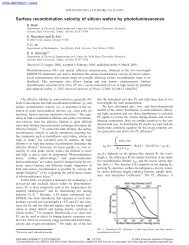
ECS Transactions 3, (4), 321-337 (2006) - Dieter Schroder - Arizona ...
ECS Transactions 3, (4), 321-337 (2006) - Dieter Schroder - Arizona ...
ECS Transactions 3, (4), 321-337 (2006) - Dieter Schroder - Arizona ...

Create successful ePaper yourself
Turn your PDF publications into a flip-book with our unique Google optimized e-Paper software.
<strong>ECS</strong> <strong>Transactions</strong> 3, (4), <strong>321</strong>-<strong>337</strong> (<strong>2006</strong>)<br />
that are near defects and those that are not. The resulting differences in ∆Rs are converted<br />
to ∆Is or ∆Vs and displayed on a cathode ray tube in the form of brightness changes leading<br />
to failure analysis location determination.<br />
A 1.3-µm laser (hν < E G ) does not generate electron-hole pairs in Si, i.e., it does not<br />
give an optical beam-induced current signal. For chips with multiple metal layers,<br />
OBIRCH images are observed by shining the 1.3-µm laser beam from the rear of a chip,<br />
because the 1.3 µm laser penetrates the Si substrate with about 40% power loss for moderately-doped<br />
Si about 500 µm thick. Materials with negative TCR include W (containing<br />
Ga), Ti (containing O) unintentionally left to be etched, and Ti-Al amorphous layers<br />
(containing O). The maximum temperature increase on the Al line is on the order of 10 K<br />
making the method nondestructive. To increase the resolution, OBIRCH has been combined<br />
with a near-field optical probe. 21 The OBIRCH image in Fig. 9(b) shows a leakage<br />
current path as the dark contrast and the defective part as the bright contrast. 22 FIB crosssectioning<br />
the bright area for TEM observation revealed a short between Al lines. Subsequent<br />
energy-dispersive X-ray analysis showed the existence of Ti and O in that region<br />
with a negative temperature coefficient leading to the bright contrast in the OBIRCH image.<br />
Scanning Kelvin Probe Microscopy (SKPM)<br />
Scanning Kelvin probe microscopy falls in the category of electrostatic force microscopy<br />
(EFM) techniques. EFM can be divided into three regimes based on tip-sample separation:<br />
long range, intermediate, and short range. 23 The SKPM probe, typically held 30-50<br />
nm above the sample, is scanned across the surface and the potential is measured. Frequently<br />
this measurement is combined with atomic force microscopy (AFM) measurements.<br />
During the first AFM scan, the sample topography is measured and during the<br />
second scan, in the SKPM mode, the surface potential is determined. 24<br />
The conducting probe and conducting substrate can be treated as a capacitor with the<br />
gap spacing being the spacing between probe and sample surface. A dc and ac voltage,<br />
applied to the tip leads to an oscillating electrostatic force between tip and sample from<br />
which the surface potential is determined. An advantage of force over current measurements<br />
is that the latter is proportional to the probe size while the former is independent of<br />
it. The frequency is chosen to be equal or close to the cantilever resonance frequency,<br />
which it typically around several 100 kHz.<br />
For a dc and ac tip voltage, the force F is 23<br />
⎡<br />
⎤<br />
= 1 dC<br />
2 1<br />
F ⎢(<br />
V − ) +<br />
2<br />
dc Vsurf<br />
Vac<br />
(1 − cos(2ω t))<br />
+ 2( Vdc<br />
− Vsurf<br />
) Vac<br />
sin( ωt)<br />
2 dz<br />
⎥ [8]<br />
⎣<br />
2<br />
⎦<br />
where V surf is the surface potential. F consists of static, first harmonic, and second harmonic<br />
components. The relevant first harmonic is given by<br />
dC<br />
Fω<br />
= ( Vdc<br />
− Vsurf<br />
) Vac<br />
sin( ωt)<br />
[9]<br />
dz<br />
which becomes zero when V dc =V surf .<br />
The method consists of applying an ac voltage of constant amplitude together with a<br />
dc voltage. A lock-in technique allows extraction of the first harmonic signal in the form<br />
of the first harmonic tip deflection proportional to F ω . The oscillation amplitude is minimized<br />
by adjusting V dc . A measure of the feedback voltage V dc is a measure of the surface<br />
potential. SKPM has also been combined with optical excitation. 25<br />
329