scanora - Soredex
scanora - Soredex
scanora - Soredex

Create successful ePaper yourself
Turn your PDF publications into a flip-book with our unique Google optimized e-Paper software.
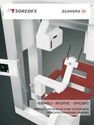
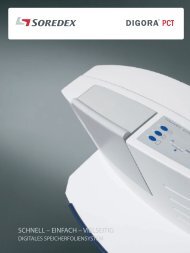
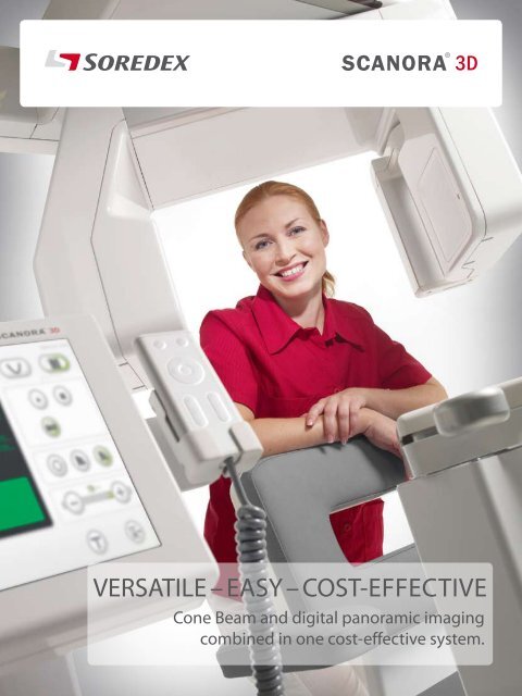
VERSATILE – EASY – COST-effEctIVE<br />
Cone Beam and digital panoramic imaging<br />
combined in one cost-effective system.
With thirty years of experience in designing and<br />
manufacturing state-of-the-art dental panoramic and<br />
tomographic systems SOREDEX turns another page in<br />
dental imaging history by introducing the SCANORA® 3D,<br />
a Cone Beam 3D system that combines versatile fields of<br />
view and standard digital panoramic with a dedicated<br />
pan sensor.<br />
The SCANORA® 3D follows the lead of the original<br />
SCANORA® system introduced twenty years ago.<br />
Then, for the first time, spiral tomography was used in<br />
dental imaging to generate cross-sectional tomograms,<br />
that produced valuable diagnostic information, especially<br />
useful for implants.
Versatile – easy – cost-effective<br />
Two systems combined. Real digital<br />
panoramic and Cone Beam 3D<br />
in one unit.<br />
The SCANORA® 3D takes dental<br />
imaging and implant planning to<br />
the next level.<br />
The new SCANORA® 3D opens a new<br />
world of diagnostic possibilities.
Superior versatility<br />
The SCANORA® 3D offers superior versatility by combining cone beam 3D imaging, with three selectable<br />
fields of view, plus optional dedicated panoramic imaging. At the press of a button, the unit automatically<br />
switches between 3D and panoramic imaging modes, making it quick and efficient to use.<br />
The smallest FOV (6 cm x 6 cm) is<br />
ideal for single implant operations,<br />
localized dental examinations and<br />
TM joints.<br />
User-selectable 3D<br />
fields of view<br />
With three fields of view (FOV) the proper<br />
image volume can be selected for each<br />
specific diagnostic task. The field of view<br />
can be positioned anywhere within the<br />
maxillofacial area.<br />
The medium FOV (7.5 cm x 10 cm)<br />
is suitable when the entire dental<br />
complex, including the mental<br />
and mandibular foramen, need<br />
to be examined. This field of view<br />
can also provide information for<br />
drill guide fabrication.<br />
Selectable 3D resolution<br />
The SCANORA® 3D combines low dose,<br />
fast imaging and high accuracy. Standard<br />
resolution offers fast imaging with low<br />
dose, suitable for most diagnostic tasks.<br />
High resolution improves accuracy with<br />
slightly higher imaging time and dose.<br />
The smallest attainable voxel (volume<br />
element) size is 0.133 mm.<br />
The largest FOV (7.5 cm x 14.5<br />
cm) is ideal when the complete<br />
dentition, both TM joints and<br />
upper cervical spine, must be<br />
examined. This field of view<br />
is also suitable for studying<br />
maxillofacial area with airways.<br />
• Superior quality, traditional full<br />
field panoramic images – not only<br />
low resolution synthesized<br />
panoramic images<br />
• No need to overexpose the patient<br />
for a standard panoramic view<br />
• No risk of dropping or damaging<br />
an expensive sensor
Two machines in ONE<br />
In most examinations a panoramic image is the first step and provides an overview of the whole dentition.<br />
SCANORA® 3D provides the speed and efficiency of traditional panoramic imaging in conjunction with<br />
advanced 3D technology. This combination speeds office workflow and produces superior panoramic<br />
image quality. 90% of your extraoral imaging needs remain a standard panoramic view. Why overexpose<br />
your patients while settling for a low resolution synthesized panoramic imaging? SCANORA® 3D gives you<br />
a standard, full field panoramic view plus three FOV Cone Beam images in one, easy-to-use fully automated<br />
system.<br />
Panoramic imaging<br />
with AutoSwitch TM<br />
The SCANORA® 3D uses a<br />
dedicated CCD sensor for highquality<br />
panoramic imaging.<br />
The unique AutoSwitch feature<br />
changes between panoramic<br />
and 3D modes making the<br />
SCANORA® 3D quick and easy<br />
to set up. There is no need to<br />
manually change detectors nor<br />
completely reposition the patient.<br />
AutoSwitch changes the detectors between 3D and panoramic.<br />
NO expensive sensors to exchange, move, drop or damage.<br />
The unit set up for panoramic imaging.<br />
The panoramic preview image on the unit display.
Excellent diagnostic performance<br />
The SCANORA® 3D system provides a new and innovative way of seeing dentomaxillofacial anatomy<br />
and solving diagnostic tasks. The high-definition panoramic image shows the regions that need further<br />
investigation. The optimum 3D technique for a specific task can be easily selected, treatment planned and<br />
finally follow up studies done, all with one efficient unit. The system contains user-selectable features that<br />
contribute to excellent diagnostic performance.<br />
The viewing software has complete selection of powerful tools for utilizing the diagnostic information.<br />
Outstanding computing intelligence<br />
For the first time in dental imaging, the SCANORA® 3D<br />
uses a sophisticated algebraic reconstruction technique<br />
(ART) for reconstructing the volume data. ART improves<br />
image quality and is less sensitive to the main causes of<br />
image artifacts such as patient movement, restorations and<br />
implants. Reconstruction times are super fast, starting from<br />
one minute, due to advanced computing technology.<br />
High technology flat-panel detector<br />
The flat-panel detector is a masterpiece of modern CMOS<br />
technology. Compared to traditional image intensifiers,<br />
flat-panel detectors offer superior image quality due<br />
to their large dynamic range, better contrast and lack<br />
of image distortion. Additionally they are insensitive to<br />
electromagnetic interference, are compact in size and have<br />
a very long service life.
Uncompromized quality<br />
The SCANORA® 3D system has been designed<br />
from the ground up using the very latest 3D<br />
imaging technology. The SCANORA® 3D is an<br />
extremely versatile Cone Beam 3D X-ray system,<br />
capable of variable size volume imaging of the<br />
dentomaxillofacial area as well as high-resolution<br />
classic digital panoramic imaging.<br />
Full diagnostic information<br />
After scanning and image reconstruction, a full range<br />
of diagnostic options can be utilized. The diagnostic<br />
information can be thoroughly examined with the many<br />
powerful software tools and features. For instance, a<br />
complete set of cross-sectional views of the jaw can be<br />
automatically generated by drawing a center line along<br />
the axial slice.<br />
Always the lowest possible X-ray dose<br />
Three selectable 3D fields of view allow the<br />
examination area to be precisely selected, thus<br />
reducing patient dose to an absolute minimum.<br />
Pulsed X-ray generation reduces dose because the<br />
effective exposure time is only a fraction of the<br />
scanning time. Whenever a high-quality panoramic<br />
image is needed, the built-in, low-dose CCD sensor<br />
can be used.<br />
Dose levels of the SCANORA® 3D are considerably<br />
lower than with medical CT imaging and carefully<br />
follow the ALARA principle – X-ray dose “As Low As<br />
Reasonably Achievable”.<br />
The smallest field of view 6x6 cm is ideal for studying local<br />
regions of interest.<br />
The largest field of view 7.5 cm x 14.5 cm clearly<br />
shows the whole dental area.<br />
Visualization can be done in many different ways. Enhancing<br />
the visibility of details helps diagnostic decisions to be made.
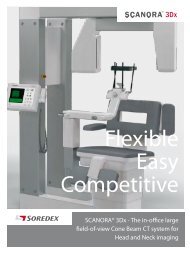
Cone Beam 3D imaging made easy<br />
The SCANORA® 3D system has been designed to make your workflow as fast and efficient as possible.<br />
The unique AutoSwitch TM feature selects the detector according to the required imaging mode.<br />
There is no need to change sensors nor radically reposition the patient or realign the unit. Short scan<br />
and reconstruction times further increase the efficiency and usability of the unit.<br />
In Cone Beam imaging<br />
a patient movement<br />
tolerance of less than<br />
one voxel’s dimension is<br />
desirable. A seated patient<br />
is important in achieving<br />
this and reducing<br />
movement artifacts.<br />
Rigid patient support<br />
The SCANORA® 3D uses an integrated<br />
motorized seat for superior patient<br />
stability and accurate patient<br />
positioning. The chin rest and temporal<br />
supports firmly hold the patient’s head<br />
to eliminate movement artifacts in 3D<br />
and panoramic imaging.<br />
Easy preparation for imaging<br />
The Scanora® 3D offers preset<br />
imaging programs that cover most<br />
common diagnostic examinations.<br />
All the imaging parameters can also<br />
be freely selected using the large<br />
ClearTouch TM control panel. The<br />
patient is easily positioned using<br />
the remote control and positioning<br />
lights.<br />
ClearTouch TM<br />
control panel<br />
intuitively quides<br />
the user through<br />
the examination<br />
sequence.
Image preview<br />
After exposure the preview image appears on the<br />
ClearTouch TM control panel. The user can easily verify the<br />
imaging procedure has been successful.<br />
Ready for diagnosis<br />
Once the preview image has been accepted, the 3D image set<br />
appears for examination and diagnosis on the workstation.<br />
Innovative and sophisticated ART reconstruction algorithm<br />
produces superior image quality with less artifacts.<br />
It takes only 1-2 minutes for the primary reconstruction to<br />
create the image. The visualization can be altered in real time.
True diagnostic value<br />
The SCANORA® 3D system provides true diagnostic value with real benefits to patients and dental<br />
professionals. The versatile X-ray system comes with an integrated software solution, designed<br />
together with dental clinicians.<br />
The MPR<br />
( Multi Planar<br />
Reconstruction)<br />
is the basic mode<br />
of visualizing the<br />
anatomy. The<br />
sections can be<br />
customized to<br />
show the region<br />
of interest from<br />
multiple directions.<br />
The software<br />
automatically<br />
generates crosssectional<br />
views<br />
after a centerline<br />
has been drawn<br />
along the jaw. No<br />
time consuming<br />
reformatting<br />
needed.
The nerve pathway<br />
can be clearly<br />
marked in the<br />
images.<br />
In the implant<br />
design mode the<br />
position of the<br />
implant can be<br />
freely navigated<br />
in relation to<br />
critical anatomical<br />
landmarks.<br />
Comprehensive set of<br />
diagnostic tools<br />
The software includes image<br />
capturing, database as well as image<br />
visualization.<br />
In addition to regular image handling<br />
functions there is an extensive range<br />
of powerful diagnostic tools. These<br />
include surface and volume rendering<br />
with anatomic plane clipping, which<br />
is used to virtually remove covering<br />
structures to show regions of interest.<br />
Other tools allow enhancement of<br />
the visibility of teeth, bones and soft<br />
tissue with several pre-set controls.<br />
Also color mapping and transparency<br />
levels can be adjusted.<br />
Synthesized panoramic images, where<br />
the layer is freely adjustable, can also<br />
be produced by reconstructing the 3D<br />
volume.<br />
Valuable tools for implant planning<br />
For proper implant site selection,<br />
accurate information is needed about<br />
the available bone, its quality, and<br />
the exact location of critical areas.<br />
Location of mandibular nerve canal<br />
or maxillary sinus can be obtained<br />
accurately and easily. With the help<br />
of a multiplanar slice display, 3D<br />
rendering, measurement tools, and<br />
the comprehensive implant symbol<br />
library, implant planning and surgery<br />
can be carried out efficiently and<br />
reliably.<br />
For third party drill guide systems<br />
the volume data can be exported in<br />
DICOM format.
Open software architecture<br />
The SCANORA® 3D produces image data in DICOM* format. This facilitates the open architecture that allows versatile<br />
and optimized software solutions to be tailored for your practice. The local area network (LAN) with several viewing<br />
stations is the solution for most practice applications allowing the system to be linked with the network and system<br />
server.<br />
The SCANORA® 3.0 software is the main platform, including the local patient image database and panoramic image<br />
handling. 3D visualization software provides 3D image handling, diagnostics and implant planning. This system forms<br />
an effective means for solving diagnostic problems, planning surgical procedures and making patient education and<br />
case acceptance.<br />
Freely distribute clinical cases on CD to referring clinicians. The referring clinician can utilize the free viewer without<br />
investing in special software or import the images in DICOM format into their own 3D software.<br />
LAN<br />
SCANORA® 3D<br />
X-ray unit<br />
PACS<br />
**<br />
Modality<br />
workstation PC<br />
SCANORA® 3.0<br />
Image management<br />
Local database<br />
Panoramic image handling<br />
DICOM components<br />
3D viewing software<br />
3D image handling<br />
Implant planning<br />
Reporting<br />
Other 3rd<br />
party softwares<br />
- Implant planning<br />
- Image handling<br />
- Drill and surgical guides<br />
- Other applications<br />
In addition to the standard 3D visualization software<br />
that comes with the unit, most other software offerings<br />
supporting the DICOM format can also be used. So the<br />
system is open and flexible, ready for globally rapid<br />
software development, and offers you the possibility to<br />
selectively take advantage of most third party diagnostic<br />
and surgical guide applications, now and in the future.<br />
* Digital Imaging and Communication in Medicine<br />
** Picture Archiving and Communication System
Low dose 3D imaging<br />
X-ray imaging is a compromise between<br />
image quality and x-ray dose. In SCANORA® 3D<br />
this dilemma has been successfully resolved<br />
by combining high image quality with low<br />
dose. The key factors in achieving this are<br />
sophisticated pulsed x-ray generation,<br />
selectable imaging modes and the innovative<br />
ART image reconstruction method.<br />
ART needs less dose than conventional<br />
algorithms. And further, because the system<br />
is less sensitive to patient movement and<br />
metal artifacts, the image quality is consistent<br />
and the success rate is very high, minimizing<br />
retakes.<br />
The x-ray dose in all the fields of view of the<br />
SCANORA® 3D is low. The minimum effective<br />
dose can be compared to one digital<br />
panoramic exposure and, at maximum, to a<br />
few panoramic exposures in a larger field of<br />
view and higher resolution.
DOSE COMPARISON<br />
SCANORA® 3D<br />
PANORAMIC<br />
AVERAGE CBCT<br />
SCANORA® 3D gives you the ability<br />
to carefully minimize the dose<br />
according to the diagnostic task,<br />
whether it is a question of detailed<br />
primary diagnostics or a follow-up<br />
study.<br />
SCANORA® 3D is a safe and efficient<br />
diagnostic tool for your clinic.<br />
MEDICAL CT
More value for your money<br />
SOREDEX has been designing and manufacturing high quality dental<br />
imaging systems for thirty years. The superior versatility, features<br />
and design of the SCANORA® 3D make it an attractive 3D imaging<br />
unit. Your investment is protected, because SOREDEX products are<br />
renowned for their excellent quality, reliability and long service life.
With SCANORA® 3D, the advanced dental imaging required for diagnostics,<br />
implant treatment planning and oral surgery can now be done in your practice.<br />
More advanced procedures can be performed efficiently and safely. Diagnostic<br />
information can be obtained without delay and with fewer referrals to outside<br />
facilities, for procedures such as medical CT examinations. The whole treatment<br />
planning process, from the first contact through radiological examinations, case<br />
planning, treatment acceptance, and follow-up, can be handled in one practice<br />
– yours.<br />
3D imaging is not just prestigious, it is clinically necessary. It allows you to improve<br />
patient care by enhancing diagnostic accuracy and performance. 3D imaging helps<br />
you work closely with your patients to plan and implement the best alternatives;<br />
all of which differentiates your practice from those using conventional imaging.<br />
The SCANORA® 3D is a total 2D and 3D imaging solution that comes with a complete<br />
3D software package for advanced diagnostics and treatment planning. Through<br />
DICOM support, the SCANORA® 3D system integrates with other imaging software<br />
and modalities and is compatible with most specialty third party software, drill<br />
and surgical guide applications.<br />
The SCANORA® 3D system makes advanced dental imaging fast and easy. We let<br />
you concentrate on your most important activity - treating your patient.
Technical data<br />
(77.7")<br />
1940 (76.4")<br />
1600 (63")<br />
1740 (68.5")<br />
1400 (55.1")<br />
Minimum space requirements<br />
194 x 174 cm (76.4 x 68.5”).<br />
Room height 235 cm (92.5”).
Technical data<br />
3D imaging fields of view (height x diameter)<br />
Small field of view<br />
60 mm x 60 mm<br />
Medium field of view<br />
75 mm x 100 mm<br />
Large field of view<br />
75 mm x145 mm<br />
All fields of view support both standard and high resolution modes<br />
3D imaging parameters<br />
Voxel size<br />
Scan time<br />
Effective exposure time<br />
Reconstruction time<br />
3D image receptor<br />
Receptor type<br />
Receptor active area<br />
Pixel size<br />
133 μm-350 μm<br />
10-20 seconds<br />
2-5 seconds<br />
1-2 minutes<br />
CMOS Flat Panel<br />
124 mm x 124 mm<br />
200 μm<br />
Panoramic imaging programs (Optional)<br />
Adult panoramic program<br />
Pediatric panoramic program<br />
TMJ programs<br />
Panoramic image receptor (Optional)<br />
Technology<br />
CCD<br />
Detector size (H x W)<br />
146 mm x 6 mm<br />
Detector pixel size<br />
48 μm<br />
X-ray generator<br />
Tube<br />
Fixed anode tube<br />
Focal spot 0.5 mm IEC 336<br />
Target angle<br />
5 degrees<br />
kV 60-85<br />
Average mA 1.0-8.0<br />
Software<br />
Primary reconstruction<br />
Workstation software<br />
ART (Algebraic Reconstruction Technique)<br />
Image capture, patient and image database, 3D slice views, 3D rendering,<br />
implant planning and DICOM connectivity<br />
Minimum system requirements for acquisition server computer<br />
Memory<br />
2 Gb<br />
Hard disk<br />
500 Gb<br />
Processor<br />
2.4 GHz dual core processor<br />
Motherboard<br />
PCI-Express bus for GPU<br />
Operating system Windows XP or 2000<br />
Local area network<br />
Copper Gigabit Ethernet<br />
General<br />
Weight<br />
310 kg (690 lbs)<br />
Dimensions (H x W x D) 1973 mm x 1600 mm x 1400 mm (77.7” x 63” x 55.1”)<br />
Power requirements<br />
Line voltage<br />
230-240 VAC (±10 %), 50/60 Hz
Digital imaging made easy<br />
Head office and factory:<br />
SOREDEX<br />
Nahkelantie 160, Tuusula<br />
P.O. Box 148, FI-04301 Tuusula<br />
Finland<br />
Tel. +358 45 7882 2000<br />
Fax +358 9 701 5261<br />
info@soredex.com<br />
SOREDEX USA<br />
300 W. Edgerton Ave.<br />
Milwaukee,WI 53207<br />
U.S.A.<br />
Tel. +1 800 235 8854<br />
Fax +1 414 481 8665<br />
info@soredexusa.com<br />
SOREDEX Germany<br />
Schutterstrasse 12<br />
77746 Schutterwald<br />
Germany<br />
Tel: +49 (0) 781 28 41 98-0<br />
Fax: +49 (0) 781 28 41 98-30<br />
kontakt@soredex.de<br />
SOREDEX designs, develops, manufactures, and markets dental imaging systems,<br />
with an emphasis on innovative digital solutions. Operating worldwide, SOREDEX<br />
offers quality imaging systems of true diagnostic value, based on an in-depth<br />
understanding of the dental practice. Applying over three decades of experience<br />
in imaging excellence, we offer reliable and easy-to-use solutions that help you<br />
focus on patient care.<br />
SOREDEX digital imaging systems are innovative and accurate diagnostic tools<br />
that are incorporated seamlessly and easily into your dental practice, enhancing<br />
the imaging process and improving workflow. Our systems are designed to be<br />
simple and easy to use. They will make your dental practice more efficient and<br />
ultimately give you more time to provide better care to your patients.<br />
SOREDEX stands for innovation and value in dental X-ray technology.<br />
SCANORA® is a registered trademark of SOREDEX, PaloDEx Group Oy. Other product names and<br />
trademarks are the property of their respective owners. CE-marked, NB (CE) number 0537. Electrical<br />
safety meets the IEC 60601-1 standard. Manufacturing complies with ISO 13485:2003, ISO 9001:2000,<br />
and ISO 14001:2004.<br />
SOREDEX reserves the right to make changes in specifications and features shown herein at any time<br />
without notice or obligation. Contact your SOREDEX representative for the most up-to-date information.<br />
© 2008 SOREDEX<br />
72125-2 04/08 Printed in Finland<br />
www.soredex.com • www.soredex.de • www.soredexusa.com