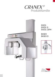
Brochure (pdf) - Soredex
Brochure (pdf) - Soredex
Brochure (pdf) - Soredex

Create successful ePaper yourself
Turn your PDF publications into a flip-book with our unique Google optimized e-Paper software.
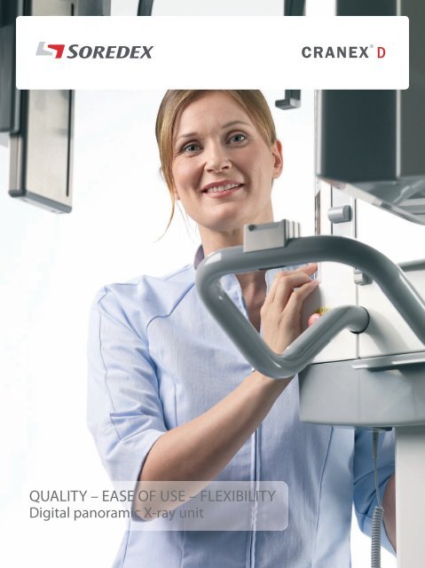
QUALITY – EASE OF USE – FLEXIBILITY<br />
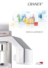
Digital panoramic X-ray unit
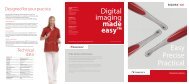
Digital imaging made easy<br />
SUMMARY OF BENEFITS<br />
QUALITY<br />
• Superior diagnostic image quality<br />
• Unique VPC panoramic collimator<br />
• Wide anterior layer thickness<br />
• Enhanced imaging geometry<br />
EASE OF USE<br />
• Excellent usability<br />
• Easy, stable patient positioning<br />
• Automatic exposure value selection<br />
FLEXIBILITY<br />
• Upgradeability<br />
• Single or dual sensor design<br />
• Small footprint and compact design
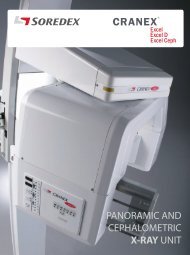
SOREDEX® has over 30 years of expertise in state-of-the-art dental imaging<br />
systems. The CRANEX® D – a direct digital panoramic and cephalometric X-ray<br />
system – is the high-end unit of the famous CRANEX® family.
True diagnostic value<br />
CRANEX® D’s excellent signal-to-noise ratio, high resolution, wide dynamic range, wide anterior layer<br />
thickness and many other features provide superior image quality and more diagnostic value.<br />
Enhanced cephalometric<br />
imaging geometry<br />
The CRANEX® D features advanced<br />
cephalometric imaging movements<br />
that provide a true central projection<br />
image. This results in non-distorted<br />
constant magnification in both the<br />
vertical and horizontal planes.
QUALITY – EASE OF USE – FLEXIBILITY<br />
The unique Variable Panoramic Collimator (VPC) is one of the keys to the outstanding panoramic image<br />
quality of the CRANEX® D. By narrowing the width of the collimator while imaging the anterior teeth, we<br />
are able to expand the focal trough by 50% compared to conventional fixed collimation. The advantage is<br />
superb anterior image quality regardless of the patient’s dentition.<br />
Typical panoramic system<br />
Most panoramic systems have a narrow anterior<br />
layer thickness, which leads to difficulty in<br />
positioning all of the patient´s dentition within<br />
the focal trough.<br />
+50%<br />
CRANEX® D<br />
CRANEX® D’s anterior layer thickness is 50% wider<br />
than traditional techniques. The result is superb<br />
anterior image quality.<br />
+50%
Quick and easy-to-use
QUALITY – EASE OF USE – FLEXIBILITY<br />
11 second fast scan – 17 second HiQ scan<br />
Superb panoramic image quality with fast<br />
panoramic programs.<br />
• Adult program in 11 seconds<br />
• Pediatric program in 8,6 seconds<br />
• Especially suitable for follow-up patients<br />
and in busy clinics<br />
Even better high quality images achieved with<br />
High Quality (HiQ) panoramic programs.<br />
• Adult program in 17 seconds<br />
• Pediatric program in 13,8 seconds<br />
• Especially suitable for complicated diagnostic cases<br />
Easy-to-use control panel<br />
• Imaging program selection<br />
• Exposure value selection<br />
• Fast and High Quality panoramic program<br />
• Same selections available in the Graphical<br />
User Interface of the computer<br />
• All needed settings visible
Patient positioning made easy<br />
Patient positioning<br />
• 3 positioning lights<br />
for accurate positioning<br />
• 4-point head support<br />
for patient stability<br />
• Open design for<br />
accessibility
QUALITY – EASE OF USE – FLEXIBILITY<br />
Optimal image quality can only be achieved if the patient is positioned correctly and held stationary during<br />
imaging. CRANEX® D’s three positioning lights, swivel mirror and 4-point head support help you to attain<br />
the needed optimal image quality for your diagnostic purposes. The open design and the unique column<br />
increase patient comfort and allow easy access for handicapped or wheelchair bound patients.<br />
Automated ease fo use<br />
The CRANEX® D automatically selects proper<br />
collimation for the selected imaging program<br />
by using the Automatic Collimator Selector (ACS).<br />
The Automatic Exposure Setting (AES) function<br />
automatically recommends proper exposure<br />
values based on the size of the patient’s head.<br />
The operator can also override the recommended<br />
values if needed before the exposure.
Cephalometric imaging<br />
CRANEX® D’s cephalometic option can be configured as either left-handed or<br />
right-handed handed for flexible installation. Soft tissue filtration is adjusted automatically<br />
for the best diagnostic quality possible. With the use of the AES function, exposure<br />
values are selected according to patient size.<br />
Cephalometric field sizes<br />
Full width: 22 x 26 cm (8.66” x 10.24”)<br />
Reduced width: 22 x 18 cm (8.66” x 7.09”)
QUALITY – EASE OF USE – FLEXIBILITY<br />
Maximum flexibility<br />
The CRANEX® D Ceph can be equipped as<br />
a single or dual sensor system for optimal<br />
workflow. Cephalometric capabilities can<br />
be added at any time to the CRANEX® D.<br />
This allows the CRANEX® D to grow along<br />
with your practice.<br />
CRANEX® D Ceph<br />
head support in the PA<br />
projection. The nasion<br />
support is rotated out<br />
of the way.<br />
Upgradeability<br />
The CRANEX® D panoramic unit is easily<br />
upgradeable to left- or right-handed<br />
cephalometric unit in the field.<br />
CRANEX® D Ceph<br />
head support<br />
in the lateral position.
Compact design<br />
Positioning controls and accessories<br />
are located close to the operator for<br />
improved workflow.<br />
The CRANEX® D can<br />
be equipped with<br />
either the unique<br />
open column<br />
(standard) or<br />
optional traditional<br />
column design.<br />
The stainless steel CCD housing is<br />
designed to last.
QUALITY – EASE OF USE – FLEXIBILITY<br />
The CRANEX® D is compatible with your<br />
choice of several software options.<br />
PACS / DICOM<br />
enviroment<br />
• DIGORA® for Windows<br />
offers a comprehensive<br />
set of imaging tools<br />
for your day-to-day<br />
imaging needs. Network<br />
multi-user versions and<br />
full DICOM support are<br />
available as options.<br />
• CliniView XV (US only)<br />
offers the capability of<br />
capturing images from<br />
all of the digital treatment<br />
planning devices.<br />
It is geared to be used<br />
in operatories for quick<br />
and easy image capture,<br />
viewing and treatment<br />
planning.<br />
• SOREDEX TWAIN<br />
utilizes the industry<br />
standard TWAIN<br />
interface to capture<br />
images directly into<br />
3rd party imaging<br />
applications.<br />
• SorCom<br />
provides a simple<br />
“DICOM bridge” for<br />
capturing images<br />
to a PACS/DICOM<br />
environment, where<br />
3rd party DICOM<br />
imaging software<br />
is used for viewing<br />
images.
Imaging programs<br />
With the CRANEX® D you can perform standard, pediatric or sectional panoramic<br />
examinations in addition to TMJ, sinus and optional cephalometric examinations.<br />
Adult panoramic<br />
All panoramic programs include automatic spinal compensation<br />
for an excellent view of the anterior teeth without a distracting<br />
spinal shadow.<br />
Child panoramic<br />
A shorter exposure time and reduced exposure field lowers<br />
patient dose.<br />
TMJ<br />
TMJ images are taken mouth open and/or closed for functional<br />
evaluation and condyle anatomy.<br />
Sectional Imaging<br />
Any combination of 1 to 5 panoramic image sections can be<br />
selected. This reduces the patient radiation dose as only the<br />
region of interest is exposed.<br />
Cephalometric<br />
Lateral and PA images can be taken<br />
with the CRANEX® D.<br />
Carpus<br />
Optional Carpus imaging is also available<br />
with the CRANEX® D Ceph.
Technical data<br />
General<br />
Generator<br />
High frequency DC generator, operating frequency 40 kHz<br />
Focal spot size<br />
0.5 mm<br />
Minimum total filtration<br />
2.7 mm Al<br />
Line voltage<br />
230/240 Vac ±10% / 115 Vac (50/60 HZ)<br />
Anode voltage<br />
57 - 85 kV<br />
Anode current<br />
10 mA<br />
Exposure time<br />
17.6 s High Quality panoramic, 11 s fast panoramic, 8 - 20 s cephalometric<br />
SID<br />
520 mm (20.47”) panoramic, 1721 mm (67.75”) cephalometric<br />
Fusing<br />
8 A / 16 A slow (230/115 Vac)<br />
Weight<br />
Pan 120 kg (264 lbs), Ceph 165 kg (363 lbs)<br />
Electrical safety classification EN 60601-1 class 1/B<br />
Color RAL 7040, RAL 9003<br />
Digital Unit<br />
Sensor<br />
Active sensor surface<br />
Pixel size sensor<br />
Pixel size image<br />
File size<br />
CCD-detector<br />
PAN: 147.5 x 6.1 mm, CEPH: 221.2 x 6.1 mm<br />
48 μm<br />
96 μm<br />
PAN max: 9.5 MB, CEPH max: 11.5 MB<br />
Workstation computer recommendations<br />
Windows Vista / Windows XP Professional / Home /<br />
Operating system<br />
SP1 or SP2, Windows 2000 Professional / SP4
DIGITAL IMAGING<br />
MADE EASY<br />
Head Office and Factory:<br />
SOREDEX<br />
Nahkelantie 160, Tuusula<br />
P.O. Box 148, FI-04301 Tuusula<br />
Finland<br />
Tel. +358 45 7882 2000<br />
Fax +358 9 701 5261<br />
info@soredex.com<br />
Subsidiary: SOREDEX USA<br />
300 W. Edgerton Ave.<br />
Milwaukee, WI 53207 U.S.A.<br />
Tel. +1 800 235 8854<br />
Fax +1 414 481 8665<br />
info@soredexusa.com<br />
Subsidiary: SOREDEX Germany<br />
Schutterstrasse 12<br />
77746 Schutterwald<br />
Germany<br />
Tel: +49 (0) 781 28 41 98 0<br />
Fax: +49 (0) 781 28 41 98 30<br />
kontakt@soredex.de<br />
SOREDEX® designs, develops, manufactures and markets dental imaging systems,<br />
with an emphasis on innovative digital solutions. Operating worldwide, SOREDEX®<br />
offers quality imaging systems of true diagnostic value, based on an in-depth<br />
understanding of the dental practice. Applying three decades of experience of<br />
imaging excellence, we offer reliable and easy-to-use solutions that help you focus<br />
on patient care.<br />
SOREDEX® digital imaging systems are innovative and accurate diagostic tools<br />
that integrate seamlessly and easily into a dental practice, enhancing the imaging<br />
process and improving workflow. Our systems are designed to be simple and easy<br />
to use. They will make your dental practice more efficient and ultimately give you<br />
more time for your patients.<br />
SOREDEX® stands for innovation and value in dental x-ray technology.<br />
MINRAY®, CRANEX® and DIGORA® are registered trademarks of SOREDEX®, PaloDEx Group Oy.<br />
Other product names and trademarks are the property of their respective owners. CE marked,<br />
NB (CE) number 0537. Electric safety according to IEC 60601-1. Manufacturing complies with<br />
ISO 13485:2003, ISO 9001:2000 and ISO 14001:2004.<br />
SOREDEX® reserves the right to make changes in specification and features shown herein at any time<br />
without notice or obligation. Contact your SOREDEX® representative for the most current information.<br />
© 2008 SOREDEX®<br />
72049-3 11/08 Printed in Finland