Cladosporium fulvum (syn. Passalora fulva), a highly specialized - Cbs
Cladosporium fulvum (syn. Passalora fulva), a highly specialized - Cbs
Cladosporium fulvum (syn. Passalora fulva), a highly specialized - Cbs

Create successful ePaper yourself
Turn your PDF publications into a flip-book with our unique Google optimized e-Paper software.
MOLECULAR PLANT PATHOLOGY (2005) 6(4),<br />
379–393 DOI: 10.1111/J.1364-3703.2005.00292.X<br />
Blackwell Publishing, Ltd.<br />
Pathogen profile<br />
<strong>Cladosporium</strong> <strong>fulvum</strong> (<strong>syn</strong>. <strong>Passalora</strong> <strong>fulva</strong>),<br />
a <strong>highly</strong> <strong>specialized</strong><br />
plant pathogen as a model for functional studies on plant<br />
pathogenic Mycosphaerellaceae<br />
1<br />
1<br />
1,<br />
2<br />
1<br />
BART P. H. J. THOMMA *†, H. PETER VAN ESSE †, PEDRO W. CROUS AND PIERRE J. G. M. DE WIT<br />
Laboratory of Phytopathology, Wageningen University, Binnenhaven 5, 6709 PD Wageningen, The Netherlands<br />
Centraalbureau voor Schimmelcultures, PO Box 85167, 3508 AD Utrecht, The Netherlands<br />
1<br />
2<br />
SUMMARY<br />
Taxonomy: <strong>Cladosporium</strong> <strong>fulvum</strong> is an asexual fungus for which<br />
no sexual stage is currently known. Molecular data, however,<br />
support C. <strong>fulvum</strong> as a member of the Mycosphaerellaceae,<br />
clustering with other taxa having Mycosphaerella teleomorphs.<br />
C. <strong>fulvum</strong> has recently been placed in the anamorph genus<br />
<strong>Passalora</strong> as P. <strong>fulva</strong>.<br />
Its taxonomic disposition is supported by its<br />
DNA phylogeny, as well as the distinct scars on its conidial hila,<br />
which are typical of <strong>Passalora</strong>,<br />
and unlike <strong>Cladosporium</strong> s.s. , which<br />
has teleomorphs that reside in Davidiella,<br />
and not Mycosphaerella.<br />
Host range and disease symptoms: The presently known<br />
sole host of C. <strong>fulvum</strong> is tomato (members of the genusLycoper<br />
sicon).<br />
C. <strong>fulvum</strong> is mainly a foliar pathogen. Disease symptoms<br />
are most obvious on the abaxial side of the leaf and include<br />
patches of white mould that turn brown upon sporulation. Due<br />
to stomatal clogging, curling of leaves and wilting can occur,<br />
leading to defoliation.<br />
C. <strong>fulvum</strong> as a model pathogen: The interaction between<br />
C. <strong>fulvum</strong> and tomato is governed by a gene-for-gene relationship.<br />
A total of eight Avr and Ecp genes, and for four of these also<br />
the corresponding plant Cf genes, have been cloned. Obtaining<br />
conclusive evidence for gene-for-gene relationships is complicated<br />
by the poor availability of genetic tools for most Mycosphaerellaceae–<br />
plant interactions. Newly developed tools, including<br />
Agrobacterium-mediated<br />
transformation and RNAi, added to the<br />
genome sequence of its host tomato, which will be available<br />
within a few years, render C. <strong>fulvum</strong> attractive as a model species<br />
for plant pathogenic Mycosphaerellaceae.<br />
Useful websites: http://www.sgn.cornell.edu/help/about/<br />
index.html; http://cogeme.ex.ac.uk<br />
* Correspondence : Tel.: +31 317 484536; fax: +31 317 483412; e-mail: bart.thomma@wur.nl).<br />
†These authors contributed equally to this work.<br />
© 2005 BLACKWELL PUBLISHING LTD<br />
INTRODUCTION<br />
<strong>Cladosporium</strong> <strong>fulvum</strong> [<strong>syn</strong>. <strong>Passalora</strong> <strong>fulva</strong> (Braun et al.,<br />
2003)]<br />
is the causal organism of tomato leaf mould, a fungal disease first<br />
described by Cooke (1883). Generally, foliage is the only tissue<br />
affected by the fungus, although occasionally also stems, blossoms,<br />
petioles and fruit are attacked (Butler and Jones, 1949;<br />
Jones et al.,<br />
1997). Conidia of the fungus can infect successfully<br />
if they settle on the abaxial side of a leaf, germinate, and subsequently<br />
enter through open stomata. Initial disease symptoms<br />
occur at the earliest 1 week after the start of infection as pale<br />
green or yellowish diffuse spots on the upper leaf surface, which<br />
later enlarge, turning into distinctive yellow spots (Fig. 1A). This<br />
appearance is the effect of cell death in the palisade parenchyma.<br />
The abaxial side of the leaf shows the most distinct symptoms:<br />
patches of white to olive-green mould that turn brown once<br />
sporulation commences (Fig. 1B). In advanced stages of disease<br />
development stomata do not function properly, because they are<br />
blocked by aggregations of conidiophores (Fig. 1E) that use the<br />
stomata to exit the leaf and liberate conidia. These subsequently<br />
contribute to spread of the disease. As a result of stomatal clogging,<br />
plant respiration is severely hampered (Butler and Jones,<br />
1949). This can result in wilting of leaves, partial defoliation and,<br />
in severe infections, death of the host (Jones et al.,<br />
1997).<br />
Although Lycopersicon esculentum (tomato) is susceptible to<br />
the fungus, many other Lycopersicon species are often resistant<br />
(Butler and Jones, 1949). About 100 years ago it was discovered<br />
that resistance against C. <strong>fulvum</strong> is genetically determined by the<br />
presence of Cf resistance genes (Lind, 1909; Norton, 1914). Later<br />
it was found that the relationship between host and pathogen is<br />
governed by a so-called ‘gene-for-gene’ relationship. The gene-forgene<br />
hypothesis states that each dominant pathogen avirulence<br />
( Avr)<br />
gene confers recognition to a corresponding dominant<br />
host resistance ( R)<br />
gene (Flor, 1942, 1946; Oort, 1944). Although<br />
C. <strong>fulvum</strong> most likely originates from the natural habitat of<br />
Lycopersicon species in South America, greenhouse cultivation<br />
379
380<br />
B. P. H. J. THOMMA et al.<br />
MOLECULAR PLANT PATHOLOGY (2005) 6(4),<br />
379–393 © 2005 BLACKWELL PUBLISHING LTD
has also generated favourable conditions for the pathogen in<br />
temperate climate areas. As a result, for decades yearly outbreaks<br />
of the disease occurred also in these regions in greenhouses and<br />
tomato leaf mould became a persistent disease. However, the<br />
introduction of resistance loci from related wild species of tomato<br />
( Cf-1 to Cf-5)<br />
into cultivated tomato has resulted in efficient<br />
containment of the pathogen (Boukema and Garretsen, 1975;<br />
Boukema, 1977; Hubbeling, 1978; Kerr et al.,<br />
1971; Langford,<br />
1937). Since the introduction of the Cf-9 resistance gene in the<br />
late 1970s in currently grown tomato cultivars in the late 1970s,<br />
C. <strong>fulvum</strong> no longer poses a serious threat to commercial tomato<br />
cultivation. Despite its limited agronomic importance, the C. <strong>fulvum</strong>–<br />
tomato interaction has become a model to study plant–pathogen<br />
interactions after intensive studies by the research groups of Drs<br />
Higgins (Higgins et al.,<br />
1998), Oliver (Oliver et al.,<br />
2000) and de<br />
Wit (Joosten and de Wit, 1999).<br />
This review will mainly focus on the pathogenic properties of<br />
C. <strong>fulvum</strong> and the mechanisms deployed by the fungus to establish<br />
pathogenicity. In addition, the properties of this interaction to<br />
serve as a model system for the interaction between plants and<br />
other members of Mycosphaerella will be discussed. As recent<br />
advances of the research on the tomato Cf resistance genes and<br />
homologous genes that act in pathogen defence from other plant<br />
species have been extensively reviewed (Kruijt et al.,<br />
2005; Rivas<br />
and Thomas, 2002), we will not address Cf-gene<br />
structures and<br />
Cf-mediated<br />
downstream defence signalling.<br />
THE INFECTION CYCLE ON SUSCEPTIBLE<br />
PLANTS: THE COMPATIBLE INTERACTION<br />
The conidia of C. <strong>fulvum</strong> are generally spread by wind or water<br />
splash. If conidia land on the abaxial side of a leaf, successful<br />
infection can occur. At high relative humidity (over 85%) conidia<br />
germinate and form thin runner hyphae that grow randomly<br />
© 2005 BLACKWELL PUBLISHING LTD<br />
MOLECULAR PLANT PATHOLOGY (2005) 6(4),<br />
379–393<br />
<strong>Cladosporium</strong> <strong>fulvum</strong> (<strong>syn</strong>. <strong>Passalora</strong> <strong>fulva</strong>)<br />
381<br />
(undirectional) over the leaf surface (Bond, 1938; de Wit, 1977;<br />
Lazarovits and Higgins, 1976a). After approximately 3 days,<br />
a main germ tube or a lateral branch of the hyphae enters the<br />
tomato leaf upon encountering an open stoma (Fig. 1C). From<br />
this stage onward, the diameter of fungal hyphae enlarges at<br />
least two-fold. Subsequently, hyphal growth continues from the<br />
substomatal cavity into the intercellular space between the spongy<br />
mesophyll cells (apoplast) by the formation of long, branched<br />
hyphal structures (Bond, 1938; de Wit, 1977; Lazarovits and<br />
Higgins, 1976a). Fungal growth appears to be preferentially directed<br />
towards the vascular tissues, probably triggered by a sucrose<br />
gradient around the phloem (van den Ackerveken et al.,<br />
1994;<br />
Wubben et al.,<br />
1994). Sometimes, but only in later stages of the<br />
infection, the palisade parenchyma is invaded (Lazarovits and<br />
Higgins, 1976a).<br />
Although no obvious feeding structures such as haustoria can<br />
be observed, growth of the fungus appears to depend on maintenance<br />
of close contact between fungal hyphae and host cells<br />
(Fig. 1D). This can sometimes be observed as slight indentations<br />
where fungal hyphae touch host cells (de Wit, 1977). This close<br />
contact suggests that the pathogen actively withdraws nutrients<br />
from the host (Bond, 1938; Lazarovits and Higgins, 1976b). No<br />
visible reaction of the host cells other than occasional callose<br />
deposition on the mesophyll cell walls can be observed during<br />
these stages of infection (de Wit, 1977; Lazarovits and Higgins,<br />
1976a,b). However, several ultrastructural changes have been<br />
described, including the occurrence of endoplasmic reticulum<br />
parallel to the plasmamembrane at the site of fungal contact, and<br />
cytoplasmic lipid bodies and microbodies containing crystalline<br />
inclusions (Lazarovits and Higgins, 1976b). In mature lesions,<br />
mesophyll cells display various signs of degeneration of cell<br />
organelles, more specifically the mitochondria and chloroplasts<br />
(Lazarovits and Higgins, 1976b). Occasionally, the release of cytoplasmic<br />
contents due to damage to the plasmamembrane and<br />
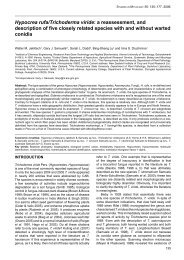
Fig. 1 Physiology of the C. <strong>fulvum</strong> infection on host and non-host plants (A–H) and typical symptoms on host plants caused by other plant pathogenic<br />
Mycosphaerellaceae as found in nature (I–N). (A) Adaxial side of a tomato leaf (MoneyMaker Cf-0)<br />
18 days after inoculation with a compatible race of C. <strong>fulvum</strong>.<br />
Distinctive yellow spots can be seen as a result of dead palisade parenchyma cells. (B) Abaxial side of a tomato leaf (MoneyMaker Cf-0)<br />
18 days after inoculation<br />
with a compatible race of C. <strong>fulvum</strong>.<br />
White mould can be seen developing into light brown patches where sporulation takes place. (C–E) SEM images from<br />
C. <strong>fulvum</strong>-infected<br />
tomato leaves in a compatible interaction at different timepoints after inoculation (pictures are taken from: de Wit, P.J.G.M. Light and<br />
scanning-electron microscopic study of infection of tomato plants by virulent and avirulent races of <strong>Cladosporium</strong> <strong>fulvum</strong>.<br />
Neth. J. Plant Pathol. (1977) 83, 109–122,<br />
with permission). (C) C. <strong>fulvum</strong>-infected<br />
tomato leaf in a compatible interaction 2 days post inoculation with fungal hyphae entering a stoma. (D) C. <strong>fulvum</strong>-infected<br />
tomato leaf in a compatible interaction 7 days post inoculation. In the spongy mesophyll hyphae (h) grow in close contact with the plant cells. (E) C. <strong>fulvum</strong>-infected<br />
tomato leaf in a compatible interaction 12 days post inoculation. Young conidiophores emerging from the stomata are observed. (F–H) Drawings upon microscopic<br />
analysis of lactophenol-stained leaf material of several plant species upon inoculation with C. <strong>fulvum</strong> (drawings are reproduced from: Bond, T.E.T. Infection experiments<br />
with <strong>Cladosporium</strong> <strong>fulvum</strong> Cooke and related species. Ann. Appl. Biol. (1938) 25, 277–307, by permission of Oxford University Press). (F) Growth of C. <strong>fulvum</strong><br />
mycelium in the tomato cultivar ‘Giant red’ 7 days after inoculation. The growth is characterized by long runner hyphae that pass between spongy mesophyll cells to<br />
send out ascending branches. (G) Limited growth of mycelium in Hyoscyamus niger (Solanaceae) 6 days after inoculation. Fungal growth does not go further than<br />
the substomatal cavity and a ring of discolored cells is observed. (H) Penetration of C. <strong>fulvum</strong> in so-called inappropriate hosts (or non-hosts) 6 days after inoculation:<br />
Anthirrhinum majus (a), Bryonia dioica (b) and Callistephus sp. (c). Mycelium is confined to single peg-like branches. (I) Cercospora beticola sporulating on sugarbeet<br />
leaves ( Beta vulgaris).<br />
(J) Fasciculate conidiophores of Pseudocercospora fijiensis on banana ( Musa)<br />
leaves. (K) Pycnidia of Mycosphaerella graminicola on wheat.<br />
(L) Angular leaf spots of Cercospora zeae-maydis on maize ( Zea mays).<br />
(M) Conidiomata of Dothistroma septospora,<br />
causing red band needle disease of Pinus sp.<br />
(N) <strong>Passalora</strong> perplexa causing Crassicarpa leaf blight on Acacia crassicarpa.
382<br />
B. P. H. J. THOMMA et al.<br />
sometimes even the tonoplast has been observed (Lazarovits and<br />
Higgins, 1976b). Nine to 10 days after the onset of the infection,<br />
hyphal aggregations (stromatic bodies) are produced by the<br />
fungus in the substomatal spaces. Subsequently, aerial mycelium<br />
is formed from which conidiophores protrude through the stomata<br />
to the exterior where they produce chains of mostly two-celled<br />
conidia (Fig. 1E). After dispersal these conidia can contribute to<br />
the spread of the disease (Bond, 1938).<br />
THE INFECTION ON RESISTANT PLANTS: THE<br />
INCOMPATIBLE INTERACTION<br />
No differences are generally observed between compatible and<br />
incompatible interactions with regard to conidial germination,<br />
formation of runner hyphae and stomatal penetration (de Wit,<br />
1977; Lazarovits and Higgins, 1976a). Although the initial stages<br />
of incompatible and compatible interactions are very similar,<br />
occasionally in incompatible interactions the fungus grows out of<br />
the stoma again after having entered it (de Wit, 1977). This suggests<br />
that runner hyphae entering the apoplast through an open<br />
stoma elicit host defence responses that successfully repel the<br />
fungus. However, the majority of hyphae do not grow out of the<br />
stomata, and host defence results in arrest of fungal growth 1 or<br />
2 days after penetration (de Wit, 1977). By then, the fungus has<br />
hardly grown from the stomatal cavity into the apoplast and<br />
hyphae appear swollen and curled. Hyphal cells that are in close<br />
contact with host mesophyll cells often collapse. Cell wall depositions<br />
containing callose are formed, leading to increased cell<br />
wall thickness, and deposits of extracellular material in the vicinity<br />
of fungal hyphae (de Wit, 1977; Lazarovits and Higgins, 1976a).<br />
At the molecular level, phytoalexins as well as pathogenesisrelated<br />
(PR)-proteins accumulate (de Wit and Flach, 1979; de Wit<br />
and Kodde, 1981; de Wit and van der Meer, 1986; Joosten and de<br />
Wit, 1989). Although the accumulation of PR-proteins also occurs<br />
in compatible interactions, in incompatible interactions the<br />
accumulation usually is faster. Chitinases and β-1,3-glucanases<br />
were found to accumulate in vacuolar protein aggregates and in<br />
extracellular material surrounding mesophyll cells (Wubben et al.,<br />
1992). In addition, accumulation of PR-proteins near the stomata<br />
is observed, although this phenomenon also occurs in compatible<br />
interactions (Wubben et al.,<br />
1993). Therefore, it can be concluded<br />
that the accumulation of PR-proteins by itself does not contain<br />
the fungus, although the speed at which the accumulation takes<br />
place might influence the outcome of the interaction (Wubben<br />
et al.,<br />
1993).<br />
The most striking feature of host defence in the incompatible<br />
interaction is the hypersensitive response (HR), in which mesophyll<br />
cells adjacent to the intracellular hyphae (and in addition<br />
occasionally guard cells and some epidermal cells) collapse in<br />
a manner that is reminiscent of apoptosis. As a result of this<br />
defence response the fungus is contained in a limited area of<br />
infection sites, exposed to components that are released upon<br />
host cell disruption, and thus cannot establish a successful<br />
infection (Joosten and de Wit, 1999).<br />
THE INFECTION ON NON-HOST PLANTS: BASIC<br />
INCOMPATIBILITY<br />
As mentioned before, the host range of C. <strong>fulvum</strong> is restricted<br />
to Lycopersicon species and thus species from other plants<br />
are non-hosts (Bond, 1938). As early as 1938, experiments were<br />
described measuring the growth of C. <strong>fulvum</strong> on host plants,<br />
resistant plants and diverse ‘inappropriate hosts’ (Fig. 1F–H).<br />
Although no visual symptoms were recorded upon inoculation<br />
of non-host plants, stomatal penetration occurred in almost all<br />
species, although usually less frequent than on tomato. Maximal<br />
growth was recorded in a number of solanaceous species that<br />
allowed some growth on young tissues, whereas mature tissues<br />
allowed almost no fungal growth outside substomatal cavities,<br />
and often necrosis was observed (Fig. 1G). The most restricted<br />
fungal growth was reported in Callistephus sp. (aster), Antirrhinium<br />
majus (snapdragon) and Bryonia dioica (white bryony) where<br />
hyphae were hardly able even to enter the substomatal cavity<br />
(Fig. 1H). It was noted that in those interactions, fungal mycelium<br />
was confined to single peg-like branches and that host cell death<br />
did not occur (Bond, 1938).<br />
Even at present, non-host resistance is a poorly understood<br />
defence mechanism (Mysore and Ryu, 2004). When assessing<br />
HR-associated recognition of extracellular C. <strong>fulvum</strong> components<br />
in non-host plants it was noted that ECP2 displayed elicitor activity<br />
in several Nicotiana species (de Kock et al.,<br />
2004; Laugé et al.,<br />
2000). This observation justifies the question of whether ECP2<br />
recognition establishes non-host resistance in those species.<br />
This appeared not to be the case. On non-host Nicotiana species<br />
C. <strong>fulvum</strong> conidia did germinate and produce runner hyphae.<br />
Subsequent stomatal penetration was observed in rare cases, but<br />
hyphal growth always arrested very soon thereafter; by no means<br />
was the fungus able to grow further than the substomatal cavity.<br />
No differences were observed between ECP2-recognizing and<br />
non-recognizing accessions and it is unclear what controls fungal<br />
arrest (de Kock et al.,<br />
2004). It is speculated that this is due to<br />
lack of production of the essential pathogenicity factors by the<br />
pathogen or due to the production and accumulation of effective<br />
defence components by the plant (Bond, 1938; de Kock et al.,<br />
2004).<br />
TAXOMONY OF CLADOSPORIUM FULVUM<br />
C. <strong>fulvum</strong> is an asexual fungal species. The genus <strong>Cladosporium</strong><br />
is extremely heterogeneous, containing more than 700 names,<br />
and consisting of close to 20 distinct, as yet undescribed genera<br />
(P. W. Crous, unpublished data). The genus <strong>Cladosporium</strong> s.s.,<br />
MOLECULAR PLANT PATHOLOGY (2005) 6(4),<br />
379–393 © 2005 BLACKWELL PUBLISHING LTD
which has teleomorphs in Davidiella (Mycosphaerellaceae),<br />
contains saprobic as well as pathogenic taxa. C. <strong>fulvum</strong><br />
(<strong>syn</strong>. P. <strong>fulva</strong>) is a typical species of <strong>Passalora</strong>, belonging to<br />
Mycosphaerella s.s. As C. <strong>fulvum</strong> is a biotrophic fungus of the<br />
non-obligate type, it can be cultured in vitro on simple media. The<br />
colonies that appear are strongly pigmented, greenish to black,<br />
and relatively slow-growing. The one- or two-celled, pigmented<br />
conidia are present in long, branched chains, arising from pigmented<br />
conidiophores. The superficial mycelium of C. <strong>fulvum</strong> is well<br />
developed, and consists of branched, septate hyphae, with cell<br />
walls consisting mainly of glucan and chitin polysaccharides<br />
(Joosten and de Wit, 1999).<br />
As is often the case for asexual fungal species, classification is<br />
ambiguous, as it has in the past mostly been based on the phenotype,<br />
which was rarely supported by DNA phylogeny, or links<br />
to known teleomorph states. Earlier attempts to reduce heterogeneity<br />
of the genus <strong>Cladosporium</strong> by placing taxa in genera<br />
such as Fulvia or Mycovellosiela (Ciferri, 1952; von Arx, 1983)<br />
never gained broad acceptance. The introduction of a more<br />
phylogenetic approach has resulted in a simplification in many of<br />
these anamorph generic concepts in the Mycosphaerellaceae<br />
(Crous et al., 2000, 2001). Based on phylogenetic analysis of<br />
internal transcribed spacer (ITS) regions from ribosomal DNA<br />
(rDNA) it was anticipated only a decade ago that <strong>Cladosporium</strong><br />
species, inluding C. <strong>fulvum</strong>, comprised a monophyletic group<br />
(Curtis et al., 1994). In addition, C. <strong>fulvum</strong> was found by molecular<br />
data to belong to the genus Mycosphaerella, the most numerous<br />
genus of the Ascomycetes with more than 2000 described species<br />
(Crous et al., 2001; Goodwin et al., 2001). Recently, it has again<br />
been questioned whether C. <strong>fulvum</strong> should indeed be assigned<br />
to <strong>Cladosporium</strong>, as morphological and molecular data did not<br />
clearly support this link (Wirsel et al., 2002). Furthermore, as part<br />
of a taxonomic revision of <strong>Cladosporium</strong>, Braun et al. (2003)<br />
restricted <strong>Cladosporium</strong> to C. herbarum and its allies, and placed<br />
their teleomorphs in the newly formed teleomorph genus Davidiella.<br />
The genus <strong>Passalora</strong> is distinguished from <strong>Cladosporium</strong> by<br />
having conidial hila that are darkened, thickened and refractive,<br />
but not protuberant as in the case of <strong>Cladosporium</strong>, and having<br />
Mycosphaerella teleomorphs, while those of <strong>Cladosporium</strong> belong<br />
to Davidiella (Braun et al., 2003; Crous & Braun 2003). By resolving<br />
C. <strong>fulvum</strong> to be a species of <strong>Passalora</strong>, and thus a true<br />
Mycosphaerella anamorph, it also suggests that many of the<br />
host-pathogen mechanisms resolved in this pathosystem should<br />
also be active in other Mycosphaerella pathosystems. An<br />
extremely high percentage of DNA similarity (ITS1, 5.8S, ITS2) is<br />
observed between the DNA sequences of C. <strong>fulvum</strong> and other<br />
well-known Mycosphaerella pathogens lodged in GenBank,<br />
such as those causing Mycosphaerella leaf blotch of Eucalyptus<br />
(M. aurantia, M. ellipsoidea, M. kensiensis, 96–98%) (Crous et al.,<br />
2004a), leaf spot of grapevines (P. dissiliens, 97%), red band needle<br />
disease of pines (Dothistroma spp., 96%) (Barnes et al., 2004;<br />
© 2005 BLACKWELL PUBLISHING LTD MOLECULAR PLANT PATHOLOGY (2005) 6(4), 379–393<br />
<strong>Cladosporium</strong> <strong>fulvum</strong> (<strong>syn</strong>. <strong>Passalora</strong> <strong>fulva</strong>) 383<br />
see Fig. 1M), leaf and sheath red spot of sugarcane (P. vaginae,<br />
97%), purple seed stain and leaf blight of soybean (Cercospora<br />
kikuchii, 92%), crassicarpa leaf blight of acacia (P. perplexa,<br />
95%) (Beilharz et al., 2004; see Fig. 1N), leaf spot of cassava<br />
(P. henningsii, 97%), ivy (M. hedericola, 97%), lupin (M. lupini,<br />
95%) (Kaiser and Crous, 1998), peanuts (P. arachidicola, 94%),<br />
sugarbeet (Cercospora beticola, 92%) and Acacia (C. acaciae–<br />
mangii, 92%) (Crous et al., 2004b).<br />
The genus Mycosphaerella contains numerous economically<br />
important non-obligate hemi-biotrophic plant pathogens. These<br />
include M. fijiensis, Cercospora zeae–maydis and M. graminicola,<br />
the causal agents of black Sigatoka on banana (Fig. 1J), grey leaf<br />
spot disease on maize (Fig. 1L) and Septoria leaf blotch on wheat<br />
(Fig. 1K), respectively (Balint-Kurti et al., 2001; Palmer and Skinner,<br />
2002; Ward et al., 1999) to name but a few. Plant pathogenic<br />
Mycosphaerellaceae species seem to share a number of characteristics:<br />
penetration through natural openings like stomata,<br />
extracellular growth between mesophyll cells without forming<br />
obvious feeding structures, and lack of obvious disease symptoms<br />
until re-emergence of conidiophores from stomata to release<br />
conidia. In all cases active penetration by appressoria and formation<br />
of haustoria has never been observed; colonization is strictly<br />
intercellular and mainly restricted to the mesophyll. Intriguingly,<br />
plant pathogenic Mycosphaerellaceae species have narrow host<br />
ranges, their hosts are <strong>highly</strong> divergent plant species, and they<br />
are found on all continents. The genetic relationship between different<br />
Mycosphaerellaceae species and the high degree of host<br />
specialization suggests an evolutional lineage from a common<br />
fungal ancestor. This ancestor might have been a pathogen of an<br />
ancestral plant species that existed before the divergence into<br />
the many different plant species that are attacked by the different<br />
Mycosphaerellaceae species today. Co-evolution between host and<br />
pathogen has since then resulted in the high degree of specialization<br />
among species that all have a narrow host range.<br />
UPTAKE OF NUTRIENTS BY C. FULVUM<br />
C. <strong>fulvum</strong> prefers to colonize a well-nourished host. On weak,<br />
starved, chlorotic or senescent plants growth of the fungus is<br />
severely limited (Butler and Jones, 1949). In advanced infections,<br />
most fungal biomass is concentrated around vascular tissues (van<br />
den Ackerveken et al., 1994; Wubben et al., 1994). In most plant<br />
species sucrose is the major sugar translocated in the phloem,<br />
and thus the concentration of fungal biomass around the vascular<br />
tissues is most likely caused by the gradient of apoplastic sucrose,<br />
of which the highest concentrations can be found near the<br />
phloem cells. C. <strong>fulvum</strong> is able to convert apoplastic sucrose into the<br />
hexose monomers glucose and fructose using a cell-wall-bound<br />
invertase and store it as mannitol (Joosten et al., 1990). Furthermore,<br />
it has been demonstrated that during colonization of the<br />
leaf the concentration of apoplastic sucrose decreases (Joosten
384 B. P. H. J. THOMMA et al.<br />
et al., 1990; Noeldner et al., 1994), indicating that there is an<br />
increase of invertase activity. It is not known whether the<br />
increased invertase activity in the C. <strong>fulvum</strong>–tomato interaction<br />
is solely due to fungal invertases because host cell invertase<br />
activity is also generally found to increase upon pathogen infections<br />
(Berger et al., 2004; Roitsch et al., 2003; Sturm and Chrispeels,<br />
1990). The increase in extracellular invertase activity by the<br />
host is a common response to pathogen challenge (Hall and<br />
Williams, 2000; Roitsch et al., 2003). Because the activation of plant<br />
defence responses triggered upon pathogen detection requires<br />
energy, the local increase of invertase activity could meet the<br />
increased demand for carbohydrates in tissues invaded by<br />
pathogens (Roitsch et al., 2003). Furthermore, an increase in<br />
carbohydrates generates a metabolic signal for the expression of<br />
defence-related genes (Roitsch et al., 2003). In turn, fungi can take<br />
up and convert hexose monomers such as glucose and fructose<br />
into polyhydroxy alcohols (polyols) such as mannitol (the<br />
predominant polyol stored by C. <strong>fulvum</strong>), glycerol or sorbitol. As<br />
many plants (including tomato) are not able to metabolize sugar<br />
alcohols, the accumulation of polyols allows fungi to store carbon<br />
in such a way that it is inaccessible to the host (Lewis and Smith,<br />
1967). C. <strong>fulvum</strong> displays mannitol dehydrogenase activity, leading<br />
to a significant increase of mannitol concentrations during<br />
infection (Joosten et al., 1990; Noeldner et al., 1994). Polyols have<br />
been implicated in diverse roles in fungi, including contribution<br />
to the osmotic balance, antioxidants (quenchers of host-produced<br />
reactive oxygen species), facilitation of carbon transportation<br />
through the hyphae and storage (Jennings, 1984; Lewis and<br />
Smith, 1967). The observation that mannitol is found in fungal<br />
conidia where it is metabolized at a very early stage of germination<br />
and the finding that polyols are metabolized under starvation<br />
conditions has strengthened the view that polyols are indeed<br />
used as storage compounds (Dijkema et al., 1985; Horikoshi<br />
et al., 1965; Witteveen and Visser, 1995). In some cases a role in<br />
fungal virulence has been shown for polyols. For instance, in<br />
Magnaporthe grisea, the polyol glycerol is required to build up<br />
the osmotic pressure in the appressorium that is required for<br />
epidermal penetration (de Jong et al., 1997). In C. <strong>fulvum</strong> mannitol<br />
most likely accumulates as a carbon storage compound and a<br />
role in fungal virulence has not been established yet.<br />
VIRULENCE OF CLADOSPORIUM FULVUM<br />
C. <strong>fulvum</strong> is a pathogen that does not penetrate host cells at any<br />
stage of its life cycle. Although hyphae are observed to grow<br />
in close contact with mesophyll cells, all communication and<br />
exchange of components between pathogen and host occurs in<br />
the apoplastic space and the extracellular matrices of both pathogen<br />
and host. Because the apoplastic fluids can be harvested by<br />
vacuum infiltration of infected tomato leaves with water or buffer<br />
followed by low-speed centrifugation, these components can be<br />
identified fairly easily (de Wit and Spikman, 1982). Considerable<br />
efforts have been made to isolate fungal components that<br />
contribute to virulence in this way.<br />
THE ROLE OF NITROGEN IN CLADOSPORIUM<br />
FULVUM PATHOGENICITY<br />
Although knowledge of nitrogen metabolism of plant pathogens<br />
is limited, nitrogen seems to play an important role in pathogenesis<br />
(Snoeijers et al., 2000). A large proportion of genes that exhibit<br />
in planta-induced expression are also expressed in vitro under<br />
nutrient-deprived conditions both in C. <strong>fulvum</strong> and in other fungi<br />
(Coleman et al., 1997; Pieterse et al., 1994; Talbot et al., 1993;<br />
van den Ackerveken et al., 1993b). For instance, the race-specific<br />
elicitor gene Avr9 is <strong>highly</strong> induced in planta and was found to<br />
be induced by nitrogen limitation in vitro (van den Ackerveken<br />
et al., 1993b). This suggests that during in planta growth, limited<br />
nitrogen is available for the colonizing pathogen (Snoeijers et al.,<br />
2000). Several studies have shown that plant pathogens have<br />
found ways to alter their host’s nitrogen metabolism to their<br />
own benefit (Hall and Williams, 2000; Snoeijers et al., 2000). In<br />
C. <strong>fulvum</strong> there are indications for such a mechanism with respect<br />
to production of γ-aminobutyric acid (GABA), a non-protein-type<br />
amino acid that is produced in organisms ranging from microbes<br />
to plants and mammals (Bouché and Fromm, 2004). It is a predominant<br />
metabolite in plants and is expected to be involved in<br />
many processes. It is suggested that GABA, similar to its neurotransmitter<br />
role in animals, acts as a signalling molecule. In addition,<br />
GABA has been suggested to play a role in osmoregulation,<br />
pH regulation, nitrogen metabolism, and in defence against insects<br />
and oxidatve stress (Bouché and Fromm, 2004). In uninfected<br />
plants GABA is already the most abundant non-protein amino<br />
acid in the tomato apoplast, and during infection its concentration<br />
rises three- to four-fold (Solomon and Oliver, 2001). A GABA<br />
transaminase involved in metabolizing GABA has been isolated<br />
from C. <strong>fulvum</strong> and was found to be induced by the addition of<br />
GABA in vitro. In addition, the tomato GABA bio<strong>syn</strong>thetic enzyme<br />
glutamate decarboxylase is induced during infection (Solomon<br />
and Oliver, 2002). It was suggested that C. <strong>fulvum</strong> manipulates<br />
the host metabolism to release nutrients because the presence of<br />
C. <strong>fulvum</strong> in the apoplast leads to enhanced GABA production by<br />
the plant and in vitro assays indicate that C. <strong>fulvum</strong> can utilize<br />
GABA both as a nitrogen and as a carbon source (Oliver and<br />
Solomon, 2004; Solomon and Oliver, 2002). In addition, both GABA<br />
and mannitol can act as a protection agent for plant cells against<br />
oxidative damage caused by the oxidative burst that is elicited as<br />
a defence response against the invading pathogen (Bouché et al.,<br />
2003; Coleman et al., 2001). In a compatible interaction, however,<br />
the oxidative burst is not effective as a defence response and<br />
the fungus may have developed means to utilize the secreted<br />
GABA as a nutritional source.<br />
MOLECULAR PLANT PATHOLOGY (2005) 6(4), 379–393 © 2005 BLACKWELL PUBLISHING LTD
NITROGEN-CONTROLLED PATHOGENICITY<br />
GENES<br />
As mentioned above, the avirulence gene Avr9 is induced both<br />
in planta and in vitro during nitrogen starvation (Snoeijers et al.,<br />
1999; van den Ackerveken et al., 1994). Analysis of the Avr9<br />
promoter showed the presence of 12 (TA)GATA boxes (Snoeijers<br />
et al., 1999). These are known to act as binding sites for GATAtype<br />
regulators such as AREA in Aspergillus nidulans or NIT2 in<br />
Neurospora crassae (Chiang and Marzluf, 1995; Punt et al., 1995).<br />
Indeed, it has been shown using a reporter construct in A. nidulans<br />
that the C. <strong>fulvum</strong> Avr9 promoter is induced during nitrogen<br />
starvation, but remains inactivated in an areA null mutant (Snoeijers<br />
et al., 1999; van den Ackerveken et al., 1994). Subsequently, from<br />
C. <strong>fulvum</strong> the AREA/NIT2 homologue Nrf1 (for Nitrogen response<br />
factor 1) was isolated (Pérez-García et al., 2001). As expected,<br />
this transcription factor was found to regulate Avr9 transcription<br />
as C. <strong>fulvum</strong> transgenes deleted for Nrf1 show severely reduced<br />
Avr9 induction in vitro under nitrogen limitation and in planta<br />
during infection. Nevertheless, residual production of Avr9 in<br />
the Nrf1 knockout line suggests that additional regulators of<br />
Avr9 exist (Pérez-García et al., 2001). Although initial data suggested<br />
that deletion of Nrf1 did not affect pathogenic capacity<br />
(Pérez-García et al., 2001), recent results indicate that virulence<br />
of Nrf1 knockout strains is actually decreased (B.P.H.J.T., unpublished<br />
data). In addition, the virulence of Nrf1 knockout strains<br />
was compared with a strain in which only the Avr9 gene is<br />
deleted. The results show that Avr9 deletion lines, in contrast to<br />
the Nrf1 knockouts, show a level of virulence that is similar to the<br />
parental lines (B.P.H.J.T., unpublished data). This leads to the conclusion<br />
that Nrf1 is a virulence factor that controls, in addition to<br />
Avr9, other fungal components that are involved in the establishment<br />
of successful colonization.<br />
Seven (TA)GATA consensus sequences are present in the<br />
promoter of the avirulence gene Avr4E (Westerink et al., 2004),<br />
suggesting that Avr4E expression might be controlled by Nrf1<br />
in a similar fashion. It has been noted that overlapping TAGATA<br />
sequences contribute to the inducibility of the Avr9 promoter<br />
(Snoeijers et al., 2003). However, in contrast to the two overlapping<br />
TAGATA boxes present in the Avr9 promoter, the Avr4E promoter<br />
lacks overlapping boxes. At present is unclear whether the Avr4E<br />
promoter is induced under low nitrogen conditions. None of the<br />
promoters of other known genes encoding secreted elicitor<br />
peptides carries (TA)GATA boxes. Avr9 is the only elicitor gene for<br />
which there is evidence that it is induced by nitrogen starvation.<br />
This also suggests that factors other than nitrogen depletion are<br />
involved in regulation of C. <strong>fulvum</strong> pathogenicity.<br />
Several other starvation-induced genes, in addition to Avr9,<br />
include an alcohol dehydrogenase (Adh1), an alcohol oxidase<br />
(Aox1) and an acetaldehyde dehydrogenase (Aldh1) (Coleman<br />
et al., 1997; Oliver and Solomon, 2004). Aox1 was found to be<br />
© 2005 BLACKWELL PUBLISHING LTD MOLECULAR PLANT PATHOLOGY (2005) 6(4), 379–393<br />
<strong>Cladosporium</strong> <strong>fulvum</strong> (<strong>syn</strong>. <strong>Passalora</strong> <strong>fulva</strong>) 385<br />
inducible by carbon starvation but repressed by nitrogen starvation<br />
in vitro (Segers et al., 2001). Remarkably, in planta Aox1 is<br />
<strong>highly</strong> expressed, which could mean that either sucrose levels are<br />
depleted at sites of fungal growth or that factors other than<br />
carbon starvation trigger expression of Aox1. Targeted disruption of<br />
Aox1 resulted in decreased growth in planta and reduced sporulation.<br />
Currently, the role of alcohol oxidases in pathogenicity is<br />
not clear. In general these enzymes catalyse the conversion of<br />
ethanol or methanol to hydrogen peroxide and acetaldehyde or<br />
formaldehyde, respectively. Contribution to pathogenicity could<br />
be due to its contribution to carbon metabolism, the removal of<br />
(m)ethanol present in tomato leaves, or the production of H 2O 2<br />
(Segers et al., 2001). Although disruption of the acetaldehyde<br />
dehydrogenase 1 gene (Aldh1) was not found to affect pathogenicity,<br />
its expression was also found to be <strong>highly</strong> induced in<br />
planta (Segers et al., 2001). Possibly, the acetaldehyde that is<br />
generated by Aox1-mediated oxidation of ethanol is oxidized to<br />
acetate by the ALDH1-enzyme or, alternatively, reduced to ethanol<br />
by alchohol dehydrogenase (ADH1).<br />
OTHER PUTATIVE VIRULENCE PROTEINS: AVRS<br />
AND ECPS<br />
A number of proteins have been identified that are secreted by<br />
C. <strong>fulvum</strong> in the apoplast of susceptible tomato leaves. Apparently,<br />
tomato has built at least part of its surveillance system on<br />
recognizing these peptides as resistance depends on the perception<br />
of the presence or activity of these proteins mediated by the<br />
Cf resistance genes (Kruijt et al., 2005). The proteins secreted by<br />
C. <strong>fulvum</strong> are divided into extracellular proteins (Ecps) and<br />
avirulence proteins (Avrs) based on the observation that some of<br />
them are produced by all strains (Ecps) whereas others are racespecific<br />
(Avrs). However, this largely is a matter of semantics as<br />
Ecps, like Avrs, are specific elicitors that are recognized only by a<br />
few plants (Laugé et al., 1998). All currently known Avr and Ecp<br />
genes are <strong>highly</strong> expressed in planta but hardly any expression is<br />
detected in vitro. This has led to the idea that these proteins play<br />
a central role in the establishment of disease and all have been<br />
recognized by some genotypes that occurred during the tomato<br />
population evolution.<br />
Although the degree of sequence conservation is very limited<br />
between individual Avrs and Ecps, the proteins are small (varying<br />
between 3 and 15 kDa) and contain an even number of cysteines<br />
(varying between four and eight). These cysteines are connected by<br />
disulphide bridges that contribute to the stability and activity of<br />
these proteins in the harsh protease-rich environment of the host<br />
apoplast (Kooman-Gersmann et al., 1997; Luderer et al., 2002a;<br />
van den Burg et al., 2003; van den Hooven et al., 2001). For Avr9<br />
it has indeed been shown that the three-dimensional structure of<br />
the 28 amino acid peptide contains three anti-parallel beta-sheets<br />
with two solvent-exposed loops, which are stabilized by three
386 B. P. H. J. THOMMA et al.<br />
disulphide bridges (Mahé et al., 1998; van den Hooven et al., 2001;<br />
Vervoort et al., 1997). This overall structure is typical for cystineknotted<br />
peptides, which, although structurally related, share very<br />
little sequence homology and display very diverse biological<br />
functions (Pallaghy et al., 1994).<br />
Despite the absence of clear homology between Avr and Ecp<br />
genes and absence of sequence homology with other proteins in<br />
public databases, some of their properties point towards putative<br />
intrinsic functions. Avr9 encodes a 63 amino acid protein that is<br />
C- and N-terminally processed by fungal as well as plant proteases,<br />
leading to a 28 amino acid peptide containing six cysteine<br />
residues (van den Ackerveken et al., 1993b; van Kan et al., 1991).<br />
Based on length, cysteine spacing and beta-sheet character,<br />
homology of Avr9 with peptidase inhibitors was suggested and<br />
indeed a high structural homology to a carboxypeptidase inhibitor<br />
was found (van den Hooven et al., 2001; Vervoort et al., 1997).<br />
Functional assays, however, could not show inhibition of carboxypeptidases<br />
by Avr9 (van den Hooven et al., 2001). Nevertheless,<br />
it was demonstrated that Avr9 can bind to a component that is<br />
present in the plasma membrane of tomato and other Solanaceous<br />
plants (Kooman-Gersmann et al., 1996). The binding is probably<br />
independent of expression of the Cf-9 resistance gene, as experiments<br />
to establish binding between Cf-9 and Avr9 were unsuccessful<br />
(Luderer et al., 2001). Despite many efforts, the nature of<br />
this binding site is not known yet. Possibly, Avr9 acts as a blocker<br />
of specific membrane channels as has been found for other cystineknotted<br />
peptides.<br />
Like Avr9, Avr4 was found to attach to membrane components.<br />
However, unlike Avr9, Avr4 binds to those of fungal rather than<br />
of plant origin (Westerink et al., 2002). Nevertheless, because<br />
Avr4 triggers an HR in Cf-4-carrying plants, it can be anticipated<br />
that Avr4 also binds to a component of plant origin. Avr4 encodes<br />
a 135 amino acid pre-pro-protein, which is C- and N-terminally<br />
processed upon secretion in the apoplast, resulting in an 86 amino<br />
acid mature protein carrying eight cysteine residues (Joosten<br />
et al., 1994, 1997; Laugé et al., 1997; Vervoort et al., 1997).<br />
Based on the disulphide pattern of Avr4, a homologous sequence<br />
designated as an invertebrate chitin-binding domain (inv ChBD,<br />
Shen and Jacobs-Lorena, 1999) was identified. Binding of Avr4 to<br />
chitin was confirmed experimentally (van den Burg et al., 2003,<br />
2004). Interestingly, Avr4 was found to protect effectively the cell<br />
wall of the fungi Trichoderma viride and Fusarium solani against<br />
antifungal activity by basic chitinases in vitro (van den Burg et al.,<br />
2003). Although the chitin-binding domain of plant chitinases<br />
(also called the Hevein domain) and the inv ChBD are sequentially<br />
unrelated, they do show strong structural homology. Remarkably,<br />
and in contrast to plant chitin-binding proteins, positive allosteric<br />
interactions were observed between chitin-binding Avr4<br />
molecules (van den Burg et al., 2004). During growth in vitro<br />
C. <strong>fulvum</strong> does not produce Avr4 and its chitin is inaccessible.<br />
However, during infection of tomato, chitin in the fungal cell walls<br />
is accessible and AVR4 is produced (H. A. van den Burg and<br />
P. J. G. M. de Wit, unpublished data). This all suggests that Avr4<br />
shields fungal cell walls against activated host enzymes during<br />
infection. Apparently, some tomato plants have developed means<br />
(i.e. Cf-4) to recognize Avr4, recognition of which results in HR.<br />
Natural isoforms of Avr4 that are no longer recognized by plants<br />
carrying the resistance gene Cf-4 exist that are still able to bind<br />
chitin. This shows that in some mutant alleles the intrinsic function<br />
of Avr4 seems to be preserved while unstable and proteasesensitive<br />
Avr4 variants still show chitin binding capability (van<br />
den Burg et al., 2003). Despite this, the absence of functional<br />
AVR4 in a mutant carrying a single nucleotide deletion does not<br />
lead to a compromised virulence phenotype, indicating that Avr4<br />
is dispensable for full virulence (Joosten et al., 1997).<br />
Dispensability for full fungal virulence also holds true for AVR9,<br />
as fungal strains in which the Avr9 gene is either absent or<br />
replaced do not display markedly decreased virulence (Marmeisse<br />
et al., 1993; van Kan et al., 1991). It is therefore not unlikely that<br />
functional redundancy occurs for Avr genes as they are not<br />
uniformly present throughout all C. <strong>fulvum</strong> strains.<br />
Another avirulence protein for which there are leads towards<br />
a function is Avr2. The corresponding Avr2 gene was cloned and<br />
found to encode a 58 amino acid mature protein that contains eight<br />
cysteine residues (Luderer et al., 2002b). The expression of Avr2<br />
leads to an HR in plants carrying the resistance gene Cf-2 (Dixon<br />
et al., 1996; Luderer et al., 2002b). In addition, a gene has been<br />
identified that is required for Cf-2-mediated resistance called Rcr3<br />
(Dixon et al., 2000; Krüger et al., 2002). As Rcr3 only plays a role<br />
in Cf-2-mediated resistance, and not in resistance mediated by<br />
other Cf genes, it is anticipated that this component functions<br />
upstream of the signalling cascade that leads to the HR (Dixon<br />
et al., 2000). Moreover, the predicted apoplastic localization of<br />
Rcr3 suggests that this protein is involved in the interaction<br />
between Avr2 and Cf-2, perhaps mediating the actual perception<br />
of the AVR protein by the Cf protein (Krüger et al., 2002; Luderer<br />
et al., 2002b). This would be in agreement with the ‘guard<br />
hypothesis’. This hypothesis suggests that R gene products can<br />
act as guards that sense the modification of specific plant components<br />
that are targets of pathogen virulence components (van<br />
der Biezen and Jones, 1998). Rcr3 was cloned and it was found<br />
to encode a cysteine protease (Krüger et al., 2002). Recent evidence<br />
indeed points towards a function of AVR2 as a cysteine protease<br />
inhibitor (Rooney et al., 2005). How AVR2 enhances virulence of<br />
the fungus in susceptible tomato plants that do not carry the<br />
Cf-2 resistance gene still remains to be determined.<br />
Five Ecps have been isolated from the apoplast of C. <strong>fulvum</strong>colonized<br />
tomato leaves and four of the corresponding genes<br />
have been cloned (Laugé et al., 2000; van den Ackerveken et al.,<br />
1993a). In contrast to Avr genes, all Ecp genes are consistently<br />
present throughout the C. <strong>fulvum</strong> isolates. This observation, in<br />
addition to the finding that these genes are <strong>highly</strong> expressed in<br />
MOLECULAR PLANT PATHOLOGY (2005) 6(4), 379–393 © 2005 BLACKWELL PUBLISHING LTD
planta (Wubben et al., 1994), has led to the idea that Ecp genes<br />
are essential for virulence. This has indeed been shown for Ecp1<br />
and Ecp2 as virulence assays on 6-week-old soil-grown plants<br />
showed a significant decrease in fungal growth of Ecp1- and<br />
Ecp2-disruptants (Laugé et al., 1997; Marmeisse et al., 1994).<br />
For Ecp4 and Ecp5 a contribution to virulence needs yet to be<br />
established.<br />
Structural analysis showed that the cysteine spacing of<br />
Ecp1 has remarkable similarity to the cysteine spacing of tumour<br />
necrosis factor receptors (TNFRs) (Bazan, 1993). One of the functions<br />
of TNFR family proteins is to initiate programmed cell death<br />
(Itoh et al., 1991). This is typically achieved by signalling through<br />
a ligand passing mechanism, meaning that a first accessory<br />
receptor recruits the ligand and regulates the association with<br />
the second receptor (Tartaglia et al., 1993). Pathogen-derived<br />
TNFRs that have been found in mammalian viruses interfere in<br />
the function of mammalian cytokines by mimicking their receptors<br />
(the endogenous TNFRs) and thus preventing the cytokines<br />
from reaching their endogenous targets and eliciting defence<br />
(Alcami and Smith, 1992). Interestingly, receptor molecules that<br />
share homology with mammalian TNFR molecules have also been<br />
identified in plants (Becraft et al., 1996). Experimental evidence<br />
establishing this particular function for ECP1 is still lacking.<br />
Intriguingly, in contrast to Avr encoding genes, no significant<br />
sequence variation has been found in Ecp genes of C. <strong>fulvum</strong><br />
isolates gathered from tomato fields and greenhouses. The high<br />
mutation frequency for Avr genes is thought to be due to selection<br />
pressure imposed by the use of Cf resistance genes in<br />
commercial tomato cultivation. Although Cf-Ecp resistance genes<br />
have been identified (Laugé et al., 2000), they have not been<br />
used on a large scale in tomato cultivars, resulting in absence of<br />
selection pressure on Ecp genes.<br />
Because Avr genes are not ubiquitously represented throughout<br />
the C. <strong>fulvum</strong> species, it can be argued that none of the individual<br />
Avr genes is absolutely required for the establishment of<br />
disease. It is likely that redundancy occurs within the total pool<br />
of Avr genes present in the fungal genome, making individual Avr<br />
genes dispensable. As a consequence, fungal pathogenicity could<br />
rely on a set of virulence factors that are partially dispensable,<br />
although their combination is required for full virulence. Possible<br />
intrinsic functions could be the induction of nutrient leakage, the<br />
suppression of defence responses or the establishment of protection<br />
against host defence. In addition, it cannot be excluded that<br />
some of these factors have an important function for survival<br />
or competition in a specific habitat outside the natural host,<br />
although the specific plant-induced expression patterns suggest<br />
differently. This plant-induced expression could, however, also be<br />
explained as an induction that is caused upon monitoring the<br />
presence of specific antagonists of C. <strong>fulvum</strong> in the apoplast<br />
of tomato leaves, a phenomenon that has not yet been studied.<br />
It is expected that the use of Arabidopsis thaliana can greatly<br />
© 2005 BLACKWELL PUBLISHING LTD MOLECULAR PLANT PATHOLOGY (2005) 6(4), 379–393<br />
<strong>Cladosporium</strong> <strong>fulvum</strong> (<strong>syn</strong>. <strong>Passalora</strong> <strong>fulva</strong>) 387<br />
facilitate investigations into the intrinsic function of these<br />
secreted proteins and can help to determine the effects of<br />
these proteins on plants that do not carry corresponding Cf<br />
resistance genes. This would be facilitated even more with<br />
the availability of Mycosphaerella species that are able to infect<br />
Arabidopsis.<br />
OTHER PUTATIVE VIRULENCE PROTEINS:<br />
HYDROPHOBINS<br />
Many if not all filamentous fungi produce cell wall proteins that<br />
confer a water repellent nature to conidia and mycelium called<br />
hydrophobins. They are relatively small proteins that display a<br />
low level of sequence conservation but share similar hydropathic<br />
profiles and contain eight cysteine residues arranged in a strictly<br />
conserved manner (Whiteford and Spanu, 2002). They cover the<br />
surface of fungal structures by spontaneous polymerization<br />
into amphipathic bilayers. Hydrophobins are involved in various<br />
developmental processes such as the formation of aerial mycelium,<br />
sporulation, formation of infection structures, formation of<br />
fruit bodies and dispersal of conidia (Whiteford and Spanu, 2002;<br />
Wösten, 2001). In some cases it has also been demonstrated that<br />
hydrophobins contribute to fungal virulence. The rice blast fungus<br />
Magnaporthe grisea, for instance, requires the hydrophobin MPG1<br />
for attachment and appressorium formation (Talbot et al., 1993,<br />
1996). In addition, a hydrophobin appears to act as a virulence<br />
factor by increasing pathogen fitness in the causal agents of<br />
Dutch elm disease, Ophiostoma ulmi and O. novo-ulmi (del Sorbo<br />
et al., 2000; Temple and Horgen, 2000).<br />
In C. <strong>fulvum</strong> six hydrophobin genes (HCf-1 to -6) have been<br />
identified, each showing different expression profiles (Nielsen<br />
et al., 2001; Segers et al., 1999; Spanu, 1997). HCf-1 appears to<br />
be specifically expressed after emergence of the conidiophores<br />
from the plant and during the start of the production of conidia,<br />
and was found to play a role in water-mediated dispersal of<br />
conidia (Whiteford and Spanu, 2001; Whiteford et al., 2004).<br />
HCf-6 is specifically expressed in runner hyphae that enter<br />
stomata and it is speculated that HCf-6 may act as a primer for<br />
hydrophilic molecules that help the fungus to attach to the leaf<br />
surface. Alternatively, HCf-6 could be involved in preventing<br />
elicitation of host defence responses by helping to mask the<br />
presence of the pathogen (Whiteford et al., 2004). Single deletion<br />
mutants of the C. <strong>fulvum</strong> hydrophobins HCf-1, HCf-2 or HCf-6 did<br />
not display a reduction in virulence (Spanu, 1998; Whiteford and<br />
Spanu, 2001; Whiteford et al., 2004). This suggests that a high<br />
degree of functional redundancy exists between different hydrophobins<br />
although a double knock-out of HCf-1 and HCf-2 did<br />
not show altered virulence (Whiteford and Spanu, 2001). Nevertheless,<br />
redundancy with other hydrophobin genes or functional<br />
homologues can occur or the role of hydrophobins in virulence is<br />
indeed not imperative.
388 B. P. H. J. THOMMA et al.<br />
THE C. FULVUM–TOMATO PATHOSYSTEM AS A<br />
FUTURE EXPERIMENTAL MODEL FOR<br />
STUDYING THE MYCOSPHAERELLACEAE<br />
Despite the study of many pathosystems, there is still little insight<br />
into what determines pathogenicity of a filamentous fungus,<br />
which are the required virulence factors and what determines its<br />
host range. C. <strong>fulvum</strong> is no exception. Through selection pressure<br />
new C. <strong>fulvum</strong> strains have emerged that have overcome the<br />
introgressed resistance traits and thus regained virulence by<br />
modification of Avr genes (Day, 1957). Although in these virulent<br />
C. <strong>fulvum</strong> strains Avr genes were sometimes found to be absent<br />
(van Kan et al., 1991), others contained point mutations (Joosten<br />
et al., 1994) or transposon insertions (Luderer et al., 2002b). In<br />
addition, mutagenesis experiments have yielded large sets of<br />
C. <strong>fulvum</strong> mutants that display reduced virulence, but the affected<br />
genes have not been characterized (Kenyon et al., 1993). The<br />
study of virulence mechanisms of fungal pathogens should greatly<br />
be facilitated with the increasing availability of fungal genome<br />
sequences. With sequencing and annotation of microbial genomes<br />
becoming more and more common practice, establishment of<br />
the C. <strong>fulvum</strong> genome sequence will also become more feasible.<br />
In the meantime, research on C. <strong>fulvum</strong> will benefit from genome<br />
sequences that are currently generated for the Mycosphaerellaceae<br />
species M. graminicola and M. fijiensis and vice versa when functional<br />
analysis of the latter species will have to be carried out.<br />
Based on phylogenetic data, C. <strong>fulvum</strong> is found to be closely<br />
related to a number of economically important Mycosphaerella<br />
pathogens (Braun et al., 2003; Crous et al., 2001; Goodwin et al.,<br />
2001). This phylogenetic relationship is supported by morphological<br />
observations on the interactions of these pathogens with<br />
their respective host plants. For instance, cytological studies of<br />
the interaction between M. fijiensis (the causal agent of the devastating<br />
black Sigatoka disease; Fig. 1J) and Musa spp. (banana<br />
and plantain) revealed that M. fijiensis, like C. <strong>fulvum</strong>, behaves<br />
as a biotrophic pathogen, entering the leaf through open stomata<br />
and exclusively colonizing the intercellular space between<br />
mesophyll cells without forming haustoria (Beveraggi et al., 1995).<br />
In susceptible cultivars the interaction is characterized by a long<br />
biotrophic stage before morphological distortions are observed;<br />
in resistant cultivars depositions of fluorescent materials near<br />
the entry sites of the fungus are observed as early as 7 days post<br />
inoculation and early necrosis of guard cells also occurs, reminiscent<br />
of an HR mediated by a gene-for-gene relationship (Beveraggi<br />
et al., 1995). Another example is the Septoria wheat blotch pathogen,<br />
M. graminicola, which is a major foliar wheat pathogen<br />
(Fig. 1K) in temperate and subtropical regions, and employs similar<br />
infection mechanisms: no active penetration, purely extracellular<br />
growth, a lack of feeding structures and eventually the fungus<br />
causes, like C. <strong>fulvum</strong>, wilting as a consequence of non-functioning<br />
stomata (Palmer and Skinner, 2002).<br />
Cercospora leaf spot disease (Fig. 1I) is considered to be the<br />
most important foliar disease of sugar beet (Beta vulgaris) worldwide<br />
(Weiland and Koch, 2004). The disease is caused by the<br />
asexual fungus Cercospora beticola that, apart from species of<br />
the genus Beta, also infects a number of Chenopodiaceae species.<br />
Although this fungal species appears to have a less narrow host<br />
range than many of the other Mycosphaerella pathogens, again,<br />
in addition to the taxonomic relationship, the cytology of infection<br />
of C. beticola resembles that of C. <strong>fulvum</strong>. The fungus penetrates<br />
the abaxial side of the leaf through stomata and grows within the<br />
intercellular space of the leaf during the biotrophic stage of its<br />
infection cycle. After intense colonization of the leaf tissue, the<br />
parenchyma and epidermal cells collapse in the vicinity of the<br />
fungal hyphae and the final necrotic zone appears, causing<br />
typical sporulating leaf spots (Feindt et al., 1981; Steinkamp<br />
et al., 1979).<br />
Another interesting feature that many of these pathogens have<br />
in common is their appearance as epi- or endophytes that become<br />
pathogenic only under certain conditions. Endophytic growth of<br />
C. beticola has been reported upon root-inoculation of sugarbeet<br />
seedlings prior to the pathogenic stages (Vereijsen et al., 2004).<br />
In addition, such an endophytic lifestyle has been demonstrated<br />
for M. buna, which colonizes foliage of Japanese beech (Fagus<br />
crenata), and also for the type species of Mycosphaerella,<br />
M. punctiformis, which was isolated from asymptomatic living oak<br />
(Quercus robur) leaves (Kaneko and Kakishima, 2001; Verkley<br />
et al., 2004).<br />
Despite the lack of a genome sequence, C. <strong>fulvum</strong> is a baseline<br />
Mycosphaerella pathogen that provides an ideal model to investigate<br />
basic pathogenicity mechanisms. As a result of the limited<br />
contact between pathogen and host, the lack of complicated<br />
feeding structures, and because host cells stay intact during the<br />
major part of the interaction, communication signals of the two<br />
interacting organisms present in the apoplast can easily be isolated<br />
by harvesting intercellular washing fluids. This has led to the<br />
identification of many secreted proteins and the corresponding<br />
genes as discussed above.<br />
Although one major disadvantage of C. <strong>fulvum</strong> is the lack of a<br />
sexual stage and thus the inability to generate the crossings that<br />
are imperative for gene mapping studies, a number of important<br />
genomics tools have been developed in recent years. Although<br />
genomic transformation has long been possible in C. <strong>fulvum</strong>,<br />
recently an Agrobacterium tumefaciens-mediated transformation<br />
protocol was established facilitating transformation procedures<br />
and reducing artefacts as protoplasting is no longer required<br />
(B. F. Brandwagt, B. P. H. J. Thomma and P. J. G. M. de Wit, unpublished<br />
data). In addition, RNAi-technology has been established<br />
which, in combination with Agrobacterium-mediated transformation,<br />
should facilitate the study of putative pathogenicity<br />
genes (B. F. Brandwagt, B. P. H. J. Thomma and P. J. G. M. de Wit,<br />
unpublished data).<br />
MOLECULAR PLANT PATHOLOGY (2005) 6(4), 379–393 © 2005 BLACKWELL PUBLISHING LTD
Another important advantage is the considerable effort that<br />
has been made to unravel disease resistance signalling in the<br />
interaction between C. <strong>fulvum</strong> and its host (Rivas et al., 2004;<br />
Rowland et al., 2005). C. <strong>fulvum</strong> was the first biotrophic fungus<br />
for which not only the first Avr genes were isolated but also the<br />
first corresponding R gene was cloned (Jones et al., 1994) and by<br />
now quite a number of Cf genes, and even complete Cf-clusters<br />
of genes, have been isolated (Kruijt et al., 2005). In total, four Avr<br />
genes and their corresponding plant Cf genes have been cloned.<br />
Apart from the C. <strong>fulvum</strong>–tomato interaction, for most other<br />
interactions between Mycosphaerellaceae and their hosts,<br />
conclusive evidence for gene-for-gene relationships is lacking.<br />
Nevertheless, recently such an interaction was demonstrated for<br />
resistance of wheat against a specific isolate of M. graminicola<br />
(Brading et al., 2002). For the other interactions, although suggested,<br />
such a relationship has never been proven, probably<br />
because of the poor availability of genetic tools for these plant–<br />
pathogen interactions (Harelimana et al., 1997; Lewellen and<br />
Whitney, 1976; Weiland and Koch, 2004).<br />
More recent efforts on the C. <strong>fulvum</strong>–tomato interaction are<br />
directed towards downstream signalling that establishes the final<br />
resistance. The sequencing of the tomato genome by an international<br />
consortium (http://www.sgn.cornell.edu/ help/about /index.html)<br />
will greatly facilitate this research. Tomato will most likely be the<br />
first dicotyledenous crop plant for which a genome sequence is<br />
available and is therefore likely to develop even more into a<br />
model plant for the Solanaceae than it is today.<br />
In light of these advancements, we anticipate that C. <strong>fulvum</strong><br />
can act as a model for many fungus–pathogen interactions in<br />
general and Mycosphaerella–plant interactions more specifically.<br />
ACKNOWLEDGEMENTS<br />
B.P.H.J.T. is supported through receipt of a VENI grant by the<br />
Research Council for Earth and Life sciences (ALW) with financial<br />
aid from the Netherlands Organization for Scientific Research<br />
(NWO). B.P.H.J.T and H.P.v.E. are financially supported by the<br />
Centre for Biosystems Genomics (CBSG). Melvin Bolton, a visiting<br />
Fulbright fellow, is acknowledged for critically reading the<br />
manuscript and we thank I. Stergiopoulos for providing the<br />
wheat blotch picture.<br />
REFERENCES<br />
van den Ackerveken, G.F.J.M., Dunn, R.M., Cozijnsen, A.J., Vossen,<br />
J.P.M.J., van den Broek, H.W.J. and de Wit, P.J.G.M. (1994) Nitrogen<br />
limitation induces expression of the avirulence gene Avr9 in the tomato<br />
pathogen <strong>Cladosporium</strong> <strong>fulvum</strong>. Mol. Gen. Genet. 243, 277–285.<br />
van den Ackerveken, G.F.J.M., van Kan, J.A.L., Joosten, M.H.A.J.,<br />
Muisers, J.M., Verbakel, H.M. and de Wit, P.J.G.M. (1993a) Characterization<br />
of two putative pathogenicity genes of the fungal tomato<br />
pathogen <strong>Cladosporium</strong> <strong>fulvum</strong>. Mol. Plant–Microbe Interact. 6, 210–215.<br />
© 2005 BLACKWELL PUBLISHING LTD MOLECULAR PLANT PATHOLOGY (2005) 6(4), 379–393<br />
<strong>Cladosporium</strong> <strong>fulvum</strong> (<strong>syn</strong>. <strong>Passalora</strong> <strong>fulva</strong>) 389<br />
van den Ackerveken, G.F.J.M., Vossen, P.J.M.J. and de Wit, P.J.G.M.<br />
(1993b) The Avr9 race-specific elicitor of <strong>Cladosporium</strong> <strong>fulvum</strong> is processed<br />
by endogenous and plant proteases. Plant Physiol. 103, 91–96.<br />
Alcami, A. and Smith, G.L. (1992) A soluble receptor for interleukin-1-beta<br />
encoded by vaccinia virus—a novel mechanism of virus modulation of<br />
the host response to infection. Cell, 71, 153–167.<br />
von Arx, J.A. (1983) Mycosphaerella and its anamorphs. Mycology, 86,<br />
15–54.<br />
Balint-Kurti, P.J., May, G.D. and Churchill, A.C.L. (2001) Development of<br />
a transformation system for Mycosphaerella pathogens of banana: a tool<br />
for the study of host/pathogen interactions. FEMS Microbiol. Lett. 195,<br />
9–15.<br />
Barnes, I., Crous, P.W., Wingfield, B.D. and Wingfield, M.J. (2004)<br />
Multigene phylogenies reveal that red band needle blight of Pinus is<br />
caused by two distinct species of Dothistroma, D. septosporum and<br />
D. pini. Stud. Mycol. 50, 551–565.<br />
Bazan, J.F. (1993) Emerging families of cytokines and receptors. Curr. Biol.<br />
3, 603–606.<br />
Becraft, P.W., Stinard, P.S. and McCarty, D.R. (1996) CRINKLY4: a TNFRlike<br />
receptor kinase involved in maize epidermal differentiation. Science,<br />
273, 1406–1409.<br />
Beilharz, V.C., Pascoe, I.G., Wingfield, M.J., Tjahjono, B. and<br />
Crous, P.W. (2004) <strong>Passalora</strong> perplexa, an important pleoanamorphic<br />
leaf blight pathogen of Acacia crassicarpa in Australia and Indonesia.<br />
Stud. Mycol. 50, 471–479.<br />
Berger, S., Papadopoulos, M., Schreiber, U., Kaiser, W. and Roitsch, T.<br />
(2004) Complex regulation of gene expression, photo<strong>syn</strong>thesis and sugar<br />
levels by pathogen infection in tomato. Physiol. Plant. 122, 419–428.<br />
Beveraggi, A., Mourichon, X. and Sallé, G. (1995) Etude comparée des<br />
premières étapes de l’infection chez des bananes sensibles et résistents<br />
infectés par Cercospora fijiensis (Mycosphaerella fijiensis), agent responsible<br />
la maladies des raies noires. Can. J. Bot. 73, 1328–1337.<br />
van der Biezen, E.A. and Jones, J.D.G. (1998) Plant disease-resistance<br />
proteins and the gene-for-gene concept. Trends Biochem. Sci. 23, 454–456.<br />
Bond, T.E.T. (1938) Infection experiments with <strong>Cladosporium</strong> <strong>fulvum</strong><br />
Cooke and related species. Ann. Appl. Biol. 25, 277–307.<br />
Bouché, N. and Fromm, H. (2004) GABA in plants: just a metabolite?<br />
Trends Plant Sci. 9, 110–115.<br />
Bouché, N., Lacombe, B. and Fromm, H. (2003) GABA signalling: a<br />
conserved and ubiquitous mechanism. Trends Cell Biol. 13, 607–610.<br />
Boukema, I.W. (1977) Breeding for resistance to <strong>Cladosporium</strong> <strong>fulvum</strong> in<br />
tomato. Acta Bot. Neerl. 26, 425–426.<br />
Boukema, I.W. and Garretsen, F. (1975) Uniform resistance to <strong>Cladosporium</strong><br />
<strong>fulvum</strong> Cooke in tomato (Lycopersicon esculentum Mill).1. Investigations<br />
on varieties and progenies of diallel crosses. Euphytica, 24, 99–104.<br />
Brading, P.A., Verstappen, E.C.P., Kema, G.H.J., Brown, J.K.M. (2002)<br />
A gene-for-gene relationship between wheat and Mycosphaerella<br />
graminicola, the Septoria tritici blotch pathogen. Phytopathology, 92,<br />
439–445.<br />
Braun, U., Crous, P.W., Dugan, F., Groenewald, J.Z. and de Hoog, G.S.<br />
(2003) Phylogeny and taxonomy of <strong>Cladosporium</strong>-like hyphomycetes,<br />
including Davidiella gen. nov., the teleomorph of <strong>Cladosporium</strong> s. str.<br />
Mycol. Progr. 2, 3–18.<br />
van den Burg, H.A., Spronk, C.A.E.M., Boeren, S., Kennedy, M.A.,<br />
Vissers, J.P.C., Vuister, G.W., de Wit, P.J.G.M. and Vervoort, J. (2004)<br />
Binding of the AVR4 elicitor of <strong>Cladosporium</strong> <strong>fulvum</strong> to chitotriose units<br />
is facilitated by positive allosteric protein–protein interactions. J. Biol.<br />
Chem. 279, 16786–16796.
390 B. P. H. J. THOMMA et al.<br />
van den Burg, H.A., Westerink, N., Francoijs, K.J., Roth, R.,<br />
Woestenenk, E., Boeren, S., de Wit, P.J.G.M., Joosten, M.H.A.J. and<br />
Vervoort, J. (2003) Natural disulfide bond-disrupted mutants of AVR4 of<br />
the tomato pathogen <strong>Cladosporium</strong> <strong>fulvum</strong> are sensitive to proteolysis,<br />
circumvent Cf-4-mediated resistance, but retain their chitin binding ability.<br />
J. Biol. Chem. 278, 27340–27346.<br />
Butler, E.J. and Jones, S.G. (1949) Tomato Leaf Mould, <strong>Cladosporium</strong> <strong>fulvum</strong><br />
Cooke. London: Macmillan.<br />
Chiang, T.Y. and Marzluf, G.A. (1995) Binding-affinity and functionalsignificance<br />
of Nit2 and Nit4 binding-sites in the promoter of the <strong>highly</strong><br />
regulated Nit-3 gene, which encodes nitrate reductase in Neurospora<br />
crassa. J. Bacteriol. 177, 6093–6099.<br />
Ciferri, R. (1952) A few critical Italian fungi. Atti, 10, 237–247.<br />
Coleman, S.T., Fang, T.K., Rovinsky, S.A., Turano, F.J. and Moye-<br />
Rowley, W.S. (2001) Expression of a glutamate decarboxylase homologue<br />
is required for normal oxidative stress tolerance in Saccharomyces<br />
cerevisiae. J. Biol. Chem. 276, 244–250.<br />
Coleman, M., Henricot, B., Arnau, J. and Oliver, R.P. (1997) Starvationinduced<br />
genes of the tomato pathogen <strong>Cladosporium</strong> <strong>fulvum</strong> are also<br />
induced during growth in planta. Mol. Plant–Microbe Interact. 10,<br />
1106–1109.<br />
Cooke, M.C. (1883) New American fungi. Grevillea, 12, 32.<br />
Crous, P.W., Aptroot, A., Kang, J.-C., Braun, U. and Wingfield, M.J.<br />
(2000) The genus Mycosphaerella and its anamorphs. Stud. Mycol. 45,<br />
107–121.<br />
Crous, P.W. and Braun, U. (2003) Mycosphaerella and its anamorphs. 1.<br />
Names published in Cercospora and <strong>Passalora</strong>. CBS Biodiv. Series, 1,<br />
1–571.<br />
Crous, P.W., Groenewald, J.G., Mansilla, J.P., Hunter, G.C. and Wingfield,<br />
M.J. (2004a) Phylogenetic reassessment of Mycosphaerella spp.<br />
and their anamorphs occurring on Eucalyptus. Stud. Mycol. 50, 195–214.<br />
Crous, P.W., Groenewald, J.G., Pongpanich, K., Himaman, W.,<br />
Arzanlou, M. and Wingfield, M.J. (2004b) Cryptic speciation and host<br />
specificity among Mycosphaerella spp. occurring on Australian Acacia<br />
species grown as exotics in the tropics. Stud. Mycol. 50, 457–469.<br />
Crous, P.W., Kang, J.C. and Braun, U. (2001) A phylogenetic redefinition<br />
of anamorph genera in Mycosphaerella based on ITS rDNA sequence and<br />
morphology. Mycologia, 93, 1081–1101.<br />
Curtis, M.D., Gore, J. and Oliver, R.P. (1994) The phylogeny of the tomato<br />
leaf mold fungus Cladosporim <strong>fulvum</strong> <strong>syn</strong> Fulvia <strong>fulva</strong> by analysis of<br />
rDNA sequences. Curr. Genet. 25, 318–322.<br />
Day, P.R. (1957) Mutation to virulence in <strong>Cladosporium</strong> <strong>fulvum</strong>. Nature,<br />
179, 1141–1142.<br />
Dijkema, C., Kester, H.C.M. and Visser, J. (1985) C-13 NMR-studies of<br />
carbon metabolism in the hyphal fungus Aspergillus nidulans. Proc. Natl<br />
Acad. Sci. USA, 82, 14–18.<br />
Dixon, M.S., Golstein, C., Thomas, C.M., van der Biezen, E.A. and<br />
Jones, J.D.G. (2000) Genetic complexity of pathogen perception by<br />
plants: the example of Rcr3, a tomato gene required specifically by Cf-2.<br />
Proc. Natl Acad. Sci. USA, 97, 8807–8814.<br />
Dixon, M.S., Jones, D.A., Keddie, J.S., Thomas, C.M., Harrison, K. and<br />
Jones, J.D.G. (1996) The tomato Cf-2 disease resistance locus comprises<br />
two functional genes encoding leucine-rich repeat proteins. Cell, 84,<br />
451–459.<br />
Feindt, F., Mendgen, K. and Heitefuss, R. (1981) Feinstruktur unterschiedlicher<br />
Zellwandreaktionen im Blattparenchym anfälliger und<br />
resistenter Rüben (Beta vulgaris L.) nach Infection durch Cercospora<br />
beticola Sacc. Phytopath. Z. 101, 248–264.<br />
Flor, H.H. (1942) Inheritance of pathogenicity in Melampsora lini. Phytopathology,<br />
32, 653–669.<br />
Flor, H.H. (1946) Genetics of pathogenicity in Melampsora lini. J. Agric.<br />
Res. 73, 335–357.<br />
Goodwin, S.B., Dunkle, L.D. and Zismann, V.L. (2001) Phylogenetic analysis<br />
of Cercospora and Mycosphaerella based on the internal transcribed<br />
spacer region of ribosomal DNA. Phytopathology, 91, 648–658.<br />
Hall, J.L. and Williams, L.E. (2000) Assimilate transport and partitioning<br />
in fungal biotrophic interactions. Austral. J. Plant Physiol. 27, 549–560.<br />
Harelimana, G., Lepoivre, P., Jijakli, H. and Mourichon, X. (1997) Use<br />
of Mycosphaerella fijiensis toxins for the selection of banana cultivars<br />
resistant to black leaf streak. Euphytica, 96, 125–128.<br />
Higgins, V.J., Lu, H.G., Xing, T., Gelli, A. and Blumwald, E. (1998) The<br />
gene-for-gene concept and beyond: interactions and signals. Can. J.<br />
Plant Pathol. 20, 150–157.<br />
van den Hooven, H.W., van den Burg, H.A., Vossen, P.J.M.J., Boeren,<br />
S., de Wit, P.J.G.M. and Vervoort, J. (2001) Disulfide bond structure of<br />
the AVR9 elicitor of the fungal tomato pathogen <strong>Cladosporium</strong> <strong>fulvum</strong>:<br />
evidence for a cystine knot. Biochemistry, 40, 3458–3466.<br />
Horikoshi, K., Iida, S. and Ikeda, Y. (1965) Mannitol and mannitol dehydrogenases<br />
in conidia of Aspergillus oryzae. J. Bacteriol. 89, 326–330.<br />
Hubbeling, N. (1978) Breakdown of resistance of the Cf-5 gene in tomato by<br />
another new race of Fulvia <strong>fulva</strong>. Med. Fac. Landbouww. Rijksuniversity<br />
Gent, 43, 891–894.<br />
Itoh, N., Yonehara, S., Ishii, A., Yonehara, M., Mizushima, S.,<br />
Sameshima, M., Hase, A., Seto, Y. and Nagata, S. (1991) The<br />
polypeptide encoded by the cDNA for human cell-surface antigen fas can<br />
mediate apoptosis. Cell, 66, 233–243.<br />
Jennings, D.H. (1984) Polyol metabolism in fungi. Adv. Microb. Physiol. 25,<br />
149–193.<br />
Jones, J.B., Jones, J.P., Stall, R.E. and Zitter, T.A. (1997) Compendium of<br />
Tomato Diseases. St. Paul, MN: APS Press.<br />
Jones, D.A., Thomas, C.M., Hammond-Kosack, K.E., Balint-Kurti, P.J.<br />
and Jones, J.D.G. (1994) Isolation of the tomato Cf-9 gene for resistance<br />
to <strong>Cladosporium</strong> <strong>fulvum</strong> by transposon tagging. Science, 266, 789–793.<br />
de Jong, J.C., McCormack, B.J., Smirnoff, N. and Talbot, N.J. (1997)<br />
Glycerol generates turgor in rice blast. Nature, 389, 244–245.<br />
Joosten, M.H.A.J., Cozijnsen, T.J. and de Wit, P.J.G.M. (1994) Host<br />
resistance to a fungal tomato pathogen lost by a single base-pair change<br />
in an avirulence gene. Nature, 367, 384–386.<br />
Joosten, M.H.A.J. and de Wit, P.J.G.M. (1989) Identification of several<br />
pathogenesis-related proteins in tomato leaves inoculated with <strong>Cladosporium</strong><br />
<strong>fulvum</strong> (<strong>syn</strong> Fulvia <strong>fulva</strong>) as 1,3-β-glucanases and chitinases. Plant Physiol.<br />
89, 945–951.<br />
Joosten, M.H.A.J. and de Wit, P.J.G.M. (1999) The tomato–<strong>Cladosporium</strong><br />
<strong>fulvum</strong> interaction: a versatile experimental system to study plant–<br />
pathogen interactions. Annu. Rev. Phytopathol. 37, 335–367.<br />
Joosten, M.H.A.J., Hendrickx, L.J.M. and de Wit, P.J.G.M. (1990)<br />
Carbohydrate-composition of apoplastic fluids isolated from tomato<br />
leaves inoculated with virulent or avirulent races of <strong>Cladosporium</strong> <strong>fulvum</strong><br />
(<strong>syn</strong> Fulvia <strong>fulva</strong>). Neth. J. Plant Pathol. 96, 103–112.<br />
Joosten, M.H.A.J., Vogelsang, R., Cozijnsen, T.J., Verberne, M.C. and<br />
de Wit, P.J.G.M. (1997) The biotrophic fungus <strong>Cladosporium</strong> <strong>fulvum</strong><br />
circumvents Cf-4-mediated resistance by producing unstable AVR4<br />
elicitors. Plant Cell, 9, 367–379.<br />
Kaiser, W. and Crous, P.W. (1998) Mycosphaerella lupini sp. nov., a<br />
serious leaf spot disease of perennial lupin in Southcentral Idaho, USA<br />
Mycologia, 90, 726–731.<br />
MOLECULAR PLANT PATHOLOGY (2005) 6(4), 379–393 © 2005 BLACKWELL PUBLISHING LTD
van Kan, J.A.L., van den Ackerveken, G.F.J.M. and de Wit, P.J.G.M.<br />
(1991) Cloning and characterization of cDNA of avirulence gene Avr9 of<br />
the fungal pathogen <strong>Cladosporium</strong> <strong>fulvum</strong>, causal agent of tomato leaf<br />
mold. Mol. Plant–Microbe Interact. 4, 52–59.<br />
Kaneko, R. and Kakishima, M. (2001) Mycosphaerella buna sp. nov. with<br />
a Pseudocercospora anamorph isolated from the leaves of Japanese<br />
beech. Mycoscience, 42, 59–66.<br />
Kenyon, L., Lewis, B.G., Coddington, A., Harling, R. and Turner, J.G.<br />
(1993) Pathogenicity mutants of the tomato leaf mold fungus Fulvia <strong>fulva</strong><br />
(cooke) Ciferri (<strong>syn</strong> <strong>Cladosporium</strong> <strong>fulvum</strong> Cooke). Physiol. Mol. Plant<br />
Pathol. 43, 173–191.<br />
Kerr, E.A., Patrick, Z.A. and Bailey, D.L. (1971) Resistance in tomato<br />
species to new races of leaf mold (<strong>Cladosporium</strong> <strong>fulvum</strong> Cke). Hortic. Res.<br />
11, 84–92.<br />
de Kock, M.J.D., Iskander, H.M., Brandwagt, B.F., Lauge, R., de Wit,<br />
P.J.G.M. and Lindhout, P. (2004) Recognition of <strong>Cladosporium</strong> <strong>fulvum</strong><br />
Ecp2 elicitor by non-host Nicotiana spp. is mediated by a single dominant<br />
gene that is not homologous to known Cf-genes. Mol. Plant Pathol. 5,<br />
397–408.<br />
Kooman-Gersmann, M., Honee, G., Bonnema, G. and de Wit, P.J.G.M.<br />
(1996) A high-affinity binding site for the AVR9 peptide elicitor of<br />
<strong>Cladosporium</strong> <strong>fulvum</strong> is present on plasma membranes of tomato and<br />
other Solanaceous plants. Plant Cell, 8, 929–938.<br />
Kooman-Gersmann, M., Vogelsang, R., Hoogendijk, E.C.M. and de<br />
Wit, P.J.G.M. (1997) Assignment of amino acid residues of the AVR9<br />
peptide of <strong>Cladosporium</strong> <strong>fulvum</strong> that determine elicitor activity. Mol.<br />
Plant–Microbe Interact. 10, 821–829.<br />
Krüger, J., Thomas, C.M., Golstein, C., Dixon, M.S., Smoker, M., Tang, S.K.,<br />
Mulder, L. and Jones, J.D.G. (2002) A tomato cysteine protease required<br />
for Cf-2-dependent disease resistance and suppression of autonecrosis.<br />
Science, 296, 744–747.<br />
Kruijt, M., de Kock, M.J.D. and de Wit, P.J.G.M. (2005) Receptor-like<br />
proteins involved in plant disease resistance. Mol. Plant Pathol. 6, 85–97.<br />
Langford, A.N. (1937) The parasitism of <strong>Cladosporium</strong> <strong>fulvum</strong> and the<br />
genetics of resistance to it. Can. J. Res. 15, 108–128.<br />
Laugé, R., Goodwin, P.H., de Wit, P.J.G.M. and Joosten, M.H.A.J.<br />
(2000) Specific HR-associated recognition of secreted proteins from<br />
<strong>Cladosporium</strong> <strong>fulvum</strong> occurs in both host and non-host plants. Plant J.<br />
23, 735–745.<br />
Laugé, R., Joosten, M.H.A.J., Haanstra, J.P.W., Goodwin, P.H., Lindhout, P.<br />
and De Wit, P.J.G.M. (1998) Successful search for a resistance gene in<br />
tomato targeted against a virulence factor of a fungal pathogen. Proc.<br />
Natl Acad. Sci. USA, 95, 9014–9018.<br />
Laugé, R., Joosten, M.H.A.J., van den Ackerveken, G.F.J.M., van den<br />
Broek, H.W.J. and de Wit, P.J.G.M. (1997) The in planta-produced<br />
extracellular proteins ECP1 and ECP2 of <strong>Cladosporium</strong> <strong>fulvum</strong> are virulence<br />
factors. Mol. Plant–Microbe Interact. 10, 725–734.<br />
Lazarovits, G. and Higgins, V.J. (1976a) Histological comparison of<br />
<strong>Cladosporium</strong> <strong>fulvum</strong> race 1 on immune, resistant, and susceptible<br />
tomato varieties. Can. J. Bot. 54, 224–234.<br />
Lazarovits, G. and Higgins, V.J. (1976b) Ultrastructure of susceptible,<br />
resistant, and immune-reactions of tomato to races of <strong>Cladosporium</strong><br />
<strong>fulvum</strong>. Can. J. Bot. 54, 235–249.<br />
Lewellen, R.T. and Whitney, E.D. (1976) Inheritance of resistance to race<br />
C2 of Cercospora beticola in sugarbeet. Crop Sci. 16, 558–561.<br />
Lewis, D.H. and Smith, D.C. (1967) Sugar alcohols (polyols) in fungi and<br />
green plants. 2. Methods of detection and quantitative estimation in<br />
plant extracts. New Phytol. 66, 185–204.<br />
© 2005 BLACKWELL PUBLISHING LTD MOLECULAR PLANT PATHOLOGY (2005) 6(4), 379–393<br />
<strong>Cladosporium</strong> <strong>fulvum</strong> (<strong>syn</strong>. <strong>Passalora</strong> <strong>fulva</strong>) 391<br />
Lind, J. (1909) En tomatsort der ikke angribes af sygdom tomatbladenes<br />
fløjlsplet. Gardner Tidende, 25, 201.<br />
Luderer, R., de Kock, M.J.D., Dees, R.H.L., de Wit, P.J.G.M. and<br />
Joosten, M.H.A.J. (2002a) Functional analysis of cysteine residues of<br />
ECP elicitor proteins of the fungal tomato pathogen <strong>Cladosporium</strong> <strong>fulvum</strong>.<br />
Mol. Plant Pathol. 3, 91–95.<br />
Luderer, R., Rivas, S., Nürnberger, T., Mattei, B., van den Hooven, H.W.,<br />
van der Hoorn, R.A.L., Romeis, T., Wehrfritz, J.M., Blume, B.,<br />
Nennstiel, D., Zuidema, D., Vervoort, J., De Lorenzo, G., Jones, J.D.G.,<br />
de Wit, P.J.G.M. and Joosten, M.H.A.J. (2001) No evidence for binding<br />
between resistance gene product Cf-9 of tomato and avirulence gene<br />
product AVR9 of <strong>Cladosporium</strong> <strong>fulvum</strong>. Mol. Plant–Microbe Interact. 14,<br />
867–876.<br />
Luderer, R., Takken, F.L.W., de Wit, P.J.G.M. and Joosten, M.H.A.J.<br />
(2002b) <strong>Cladosporium</strong> <strong>fulvum</strong> overcomes Cf-2-mediated resistance by<br />
producing truncated AVR2 elicitor proteins. Mol. Microbiol. 45, 875–884.<br />
Mahé, E., Vossen, P.J.M.J., van den Hooven, H.W., Le-Nguyen, D.,<br />
Vervoort, J. and de Wit, P.J.G.M. (1998) Solid-phase <strong>syn</strong>thesis, conformational<br />
analysis, and biological activity of AVR9 elicitor peptides of<br />
the fungal tomato pathogen <strong>Cladosporium</strong> <strong>fulvum</strong>. J. Peptide Res. 52,<br />
482–494.<br />
Marmeisse, R., van den Ackerveken, G.F.J.M., Goosen, T., de Wit,<br />
P.J.G.M. and van den Broek, H.W.J. (1993) Disruption of the avirulence<br />
gene Avr9 in 2 races of the tomato pathogen <strong>Cladosporium</strong> <strong>fulvum</strong> causes<br />
virulence on tomato genotypes with the complementary resistance gene<br />
Cf9. Mol. Plant–Microbe Interact. 6, 412–417.<br />
Marmeisse, R., van den Ackerveken, G.F.J.M., Goosen, T., de Wit,<br />
P.J.G.M. and van den Broek, H.W.J. (1994) The in-planta induced Ecp2<br />
gene of the tomato pathogen <strong>Cladosporium</strong> <strong>fulvum</strong> is not essential for<br />
pathogenicity. Curr. Genet. 26, 245–250.<br />
Mysore, K.S. and Ryu, C.M. (2004) Nonhost resistance: how much do we<br />
know? Trends Plant Sci. 9, 97–104.<br />
Nielsen, P.S., Clark, A.J., Oliver, R.P., Huber, M. and Spanu, P.D. (2001)<br />
HCf-6, a novel class II hydrophobin from <strong>Cladosporium</strong> <strong>fulvum</strong>. Microbiol.<br />
Res. 156, 59–63.<br />
Noeldner, P.K.M., Coleman, M.J., Faulks, R. and Oliver, R.P. (1994)<br />
Purification and characterization of mannitol dehydrogenase from the<br />
fungal tomato pathogen <strong>Cladosporium</strong> <strong>fulvum</strong> (<strong>syn</strong> Fulvia <strong>fulva</strong>). Physiol.<br />
Mol. Plant Pathol. 45, 281–289.<br />
Norton, J. (1914) Resistance to <strong>Cladosporium</strong> <strong>fulvum</strong> in tomato varieties.<br />
Phytopathology, 4, 398.<br />
Oliver, R.P., Henricot, B. and Segers, G. (2000) <strong>Cladosporium</strong> <strong>fulvum</strong>,<br />
cause of leaf mould of tomato. In Fungal Pathology (Kronstad, J.W., ed.).<br />
Dordrecht: Kluwer Academic Publishers, pp. 65–91.<br />
Oliver, R.P. and Solomon, P.S. (2004) Does the oxidative stress used by<br />
plants for defence provide a source of nutrients for pathogenic fungi?<br />
Trends Plant Sci. 9, 472–473.<br />
Oort, A.J.P. (1944) Onderzoekingen over stuifbrand. II. Overgevoeligheid<br />
voor stuifbrand (Ustilago tritici). Tijdschr. Plantenziekten, 50, 73–106.<br />
Pallaghy, P.K., Nielsen, K.J., Craik, D.J. and Norton, R.S. (1994) A common<br />
structural motif incorporating a cystine knot and a triple-stranded<br />
beta-sheet in toxic and inhibitory polypeptides. Prot. Sci. 3, 1833–1839.<br />
Palmer, C.L. and Skinner, W. (2002) Mycosphaerella graminicola: latent<br />
infection, crop devastation and genomics. Mol. Plant Pathol. 3, 63–70.<br />
Pérez-García, A., Snoeijers, S.S., Joosten, M.H.A.J., Goosen, T. and<br />
de Wit, P.J.G.M. (2001) Expression of the avirulence gene Avr9 of the fungal<br />
tomato pathogen <strong>Cladosporium</strong> <strong>fulvum</strong> is regulated by the global nitrogen<br />
response factor NRF1. Mol. Plant–Microbe Interact. 14, 316–325.
392 B. P. H. J. THOMMA et al.<br />
Pieterse, C.M.J., Derksen, A.M.C.E., Folders, J. and Govers, F. (1994)<br />
Expression of the Phytophthora infestans ipiB and ipiO genes in planta<br />
and in vitro. Mol. Gen. Genet. 244, 269–277.<br />
Punt, P.J., Kuyvenhoven, A. and van den Hondel, C.A.M.J.J. (1995) A<br />
mini-promoter lacZ gene fusion for the analysis of fungal transcription<br />
control sequences. Gene, 158, 119–123.<br />
Rivas, S., Rougon-Cardoso, A., Smoker, M., Schauser, L., Yoshioka, H.<br />
and Jones, J.D.G. (2004) CITRX thioredoxin interacts with the tomato<br />
Cf-9 resistance protein and negatively regulates defence. EMBO J. 23,<br />
2156–2165.<br />
Rivas, S. and Thomas, C.M. (2002) Recent advances in the study of<br />
tomato Cf resistance genes. Mol. Plant Pathol. 3, 277–282.<br />
Roitsch, T., Balibrea, M.E., Hofmann, M., Proels, R. and Sinha, A.K.<br />
(2003) Extracellular invertase: key metabolic enzyme and PR protein. J.<br />
Exp. Bot. 54, 513–524.<br />
Rooney, H.C.E., van ’t Klooster, J.W., van der Hoorn, R.A.L., Joosten,<br />
M.H.A.J., Jones, J.D.G. and de Wit, P.J.G.M. (2005) <strong>Cladosporium</strong><br />
Avr2inhibits tomato Rcr3 protease required for Cf-2-dependent disease<br />
resistance. Science, doi: 10.1126/science.1111404.<br />
Rowland, O., Ludwig, A.A., Merrick, C.J., Baillieul, F., Tracy, F.E., Durrant,<br />
W.E., Fritz-Laylin, L., Nekrasov, V., Sjölander, K., Yoshioka, H. and<br />
Jones, J.D.G. (2005) Functional analysis of Avr9/Cf-9 rapidly elicited<br />
genes identifies a protein kinase, ACIK1, that is essential for full Cf-9dependent<br />
disease resistance in tomato. Plant Cell, 17, 295–310.<br />
Segers, G., Bradshaw, N., Archer, D., Blissett, K. and Oliver, R.P.<br />
(2001) Alcohol oxidase is a novel pathogenicity factor for <strong>Cladosporium</strong><br />
<strong>fulvum</strong>, but aldehyde dehydrogenase is dispensable. Mol. Plant–Microbe<br />
Interact. 14, 367–377.<br />
Segers, G.C., Hamada, W., Oliver, R.P. and Spanu, P.D. (1999) Isolation<br />
and characterisation of five different hydrophobin-encoding cDNAs from<br />
the fungal tomato pathogen <strong>Cladosporium</strong> <strong>fulvum</strong>. Mol. Gen. Genet.<br />
261, 644–652.<br />
Shen, Z.C. and Jacobs-Lorena, M. (1999) Evolution of chitin-binding<br />
proteins in invertebrates. J. Mol. Evol. 48, 341–347.<br />
Snoeijers, S.S., Perez-Garcia, A., Goosen, T. and de Wit, P.J.G.M. (2003)<br />
Promoter analysis of the avirulence gene Avr9 of the fungal tomato<br />
pathogen <strong>Cladosporium</strong> <strong>fulvum</strong> in the model filamentous fungus<br />
Aspergillus nidulans. Curr. Genet. 43, 96–102.<br />
Snoeijers, S.S., Perez-Garcia, A., Joosten, M.H.A.J. and de Wit, P.J.G.M.<br />
(2000) The effect of nitrogen on disease development and gene expression<br />
in bacterial and fungal plant pathogens. Eur. J. Plant Pathol. 106, 493–506.<br />
Snoeijers, S.S., Vossen, P.J.M.J., Goosen, T., van den Broek, H.W.J. and<br />
de Wit, P.J.G.M. (1999) Transcription of the avirulence gene Avr9 of the<br />
fungal tomato pathogen <strong>Cladosporium</strong> <strong>fulvum</strong> is regulated by a GATA-type<br />
transcription factor in Aspergillus nidulans. Mol. Gen. Genet. 261, 653–659.<br />
Solomon, P.S. and Oliver, R.P. (2001) The nitrogen content of the tomato<br />
leaf apoplast increases during infection by <strong>Cladosporium</strong> <strong>fulvum</strong>. Planta,<br />
213, 241–249.<br />
Solomon, P.S. and Oliver, R.P. (2002) Evidence that γ-aminobutyric acid<br />
is a major nitrogen source during <strong>Cladosporium</strong> <strong>fulvum</strong> infection of<br />
tomato. Planta, 214, 414–420.<br />
del Sorbo, G., Scala, F., Parrella, G., Lorito, M., Comparini, C., Ruocco, M.<br />
and Scala, A. (2000) Functional expression of the gene cu, encoding<br />
the phytotoxic hydrophobin cerato-ulmin, enables Ophiostoma quercus,<br />
a nonpathogen on elm, to cause symptoms of Dutch elm disease. Mol.<br />
Plant–Microbe Interact. 13, 43–53.<br />
Spanu, P. (1997) HCf-1, a hydrophobin from the tomato pathogen<br />
<strong>Cladosporium</strong> <strong>fulvum</strong>. Gene, 193, 89–96.<br />
Spanu, P. (1998) Deletion of HCf-1, a hydrophobin gene of <strong>Cladosporium</strong><br />
<strong>fulvum</strong>, does not affect pathogenicity in tomato. Physiol. Mol. Plant<br />
Pathol. 52, 323–334.<br />
Steinkamp, M.P., Martin, S.S., Hoefert, L.L. and Ruppel, E.G. (1979)<br />
Ultrastructure of lesions produced by Cercospora beticola in leaves of<br />
Beta vulgaris. Physiol. Plant Pathol. 15, 13–26.<br />
Sturm, A. and Chrispeels, M.J. (1990) cDNA cloning of carrot extracellular<br />
α-fructosidase and its expression in response to wounding and infection.<br />
Plant Cell, 2, 1107–1119.<br />
Talbot, N.J., Ebbole, D.J. and Hamer, J.E. (1993) Identification and<br />
characterization of MPG1, a gene involved in pathogenicity from the rice<br />
blast fungus Magnaporthe grisea. Plant Cell, 5, 1575–1590.<br />
Talbot, N.J., Kershaw, M.J., Wakley, G.E., deVries, O.M.H., Wessels,<br />
J.G.H. and Hamer, J.E. (1996) MPG1 encodes a fungal hydrophobin<br />
involved in surface interactions during infection-related development of<br />
Magnaporthe grisea. Plant Cell, 8, 985–999.<br />
Tartaglia, L.A., Pennica, D. and Goeddel, D.V. (1993) Ligand passing: the<br />
75-kda tumor-necrosis-factor (TNF) receptor recruits TNF for signalling by<br />
the 55-kda TNF receptor. J. Biol. Chem. 268, 18542–18548.<br />
Temple, B. and Horgen, P.A. (2000) Biological roles for cerato-ulmin, a<br />
hydrophobin secreted by the elm pathogens, Ophiostoma ulmi and<br />
O-novo-ulmi. Mycologia, 92, 1–9.<br />
Vereijsen, J., Schneider, H.J.H.M. and Termorshuizen, A.A.J. (2004)<br />
Possible root infection of Cercospora beticola in sugar beet. Eur. J. Plant<br />
Pathol. 110, 103–106.<br />
Verkley, G.J.M., Crous, P.W., Groenewald, J.Z., Braun, U. and Aptroot, A.<br />
(2004) Mycosphaerella punctiformis revisited: morphology, phylogeny,<br />
and epitypification of the type species of the genus Mycosphaerella<br />
(Dothideales, Ascomycota). Mycol. Res. 108, 1271–1282.<br />
Vervoort, J., van den Hooven, H.W., Berg, A., Vossen, P.J.M.J.,<br />
Vogelsang, R., Joosten, M.H.A.J. and de Wit, P.J.G.M. (1997) The<br />
race-specific elicitor AVR9 of the tomato pathogen <strong>Cladosporium</strong> <strong>fulvum</strong>:<br />
a cystine knot protein. FEBS Lett. 404, 153–158.<br />
Ward, J.M.J., Stromberg, E.L., Nowell, D.C. and Nutter, F.W. Jr (1999)<br />
Gray leaf spot: a disease of global importance in maize production. Plant<br />
Dis. 83, 884–895.<br />
Weiland, J. and Koch, G. (2004) Sugarbeet leaf spot disease (Cercospora<br />
beticola Sacc.). Mol. Plant Pathol. 5, 157–166.<br />
Westerink, N., Brandwagt, B.F., de Wit, P.J.G.M. and Joosten, M.H.A.J.<br />
(2004) <strong>Cladosporium</strong> <strong>fulvum</strong> circumvents the second functional resistance<br />
gene homologue at the Cf-4 locus (Hcr9–4E) by secretion of a stable<br />
avr4E isoform. Mol. Microbiol. 54, 533–545.<br />
Westerink, N., Roth, R., van den Burg, H.A., de Wit, P.J.G.M. and<br />
Joosten, M.H.A.J. (2002) The AVR4 elicitor protein of <strong>Cladosporium</strong><br />
<strong>fulvum</strong> binds to fungal components with high affinity. Mol. Plant–Microbe<br />
Interact. 15, 1219–1227.<br />
Whiteford, J.R., Lacroix, H., Talbot, N.J. and Spanu, P.D. (2004) Stagespecific<br />
cellular localisation of two hydrophobins during plant infection<br />
by the pathogenic fungus <strong>Cladosporium</strong> <strong>fulvum</strong>. Fungal Genet. Biol. 41,<br />
624–634.<br />
Whiteford, J.R. and Spanu, P.D. (2001) The hydrophobin HCf-1 of<br />
<strong>Cladosporium</strong> <strong>fulvum</strong> is required for efficient water-mediated dispersal<br />
of conidia. Fungal Genet. Biol. 32, 159–168.<br />
Whiteford, J.R. and Spanu, P.D. (2002) Hydrophobins and the interactions<br />
between fungi and plants. Mol. Plant Pathol. 3, 391–400.<br />
Wirsel, S.G.R., Runge-Frobose, C., Ahren, D.G., Kemen, E., Oliver, R.P.<br />
and Mendgen, K.W. (2002) Four or more species of <strong>Cladosporium</strong><br />
sympatrically colonize Phragmites australis. Fungal Genet. Biol. 35, 99–113.<br />
MOLECULAR PLANT PATHOLOGY (2005) 6(4), 379–393 © 2005 BLACKWELL PUBLISHING LTD
de Wit, P.J.G.M. (1977) Light and scanning-electron microscopic study of<br />
infection of tomato plants by virulent and avirulent races of <strong>Cladosporium</strong><br />
<strong>fulvum</strong>. Neth. J. Plant Pathol. 83, 109–122.<br />
de Wit, P.J.G.M. and Flach, W. (1979) Differential accumulation of phytoalexins<br />
in tomato leaves but not in fruits after inoculation with virulent<br />
and avirulent races of <strong>Cladosporium</strong> <strong>fulvum</strong>. Physiol. Plant Pathol. 15,<br />
257–267.<br />
de Wit, P.J.G.M. and Kodde, E. (1981) Further characterization and<br />
cultivar-specificity of glycoprotein elicitors from culture filtrates and<br />
cell-walls of <strong>Cladosporium</strong> <strong>fulvum</strong> (<strong>syn</strong> Fulvia <strong>fulva</strong>). Physiol. Plant Pathol.<br />
18, 297–314.<br />
de Wit, P.J.G.M. and Spikman, G. (1982) Evidence for the occurrence of<br />
race and cultivar-specific elicitors of necrosis in inter-cellular fluids of<br />
compatible interactions of <strong>Cladosporium</strong> <strong>fulvum</strong> and tomato. Physiol.<br />
Plant Pathol. 21, 1–11.<br />
de Wit, P.J.G.M. and van der Meer, F.E. (1986) Accumulation of the<br />
pathogenesis-related tomato leaf protein P14 as an early indicator of<br />
© 2005 BLACKWELL PUBLISHING LTD MOLECULAR PLANT PATHOLOGY (2005) 6(4), 379–393<br />
<strong>Cladosporium</strong> <strong>fulvum</strong> (<strong>syn</strong>. <strong>Passalora</strong> <strong>fulva</strong>) 393<br />
incompatibility in the interaction between <strong>Cladosporium</strong> <strong>fulvum</strong> (<strong>syn</strong><br />
Fulvia <strong>fulva</strong>) and tomato. Physiol. Mol. Plant Pathol. 28, 203–214.<br />
Witteveen, C.F.B. and Visser, J. (1995) Polyol pools in Aspergillus niger.<br />
FEMS Microbiol. Lett. 134, 57–62.<br />
Wösten, H.A.B. (2001) Hydrophobins: multipurpose proteins. Annu. Rev.<br />
Microbiol. 55, 625–646.<br />
Wubben, J.P., Eijkelboom, C.A. and de Wit, P.J.G.M. (1993) Accumulation<br />
of pathogenesis-related proteins in the epidermis of tomato leaves<br />
infected by <strong>Cladosporium</strong> <strong>fulvum</strong>. Neth. J. Plant Pathol. 99, 231–239.<br />
Wubben, J.P., Joosten, M.H.A.J. and de Wit, P.J.G.M. (1994) Expression<br />
and localization of two in planta induced extracellular proteins of the fungal<br />
tomato pathogen <strong>Cladosporium</strong> <strong>fulvum</strong>. Mol. Plant–Microbe Interact. 7,<br />
516–524.<br />
Wubben, J.P., Joosten, M.H.A.J., van Kan, J.A.L. and de Wit, P.J.G.M.<br />
(1992) Subcellular-localization of plant chitinases and,-1,3-glucanases in<br />
<strong>Cladosporium</strong> <strong>fulvum</strong> (<strong>syn</strong> Fulvia <strong>fulva</strong>)-infected tomato leaves. Physiol.<br />
Mol. Plant Pathol. 41, 23–32.