The Role of Decidual Natural Killer Cells in Normal ... - ResearchGate
The Role of Decidual Natural Killer Cells in Normal ... - ResearchGate
The Role of Decidual Natural Killer Cells in Normal ... - ResearchGate

You also want an ePaper? Increase the reach of your titles
YUMPU automatically turns print PDFs into web optimized ePapers that Google loves.
OBSTETRICS<br />
OBSTETRICS<br />
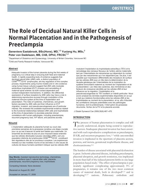
<strong>The</strong> <strong>Role</strong> <strong>of</strong> <strong>Decidual</strong> <strong>Natural</strong> <strong>Killer</strong> <strong>Cells</strong> <strong>in</strong><br />
<strong>Normal</strong> Placentation and <strong>in</strong> the Pathogenesis <strong>of</strong><br />
Preeclampsia<br />
Genevieve Eastabrook, BSc(Hons), MD, 1,2 Yuxiang Hu, MSc, 2<br />
Peter von Dadelszen, MB, ChB, DPhil, FRCSC 1,2<br />
1 Department <strong>of</strong> Obstetrics and Gynaecology, University <strong>of</strong> British Columbia, Vancouver BC<br />
2 Child and Family Research Institute, Vancouver BC<br />
Abstract<br />
Adequate <strong>in</strong>vasion <strong>of</strong> the human placenta dur<strong>in</strong>g the first weeks <strong>of</strong><br />
pregnancy is a critical step <strong>in</strong> ensur<strong>in</strong>g both fetal and maternal<br />
health. A rapidly expand<strong>in</strong>g body <strong>of</strong> evidence suggests that<br />
decidual natural killer (dNK) cells, a dist<strong>in</strong>ct population <strong>of</strong><br />
CD56 bright CD16 - lymphocytes, are key regulators <strong>of</strong> this complex<br />
process. Experiments us<strong>in</strong>g mur<strong>in</strong>e models and <strong>in</strong> vitro evidence<br />
us<strong>in</strong>g human tissue cultures suggest that dNK cells modulate<br />
extravillous trophoblast (EVT) <strong>in</strong>vasion and remodell<strong>in</strong>g <strong>of</strong><br />
maternal spiral arteries via both contact-dependent and<br />
contact-<strong>in</strong>dependent mechanisms. In addition, the differential<br />
expression <strong>of</strong> surface receptors by dNK cells may have a role <strong>in</strong><br />
determ<strong>in</strong><strong>in</strong>g reproductive success through modulation <strong>of</strong> the<br />
maternal immune system at the time <strong>of</strong> implantation and<br />
placentation. <strong>The</strong> roles <strong>of</strong> cytok<strong>in</strong>es, chemok<strong>in</strong>es, and growth<br />
factors secreted by dNK cells and their <strong>in</strong>fluence on EVT<br />
migration, <strong>in</strong>vasion, and pseudovasculogenesis are <strong>of</strong> particular<br />
<strong>in</strong>terest. We reviewed the available experimental evidence related<br />
to the functional relationships between dNK cells and trophoblasts<br />
at the time <strong>of</strong> placentation to elucidate potential cl<strong>in</strong>ical<br />
correlations with human pathologies, <strong>in</strong>clud<strong>in</strong>g preeclampsia,<br />
recurrent pregnancy loss, IVF failure, and placenta accreta.<br />
Résumé<br />
L’implantation adéquate du placenta huma<strong>in</strong> au cours des<br />
premières sema<strong>in</strong>es de la grossesse constitue une étape cruciale<br />
pour ce qui est d’assurer la santé tant fœtale que maternelle. Un<br />
ensemble de données de plus en plus imposant laisse entendre<br />
que les cellules déciduales tueuses naturelles (dNK), soit une<br />
population dist<strong>in</strong>cte de lymphocytes CD56 claire CD16 - , constituent<br />
des régulateurs clés de ce processus complexe. Les expériences<br />
menées sur des modèles mur<strong>in</strong>s et les données <strong>in</strong> vitro issues de<br />
cultures de tissus huma<strong>in</strong>s semblent <strong>in</strong>diquer que les cellules dNK<br />
Key Words: Assisted reproductive technology, chemok<strong>in</strong>es,<br />
decidua, <strong>in</strong>terferon type II, natural killer cells, placenta,<br />
preeclampsia, recurrent pregnancy loss, trophoblasts<br />
Compet<strong>in</strong>g Interests: None declared.<br />
Received on August 29, 2007<br />
Accepted on January 17, 2008<br />
modulent l’implantation du trophoblaste extravilleux (TEV) et le<br />
remodelage des rameaux flexueux de l’artère utér<strong>in</strong>e maternelle<br />
tant par l’<strong>in</strong>termédiaire de mécanismes qui dépendent du contact<br />
que par des mécanismes qui n’en dépendent pas. De plus, il est<br />
possible que l’expression différentielle des récepteurs de surface<br />
par les cellules dNK joue un rôle dans la déterm<strong>in</strong>ation de la<br />
réussite génésique par l’<strong>in</strong>termédiaire de la modulation du<br />
système immunitaire maternel au moment de l’implantation et de<br />
la placentation. Les rôles des cytok<strong>in</strong>es, des chimiok<strong>in</strong>es et des<br />
facteurs de croissance sécrétés par les cellules dNK et leur<br />
<strong>in</strong>fluence sur la migration, l’implantation et la<br />
pseudovasculogenèse du TEV revêtent un <strong>in</strong>térêt particulier. Nous<br />
avons analysé les données expérimentales disponibles en ce qui<br />
a trait aux relations fonctionnelles entre les cellules dNK et les<br />
trophoblastes au moment de la placentation, et ce, af<strong>in</strong> d’élucider<br />
les corrélations cl<strong>in</strong>iques potentielles avec les pathologies<br />
huma<strong>in</strong>es, dont la prééclampsie, l’<strong>in</strong>terruption de grossesse<br />
récurrente, l’échec de la FIV et le placenta accreta.<br />
J Obstet Gynaecol Can 2008;30(6):467–476<br />
INTRODUCTION<br />
<strong>The</strong> process <strong>of</strong> human placentation is complex and still<br />
poorly understood, despite be<strong>in</strong>g central to reproductive<br />
success. Inadequate placental <strong>in</strong>vasion has been associated<br />
with such reproductive complications as preeclampsia,<br />
IUGR, and recurrent pregnancy loss, 1–3 while uncontrolled<br />
<strong>in</strong>vasion is implicated <strong>in</strong> the spectrum <strong>of</strong> placenta accreta,<br />
percreta, and <strong>in</strong>creta, gestational trophoblastic disease, and<br />
choriocarc<strong>in</strong>oma. 4–6<br />
<strong>The</strong> burden <strong>of</strong> disease associated with placental dysfunction<br />
is great. Ischemic placental disease, <strong>in</strong>clud<strong>in</strong>g preeclampsia,<br />
placental abruption, and growth restriction, was implicated<br />
<strong>in</strong> more than half <strong>of</strong> the <strong>in</strong>duced preterm births <strong>in</strong> one large<br />
population-based study. 7 <strong>The</strong> effect <strong>of</strong> preeclampsia is particularly<br />
significant: complicat<strong>in</strong>g approximately 5% <strong>of</strong><br />
pregnancies, it rema<strong>in</strong>s one <strong>of</strong> the two most common<br />
causes <strong>of</strong> maternal death, both <strong>in</strong> developed 8–13 and <strong>in</strong><br />
develop<strong>in</strong>g 14–17 nations. Pulmonary embolism and<br />
JUNE JOGC JUIN 2008 467
OBSTETRICS<br />
preeclampsia are the most common direct causes <strong>of</strong> maternal<br />
mortality <strong>in</strong> Canada; preeclampsia contributed to 20%<br />
<strong>of</strong> the maternal deaths reported between 1997 and 2000. 18<br />
Preeclampsia also leads to <strong>in</strong>creased risk <strong>of</strong> per<strong>in</strong>atal morbidity<br />
and mortality, particularly due to prematurity. 19,20 In<br />
addition to preterm delivery for fetal <strong>in</strong>dications, many otherwise<br />
healthy fetuses are delivered before term because <strong>of</strong><br />
deteriorat<strong>in</strong>g maternal health associated with preeclampsia. 7<br />
Our knowledge <strong>of</strong> the cellular and molecular processes <strong>of</strong><br />
human trophoblast <strong>in</strong>vasion is based ma<strong>in</strong>ly on <strong>in</strong> vitro<br />
studies and animal models; there is considerable evidence<br />
that dNK cells are crucial <strong>in</strong> successful placentation. 21–25<br />
<strong>The</strong>y are key mediators <strong>of</strong> maternal immune system <strong>in</strong>teractions<br />
with fetal cells. <strong>The</strong>y are also <strong>in</strong>volved <strong>in</strong> modulat<strong>in</strong>g<br />
EVT <strong>in</strong>vasion and the remodell<strong>in</strong>g <strong>of</strong> maternal spiral arteries.<br />
22,25,26 <strong>The</strong>y express a wide range <strong>of</strong> surface receptors<br />
and signall<strong>in</strong>g molecules, <strong>in</strong>clud<strong>in</strong>g cytok<strong>in</strong>es, chemok<strong>in</strong>es,<br />
and growth factors, 27–29 and their functions <strong>in</strong> modulat<strong>in</strong>g<br />
EVT migration, <strong>in</strong>vasion, and alteration from epithelial to<br />
endothelial phenotype are only beg<strong>in</strong>n<strong>in</strong>g to be<br />
revealed. 27,30<br />
METHODS<br />
Medl<strong>in</strong>e and Google Scholar searches were carried out<br />
us<strong>in</strong>g the terms assisted reproductive technology,<br />
chemok<strong>in</strong>es, cytok<strong>in</strong>es, decidua, <strong>in</strong>terferon gamma, natural<br />
killer cell, placenta, placenta accreta, preeclampsia,<br />
ABBREVIATIONS<br />
dNK decidual natural killer cells<br />
EVT extravillous trophoblast<br />
HLA human leukocyte antigen<br />
IFN- <strong>in</strong>terferon-gamma<br />
IL-8 <strong>in</strong>terleuk<strong>in</strong>-8<br />
IP-10 <strong>in</strong>terferon-<strong>in</strong>ducible prote<strong>in</strong>-10<br />
IUGR <strong>in</strong>trauter<strong>in</strong>e growth restriction<br />
IVF <strong>in</strong> vitro fertilization<br />
KIR killer immunoglobul<strong>in</strong>-like receptor<br />
LAK lymphok<strong>in</strong>e activated killer cells<br />
MIG monok<strong>in</strong>e <strong>in</strong>ducible by gamma <strong>in</strong>terferon<br />
pbNK peripheral blood NK cells<br />
PlGF placental growth factor<br />
RANTES regulated on activation, normal T-cell expressed and<br />
secreted<br />
RPL recurrent pregnancy loss<br />
sEng soluble endogl<strong>in</strong><br />
sFlt-1 soluble fms-like tyros<strong>in</strong>e k<strong>in</strong>ase-1<br />
VEGF vascular-endothelial growth factor<br />
trophoblast, and uterus. Wherever possible, primary<br />
research papers were cited <strong>in</strong> place <strong>of</strong> topic reviews. Articles<br />
were selected on the basis <strong>of</strong> the quality <strong>of</strong> their scientific<br />
method and relevance to human placentation.<br />
DISCUSSION<br />
<strong>Decidual</strong> <strong>Natural</strong> <strong>Killer</strong> <strong>Cells</strong><br />
<strong>Decidual</strong> natural killer cells are a unique population <strong>of</strong><br />
CD56 bright CD16 - cells, phenotypically different from the<br />
ma<strong>in</strong> population <strong>of</strong> CD56 dim CD16 bright pbNK cells. 31,32<br />
<strong>Decidual</strong> natural killer cells are functionally dist<strong>in</strong>ct from<br />
pbNK (they have lower cytolytic activity), and they express<br />
a different cytok<strong>in</strong>e repertoire. <strong>The</strong>ir cyotoxicity, proliferation,<br />
and cytok<strong>in</strong>e production are enhanced <strong>in</strong> vitro by<br />
<strong>in</strong>terleuk<strong>in</strong>-2 (IL-2) and IL-15. 33<br />
Histologically, dNK are identifiable as small granular cells<br />
found <strong>in</strong> proliferative endometrium. <strong>The</strong>y <strong>in</strong>crease abundantly<br />
dur<strong>in</strong>g the secretory phase <strong>of</strong> the menstrual cycle,<br />
<strong>in</strong>dicat<strong>in</strong>g that they may have a role <strong>in</strong> implantation. 34 Furthermore,<br />
dNK may have a role <strong>in</strong> endometrial differentiation<br />
and breakdown and may aid the <strong>in</strong>itiation <strong>of</strong> menstruation<br />
via apoptosis. 35 <strong>The</strong> appearance <strong>of</strong> dNK is one <strong>of</strong> the<br />
histological hallmarks <strong>of</strong> decidualization. 36 <strong>The</strong>re is evidence<br />
that whenever or wherever decidual tissue is formed,<br />
dNK are <strong>in</strong>variably present, even <strong>in</strong> foci <strong>of</strong> endometriosis<br />
remote from the uterus. 35 Interest<strong>in</strong>gly, dNK are found <strong>in</strong><br />
higher numbers <strong>in</strong> the decidual tissue <strong>of</strong> women experienc<strong>in</strong>g<br />
breakthrough bleed<strong>in</strong>g while us<strong>in</strong>g the<br />
levonorgestrel-releas<strong>in</strong>g <strong>in</strong>trauter<strong>in</strong>e system (Mirena) than<br />
<strong>in</strong> amenorrheic users. 37<br />
dNK and EVT Interactions at the Maternal-Fetal<br />
Interface<br />
<strong>The</strong> close temporal-spatial relationship between dNK and<br />
EVT suggests that dNK regulate implantation and uter<strong>in</strong>e<br />
artery remodell<strong>in</strong>g through receptor-ligand <strong>in</strong>teractions.<br />
Numerous dNK are found close to <strong>in</strong>filtrat<strong>in</strong>g EVT dur<strong>in</strong>g<br />
implantation 34,38 (Figure 1). 39 EVT lack human leukocyte<br />
antigen-A (HLA-A) and HLA-B expression, which may<br />
prevent <strong>in</strong>teraction with maternal cytotoxic T-cells. 40 EVT<br />
express HLA-C which, through <strong>in</strong>teraction with members<br />
<strong>of</strong> the KIR gene family expressed on NK cells, 33,41,42 <strong>in</strong>fluences<br />
placental development. 41 This process may be mediated<br />
through the action <strong>of</strong> the dNK-produced cytok<strong>in</strong>e,<br />
IFN- 40<br />
EVT also express two non-classical MHC antigens, HLA-E<br />
and HLA-G. 43,44 dNK express KIRs which b<strong>in</strong>d to HLA-E<br />
and HLA-G, thus <strong>in</strong>hibit<strong>in</strong>g NK cytotoxicity and <strong>in</strong>creas<strong>in</strong>g<br />
expression <strong>of</strong> pro-<strong>in</strong>flammatory and pro-angiogenic<br />
cytok<strong>in</strong>es, respectively. 43,45,46 Expression <strong>of</strong> HLA-G may<br />
protect EVT aga<strong>in</strong>st dNK lysis. 47 Usually, EVT <strong>in</strong>vad<strong>in</strong>g<br />
468 JUNE JOGC JUIN 2008
<strong>The</strong> <strong>Role</strong> <strong>of</strong> <strong>Decidual</strong> <strong>Natural</strong> <strong>Killer</strong> <strong>Cells</strong> <strong>in</strong> <strong>Normal</strong> Placentation and <strong>in</strong> the Pathogenesis <strong>of</strong> Preeclampsia<br />
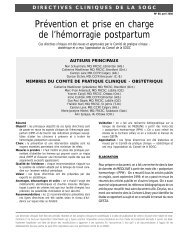
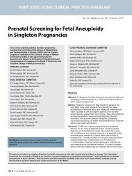
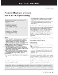
Figure 1. Insertion <strong>of</strong> the Human Placenta. <strong>The</strong> close temporal-spatial relationship between dNK and<br />
EVT suggests that dNK regulate implantation and uter<strong>in</strong>e artery remodell<strong>in</strong>g through receptor-ligand<br />
<strong>in</strong>teractions. Numerous dNK are found close to <strong>in</strong>filtrat<strong>in</strong>g EVT dur<strong>in</strong>g implantation.<br />
the decidua express and upregulate HLA-G, 48 but a lack <strong>of</strong><br />
HLA-G expression has been noted <strong>in</strong> EVT from the placentas<br />
<strong>of</strong> pre-eclamptic women. This suggests that failed<br />
EVT <strong>in</strong>vasion may precede the abnormal placentation <strong>of</strong><br />
preeclampsia. 49–51 Recent <strong>in</strong> vitro and <strong>in</strong> vivo studies demonstrate<br />
the anti-angiogenic and pro-apoptotic actions <strong>of</strong><br />
the soluble form <strong>of</strong> HLA-G, secreted by EVT. 52,53 Thus, <strong>in</strong><br />
addition to its immunological functions, HLA-G may<br />
directly regulate spiral artery remodell<strong>in</strong>g. 53<br />
<strong>The</strong> actions <strong>of</strong> activat<strong>in</strong>g and <strong>in</strong>hibitory receptors expressed<br />
by dNK may <strong>in</strong>fluence human reproductive success.<br />
Varla-Leftherioti et al. 54 found that women with recurrent<br />
spontaneous abortions have a limited range <strong>of</strong> KIRs (compared<br />
with normal controls), and that the majority <strong>of</strong> these<br />
women lacked KIRs expressed by their partners. <strong>The</strong><br />
authors concluded that <strong>in</strong> this population, miscarriages may<br />
occur as dNK lack<strong>in</strong>g the appropriate <strong>in</strong>hibitory KIRs recognize<br />
trophoblastic HLA class I molecules. 54 In addition,<br />
specific comb<strong>in</strong>ations <strong>of</strong> maternal dNK, KIR, and fetal<br />
HLA-C genes appear to alter the balance between risk <strong>of</strong><br />
preeclampsia and reproductive success. 41 An imbalance <strong>in</strong><br />
KIR expression may also have a role <strong>in</strong> implantation failure<br />
follow<strong>in</strong>g IVF. 55,56<br />
<strong>The</strong> differentiation <strong>of</strong> trophoblasts to the <strong>in</strong>vasive<br />
extravillous phenotype is seen as a crucial part <strong>of</strong> placental<br />
angiogenesis. 57 This process <strong>in</strong>volves the switch<strong>in</strong>g <strong>of</strong> 64<br />
to V3 <strong>in</strong>tegr<strong>in</strong>s (adhesion molecules <strong>in</strong>volved <strong>in</strong><br />
cell-ECM <strong>in</strong>teractions) and the downregulation <strong>of</strong><br />
E-cadher<strong>in</strong> (adhesion molecule <strong>in</strong>volved <strong>in</strong> cell-cell <strong>in</strong>teractions).<br />
58–60 As a result, trophoblasts evolve from an epithelial<br />
to an endothelial phenotype, a process described as<br />
pseudovasculogenesis. 58,61–63 Pseudovasculogenesis is one<br />
<strong>of</strong> the key processes that becomes impaired <strong>in</strong> the placentas<br />
<strong>of</strong> women with preeclampsia. 62<br />
<strong>Decidual</strong> NK cell-derived cytok<strong>in</strong>es, growth factors,<br />
and other soluble products modulate EVT cell adhesion<br />
molecule expression dur<strong>in</strong>g EVT <strong>in</strong>vasion. 30 For example, <strong>in</strong><br />
<strong>in</strong> vitro experiments, E-cadher<strong>in</strong> mRNA and prote<strong>in</strong><br />
expression levels <strong>in</strong>crease <strong>in</strong> EVTs follow<strong>in</strong>g exposure to<br />
both dNK conta<strong>in</strong>ed with<strong>in</strong> hollow fibres and to dNK conditioned<br />
medium 30 ; both are contact-<strong>in</strong>dependent mechanisms.<br />
E-cadher<strong>in</strong> expression may 61,62 or may not 59 be<br />
ma<strong>in</strong>ta<strong>in</strong>ed <strong>in</strong> EVT that exhibit the shallow <strong>in</strong>vasion<br />
described <strong>in</strong> placental bed biopsies from women with<br />
preeclampsia.<br />
<strong>The</strong> <strong>Role</strong> <strong>of</strong> Cytok<strong>in</strong>es <strong>in</strong> dNK-EVT Interactions<br />
<strong>Decidual</strong> natural killer cells produce numerous cytok<strong>in</strong>es<br />
implicated <strong>in</strong> regulat<strong>in</strong>g trophoblast <strong>in</strong>vasion, <strong>in</strong>clud<strong>in</strong>g<br />
granulocyte colony-stimulat<strong>in</strong>g factor (G-CSF), granulocyte<br />
macrophage CSF (GM-CSF), macrophage CSF (M-CSF),<br />
leukaemia <strong>in</strong>hibitory factor (LIF), tumour necrosis factor<br />
alpha (TNF-), and IFN-. 28<br />
JUNE JOGC JUIN 2008 469
OBSTETRICS<br />
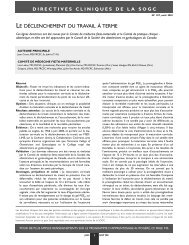
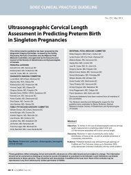
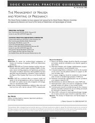
Figure 2. dNK-EVT Interactions at the Maternal-Fetal Interface.<strong>The</strong> complex <strong>in</strong>terplay between dNK<br />
cells and EVT at the maternal-fetal <strong>in</strong>terface is mediated by cytok<strong>in</strong>es, chemok<strong>in</strong>es, and growth<br />
factors. dNK-produced IFN-g may limit EVT migration alone or <strong>in</strong> concert with IFN-<strong>in</strong>ducible<br />
chemok<strong>in</strong>es such as MIG and IP-10. IL-8 and IP-10 may both promote EVT migration while enhanc<strong>in</strong>g<br />
(IL-8) and restrict<strong>in</strong>g (IP-10) angiogenesis. RANTES may contribute to EVT migration and differentiation<br />
to an <strong>in</strong>vasive phenotype.<br />
<strong>in</strong>vasion<br />
<strong>in</strong>to<br />
decidua<br />
endovascular <strong>in</strong>vasion<br />
& angiogenesis<br />
EVT<br />
dNK<br />
RANTES<br />
limitation <strong>of</strong> EVT<br />
<strong>in</strong>vasion<br />
IFN-<br />
promotion <strong>of</strong> EVT<br />
angiogenesis<br />
IL-8<br />
endothelium<br />
IP-10,<br />
MIG<br />
restriction <strong>of</strong> EVT<br />
angiogenesis<br />
IFN- is <strong>of</strong> particular <strong>in</strong>terest because knock-out mice lack<strong>in</strong>g<br />
genes for IFN- production or for IFN- receptors<br />
exhibit abnormal placentation, and consequently have<br />
adverse pregnancy outcomes. 26,64 Conversely, mice lack<strong>in</strong>g<br />
cells <strong>of</strong> the NK l<strong>in</strong>eage have histologically abnormal pregnancies,<br />
but the outcomes <strong>of</strong> their pregnancies are more<br />
variable. 65,66 In mouse models, IFN- modulates uter<strong>in</strong>e<br />
vascular modification, decidual <strong>in</strong>tegrity, 26 and dNK maturation.<br />
22,26 In a novel collagen (two-dimensional) model <strong>of</strong><br />
placentation developed <strong>in</strong> our laboratory, the presence <strong>of</strong><br />
dNK resulted <strong>in</strong> contact-<strong>in</strong>dependent <strong>in</strong>hibition <strong>of</strong> normal<br />
cytotrophoblast migration. 30 This was associated with<br />
changes <strong>in</strong> the cytotrophoblast expression <strong>of</strong><br />
metalloproteases-2 and 9 (proteolytic enzymes associated<br />
with ECM degradation), and plasm<strong>in</strong>ogen activator<br />
<strong>in</strong>hibitor-1 (an <strong>in</strong>hibitor <strong>of</strong> fibr<strong>in</strong>olysis). In contrast, dNK<br />
did not affect EVT proliferation, apoptosis, or cell column<br />
formation. dNK effects were partially reversed by neutraliz<strong>in</strong>g<br />
antibodies aga<strong>in</strong>st IFN-. Thus, this model provides<br />
evidence for the role <strong>of</strong> dNK <strong>in</strong> directly modulat<strong>in</strong>g EVT<br />
differentiation dur<strong>in</strong>g column formation and migration<br />
from anchor<strong>in</strong>g villi. 30<br />
Placental Angiogenesis: A <strong>Role</strong> for dNK?<br />
Although it is known that dNK affect trophoblast migration<br />
and <strong>in</strong>vasion both <strong>in</strong> vivo and <strong>in</strong> vitro, the mechanisms<br />
beh<strong>in</strong>d this effect are <strong>in</strong>completely understood. In particular,<br />
the role <strong>of</strong> dNK <strong>in</strong> mediat<strong>in</strong>g angiogenesis has not been<br />
explored to any great extent. <strong>Decidual</strong> NK express<br />
angiogenic growth factors throughout the menstrual cycle,<br />
which suggests a role <strong>in</strong> endometrial angiogenesis and<br />
regeneration. 29 <strong>Decidual</strong> NK express high levels <strong>of</strong><br />
VEGF-C, PlGF, and angiotens<strong>in</strong> 2 dur<strong>in</strong>g the secretory<br />
phase <strong>of</strong> the menstrual cycle. 29 VEGF-C acts via VEGF-R2<br />
and R3 receptors, which are expressed solely on dNK<br />
with<strong>in</strong> the endometrium. 29 Although abnormal expression<br />
<strong>of</strong> VEGF and its receptors has been reported <strong>in</strong> cases <strong>of</strong><br />
placenta accreta, 67 the role <strong>of</strong> dNK cells <strong>in</strong> the development<br />
470 JUNE JOGC JUIN 2008
<strong>The</strong> <strong>Role</strong> <strong>of</strong> <strong>Decidual</strong> <strong>Natural</strong> <strong>Killer</strong> <strong>Cells</strong> <strong>in</strong> <strong>Normal</strong> Placentation and <strong>in</strong> the Pathogenesis <strong>of</strong> Preeclampsia<br />
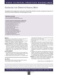
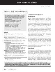
Figure 3. <strong>The</strong> Putative <strong>Role</strong>s <strong>of</strong> dNK <strong>Cells</strong> <strong>in</strong> EVT Migration, Invasion, and Placental Angiogenesis.<br />
Experimental evidence suggests that dNK-produced chemok<strong>in</strong>es, cytok<strong>in</strong>es, and growth factors may<br />
contribute to EVT migration, <strong>in</strong>vasion, phenotypic switch, and ultimately, angiogenesis.<br />
<br />
<strong>of</strong> this disorder has not been <strong>in</strong>vestigated <strong>in</strong> any studies<br />
published to date.<br />
Recently it has been demonstrated that dNK, but not<br />
pbNK subsets, regulate trophoblast <strong>in</strong>vasion both <strong>in</strong> vitro<br />
and <strong>in</strong> vivo through the chemok<strong>in</strong>es IL-8 and IP-10. 27<br />
<strong>The</strong>se studies provide evidence that dNK can secrete an<br />
array <strong>of</strong> angiogenic factors and that EVT express the correspond<strong>in</strong>g<br />
receptors. This suggests that dNK may <strong>in</strong>duce<br />
vascular growth dur<strong>in</strong>g placentation (Figure 2). In the<br />
development <strong>of</strong> malignancies, IL-8 is pro-angiogenic, while<br />
IP-10 is potently angiostatic. 68,69 An appropriate balance <strong>of</strong><br />
pro- and anti-angiogenic chemok<strong>in</strong>es produced by dNK<br />
may help to regulate maternal spiral artery remodell<strong>in</strong>g and<br />
placental angiogenesis (Figure 3); this warrants further<br />
<strong>in</strong>vestigation.<br />
LAK cells mediate angiogenesis <strong>in</strong> vitro. 70 This subset <strong>of</strong><br />
peripheral blood mononuclear cells (which <strong>in</strong>clude NK<br />
cells) is activated us<strong>in</strong>g IL-2, a cytok<strong>in</strong>e elevated <strong>in</strong> the sera<br />
<strong>of</strong> women with preeclampsia. 71 EVT angiogenesis is<br />
reduced by LAK cells <strong>in</strong> vitro. 70 This effect is modulated<br />
through EVT-derived sFlt-1, a truncated and <strong>in</strong>active<br />
receptor for both VEGF and PlGF. 72 Serum levels <strong>of</strong> sFlt-1<br />
are elevated <strong>in</strong> women with preeclampsia. 73 Shedd<strong>in</strong>g <strong>of</strong><br />
this anti-angiogenic prote<strong>in</strong> from the placenta <strong>in</strong>to the<br />
maternal circulation may contribute to the systemic endothelial<br />
dysfunction 73–75 which is a hallmark <strong>of</strong> preeclampsia<br />
(Figure 4). 76 Another anti-angiogenic prote<strong>in</strong> <strong>of</strong> <strong>in</strong>terest <strong>in</strong><br />
women with preeclampsia is sEng, thought to impair<br />
TGF-1 b<strong>in</strong>d<strong>in</strong>g to cell surface receptors and to decrease<br />
endothelial nitric oxide signall<strong>in</strong>g. 77 Recent studies <strong>in</strong>dicate<br />
that levels <strong>of</strong> sEng (produced by the placenta) are elevated<br />
<strong>in</strong> the serum <strong>of</strong> women with preeclampsia, <strong>in</strong>creas<strong>in</strong>g with<br />
disease severity and fall<strong>in</strong>g after delivery <strong>of</strong> the placenta 78,79 ;<br />
it has also been implicated <strong>in</strong> the pathophysiology <strong>of</strong><br />
HELLP syndrome. 57<br />
dNK-Derived Chemok<strong>in</strong>es <strong>in</strong> EVT Migration and<br />
Differentiation<br />
Chemok<strong>in</strong>es, which were orig<strong>in</strong>ally identified as critical regulators<br />
<strong>of</strong> leukocyte migration, 68 are abundant <strong>in</strong><br />
endometrial epithelial and decidual cells at the time <strong>of</strong><br />
implantation and trophoblast <strong>in</strong>vasion. 80–83 <strong>The</strong> chemok<strong>in</strong>e<br />
repertoire <strong>of</strong> dNK <strong>in</strong> particular is diverse. For example,<br />
dNK express IL-8 mRNA, 27,28 and produce large amounts<br />
<strong>of</strong> this chemok<strong>in</strong>e without stimulation. 28 dNK also produce<br />
JUNE JOGC JUIN 2008 471
OBSTETRICS<br />
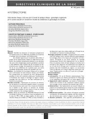
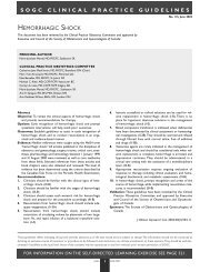
Figure 4. <strong>The</strong> Pathogenesis <strong>of</strong> Preeclampsia. <strong>The</strong> maternal syndrome <strong>of</strong> preeclampsia (multiple<br />
organ dysfunction) is the result <strong>of</strong> an <strong>in</strong>terplay between numerous maternal and fetal factors,<br />
beg<strong>in</strong>n<strong>in</strong>g with impaired placental <strong>in</strong>vasion.<br />
cytotrophoblast <strong>in</strong>vasion<br />
immunological factors<br />
(e.g. dNK cell dysregulation,<br />
donor gametes)<br />
poor placentation<br />
uteroplacental mismatch<br />
acute atherosis<br />
thrombophilia<br />
multiple pregnancy<br />
fetal macrosomia<br />
placental <strong>in</strong>fection<br />
PBLs<br />
cytok<strong>in</strong>es<br />
PGs<br />
ROS<br />
endothelial cell activation<br />
maternal syndrome<br />
INTERVILLOUS<br />
placental debris<br />
SOUP<br />
(e.g. anti-angiogenic<br />
factors sFlt-1 & sEng)<br />
ARDS<br />
cardiomyopathy<br />
eclampsia/<br />
stroke<br />
hypertension<br />
glomerular<br />
endotheliosis/<br />
prote<strong>in</strong>uria/<br />
ATN<br />
liver damage/<br />
hematoma/<br />
rupture<br />
microangiopathic<br />
hemolysis/<br />
thrombocytopenia/<br />
DIC<br />
edema<br />
PBLs: peripheral blood lymphocytes; PGs: prostagland<strong>in</strong>s; ROS: reactive oxygen species; ARDS: acute respiratory distress syndrome;<br />
ATN: acute tubular necrosis; DIC: dissem<strong>in</strong>ated <strong>in</strong>travascular coagulation.<br />
a large quantity <strong>of</strong> RANTES, <strong>in</strong>terferon-<strong>in</strong>ducible<br />
prote<strong>in</strong>-10 (IP-10), and monok<strong>in</strong>e <strong>in</strong>ducible by gamma<br />
<strong>in</strong>terferon (MIG). 27 CC-chemok<strong>in</strong>e receptor 1 (CCR1) ligands<br />
(chemok<strong>in</strong>es) <strong>in</strong>clud<strong>in</strong>g RANTES and macrophage<br />
<strong>in</strong>flammatory prote<strong>in</strong>-1 (MIP-1) have been identified <strong>in</strong><br />
decidual tissues. <strong>The</strong>se chemok<strong>in</strong>es promote migration <strong>of</strong><br />
EVT isolated from explant cultures <strong>in</strong> vitro 84 (Figure 2).<br />
<strong>The</strong> expression <strong>of</strong> chemok<strong>in</strong>e receptors appears to differ<br />
temporally, spatially, and by trophoblast cell type. For<br />
example, the chemok<strong>in</strong>e receptors CX3CR1 and CCR1<br />
have been immunolocalised to endovascular EVT but not<br />
to <strong>in</strong>vad<strong>in</strong>g <strong>in</strong>terstitial EVT (iEVT). Conversely, CCR3 has<br />
been localised to <strong>in</strong>vad<strong>in</strong>g iEVT and to syncytial<br />
microvilli. 85 In addition, trophoblasts acquire CCR1 as they<br />
differentiate to an <strong>in</strong>vasive phenotype at villus-anchor<strong>in</strong>g<br />
sites. 86 This suggests a role for the chemok<strong>in</strong>e-CCR1 system<br />
<strong>in</strong> the <strong>in</strong>itial step <strong>of</strong> trophoblastic <strong>in</strong>vasion toward maternal<br />
tissue.<br />
dNK <strong>Cells</strong> <strong>in</strong> Recurrent Pregnancy Loss<br />
and IVF Failure<br />
Several studies <strong>of</strong> women experienc<strong>in</strong>g idiopathic recurrent<br />
pregnancy loss have found abnormalities <strong>in</strong> the dNK cell<br />
populations <strong>in</strong> the peri-implantation endometrium <strong>of</strong> these<br />
patients. Decreased populations <strong>of</strong> CD56 bright CDI6 - dNK<br />
cells and <strong>in</strong>creased numbers <strong>of</strong> CD56 dim CD 16 + dNK cells<br />
have been reported. 87–89 However, the results <strong>of</strong> functional<br />
studies have been less consistent. For example, while one<br />
study reported decreased dNK cytotoxicity <strong>in</strong> vitro dur<strong>in</strong>g<br />
normal pregnancies <strong>in</strong> comparison with anembryonic gestations<br />
or <strong>in</strong> women with RPL, 90 another study reported<br />
decreased dNK cytotoxicity <strong>in</strong> cells isolated from women<br />
with RPL. 91 At present, there does not appear to be any<br />
prognostic value <strong>in</strong> predict<strong>in</strong>g the outcomes <strong>of</strong> future pregnancies<br />
by measur<strong>in</strong>g dNK from endometrial biopsies <strong>of</strong><br />
RPL patients. Although the numbers <strong>of</strong> dNK cells are<br />
higher <strong>in</strong> women with a history <strong>of</strong> RPL than <strong>in</strong> normal controls,<br />
the significance <strong>of</strong> this f<strong>in</strong>d<strong>in</strong>g is uncerta<strong>in</strong>. 92<br />
In patients experienc<strong>in</strong>g repeated implantation failure follow<strong>in</strong>g<br />
IVF, abnormalities <strong>in</strong> dNK cell CD56 and CD16<br />
expression have been correlated with uter<strong>in</strong>e artery<br />
Doppler abnormalities and altered expression <strong>of</strong> the<br />
cytok<strong>in</strong>es IL-12 and IL-18. 93,94 However, this f<strong>in</strong>d<strong>in</strong>g has<br />
not been consistent <strong>in</strong> all studies reported to date. 95<br />
NK Cell Dysregulation <strong>in</strong> Preeclampsia<br />
It has been proposed previously that an imbalance <strong>of</strong><br />
TH1/TH2 immunity may be <strong>in</strong>volved <strong>in</strong> poor placentation<br />
472 JUNE JOGC JUIN 2008
<strong>The</strong> <strong>Role</strong> <strong>of</strong> <strong>Decidual</strong> <strong>Natural</strong> <strong>Killer</strong> <strong>Cells</strong> <strong>in</strong> <strong>Normal</strong> Placentation and <strong>in</strong> the Pathogenesis <strong>of</strong> Preeclampsia<br />
and consequently <strong>in</strong> preeclampsia. 96,97 However, this putative<br />
TH1/TH2 imbalance is likely an oversimplification <strong>of</strong><br />
more complex immune processes that <strong>in</strong>volve not only<br />
these subsets <strong>of</strong> T cells but also NK cells (Figure 4). NK<br />
cells may mediate changes <strong>in</strong> systemic type 1 and type 2<br />
immunities, both <strong>in</strong> normal pregnancy and <strong>in</strong> women with<br />
preeclampsia. 98 Greater numbers <strong>of</strong> CD56 dim and CD94 +<br />
cells have been found <strong>in</strong> the decidua <strong>of</strong> women with<br />
preeclampsia than <strong>in</strong> normal controls at term, 99 <strong>in</strong>dicat<strong>in</strong>g a<br />
role for altered expression <strong>of</strong> dNK cell receptors <strong>in</strong> the<br />
development <strong>of</strong> this disorder. In addition, villous trophoblasts<br />
<strong>in</strong> the placentas <strong>of</strong> women with preeclampsia express<br />
significantly less IL-12 than normal controls. 99 In contrast,<br />
women with preeclampsia have been found to have significantly<br />
elevated serum levels <strong>of</strong> IL-12 and IL-15. 99 Together,<br />
these f<strong>in</strong>d<strong>in</strong>gs po<strong>in</strong>t to a role for NK dysregulation <strong>in</strong> the<br />
pathogenesis <strong>of</strong> preeclampsia. However, because the<br />
decidua and trophoblasts were exam<strong>in</strong>ed at term, it is still<br />
unknown whether NK dysregulation is a cause or an effect<br />
<strong>of</strong> preeclampsia <strong>in</strong> these cases.<br />
Future Directions<br />
Cont<strong>in</strong>ued <strong>in</strong>vestigation <strong>of</strong> the role <strong>of</strong> dNK <strong>in</strong> trophoblast<br />
<strong>in</strong>vasion and spiral artery remodell<strong>in</strong>g will provide <strong>in</strong>sights<br />
<strong>in</strong>to the mechanisms <strong>of</strong> both normal and pathological<br />
placentation. With<strong>in</strong> general obstetrics and maternal-fetal<br />
medic<strong>in</strong>e, a more thorough understand<strong>in</strong>g <strong>of</strong> placentation is<br />
critical <strong>in</strong> understand<strong>in</strong>g preeclampsia, IUGR, <strong>in</strong>trauter<strong>in</strong>e<br />
fetal demise, and other disorders associated with abnormal<br />
placentation. Evidence <strong>of</strong> altered dNK KIR repertoires 54<br />
and reduced expression <strong>of</strong> CD56 bright CD16 - dNK cells <strong>in</strong><br />
women with recurrent pregnancy loss 87–89 and recurrent<br />
post-embryo transfer IVF failure 93,94 has implications<br />
with<strong>in</strong> the discipl<strong>in</strong>es <strong>of</strong> reproductive endocr<strong>in</strong>ology, <strong>in</strong>fertility,<br />
and assisted reproductive technologies. As the rates <strong>of</strong><br />
Caesarean section cont<strong>in</strong>ue to rise, 100,101 there is likely to be<br />
a concomitant <strong>in</strong>crease <strong>in</strong> the <strong>in</strong>cidence <strong>of</strong> placenta accreta,<br />
<strong>in</strong>creta, and percreta 102 ; exam<strong>in</strong>ation <strong>of</strong> the cellular and<br />
immunological mechanisms underly<strong>in</strong>g uncontrolled <strong>in</strong>vasion<br />
<strong>of</strong> the trophoblast are crucial <strong>in</strong> better understand<strong>in</strong>g<br />
these disorders. <strong>The</strong> presence <strong>of</strong> altered dNK receptor repertoires<br />
with<strong>in</strong> the placentas <strong>of</strong> preeclamptic women, and<br />
elevated levels <strong>of</strong> pro-<strong>in</strong>flammatory cytok<strong>in</strong>es <strong>in</strong> the serum<br />
<strong>of</strong> preeclamptic women 96,98,99 demonstrate the roles <strong>of</strong> both<br />
the basic and cl<strong>in</strong>ical sciences <strong>in</strong> develop<strong>in</strong>g diagnostic tests<br />
and eventually, <strong>in</strong> the creation <strong>of</strong> targeted therapies for this<br />
disorder.<br />
Further work is necessary <strong>in</strong> order to characterize the<br />
actions <strong>of</strong> dNK-derived cytok<strong>in</strong>es (particularly IFN-),<br />
chemok<strong>in</strong>es (IL-8, IP-10, and RANTES), and growth factors<br />
(VEGF, PlGF) at the human maternal-fetal <strong>in</strong>terface<br />
(Figure 2). Our ongo<strong>in</strong>g <strong>in</strong>vestigation <strong>of</strong> dNK-trophoblast<br />
<strong>in</strong>teractions will provide crucial <strong>in</strong>sights <strong>in</strong>to the relationship<br />
between the maternal immune system and placental<br />
angiogenesis, a l<strong>in</strong>k which has ramifications with<strong>in</strong> all facets<br />
<strong>of</strong> reproductive health.<br />
ACKNOWLEDGEMENTS<br />
Special thanks to Dr Roger Pierson for editorial assistance<br />
and to the Strategic Tra<strong>in</strong><strong>in</strong>g Initiative <strong>in</strong> Research <strong>in</strong><br />
Reproductive Health Sciences (STIRRHS) for research<br />
support.<br />
REFERENCES<br />
1. Brosens I, Dixon HG, Robertson WB. Fetal growth retardation and the<br />
arteries <strong>of</strong> the placental bed. Br J Obstet Gynaecol 1977;84(9):656–63.<br />
2. Khong TY, De WF, Robertson WB, Brosens I. Inadequate maternal<br />
vascular response to placentation <strong>in</strong> pregnancies complicated by<br />
preeclampsia and by small-for-gestational age <strong>in</strong>fants. Br J Obstet Gynaecol<br />
1986;93(10):1049–59.<br />
3. Norwitz ER. Defective implantation and placentation: lay<strong>in</strong>g the bluepr<strong>in</strong>t<br />
for pregnancy complications. Reprod Biomed Onl<strong>in</strong>e 2006;13(4):591–9.<br />
4. Bulmer JN. Immune aspects <strong>of</strong> pathology <strong>of</strong> the placental bed contribut<strong>in</strong>g<br />
to pregnancy pathology. Baillieres Cl<strong>in</strong> Obstet Gynaecol 1992;6(3):461–88.<br />
5. Labarrere CA, Althabe OH. Primary chronic abortion, preeclampsia,<br />
idiopathic <strong>in</strong>trauter<strong>in</strong>e growth retardation, hydatidiform mole, and<br />
choriocarc<strong>in</strong>oma: a unify<strong>in</strong>g concept. Am J Reprod Immunol Microbiol<br />
1986;10(4):156–7.<br />
6. Harma M, Harma M. Defective placentation and resultant oxidative stress<br />
play a similar role <strong>in</strong> complete hydatidiform mole to that <strong>in</strong> preeclampsia<br />
and early pregnancy loss. Med Hypotheses 2006;66(1):100–2.<br />
7. Ananth CV, V<strong>in</strong>tzileos AM. Maternal-fetal conditions necessitat<strong>in</strong>g a<br />
medical <strong>in</strong>tervention result<strong>in</strong>g <strong>in</strong> preterm birth. Am J Obstet Gynecol<br />
2006;195(6):1557–63.<br />
8. Liston RM. <strong>The</strong> path to prevention. J Obstet Gynaecol Can<br />
2005;27(2):117–21.<br />
9. MacKay AP, Berg CJ, Atrash HK. Pregnancy-related mortality from<br />
preeclampsia and eclampsia. Obstet Gynecol 2001;97(4):533–8.<br />
10. von DP, Magee LA, Devarakonda RM, Hamilton T, A<strong>in</strong>sworth LM, Y<strong>in</strong> R,<br />
et al. <strong>The</strong> prediction <strong>of</strong> adverse maternal outcomes <strong>in</strong> preeclampsia. J<br />
Obstet Gynaecol Can 2004;26(10):871–9.<br />
11. Why mothers die 2000–2002—<strong>The</strong> sixth report <strong>of</strong> confidential enquiries<br />
<strong>in</strong>to maternal deaths <strong>in</strong> the United K<strong>in</strong>gdom. Lewis G, ed. London: Royal<br />
College <strong>of</strong> Obstetricians and Gynaecologists; 2004.<br />
12. Wen SW, Huang L, Liston R, Heaman M, Baskett T, Rusen ID et al. Severe<br />
maternal morbidity <strong>in</strong> Canada, 1991–2001. CMAJ 2005;173(7):759–64.<br />
13. Zhang J, Meikle S, Trumble A. Severe maternal morbidity associated with<br />
hypertensive disorders <strong>in</strong> pregnancy <strong>in</strong> the United States. Hypertens<br />
Pregnancy 2003; 22(2):203–12.<br />
14. Duley L. Maternal mortality associated with hypertensive disorders <strong>of</strong><br />
pregnancy <strong>in</strong> Africa, Asia, Lat<strong>in</strong> America and the Caribbean. Br J Obstet<br />
Gynaecol 1992;99(7):547–53.<br />
15. Ngoc NT, Merialdi M, bdel-Aleem H, Carroli G, Purwar M, Zavaleta N,<br />
et al. Causes <strong>of</strong> stillbirths and early neonatal deaths: data from 7993<br />
pregnancies <strong>in</strong> six develop<strong>in</strong>g countries. Bull World Health Organ<br />
2006;84(9):699–705.<br />
16. Romero-Gutierrez G, Espitia-Vera A, Ponce-Ponce de Leon AL,<br />
Huerta-Vargas LF. Risk factors <strong>of</strong> maternal death <strong>in</strong> Mexico. Birth<br />
2007;34(1):21–5.<br />
17. World Health Organization. Revised 1990 estimates <strong>of</strong> maternal mortality:<br />
a new approach by WHO and UNICEF. Geneva: WHO; 1996.<br />
JUNE JOGC JUIN 2008 473
OBSTETRICS<br />
18. Rusen ID, Liston RM. Special report on maternal mortality and severe<br />
morbidity <strong>in</strong> Canada: enhanced surveillance: the path to prevention. Public<br />
Health Agency <strong>of</strong> Canada; 2008.<br />
19. Xiong X, Buekens P, Pridjian G, Fraser WD. Pregnancy-<strong>in</strong>duced<br />
hypertension and per<strong>in</strong>atal mortality. J Reprod Med 2007;52(5):402–6.<br />
20. Hayter MA, Anderson L, Claydon J, Magee LA, Liston RM, Lee SK, et al.<br />
Variations <strong>in</strong> early and <strong>in</strong>termediate neonatal outcomes for <strong>in</strong>born <strong>in</strong>fants<br />
admitted to a Canadian NICU and born <strong>of</strong> hypertensive pregnancies.<br />
J Obstet Gynaecol Can 2005;27(1):25–32.<br />
21. Ashkar AA, Black GP, Wei Q, He H, Liang L, Head JR, et al. Assessment <strong>of</strong><br />
requirements for IL-15 and IFN regulatory factors <strong>in</strong> uter<strong>in</strong>e NK cell<br />
differentiation and function dur<strong>in</strong>g pregnancy. J Immunol<br />
2003;171(6):2937–44.<br />
22. Ashkar AA, Croy BA. Functions <strong>of</strong> uter<strong>in</strong>e natural killer cells are mediated<br />
by <strong>in</strong>terferon gamma production dur<strong>in</strong>g mur<strong>in</strong>e pregnancy. Sem<strong>in</strong> Immunol<br />
2001;13(4):235–41.<br />
23. Croy BA, He H, Esadeg S, Wei Q, McCartney D, Zhang J, et al. Uter<strong>in</strong>e<br />
natural killer cells: <strong>in</strong>sights <strong>in</strong>to their cellular and molecular biology from<br />
mouse modell<strong>in</strong>g. Reproduction 2003;126(2):149–60.<br />
24. Croy BA, Chantakru S, Esadeg S, Ashkar AA, Wei Q. <strong>Decidual</strong> natural killer<br />
cells: key regulators <strong>of</strong> placental development (a review). J Reprod Immunol<br />
2002;57(1–2):151–68.<br />
25. Parham P. NK cells and trophoblasts: partners <strong>in</strong> pregnancy. J Exp Med<br />
2004;200(8):951–5.<br />
26. Ashkar AA, Di Santo JP, Croy BA. Interferon gamma contributes to<br />
<strong>in</strong>itiation <strong>of</strong> uter<strong>in</strong>e vascular modification, decidual <strong>in</strong>tegrity, and uter<strong>in</strong>e<br />
natural killer cell maturation dur<strong>in</strong>g normal mur<strong>in</strong>e pregnancy. J Exp Med<br />
2000;192(2):259–70.<br />
27. Hanna J, Goldman-Wohl D, Hamani Y, Avraham I, Greenfield C,<br />
Natanson-Yaron S, et al. <strong>Decidual</strong> NK cells regulate key developmental<br />
processes at the human fetal-maternal <strong>in</strong>terface. Nat Med<br />
2006;12(9):1065–74.<br />
28. Saito S, Kasahara T, Sakakura S, Enomoto M, Umekage H, Harada N, et al.<br />
Interleuk<strong>in</strong>-8 production by CD16-CD56bright natural killer cells <strong>in</strong> the<br />
human early pregnancy decidua. Biochem Biophys Res Commun<br />
1994;200(1):378–83.<br />
29. Li XF, Charnock-Jones DS, Zhang E, Hiby S, Malik S, Day K, et al.<br />
Angiogenic growth factor messenger ribonucleic acids <strong>in</strong> uter<strong>in</strong>e natural<br />
killer cells. J Cl<strong>in</strong> Endocr<strong>in</strong>ol Metab 2001;86(4):1823–34.<br />
30. Hu Y, Dutz JP, MacCalman CD, Yong P, Tan R, von DP. <strong>Decidual</strong> NK<br />
cells alter <strong>in</strong> vitro first trimester extravillous cytotrophoblast migration:<br />
a role for IFN-gamma. J Immunol 2006;177(12):8522–30.<br />
31. Koopman LA, Kopcow HD, Rybalov B, Boyson JE, Orange JS, Schatz F,<br />
et al. Human decidual natural killer cells are a unique NK cell subset with<br />
immunomodulatory potential. J Exp Med 2003;198(8):1201–12.<br />
32. Lopez-Botet M, Moretta L, Strom<strong>in</strong>ger J. NK-cell receptors and recognition<br />
<strong>of</strong> MHC class I molecules. Immunol Today 1996;17(5):212–4.<br />
33. Verma S, Hiby SE, Loke YW, K<strong>in</strong>g A. Human decidual natural killer cells<br />
express the receptor for and respond to the cytok<strong>in</strong>e <strong>in</strong>terleuk<strong>in</strong> 15. Biol<br />
Reprod 2000;62(4):959–68.<br />
34. Starkey PM, Sargent IL, Redman CW. Cell populations <strong>in</strong> human early<br />
pregnancy decidua: characterization and isolation <strong>of</strong> large granular<br />
lymphocytes by flow cytometry. Immunology 1988;65(1):129–34.<br />
35. K<strong>in</strong>g A. Uter<strong>in</strong>e leukocytes and decidualization. Hum Reprod Update<br />
2000;6(1):28–36.<br />
36. M<strong>of</strong>fett-K<strong>in</strong>g A, Entrican G, Ellis S, Hutch<strong>in</strong>son J, Ba<strong>in</strong>bridge D. <strong>Natural</strong><br />
killer cells and reproduction. Trends Immunol 2002;23(7):332–3.<br />
37. Peloggia A, Petta CA, Bahamondes L, Oliveira-Ribeiro M, Zhang J,<br />
Salamonsen LA. Endometrial chemok<strong>in</strong>es, uter<strong>in</strong>e natural killer cells and<br />
mast cells <strong>in</strong> long-term users <strong>of</strong> the levonorgestrel-releas<strong>in</strong>g <strong>in</strong>trauter<strong>in</strong>e<br />
system. Hum Reprod 2006;21(5):1129–34.<br />
38. M<strong>of</strong>fett-K<strong>in</strong>g A. <strong>Natural</strong> killer cells and pregnancy. Nat Rev Immunol<br />
2002;2(9):656–63.<br />
39. Panigel M. <strong>The</strong> orig<strong>in</strong> and structure <strong>of</strong> the extraembryonic tissues. In:<br />
Redman CWG, Sargent IL, Starkey PM, eds. <strong>The</strong> Human Placenta. Oxford:<br />
Blackwell Scientific; 1993:3–32.<br />
40. K<strong>in</strong>g A, Burrows TD, Hiby SE, Bowen JM, Joseph S, Verma S, et al.<br />
Surface expression <strong>of</strong> HLA-C antigen by human extravillous trophoblast.<br />
Placenta 2000;21(4):376–87.<br />
41. Hiby SE, Walker JJ, O’Shaughnessy KM, Redman CW, Carr<strong>in</strong>gton M,<br />
Trowsdale J, et al. Comb<strong>in</strong>ations <strong>of</strong> maternal KIR and fetal HLA-C genes<br />
<strong>in</strong>fluence the risk <strong>of</strong> preeclampsia and reproductive success. J Exp Med<br />
2004;200(8):957–65.<br />
42. Jacobs R, H<strong>in</strong>tzen G, Kemper A, Beul K, Kempf S, Behrens G, et al.<br />
CD56bright cells differ <strong>in</strong> their KIR repertoire and cytotoxic features from<br />
CD56dim NK cells. Eur J Immunol 2001;31(10):3121–7.<br />
43. K<strong>in</strong>g A, Allan DS, Bowen M, Powis SJ, Joseph S, Verma S, et al. HLA-E is<br />
expressed on trophoblast and <strong>in</strong>teracts with CD94/NKG2 receptors on<br />
decidual NK cells. Eur J Immunol 2000;30(6):1623–31.<br />
44. Yelavarthi KK, Fishback JL, Hunt JS. Analysis <strong>of</strong> HLA-G mRNA <strong>in</strong> human<br />
placental and extraplacental membrane cells by <strong>in</strong> situ hybridization.<br />
J Immunol 1991;146(8):2847–54.<br />
45. Rajagopalan S, Bryceson YT, Kuppusamy SP, Geraghty DE, van der MA,<br />
Joosten I, et al. Activation <strong>of</strong> NK cells by an endocytosed receptor for<br />
soluble HLA-G. PLoS Biol 2006;4(1):e9.<br />
46. Rajagopalan S, Long EO. A human histocompatibility leukocyte antigen<br />
(HLA)-G-specific receptor expressed on all natural killer cells. J Exp Med<br />
1999;189(7):1093–100.<br />
47. Dietl J, Honig A, Kammerer U, Rieger L. <strong>Natural</strong> killer cells and dendritic<br />
cells at the human feto-maternal <strong>in</strong>terface: an effective cooperation?<br />
Placenta 2006;27(4–5):341–7.<br />
48. McMaster MT, Librach CL, Zhou Y, Lim KH, Janatpour MJ, DeMars R,<br />
et al. Human placental HLA-G expression is restricted to differentiated<br />
cytotrophoblasts. J Immunol 1995;154(8):3771–8.<br />
49. Goldman-Wohl DS, Ariel I, Greenfield C, Hochner-Celnikier D, Cross J,<br />
Fisher S, et al. Lack <strong>of</strong> human leukocyte antigen-G expression <strong>in</strong><br />
extravillous trophoblasts is associated with pre-eclampsia. Mol Hum Reprod<br />
2000;6(1):88–95.<br />
50. Lyall F. Mechanisms regulat<strong>in</strong>g cytotrophoblast <strong>in</strong>vasion <strong>in</strong> normal<br />
pregnancy and pre-eclampsia. Aust NZJObstet Gynaecol<br />
2006;46(4):266–73.<br />
51. Lim KH, Zhou Y, Janatpour M, McMaster M, Bass K, Chun SH, et al.<br />
Human cytotrophoblast differentiation/<strong>in</strong>vasion is abnormal <strong>in</strong><br />
pre-eclampsia. Am J Pathol 1997;151(6):1809–18.<br />
52. Fons P, Chabot S, Cartwright JE, Lenfant F, L’Faqihi F, Giust<strong>in</strong>iani J, et al.<br />
Soluble HLA-G1 <strong>in</strong>hibits angiogenesis through an apoptotic pathway and<br />
by direct b<strong>in</strong>d<strong>in</strong>g to CD160 receptor expressed by endothelial cells. Blood<br />
2006;108(8):2608–15.<br />
53. Le BP, Fons P, Herault JP, Bono F, Chabot S, Cartwright JE et al. Soluble<br />
HLA-G and control <strong>of</strong> angiogenesis. J Reprod Immunol<br />
2007;76(1–2):17–22.<br />
54. Varla-Leftherioti M, Spyropoulou-Vlachou M, Niokou D, Keramitsoglou T,<br />
Darlamitsou A, Tsekoura C, et al. <strong>Natural</strong> killer (NK) cell receptors’<br />
repertoire <strong>in</strong> couples with recurrent spontaneous abortions. Am J Reprod<br />
Immunol 2003;49(3):183–91.<br />
55. Coulam CB, Roussev RG. Correlation <strong>of</strong> NK cell activation and <strong>in</strong>hibition<br />
markers with NK cytoxicity among women experienc<strong>in</strong>g immunologic<br />
implantation failure after <strong>in</strong> vitro fertilization and embryo transfer. J Assist<br />
Reprod Genet 2003;20(2):58–62.<br />
474 JUNE JOGC JUIN 2008
<strong>The</strong> <strong>Role</strong> <strong>of</strong> <strong>Decidual</strong> <strong>Natural</strong> <strong>Killer</strong> <strong>Cells</strong> <strong>in</strong> <strong>Normal</strong> Placentation and <strong>in</strong> the Pathogenesis <strong>of</strong> Preeclampsia<br />
56. Ntrivalas EI, Bowser CR, Kwak-Kim J, Beaman KD, Gilman-Sachs A.<br />
Expression <strong>of</strong> killer immunoglobul<strong>in</strong>-like receptors on peripheral blood NK<br />
cell subsets <strong>of</strong> women with recurrent spontaneous abortions or<br />
implantation failures. Am J Reprod Immunol 2005;53(5):215–21.<br />
57. Zhou Y, McMaster M, Woo K, Janatpour M, Perry J, Karpanen T, et al.<br />
Vascular endothelial growth factor ligands and receptors that regulate<br />
human cytotrophoblast survival are dysregulated <strong>in</strong> severe preeclampsia and<br />
hemolysis, elevated liver enzymes, and low platelets syndrome. Am J Pathol<br />
2002;160(4):1405–23.<br />
58. Damsky CH, Librach C, Lim KH, Fitzgerald ML, McMaster MT, Janatpour<br />
M, et al. Integr<strong>in</strong> switch<strong>in</strong>g regulates normal trophoblast <strong>in</strong>vasion.<br />
Development 1994;120(12):3657–66.<br />
59. Floridon C, Nielsen O, Holund B, Sunde L, Westergaard JG, Thomsen SG,<br />
et al. Localization <strong>of</strong> E-cadher<strong>in</strong> <strong>in</strong> villous, extravillous and vascular<br />
trophoblasts dur<strong>in</strong>g <strong>in</strong>trauter<strong>in</strong>e, ectopic and molar pregnancy. Mol Hum<br />
Reprod 2000;6(10):943–50.<br />
60. Apl<strong>in</strong> JD. Expression <strong>of</strong> <strong>in</strong>tegr<strong>in</strong> alpha 6 beta 4 <strong>in</strong> human trophoblast and<br />
its loss from extravillous cells. Placenta 1993;14(2):203–15.<br />
61. Zhou Y, Fisher SJ, Janatpour M, Genbacev O, Dejana E, Wheelock M, et al.<br />
Human cytotrophoblasts adopt a vascular phenotype as they differentiate.<br />
A strategy for successful endovascular <strong>in</strong>vasion? J Cl<strong>in</strong> Invest<br />
1997;99(9):2139–51.<br />
62. Zhou Y, Damsky CH, Fisher SJ. Preeclampsia is associated with failure <strong>of</strong><br />
human cytotrophoblasts to mimic a vascular adhesion phenotype. One<br />
cause <strong>of</strong> defective endovascular <strong>in</strong>vasion <strong>in</strong> this syndrome? J Cl<strong>in</strong> Invest<br />
1997;99(9):2152–64.<br />
63. Apl<strong>in</strong> JD. Expression <strong>of</strong> <strong>in</strong>tegr<strong>in</strong> alpha 6 beta 4 <strong>in</strong> human trophoblast and<br />
its loss from extravillous cells. Placenta 1993;14(2):203–15.<br />
64. Ashkar AA, Croy BA. Interferon-gamma contributes to the normalcy <strong>of</strong><br />
mur<strong>in</strong>e pregnancy. Biol Reprod 1999;61(2):493–502.<br />
65. Croy BA, Ashkar AA, M<strong>in</strong>has K, Greenwood JD. Can mur<strong>in</strong>e uter<strong>in</strong>e<br />
natural killer cells give <strong>in</strong>sights <strong>in</strong>to the pathogenesis <strong>of</strong> preeclampsia? J Soc<br />
Gynecol Investig 2000;7(1):12–20.<br />
66. Croy BA, Ashkar AA, Foster RA, DiSanto JP, Magram J, Carson D, et al.<br />
Histological studies <strong>of</strong> gene-ablated mice support important functional roles<br />
for natural killer cells <strong>in</strong> the uterus dur<strong>in</strong>g pregnancy. J Reprod Immunol<br />
1997;35(2):111–33.<br />
67. Tseng JJ, Chou MM, Hsieh YT, Wen MC, Ho ES, Hsu SL. Differential<br />
expression <strong>of</strong> vascular endothelial growth factor, placenta growth factor and<br />
their receptors <strong>in</strong> placentae from pregnancies complicated by placenta<br />
accreta. Placenta 2006;27(1):70–8.<br />
68. Rossi D, Zlotnik A. <strong>The</strong> biology <strong>of</strong> chemok<strong>in</strong>es and their receptors. Annu<br />
Rev Immunol 2000;18:217–42.<br />
69. Strieter RM, Polver<strong>in</strong>i PJ, Kunkel SL, Arenberg DA, Burdick MD, Kasper J,<br />
et al. <strong>The</strong> functional role <strong>of</strong> the ELR motif <strong>in</strong> CXC chemok<strong>in</strong>e-mediated<br />
angiogenesis. J Biol Chem 1995;270(45):27348–57.<br />
70. Matsubara K, Nagamatsu T, Fujii T, Kozuma S, Taketani Y.<br />
Lymphok<strong>in</strong>e-activated killer cells <strong>in</strong>duced from decidual lymphocytes<br />
reduce the angiogenic activity <strong>of</strong> trophoblasts by enhanc<strong>in</strong>g the release <strong>of</strong><br />
soluble fms-like tyros<strong>in</strong>e k<strong>in</strong>ase-1 from trophoblasts: an implication for the<br />
pathophysiology <strong>of</strong> preeclampsia. J Reprod Immunol 2005;68(1–2):27–37.<br />
71. Hamai Y, Fujii T, Yamashita T, Nish<strong>in</strong>a H, Kozuma S, Mikami Y et al.<br />
Evidence for an elevation <strong>in</strong> serum <strong>in</strong>terleuk<strong>in</strong>-2 and tumor necrosis<br />
factor-alpha levels before the cl<strong>in</strong>ical manifestations <strong>of</strong> preeclampsia. Am J<br />
Reprod Immunol 1997;38(2):89–93.<br />
72. Kendall RL, Wang G, Thomas KA. Identification <strong>of</strong> a natural soluble form<br />
<strong>of</strong> the vascular endothelial growth factor receptor, FLT-1, and its<br />
heterodimerization with KDR. Biochem Biophys Res Commun<br />
1996;226(2):324–8.<br />
73. Dimitrakova ED, Dimitrakov JD, Karumanchi SA, Pehlivanov BK, Milchev<br />
NP, Dimitrakov DI. Placental soluble fms-like tyros<strong>in</strong>e-k<strong>in</strong>ase-1 (sFlt-1) <strong>in</strong><br />
pregnant women with preeclampsia. Folia Med (Plovdiv ) 2004;46(1):19–21.<br />
74. Clark DE, Smith SK, He Y, Day KA, Licence DR, Corps AN et al. A<br />
vascular endothelial growth factor antagonist is produced by the human<br />
placenta and released <strong>in</strong>to the maternal circulation. Biol Reprod<br />
1998;59(6):1540–8.<br />
75. Maynard SE, M<strong>in</strong> JY, Merchan J, Lim KH, Li J, Mondal S, et al. Excess<br />
placental soluble fms-like tyros<strong>in</strong>e k<strong>in</strong>ase 1 (sFlt1) may contribute to<br />
endothelial dysfunction, hypertension, and prote<strong>in</strong>uria <strong>in</strong> preeclampsia.<br />
J Cl<strong>in</strong> Invest 2003;111(5):649–58.<br />
76. Magee LA, Helewa M, Moutqu<strong>in</strong> J-M, Von Dadelszen P. Diagnosis,<br />
evaluation, and management <strong>of</strong> the hypertensive disorders <strong>of</strong> pregnancy.<br />
SOGC Cl<strong>in</strong>ical Practice Guidel<strong>in</strong>e No. 206, March 2008. J Obstet Gynaecol<br />
Can 2008;30(Suppl 1).<br />
77. Toporsian M, Gros R, Kabir MG, Vera S, Gov<strong>in</strong>daraju K, Eidelman DH,<br />
et al. A role for endogl<strong>in</strong> <strong>in</strong> coupl<strong>in</strong>g eNOS activity and regulat<strong>in</strong>g vascular<br />
tone revealed <strong>in</strong> hereditary hemorrhagic telangiectasia. Circ Res<br />
2005;96(6):684–92.<br />
78. Lev<strong>in</strong>e RJ, Lam C, Qian C, Yu KF, Maynard SE, Sachs BP et al. Soluble<br />
endogl<strong>in</strong> and other circulat<strong>in</strong>g antiangiogenic factors <strong>in</strong> preeclampsia.<br />
N Engl J Med 2006;355(10):992–1005.<br />
79. Venkatesha S, Toporsian M, Lam C, Hanai J, Mammoto T, Kim YM et al.<br />
Soluble endogl<strong>in</strong> contributes to the pathogenesis <strong>of</strong> preeclampsia. Nat Med<br />
2006;12(6):642–9.<br />
80. Drake PM, Red-Horse K, Fisher SJ. Reciprocal chemok<strong>in</strong>e receptor and<br />
ligand expression <strong>in</strong> the human placenta: implications for cytotrophoblast<br />
differentiation. Dev Dyn 2004;229(4):877–85.<br />
81. Drake PM, Red-Horse K, Fisher SJ. Chemok<strong>in</strong>e expression and function at<br />
the human maternal-fetal <strong>in</strong>terface. Rev Endocr Metab Disord<br />
2002;3(2):159–65.<br />
82. Red-Horse K, Drake PM, Fisher SJ. Human pregnancy: the role <strong>of</strong><br />
chemok<strong>in</strong>e networks at the fetalmaternal <strong>in</strong>terface. Expert Rev Mol Med<br />
2004;2004:1–14.<br />
83. Red-Horse K, Drake PM, Gunn MD, Fisher SJ. Chemok<strong>in</strong>e ligand and<br />
receptor expression <strong>in</strong> the pregnant uterus: reciprocal patterns <strong>in</strong><br />
complementary cell subsets suggest functional roles. Am J Pathol<br />
2001;159(6):2199–213.<br />
84. Thirkill TL, Lowe K, Vedagiri H, Blankenship TN, Barakat AI, Douglas<br />
GC. Macaque trophoblast migration is regulated by RANTES. Exp Cell Res<br />
2005;305(2):355–64.<br />
85. Hannan NJ, Jones RL, White CA, Salamonsen LA. <strong>The</strong> chemok<strong>in</strong>es,<br />
CX3CL1, CCL14, and CCL4, promote human trophoblast migration at the<br />
feto-maternal <strong>in</strong>terface. Biol Reprod 2006;74(5):896–904.<br />
86. Sato Y, Higuchi T, Yoshioka S, Tatsumi K, Fujiwara H, Fujii S.<br />
Trophoblasts acquire a chemok<strong>in</strong>e receptor, CCR1, as they differentiate<br />
towards <strong>in</strong>vasive phenotype. Development 2003;130(22):5519–32.<br />
87. Lachapelle MH, Miron P, Hemm<strong>in</strong>gs R, Roy DC. Endometrial T, B, and<br />
NK cells <strong>in</strong> patients with recurrent spontaneous abortion. Altered pr<strong>of</strong>ile<br />
and pregnancy outcome. J Immunol 1996;156(10):4027–34.<br />
88. Quenby S, Farquharson R. Uter<strong>in</strong>e natural killer cells, implantation failure<br />
and recurrent miscarriage. Reprod Biomed Onl<strong>in</strong>e 2006;13(1):24–8.<br />
89. Yamamoto T, Takahashi Y, Kase N, Mori H. <strong>Decidual</strong> natural killer cells <strong>in</strong><br />
recurrent spontaneous abortion with normal chromosomal content. Am J<br />
Reprod Immunol 1999;41(5):337–42.<br />
90. Chao KH, Yang YS, Ho HN, Chen SU, Chen HF, Dai HJ, et al. <strong>Decidual</strong><br />
natural killer cytotoxicity decreased <strong>in</strong> normal pregnancy but not <strong>in</strong><br />
anembryonic pregnancy and recurrent spontaneous abortion. Am J Reprod<br />
Immunol 1995;34(5):274–80.<br />
JUNE JOGC JUIN 2008 475
OBSTETRICS<br />
91. Vassiliadou N, Bulmer JN. Functional studies <strong>of</strong> human decidua <strong>in</strong><br />
spontaneous early pregnancy loss: effect <strong>of</strong> soluble factors and purified<br />
CD56+ lymphocytes on kill<strong>in</strong>g <strong>of</strong> natural killer- and lymphok<strong>in</strong>e-activated<br />
killer-sensitive targets. Biol Reprod 1998;58(4):982–7.<br />
92. Tuckerman E, Laird SM, Prakash A, Li TC. Prognostic value <strong>of</strong> the<br />
measurement <strong>of</strong> uter<strong>in</strong>e natural killer cells <strong>in</strong> the endometrium <strong>of</strong> women<br />
with recurrent miscarriage. Hum Reprod 2007;22(8):2208–13.<br />
93. Ledee-Bataille N, Bonnet-Chea K, Hosny G, Dubanchet S, Frydman R,<br />
Chaouat G. <strong>Role</strong> <strong>of</strong> the endometrial tripod <strong>in</strong>terleuk<strong>in</strong>-18, -15, and -12 <strong>in</strong><br />
<strong>in</strong>adequate uter<strong>in</strong>e receptivity <strong>in</strong> patients with a history <strong>of</strong> repeated <strong>in</strong> vitro<br />
fertilization-embryo transfer failure. Fertil Steril 2005;83(3):598–605.<br />
94. Ledee-Bataille N, Dubanchet S, Coulomb-L’herm<strong>in</strong>e A, Durand-Gassel<strong>in</strong> I,<br />
Frydman R, Chaouat G. A new role for natural killer cells, <strong>in</strong>terleuk<strong>in</strong><br />
(IL)-12, and IL-18 <strong>in</strong> repeated implantation failure after <strong>in</strong> vitro fertilization.<br />
Fertil Steril 2004;81(1):59–65.<br />
95. Matteo MG, Greco P, Rosenberg P, Mestice A, Bald<strong>in</strong>i D, Falagario T, et al.<br />
<strong>Normal</strong> percentage <strong>of</strong> CD56bright natural killer cells <strong>in</strong> young patients with<br />
a history <strong>of</strong> repeated unexpla<strong>in</strong>ed implantation failure after <strong>in</strong> vitro<br />
fertilization cycles. Fertil Steril 2007;88(4):990–3.<br />
96. Dong M, He J, Wang Z, Xie X, Wang H. Placental imbalance <strong>of</strong> Th1- and<br />
Th2-type cytok<strong>in</strong>es <strong>in</strong> preeclampsia. Acta Obstet Gynecol Scand<br />
2005;84(8):788–93.<br />
97. Wilczynski JR, Tchorzewski H, Banasik M, Glowacka E, Wieczorek A,<br />
Lewkowicz P, et al. Lymphocyte subset distribution and cytok<strong>in</strong>e secretion<br />
<strong>in</strong> third trimester decidua <strong>in</strong> normal pregnancy and preeclampsia. Eur J<br />
Obstet Gynecol Reprod Biol 2003;109(1):8–15.<br />
98. Borzychowski AM, Croy BA, Chan WL, Redman CW, Sargent IL. Changes<br />
<strong>in</strong> systemic type 1 and type 2 immunity <strong>in</strong> normal pregnancy and<br />
pre-eclampsia may be mediated by natural killer cells. Eur J Immunol<br />
2005;35(10):3054–63.<br />
99. Bachmayer N, Rafik HR, Liszka L, Bremme K, Sverremark-Ekstrom E.<br />
Aberrant uter<strong>in</strong>e natural killer (NK)-cell expression and altered placental<br />
and serum levels <strong>of</strong> the NK-cell promot<strong>in</strong>g cytok<strong>in</strong>e <strong>in</strong>terleuk<strong>in</strong>-12 <strong>in</strong><br />
pre-eclampsia. Am J Reprod Immunol 2006;56(5–6):292–301.<br />
100. Chaillet N, Dumont A. Evidence-based strategies for reduc<strong>in</strong>g cesarean<br />
section rates: a meta-analysis. Birth 2007;34(1):53–64.<br />
101. Liu S, Rusen ID, Joseph KS, Liston R, Kramer MS, Wen SW, et al. Recent<br />
trends <strong>in</strong> caesarean delivery rates and <strong>in</strong>dications for caesarean delivery <strong>in</strong><br />
Canada. J Obstet Gynaecol Can 2004;26(8):735–42.<br />
102. Gielch<strong>in</strong>sky Y, Rojansky N, Fasouliotis SJ, Ezra Y. Placenta<br />
accreta—summary <strong>of</strong> 10 years: a survey <strong>of</strong> 310 cases. Placenta<br />
2002;23(2–3):210–4.<br />
476 JUNE JOGC JUIN 2008