Protist Lab
Protist Lab
Protist Lab

You also want an ePaper? Increase the reach of your titles
YUMPU automatically turns print PDFs into web optimized ePapers that Google loves.
<strong>Protist</strong> <strong>Lab</strong><br />
Name________________________<br />
Date ________________ Period _____<br />
Name ___________ ____________<br />
Background:<br />
The <strong>Protist</strong> Kingdom is made up of a variety of unicellular organisms, which are sometimes<br />
referred to as “protozoans” or “algae”. Some of these one-celled organisms are capable of<br />
making their own food by photosynthesis. Others have developed methods of ingesting food by<br />
means of specialized organelles. (Some protists make their own food and eat other food.)<br />
<strong>Protist</strong>s have a variety of appearances and methods of locomotion.<br />
Materials:<br />
Cultures of: Ameba (Amoeba), Paramecium, Euglena, microscopes, slides, cover slips,<br />
droppers. Optional: other assorted <strong>Protist</strong>s, methyl cellulose.<br />
Special Note:<br />
This lab is made up of Three Main Sections, which can be done in any order. In order to avoid<br />
extra waiting time, it is a good idea to not work on the same section of the lab that other groups<br />
near you are doing. You may begin with any of the three sections.<br />
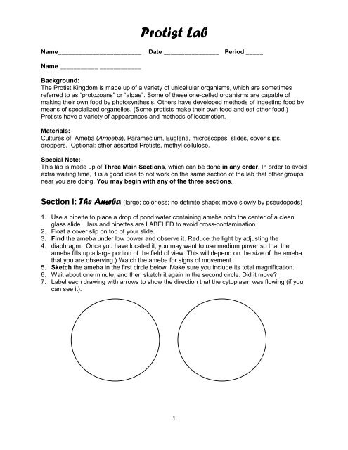
Section I: The Ameba (large; colorless; no definite shape; move slowly by pseudopods)<br />
1. Use a pipette to place a drop of pond water containing ameba onto the center of a clean<br />
glass slide. Jars and pipettes are LABELED to avoid cross-contamination.<br />
2. Float a cover slip on top of your slide.<br />
3. Find the ameba under low power and observe it. Reduce the light by adjusting the<br />
4. diaphragm. Once you have located it, you may want to use medium power so that the<br />
ameba fills up a large portion of the field of view. This will depend on the size of the ameba<br />
that you are observing.) Watch the ameba for signs of movement.<br />
5. Sketch the ameba in the first circle below. Make sure you include its total magnification.<br />
6. Wait about one minute, and then sketch it again in the second circle. Did it move?<br />
7. <strong>Lab</strong>el each drawing with arrows to show the direction that the cytoplasm was flowing (if you<br />
can see it).<br />
1
<strong>Protist</strong> <strong>Lab</strong><br />
IF YOU CAN’T FIND A LIVE AMEBA, ASK YOUR INSTRUCTOR FOR A STAINED, PREPARED SLIDE<br />
(DEAD) TO DRAW IN ONE OF THE CIRCLES.<br />
8. Look up contractile vacuole in your textbook, and define it in your own words:<br />
9. The ameba’s contractile vacuole appears as a clear circle. Look at your ameba and try to<br />
find what looks like a small bubble inside. If you find it, label it.<br />
10. Look up the word pseudopod in your textbook, and define it in your own words:<br />
11. After an ameba has engulfed its food, a food vacuole is formed. Look at the ameba in the<br />
microscope, and try to find a vacuole with material inside. (Do not confuse it with the nucleus,<br />
which is the largest dark object in the ameba.) <strong>Lab</strong>el it if you can see it.<br />
12. Below, draw a picture of the ameba from your textbook, labeling the following:<br />
pseudopod, nucleus, cell membrane, cytoplasm, contractile vacuole and food vacuole.<br />
13. Wash you ameba down the drain and clean off your slide and cover slip thoroughly. Return<br />
any prepared (stained, fixed) slides to your teacher.<br />
Section II: The Paramecium (medium size; clear; slipper-shaped; move quickly by<br />
cilia)<br />
Look up the word cilium in your textbook, and define it in your own words:<br />
1. Use the dropper in the paramecium culture to get a drop of “scum” out of the container, and<br />
place it on a microscope slide. Place a cover slip on top of the mixture. Optional: you can<br />
slow the protist down by adding one drop of methyl cellulose on top of the paramecium<br />
(this is a “syrup” that makes it hard for them to swim).<br />
2. Find a paramecium under low power and observe it.<br />
Then, change to medium power to see the details of the paramecium better.<br />
2
<strong>Protist</strong> <strong>Lab</strong><br />
Reduce the light by adjusting the diaphragm.<br />
You may need to move the slide to keep the paramecium in the field of view.<br />
3. Carefully draw the paramecium in the circle below (include magnification power).<br />
IF YOU CAN’T FIND A LIVE PARAMECIUM, ASK YOUR INSTRUCTOR FOR A STAINED, PREPARED<br />
SLIDE (DEAD) TO DRAW.<br />
4. Look up contractile vacuole in your textbook, and define it in your own words:<br />
5. The paramecium’s contractile vacuoles appear as star-shaped structures at each end. Try to<br />
find the contractile vacuoles in your specimen. <strong>Lab</strong>el them if you can see them.<br />
6. To eat, a paramecium collects food in its oral groove. When it has eaten, a food vacuole is<br />
formed. Look at the paramecium in the microscope, and try to find a vacuole with material<br />
inside. (Do not confuse it with the nucleus, which is the largest dark object in the paramecium.)<br />
7. Below, draw a picture of paramecium from your textbook, labeling the following: cilia,<br />
nucleus (macro/micro), cell membrane, pellicle, oral groove, cytoplasm, contractile vacuole and<br />
food vacuole.<br />
3
<strong>Protist</strong> <strong>Lab</strong><br />
8. Wash the paramecium down the drain. Wash and dry the slide and cover slip.<br />
Section III: The Euglena (small; green; oval; move quickly by flagellum)<br />
1. Look up the word flagellum in your textbook, and define it in your own words:<br />
2. Use the dropper in the euglena culture to get a drop out of the container, and place it on a<br />
microscope slide. Place a cover slip on top of the drop.<br />
3. Find euglena under low power and observe them. Shift to high power, and get a closer look.<br />
4. Carefully draw a few euglena in the circle below. Include magnification power.<br />
IF YOU CAN’T FIND A LIVE EUGLENA, ASK YOUR INSTRUCTOR FOR A STAINED, PREPARED<br />
SLIDE (DEAD) TO DRAW.<br />
5. Look up chloroplast in your textbook, and define it in your own words:<br />
6. The euglenas’ chloroplasts appear as green objects inside the cells. Euglenas make their<br />
own food by photosynthesis, but may also eat if they choose to or if sunlight is not available.<br />
7. Below, draw a picture of euglena from your textbook, labeling the following: flagellum,<br />
nucleus, chloroplasts, cell membrane, pellicle, eyespot & cytoplasm<br />
4
<strong>Protist</strong> <strong>Lab</strong><br />
8. Wash the euglenas down the drain. Wash and dry the slide and cover slip.<br />
5