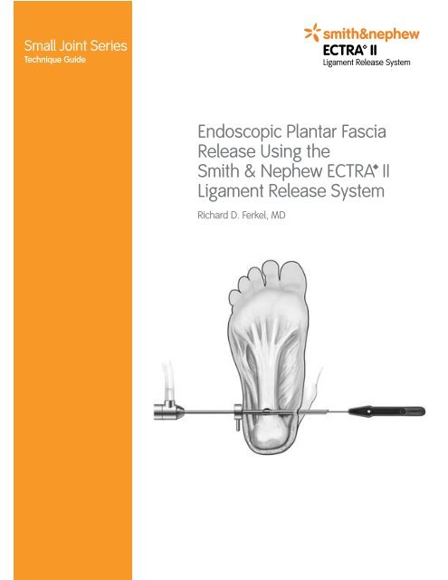
Endoscopic Plantar Fascia Release Using the Smith & Nephew ...
Endoscopic Plantar Fascia Release Using the Smith & Nephew ... Endoscopic Plantar Fascia Release Using the Smith & Nephew ...
Small Joint Series Technique Guide Endoscopic Plantar Fascia Release Using the Smith & Nephew ECTRA II Ligament Release System Richard D. Ferkel, MD
- Page 2 and 3: As Described by: Richard D. Ferkel,
- Page 4 and 5: Patient Preparation 1. Place the pa
- Page 6 and 7: 16. Insert the retrograde knife thr
- Page 8: Additional Instruction Prior to per
Small Joint Series<br />
Technique Guide<br />
<strong>Endoscopic</strong> <strong>Plantar</strong> <strong>Fascia</strong><br />
<strong>Release</strong> <strong>Using</strong> <strong>the</strong><br />
<strong>Smith</strong> & <strong>Nephew</strong> ECTRA II<br />
Ligament <strong>Release</strong> System<br />
Richard D. Ferkel, MD
As Described by:<br />
Richard D. Ferkel, MD<br />
Sou<strong>the</strong>rn California Orthopedic Institute<br />
Van Nuys, California
Ragnell retractors<br />
VideoEndoscope<br />
Slotted cannula<br />
Dissecting obturator<br />
with detachable<br />
handle<br />
Conical obturator<br />
Boat nose obturator<br />
Probe<br />
Blunt dissector, curved<br />
Figure 1. ECTRA II Ligament <strong>Release</strong> System components used in endoscopic plantar<br />
fascia release<br />
Triangle knife<br />
Probe knife<br />
Retrograde knife<br />
<strong>Endoscopic</strong> <strong>Plantar</strong> <strong>Fascia</strong> <strong>Release</strong><br />
<strong>Using</strong> <strong>the</strong> <strong>Smith</strong> & <strong>Nephew</strong><br />
ECTRA II Ligament <strong>Release</strong> System<br />
Introduction<br />
The <strong>Smith</strong> & <strong>Nephew</strong> ECTRA II Ligament <strong>Release</strong><br />
System can be used as a simple, effective means of<br />
performing endoscopic plantar fascia release.<br />
The ECTRA II System provides <strong>the</strong> specific reusable<br />
instrumentation required for endoscopic plantar<br />
fascia release, including a VideoEndoscope<br />
(4 mm x 30˚, 35 mm focal length, same-side light<br />
post direction), slotted cannula, obturator set,<br />
Ragnell retractors, blunt dissector (curved), and<br />
probe (Figure 1). The ECTRA II System also includes<br />
a reusable, detachable obturator handle. During<br />
endoscopic plantar fascia release, this handle may<br />
be interchangeably attached to <strong>the</strong> conical obturator,<br />
boat nose obturator, or dissecting obturator to<br />
enhance ease of obturator use.<br />
Designed to be used with <strong>the</strong> ECTRA II System,<br />
<strong>the</strong> ECTRA II Disposable Kit includes <strong>the</strong> specific<br />
disposable instrumentation required for endoscopic<br />
plantar fascia release: a probe knife, triangle knife,<br />
and retrograde knife (Figure 2).<br />
This two-portal plantar fasciotomy technique<br />
employs medial and lateral portals with blunt<br />
dissection transversing <strong>the</strong> heel superficial to <strong>the</strong><br />
plantar fascia. <strong>Using</strong> <strong>the</strong> ECTRA II System for this<br />
technique, <strong>the</strong> surgeon is able to cut from ei<strong>the</strong>r<br />
portal, maintain constant visualization, and minimize<br />
endoscopic lens fogging with good air flow. The<br />
medial 1/3–1/2 of <strong>the</strong> plantar fascia is released (<strong>the</strong><br />
entire plantar fascia should never be released).<br />
This procedure is most appropriate in carefully<br />
selected patients for whom a 6–9 month course of<br />
conservative, non-operative treatment has failed<br />
to provide relief. Patients should have no signs of<br />
nerve entrapment upon physical exam and/or nerve<br />
testing and should have negative blood tests for<br />
inflammatory arthritis.<br />
Figure 2. ECTRA II Disposable Kit knives
Patient Preparation<br />
1. Place <strong>the</strong> patient in <strong>the</strong> supine position<br />
on <strong>the</strong> table.<br />
2. Use an ankle block, regional, or general<br />
anes<strong>the</strong>tic.<br />
3. Apply an ankle or calf tourniquet.<br />
Technique<br />
1. Mark <strong>the</strong> location of <strong>the</strong> medial portal. Draw<br />
a line distally from <strong>the</strong> posterior aspect of <strong>the</strong><br />
medial malleolus to <strong>the</strong> intersection of <strong>the</strong><br />
medial origin of <strong>the</strong> plantar fascia at <strong>the</strong><br />
calcaneal tuberosity (Figure 3).<br />
2. Create an incision through <strong>the</strong> skin at <strong>the</strong><br />
location of <strong>the</strong> medial portal (Figure 4).<br />
3. Perform blunt dissection to <strong>the</strong> medial edge<br />
of <strong>the</strong> plantar fascia, being careful to avoid <strong>the</strong><br />
calcaneal nerve branch (Figure 4).<br />
4. Use a Freer elevator to clear <strong>the</strong> subcutaneous<br />
tissue from <strong>the</strong> plantar fascia transversely.<br />
5. Establish <strong>the</strong> location of <strong>the</strong> lateral portal by<br />
placing <strong>the</strong> obturator through <strong>the</strong> medial portal<br />
under <strong>the</strong> plantar fascia until it is palpable<br />
against <strong>the</strong> lateral skin. Keep <strong>the</strong> obturator<br />
parallel to <strong>the</strong> floor (Figure 5).<br />
6. Make an incision over <strong>the</strong> protruding obturator<br />
(Figure 5).<br />
Figure 3. Marking for <strong>the</strong> medial portal<br />
<strong>Plantar</strong><br />
fascia<br />
Lateral<br />
plantar<br />
nerve<br />
Medial<br />
portal<br />
Medial plantar nerve<br />
Medial<br />
calcaneal nerve<br />
Medial malleolus<br />
Posterior<br />
tibial nerve<br />
Figure 4. Avoiding injury to <strong>the</strong> medial calcaneal and lateral plantar nerves<br />
<strong>Plantar</strong> fascia<br />
Medial portal<br />
Figure 5. Establishing <strong>the</strong> lateral portal
VideoEndoscope<br />
Fasciotomy<br />
Slotted<br />
cannula<br />
Figure 6. Releasing <strong>the</strong> medial aspect of <strong>the</strong> plantar fascia<br />
Toes extended,<br />
ankle dorsiflexed<br />
Triangle knife<br />
7. Place <strong>the</strong> slotted cannula over <strong>the</strong> obturator<br />
through <strong>the</strong> lateral portal towards <strong>the</strong> medial<br />
portal (Figure 6).<br />
8. Place <strong>the</strong> VideoEndoscope laterally (Figure 6).<br />
9. Remove fluid and debris from <strong>the</strong> visual field<br />
using cotton swabs.<br />
10. Measure <strong>the</strong> width of <strong>the</strong> plantar fascia by<br />
placing a ruler over <strong>the</strong> posterior heel.<br />
11. For a point of reference, mark <strong>the</strong> probe and<br />
knives to <strong>the</strong> length of <strong>the</strong> plantar fascia that<br />
will be released.<br />
12. Insert <strong>the</strong> probe through <strong>the</strong> medial portal to<br />
check <strong>the</strong> measurements of <strong>the</strong> plantar fascia.<br />
13. Dorsiflex <strong>the</strong> ankle, foot, and toes to place<br />
tension on <strong>the</strong> plantar fascia (Figure 6).<br />
14. Insert <strong>the</strong> triangle knife through <strong>the</strong> medial<br />
portal (Figure 6).<br />
15. Make an incision at <strong>the</strong> predetermined lateral<br />
border of <strong>the</strong> fasciotomy (Figure 7).<br />
Figure 7. <strong>Using</strong> <strong>the</strong> triangle knife to initiate <strong>the</strong> release
16. Insert <strong>the</strong> retrograde knife through <strong>the</strong> medial<br />
portal and release <strong>the</strong> plantar fascia, pulling<br />
<strong>the</strong> knife from lateral to medial. The release<br />
is complete when <strong>the</strong> flexor digitorum brevis<br />
muscle is seen (Figures 8 and 9).<br />
17. <strong>Release</strong> <strong>the</strong> tourniquet, irrigate <strong>the</strong> wounds,<br />
and close with non-absorbable suture.<br />
18. Apply a compression dressing and place <strong>the</strong><br />
patient into a walker boot.<br />
19. Patients start partial weight bearing in <strong>the</strong> boot<br />
with crutches 1–2 days after surgery.<br />
20. Full weight bearing is initiated after <strong>the</strong> wound<br />
has healed.<br />
Figure 8. Visualizing <strong>the</strong> flexor digitorum brevis muscle<br />
Flexor digitorum<br />
brevis m.<br />
<strong>Plantar</strong> fascia<br />
Slotted cannula<br />
Retrograde knife<br />
Figure 9. Visualizing <strong>the</strong> completed release
References<br />
Ferkel, Richard D. Future Developments.<br />
Arthroscopic Surgery: The Foot and Ankle<br />
(pp. 317–324). Philadelphia: Lippincott-Raven, 1996.<br />
Bazaz, Rajesh; Ferkel, Richard D. Results of<br />
<strong>Endoscopic</strong> <strong>Plantar</strong> <strong>Fascia</strong> <strong>Release</strong>.<br />
Foot and Ankle International. 28:549–556, 2007.
Additional Instruction<br />
Prior to performing this technique, consult<br />
<strong>the</strong> Instructions For Use provided with<br />
individual components, including indications,<br />
contraindications, warnings, cautions, and<br />
instructions.<br />
Ordering Information<br />
Call +1 800 343 5717 in <strong>the</strong> U.S. or contact your<br />
authorized <strong>Smith</strong> & <strong>Nephew</strong> representative to<br />
order <strong>the</strong> ECTRA II System, 35 mm (REF 4143) or<br />
any of <strong>the</strong> following components:<br />
REF Description<br />
3896 VideoEndoscope, 35 mm<br />
4120 Slotted cannula<br />
4101 Blunt dissector, curved<br />
3856 Probe<br />
4283 Palmer arch suppressor<br />
4450 Hand holder and sterilization tray with cover<br />
4432 Detachable obturator handle<br />
4433 Conical obturator<br />
4434 Boat nose obturator<br />
4435 Dissecting obturator<br />
4452 Ragnell retractor (2 included with system)<br />
The ECTRA II Disposable Kit (REF 4116 – includes 8<br />
swabs, 3 disposable knives, and 1 sterile hand pad)<br />
or any of <strong>the</strong> following components may also be<br />
ordered:<br />
REF Description<br />
4447 Probe knife, beige, sterile, box of 6<br />
4448 Triangle knife, green, sterile, box of 6<br />
4449 Retrograde knife, blue, sterile, box of 6<br />
Courtesy of <strong>Smith</strong> & <strong>Nephew</strong>, Inc.,<br />
Endoscopy Division<br />
Caution: U.S. Federal law restricts this device to sale by or on<br />
<strong>the</strong> order of a physician.<br />
Trademark of <strong>Smith</strong> & <strong>Nephew</strong>, registered<br />
U.S. Patent & Trademark Office<br />
Endoscopy<br />
<strong>Smith</strong> & <strong>Nephew</strong>, Inc.<br />
Andover, MA 01810<br />
USA<br />
www.smith-nephew.com<br />
+1 978 749 1000 Telephone<br />
+1 978 749 1108 Fax<br />
+1 800 343 5717 U.S. Customer Service<br />
©2007 <strong>Smith</strong> & <strong>Nephew</strong>, Inc.<br />
All rights reserved.<br />
12/2007 10600321 Rev. A


