Secondary Chondrosarcoma
Secondary Chondrosarcoma
Secondary Chondrosarcoma

You also want an ePaper? Increase the reach of your titles
YUMPU automatically turns print PDFs into web optimized ePapers that Google loves.
<strong>Secondary</strong> <strong>Chondrosarcoma</strong><br />
lished data of the large series that we<br />
reviewed, more than three fourths of<br />
the cases were grade 1 tumors, and<br />
nearly all of the remaining lesions<br />
were grade 2. 4,5,7,11,13 Only 1% of<br />
cases were reported to be grade 3.<br />
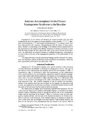
Figure 2<br />
Genetics and Mutations<br />
Histologic comparison of osteochondroma and secondary chondrosarcoma.<br />
A, Cartilaginous tissue of an osteochondroma demonstrating features that<br />
recapitulate the hyaline cartilage of articular surfaces. Note the gradual<br />
enlargement of cells as they near the subchondral bone and become<br />
hypertrophic. Most cells reside in solitary lacunae, with occasional lacunae<br />
having two cells (arrowhead). The cells are relatively sparse in the abundant<br />
extracellular matrix (hematoxylin-eosin, original magnification ×40). B, Grade<br />
2 secondary chondrosarcoma demonstrating greater cellularity and less<br />
discernible organization of the chondrocytes. Lacunae with multiple cells are<br />
more common. Because cells retract from the walls of the lacunae during the<br />
fixation process, pleomorphism of cells is not easy to identify (hematoxylineosin,<br />
original magnification ×20). C, Osteochondroma under high-power<br />
magnification demonstrating cells that are fairly well-spaced in their lacunae.<br />
Slight atypia may be visible, making the distinction between benign lesions<br />
and low-grade chondrosarcoma difficult on the basis of histology alone.<br />
Certain lacunae contain two cells (hematoxylin-eosin, original magnification<br />
×400). D, Grade 1 secondary chondrosarcoma under high-power<br />
magnification demonstrating atypical features, such as the binucleate cell in<br />
the center of the field. No significant nuclear pleomorphism is present<br />
(hematoxylin-eosin, original magnification ×400).<br />
Hereditary multiple exostosis<br />
(HME), also referred to as multiple<br />
hereditary exostosis and hereditary<br />
multiple osteochondromatosis, is a<br />
rare familial disease characterized by<br />
multiple osteochondromas throughout<br />
the skeleton. The disease is inherited<br />
as an autosomal dominant condition<br />
with high penetrance. Three<br />
related genes have been implicated in<br />
the disorder, EXT1, EXT2, and<br />
EXT3, which are located on 8q24,<br />
11p13, and 19p, respectively. 29-31<br />
EXT1 and EXT2 mutations are<br />
more common and make up most<br />
cases. 32 Patients with EXT1 mutation<br />
tend to have more severe phenotypes<br />
than do patients with EXT2<br />
mutation. 32 It is not clear how the<br />
EXT genes are related to the pathophysiology<br />
of osteochondromas.<br />
Mutation of the genes usually results<br />
in truncated forms of the proteins,<br />
which are needed for the synthesis of<br />
heparan sulfate. This may secondarily<br />
impair diffusion of cellsignaling<br />
molecules. 33<br />
A genetic model for cartilaginous<br />
tumorigenesis in the setting of HME<br />
has been proposed. 34 First, inactivation<br />
of both copies of the EXT1 gene<br />
in cartilage cells is essential for the<br />
formation of osteochondroma. This<br />
typically occurs via loss of heterozygosity<br />
in sporadic cells. Cells that<br />
maintain one normal copy of the<br />
gene are apparently normal. Of note,<br />
it appears that solitary osteochondromas<br />
also develop as a result of<br />
mutation of both copies of an EXT<br />
gene, the difference being that this<br />
occurs in isolated cells and no germline<br />
mutation is inherited. Without<br />
further mutations in other genes, the<br />
lesion remains benign; most osteochondromas<br />
remain in this state and<br />
do not progress further. One or more<br />
additional mutations in other genes<br />
612 Journal of the American Academy of Orthopaedic Surgeons