Secondary Chondrosarcoma
Secondary Chondrosarcoma
Secondary Chondrosarcoma

You also want an ePaper? Increase the reach of your titles
YUMPU automatically turns print PDFs into web optimized ePapers that Google loves.
<strong>Secondary</strong> <strong>Chondrosarcoma</strong><br />
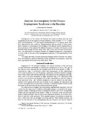
Figure 1<br />
<strong>Secondary</strong> chondrosarcoma arising from a pelvic osteochondroma. A, Preoperative AP pelvic radiograph<br />
demonstrating a large calcified tumor arising from the iliac wing. The surfaces of the calcified areas are indistinct and<br />
not well-delineated. B, Postoperative AP pelvic radiograph following wide excision of the tumor. C, Photograph of the<br />
cut specimen after resection demonstrating a thick layer of white, glistening hyaline cartilage. The central area shows a<br />
small area of ossified bone (arrow), which represents the stalk of the preexisting osteochondroma. D, T1-weighted<br />
axial magnetic resonance image demonstrating fatty marrow in the central portion of the sessile stalk emanating from<br />
the iliac crest, indicating the presence of an ossified central portion of the original osteochondroma. E, T2-weighted<br />
axial magnetic resonance image demonstrating the thick layer of cartilaginous tumor surrounding the central ossified<br />
stalk, which appears dark. F, Gadolinium-enhanced fat-saturated T1-weighted magnetic resonance image showing a<br />
subtle speckled pattern of enhancement within the tumor.<br />
Radiologic Features<br />
Conventional radiography may offer<br />
important clues regarding the diagnosis<br />
of secondary chondrosarcoma.<br />
Osteochondromas have well-defined<br />
bony edges, including the subchondral<br />
bone of the cartilaginous cap. In<br />
contrast, secondary chondrosarcomas<br />
demonstrate irregularity or blurriness<br />
of the surface of the osteochondroma<br />
(Figure 1). A soft-tissue<br />
mass outside the osseous portion of<br />
the osteochondroma may be subtly<br />
appreciable, and this mass may exhibit<br />
scattered foci of calcification. 4,5<br />
CT and MRI are important in<br />
demonstrating the malignant features<br />
of the tumor. Most critically,<br />
they reveal an abnormally large cartilaginous<br />
cap on the osteochondroma<br />
(Figure 1, D through F). The<br />
portion of the osteochondroma that<br />
becomes malignant is the cartilaginous<br />
tissue that forms the cap of the<br />
lesion. The osseous tissue that makes<br />
up the base or stalk is not thought to<br />
610 Journal of the American Academy of Orthopaedic Surgeons