Crescent fracture-dislocation of the sacroiliac joint
Crescent fracture-dislocation of the sacroiliac joint
Crescent fracture-dislocation of the sacroiliac joint

Create successful ePaper yourself
Turn your PDF publications into a flip-book with our unique Google optimized e-Paper software.
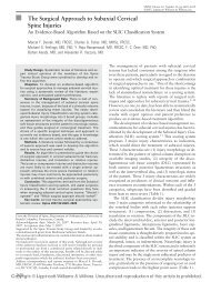
<strong>Crescent</strong> <strong>fracture</strong>-<strong>dislocation</strong> <strong>of</strong> <strong>the</strong> <strong>sacroiliac</strong><br />
<strong>joint</strong><br />
A FUNCTIONAL CLASSIFICATION<br />
A. C. Day,<br />
C. Kinmont,<br />
M. D. Bircher,<br />
S. Kumar<br />
From St. George’s<br />
Healthcare NHS<br />
Trust, London,<br />
England<br />
<strong>Crescent</strong> <strong>fracture</strong> <strong>dislocation</strong>s are a well-recognised subset <strong>of</strong> pelvic ring injuries which<br />
result from a lateral compression force. They are characterised by disruption <strong>of</strong> <strong>the</strong><br />
<strong>sacroiliac</strong> <strong>joint</strong> and extend proximally as a <strong>fracture</strong> <strong>of</strong> <strong>the</strong> posterior iliac wing. We describe a<br />
classification with three distinct types. Type I is characterised by a large crescent fragment<br />
and <strong>the</strong> <strong>dislocation</strong> comprises no more than one-third <strong>of</strong> <strong>the</strong> <strong>sacroiliac</strong> <strong>joint</strong>, which is<br />
typically inferior. Type II <strong>fracture</strong>s are associated with an intermediate-size crescent<br />
fragment and <strong>the</strong> <strong>dislocation</strong> comprises between one- and two-thirds <strong>of</strong> <strong>the</strong> <strong>joint</strong>. Type III<br />
<strong>fracture</strong>s are associated with a small crescent fragment where <strong>the</strong> <strong>dislocation</strong> comprises<br />
most, but not all <strong>of</strong> <strong>the</strong> <strong>joint</strong>. The principal goals <strong>of</strong> surgical intervention are <strong>the</strong> accurate<br />
and stable reduction <strong>of</strong> <strong>the</strong> <strong>sacroiliac</strong> <strong>joint</strong>. This classification proves useful in <strong>the</strong> selection<br />
<strong>of</strong> both <strong>the</strong> surgical approach and <strong>the</strong> reduction technique. A total <strong>of</strong> 16 patients were<br />
managed according to this classification and achieved good functional results<br />
approximately two years from <strong>the</strong> time <strong>of</strong> <strong>the</strong> index injury. Confounding factors<br />
compromise <strong>the</strong> summary short-form-36 and musculoskeletal functional assessment<br />
instrument scores, which is a well-recognised phenomenon when reporting <strong>the</strong> outcome <strong>of</strong><br />
high-energy trauma.<br />
A. C. Day, FRCS(Orth),<br />
Consultant Trauma and<br />
Orthopaedic Surgeon<br />
M. D. Bircher, FRCS,<br />
Consultant Trauma and<br />
Orthopaedic Surgeon<br />
St George’s Healthcare NHS<br />
Trust, Blackshaw Road,<br />
London, SW17 0QT, United<br />
Kingdom.<br />
C. Kinmont, FRCS(Orth),<br />
Consultant Trauma and<br />
Orthopaedic Surgeon<br />
Mayday Hospital, London<br />
Road, Croydon, CR7 7YE,<br />
United Kingdom.<br />
S. Kumar, FRCS(Orth),<br />
Consultant Trauma and<br />
Orthopaedic Surgeon<br />
Kent and Sussex Hospital,<br />
Mount Ephraim, Tunbridge<br />
Wells, Kent, TN4 8AT, United<br />
Kingdom.<br />
Correspondence should be sent<br />
to Mr A. C. Day; e-mail:<br />
adriancday@hotmail.com<br />
©2007 British Editorial Society<br />
<strong>of</strong> Bone and Joint Surgery<br />
doi:10.1302/0301-620X.89B5.<br />
18129 $2.00<br />
J Bone Joint Surg [Br]<br />
2007;89-B:651-8.<br />
Received 15 May 2006;<br />
Accepted after revision 19<br />
December 2006<br />
Lateral compression <strong>fracture</strong>s comprise up to<br />
80% <strong>of</strong> all pelvic ring injuries. 1-4 Of this group,<br />
crescent <strong>fracture</strong>s form a less common subset, 1-<br />
4 although <strong>the</strong> exact proportion is difficult to<br />
determine from <strong>the</strong> literature. The Pelvic and<br />
Acetabular Reconstruction Unit at St George’s<br />
Healthcare NHS Trust provides a tertiary<br />
referral service. A review <strong>of</strong> our database identified<br />
37 lateral compression-type pelvic ring<br />
injuries in a consecutive series <strong>of</strong> 118 <strong>fracture</strong>s.<br />
A total <strong>of</strong> 14 may be classified as crescent <strong>fracture</strong>s<br />
according to <strong>the</strong> definition <strong>of</strong> Borrelli et<br />
al. 1,2 Therefore, crescent <strong>fracture</strong>s are not<br />
uncommon and occur in approximately 12%<br />
<strong>of</strong> pelvic ring <strong>fracture</strong>s admitted to a specialist<br />
unit. They are, in fact, <strong>fracture</strong>-<strong>dislocation</strong>s <strong>of</strong><br />
<strong>the</strong> <strong>sacroiliac</strong> <strong>joint</strong> in which <strong>the</strong>re is variable<br />
disruption <strong>of</strong> <strong>the</strong> <strong>sacroiliac</strong> ligament complex,<br />
extending proximally as a <strong>fracture</strong> <strong>of</strong> <strong>the</strong> posterior<br />
iliac crest. A crescent-shaped segment <strong>of</strong><br />
<strong>the</strong> iliac wing remains attached to <strong>the</strong> sacrum<br />
by <strong>the</strong> intact portion <strong>of</strong> <strong>the</strong> posterior ligament<br />
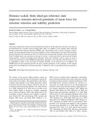
complex. 1,4 The anteroposterior (AP) radiograph<br />
(Fig. 1) demonstrates <strong>the</strong> topography <strong>of</strong><br />
a typical crescent <strong>fracture</strong>. The posterior pelvic<br />
ring injury is <strong>of</strong>ten accompanied by <strong>fracture</strong>s<br />
<strong>of</strong> <strong>the</strong> pubic rami or a symphyseal diastasis. A<br />
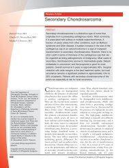
three-dimensional (3D) reformat <strong>of</strong> spiral CT<br />
data <strong>of</strong> a crescent <strong>fracture</strong> is shown in Figure 2.<br />
<strong>Crescent</strong> <strong>fracture</strong>s are not commonly associated<br />
with life-threatening pelvic vascular injuries<br />
and <strong>the</strong> urgent application <strong>of</strong> a pelvic<br />
external fixator device in compression is not<br />
generally required. 1-3,5,6 However, urogenital<br />
and vascular injuries can complicate any <strong>fracture</strong><br />
or <strong>dislocation</strong> <strong>of</strong> <strong>the</strong> pelvic ring and<br />
should not be overlooked. The value <strong>of</strong><br />
contrast-enhanced CT scans is established and<br />
facilitates <strong>the</strong> selection <strong>of</strong> patients who may<br />
benefit from angiography and arterial embolisation<br />
techniques. 7<br />
Operative intervention is recommended for<br />
<strong>the</strong> anatomical reduction and stable fixation <strong>of</strong><br />
a crescent <strong>fracture</strong>-<strong>dislocation</strong>. Restoration <strong>of</strong><br />
<strong>the</strong> normal anatomy should reduce <strong>the</strong> incidence<br />
<strong>of</strong> malunion, post-traumatic <strong>sacroiliac</strong><br />
<strong>joint</strong> arthritis, painful stance phase gait cycle<br />
instability and seating obliquity. 5,6,8 Despite<br />
satisfactory reduction and stabilisation, some<br />
patients may experience persistent pain and<br />
dysfunction, which is commonly observed in<br />
high-energy injuries. 9<br />
A number <strong>of</strong> surgical techniques have been<br />
described. 1,2,5,9-16 To <strong>the</strong> authors’ knowledge,<br />
<strong>the</strong>re are only two published articles that spe-<br />
VOL. 89-B, No. 5, MAY 2007 651
652 A. C. DAY, C. KINMONT, M. D. BIRCHER, S. KUMAR<br />
Fig. 1<br />
Radiograph <strong>of</strong> a crescent <strong>fracture</strong>-<strong>dislocation</strong> <strong>of</strong> <strong>the</strong> left <strong>sacroiliac</strong> <strong>joint</strong>.<br />
The crescent fragment (black arrow) remains attached to <strong>the</strong> sacrum by<br />
<strong>the</strong> intact portion <strong>of</strong> <strong>the</strong> posterior <strong>sacroiliac</strong> ligament complex. The left<br />
hemipelvis (white arrow) is rotationally unstable, but vertical displacement<br />
is limited by <strong>the</strong> intact sacrospinous and sacrotuberous ligament<br />
complexes. 1,3,5 There is an associated <strong>fracture</strong> <strong>of</strong> both <strong>the</strong> superior and<br />
inferior pubic ramus.<br />
Fig. 2<br />
This 3D reformat <strong>of</strong> spiral CT data demonstrates a crescent <strong>fracture</strong> <strong>of</strong><br />
<strong>the</strong> right hemipelvis in which two-thirds <strong>of</strong> <strong>the</strong> <strong>sacroiliac</strong> <strong>joint</strong> was dislocated<br />
and is associated with a medium-sized crescent fragment.<br />
cifically address <strong>the</strong> management <strong>of</strong> this <strong>fracture</strong>. Borrelli et<br />
al 1,2 describe a <strong>fracture</strong>-<strong>dislocation</strong> involving approximately<br />
50% <strong>of</strong> <strong>the</strong> <strong>sacroiliac</strong> <strong>joint</strong>, and restoration <strong>of</strong> a stable<br />
pelvic ring is advocated by means <strong>of</strong> a posterior<br />
subgluteal approach supplemented by a lag screw and plating<br />
technique. This approach is entirely appropriate for <strong>the</strong><br />
type <strong>of</strong> <strong>fracture</strong>-<strong>dislocation</strong> described. 1,2<br />
There are a number <strong>of</strong> subtypes that may reasonably be<br />
described as crescent <strong>fracture</strong>s. They share a common<br />
mechanism <strong>of</strong> injury and <strong>the</strong> <strong>fracture</strong> morphology is similar.<br />
Operative management is generally to be recommended,<br />
but, significantly, <strong>the</strong> operative approach and<br />
technique differ substantially for each subtype. We propose<br />
three distinct categories according to <strong>the</strong> extent <strong>of</strong> <strong>sacroiliac</strong><br />
<strong>joint</strong> involvement, and <strong>the</strong> classification is inclusive <strong>of</strong><br />
<strong>the</strong> <strong>fracture</strong> type described by Borrelli et al. 1,2<br />
<strong>Crescent</strong> <strong>fracture</strong> classification. Imaging should include plain<br />
radiographs comprising AP, inlet and outlet views. Additionally,<br />
spiral CT data sets should be reformatted in <strong>the</strong> axial<br />
plane with <strong>the</strong> scout grid lines orientated parallel to <strong>the</strong> superior<br />
end-plate <strong>of</strong> S1. Where appropriate s<strong>of</strong>tware is available,<br />
3D reformatting is particularly helpful in <strong>the</strong> radiological<br />
assessment <strong>of</strong> <strong>the</strong>se injuries and should be orientated in projections<br />
similar to <strong>the</strong> plain radiograph series.<br />
We identified three groups <strong>of</strong> crescent <strong>fracture</strong> according to<br />
<strong>the</strong> extent <strong>of</strong> <strong>sacroiliac</strong> <strong>joint</strong> involvement (Fig. 3).<br />
Type I <strong>fracture</strong>s involve less than one-third <strong>of</strong> <strong>the</strong> <strong>joint</strong> and<br />
enter it inferiorly. A large crescent fragment is observed and<br />
<strong>the</strong> <strong>fracture</strong> line enters <strong>the</strong> <strong>joint</strong> close to <strong>the</strong> foramen <strong>of</strong> <strong>the</strong><br />
anterior S2 nerve root, a feature best appreciated from 3D CT<br />
data sets and <strong>the</strong> pelvic outlet view. This <strong>fracture</strong> type may be<br />
surgically addressed by means <strong>of</strong> an ilioinguinal approach.<br />
The lateral window usually gives sufficient exposure and facilitates<br />
an anterior plating technique, with direct visualisation <strong>of</strong><br />
both <strong>the</strong> <strong>fracture</strong> and <strong>the</strong> <strong>sacroiliac</strong> <strong>joint</strong>.<br />
Type II <strong>fracture</strong>s involve between one-third and twothirds<br />
<strong>of</strong> <strong>the</strong> <strong>joint</strong>. The crescent fragment is <strong>of</strong> moderate size<br />
and <strong>the</strong> <strong>fracture</strong> line enters <strong>the</strong> <strong>joint</strong> between <strong>the</strong> anterior S1<br />
and S2 foramina. This is, in effect, <strong>the</strong> <strong>fracture</strong> type<br />
described by Borrelli et al, 1,2 and it may be surgically<br />
addressed by means <strong>of</strong> a posterior approach which facilitates<br />
<strong>the</strong> placement <strong>of</strong> interfragmentary screws and supplementary<br />
plates.<br />
Borrelli et al 1,2 comment that <strong>the</strong>se lateral compression injuries<br />
may be associated with significant s<strong>of</strong>t-tissue injuries, such<br />
as <strong>the</strong> Morel-Lavallee 17 lesion.<br />
Type III <strong>fracture</strong>s involve more than two-thirds <strong>of</strong> <strong>the</strong> <strong>joint</strong><br />
and are associated with a small, superior crescent fragment.<br />
The <strong>fracture</strong> line enters <strong>the</strong> <strong>joint</strong> posterior and superior to <strong>the</strong><br />
anterior S1 nerve root foramen. This <strong>fracture</strong> type may <strong>the</strong>refore<br />
be surgically addressed by means <strong>of</strong> a closed or percutaneous<br />
reduction technique, supplemented by <strong>the</strong> placement <strong>of</strong><br />
percutaneous iliosacral screws. The technique should only be<br />
undertaken by appropriately-trained practitioners, and in <strong>the</strong><br />
case <strong>of</strong> delayed presentation, closed reduction may not be possible.<br />
Under <strong>the</strong>se circumstances it may be argued that <strong>the</strong> lateral<br />
window <strong>of</strong> an ilioinguinal approach <strong>of</strong>fers <strong>the</strong> best chance<br />
<strong>of</strong> accurate reduction with stable anterior plate fixation.<br />
The principal <strong>fracture</strong> line is oblique, and its orientation<br />
will <strong>the</strong>refore vary according to <strong>the</strong> virtual gantry angle and<br />
<strong>the</strong> level <strong>of</strong> <strong>the</strong> axial CT cut. The S1 nerve root canal itself is<br />
oblique to <strong>the</strong> coronal and sagittal planes, and its appearance<br />
will <strong>the</strong>refore also vary according to similar criteria. The <strong>fracture</strong><br />
type is determined not so much by <strong>the</strong> precise plane in<br />
which <strong>the</strong> <strong>fracture</strong> enters <strong>the</strong> <strong>sacroiliac</strong> <strong>joint</strong> at a carefully<br />
defined axial level, but ra<strong>the</strong>r by its overall morphology and<br />
<strong>the</strong> way in which this influences <strong>the</strong> choice <strong>of</strong> surgical<br />
approach. Examples <strong>of</strong> <strong>the</strong> three <strong>fracture</strong> types are shown in<br />
Figure 3, and are derived from axial reformats which demon-<br />
THE JOURNAL OF BONE AND JOINT SURGERY
CRESCENT FRACTURE-DISLOCATION OF THE SACROILIAC JOINT 653<br />
Fig. 3a<br />
Fig. 3c<br />
Fig. 3b<br />
Fig. 3d<br />
Type I<br />
Type II<br />
Type III<br />
The positions <strong>of</strong> <strong>the</strong> principal <strong>fracture</strong> lines are shown for crescent <strong>fracture</strong>-<strong>dislocation</strong><br />
types I, II and III, as defined by axial CT sections, reformatted<br />
parallel to <strong>the</strong> <strong>sacroiliac</strong> superior end-plate. a) Type I - <strong>the</strong> <strong>fracture</strong><br />
enters <strong>the</strong> anterior third <strong>of</strong> <strong>the</strong> <strong>sacroiliac</strong> <strong>joint</strong>. b) Type II - <strong>the</strong> <strong>fracture</strong><br />
enters <strong>the</strong> middle third <strong>of</strong> <strong>the</strong> <strong>sacroiliac</strong> <strong>joint</strong>. c) Type III- <strong>the</strong> <strong>fracture</strong><br />
enters <strong>the</strong> posterior third <strong>of</strong> <strong>the</strong> <strong>sacroiliac</strong> <strong>joint</strong>. d) All three <strong>fracture</strong> types<br />
are shown diagrammatically.<br />
strate <strong>the</strong> coronal plane in which <strong>the</strong> <strong>fracture</strong> enters <strong>the</strong> <strong>sacroiliac</strong><br />
<strong>joint</strong> and its relationship to both <strong>the</strong> S1 nerve root canal<br />
and <strong>the</strong> sacral alar recess. It should be noted that <strong>the</strong> coronal<br />
plane in which <strong>the</strong> <strong>fracture</strong> enters <strong>the</strong> <strong>joint</strong> may differ from <strong>the</strong><br />
<strong>fracture</strong> plane if <strong>the</strong> <strong>fracture</strong> is oblique to <strong>the</strong> <strong>joint</strong>, which is<br />
<strong>of</strong>ten <strong>the</strong> case.<br />
Patients and Methods<br />
Our classification system was applied to a consecutive<br />
series <strong>of</strong> 16 patients with crescent <strong>fracture</strong>s admitted to St<br />
George’s Healthcare NHS Trust between 1999 and 2001.<br />
One case was managed initially by non-operative means<br />
and presented late. The o<strong>the</strong>r cases were transferred within<br />
three weeks <strong>of</strong> injury. The mean age <strong>of</strong> <strong>the</strong> patient group<br />
was 25 years (16 to 63). There were eight males and eight<br />
females. Five patients sustained multiple musculoskeletal<br />
injuries, and <strong>of</strong> <strong>the</strong>se, two sustained associated <strong>fracture</strong>s <strong>of</strong><br />
<strong>the</strong> acetabulum.<br />
Pelvic external fixators had been applied in two cases at <strong>the</strong><br />
referring hospital. The mechanism <strong>of</strong> injury was recorded as<br />
lateral compression in all cases, and <strong>the</strong>re was no evidence <strong>of</strong><br />
significant haemodynamic compromise. All <strong>of</strong> <strong>the</strong> patients<br />
were assessed at approximately two years after <strong>the</strong> index<br />
injury.<br />
Radiological assessment. The <strong>fracture</strong>s were evaluated by<br />
means <strong>of</strong> plain anteroposterior, inlet and outlet pelvic<br />
radiographs. For those cases associated with a <strong>fracture</strong> <strong>of</strong><br />
<strong>the</strong> acetabulum, 45˚ oblique views were obtained as<br />
described by Judet. 18 The extent <strong>of</strong> each principal <strong>fracture</strong><br />
line was assessed by spiral CT; 3 mm contiguous cuts were<br />
reformatted in an axial plane, parallel to <strong>the</strong> S1 superior<br />
end-plate, as determined from <strong>the</strong> lateral scout projection.<br />
Additional 3D reformatting was undertaken for selected<br />
cases in order to clarify <strong>the</strong> <strong>fracture</strong> morphology.<br />
Post-operative assessment. Patients were reviewed both clinically<br />
and radiologically. Radiological criteria as assessed by<br />
plain AP, inlet and outlet projections, included <strong>the</strong> quality <strong>of</strong><br />
<strong>fracture</strong> reduction and evidence <strong>of</strong> union.<br />
CT scans were obtained if <strong>the</strong>re was any doubt about<br />
<strong>fracture</strong> union. Clinical outcome was assessed by means <strong>of</strong><br />
<strong>the</strong> validated short-form (SF)36 19 and musculoskeletal<br />
functional assessment instruments. 20 These were administered<br />
between 12 and 18 months after surgery, by which<br />
time <strong>the</strong> patients were expected to be independently mobile,<br />
and <strong>fracture</strong> consolidation was anticipated. Ethical<br />
approval was obtained for <strong>the</strong> study. The data were rendered<br />
anonymous.<br />
Results<br />
Type I <strong>fracture</strong>s. We identified four type I crescent <strong>fracture</strong>s<br />
(Fig. 4). In addition to a crescent <strong>fracture</strong>, one patient sustained<br />
<strong>fracture</strong>s <strong>of</strong> all four limbs, an ipsilateral <strong>fracture</strong> <strong>of</strong><br />
<strong>the</strong> acetabulum involving both columns, a <strong>fracture</strong> <strong>of</strong> <strong>the</strong><br />
ipsilateral pubic ramus and an extensive posterior Morel-<br />
Lavallee lesion. One patient sustained ipsilateral pubic<br />
ramus <strong>fracture</strong>s, and <strong>the</strong> remaining two had a symphyseal<br />
diastasis.<br />
Figure 4a illustrates a type I <strong>fracture</strong>. A large crescent fragment<br />
remains congruent with <strong>the</strong> <strong>sacroiliac</strong> <strong>joint</strong> and <strong>the</strong> superior<br />
portion <strong>of</strong> <strong>the</strong> ligament complex is intact. The axial CT<br />
scan (Fig. 4b) demonstrates a <strong>fracture</strong> <strong>of</strong> <strong>the</strong> iliac crest which<br />
enters <strong>the</strong> <strong>sacroiliac</strong> <strong>joint</strong> well within <strong>the</strong> anterior third, and<br />
<strong>the</strong> associated <strong>sacroiliac</strong> <strong>dislocation</strong> is noted. The inferior portion<br />
<strong>of</strong> <strong>the</strong> <strong>sacroiliac</strong> ligament complex may have been injured,<br />
but type I <strong>fracture</strong>s cause <strong>the</strong> least ligamentous injury <strong>of</strong> <strong>the</strong><br />
three subtypes. Traction was applied in flexion and <strong>the</strong> <strong>fracture</strong><br />
was addressed through <strong>the</strong> lateral window <strong>of</strong> an ilioinguinal<br />
approach, controlling rotation with a Schanz pin,<br />
which was placed in <strong>the</strong> anteroinferior iliac spine by means <strong>of</strong><br />
a percutaneous technique.<br />
The united <strong>fracture</strong> and fixation in situ is illustrated in<br />
Figure 4c. The use <strong>of</strong> two plates is recommended, as stability<br />
in 6 degrees <strong>of</strong> freedom is required. In one case, <strong>the</strong> ilioinguinal<br />
approach was extended to address an acetabular<br />
<strong>fracture</strong>, and for ano<strong>the</strong>r case a posterior approach was<br />
selected to avoid infected pin track sites following <strong>the</strong><br />
removal <strong>of</strong> an external fixator device.<br />
In <strong>the</strong> latter situation, <strong>the</strong> dissection required was more<br />
extensive than is typical <strong>of</strong> <strong>the</strong> classic crescent <strong>fracture</strong><strong>dislocation</strong><br />
described by Borrelli et al. 1,2 It is for this reason<br />
that <strong>the</strong> lateral window <strong>of</strong> an ilioinguinal approach is preferred<br />
when <strong>the</strong> clinical circumstances allow. To summarise,<br />
three cases were managed through <strong>the</strong> lateral<br />
window <strong>of</strong> an ilioinguinal approach. This was extended in<br />
<strong>the</strong> first case to facilitate access to a <strong>fracture</strong> <strong>of</strong> <strong>the</strong> ipsilat-<br />
VOL. 89-B, No. 5, MAY 2007
654 A. C. DAY, C. KINMONT, M. D. BIRCHER, S. KUMAR<br />
Fig. 4a<br />
Fig. 4b<br />
Fig. 4c<br />
Type I crescent <strong>fracture</strong>. a) Anteroposterior radiograph, b) axial CT image and c) post-operative anteroposterior radiograph<br />
following union.<br />
eral acetabulum, and an ipsilateral pubic ramus <strong>fracture</strong><br />
was addressed simultaneously. An AO (Syn<strong>the</strong>s, Stratec<br />
Medical, Oberdorf, Switzerland) external fixator device<br />
was applied in a second case, associated with <strong>fracture</strong>s <strong>of</strong><br />
all four pubic rami. The ‘Delta’ configuration was selected<br />
in distraction mode. This technique facilitates <strong>the</strong> reduction<br />
<strong>of</strong> bilateral pubic ramus <strong>fracture</strong>s, sustained as a result <strong>of</strong><br />
<strong>the</strong> internal rotation mechanism, and relies on <strong>the</strong> presence<br />
<strong>of</strong> an intact periosteal hinge. It provides reasonable stability<br />
without recourse to an extensive anterior approach. The<br />
third case was associated with a symphyseal diastasis which<br />
was stabilised by means <strong>of</strong> a four-hole AO (Syn<strong>the</strong>s) large<br />
fragment reconstruction plate, using a separate Pfannenstiel<br />
incision. A posterior approach was selected for <strong>the</strong><br />
remaining case to avoid infected external fixator pin sites.<br />
Sound union was achieved in all four cases.<br />
Type II <strong>fracture</strong>s. Of <strong>the</strong> four type II crescent <strong>fracture</strong>s in this<br />
series, three were isolated pelvic <strong>fracture</strong>s and all had pubic<br />
ramus <strong>fracture</strong>s (Fig. 5). The fourth patient sustained multiple<br />
injuries and <strong>the</strong> crescent <strong>fracture</strong> was associated with a transverse<br />
<strong>fracture</strong> <strong>of</strong> <strong>the</strong> ipsilateral acetabulum. Figure 5a demonstrates<br />
a typical type II <strong>fracture</strong>. The axial CT reformat (Fig.<br />
5b) demonstrates a <strong>fracture</strong> line which enters <strong>the</strong> middle third<br />
<strong>of</strong> <strong>the</strong> <strong>sacroiliac</strong> <strong>joint</strong>. Injury to <strong>the</strong> posterior <strong>sacroiliac</strong> ligament<br />
complex is more extensive in this case, and <strong>the</strong> crescent<br />
fragment is <strong>of</strong> intermediate size.<br />
The <strong>fracture</strong> line, which is best appreciated on <strong>the</strong> axial<br />
CT scan, runs in a relatively oblique plane. A posterior<br />
approach was selected, as described by Borrelli et al. 1,2 It<br />
was considered that an interfragmentary screw might result<br />
in shear ra<strong>the</strong>r than compression <strong>of</strong> <strong>the</strong> <strong>fracture</strong>, and <strong>the</strong><br />
two ACE ribbon plates (DePuy Orthopaedics, Warsaw,<br />
Indiana) <strong>the</strong>refore incorporate interfragmentary screws,<br />
placed orthogonal to <strong>the</strong> <strong>fracture</strong> line (Fig. 5c). A fur<strong>the</strong>r<br />
case (Fig. 5d) illustrates <strong>the</strong> classic technique, where it has<br />
been possible to place interfragmentary screws across an<br />
orthogonal <strong>fracture</strong> line with <strong>the</strong> addition <strong>of</strong> two contoured<br />
reconstruction plates (Syn<strong>the</strong>s). Once again, <strong>the</strong> posterior<br />
approach was selected and proved satisfactory. In one case,<br />
<strong>the</strong> posterior fixation was supplemented by an external<br />
fixator device, applied in ‘distraction mode’ as described<br />
previously. If <strong>the</strong> pubic ramus <strong>fracture</strong>s are well aligned,<br />
retrograde anterior column screws may be considered a less<br />
cumbersome alternative. It should be noted, however, that<br />
THE JOURNAL OF BONE AND JOINT SURGERY
CRESCENT FRACTURE-DISLOCATION OF THE SACROILIAC JOINT 655<br />
Fig. 5a<br />
Fig. 5b<br />
Fig. 5c<br />
Fig. 5d<br />
Type II crescent <strong>fracture</strong>s, a)<br />
anteroposterior radiograph,<br />
b) axial CT image, c) postoperative<br />
anteroposterior<br />
radiograph following union,<br />
and d) a fur<strong>the</strong>r example<br />
demonstrating <strong>the</strong> classic<br />
technique described by Borrelli<br />
et al. 1,2 The inset demonstrates<br />
<strong>the</strong> technique <strong>of</strong><br />
internal fixation by means<br />
<strong>of</strong> an interfragmentary<br />
screw and neutralisation<br />
plate, as described by Borrelli<br />
et al. 1,2<br />
satisfactory alignment cannot always be achieved. The<br />
patient sustaining an ipsilateral transverse <strong>fracture</strong> <strong>of</strong> <strong>the</strong><br />
acetabulum required both ilioinguinal and Kocher-<br />
Langenbeck approaches. An anterior plating technique was<br />
<strong>the</strong>refore selected in respect <strong>of</strong> <strong>the</strong> crescent <strong>fracture</strong>.<br />
In summary, three <strong>of</strong> <strong>the</strong> four cases were managed by<br />
means <strong>of</strong> <strong>the</strong> classic posterior approach and a technique<br />
similar to that described by Borrelli et al. 1,2 All <strong>of</strong> <strong>the</strong> <strong>fracture</strong>s<br />
in this group united uneventfully.<br />
Type III <strong>fracture</strong>s. Eight type III crescent <strong>fracture</strong>s were identified.<br />
In one case <strong>the</strong>re was an associated symphyseal diastasis,<br />
and pubic ramus <strong>fracture</strong>s occurred in all <strong>of</strong> <strong>the</strong> o<strong>the</strong>r cases. A<br />
typical example is presented in Figure 6.<br />
The now familiar pattern is observed in Figure 6, but in<br />
this case <strong>the</strong> <strong>dislocation</strong> encompasses more than two-thirds<br />
<strong>of</strong> <strong>the</strong> <strong>sacroiliac</strong> <strong>joint</strong>. The axial CT (Fig. 6b) highlights <strong>the</strong><br />
small posterior crescent (white arrow) and <strong>the</strong> projected<br />
track <strong>of</strong> a percutaneous iliosacral screw (black arrow).<br />
These <strong>fracture</strong>s are associated with more extensive disruption<br />
<strong>of</strong> <strong>the</strong> <strong>sacroiliac</strong> ligament complex, but are never<strong>the</strong>less<br />
distinct from pure <strong>sacroiliac</strong> <strong>dislocation</strong>s and are consistently<br />
lateral compression injuries in which <strong>the</strong> degree <strong>of</strong><br />
vertical displacement is limited. Six cases were treated with<br />
closed reduction and percutaneous iliosacral screw placement.<br />
This technique requires skeletal traction by means <strong>of</strong><br />
a traction pin placed in <strong>the</strong> proximal tibia. Traction is<br />
applied with <strong>the</strong> hip in flexion on a specialised radiolucent<br />
pelvic reduction table (OSI, Union City, California) allowing<br />
correction <strong>of</strong> both <strong>the</strong> vertical and <strong>the</strong> posterior displacement.<br />
The internal rotation deformity may be<br />
corrected by means <strong>of</strong> a percutaneous Schanz pin, applied<br />
to ei<strong>the</strong>r <strong>the</strong> anterior iliac crest or <strong>the</strong> anteroinferior iliac<br />
spine. The use <strong>of</strong> two Schanz pins facilitates fine adjustment<br />
<strong>of</strong> <strong>the</strong> reduction in 6 degrees <strong>of</strong> freedom, and translational<br />
reduction may be fur<strong>the</strong>r supplemented by a ball spike<br />
pusher (Syn<strong>the</strong>s, Stratec Medical).<br />
The inlet, outlet and lateral projections required for this<br />
technique are <strong>the</strong>n acquired by image intensifier, and navigation<br />
may prove to be a useful supplementary technique.<br />
The 3.2 mm guide wires for ACE (DePuy) 8 mm cannulated<br />
Timax screws, are <strong>the</strong>n passed across <strong>the</strong> <strong>sacroiliac</strong> <strong>joint</strong><br />
under image intensifier control. The use <strong>of</strong> a washer at <strong>the</strong><br />
level <strong>of</strong> S1 is recommended. Stable reduction and internal<br />
fixation is <strong>the</strong>refore achieved by a minimally-invasive and<br />
indirect method. This technique requires specific training<br />
and regular surgical experience. Accurate closed reduction<br />
proved impossible in one case as a result <strong>of</strong> callus around<br />
<strong>the</strong> anterior column. The lateral two windows <strong>of</strong> an ilioinguinal<br />
approach were <strong>the</strong>refore selected.<br />
An external fixator was applied as supplementary fixation<br />
in four cases. One fur<strong>the</strong>r case was treated at <strong>the</strong> referring unit<br />
with an isolated external fixator. The case was <strong>the</strong>n referred to<br />
VOL. 89-B, No. 5, MAY 2007
656 A. C. DAY, C. KINMONT, M. D. BIRCHER, S. KUMAR<br />
Fig. 6a<br />
Fig. 6b<br />
Fig. 6c<br />
Type III crescent <strong>fracture</strong>. a) Anteroposterior radiograph, b) axial CT image showing a small crescent fragment (white arrow) and <strong>the</strong> track <strong>of</strong> an<br />
S1 level iliosacral screw (black arrow), and c) post-operative anteroposterior radiograph following union.<br />
<strong>the</strong> specialist unit with pelvic obliquity four weeks after injury.<br />
Closed reduction proved impossible as a result <strong>of</strong> callus formation,<br />
and open reduction was precluded by <strong>the</strong> presence <strong>of</strong><br />
pin track infections. A non-operative course <strong>of</strong> management<br />
was <strong>the</strong>refore selected and malunion occurred with approximately<br />
2 cm <strong>of</strong> vertical displacement at <strong>the</strong> <strong>sacroiliac</strong> <strong>joint</strong>,<br />
and with a similar leg-length discrepancy. There was no significant<br />
pain, and as seating obliquity proved to be less <strong>of</strong> a problem,<br />
<strong>the</strong> patient elected to accept <strong>the</strong> result.<br />
The SF-36 and musculoskeletal functional assessment<br />
instrument scores for <strong>the</strong> data set are presented in Figures 7<br />
and 8 respectively.<br />
Discussion<br />
This series demonstrates <strong>the</strong> heterogeneous nature <strong>of</strong> crescent<br />
<strong>fracture</strong>s. In all cases, lateral compression proves to be <strong>the</strong><br />
mechanism <strong>of</strong> injury and rotational instability is observed.<br />
Some vertical displacement does occur, but it appears to be<br />
limited by <strong>the</strong> sacrospinous and sacrotuberous ligaments.<br />
Operative stabilisation <strong>of</strong> crescent <strong>fracture</strong>s is recommended<br />
in order to reduce <strong>the</strong> risk <strong>of</strong> malunion and pain. The<br />
suggested classification <strong>of</strong>fers guidance on <strong>the</strong> choice <strong>of</strong> surgical<br />
approach. Type I <strong>fracture</strong>s entering <strong>the</strong> anterior third <strong>of</strong> <strong>the</strong><br />
<strong>sacroiliac</strong> <strong>joint</strong> may be addressed through <strong>the</strong> lateral window<br />
<strong>of</strong> an ilioinguinal approach. Type II <strong>fracture</strong>s involving <strong>the</strong><br />
middle third <strong>of</strong> <strong>the</strong> <strong>joint</strong> are generally addressed through a<br />
posterior approach. Type III <strong>fracture</strong>s limited to <strong>the</strong> posterior<br />
third <strong>of</strong> <strong>the</strong> <strong>sacroiliac</strong> <strong>joint</strong> are <strong>of</strong>ten amenable to closed reduction<br />
and percutaneous iliosacral screw fixation.<br />
Pelvic crescent <strong>fracture</strong>s are a relatively uncommon subtype<br />
<strong>of</strong> lateral compression <strong>fracture</strong> and are rotationally<br />
unstable. There may be some limited vertical displacement,<br />
but in contrast to Tile type C <strong>fracture</strong>s, <strong>the</strong> vertical displacement<br />
is limited by <strong>the</strong> sacrotuberous and sacrospinous<br />
ligaments, which typically remain intact. 3,5 Operative intervention<br />
in <strong>the</strong>se patients aims to achieve accurate reduction<br />
<strong>of</strong> <strong>the</strong> <strong>sacroiliac</strong> <strong>joint</strong> and stabilisation <strong>of</strong> <strong>the</strong> associated pelvic<br />
ring <strong>fracture</strong>s or <strong>dislocation</strong>s. This facilitates early<br />
THE JOURNAL OF BONE AND JOINT SURGERY
CRESCENT FRACTURE-DISLOCATION OF THE SACROILIAC JOINT 657<br />
100<br />
100<br />
90<br />
90<br />
80<br />
80<br />
70<br />
70<br />
60<br />
60<br />
Score<br />
50<br />
40<br />
Score<br />
50<br />
40<br />
30<br />
30<br />
20<br />
20<br />
10<br />
10<br />
0<br />
health function physical emotional social pain vitality mental<br />
Domain<br />
0<br />
arms<br />
+ legs<br />
arms<br />
home<br />
work<br />
self<br />
care<br />
sleep leisure relationships<br />
thinking feeling work<br />
Domain<br />
Fig. 7<br />
Fig. 8<br />
Short-form-36 scores. The mean score and range are recorded for<br />
each domain. The lowest score represents <strong>the</strong> greatest level <strong>of</strong><br />
disability.<br />
Musculoskeletal functional assessment instrument scores. The mean<br />
score and range are recorded for each domain. The highest score<br />
represents <strong>the</strong> greatest level <strong>of</strong> disability.<br />
mobilisation and minimises disability due to post-traumatic<br />
malunion and osteoarthritis or instability <strong>of</strong> <strong>the</strong> <strong>sacroiliac</strong><br />
<strong>joint</strong>. 1,2,5-7,10 We have observed rotational malalignment<br />
and limited vertical displacement, leading to leg-length discrepancy<br />
and seating obliquity in cases where nonoperative<br />
management proved necessary as a consequence<br />
<strong>of</strong> late referral. Burgess et al 3 have observed horizontallyorientated<br />
<strong>fracture</strong>s <strong>of</strong> <strong>the</strong> pubic rami in association with<br />
lateral compression injuries and vertically-orientated <strong>fracture</strong>s<br />
in association with <strong>the</strong> AP compression and vertical<br />
shear injuries. Although <strong>the</strong> argument is intuitive, <strong>the</strong> orientation<br />
<strong>of</strong> pubic ramus <strong>fracture</strong>s in this series proves heterogeneous<br />
and <strong>the</strong> recorded mechanism <strong>of</strong> injury is lateral<br />
compression in all cases. No significant case <strong>of</strong> pelvic vascular<br />
injury occurred in this series, which is consistent with<br />
<strong>the</strong> mechanism <strong>of</strong> injury, and similar findings have been<br />
reported previously. 1-3,5,6 It should be emphasised, however,<br />
that bleeding may occur. 17 It is <strong>the</strong>refore recommended<br />
that haemodynamic instability be treated on its<br />
merits. The application <strong>of</strong> an external fixator in compression<br />
mode should not be considered a reflex action, as contaminated<br />
pin sites may adversely affect <strong>the</strong> subsequent<br />
choice <strong>of</strong> surgical approach. An external fixator may, however,<br />
prove useful when applied in distraction mode, as<br />
described above. The technique takes advantage <strong>of</strong> <strong>the</strong> fact<br />
that <strong>the</strong> thick anterior periosteum around <strong>the</strong> pubic rami<br />
remains largely intact and may serve as a tension band.<br />
In <strong>the</strong> absence <strong>of</strong> haemodynamic instability, it makes sense<br />
to apply this device at <strong>the</strong> same time as definitive surgical management.<br />
A number <strong>of</strong> surgical techniques have been described for<br />
<strong>the</strong> reduction and stabilisation <strong>of</strong> <strong>sacroiliac</strong> <strong>fracture</strong><strong>dislocation</strong>s.<br />
Each has merit, but <strong>the</strong> variety <strong>of</strong> techniques<br />
suggests a variety <strong>of</strong> <strong>fracture</strong> patterns. Only two published<br />
articles specifically address <strong>the</strong> morphology and surgical<br />
management <strong>of</strong> crescent <strong>fracture</strong>s. 1,2 The posterior<br />
approach as described by Borrelli et al 1,2 is considered to be<br />
a safe and reliable method <strong>of</strong> fixation in type II <strong>fracture</strong>s.<br />
The crescent fragment will be <strong>of</strong> sufficient size to allow <strong>the</strong><br />
insertion <strong>of</strong> stable inter-table lag screws and supplementary<br />
plates without <strong>the</strong> need for excessive s<strong>of</strong>t-tissue stripping.<br />
On <strong>the</strong> o<strong>the</strong>r hand, type III <strong>fracture</strong>s are associated with<br />
small crescent fragments and are less amenable to stable fixation<br />
by this technique. Supplementary transarticular fixation<br />
may be required, 1,2,15 and closed degloving injuries<br />
such as <strong>the</strong> Morel-Lavallee lesion 17 may be present, <strong>the</strong>reby<br />
increasing <strong>the</strong> surgical risk. We suggest that <strong>the</strong>se injuries<br />
may be better managed by percutaneous iliosacral screw<br />
fixation. Although technically demanding, percutaneous<br />
iliosacral screws require minimum s<strong>of</strong>t-tissue dissection<br />
and allow stable reduction <strong>of</strong> <strong>the</strong> <strong>sacroiliac</strong> <strong>dislocation</strong> in<br />
<strong>the</strong> presence <strong>of</strong> very small crescent fragments. 14,16 The iliac<br />
vessels, toge<strong>the</strong>r with <strong>the</strong> L5, S1 and S2 nerve roots, are <strong>the</strong>oretically<br />
at risk. This method may be usefully supple-<br />
VOL. 89-B, No. 5, MAY 2007
658 A. C. DAY, C. KINMONT, M. D. BIRCHER, S. KUMAR<br />
mented by surgical navigation techniques. Proponents <strong>of</strong><br />
<strong>the</strong> anterior approach for <strong>sacroiliac</strong> <strong>joint</strong> fixation stress <strong>the</strong><br />
importance <strong>of</strong> visualising <strong>the</strong> anterior face <strong>of</strong> <strong>the</strong> <strong>joint</strong> as an<br />
aid to accurate reduction. 11,14 The posterior approach does<br />
not allow <strong>the</strong> surgeon to assess <strong>joint</strong> congruence accurately,<br />
and relies on an indirect reduction technique which may be<br />
compromised by plastic deformation, comminution, and<br />
small key-in areas.<br />
The anterior approach (effectively <strong>the</strong> lateral window <strong>of</strong><br />
an ilioinguinal approach) is well suited to type I <strong>fracture</strong>s.<br />
The L5 nerve root running in <strong>the</strong> lumbosacral trunk must<br />
be respected. 1,2,4<br />
This classification is pragmatic in that it facilitates <strong>the</strong> selection<br />
<strong>of</strong> an appropriate approach and surgical technique for<br />
each <strong>fracture</strong> within a heterogeneous group. Surgeons, should,<br />
however, remain flexible, as <strong>the</strong> choice <strong>of</strong> approach may also<br />
be influenced by associated s<strong>of</strong>t-tissue injuries, contaminated<br />
external fixator pin sites, an ipsilateral acetabular <strong>fracture</strong>, or<br />
a delay in referral. All <strong>fracture</strong>s in this series achieved union,<br />
and malunion was prevented in <strong>the</strong> cases which were managed<br />
operatively. Despite satisfactory technical outcome measures,<br />
<strong>the</strong> functional assessment scores demonstrate significant residual<br />
disability throughout <strong>the</strong> groups and within domains.<br />
There is no evidence that <strong>the</strong> three <strong>fracture</strong> types have different<br />
functional outcomes, and <strong>the</strong>re is no obvious relationship<br />
between age and outcome, although <strong>the</strong> maximum age in <strong>the</strong><br />
series was only 63. All <strong>of</strong> <strong>the</strong> <strong>fracture</strong>s united with only one<br />
malunion. This case was managed non-operatively for reasons<br />
described in <strong>the</strong> text. All patients returned to work, but five<br />
with multiple injuries including two with ipsilateral acetabular<br />
<strong>fracture</strong>s and <strong>the</strong> one with malunion, recorded a higher level<br />
<strong>of</strong> residual disability on SF-36 and musculoskeletal functional<br />
assessment instrument scores, particularly for <strong>the</strong> role physical<br />
and pain domains in <strong>the</strong> case <strong>of</strong> <strong>the</strong> SF-36. The leisure and<br />
recreational activity domains were <strong>the</strong> worst affected for <strong>the</strong><br />
musculoskeletal functional assessment instrument.<br />
Overall, <strong>the</strong> highest mean levels <strong>of</strong> dysfunction occurred<br />
in <strong>the</strong> role physical and pain domains for <strong>the</strong> SF-36 and <strong>the</strong><br />
leisure and recreational activities for <strong>the</strong> musculoskeletal<br />
functional assessment instrument. Although <strong>the</strong> technical<br />
outcome following surgery was satisfactory in o<strong>the</strong>r published<br />
series, pain requiring regular analgesia and a return<br />
to less strenuous employment are commonly reported. 5,10<br />
The use <strong>of</strong> functional assessment instruments is clearly beneficial<br />
in determining <strong>the</strong> outcome, in view <strong>of</strong> <strong>the</strong> fact that<br />
<strong>the</strong> majority <strong>of</strong> <strong>the</strong>se patients achieve good or excellent<br />
results on <strong>the</strong> basis <strong>of</strong> radiographs and clinical assessment<br />
alone. This phenomenon is now well accepted in <strong>the</strong> assessment<br />
<strong>of</strong> trauma, and in particular where <strong>the</strong>re are multiple<br />
injuries.<br />
Scores demonstrating <strong>the</strong> highest overall level <strong>of</strong> dysfunction<br />
in this series relate to those patients with failure <strong>of</strong> fixation,<br />
multiple injuries and malunion following non-operative<br />
management.<br />
The outcome may <strong>the</strong>refore be optimised by accurate<br />
reduction and stable internal fixation by means <strong>of</strong> an appropriate<br />
approach. The objective <strong>of</strong> surgical management is to<br />
prevent malunion which may result in pelvic obliquity and<br />
pain. Surgeons may find this classification helpful in <strong>the</strong> selection<br />
<strong>of</strong> an appropriate surgical technique.<br />
No benefits in any form have been received or will be received from a commercial<br />
party related directly or indirectly to <strong>the</strong> subject <strong>of</strong> this article.<br />
References<br />
1. Borrelli J, Koval KJ, Helfet DL. The crescent <strong>fracture</strong>: a posterior <strong>fracture</strong> <strong>dislocation</strong><br />
<strong>of</strong> <strong>the</strong> <strong>sacroiliac</strong> <strong>joint</strong>. J Orthop Trauma 1996;10:165-70.<br />
2. Borrelli J Jr, Koval KJ, Helfet DL. Operative stabilization <strong>of</strong> <strong>fracture</strong> <strong>dislocation</strong>s<br />
<strong>of</strong> <strong>the</strong> <strong>sacroiliac</strong> <strong>joint</strong>. Clin Orthop 1996;329:141-6.<br />
3. Burgess AR, Eastridge BJ, Young JW, et al. Pelvic ring disruptions: effective<br />
classification system and treatment protocols. J Trauma 1990;30:848-56.<br />
4. Dalal SA, Burgess AR, Young JW, et al. Pelvic <strong>fracture</strong> in multiple trauma:<br />
classification by mechanism is key to pattern <strong>of</strong> organ injury, resuscitative<br />
requirements and outcome. J Trauma 1989;29:981-1000.<br />
5. Tile M. Pelvic <strong>fracture</strong>s: operative versus non-operative treatment. Orthop Clin<br />
North Am 1980;11:423-64.<br />
6. Holdsworth FW. Dislocation and <strong>fracture</strong>-<strong>dislocation</strong> <strong>of</strong> <strong>the</strong> pelvis. J Bone Joint<br />
Surg [Br] 1948;30-B:461-6.<br />
7. Stephen DJ, Kreder HJ, Day AC, et al. Early detection <strong>of</strong> arterial bleeding in<br />
acute pelvic trauma. J Trauma 1999;47:638-42.<br />
8. Dujardin FH, Hossenbaccus M, Duparc F, Biga N, Thomine JM. Long-term<br />
functional prognosis <strong>of</strong> posterior injuries in high-energy pelvic disruption. J<br />
Orthop Trauma 1998;12:145-50.<br />
9. Pattee GA, Bohlman HH, McAfee PC. Compression <strong>of</strong> a sacral nerve as a complication<br />
<strong>of</strong> screw fixation <strong>of</strong> <strong>the</strong> sacro-iliac <strong>joint</strong>: a case report. J Bone Joint Surg<br />
[Am] 1986;68-A:769-71.<br />
10. Kellam JF, McMurtry RY, Paley D, Tile M. The unstable pelvic <strong>fracture</strong>: operative<br />
treatment. Orthop Clin North Am 1987;18:25-41.<br />
11. Simpson LA, Waddell JP, Leighton RK, Celom JG, Tile M. Anterior approach<br />
and stabilization <strong>of</strong> <strong>the</strong> disrupted <strong>sacroiliac</strong> <strong>joint</strong>. J Trauma 1987;27:1332-9.<br />
12. Mears DC, Capito CP, Deleeuw A. Post-pelvic disruptions managed by <strong>the</strong> use<br />
<strong>of</strong> <strong>the</strong> cobra plate. Instr Course Lect 1988;37:143-50.<br />
13. Dabezies SA, Millet CW, Murphy CW, et al. Stabilisation <strong>of</strong> <strong>sacroiliac</strong> <strong>joint</strong><br />
disruption with threaded compression rods. Clin Orthop 1989;246:165-71.<br />
14. Lange RH, Webb LX, Mayo KA. Efficacy <strong>of</strong> <strong>the</strong> anterior approach for fixation <strong>of</strong><br />
<strong>sacroiliac</strong> <strong>dislocation</strong>s and <strong>fracture</strong> <strong>dislocation</strong>s [abstract]. J Orthop Trauma<br />
1990;4:220-21.<br />
15. Routt MLC Jr, Simonia PT, Mills WJ. Iliosacral screw fixation: early complications<br />
<strong>of</strong> <strong>the</strong> percutaneous technique. J Orthop Trauma 1997;11:584-9.<br />
16. Starr AJ, Walter JC, Harris RW, Reinert CM, Jones AL. Percutaneous screw<br />
fixation <strong>of</strong> <strong>fracture</strong>s <strong>of</strong> <strong>the</strong> iliac wing and <strong>fracture</strong>-<strong>dislocation</strong>s <strong>of</strong> <strong>the</strong> sacro-iliac<br />
<strong>joint</strong> (OTA Types 61-B2.2 and 61-B2.3, or Young-Burgess “lateral compression<br />
Type 11” pelvic <strong>fracture</strong>s). J Orthop Trauma 2002;16:116-23.<br />
17. Tseng S, Tornetta P. Percutaneous management <strong>of</strong> Morel-Lavallee lesions. J<br />
Bone Joint Surg [Am] 2006;88-A:92-105.<br />
18. Judet R. Radiology <strong>of</strong> <strong>the</strong> normal acetabulum. In: Letournel E, Judet R eds. Fractures<br />
<strong>of</strong> <strong>the</strong> acetabulum. Second ed. Berlin: Springer Verlag, 1993.<br />
19. Garratt AM, Ruta DA, Abdalla MI, Buckingham JK, Russell IT. The SF36<br />
health survey questionnaire: an outcome measure suitable for routine use within<br />
<strong>the</strong> NHS? BMJ 1993;306:1440-4.<br />
20. Engelberg R, Martin DP, Agel J, et al. Musculoskeletal function assessment<br />
instrument: criterion and construct validity. J Orthop Res 1996;14:182-92.<br />
THE JOURNAL OF BONE AND JOINT SURGERY