

Ultrasound Blocks for the Anterior Abdominal Wall
Ultrasound Blocks for the Anterior Abdominal Wall
Ultrasound Blocks for the Anterior Abdominal Wall

Create successful ePaper yourself
Turn your PDF publications into a flip-book with our unique Google optimized e-Paper software.
13. Complications | 95<br />
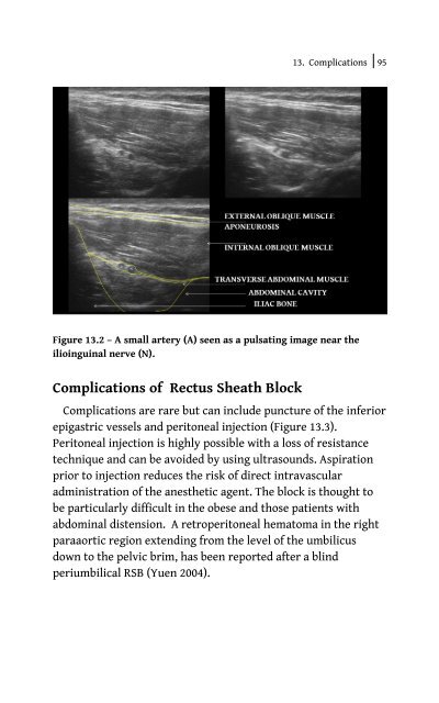
Figure 13.2 – A small artery (A) seen as a pulsating image near <strong>the</strong><br />
ilioinguinal nerve (N).<br />
Complications of Rectus Sheath Block<br />
Complications are rare but can include puncture of <strong>the</strong> inferior<br />
epigastric vessels and peritoneal injection (Figure 13.3).<br />
Peritoneal injection is highly possible with a loss of resistance<br />
technique and can be avoided by using ultrasounds. Aspiration<br />
prior to injection reduces <strong>the</strong> risk of direct intravascular<br />
administration of <strong>the</strong> anes<strong>the</strong>tic agent. The block is thought to<br />
be particularly difficult in <strong>the</strong> obese and those patients with<br />
abdominal distension. A retroperitoneal hematoma in <strong>the</strong> right<br />
paraaortic region extending from <strong>the</strong> level of <strong>the</strong> umbilicus<br />
down to <strong>the</strong> pelvic brim, has been reported after a blind<br />
periumbilical RSB (Yuen 2004).


