

Ultrasound Blocks for the Anterior Abdominal Wall
Ultrasound Blocks for the Anterior Abdominal Wall
Ultrasound Blocks for the Anterior Abdominal Wall

Create successful ePaper yourself
Turn your PDF publications into a flip-book with our unique Google optimized e-Paper software.
1. Anatomy <strong>for</strong> Anes<strong>the</strong>siologists | 17<br />
between <strong>the</strong> IOM and <strong>the</strong> TAM (Rozen 2008). TAM plane is<br />
delimitated superiorly by <strong>the</strong> costal margin, inferiorly by <strong>the</strong><br />
iliac crest, medially by <strong>the</strong> lateral border of <strong>the</strong> RAM, posteriorly<br />
by <strong>the</strong> lumbodorsal fascia, superficially by <strong>the</strong> IOM and deeply by<br />
<strong>the</strong> TAM.<br />
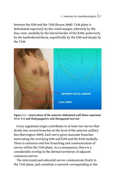
Figure 1.5 – Innervation of <strong>the</strong> anterior abdominal wall (lines represent<br />
T6 to T12 and iliohypogastric and ilioinguinal nerves).<br />
Every segmental origin contributes to at least two nerves that<br />
divide into several branches at <strong>the</strong> level of <strong>the</strong> anterior axillary<br />
line (Barrington 2009). Each nerve gives muscular branches<br />
innervating <strong>the</strong> overlying IOM and EOM and <strong>the</strong> RAM medially.<br />
There is extensive and free branching and communication of<br />
nerves within <strong>the</strong> TAM plane. As a consequence, <strong>the</strong>re is a<br />
considerable overlap in <strong>the</strong> dermal territories of adjacent<br />
cutaneous nerves.<br />
The intercostal and subcostal nerves communicate freely in<br />
<strong>the</strong> TAM plane, and constitute a network corresponding to <strong>the</strong>


